student - koerperwelten.de · diseases of the cardiovascular system are the leading cause of death...
TRANSCRIPT

ST
UD
EN
T
GU
IDE

A WORD OF APPRECIATIONWe would like to thank all those
who have donated their bodies,
without whom this exhibition
would not have been possible.

CONTENTS
Frequently Asked Questions
What is Plastination?
Interview with Gunther von Hagens
Welcome
Exhibition Overview
The Locomotive System
The Nervous System
The Respiratory System
The Cardiovascular System
The Digestive System
Embryonic & Fetal Development
Art in Science
Would You Do It?
4
8
10
12
13
14
16
18
20
22
24
26
27
This material is protected under copyright laws and may not be reproduced in any manner without the express written permission of the Institute for Plastination.
MARCH, 2017 US

FREQUENTLY ASKED QUESTIONS
4
What is BODY WORLDS?The exhibition BODY WORLDS, internationally known as BODY WORLDS: The Original Exhibition of Real Human Bodies, is the first exhibition of its kind to inform the visi-tor about anatomy, physiology, and health by viewing real human bodies. The specimens on display were preserved through Plastination, the preservation process invented by Dr. Gunther von Hagens in 1977, while he was working as an anatomist at the University of Heidelberg. Since the begin-ning of the exhibition series in Japan in 1995, more than 44 million visitors in more than 115 cities in America, Europe, Asia and Africa have seen the world’s most successful travel-ing exhibition.
What does BODY WORLDS show?Each BODY WORLDS exhibition contains real human speci-mens, including whole-body plastinates as well as individual organs, organ configurations, and transparent body slices. The spectacular plastinates in the exhibition take the visitor on an exciting journey of discovery under the skin. It pro-vides wide-ranging insight into the anatomy and physiology of the human body. In addition to organ functions, com-mon diseases are described in an easily understood manner by comparing healthy and affected organs. They show the long-term impact of diseases and addictions, such as tobac-co or alcohol consumption, and demonstrate the mechanics of artificial knee and hip joints.
How do the various BODY WORLDS exhibitions that are being shown differ from each other?While all of the BODY WORLDS exhibitions focus on gen-eral anatomy revealed through Plastination, each exhibition is currently being shown with dedicated themes—on cardiol-ogy and the heart (BODY WORLDS & The Story of the Heart), human development, longevity and aging (BODY WORLDS & The Cycle of Life), the body´s capability and vitality (BODY
WORLDS Vital), the story of the human body in the 21st cen-tury (BODY WORLDS: Pulse), the influence that ‘happiness’ has on our health (BODY WORLDS: The Happiness Project) and on prescriptions for healthy living (BODY WORLDS RX).
The exhibitions show a multitude of brand new plastinates and offer every visitor—even the ardent BODY WORLDS visitor—a fascinating exhibition experience.
BODY WORLDS & The Story of the Heart reveals—through the lenses of anatomy, cardiology, psychology, and culture—how the heart nourishes, regulates, and maintains life. The heart is the steady engine of our life. Due to this continuous strain, it is also vulnerable to wear and tear and to dysfunctions. Diseases of the cardiovascular system are the leading cause of death today. “The Story of the Heart“ also highlights other aspects of this vital organ. In religion, art, literature, and pop culture, the heart is seen as a symbol of love, compassion, happiness, and courage. The exhibi-tion gives visitors a deep insight into the human body, health and disease, and the complicated world of the cardiovascu-lar system.
BODY WORLDS & The Cycle of Life features a special presentation on the human life cycle and aging—from prena-tal development and infancy, to childhood and adolescence, to youth, adulthood, and old age. It shows the body living through time—at its most healthy, and as it changes, grows, matures, peaks, and finally fades. The exhibition shows the complexity, resilience, and vulnerability of the human body through anatomical studies of the body in distress, disease, and optimal health. Visitors to “The Cycle of Life” will see the body living through the span of time from the spark of conception to old age, and learn about the latest findings in longevity and aging science.
5
BODY WORLDS Vital presents a special collection of specimens designed to show visitors the basics for human health and wellness. The exhibition includes whole-body plastinates, a large arrangement of individual organs, organ and arterial configurations, and translucent slices that give a complete picture of how the human body works. Vital tells the fascinating story of how best to fight life-threatening diseases—such as cancer, diabetes, and heart ailments—through healthy choices and lifestyle changes.
BODY WORLDS: Pulse shows the science and splendor of the human body, and deconstructs its form and function through the trailblazing science of Plastination invented by anatomist, Dr. Gunther von Hagens. It presents the body in health and distress, its vulnerabilities and potential, and many of the challenges the human body faces as it navigates the 21st Century. BODY WORLDS: Pulse is a convergence of aesthetic anatomy, the latest findings in health and wellness, immersive multimedia, and narrative storytelling threaded with deep metaphors. It shows the possibility of living mind-fully, with meaning and vitality, by refracting the story of life and self-actualization, through the donors, who have com-mitted to educating future generations.
BODY WORLDS: The Happiness Project marks an en-tirely new chapter in the already impressive range of BODY WORLDS exhibitions. A permanent exhibition exploring what happiness is and the science behind it. “The Happi-ness Project” tells the story of our bodies and the influence that the emotional phenomenon of ‘happiness’ has on our health. Over 200 anatomical specimens of real human bod-ies reveal the complexity, resilience and vulnerability of the body. Visitors learn for example that people who are happy, live longer than people who are unhappy.
BODY WORLDS RX offers prescriptions for healthy living and focuses on the most prevalent contemporary diseases, that afflict children and adults alike, their causes and effects. BODY WORLDS RX is an informative and entertaining pre-sentation of the latest research on top health issues and shall inspire visitors to embrace preventive healthcare. From organs, to muscles, to the nervous system, and to skeletal structures, visitors will have an unprecedented look inside the intricate systems of the most sophisticated mechanism in the world, the human body.
All BODY WORLDS exhibitions generally present dif-ferent plastinates, which is most evident in the whole-body plastinates which each vary in pose and display.
Are there animals in the BODY WORLDS exhibitions, as well?Most BODY WORLDS exhibitions have a few animal speci-mens on display. Due to the great popularity of the animal plastinates, Dr. Gunther von Hagens and Dr. Angelina Whal-ley were encouraged to create the new exhibition BODY WORLDS of Animals which also tours the world under the name ANIMAL INSIDE OUT. The display features the most popular species in the animal kingdom. Better than any text-book, this fascinating exhibition shows the complex, amaz-ing biology of the natural world’s most remarkable creatures and their nervous system, bones, muscles, and organs. AN-IMAL INSIDE OUT also allows a peek under the elephant’s skin. Its trunk with a network of 40,000 muscles is an in-credibly unique feature that has many different uses. And who would have known that a giraffe uses its 20-inch-long bluish tongue like a hand? ANIMAL INSIDE OUT’s educa-tional approach is particularly suitable for young visitors. For more information: www.AnimalInsideOut.com.

6
What is the goal of the exhibition? BODY WORLDS aims to educate the public about the in-ner workings of the human body and shows the effects of poor health, good health, and lifestyle choices. It is also pre-sented in the hopes that it will motivate visitors to learn more about the science of anatomy and physiology.
Who should see BODY WORLDS?Anyone interested in learning what makes us human. Adults of all ages and children will find the exhibits fascinating. Given the nature of the BODY WORLDS exhibits, it is up to parents, guardians, or school staff to decide whether BODY WORLDS is appropriate for the children in their care.
Where else has BODY WORLDS been exhibited? Where will they be on display next?There are nine BODY WORLDS exhibitions including ANIMAL INSIDE OUT, which have been viewed by 40 million people throughout the world. BODY WORLDS exhibitions have been displayed in America, Europe, Asia and Africa. Additional BODY WORLDS exhibitions are planned. If you would like to know in what cities the exhibitions will be on display next, please go to our official web site, www.bodyworlds.com, where you will find an overview of past and future exhibition venues. If you are interested in additional information about BODY WORLDS current exhibitions and more, you may join our Facebook community.
Why is it important for the public to see these exhibitions? The organizers of BODY WORLDS believe that when people understand more about how the body works and how it can break down, they are more likely to choose healthy lifestyles. They also hope it will inspire visitors to learn more about the life sciences. Knowledge about what the human body looks like and how it functions is basic life science information that should be available to everyone.
Would I be able to learn just as much from books or models of the human anatomy?The use of authentic specimens allows a thorough examina-tion and study of disease, physiology, and anatomy that you cannot find in models, textbooks, or photos. In addition, the exhibition allows visitors to understand that each and every body has its own unique features, even on the inside. The experience in cities around the world has clearly demonstrat-ed that real specimens fascinate exhibit visitors in a way that models cannot.
What is Plastination? Plastination is a unique process invented by Dr. Gunther von Hagens in 1977 to preserve specimens for medical educa-tion. The process replaces bodily fluids and fat in specimens with fluid plastics that harden after so-called vacuum-forced impregnation. After the bodies are shaped into lifelike poses, they are hardened with gas, heat, or light. The plastinates show how our bodies move in everyday life, as well as during athletic activities. For more information about Plastination, go to www.bodyworlds.com.
Where did the specimens on display come from? Will we know who the plastinates are or how they died?The BODY WORLDS exhibitions rely on the generosity of body donors; individuals who requested that, upon their death, their bodies could be used for educational purpos-es in the exhibition. All the whole-body plastinates and the majority of the specimens are from these body donors; only some organs, fetuses and specific specimens that show un-usual conditions come from old anatomical collections and morphological institutes. As agreed upon by the body do-nors, their identities and causes of death are not disclosed. The exhibition focuses on the nature of our bodies, not on telling personal information. Currently there are more than 16,000 donors registered in the body donation program of the Institute for Plastination. For more information please visit the body donation section of www.bodyworlds.com.

7
BODY WORLDS exhibitions are the based on an established body donation program through which the body donors spe-cifically request that their bodies could be used in a public exhibition after their deaths.
Why are the plastinates posed the way they are? The poses of the plastinates have been carefully thought out and serve educational aims. Each plastinate is posed to show different anatomical features. For instance, the ath-letic poses illustrate the use of muscle systems while playing sports. The poses are chosen to highlight specific anatomi-cal features and allow the visitor to compare the plastinate to his or her own body.
Will I be able to touch any of the plastinates? While you will be able to get very close to the plastinates, as a rule, visitors are not allowed to touch them.
Is this exhibition appropriate for children? 44 million people, including young children, have viewed the BODY WORLDS exhibitions around the world. If you are considering bringing children or school groups to BODY WORLDS, visit our online resources section to find out how to use the exhibition as a learning experience.
Is there an audio tour?Some exhibitions offer audio guides for an additional fee. The audio tours are designed for the layman. They are avail-able in English and other languages. There are no personally guided tours through the exhibit, at this time.
Have the ethical questions about this exhibition been discussed?Before the North American premiere of BODY WORLDS, a wide committee of theologians, ethicists, academics, and
medical experts thoroughly discussed the ethical questions. Guided by the California Science Center in Los Angeles, they wrote an Ethics Review of the origins of bodies in BODY WORLDS. It can be downloaded from our website www.bodyworlds.com.
What educational materials are provided? Teachers will wish to prepare both their students and their adult supervisors carefully for their BODY WORLDS experi-ence. Educator materials are available upon request and for download on the website www.bodyworlds.com. BODY WORLDS offers preview opportunities so that teachers can see the exhibition free of charge before bringing their classes to it.
How long can you stay inside the exhibit? You can stay as long as you like, within the opening hours. We recommend allowing yourself about one to two hours. The length of time will vary on how long each visitor wishes to examine each specimen and read the information. An audio tour will add to your time in the exhibits. Reentry to the exhibition is not allowed, once you exit.
Can you take photographs or film in the exhibitions? Professional photography and filming in the exhibition is limited to registered members of the media, for editorial purposes only. Some locations allow photography with small hand-held devices for personal, non-commercial use. Please check with the location you are visiting to con-firm that photography is allowed. Out of respect to other visitors and the body donors, photography may be restricted at anytime.
Acetone bath
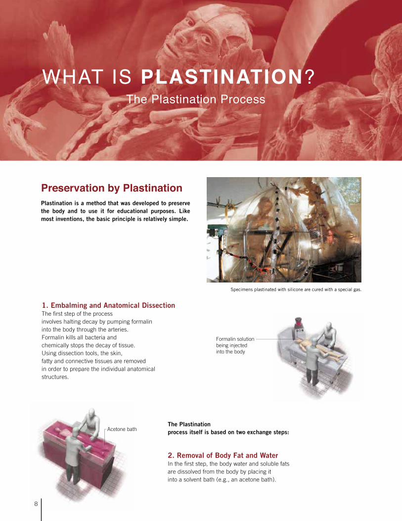
Specimens plastinated with silicone are cured with a special gas.
The Plastination ProcessWHAT IS PLASTINATION?
1. Embalming and Anatomical DissectionThe first step of the process involves halting decay by pumping formalin into the body through the arteries. Formalin kills all bacteria and chemically stops the decay of tissue. Using dissection tools, the skin, fatty and connective tissues are removed in order to prepare the individual anatomical structures.
Formalin solution being injected into the body
The Plastination process itself is based on two exchange steps:
2. Removal of Body Fat and WaterIn the first step, the body water and soluble fats are dissolved from the body by placing it into a solvent bath (e.g., an acetone bath).
Preservation by PlastinationPlastination is a method that was developed to preserve the body and to use it for educational purposes. Like most inventions, the basic principle is relatively simple.
8

Vacuum
Vacuum pump
Acetone is removed from the tissue
Silicone enters the tissue
Vacuum pump
Vacuum chamber with liquid polymer
Courtesy of The Denver Post
3. Forced ImpregnationThis second exchange process is the central step in Plastination. During forced impregnation a reactive polymer, e.g., silicone rubber, replaces the acetone. To achieve this, the specimen is immersed in a polymer solution and placed in vacuum chamber. The vacuum removes the acetone from the specimen and helps the polymer to penetrate every last cell.
4. PositioningAfter vacuum impregnation, the body is positioned as desired. Every single anatomical structure is properly aligned and fixed with the help of wires, needles, clamps, and foam blocks.
5. Curing (Hardening)In the final step, the specimen is hardened. Depending on the polymer used, this is done with gas, light, or heat.
Dissection and Plastination of an entire body requires about 1,500 working hours and normally takes about one year to complete.
Slice Plastination Slice Plastination is a special form of Plastination. First, the body is frozen and cut into 1 to 3-inch-thick slices. Instead of silicone, the body is treated with polyester or epoxy resin during this process.
9
Positioning

Were you ever scared to work with dead bodies?
Dr. von Hagens: When I was about six years old, I was very sick and nearly died. I was in hospital for many months and became very comfortable in that environment of the sick and dying. The doctors and nurses who cared for me became my heroes and I wanted to be like them. Later, when I worked in a hospital as an orderly and then a nurse, (long before I became a doctor), one of my duties was to transport the dead to the morgue. Other workers didn’t like this job because it frightened them, but I was never afraid. Being afraid of death is not a good way to live.
Were the people in the exhibit old when they died?
Dr. von Hagens: The people who donated their bodies for Plastination and to educate all of us about health are of vari-ous ages. Some were old, but others were young in the prime of their life. Each person is different, not just on the outside but also on the inside. Even after more than 40 years as an anatomist, I have never seen two hearts that look the same.
Where did the idea for BODY WORLDS come from?
Dr. von Hagens: When I used to teach anatomy to students in medical school in the 1970s, I had to use illustrated anatomy atlases and picture books to show the organs and body sys-tems. I tried to use real human organs and specimens, but at that time the specimens were preserved in blocks of plastic so you could not touch them or study the placement of the organs properly. I realized one day that if the plastic was in-side the body and not outside it, the specimen would be rigid and easy to grasp, and study and work with. I was only trying to solve a problem; I wanted to educate my students so they would become better doctors, as I don’t think doctors should be poking around inside your body and operating on you if they don’t know important things about it.
Children Interview Dr. Gunther von Hagens, Creator of BODY WORLDS & Inventor of Plastination
INTERVIEW WITH GUNTHER VON HAGENS
10
But something very unusual began to happen after I began to plastinate organs and specimens. The janitors and secretar-ies and office workers at the university began to stop by the lab; they were fascinated by the plastinates. This was when I began to think of anatomy for lay people, which is what BODY WORLDS is. It is very different from anatomy for medical pro-fessionals because it has to be interesting and dynamic and not scary to look at.
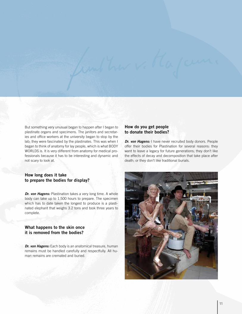
How long does it take to prepare the bodies for display?
Dr. von Hagens: Plastination takes a very long time. A whole body can take up to 1,500 hours to prepare. The specimen which has to date taken the longest to produce is a plasti-nated elephant that weighs 3.2 tons and took three years to complete.
What happens to the skin once it is removed from the bodies?
Dr. von Hagens: Each body is an anatomical treasure, human remains must be handled carefully and respectfully. All hu-man remains are cremated and buried.
How do you get people to donate their bodies?
Dr. von Hagens: I have never recruited body donors. People offer their bodies for Plastination for several reasons: they want to leave a legacy for future generations; they don’t like the effects of decay and decomposition that take place after death; or they don’t like traditional burials.
11

12
Dear Students,
Have you ever watched a professional basketball player seem to float in air as he or she leaps up to dunk the ball in the basket? Or maybe you watched athletes competing at the Olympics, and wondered “How did they do that?”
Well, our bodies are pretty amazing. And the more we learn about ourselves and how our bodies work, the better we can take care of ourselves and others. And, the healthier we will be—making us better on the football pitch, basketball or tennis court, riding a bike, or just walking down the street.
Gunther von Hagens’ BODY WORLDS: The Original Exhibition was developed by a German doctor and anatomist to help people understand how their bodies work by letting them look inside real human bodies.
When you visit with your school or family, you will see exactly how your organs look and what happens to them when certain diseases take over. You will see how smoking destroys lungs and how bones, muscles, and ligaments all work together so you can play sports, dance, or skate.
WELCOME A Letter from BODY WORLDS
Dr. Angelina Whalley
Conceptual Designer of BODY WORLDS and
President and CEO of the Institute for Plastination.
Dr. Gunther von Hagensinvented Plastination in1977.
COOL FACT
The activities inside this guide will help you learn more about the human body. Come visit us to see BODY WORLDS.
You’ll really get to know yourself!

13
Exhibition Overview
including Human Facts
Gunther von Hagens’ BODY WORLDS exhibits
use the science of Plastination
to let visitors see
how human bodies are put together.
The exhibit also teaches how different
anatomical systems work in the human body.
This special student supplement explores
several of the systems featured in the exhibit,
including the locomotive system,
the nervous system, the respiratory system,
the cardiovascular system, the digestive system,
and embryonic & fetal development.

Motion HappenTHE LOCOMOTIVE SYSTEM
The locomotive system makes movement possible. It consists of the bones that make up the skeleton, the joints that hold the bones together, and the muscles that contract and relax to actually make you move.
The skeleton is the framework of the body, and is made up of bones and cartilage. Bone is made mostly of calcium, which is why it is important to eat calcium-rich food to keep your bones strong.
Inside the bone is sponge-like matter called bone marrow. This makes bones light so people can move easily, but strong enough to support body weight. Bone marrow also produces red and white blood cells. Red blood cells have hemoglobin
and carry oxygen. White blood cells produce antibodies to attack bacteria, infections, and diseases.
The skeleton has many jobs. It provides protection to internal organs, it supports the body and gives it its shape, and it provides a place for muscles to attach.Bones are important to almost every movement we make. Bones couldn’t move a pencil, though, without help from muscles. Muscles consist of cells that contract.
Muscles and bones are connected by tendons, which are similar to ropes. When a muscle contracts, it pulls the tendon, which then tugs on the bone, and everything moves.
Although it may seem easy to do something like throw a ball, it’s actually complicated when looked at inside the body. To make the motion of throwing, many muscle groups in the shoulders, arms, chest, abdomen, and even legs must be used! Each of these groups must work together with nerves in order for motion to occur. And all this happens in a fraction of a second!
FLEXION
Triceps is relaxedBiceps is contracted
EXTENSION
Triceps is contracted
Biceps is relaxed
Skeleton
Shaft of the femur
Kneecap
Fibula
Shinbone (tibia)
Breastbone (sternum)
Collarbone (clavicle)
Cheekbone
Wing of ilium
Coccyx
SacrumPelvis
14
Upper ankle joint

At birth, humans have 300 bones. As a baby grows, however, many of the smaller bones fuse together so that adults have just 206 bones.
Learn with BODY WORLDS
The bones of the human skeleton give the body both
strength and structure. A strong and healthy skeleton is
important for every person for both work and recreation.
Think of three things that you do every day that involve
the use of certain bones.
Voluntary muscles are used when you throw a ball. These are the muscles we can control. People also have involuntary muscles, which we cannot control, such as the heart and the stomach.
Another important part of the locomotive system are the joints. Joints are positioned between major bones that come together and help you to move and bend.
UlnaRadius
Upper armbone
Elbow joint, viewed from the front
Collateral ligament
The Runner
There are different kinds of joints, including ball and socket joints in the hips and hinge joints at the knees and elbows.
Joints are surrounded by capsules containing fluid that help the bones move smoothly.
15
COOL FACT

The Messenger and the BossTHE NERVOUS SYSTEM
The nervous system is the system of the body that controls movements, thoughts, and emotions throughout the body. Without it, you wouldn’t be able to function!
There are two parts to the nervous system: the central ner-vous system and the peripheral nervous system.
The central nervous system includes the brain and the spinal cord. They work together with nerves to send messages back and forth between the brain and the rest of the body.
The brain controls the system. It has five parts: the cerebrum, the cerebellum, the brain stem, the pituitary gland, and the hypothalamus.
The cerebrum is the biggest part of the brain and controls thoughts, language, and voluntary muscles, which are the muscles you can control. You also use the cerebrum when you think hard and when you need to remember things.
The cerebellum is a lot smaller than the cerebrum, but still very important. It controls balance, movement, and coordina-tion. If it weren’t for the cerebellum, you wouldn’t be able to stand without falling!
The brain stem connects the rest of the brain to the spinal cord. It’s the part in charge of major things that keep you alive like breathing, blood pressure, and digesting food. Unlike the cerebrum, the brain stem controls the involuntary muscles— the ones that work without you thinking about it, such as the heart and stomach.
The nervous system carries messages from the brain to other parts of the body at more than 250 miles per hour.
COOL FACT
Axon terminals
Nucleus
Cell body
Dendrite
Schwann cell
Corpus callosum
Lateral ventricle
Brain stem
Medulla oblongata
Cerebellum
Left hemisphere of brain
16
Schematic illustration
of a neuron

The tiny pituitary gland produces and releases hormones into the body—hormones like those that help you grow and change.
Finally, the hypothalamus regulates your body temperature, your emotions, and hunger and thirst.
The brain has many jobs, but it needs help from nerves and the spinal cord, too. Every action you do happens because your brain, your nerves, and your spinal cord work together.
The nervous system includes millions and millions of neu-rons, which are microscopic cells. When you do something, messages travel from the neurons to your brain.The peripheral nervous system is composed of the nerves and neurons that go outside the central nervous system to operate the body’s limbs and organs. It is here that everything gets connected.
Next time you take a test, drink a glass of water, laugh, or do anything at all, thank your nervous system. Actually, you can thank it right now since it just helped you read this!
Cerebrum
Cerebellum
Dura mater
Spinal cord
Spinal nerves
Sciatic nerve
Learn with BODY WORLDSThe nervous system carries messages to the brain that make it possible for the
body’s five senses to work. The five senses are touch, taste, hearing, sight, and
smell. Explore the five senses by writing about one of your favorite things for each
sense.
For example you may enjoy listening to music, because it helps you concentrate.
This relates to your sense of hearing.
17

Oxygen In, Carbon Dioxide OutTHE RESPIRATORY SYSTEM
The organs of the respiratory system work together, along with other body systems, to ensure that the cells of the body re-ceive the oxygen they need to live.
When you breathe in, the muscles of your chest expand. Your diaphragm lowers and creates lower air pressure in your lungs than in the world outside. This causes air to enter through the nose or mouth.
Once air enters, it travels past your esophagus, sometimes called the “foodpipe,” and is moistened as it goes down the trachea, or “windpipe,” into the lungs. As the air enters the lungs, the lungs expand outward.
Once inside the lungs, the air travels through tubes, called bronchi, into smaller tubes called bronchioles, which get smaller and smaller until they reach the alveoli which are sacs about the size of a grain of sand.
It is through the walls of the alveoli that the oxygen in the air you breathe enters the body’s blood, which flows past the alveoli. The blood receives the oxygen and, in return, passes carbon dioxide into the alveoli.
The cells of your body need oxygen to live, and carbon dioxide is the waste of things the cells do. Your red blood cells are little workers that carry the oxygen to the cells and take the carbon dioxide away.
Smoking, as we all know, makes the lungs less healthy and can lead to death.
One of the reasons for this is that smoking makes little structures called cilia stop working. Cilia move within the lungs to help clear things out that enter the lungs. Smoking disables or even kills them. Then harmful particles stay in the lungs.
Lungs showing the bronchial tree in the left upper lobe
Windpipe
Main bronchi
Bronchi
18

Your left lung is a bit smaller than the right to leave room for your heart.
Another bad effect of smoking is that chemicals from ciga-rettes will build up in the lungs, and the delicate alveoli can become thickened, swollen, and unable to exchange oxygen and carbon dioxide with the blood in a healthy way. This condi-tion leads to emphysema. Severely enlarged
thyroid gland
Epiglottis
Enlarged thyroid gland (goiter)
Think about itPlants take the carbon dioxide that we release and use it, cre-ating oxygen, which we need. We in turn take oxygen and turn it into carbon dioxide, which plants need. This is what is called a symbiotic relationship—one that is good for both organisms. Try to think of other ways in which humans interact with nature in symbiotic relationships.
A healthy respiratory system makes it possible for
people to live active lives. Smoking causes prob-
lems for the respiratory system. Make a list of five
reasons why people shouldn’t smoke.
Non-smoker‘s lungs
Smoker’s lungs
19
LEARN WITH BODY WORLDS
COOL FACT

The Body’s Great Pump
THE CARDIOVASCULAR SYSTEM
The heart is the central organ of the cardiovascular system and it doesn’t look much like the drawings found on Valentines. Cardio means heart, and the cardiovascular system is essential to our survival.
The cardiovascular system is sometimes referred to as the cir-culatory system because it’s responsible for the circulation of blood through the body. It consists of the heart, which is a mu-scular pumping device, and a closed system of vessels called arteries, veins, and capillaries.The cardiovascular system’s vital role is to provide a continuous and controlled movement of blood through the thousands of miles of microscopic capillaries that reach every tissue and cell in the body.
Human survival depends on the circulation of blood to the or-gans, tissues, and cells of your body.
Arteries carry blood enriched with oxygen away from the heart and veins carry blood that has used up its oxygen back to the heart. Through the heart and lungs, the blood gets a fresh sup-ply of oxygen and delivers it to the rest of the body.
Twenty major arteries make a path through the tissues of the body. Then they branch out into smaller vessels called arterio-les. These branch further into the capillaries, most of which are thinner than a hair—some so tiny, in fact, that only one blood cell can move through at a time.
Heart, opened longitudinally
Left atrium
Mitral valve
Septum of the heart
Right atrium
Left ventricle
Tricuspid valve
Left atrium
Aortic valve
Right ventricle
Left ventricle
20

At every stage of life, your heart is about the size of the fist you make when you close your hand.
Once the blood in capillaries delivers oxygen and nutrients, it picks up carbon dioxide and other waste. Then blood moves back through wider vessels, called venules. These eventually join to form veins, which deliver the blood back to your heart to pick up oxygen.
If all the vessels of this network were laid end to end, they would extend about 60,000 miles, far enough to circle the Earth more than twice!
Because all the tissues in the body rely on it, the cardiovascu-lar system appears early in developing embryos—in the fourth week after fertilization—and reaches a functioning state long before any other major organ system.
The cardiovascular system is delicate and can be affected by many things. Fats and choles-
terol, for example, can slow or even block the flow of blood in the body. Fats and cholesterol
enter the body as food. One reason people are encouraged to limit the amount of fatty or oily
foods they eat is to help prevent blockage. Think of ten fatty foods and ten healthier options.
For example, you may think of a doughnut as a fatty food and toast as an alternative.
Blood vessel configurationof inner organs
Aortic arch
Capillary bed of the liver
Capillary bed of the right kidney
Capillary bed of the uterus
Heart
21
Learn with BODY WORLDS
COOL FACT

The body’s digestive system converts the food you eat into the energy you need to live.
The journey through your digestive system is a long one for food. It starts in the mouth, where teeth grind and tear the food into small pieces. Saliva then wets and softens the food, and begins to dissolve carbohydrates. Once the food is properly mashed and wet, it is pushed by muscle action into the pha-rynx, or throat, and down the esophagus, which leads to the stomach. When food reaches the stomach it is mixed and broken down further by acids the stomach produces. The stomach protects itself from these acids by secreting a layer of mucus that lines the inside of the stomach.
Some things, such as water and sugars, can be absorbed right out of the stomach and into the bloodstream. The things that need more digestion have further steps ahead of them. When the stomach has made the food a liquid, the food passes through a valve into the small intestine. The small intestine has a large surface area because it contains villi. Villi are tiny little structures like very short hairs that stick out into the small intestine. Through the walls of the villi nut-rients from food pass into the bloodstream. The bloodstream carries the nutrients to your cells so they can live.
Once all the useful nutrients have been taken from food in the small intestine, the unusable parts pass into the large intestine, or colon.
In the large intestine, water is extracted from the waste and the material hardens into feces. The feces are passed out of the body when you go to the restroom.
Stomach
Pancreas
Large intestine
Caecum
Appendix
Duodenum
Liver
Oesophagus
Rectum
Tongue
Digestive tract
Small intestine
Converting Food Into Energy
THE DIGESTIVE SYSTEM
22

Your mouth makes about a quart of sali-va each day, and you produce a total of about seven quarts of digestive juices.
BODY WORLDS leertip!
Digestive helpersThe pancreas, liver, and gallbladder are all organs that do things important to the digestive system. The pancreas makes enzymes that help digest proteins, fats, and carbohydrates. The liver makes bile, which helps the body absorb fat.
Parasympatheticnerve branches
Stomachs of varying size and shape
Duodenum
Blood vessel configuration of the liver (rear view)
The digestive system breaks down the food that supplies the human body with energy. What
foods would you eat if you needed energy for sports or active recreation?
Pick five foods you think would be good sources of energy. Then pair off and research your
foods. Were they all healthy choices for getting the energy you needed?
Bile is stored in the gallbladder until it is needed. Enzymes and bile travel into the small intestine through ducts. Interestingly, people don’t really need the gallbladder. If it is removed, the bile just flows right into the small intestine and does its job.
23
Learn with BODY WORLDS
COOL FACT

Life begins with a single cell, or zygote, after the father’s sperm fertilizes the mother’s egg.
The zygote contains the human genome, the individual blueprint of a human being. It consists of the parents’ gene pairs, organized in chromosomes. This special set of chromosomes, which has never existed before and will never be recreated, determines the characteristics and traits of the conceived human being.
The first weeksRoughly 30 hours after fertilization, a microscopic human egg begins to divide into two identical daughter cells. Twins will develop if these two cells separate from each other. Most of the time, however, the complete embryo will remain intact and migrate down the Fallopian tube, settling in the uterus on the sixth day. Pregnancy will last an average of 260 days from that point.
The embryo, suspended in amniotic fluid and surrounded by fetal membranes, is linked to the maternal blood supply via the umbilical cord and placenta. During the first four weeks, the embryo is roughly 0.15 inches long and will grow to 1.2 inches by the end of the eighth week, when it will weigh approximately 0.1 ounce. All of the organs will be in place by the end of this period, after which the developing child is referred to as a fetus. The length and weight of the fetus then begins to increase significantly as it proceeds through further complex stages of development.
Week 13 to 14Coordinated movements will begin, although the mother is not yet able to feel them. The relatively large head will straighten up, the lower extremities are already well developed, and the toenails will begin to grow.
Week 15 to 16The fetus is now 6 inches long and can weigh up to 7 ounces. Its gender can be detected via ultrasound, and its skeleton will show up clearly on x-rays. Its legs have begun to grow larger, and its head is smaller relative to its body as a whole. Fetal blood begins to develop in the liver. Ovaries have already developed in female fetuses.
Week 17 to 18Fetal growth has slowed; the weight of the fetus has increased to 10.5 ounces. The skin is still thin because the (white) subcutaneous fatty tissues have not yet developed. Brown fatty tissues have, however, begun to form; these will allow the small organism to produce its own heat. The uterus has developed in female fetuses. Mothers may feel the fetus move from this point on.
Week 19 to 20Toward the end of this phase, the fetus will be 11 inches long and will weigh up to 1 pound. The body and head of the fetus are now covered with fine hair (known as lanugo), which contains little pigment.
Zygote or fertilized egg (400 times magnified).
24
EMBRYONIC & FETAL DEVELOPMENT

Week 21 to 24The fetus begins to gain weight more rapidly again, and its proportions are becoming more like those of a baby. Rapid eye movement has begun, and fingernails will start to grow. The skin is still red and wrinkled. The lungs, however, are now capable of breathing, if insufficiently, because there is not yet any coordination between them and the nervous system. As a result of this lack of coordination, the exchange of gases (especially CO2 exhalation) cannot be ensured to a sufficient extent, thereby leading to an oxygen deficiency, which can cause more or less severe damage to the brain if the baby is born at this stage.
Eight-week-old embryo.
Placenta. On the surface of the fetal side, the arteries and veins of
the umbilical cord vessels branch out.
Week 25 to 28The lungs are now fully capable of breathing, which means that the fetus is capable of living outside the womb. During week 26, the eyes can open, and subcutaneous fatty tissue developed by this point has given the body a more rounded shape. Until this point, the spleen has been producing blood; during week 28, bone marrow will take over this function. A fetus will now weigh more than 2.8 pounds.
Week 29 to 32The fetus’s body will grow to over 16.5 inches, and its weight will increase to 3.3 to 4.2 pounds. The fingernails will grow to the tips of the fingers, and the skin will now be pink and smooth. The eyes will respond to light by means of the pupillary reflex, and the hands will respond to stimulus with a ‘grasping’ reflex.
When a pregnant woman consumes alco-hol, the alcohol level in the blood of her fetus will be the same as in her own.
Many factors influence the development of an unborn baby.
How do environmental influences affect the child? What
circumstances in the life of the mother have a positive or
negative effect, or can even harm the baby? How do these
influences actually reach the fetus? Discuss these questi-
ons in class.
25
LEARN WITH BODY WORLDS
IMPORTANT FACT

BODY WORLDS ex-hibitions teach us a
lot about the science and anatomy of the hu-
man body. They also teach about the form and art of the human body.
Studies of anatomy have always been an important part of art edu-
cation. Artists who know how the human body is put together and how its muscles work are better able to portray people in painting, sculp-ture, and other art forms.
This knowledge is important, even if artists choose to represent the human
form in abstract ways.
In the BODY WORLDS exhibits, Dr. Gunther von Hagens has positioned human figures to reveal how the body is put to-gether and how it performs different tasks. He has also pre-sented human figures in ways that highlight different body systems, such as muscles, internal organs or nerves, and blood vessels.
The scientific choices he has made give us a new way to un-derstand how human bodies work. At the same time, he has revealed how beautiful the form and systems of the human body are.
As visitors go through the exhibits, they learn the science and biology of anatomy. They also get to experience the artistic qualities of anatomy. This gives the exhibits appeal to all stu-dents, not just those in science classes.
The Beauty of the BodyART IN SCIENCE
Think like an artistArtists sometimes like to focus on one aspect of a figure. In art, this may be done by emphasizing one feature of a person, or showing the subject from an unusual angle or perspec-tive.
Explore this idea by thinking about someone in your family. Reflect on what this person is like, or what you admire about him or her. Then think about what you would focus on if you were to portray this person in an artwork. Draw a sketch of your artwork and explain your ideas to the class.
Photography as artNewspaper photographers often are asked to take photo por-traits of people in the news. These portraits often could be considered photographic artworks. Look through the news and features sections for several days and cut out photos portraying people. Pick the one you like the most and explain to the class what makes the portrayal effective or artistic in your eyes. Finish by giving the photo a title, and explain it to classmates.
Sports anatomyCoaches need to know how to evaluate the physical skills and talents of players. These talents often are based on anatomy. Pick an athlete you admire. Then think about the different body systems explored in this guide. Write out which systems contribute most to the success of this athlete.
Understanding how the body works is important in many
professions. Think about what you want to be when you
grow up, and write a short sentence or paragraph explain-
ing why anatomy could be important in the job, and why.
26
LEARN WITH BODY WORLDS

All specimens in Gunther von Hagens’ BODY WORLDS ex-hibits are authentic. They belonged to people who declared during their lifetime that their bodies should be made avail-able after their deaths for the instruction of doctors and the education of the public.
“BODY WORLDS is most of all a collaboration between the donors and myself, and all those who view the exhibit,” Dr. von Hagens says. “All of humanity owes the donors a great deal, for without them, there would be no BODY WORLDS.”
To ensure that donors make the decision willingly, von Hagens’ Institute for Plastination requires that all donors sign an official consent form. In the form, the donors must declare that they have made the decision “freely and voluntarily” to donate their body “for the purpose of anatomical research and education … for students and especially for the general public.”
In addition, they must check off answers to specific questions that have been raised by Plastination so there is no doubt they fully understand their decision.
“I agree for my body to be used for any purposes, provided it is to do with medical research or training” reads one ex-ample.Or “I agree that my plastinated body can be used for the medical enlightenment of laypeople and, to this end, exhib-ited in public (e.g. in a museum).”
Or “I agree that my body can be used for an anatomical work of art.”
Or “I agree that lay people be allowed to touch my plastinated body” in some exhibits.
Donors to the Institute for Plastination have the option to do-nate all useable organs to save lives before their bodies are plastinated.
Talk about itAs a class, discuss whether you would want to have your body, or the body of a relative, plastinated for education or display. Then discuss whether you think it is a good idea to exhibit plastinates for the general public. To ease discussion, you can set up a “For Chair” and an “Against Chair” to sit in at the front of the room when offering your opinion.
In your discussion: • Consider what motivates a donor to allow his/her body to be plastinated for education or an exhibit.
• Consider how the friends and relatives of a donor might feel.
• Imagine that a member of your immediate family wanted to be plastinated.
• Consider what you might learn – or did learn – about your own body from viewing the BODY WORLDS exhibits.
Thoughts about Plastination and Your Body
WOULD YOU DO IT?
After holding the class discussion, summarize the general
feelings of the class in a news story of the style found on
the front page of a newspaper. Talk about how newspaper
reporters must weigh all information before making a gen-
eral conclusion.
Then compare summaries written by different members of
the class. How similar were they?
What were some differences? What was the source
of those differences?
27
Plastination takes a very long time. A whole body can take up to 1,500 hours to prepare.
LEARN WITH BODY WORLDS
COOL FACT

www.bodyworlds.com
