ststn guidelines group · 1. cerebrovascular accident (cva) is a neurological event lasting > 24...
TRANSCRIPT

30/11/2017
1
Baba Inusa Paediatric Haematology
Evelina London Children’s Hospital
Honorary Reader, King’s College London
29th November, 2017; STSTN
Protocol for stroke management in Sickle Cell Disease
STSTN Guidelines Group
• Baba Inusa – Evelina Children’s Hospital
• Sara Stuart-Smith – King’s College Hospital
• Sue Height – King’s College Hospital
• Moira Dick – King’s College Hospital
• Alison Thomas – St George’s Hospital
• Rachel Kesse-Adu – Guy’s and St Thomas’ Hospital
30/11/2017
2
Plan
1. Introduction
2. Prevalence in SCD phenotypes
3. Role of Blood transfusion
4. Suggested Protocol
• Presentation
• Investigations
• Management –immediate
• Management- Long term
5. Conclusions- Q&A
Acute Management of Stroke in Paediatric Patients with Sickle Cell Disease 1. Cerebrovascular Accident (CVA) is a neurological event lasting > 24
hours +/- radiographic evidence of new areas of abnormality.
2. Transient Ischaemic Attack (TIA) is a focal event lasting < 24 hours with no radiographic evidence of abnormality
3. Clinical stroke is 250 times more common in children with Sickle Cell Disease (SCD) than the general paediatric population; 11% have an overt stroke by the age of 20 ( peak age 7) in absence prophylaxis
4. Vary according Phenotype. CVA highest -SCD-SS (0.61/100 person-years), compared with SCD-SC (0.15/100 person-years) or haemoglobin Sβ+ or Sβ0 thalassemia ( 0.09/100 person-years and 0.08/100 persons-year respectively)

30/11/2017
3
Stroke subtype by age
• Ischaemic stroke • 54% of CVAs
• highest in 1st decade and after 30 years
• peak at 2–5 years
• Haemorrhagic stroke • during 2nd decade
• Silent stroke • radiological findings consistent with
white matter disease
• 10–30% (not characterized as age-dependent)
• associated with cognitive deficiencies and higher stroke risk
Verduzco LA, et al. Blood. 2009;114:5117-25.
Age (years)
Ha
zard
fu
nct
ion
0.0010
0.0015
0.0020
0.0025
0.0030
0.0035
0.0040
10 20 30 40 50
Haemorrhagic stroke
Infarctive stroke
Cerebrovascular accidents in SCD • Age at first CVA and cumulative incidence of CVA in 2,436 patients with sickle cell anaemia
(Hb SS), 839 with HbSC disease, 188 with HbSβ0 thalassaemia, and 184 with HbSβ+ thalassaemia. CVAs occurred earlier and more frequently with age in patients with HbSS
Age (years)
Perc
en
tag
e w
ith
ou
t C
VA
0 10 20 30 40 50 60 70
Hb Sβ0
90
50
80
100
70
60
Hb Sβ+
Hb SC
Hb SS
Ohene-Frempong K, et al. Blood. 1998;91:288-94.
30/11/2017
4
Epidemiology
1. CSSD study of more than 4000 patients from 23 centers in the United States
2. Likelihood of developing a stroke was 11% by 20, 15% by 30 and 24% by 45 years for HbSS patients • Infarctive strokes 53.9%
• Hemorrhagic 34.2%
• TIA 10.5%
3. Positive Family History: Russell et al 1984
Ohene-Frempong K et al. Blood 1998;91:288–294
Stroke: Genetic Factors
1. Familial risk of stroke (Driscoll et al. Blood 2003;101:2401) • Number of families in which 2 children with SCD who had strokes was larger than
expected by chance
2. HLA alleles associated with stroke • Increased or decreased risk (Styles et al. Blood 2000;95:3502, Hoppe et al. Blood
2003;101:2865)
3. Adhesion molecules and stroke (Taylor et al. 2002;100:4303) • VCAM1 G1238C protected against stroke (OR 0.35 (0.15-0.83, p=0.04)

30/11/2017
5
Stroke: Genetic Factors
• Gene interactions with risk of stroke-CSSD (Hoppe et al. Blood 2004;103:2391-6)
• 104 SNPs in 65 genes
• Large vessel stroke: IL4R, TNF, ADRB2
• Small vessel stroke: VCAM1, LDLR
• TNF (-308GG) homozygosity and TNF (-308GG) carrier status strongly increased risk of stroke (OR 5.5, 2.3-13.1)
Stroke: Nocturnal Hypoxemia
1. Hypoxemia is common in SCD patients
2. Nocturnal hypoxemia (Kirkham et al. Lancet 2001;357:1656) • Associated with CNS events (stroke, TIA and seizures)
• Predictor: mean overnight oxygen saturation (hazard ratio of 0.82 for every 1% decrease in saturation)
3. Causal association not established
4. Screening for and appropriate management of nocturnal hypoxemia might be useful to predict and prevent stroke
30/11/2017
6
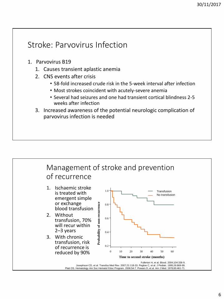
Stroke: Parvovirus Infection
1. Parvovirus B19
1. Causes transient aplastic anemia
2. CNS events after crisis • 58-fold increased crude risk in the 5-week interval after infection
• Most strokes coincident with acutely-severe anemia
• Several had seizures and one had transient cortical blindness 2-5 weeks after infection
3. Increased awareness of the potential neurologic complication of parvovirus infection is needed
Management of stroke and prevention of recurrence
1. Ischaemic stroke is treated with emergent simple or exchange blood transfusion
2. Without transfusion, 70% will recur within 2–3 years
3. With chronic transfusion, risk of recurrence is reduced by 90%
Fullerton H, et al. Blood. 2004;104:336-9.
Josephson CD, et al. Transfus Med Rev. 2007;21:118-33. Peglow C, et al. J Pediatr. 1995;26:869-99. Platt OS. Hematology Am Soc Hematol Educ Program. 2006:54-7. Powars D, et al. Am J Med. 1978;65:461-71.
Time to second stroke (months)
Pro
ba
bil
ity
of
no
n-r
ecu
rren
ce
0.2
0.4
0.6
0.8
1.0
0 10 20 30 40 50 60
Transfusion
No transfusion

30/11/2017
7
Prediction: Increased occurrence of recurrent stroke events in alternative arm counter-balanced by better management of iron overload with phlebotomy
SWiTCH: Aims and Study Design
Ware and Helms. Presented at ASH 2010 [Blood 2010;116(21):abst 844]
Aim: To compare 30 months of hydroxyurea and phlebotomy (alternative) with transfusions and deferasirox (standard) for the prevention of secondary stroke and reduction of transfusional iron
overload
161 pediatric patients with sickle cell anemia (83 male, 78 female), documented stroke and iron overload enrolled in SWiTCH (US10)
134 patients randomized 1:1
Alternative arm 67 patients
Hydroxyurea + phlebotomy
Standard arm 67 patients
Transfusions + deferasirox
US10 – SWiTCH study
ORAL
Results: Stroke Recurrence Rate
• The difference in the stroke rates between the two arms were greater than expected
Stroke incidence
Treatment arm
Transfusions +
deferasirox
Hydroxyurea +
phlebotomy
Estimated 6% 12%
Actual 0/66 (0%) 7/67 (10%)
Ware and Helms. Presented at ASH 2010 [Blood 2010;116(21):abst 844]
30/11/2017
8
Conclusions
1. Transfusions and chelation remain the gold standard treatment for secondary stroke prevention in paediatric SCD patients
2. Phlebotomy is not superior to deferasirox in reducing iron overload
3. Pre-study stroke predictions were inaccurate
4. Study was terminated early as reduction of LIC by phlebotomy could not compensate for the marked increase in secondary stroke risk with hydroxyurea
Ware and Helms. Presented at ASH 2010 [Blood 2010;116(21):abst 844]
HSCT for Stroke
1. Engrafted patients with stroke had no subsequent stroke events after BMT
2. Cerebral MRI and MRA exams demonstrated stable or improved appearance
3. In US study one with graft rejection experienced a second stroke when the Hb S fraction reached 60%

30/11/2017
9
Draft Protocol
For Discussion
When stroke is suspected in SCD
1. A full medical History, in addition:
2. Full History of onset of events
3. Detailed pre-existing neurodevelopment profile
4. Weakness, speech difficulties, changes in personality, seizures
5. Any history of possible drug abuse ( prescribed and not prescribed)
6. Any history of recent illness, recent admissions with acute chest syndrome
7. Any recent Transcranial Doppler (TCD) and/or MRI?
8. headaches?
9. History of noisy breathing or snoring at night

30/11/2017
10
Examination should include:
1. Full general systemic examination (respiratory, cardiac, GIT, MSK, ENT)
2. Detailed neurological examination including:
3. Exclusion of focal Neurological deficit
4. Exclusion of clinical features of raised intracranial pressure – papillodema, nature of pupillary response, respiratory pattern, pulse and blood pressure
5. Assessment of GCS
6. Assessment of mental state and possible aphasia
7. Assess for neck stiffness, limited straight leg raising and cranial bruit
Acute Neurological Presentations in SCD Differential Diagnosis
Diagnosis Symptoms
Meningitis/encephalitis Severe headache, neck stiffness, photophobia Rash Altered behaviour
Syncope Sudden LOC without fit? Vasovagal/cardiac
Stroke Altered mental state Aphasia, hemiparesis, ataxia, vertigo, coma
TIA Acute deficit resolves < 24 hours and normal neuro imaging
SAH Severe headache/neck stiffness +/- deficit Vaso-occlusion of calvarium Headache with tenderness +/- scalp oedema
Cerebral Malaria Altered conscious level, background history of travel to malaria prone area
Trauma Fat embolism Severe painful episode, desaturation, coma, petechial rash, multi-organ failure, DIC
Drugs Altered mental state and other related to agent Enquire about: opiates, paracetamol, NSAIDs, alcohol
Abscess Headache, fevers,. Focal signs ? background of sinusitis, otitis, mastoiditis
Tumour Headache, progressive focal signs, papilloedema

30/11/2017
11
Initial Investigations and Management-I 1. ABC
2. Commence oxygen therapy
3. Assess and Secure airway
4. Inform paediatric PNP, Paediatric SpR, PICU and paediatric consultant on-call
5. Obtain iv access x2 - Start IV fluids ( 2/3rd to full maintenance-0.9% Saline)
6. BM stix, iv access and send urgent blood tests:
7. Blood tests – see table below
Initial Investigations and Management- II 1. Admit to HDU / PICU or transfer to specialist centre
2. FBC, reticulocytes and film
3. Blood group (ABO, RhD and Kell) & antibody screen and urgent cross-match (request sickle negative blood).
4. LFTs, U&Es, blood glucose and CRP
5. INR/APTR/Fibrinogen and D-Dimers,
6. Haemoglobin analysis for HbS% if not known
7. Blood culture, urine, and throat swab for cultures and ASO titre
8. Consider malaria screen, auto- and ds DNA antibodies, cardiolipin and beta2 microglobulin antibodies

30/11/2017
12
Initial blood tests when a child with sickle cell presents with an acute neurological event
Haematology Group & save
Cross match ( see exchange transfusion protocol)
FBC and Reticulocyte count Haemoglobin analysis : HbS %, HbF %
Blood film ( Thick and thin film if malaria possibility)
Haemostasis Coagulation screen including fibrinogen
Biochemistry Venoud blood gas Blood glucose U&E
Liver profile
Bone profile, include Magnesium and Calcium
CRP
LDH
Infection Blood cultures
ASOT
HSV Serology
CMV serology
Varicella serology
Parvovirus serology
Hepatitis A,B &C serology
Immunology Full auto-immune profile
Management of acute stroke in SCD
1. Perform exchange transfusion aiming for target HbS < 30% ( follow exchange transfusion protocol) ; Top-up while waiting for exchange transfusion
2. If initial Hb is low <70g /L; top up first, then exchange-Check post exchange Hb and HbS%
3. Inform paediatric neurology team and ensure review within 24 hours of admission
4. Consider treatment with broad spectrum antibiotics and good CNS penetration and consider triple therapy-adding Acyclovir to be added and a macrolide
5. Consider LP to rule out meningitis/encephalitis
30/11/2017
13
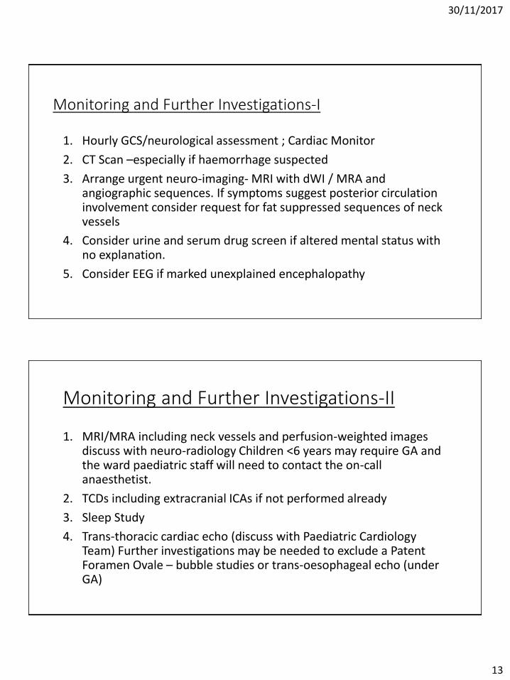
Monitoring and Further Investigations-I
1. Hourly GCS/neurological assessment ; Cardiac Monitor
2. CT Scan –especially if haemorrhage suspected
3. Arrange urgent neuro-imaging- MRI with dWI / MRA and angiographic sequences. If symptoms suggest posterior circulation involvement consider request for fat suppressed sequences of neck vessels
4. Consider urine and serum drug screen if altered mental status with no explanation.
5. Consider EEG if marked unexplained encephalopathy
Monitoring and Further Investigations-II
1. MRI/MRA including neck vessels and perfusion-weighted images discuss with neuro-radiology Children <6 years may require GA and the ward paediatric staff will need to contact the on-call anaesthetist.
2. TCDs including extracranial ICAs if not performed already
3. Sleep Study
4. Trans-thoracic cardiac echo (discuss with Paediatric Cardiology Team) Further investigations may be needed to exclude a Patent Foramen Ovale – bubble studies or trans-oesophageal echo (under GA)
30/11/2017
14
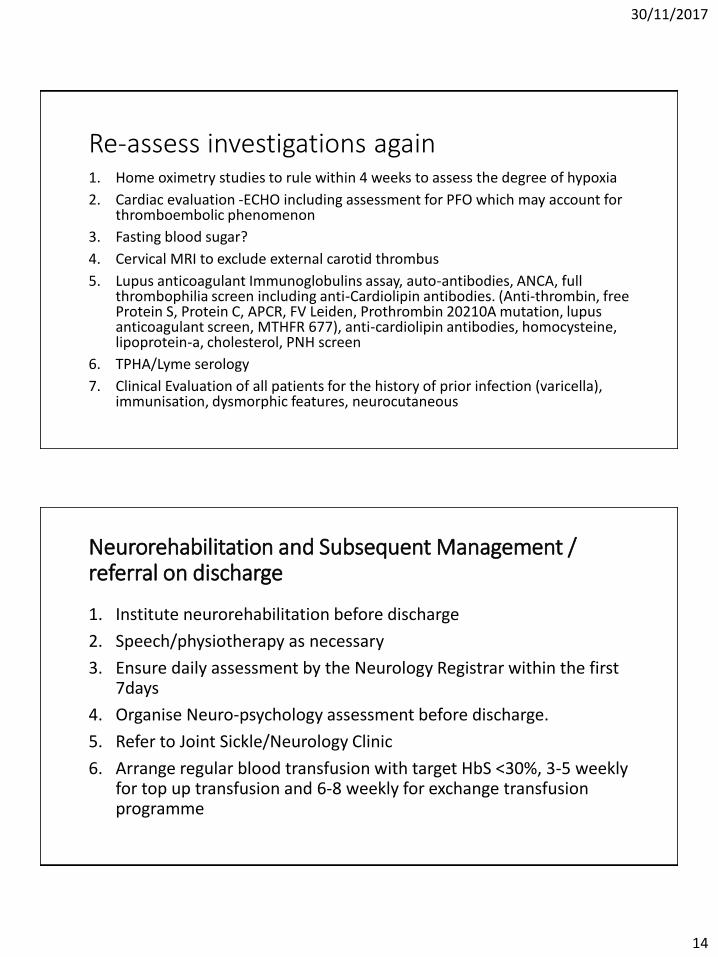
Re-assess investigations again 1. Home oximetry studies to rule within 4 weeks to assess the degree of hypoxia
2. Cardiac evaluation -ECHO including assessment for PFO which may account for thromboembolic phenomenon
3. Fasting blood sugar?
4. Cervical MRI to exclude external carotid thrombus
5. Lupus anticoagulant Immunoglobulins assay, auto-antibodies, ANCA, full thrombophilia screen including anti-Cardiolipin antibodies. (Anti-thrombin, free Protein S, Protein C, APCR, FV Leiden, Prothrombin 20210A mutation, lupus anticoagulant screen, MTHFR 677), anti-cardiolipin antibodies, homocysteine, lipoprotein-a, cholesterol, PNH screen
6. TPHA/Lyme serology
7. Clinical Evaluation of all patients for the history of prior infection (varicella), immunisation, dysmorphic features, neurocutaneous
Neurorehabilitation and Subsequent Management / referral on discharge
1. Institute neurorehabilitation before discharge
2. Speech/physiotherapy as necessary
3. Ensure daily assessment by the Neurology Registrar within the first 7days
4. Organise Neuro-psychology assessment before discharge.
5. Refer to Joint Sickle/Neurology Clinic
6. Arrange regular blood transfusion with target HbS <30%, 3-5 weekly for top up transfusion and 6-8 weekly for exchange transfusion programme

30/11/2017
15
Principles of Regular Blood Transfusions 1. Following a stroke, children are transfused regularly into adulthood
to prevent the occurrence of further strokes
2. Aim for a target pre-transfusion HbS% < 30%
3. After 3 years, of consistent transfusion, the HbS% may be allowed to rise to <50%
4. Children who cannot receive regular blood transfusion might be considered for hydroxyurea
5. Monitor ferritin and discuss iron chelation therapy (to commence when ferritin >1000), baseline liver FerriScan at onset of chelation
6. Monitoring for iron overload (see guidelines)
Long Term Management
1. Yearly joint clinic with paediatric neurologist
2. Yearly MRI including MRA scans to rule out any progression of Cerebrovascular changes
3. Annual Transcranial Doppler Scans (TCD) including assessment of extracranial vascular changes.
4. Regular Neurocognitive testing
5. Hydroxycarbamide should be considered as part of secondary prevention when blood is not suitable e.g. multiple antibodies, or as alternative where blood transfusion is not acceptable to the family

30/11/2017
16
Long Term Management
1. Consider anticoagulant therapy in presence of other risk factors
2. Consider haematopoietic stem cell transplantation in children and young people staring on regular blood transfusion
3. Silent Cerebral Infarction- Discuss the benefit of blood transfusion with the children, families and neurologists. Factors favouring transfusion:
• Impaired cognition
• Increasing size and / increase numbers of silent cerebral infarct lesions
4. Other co-existing morbidities of SCD including pain, conditional TCD, progressive renal impairment
Ischaemic stroke Hemorrhagic Silent stroke
Incidence 2–5 20–29 22% 6–12 years
Presentation Hemiparesis, aphasia,
focal seizures
HA, altered mental
status. Seizures, syncope
Neurocognitive
dysfuntion
Imaging Infarcts dICA, MCA,
stenosis, moyamoya
SAH, ICH aneurysms,
moyamoya
Arterial borderzone
infarcts deep white
matter
Pathology Intimal hyperplasia,
thrombosis, smooth
muscle hypertrophy
Aneurysmal dilation
Location Frontal, parietal,
temporal, BG, thalamus
>1.5 cm
Frontal, parietal temporal
<1.5 cm
Screening TCD MRI NeuroPsych?
Management Transfusions, SCT, HU? Surgical ?
Summary- Different Neurologic Lesions