structure of the yeast polarity protein sro7 reveals a snare regulatory mechanism
TRANSCRIPT

LETTERS
Structure of the yeast polarity protein Sro7 revealsa SNARE regulatory mechanismDouglas A. Hattendorf1, Anna Andreeva2, Akanksha Gangar2, Patrick J. Brennwald2 & William I. Weis1
Polarized exocytosis requires coordination between the actincytoskeleton and the exocytic machinery responsible for fusionof secretory vesicles at specific sites on the plasma membrane1.Fusion requires formation of a complex between a vesicle-boundR-SNARE and plasma membrane Qa, Qb and Qc SNARE proteins2.Proteins in the lethal giant larvae protein family, including lethalgiant larvae and tomosyn in metazoans and Sro7 in yeast, interactwith Q-SNAREs and are emerging as key regulators of polarizedexocytosis3. The crystal structure of Sro7 reveals two seven-bladedWD40 b-propellers followed by a 60-residue-long ‘tail’, which isbound to the surface of the amino-terminal propeller. Deletion ofthe Sro7 tail enables binding to the Qbc SNARE region of Sec9and this interaction inhibits SNARE complex assembly. The N-terminal domain of Sec9 provides a second, high-affinity Sro7interaction that is unaffected by the tail. The results suggest thatSro7 acts as an allosteric regulator of exocytosis through inter-actions with factors that control the tail. Sequence alignments indi-cate that lethal giant larvae and tomosyn have a two-b-propellerfold similar to that of Sro7, but only tomosyn appears to retain theregulatory tail.
Growth of the bud during cell division in Saccharomyces cerevisiaerequires polarized vesicle fusion at the bud tip1. Sro7, a 1,033-residueprotein, is part of the cellular machinery that facilitates this process4.Physical and genetic interactions exist between Sro7 and other exocyticproteins, including SNAREs, exocyst subunits and the Rab GTPaseSec4 (refs 4–6). In addition, Sro7 suppresses mutations in Rho3 andCdc42, two Rho GTPases necessary for exocytosis and polarizationof the actin cytoskeleton, and in Myo2, a myosin involved in vesicletransport7–9. Genetic analysis places Sro7 function downstream of theGTPases and the exocyst5. However, the molecular mechanism of Sro7in polarized bud growth remains unclear.
Like Sro7, lethal giant larvae (LGL) and tomosyn regulate targeteddelivery of secretory vesicles. LGL acts during establishment of polarityin epithelial cells and asymmetric neuroblast division3,10, and tomosynfunctions in the presynaptic nerve terminal to inhibit priming of syn-aptic vesicles11,12. Sro7, LGL and tomosyn each physically interact withQ-SNARE proteins, but with different specificities. Sro7 binds to Sec9(ref. 4), which has both Qb and Qc SNARE motifs, whereas LGL bindsto syntaxin 4, a Qa-SNARE13. Tomosyn has an R-SNARE motif at itscarboxy terminus that substitutes for synaptobrevin in the neuronalSNARE complex14,15. Sro7 and LGL lack this R-SNARE motif, although,40 amino acids at the extreme C terminus of Sro7 are predicted toform an a-helix of unknown function15.
We determined the crystal structure of a proteolytic fragment of S.cerevisiae Sro7, spanning residues 61–962, at 2.4 A resolution. Thefinal model includes residues 62–582 and 598–951. The sequencecontaining the predicted a-helix at the C terminus was protease-sensitive and is absent from the crystallized fragment. The structure
consists of 14 WD40 repeats distributed throughout almost the entiresequence, followed by a 60-residue tail (Fig. 1a–d). The WD40repeats fold into two seven-bladed b-propeller domains with a topo-logy similar to Aip1 (refs 16, 17). Blades 1–7 form the N-terminaldomain, and blades 8–14 form the C-terminal domain. Each blade isan antiparallel b-sheet, typically comprising four b-strands labelled Ato D. The bottom surface of each propeller is formed by the A–B andC–D loops, and the top surfaces are formed by the B–C and D–Aloops.
The b-propeller domains are arranged to resemble a twisted, openclamshell. A notable feature of the domain interface is the amphi-pathic 7C–8A loop, which is buried between the two domains(Fig. 1c). Its hydrophobic face packs against the N-terminal b-propeller (Supplementary Fig. 1a), and its hydrophilic face interactswith the C-terminal b-propeller through an extensive network ofdirect and water-mediated hydrogen bonds (Supplementary Fig.1b). Another feature of the interface is the 8AB loop, which isanchored by a hydrophobic a-helix that binds to the C-terminalb-propeller and which contains an extended 23-residue region thatwraps around the 1AB and 8CD loops at the bottom side of thedomain interface (Supplementary Fig. 1c). Leu 490 and Leu 491 inthe extended region act as a second anchor by packing into a hydro-phobic pocket formed by residues in the N-terminal b-propeller andthe 7C–8A loop (Supplementary Fig. 1a).
The Sro7 tail (residues 892–951) is bound to the bottom surface ofthe N-terminal b-propeller (Fig. 1e). The binding interface is extens-ive, with 4,356 A2 of buried surface area. The base of the tail (residues898–901) binds to a pocket with one side formed by residues 492–497of the 8AB loop, which links the tail to the domain interface. The tailspans the entire propeller once in an extended conformation, thenagain as two short a-helices separated by a 10-residue linker. Thesecond a-helix (residues 945–949) forms the other side of the bindingpocket for tail residues 898–901.
Sequence alignment of Sro7 with tomosyn and LGL shows signifi-cant conservation in the 14 WD40 repeats and in many elements ofthe domain interface (Fig. 1d and Supplementary Fig. 2). The meta-zoan proteins have insertions in the 10D–11A loop that containphosphorylation sites implicated in their regulation18,19. Notably,many residues in the Sro7 tail that interact with the N-terminalpropeller (Fig. 1e) are conserved in tomosyn, but not in LGL. Thisdivergence is also reflected in residues that form the binding site forthe tail: they are conserved in the yeast (Fig. 2a) and tomosyn sub-families, but vary among the LGL proteins (Supplementary Fig. 2). Incertain positions, co-variation of residues in the tail and its bindingsite in tomosyn seem to preserve the contacts observed in Sro7.
The binding site for the tail on Sro7 is largely hydrophilic (Fig. 1e),suggesting that the tail serves a regulatory rather than a structuralrole. The effect of deleting the Sro7 tail on binding of Sec9 was
1Departments of Structural Biology and of Molecular and Cellular Physiology, Stanford University School of Medicine, 299 Campus Drive West, Stanford, California 94305-5126, USA.2Department of Cell and Developmental Biology, University of North Carolina, 538 Taylor Hall, Chapel Hill, North Carolina, 27599-7090, USA.
Vol 446 | 29 March 2007 | doi:10.1038/nature05635
567Nature ©2007 Publishing Group

measured by isothermal titration calorimetry (ITC; SupplementaryFig. 3 and Supplementary Table 2). An Sro7 variant lacking the tail,Sro7(61–891), binds to Sec9 with 3.3-fold higher affinity (Kd 5800 nM) and with a 2.3-fold more favourable enthalpy change thaneither wild-type Sro7 or the crystallization construct Sro7(61–962),which contains the tail. Thus, the tail has an autoinhibitory effect onSro7 binding to Sec9.
To understand this effect of the tail, we first defined the regions ofSec9 that bind to Sro7 using a yeast two-hybrid assay. Sec9 has a 413-residue N-terminal domain that is not required for growth20, followedby the essential C-terminal region that contains the Qb and Qc SNAREmotifs, designated the Qbc-SNARE domain (Supplementary Fig. 4a).The N-terminal domain interacts strongly with Sro7, whereas the Qbc-SNARE domain shows a weaker interaction (Supplementary Fig. 4a).Residues 255–343 in the N-terminal domain were necessary for thehigh-affinity interaction, because their deletion reduced binding tothe level of the isolated Qbc domain. The region of Sec9 necessaryfor suppression of the sec4-P48 mutation20 (residues 3–82; Supplemen-tary Fig. 4a) is distinct from the high-affinity Sro7 binding site. Thetwo-hybrid results were confirmed by co-immunoprecipitation (Sup-plementary Fig. 4b). Deletion of residues 255–343 of Sec9 reduced theamount of Sro7 precipitated with Sec9, but a significant fraction of Sro7was still associated with the Sec9 mutant.
On the basis of the results of the two-hybrid assay and additionalmapping experiments (see Supplementary Methods), a fragment ofthe Sec9 N-terminal domain was purified to measure its binding toSro7 in vitro. Sec9(250–372) bound to Sro7(61–962) with a Kd of3mM, identical to that of full-length Sec9 (Supplementary Fig. 3 andSupplementary Table 2). Significantly, deletion of the Sro7 tail hadno effect on binding of Sec9(250–372) (Supplementary Fig. 3 andSupplementary Table 2). To locate the binding site for the Sec9 N-terminal domain fragment, we examined the conservation of Sro7surface residues. In addition to the highly conserved tail-binding site(Fig. 2a), there is a second cluster of conserved residues in a shallowgroove formed by the A–B and C–D loops of blades 9–12 (Fig. 2b). Ofsix mutations in the second cluster, two (Y700A and P721A) abolishedbinding to Sec9(250–372), and three others (E562A, R664A andM764A) each caused a nearly tenfold reduction of the binding affinity(Supplementary Fig. 5 and Supplementary Table 2).
Next, binding of the Sec9 Qbc domain to Sro7 was assessed in vitro.In this case, binding was only observed if the Sro7 tail was deleted(Fig. 3a). This interaction was detectable by ITC (Supplementary Fig.3), but the low affinity and small enthalpy change prevented a pre-cise determination of the binding thermodynamics. Nonetheless,two independent titrations indicated a Kd of approximately 50 mM.These results indicate that in wild-type Sro7, the tail masks the
8
910
11
12
1314
NC
*
*
b
1
23
4
5
67
N C
1062
1033
1152
S. cerevisiae Sro7
Mus musculus LGL1
Rattus norvegicus tomosyn
P
PPP
N-terminal propeller C-terminal propeller Tail
R-SNARE
α-helix
A
BC
D
d
Tail
8ABloop
Tail
7C–8A loop
8AB loopTail
Crystallized fragment 96261
Top surface
Bottom surface
S897
D898
T899L900 Y901
I902 I905
I907 R910
P911
V913
N914
Q917
W918
L930
L933
L934
N931R939
S942
K943
Y944
E946 S947
I949
L490L491
G493G494
V495R496
T497
Tail
8ABloop
a
c
e
Figure 1 | Overview of the Sro7 structure. a–c, The N-terminal b-propelleris light green, the C-terminal b-propeller is light blue, the 8AB loop is yellow,and the tail is dark blue. a, View of the bottom surface of Sro7, along the axisof the N-terminal b-propeller. WD40 blades 1–7, b-strands A–D of blade 6,the 8AB loop and the tail are labelled. b, View along the axis of the C-terminalb-propeller. WD40 blades 8–14 are labelled. Asterisks mark the loop of blade9 that is not visible in the structure (residues 583–597). c, Side-view of thestructure; the interface loops and the tail are labelled. d, Schematic of thestructure-based sequence alignment of the LGL family. Structural elementsare coloured as in a–c. The predicted C-terminal a-helix in Sro7 and theR-SNARE of tomosyn are shown in red. Light green and light blue boxescorrespond to the WD40 blades as observed structurally, rather than thestandard definition of a WD40 repeat; that is, strand D of one blade andstrands A–C of the next blade. Note that strand 14D is located near theN-terminus of Sro7, before strand 1A, and is indicated by a narrow blue box.Phosphorylation sites in tomosyn and LGL are marked by ‘P’. e, The Sro7tail-binding site, with the structure oriented as in panel a. The b-propellerdomains are shown in surface representation and coloured by electrostaticpotential25 in the range of –10kT/e (red) to 10kT/e (blue). The tail and the8AB loop are shown as ribbons. Side chains in the tail that interact with theN-terminal domain are shown as sticks. Side chains in the 8AB loop thatform part of the tail-binding site are also shown.
LETTERS NATURE | Vol 446 | 29 March 2007
568Nature ©2007 Publishing Group

Qbc-SNARE binding site, either by direct competition or through anallosteric mechanism. This interaction is not entirely specific to Sec9,because the SNARE domain of the Qa-SNARE Sso1 (residues 193–265) also binds Sro7(61–891), albeit more weakly (SupplementaryFig. 6a).
Sro7(61–891) did not bind to the pre-assembled SNARE complexof GST–Sec9 Qbc-SNARE, the Qa-SNARE Sso1 and the R-SNARESnc2 (Fig. 3a). In a kinetic assay, Sro7(61–891) slowed SNARE
complex assembly fourfold relative to assembly reactions lackingSro7 or containing Sro7(61–962) (Fig. 3b and Supplementary Fig.6b). Thus, Sro7(61–891) binds to the Sec9 Qbc-SNARE domain in amanner that competes with SNARE complex formation. At the con-centrations used in the kinetic assay, approximately 60% of the Sro7and full-length Sec9 are expected to be in a complex, whereas almostno complex would exist in the absence of the Sec9 N-terminaldomain. The Sec9 N-terminal domain may act as a high-affinity
Y700
P721M764
R664
E562
Q765
a b
Figure 2 | Surface representation of Sro7, with residues coloured fromwhite to yellow by increasing conservation. The brightest yellow represents100% identity. Because the Sec9 N-terminal domain is unique to theSaccharomycotina subphylum of fungi, the sequences used were limited tospecies in this subphylum (S. cerevisiae, Candida glabrata, Kluyveromyceslactis, Ashbya gossypii, Yarrowia lipolytica, Candida albicans and
Debaryomyces hansenii). The tail is shown as a dark blue ribbon.Conservation scores were calculated with ConSurf26. a, View along the axis ofthe N-terminal b-propeller, as in Fig. 1a, showing the conservation of thetail-binding site. b, Structure rotated from a to show the second conservedsite on the C-terminalb-propeller. Residues that were mutated to alanine arelabelled on the surface.
GST
Sro7 FL + + – – – – – –
Sro7 61–891 – – – – + + + +GST–Sec9 Qbc – – – –+ + + +Sso1/Snc2 – – – – – – –+
– – – – – – – +
a
GST–Sec9Qbc
GST
Sso1(145–265)Snc2(1–92)
Sro7 variants
37 °C
sc-URA + doxy
pCM189expressing:
25 °C
CEN vectorexpressing:
YPD,37 °C
SRO7, SRO77
19 °C
YPD,25 °C
YPD,19 °C
VectorSRO7
sro7-∆142sro7-∆42
c50
40
30
20
10
0
Bgl
2 in
tern
al (%
)
20
10
15
25
5
0
sro7∆, sro77∆
VectorSRO7
sro7-∆142sro7-∆42
VectorSRO7
sro7-∆142sro7-∆42
VectorSRO7
sro7-∆142sro7-∆42 pCM189
expressing:
CEN vectorexpressing:
sro7∆, sro77∆
SRO7, SRO77
SRO7
sro7-
∆42
sro7-
∆142sc-URA
– doxy
d
b
Time (s)
1.0
0.8
0.6
0.4
0.2
200 600 1,0000
Frac
tion
Sso
1 b
ound
Bgl
2 in
tern
al (%
)
SRO7
sro7-
∆42
sro7-
∆142
Sro7 61–962 – – – – – –+ +
Figure 3 | Interaction of Sec9 with Sro7. a, GST pull-down assays of bindingof Sro7 variants to the GST–Sec9 Qbc-SNARE domain. A gel stained withCoomassie blue is shown. b, Fraction of Sso1(145–265) pulled down byGST–Sec9 as a function of time in the absence of Sro7 (filled circles), orpresence of Sro7(61–962) (open squares) or Sro7(61–891) (filled squares).Error bars, s.d.; n 5 3. Data were fitted to a second-order rate equation (seeSupplementary Information). In the absence of Sro7, the rate constant is7,300 6 1,200 M21 s21, which is very similar to that previously determinedusing a circular dichroism assay for assembly27. Sro7(61–962) had no effecton assembly (rate constant of 7,200 6 700 M21 s21, whereas Sro7(61–891)
reduced the rate constant to 1,800 6 200 M21 s21. c, The effect of SRO7 tail-deletion mutants on growth. The top panel shows the ability of SRO7 allelesto complement the cold-sensitive growth phenotype of the sro7D,sro77Dstrain. The bottom panel examines dominant-negative effects of the SRO7alleles in wild-type yeast. In each panel, growth of three independentcolonies at the permissive (37 uC) and restrictive temperatures (25 uC and19 uC) is shown. d, The effect of SRO7 alleles on secretion. Graphs show theper cent of the secreted Bgl2 enzyme that remained inside spheroplasted cellsin either the sro7D,sro77D strain (top panel) or wild-type strain (bottompanel). Error bars, s.d.; n 5 3.
NATURE | Vol 446 | 29 March 2007 LETTERS
569Nature ©2007 Publishing Group
tether for the Qbc-SNARE domain, such that the regulatory inter-action between Sro7 and the Qbc domain occurs at a biologicallysignificant concentration. Given the sequence homology amongQ-SNAREs21, a second, related role may be to increase the specificityof Sro7 for the Qbc-SNARE over Sso1.
The Sro7 mutants that are defective in binding to the Sec9 N-terminal domain had no observable phenotype when expressed inyeast (data not shown), consistent with the fact that this domain isnonessential20. In contrast, deletion of residues 892–1033 (sro7-D142),which removes the tail, caused a loss-of-function phenotype in asro7D,sro77D strain and a dominant-negative phenotype in thewild-type strain (Fig. 3c). In the complementation assay, the sro7-D142 mutant was expressed at only 20% of wild-type Sro7 level(Supplementary Fig. 7). However, the mutant does not complementsro7D,sro77D even when expressed at a level higher than wild-type
Sro7, from the stronger tetracycline-repressible promoter (data notshown). These phenotypes were specifically associated with removalof the tail that is observed in the structure, because a smaller deletionof the predicted a-helix at the C terminus (residues 987–1,028; sro7-D42) was a much weaker loss-of-function allele with no dominant-negative effect on growth (Fig. 3c). The growth phenotypes correlatedwith defects in secretion, monitored by examining the accumulationof internal pools of the secreted enzyme Bgl2 (Fig. 3d). Taken togetherwith the biochemical data, this suggests that the tail is an essentialcomponent of Sro7 function during SNARE-mediated exocytosis.
The interaction of wild-type Sro7 with the Sec9 Qbc-SNAREdomain in vivo suggests that Sro7 exists in two conformational stateswith different affinities for the tail. The equilibrium between thesestates may be affected by other factors that regulate Sro7 activity.Such factors could bind the tail directly, or they could bind to a distantsite on Sro7 and regulate the tail through an allosteric mechanism. Thearchitecture of Sro7 is suggestive of an allosteric mechanism wherebyrearrangements in the b-propeller domain interface that are assoc-iated with ligand binding are propagated to the tail through inter-action between the tail and the 8AB loop (Fig. 1e and SupplementaryFig. 2).
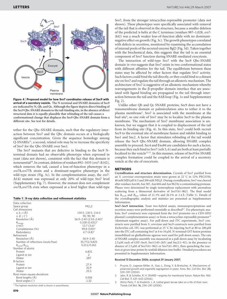
Unlike other Qb and Qc SNARE proteins, Sec9 does not have atransmembrane domain or palmitoylation sites to tether it to theplasma membrane2. Sro7 is associated with the membrane at thebud site4, so one role of Sro7 may be to localize Sec9 to the plasmamembrane. The mechanism of Sro7 membrane association is un-known, but we suggest that it is coupled to displacement of the tailfrom its binding site (Fig. 4). In this state, Sro7 could both recruitSec9 to the eventual site of membrane fusion and inhibit binding toSso1 and Snc2. A factor that stimulates rebinding of the tail wouldrelease the Sec9 Qbc-SNARE domain, allowing SNARE complexassembly to proceed. Sec4 and Exo84 are candidates for such a factor,because they each bind to Sro7 (refs 5, 6) and are both at least partiallylocalized to the vesicle22,23. In this manner, release of Sec9 for SNAREcomplex formation could be coupled to the arrival of a secretoryvesicle at the site of exocytosis.
METHODSCrystallization and structure determination. Crystals of Sro7 purified from
an S. cerevisiae overexpression strain were grown at 22 uC in 12% PEG3350,
40 mM MES pH 6.5 and 200 mM NH4F. During crystallization, Sro7 was cleaved
after residues Asn 60, Asn 587, Asn 604 and Asn 962 by a contaminating protease.
Phases were determined by single isomorphous replacement with anomalous
scattering from a thimerosal derivative of Sro7(61–962). The final model
has Rwork and Rfree values of 21.1% and 26.1% at 2.4 A (Table 1). Details of
the crystallographic analysis and statistics are presented as Supplementary
Information.
Sro7–Sec9 interaction. Yeast two-hybrid assays, immunoprecipitations and
secretion assays were performed essentially as described4,7. For phenotypic ana-
lysis, Sro7 constructs were expressed from the Sro7 promoter on a CEN-HIS3
plasmid (complementation assay) or from a tetracycline-repressible promoter24
(dominant-negative assay). For pull-down and ITC experiments, Sro7 con-
structs were purified from S. cerevisiae and Sec9 constructs were purified from
Escherichia coli. ITC was performed at 25 uC by injecting Sec9 at 80 to 200mM
into the ITC cell containing Sro7 at 4 to 10 mM. N-terminal GST fusion proteins
immobilized on gluthathione-agarose were used for pull-down assays. The rate
of SNARE complex assembly was measured in a pull-down assay by incubating
2.5mM each of GST–Sec9, Sso1(145–265) and Snc2(1–92), in the presence or
absence of 2.5mM of Sro7(61–962) or Sro7(61–891), then quenching the reac-
tion at given time points by tenfold dilution into buffer. Detailed procedures are
presented in Supplementary Information.
Received 13 December 2006; accepted 29 January 2007.
1. Pruyne, D., Legesse-Miller, A., Gao, L., Dong, Y. & Bretscher, A. Mechanisms ofpolarized growth and organelle segregation in yeast. Annu. Rev. Cell Dev. Biol. 20,559–591 (2004).
2. Jahn, R. & Scheller, R. H. SNARE—engines for membrane fusion. Nature Rev. Mol.Cell Biol. 7, 631–643 (2006).
3. Wirtz-Peitz, F. & Knoblich, J. A. Lethal giant larvae take on a life of their own.Trends Cell Biol. 16, 234–241 (2006).
Sec4
Vesicle
Sso1
Snc2
Sro7
Plasma membrane
Exocyst
Sec9
?
?
NQb
Qc
R
Qa
N
Figure 4 | Proposed model for how Sro7 coordinates release of Sec9 witharrival of a secretory vesicle. The N-terminal and SNARE domains of Sec9are indicated by N, Qb, and Qc. Although the figure depicts direct binding ofthe Sec9 Qbc-SNARE domain to the tail-binding site, in the absence of directstructural data it is equally plausible that rebinding of the tail causes aconformational change that displaces the Sec9 Qbc-SNARE domain from adifferent site. See text for details.
Table 1 | X-ray data collection and refinement statistics
Data collectionSpace group P42
12
Cell dimensionsa, b, c (A) 130.5, 130.5, 116.0a, b, c (u) 90, 90, 90
Resolution (A) 56.9–2.40 (2.53–2.40)*Rmerge 0.108 (0.431)*I/s(I) 13.7 (3.6)*Completeness (%) 99.8 (100)*Redundancy 4.7 (4.8)*
RefinementResolution (A) 56.9–2.40
Number of reflections (work/free) 35,772/3,825
Rwork/Rfree 0.211/0.261
Number of atomsProtein 6,831
Ligand or ion 2
Water 287
B-factorsProtein 27.4Ligand or ion 32.9Water 25.5
Root-mean-square deviationsBond lengths (A) 0.006
Bond angles (u) 1.32
* The highest-resolution shell is shown in parentheses.
LETTERS NATURE | Vol 446 | 29 March 2007
570Nature ©2007 Publishing Group

4. Lehman, K., Rossi, G., Adamo, J. E. & Brennwald, P. Yeast homologues of tomosynand lethal giant larvae function in exocytosis and are associated with the plasmamembrane SNARE, Sec9. J. Cell Biol. 146, 125–140 (1999).
5. Zhang, X. et al. Lethal giant larvae proteins interact with the exocyst complex andare involved in polarized exocytosis. J. Cell Biol. 170, 273–283 (2005).
6. Grosshans, B. L. et al. The yeast lgl family member Sro7p is an effector of thesecretory Rab GTPase Sec4p. J. Cell Biol. 172, 55–66 (2006).
7. Adamo, J. E., Rossi, G. & Brennwald, P. The Rho GTPase Rho3 has a direct role inexocytosis that is distinct from its role in actin polarity. Mol. Biol. Cell 10,4121–4133 (1999).
8. Adamo, J. E. et al. Yeast Cdc42 functions at a late step in exocytosis,specifically during polarized growth of the emerging bud. J. Cell Biol. 155, 581–592(2001).
9. Kagami, M., Toh-e, A. & Matsui, Y. Sro7p, a Saccharomyces cerevisiae counterpartof the tumor suppressor l(2)gl protein, is related to myosins in function. Genetics149, 1717–1727 (1998).
10. Vasioukhin, V. Lethal giant puzzle of Lgl. Dev. Neurosci. 28, 13–24 (2006).11. Gracheva, E. O. et al. Tomosyn inhibits synaptic vesicle priming in Caenorhabditis
elegans. PLoS Biol. 4, e261 (2006).12. McEwen, J. M., Madison, J. M., Dybbs, M. & Kaplan, J. M. Antagonistic
regulation of synaptic vesicle priming by Tomosyn and UNC-13. Neuron 51,303–315 (2006).
13. Musch, A. et al. Mammalian homolog of Drosophila tumor suppressor lethal (2)giant larvae interacts with basolateral exocytic machinery in Madin-Darby caninekidney cells. Mol. Biol. Cell 13, 158–168 (2002).
14. Hatsuzawa, K., Lang, T., Fasshauer, D., Bruns, D. & Jahn, R. The R-SNARE motifof tomosyn forms SNARE core complexes with syntaxin 1 and SNAP-25 anddown-regulates exocytosis. J. Biol. Chem. 278, 31159–31166 (2003).
15. Pobbati, A. V., Razeto, A., Boddener, M., Becker, S. & Fasshauer, D. Structuralbasis for the inhibitory role of tomosyn in exocytosis. J. Biol. Chem. 279,47192–47200 (2004).
16. Mohri, K., Vorobiev, S., Fedorov, A. A., Almo, S. C. & Ono, S. Identification offunctional residues on Caenorhabditis elegans actin-interacting protein 1 (UNC-78) for disassembly of actin depolymerizing factor/cofilin-bound actin filaments.J. Biol. Chem. 279, 31697–31707 (2004).
17. Voegtli, W. C., Madrona, A. Y. & Wilson, D. K. The structure of Aip1p, a WD repeatprotein that regulates cofilin-mediated actin depolymerization. J. Biol. Chem. 278,34373–34379 (2003).
18. Betschinger, J., Eisenhaber, F. & Knoblich, J. A. Phosphorylation-inducedautoinhibition regulates the cytoskeletal protein Lethal (2) giant larvae. Curr. Biol.15, 276–282 (2005).
19. Baba, T., Sakisaka, T., Mochida, S. & Takai, Y. PKA-catalyzed phosphorylation oftomosyn and its implication in Ca21-dependent exocytosis of neurotransmitter.J. Cell Biol. 170, 1113–1125 (2005).
20. Brennwald, P. et al. Sec9 is a SNAP-25-like component of a yeast SNARE complexthat may be the effector of Sec4 function in exocytosis. Cell 79, 245–258 (1994).
21. Fasshauer, D., Sutton, R. B., Brunger, A. T. & Jahn, R. Conserved structural featuresof the synaptic fusion complex: SNARE proteins reclassified as Q- and R-SNAREs.Proc. Natl Acad. Sci. USA 95, 15781–15786 (1998).
22. Goud, B., Salminen, A., Walworth, N. C. & Novick, P. J. A GTP-binding proteinrequired for secretion rapidly associates with secretory vesicles and the plasmamembrane in yeast. Cell 53, 753–768 (1988).
23. Boyd, C., Hughes, T., Pypaert, M. & Novick, P. Vesicles carry most exocystsubunits to exocytic sites marked by the remaining two subunits, Sec3p andExo70p. J. Cell Biol. 167, 889–901 (2004).
24. Gari, E., Piedrafita, L., Aldea, M. & Herrero, E. A set of vectors with a tetracycline-regulatable promoter system for modulated gene expression in Saccharomycescerevisiae. Yeast 13, 837–848 (1997).
25. Baker, N. A., Sept, D., Joseph, S., Holst, M. J. & McCammon, J. A. Electrostatics ofnanosystems: application to microtubules and the ribosome. Proc. Natl Acad. Sci.USA 98, 10037–10041 (2001).
26. Landau, M. et al. ConSurf 2005: the projection of evolutionary conservationscores of residues on protein structures. Nucleic Acids Res. 33, W299–W302(2005).
27. Nicholson, K. L. et al. Regulation of SNARE complex assembly by an N-terminaldomain of the t-SNARE Sso1p. Nature Struct. Biol. 5, 793–802 (1998).
Supplementary Information is linked to the online version of the paper atwww.nature.com/nature.
Acknowledgements We thank L. Katz for help with the yeast two-hybrid analysis,and S. Kaiser for assistance with mass spectrometry. Diffraction data weremeasured at the Advanced Light Source and the Stanford Synchrotron RadiationLaboratory. D.A.H. was supported by a fellowship from the American CancerSociety. This work was supported by NIH grants to P.J.B. and W.I.W and a grantfrom the G. Harold and Leila Y. Mathers Foundation to P.J.B.
Author Contributions Crystallographic and biochemical experiments weredesigned by D.A.H. and W.I.W. and performed by D.A.H. Plasmid and strainconstruction were designed and performed by D.A.H. and P.J.B. Yeast two-hybridanalysis, secretion assays and analysis of mutant phenotypes were designed byP.J.B. and performed by A.A. and A.G. D.A.H. and W.I.W. wrote the paper and allauthors made editorial comments.
Author Information Coordinates and structure factors have been deposited in theProtein Data Bank under accession number 2OAJ. Reprints and permissionsinformation is available at www.nature.com/reprints. The authors declare nocompeting financial interests. Correspondence and requests for materials shouldbe addressed to W.I.W. ([email protected]) or P.J.B.([email protected]).
NATURE | Vol 446 | 29 March 2007 LETTERS
571Nature ©2007 Publishing Group