structure of the skin superficial epithelial layer (epidermis) deep connective tissue layer (dermis)...
TRANSCRIPT

Structure of the skin

Structure of the skin


Structure of the skin
EpidermisEpidermis
Keratinized Keratinized stratified stratified squamous squamous epithelium epithelium devoid of blood devoid of blood vesselsvessels
DermisDermis
Connective tissue Connective tissue containing containing (bl. v. (bl. v. lymph v., sensory lymph v., sensory nerve endings, nerve endings, smooth m, hair smooth m, hair follicles, sweat and follicles, sweat and sebaceous glands) sebaceous glands)
In its deep part the In its deep part the collagencollagen bundles bundles are arranged in are arranged in parallel rowsparallel rows

Epidermis
Thickness:The epidermis is generally thin except in :• The palms of the hand.• The soles of the feet.
Why?To protect these parts and withstand friction, wear and tear that occurs in these regions.

Functions of the Skin
1-Protection abrasion, invasion, water loss, UV protection 2-Vitamin D synthesis epidermal keratinocytes when exposed to
UV light helps maintain health of skeleton by
increasing absorption of Ca2+ 3-Sensation receptors for heat, cold, touch, pressure,
vibration and pain

4- Thermoregulation thermo receptors and sweat glands hypothalamus controls cutaneous
arteries and sweat glands to retain or dissipate heat
5- Psychological and social functions appearance and social acceptance facial expression and nonverbal
communication

Lines of cleavage
• The collagen fibers, arranged in parallel rows, called:
Lines of cleavage (langer’s lines):
• The direction of the rows of collagen fibers in the dermis:
It runs
•Longitudinally in the limbs.•Circumferentially in the neck and the trunk.

Lines of cleavage
These lines are important
to determine the direction
for an incision (cut) during
a surgery to avoid obvious
scars.

• A surgical incision along or between these lines causes the minimum disruption of collagen so that the wound heals with a small scar.
• Conversely, an incision made across the rows of collagen makes a disruption resulting in the massive production of fresh collagen and the formation of a broad scar.

Skin creases
Folded skin over the
joints.
Skin is thin and is
firmly adherent to
underlying structures.

Skin Color
Due to Melanin, a pigment in the epidermis and Carotene,
a pigment in dermis as well as the blood in the capillaries
of the dermis. Melanin is synthesized in cells called Melanocytes (found in
basal layer). Number of Melanocytes is essentially the same in all races. The differences in skin color is due to the amount of pigment the
melanocytes produce. When skin is exposed to ultraviolet radiation, enzymatic activity
is increased and both the amount and darkness of melanin increase and the skin darkens as a protective measure

• Nails• Hairs• Sebaceous glands• Sweat glands
The appendages of the skin

A nail is a flat horny plate on the dorsal surface of tips of the fingers and toes
It has: Root: proximal edge (part
embedded in skin) body: exposed part & has a
free distal edge Nail fold: folds of skin
surround and overlap the nail
• Nail bed is very vascular causing pink color of the nail• The germinative zone lies beneath the root& is responsible for growth of nail
Nails

Cover whole surface of the body except some areas as lips, palms, soles, glans, clitoris, L. minora.
Hairs

• A band of smooth muscle
connects the undersurface of the
follicle to the superficial part of
the dermis.
• It is innervated by sympathetic
nerve fibers.
• It is involuntary.
Arrector Pilli muscle

Function It secrets sebum to oil
(lubricate) hair and skin.
Sebum An oily material that
keeps the flexibility of the hair and oils the epidermis around the mouth of the follicle.
Sebaceous glands
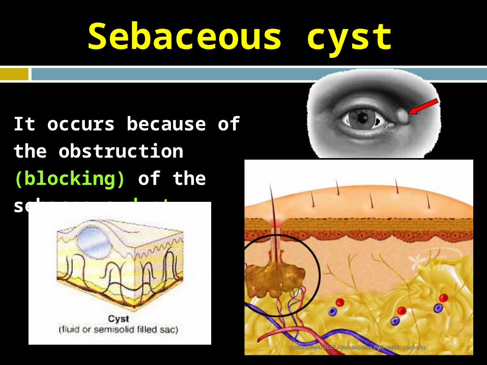
It occurs because of the
obstruction (blocking) of the
sebaceous duct.
Sebaceous cyst

• long tubular glands with deep coiled part.
• All over the body except red margins of lips, nail beds, glans penis and clitoris.
• The most deeply penetrated structure.
Sweat glands

Rule of NineRule of Nine

Burns
1st degree burn: Damages only epidermis (sunburn with reddening of the skin). 2nd degree burn: Damages much of the epidermis but leaves some epidermal remnant.
Re-growth from remnants is possible. Blisters are common and pain is often severe since the skin
nerves are irritated by the products of cellular destruction.

Burns
3rd Degree burn:
It reaches to and through dermis (May expose muscle
and bone. No epidermal remnants are present. Little or no feeling of pain because of destruction of
nerves. Treatment requires skin grafts to provide epidermal
cells.

Major Problems of Burns
Infection. Maintaining fluid. Maintaining electrolyte balance which requires food
and fluid intake. Contractures of skin and underlying connective tissue
and muscle due to intense scarring.


Skin burns
DeepDeepSuperficialSuperficial
Heals slowly from the edges.
Usually needs skin grafting.
Heals rapidly from the edges, cells of HF and glands.
Heals quickly.
Doesn’t need a skin graft.

Clinical notes
Graft is transferring tissue from one site to another.
Skin graft is needed when the skin is damaged ( usually by deep
burning )