structure of the mouthparts of (morgan) …
TRANSCRIPT
STRUCTURE OF THE MOUTHPARTS OF Frankliniella bispinosa (Morgan) (THYSANOPTERA: THRIPIDAE)
Carl C. Childers and Diann S. Achor
Citrus Research and Education Center
University of Florida-IFAS
Lake Alfred, Florida USA
Introduction
Thrips are increasingly recognized as potentially serious pests in
a number of different agricultural, ornamental and sylvan commodities
worldwide as indicated by the papers presented at this conference. The
small size of thrips, their large numbers, capacity for flight and wind
dispersal, wide host ranges, poorly understood life histories and
probable potential for pesticide resistance exacerbate the timely
development of control strategies. Thrips feed on pollen, leaves,
flowers or young fruit and oviposit in various plant tissues. Some thrips
feed on other arthropods or on fungal spores or hyphae. Plant injury is
characterized by chlorosis, distortion or galling of leaves, spotting of
leaves or fruit with fecal droplets, scarring of young fruit, premature
fruit loss, or reduced yields from excessive reduction by feeding in the
quality or quantity of viable pollen (Palmer et al. 1989). Several thrips
species also function as direct or indirect vectors of certain viral, fungal
or bacterial diseases (Ananthakrishnan 1980). These problems focus
on the need for a better understanding of thrips including the structure
and function of their mouthparts relative t o plant injury and disease
transmission.

Studies by Mound (1971), Heming (1978), Milne & Manicom
(1 978), Chisholm & Lewis (1 984) and others have conclusively shown
that thrips have piercing-sucking mouthparts. Excellent morphological
studies of a limited number of thrips species have been completed
including the larvae of Haplothr@s verbasci Osborn by Heming (1 9781,
Limothr@s cerealium (Haliday) by Chisholm & Lewis (1 984), Scirtothrips
citri (Moulton) by Wiesenborn & Morse (I 988), Frankliniella occidentalis
(Pergande) and F. schultzei (Trybom) by Hunter & Ullman (1 989).
Thirteen species of thrips have been identified in association wi th
citrus flowers and developing buds in Florida with Frankliniella bispinosa
(Morgan) the prevalent species (Childers et al. 1990). Injury to citrus
flowers and buds by F. bispinosa has been documented (C.C.C. & D.S.A, unpublished data). Chemical control programs are in progress
to evaluate the impact on fruit set following suppression of thrips during
the citrus flowering cycle.
The object of this paper is to review the structure of the
mouthparts of F. bispinosa illustrated by a series of light, scanning and
transmission electron micrographs. This will provide additional
information to identify or compare structures with those of other
species of thrips.
Materials and Methods
At frequent intervals between February and April 1988, thrips
were collected from open flowers or swollen buds in a "naval" orange
grove directly into one pint or one quart Mason jars filled wi th 70%
ethanol and returned to the laboratory. Open flowers and swollen buds
wi th live thrips were collected directly into paper bags in the field,
placed in an ice chest and returned to the laboratory for processing.
Light Microscopy. Whole-mounts of thrips were prepared using
Hoyer's mounting medium (Krantz 1978). Light micrographs were
taken through a Zeiss compound microscope equipped wi th a Minolta
35mm camera on Panatomic X ASA 3 2 film.
Scanning Electron Microscopy (SEM). Chloroform was applied
directly t o the blossom on which selected individual thrips were feeding
in the laboratory. Dead or inactive thrips were then transferred into
micro-tissue capsule tubes enclosed by 150 mesh grids and placed in
3% glutaraldehyde or Hallam's variation of Karnovsky's fixative with a
0.1 M potassium phosphate buffer at pH 7.2 for 3-4 hr (Hallam &
Chambers 1970). Post-fixation was in 2 % osmium tetroxide in the
same buffer for 4 hr at 25OC. Samples were dehydrated in ethanol and
critical point dried in a Ladd Critical Point Drier using carbon dioxide.
Thrips were mounted ventral side up on stubs, sputter coated wi th 150-
200 A gold in a Ladd Sputter Coater and examined and photographed wi th a Hitachi S530 Scanning Electron Microscope.
Transmission Electron Microscopy (TEM). Individual thrips were
fixed as above, dehydrated in acetone and embedded in Spurr's plastic
(Spurr 1969). Gold and silver sections made on an LKB Huxley
Ultramicrotome were stained with methanolic uranyl acetate (Stempack
& Ward 1964) for 15 minutes and post-stained wi th lead citrate (Reynolds 1963) for five minutes. Grids were examined with a Philips
201 electron microscope. For light microscopy, one micrometer
transverse sections of embedded thrips mouthparts were prepared using
glass knives. Each section was collected and counted from the first
identification of stylets. Sections were stained with 0.1 % toluidine blue
before observation (O'Brien et al. 1964).
I Results and Discussion
Numerous setae are evident on the frons (f) and genal (g) areas
o f the head and mouthcone of F. bispinosa (Fig. 1) while the clypeus (c) is free of setae. The mouthcone of F. bispinosa is typical for
terebrantian thrips and is characterized by the presence of the labrum
(Im) in front, t w o distally tapered lobes, the maxillary stipites (ms), that
form the sides of the mouthcone and the labium (=prementum (pm)
and postmentum (psm)) behind (Heming 1978) (Figs. 1, 2). The mouthcone is about 130 p m long in the adult female F. bispinosa, is
situated towards the basal end of the head capsule, and projects
ventrally between the prothoracic legs (Fig. 2).

Figure 1. Scanning electron micrograph (SEM) of the head
capsule of an adult female of F. bispinosa showing: (g) gena, (f) frons,
and (c) clypeus. Mouthcone showing: (Im) labrum, (ms) maxillary
stipes, (pm) prementum and protruding (mbs) mandibular stylet.

Figure 2. SEM of the lateral aspect of head capsule showing:
(ms) maxillary stipes, (pm) prementum, (psm) postmentum, (lip) labial
palp, (Im) labrum and (mp) maxillary palp.

The principal feeding structures contained within the mouthcone
resemble those of hemipterans and consist of cibarial and salivary
pumps, and elongate hypopharynx, t w o maxillary stylets and the left
mandibular stylet (Heming 1978). Thrips differ from hemipterans in
having maxillary and labial palpi, lacking both mandibular plates and a
salivary canal between their protracted maxillary stylets and in having
only the left mandibular stylet (Heming 1978).
The larger maxillary palpi (mp) are inserted into the maxillary
stipites (ms) about midway down the mouthcone while the smaller,
labial palpi (lip) are at the tip of the prementum (pm) (Fig. 2). Located
at the distal end of the mouthcone are t w o paraglossae (pg) (Fig. 3).
When a thrips is not feeding, the paraglossae press against each other
providing a protective cover over the tip of the mouthcone. Each
paraglossa bears a series of three morphologically distinct types of
sensilla comprising (1) sensilla basiconica without a distinct cuticular
collar (sbo) (2) sensilla basiconica with a distinct cuticular collar (sbc)
and (3) sensilla trichodea (st) (Figs. 3, 4) (Richards & Davis 1977,
Chisholm & Lewis 1984). Hunter & Ullman (1989) demonstrated that
these sensory pegs are innervated. Their position and proximity to the
feeding site suggest an olfactory and gustatory function (Chisholm &
Lewis 1984). Arrangement, number and type of sensory pegs in
females of F. bispinosa are similar to those of F. occidentalis (Hunter & Ullman 1989). However, Chisholm & Lewis (1984) reported only nine
individual sensilla on each paraglossa of Lirnothrips cerealiurn.

Figure 3. SEM of the t ip o f mouthcone showing: (pg) paraglossa
w i th sensory pegs, (mls) paired maxillary stylets and (mbs) single
mandibular stylet.
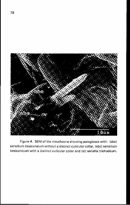
Figure 4. SEM of the mouthcone showing paraglossa with: (sbo)
sensillurn basiconicurn without a distinct cuticular collar, (sbc) sensillurn
basiconicurn with a distinct cuticular collar and (st) sensilla trichodeum.
When the paraglossae open laterally, they expose the ventral
surface of a horse-shoe shaped labral pad (Ip) bearing elongate papillae about its dorsal and lateral margins (Figs. 5, 6, 7). A second set of
more slender papillae (p) are situated along the ventral edge of the pad.
The function of these papillae is not known (Chisholm & Lewis 1984).
The labral pad of F. bispinosa females is around 11 p m across.
A thrips presses the tip of its mouthcone against the plant
substrate in preparation for feeding. This close contact is maintained
by means of the labral pad. According to Chisholm & Lewis (1984) the
only function presently attributed to the labral pad is t o support the slim
maxillary stylets as they are protracted into plant tissue. Thrips use the
single mandible to make an initial opening by punching through the plant cuticle before feeding. Entry is achieved by a characteristic
rocking of the head and a forceful downward and backward thrusting of the head capsule (Chisholm & Lewis 1984).
Based on studies by Heming (1978), the mandible is capable of
only limited movement due t o its orientation, musculature and
articulation within the head capsule. He indicated that maximum protraction of the mandible is about one-third of its length. Chisholm
& Lewis (1 984) found that adult females of L. cerealium never extended
the mandible beyond 20 p m even though its overall length was 102
pm. Maximum observed protraction of the mandibular stylet was 41 p m for one F. bispinosa adult exposed t o chloroform while feeding (Figs. 8, 9). The mandibular stylet lacks an opening or food channel
and is used principally t o punch a hole in the substrate followed by
insertion of the paired, tongue-in-grooved maxillary stylets (Fig. 10).
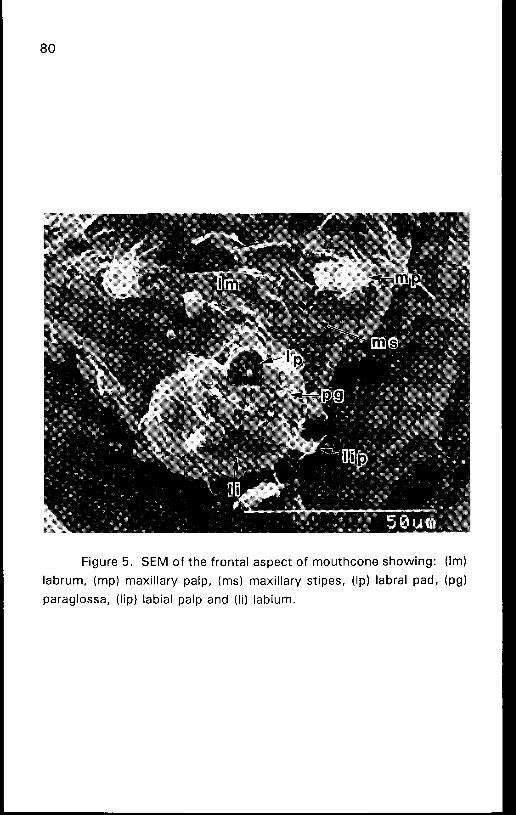
Figure 5. SEM of the frontal aspect of mouthcone showing: (Im)
labrum, (mp) maxillary palp, (ms) maxillary stipes, (Ip) labral pad, (pg)
paraglossa, (lip) labial palp and (li) labium.
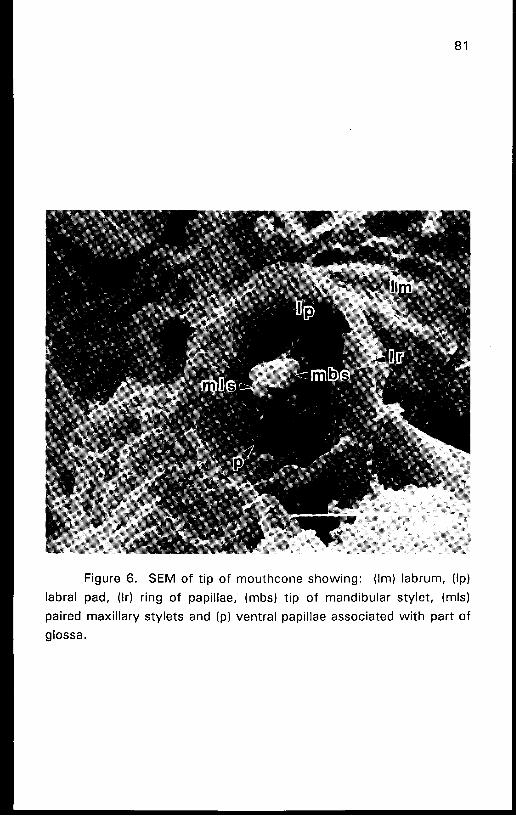
Figure 6. SEM of t ip o f mouthcone showing: (Im) labrum, (Ip)
labral pad, (Ir) ring of papillae, (mbs) t ip o f mandibular stylet, (mls)
paired maxillary stylets and (p) ventral papillae associated wi th part of
glossa.
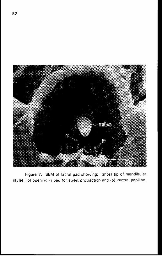
-aell!ded IeJJuaA (d) pue UO!J~~JIOJ~ JalA~s JOJ ped u! 6u!uado (0) '$alA~s
Jelnq!puew 40 d!~ (sqw) :6u!~oys ped IeJqel 40 ~3s 'L a~n6!j
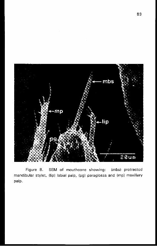
Figure 8. SEM of mouthcone showing: (mbs) protracted
mandibular stylet, (lip) labial palp, (pg) paraglossa and (mp) maxillary
P ~ I P .

Figure 9. SEM of t ip o f mouthcone showing separating
paraglossae: (mls) curved pair of maxillary stylets, (mbs) single
mandibular stylet, (sbo) sensillum basiconicum without a distinct
cuticular collar, (sbc) sensillum basiconicum wi th a distinct cuticular
collar and (st) sensilla trichodeum.

The maxillary stylets are capable of being extended or withdrawn
singly (Fig. 10) or together (Figs. 9, 1 1 ) and each is equipped with an
internal longitudinal groove that, when the t w o stylets are interlocked,
provides a hollow tube for withdrawing food material from the
underlying cells (Mound 1971). Adults of F. bispinosa have a
subterminal opening (so) on the maxillary stylets (Fig. 10) and their tips
are asymmetrical (Figs. 10, 11). The right stylet t ip is larger and
slightly broader than that of the left. Maximum observed protraction of
the maxillary stylets was 5 0 p m for one F. bispinosa specimen that had
been exposed to chloroform while feeding. This length corresponds to
the average depth of feeding injury observed in citrus flowers and
developing buds (C.C.C. & D.S.A., unpublished data).
A light micrograph of the somewhat flattened mouthcone of a
F. bispinosa female is shown in Figure 12. The labral pad (Ip) and ring
(Ir) were distorted and pulled out of position in this preparation allowing
a clearer view of several otherwise overlapping structures. The
hypopharynx (h) is exposed as well as the food canal (fc) in its anterior
surface and (e) indicates the tips of the t w o maxillary stipites (Fig. 12).
The sclerite of the labial glossae including associated sensilla is
indicated by (slg).
Cross sections were prepared for TEM and provide a clearer view
of the arrangement of the principal internal structures of the mouthcone
t o illustrate their position and relative size (Figs. 13-1 5). Figure 13
shows a cross section of the mouthcone around 1 0 pm above its apex.
The outer t w o elements are the labial paraglossae (pg). Anteriorly, the
fused glossae (fg) are surrounded by the labral pad (Ip). The labrum
(Im) is located in the center and encloses the t w o irregular, broadened
tips of the maxillary stylets (mls) and three parts of the irregular t ip of
the hypopharynx (h) in this section. The mandibular stylet (mbs)
contains three diverging sensory dendrites (d). A t this level, the food
and salivary canals are not present.

Figure 10. SEM of tip of mouthcone, showing the irregular shape
and (so) subapical opening of the right maxillary stylet with its grooved
interior (fc) forming half of the food channel.

Figure 11. SEM o f t ip of mouthcone showing: (mbs) t ip of
mandibular stylet, (mls) paired maxillary stylets and (t) irregular-shaped
t ip o f stylets.

Figure 12. Light micrograph of whole mount preparation of the
rnouthcone of F. bispinosa showing: (Ir) labral ring, (Ip) labral pad, (slg)
sclerite of labial glossa, (s) sensilla of labial paraglossae, (e) tips of
maxillary stipites, (fc) food canal in anterior wall of hypopharynx ( =
precibarium), (h) hypopharynx, (mbs) mandibular stylet, (rnls) maxillary
laciniae and (pg) base of paraglossa.

Figure 13. Transmission electron micrograph (TEM) showing
cross section of the mouthcone of F. bispinosa about 10 p m from the
tip with: (pg) labial paraglossae (Im) labrum, (fg) fused glossae, (Ip)
labral pad, (d) mandibular stylet with three sensory dendrites, (mls) tips
o f maxillary stylets and (h) hypopharynx. Large arrow points to front
of insect.

The next cross section (Fig. 14) is around 4 0 p m proximal t o the
tip of the mouthcone and clearly shows the presence of both food and
salivary canals. The labrum (Im) is situated anteriorly (Fig. 14) with the
t w o maxillary stipites (ms) on either side and the prementum behind
(pm). The hypopharynx (h) is located in the center with the food canal
(=precibarium) (fc) in its anterior wall and the salivary canal (sc) in its
posterior wall. Located on either side of the hypopharynx are the t w o
maxillary stylets (mls). The larger, single mandibular stylet (mbs) is
located anterior to the left maxillary stylet. It is supported by grooves in the hypopharynx and in the inner, membranous wall of the left
maxillary stipes (Fig. 14). A similar arrangement occurs in larvae of H. verbasci (Heming 1978). Both maxillary stylets have recessed areas
in the inner walls of the adjacent stipites that appear t o also provide a
degree of support. Closer examination of the stylets reveals the
presence of four sensory dendrites in each lacinia and a central bundle
of three dendrites (dl in the mandibular stylet (Fig. 1 5). The mandibular
stylet of H. verbasci appeared to lack sensilla when viewed by SEM (Heming 1978). The mandible of F. bispinosa is solid with the
exception of the small innervated channel and not hollow as previously
reported for L. cerealium by Chisholm & Lewis (1984) and for S. citri by Wiesenborn & Morse (1989).

Figure 14. TEM of cross section of mouthcone of F. bispinosa
taken about 40 p m from tip, showing: (Im) labrum, (ms) maxillary
stipites, (pm) prementum, (h) hypopharynx, (mbs) mandible, (mls)
maxillary laciniae, (fc) food canal and (sc) salivary canal. Large arrow
points t o front of insect.

Figure 15. Enlargement of previous TEM showing: (mbs)
niandible with (d) three sensory dendrites, (mls) maxillary laciniae with
associated dendrites, (fc) food canal, (h) hypopharynx and (sc) salivary
canal.

Acknowledgment
We thank Bruce S. Heming for his invaluable guidance and
comments on the manuscript. This research was partially supported by
a grant from J. R. Brooks and Son, Inc., Homestead, Florida. This is Florida Agricultural Experiment Station Journal Series No. N-00239.
References Cited
Ananthakrishnan, T. N. 1980. Thrips, pp. 149-1 64. In K. F. Harris & K.
Maramorosch [eds.], Vectors of plant pathogens. Academic Press, New York.
Childers, C. C., R. J. Beshear, J. R. Brushwein & H. A. Denmark. In
press. Thrips (Thysanoptera) species, their occurrence and
seasonal abundance on citrus flowers and developing buds in Florida. J. Entomol. Sci.
Chisholm, I. F. & T. Lewis. 1984. A new look at thrips (Thysanoptera)
mouthparts, their action and effects of feeding on plant tissue.
Bull. Entomol. Res. 74: 663-675.
Hallam, N. D. & T. C. Chambers. 1970. The leaf waxes of the genus
Eucalyptus LIHeritier. Aust. J. Bot. 18: 335-386.
Heming, B. S. 1978. Structure and function of the mouthparts in larvae
of Haplothrtps verbasci (Osborn) (Thysanoptera, Tubulifera,
Phlaeothripidae). J. Morph. 156: 1-38.
Hunter, W. B. & D. E. Ullman. 1989. Analysis of mouthparts
movements during feeding of Frankliniella occidentalis (Pergande)
and F. schultzei Trybom (Thysanoptera: Thripidae). Int. J. Insect Morphol. and Embryol. 18: 161 -1 71.
Krantz, G. W. 1978. A manual of acarology, 2nd ed. Oregon State
Univ. Book Stores, Inc., Corvallis. 509 pp.
Milne, D. L. & B. Q. Manicom. 1978. Feeding apparatus of the South
African citrus thrips, Scirtothrips aurantii Faure. Citrus and
Subtropical Fruit J. 535: 6-1 1.
Mound, L. A. 1971. The feeding apparatus of thrips. Bull. Entomol.
Res. 60: 547-548.
O1Brien, T. P., N. Feder & M. E. McCulley. 1964. Polychromatic
staining of plant cell walls by toluidine blue 0. Protoplasma.
59: 367-373.
Palmer, J. M., L. A. Mound & G. J. du Heaume. 1989. 2.
Thysanoptera. CIE guides to insects of importance t o man. CAB Int. Inst. Entomol. British Mus. Nat. Hist. 73 pp.
Reynolds, E. S. 1963. The use of lead citrate at high pH as an electron-
opaque stain in electron microscopy. J. Cell Biol. 17: 208-21 2.
Richards, 0. W. & R. G. Davies. 1977. Imm's general textbook of entomology. Vol. 1 10th ed. Chapman and Hall, London. 418
PP.
Spurr, A. R. 1969. A low-viscosity epoxy resin embedding medium for
electron microscopy. J. Ultrastructure Res. 26: 31-43.
Stempack, J. C. & R. T. Ward. 1964. An improved staining method
for electron microscopy. J. Cell Biol. 22: 697-701.
Wiesenborn, W. D. & J. G. Morse. 1988. The mandible and maxillary
stylets of Scirtothr@s citri (Moulton) (Thysanoptera: Thripidae). Pan-Pacific Entomol. 64: 39-42.