structure of rec2, a recombinational repair gene of ustilago maydis, and its function in
TRANSCRIPT

MOLECULAR AND CELLULAR BIOLOGY, Sept. 1994, p. 6287-62960270-7306/94/$04.00+0Copyright © 1994, American Society for Microbiology
Structure of REC2, a Recombinational Repair Gene of Ustilagomaydis, and Its Function in Homologous Recombination
between Plasmid and Chromosomal SequencesBRIAN P. RUBIN, DAVID 0. FERGUSON, AND WILLIAM K. HOLLOMAN*
Department of Microbiology, Comell University Medical College, New Yorkl New York 10021
Received 3 May 1994/Returned for modification 16 June 1994/Accepted 27 June 1994
Mutation in the REC2 gene of Ustilago maydis leads to defects in DNA repair, recombination, and meiosis.Analysis of the primary sequence of the Rec2 protein reveals a region with significant homology to bacterialRecA protein and to the yeast recombination proteins Dmcl, Rad5l, and Rad57. This homologous region in theU. maydis Rec2 protein was found to be functionally sensitive to mutation, lending support to the hypothesisthat Rec2 has a functional RecA-like domain essential for activity in recombination and repair. Homologousrecombination between plasmid and chromosomal DNA sequences is reduced substantially in the rec2 mutantfollowing transformation. The frequency can be restored to a level approaching, but not exceeding, thatobserved in the wild-type strain if transformation is performed with cells containing multiple copies of REC2.
It has become increasingly clear that the cellular response toDNA damage encompasses a complex set of processes gearednot only to preserve the integrity of the genome but also, in a
broader sense, to contribute to survival of the organism. Inwell-researched model systems such as Saccharomyces cerevi-siae, studies on DNA repair have revealed that mutation in anyone of several dozen genes can lead to radiation sensitivity.These genes can be grouped into three general categoriesbased loosely on epistatic relationships and genetic properties(11). They are referred to as the excision repair group, theerror-prone repair group, and the recombinational-repairgroup, the last group so named because the pathway of repairdisabled is governed by functions dedicated to genetic recom-bination. Mutation in the genes defining this group leads todefects in induced recombination, extreme sensitivity to ioniz-ing radiation, lethality upon mating-type switching in ho-mothallic strains, sporulation deficiency, and inability to repairdouble-strand gaps in plasmid DNA during transformation(for a review, see reference 25). All of these defects can beaccounted for by loss of a basic recombination function.Discovery that at least two genes in the recombinational repairgroup, namely RAD51 and RAD57, have extensive homologywith the Escherichia coli recA gene (1, 3, 18, 33) underscoresthe notion that this group of genes is directly involved ingenetic recombination.
Such an extensive inventory of radiation-sensitive mutants isunrivaled in any other eukaryotic experimental system. Never-theless, pathways of DNA repair as exemplified in S. cerevisiaeprovide a paradigm for thinking about DNA repair in otherorganisms and a platform for generalizing about the mecha-nisms. Radiation-sensitive mutants obtained in other less-mainstream experimental systems can also be categorizedaccording to the three classes of yeast DNA repair genes. Forinstance, mutants of Ustilago maydis that appear to be repre-sentative of all three of these groups have been isolated (15,
* Corresponding author. Mailing address: Dept. of Microbiology,Box 62, Cornell University Medical College, 1300 York Ave., NewYork, NY 10021. Phone: (212) 746-6510. Fax: (212) 746-8587. Elec-tronic mail address: [email protected].
21). The uvs3 mutant is recombination proficient, extremelysensitive to UV rather than ionizing radiation, and defective inexcision of pyrimidine photodimers (42). This mutant would beappropriately categorized in the excision repair group. Therecl mutant is sensitive to both UV and ionizing radiation andhyperactive for mitotic allelic recombination and exhibits a
mutator phenotype reminiscent of that of the error-prone
repair group (17, 37). The rec2 mutant is extremely sensitive toionizing radiation and defective in mitotic crossing over andinduced gene conversion and fails to complete meiosis (16).Thus, rec2 corresponds to the recombinational-repair class.
In a study designed to continue exploration of the mecha-nism of recombination in U. maydis, pairs of autonomouslyreplicating plasmids bearing noncomplementing alleles of aselectable marker were used to cotransform the DNA repair-deficient strains mentioned above that were deleted entirelyfor the genomic copy of the marker (10). Generation ofprototrophy proceeded through extrachromosomal recombi-nation. Introduction of double-strand breaks into the plasmidDNA greatly stimulated recombination, but no such stimula-tion was apparent in the rec2 mutant unless the geneticmarkers on the plasmids were oriented in such a way as tocircumvent recombination by a conservative pathway. It was
deemed unlikely that the defect in rec2 was in a mismatchcorrection step, since artificially formed heteroduplex DNAcontaining the two allelic markers was highly active in trans-formation to prototrophy. These results were interpreted tomean that the REC2-dependent pathway involved homologouspairing and strand exchange and that the defect in the rec2mutant was likely to involve a step in pairing ofDNA duplexes.This intriguing possibility aroused our interest in investigatingthe REC2 gene further.To learn more about the role ofREC2 in recombination, we
cloned and characterized the gene and continued efforts toanalyze recombination of plasmid DNA substrates in the rec2mutant. In the study presented in this paper, the aims were todetermine the nature of the REC2 gene product, to test thesignificance of structural motifs found in the sequence, and tomeasure recombination between a cloned gene on a plasmidand the homologous sequence in the genome.
6287
Vol. 14, No. 9
Dow
nloa
ded
from
http
s://j
ourn
als.
asm
.org
/jour
nal/m
cb o
n 26
Jan
uary
202
2 by
138
.36.
200.
100.

6288 RUBIN ET AL.
TABLE 1. Strains of U. maydis used in these studies
Strain Genotype
UCM5.... REC2 adel-I leul-la2b2UCM54.... rec2-1 panl-l narl-1 alblUCM164..... Arec2::HPH adel-1 leul-1 a2b2UCM288..... Arec2::HPHADEl/adel-J leul-] a2b2UCM289.... Arec2::HPHADEl-REC2/adel-I leul-i a2b2UCM302..... Arec2::HPHADEJ-rec2-2/adel-1 leul-1 a2b2UCM303..... Arec2::HPHADEJ-rec2-3Iadel-J leul-I a2b2UCM304..... Arec2::HPHADEI-rec2-5/adel-J leul-i a2b2UCM305..... Arec2::HPHADEJ-rec2-7/adel-i leul-la2b2UCM306..... Arec2::HPHADEI-rec2-4Iadel-J leul-1 a2b2UCM320.... Arec2::HPHADEJ-rec2-6Iadel-I leul-I a2b2UCM321..... Arec2::HPHADEI-rec2-8/adel-1leul-J a2b2
MATERMILS AND METHODS
Ustilago maydis strains and methods. All strains (Table 1)used were from laboratory stocks or were derived by standardmolecular genetic methods for U. maydis (see references 8 and41). The designations ade, leu, pan, and nar refer to require-ments for adenine, leucine, and pantothenic acid and toinability to metabolize nitrate, respectively. albi and a2b2refer to mating-type alleles. Radiation sensitivity was deter-mined as described previously (40). In general, cultures were
grown to a density of 2 x 10⁷ cells per ml and the cells were
collected by centrifugation, washed in water, and plated byspreading from 103 to 106 cells on yeast extract-peptone-sucrose (YEPS) medium. Cells were irradiated with UV with a
30-W General Electric germicidal lamp delivering 0.2 J/m2/s.Survival was determined by counting of colonies visible afterincubation of plates for 3 days at 32°C. Transformation toleucine prototrophy was performed with U. maydis protoplastsas previously described (10). Homologous recombination was
measured by transforming leul-1 strains to Leu+ with pCM381over a range of input DNA (1 to 10 ,ug of DNA) at which theresponse was approximately linear. Some variation in transfor-mation frequency resulted from differences in protoplast com-
petence and was controlled for by use of the autonomouslyreplicating plasmid pCM216 as a standard (usually 30 ng ofDNA). Recombination frequency was calculated as the num-
ber of Leu+ transformants obtained with pCM381 divided bythe number of Leu+ transformants obtained with pCM216 permicrogram of DNA. Specific recombination frequency was
calculated as the number of Leu+ transformants obtained withpCM381 per microgram of DNA divided by the number ofLeu+ transformants obtained with pCM216 per microgram ofDNA.
Plasmids. pCM54 is a pUC12 derivative containing thehygromycin phosphotransferase-hsp7O promoter gene fusion(HPH) used in selection for hygromycin resistance (44) and the383-bp SspI active subfragment from the autonomously repli-cating sequence of U. maydis inserted into the SspI site.pCM230 is a shuttle vector based on pBluescript II SK+ andcontains the U. maydis ARS (41) and the HPH gene. pCM158is pCM54 containing a 9-kbp fragment from a Sau3AI partialgenomic digest of DNA from UCM3 which is inserted at theBamHI site in multiple cloning sequence (4). This fragmentcontains the entire REC2 gene. The almost complete LEU1gene of U. maydis is contained on a 3.0-kbp EcoRI-HindIIIDNA fragment (8, 31). pCM216 (10) has this 3.0-kbp fragmentinserted into a pBluescript II SK+ derivative (Stratagene)containing the 383-bp U. maydis autonomously replicatingsequence (41). pCM216 fully complements the auxotrophy ofthe U. maydis leul-l mutant. pCM381 is pBluescript II SK+
containing a 2.7-kb HindIll portion of the LEU1 gene whichlacks a 300-bp segment essential for complementation. It doesnot replicate autonomously. pCM425 was generated by insert-ing an 823-bp HindIll fragment (+233 to + 1057 with respectto the initiation methionine of the putative Rec2 open readingframe [ORF]) from the REC2 gene into pBluescript II SK+.This plasmid was used as a template for construction ofsite-directed mutants within the span of this portion of REC2.pCM427 was made by inserting the 3.2-kbp MseI fragment,which contains the entire REC2 gene, into the HinclI site ofpCM325 (an adaptor plasmid with a duplicated multiplecloning site [39]). This enables excision of the REC2 gene on a3.2-kbp BamHI fragment. pCM430 was constructed by ablatingthe 5'-most HindIll site in the noncoding region upstreamfrom the SspI site of the REC2 gene in pCM427. This plasmidwas used as the REC2 backbone for construction of all of theREC2 site-directed mutants. The ADEI gene of U. maydis iscontained on a 5.0-kbp BamHI-XbaI fragment that was iso-lated by complementation of the adenine auxotrophy of theadel-I mutant. pCM441 is a derivative of pBluescript II SK+carrying this BamHI-XbaI fragment modified by removal of anessential 100-bp NcoI fragment from within the coding regionof the gene (6a). Cleavage of pCM441 with NcoI creates a gapwithin the ADE1 gene that does not overlap the adel-I lesionand yet enables transformation of adel-1 strains to adenineprototrophy upon transformation. pCM463 was constructed byinsertion of a 3.2-kbp BamHI fragment containing the REC2gene from pCM430 into the BamHI site of pCM441. pCM474was constructed by insertion of a 3.2-kbp BamHI fragmentcontaining the rec2-2 gene into the BamHI site of pCM441.Similarly, pCM476 contained the rec2-3 gene, pCM478 con-tained rec2-4, pCM479 contained rec2-5, pCM480 containedrec2-6, pCM481 contained rec2-7, and pCM500 containedrec2-8. All plasmids were amplified in E. coli XL-1 Blue(Stratagene) endAl hsdRJ7supE44 thi-1 A- recAl gyrA96 relA1lac [F' proAB lacIq lacZ AM15 TniOftetR}].
Nucleic acid techniques. Southern blot hybridization wascarried out as described previously (9). For Northern (RNA)blot hybridization, RNA was prepared by LiCl precipitation ofnucleic acids extracted from cells sheared open by violentagitation with glass beads in a solution containing phenol andsodium dodecyl sulfate (SDS) and was separated on agarosegels run in formaldehyde as described before (40). Both RNAand DNA samples were transferred to Zeta-Probe membranes(Bio-Rad Laboratories) in 0.4 M NaOH, and hybridizationswere carried out in 7% SDS-0.5 M sodium phosphate (pH7.2)-i mM EDTA at 65°C. DNA probes were labeled byrandom priming with the Klenow fragment of E. coli DNApolymerase I (6). Radiolabeled bands in blots were visualizedby autoradiography and quantitated by scanning with a Molec-ular Dynamics Series 400 Phosphor Imager. Autoradiographimages were digitized with a scanner and Photoshop software(Adobe Systems, Inc.).REC2 DNA sequence determination. The REC2 gene was
isolated originally on a 9-kbp genomic DNA fragment (4). A5.0-kbp KpnI-SspI fragment encompassing the REC2 gene wassubcloned from the original cloned fragment, and overlappingdeletions were prepared by controlled digestion with E. coliexonuclease III and mung bean nuclease (13). Plasmid dele-tions obtained were tested for the ability to complement theUV sensitivity of the U. maydis rec2-1 strain UCM54 afterirradiation with 40 J of 254-nm UV light per M2. The bound-aries of the complementing fragments were found to corre-spond approximately to the 3.5-kbp MluI-EcoRI fragmentdepicted in Fig. 1. DNA sequence was determined by theenzymatic chain termination method with Sequenase 2.0
MOL. CELL. BIOL.
Dow
nloa
ded
from
http
s://j
ourn
als.
asm
.org
/jour
nal/m
cb o
n 26
Jan
uary
202
2 by
138
.36.
200.
100.

U MAYDIS REC2 GENE 6289
TABLE 2. Oligonucleotide primers synthesized for site-directed mutagenesis
Primer Sequence
rec2-2 A.... 5'GAC GAC CTG TTC GGC GGT GGGrec2-2 B.... 5'CCC ACC GCC AAG CAG GTC GTC
rec2-3 A.... 5'GGC TCT GGT GCG ACC CAG ATGrec2-3 B.... 5'CAT CTG TGG CGC CCA AGA CCG
rec2-4 A.... 5'GTA TTC TCA GCC GGC TCC CGArec2-4 B.... 5'TCG GGA GCC GGC TGA GAA TAC
rec2-5 A.... 5'CGA GAG CTC GCC GAC CTG CTArec2-5 B.... 5'TAG CAG GTC GGC GAG CTC TCG
rec2-6 A.... 5'CTG CTA GGC GCT GGG GTG CGTrec2-6 B.... 5'ACG CAC CCC AGC GCC TAG CAG
rec2-7 A.... 5'CTA GGC GGT GCG GTG CGT TCCrec2-7 B.... 5'GGA ACG CAC CGC ACC GCC TAG
rec2-8 A.... 5'GGC TCT GGT AGG ACC CAG ATGrec2-8 B.... 5'CAT CTG GGT CCT ACC AGA GCC
(United States Biochemical Corp., Cleveland, Ohio) andaL-35S-dATP as described by Tabor and Richardson (36).Denatured double-stranded DNA was sequenced directly (38)on 6% polyacrylamide gels containing 7.7 M urea with oligo-nucleotide primers synthesized as needed (Oligos, Etc., Wil-sonville, Oreg.). Sequence homology searches and alignmentswere carried out with the FASTA algorithm at the RockefellerUniversity Computer Center, and the RDF2 program was usedfor statistical verification of homologies (24). Certain motifswere identified with the PROSEARCH software and PRO-SITE database (2). Restriction endonuclease mapping, DNAsequence project management, and ORF analysis were carriedout with DNASTAR (DNASTAR, Inc., Madison, Wis.).
Isolation of the rec2-1 allele. By restriction endonucleasemapping of genomic DNA extracted from the rec2-1 mutant, itwas determined that a 0.8-kbp stretch spanning a landmarkHindlIl site in the wild type was deleted. The DNA fragmentencompassing the region of deletion was isolated from asize-selected library prepared from UCM54 genomic DNAafter digestion with HindIll and PstI. DNA fragments in thesize range 0.3 to 1.5 kbp were eluted from a preparativeagarose gel after electrophoresis and ligated together withpBluescript II SK+ DNA previously cut with HindIlI and PstI.After transformation into E. coli XL-1 Blue, colonies werereplica plated onto nitrocellulose membranes (BA-85S; Schlei-cher & Schuell, Keene, N.H.), prepared for hybridization (12),and screened with a 0.3-kbp HincII-PstI fragment as probe.From approximately 400 colonies, 1 positive was found. Itsidentity was confirmed by restriction enzyme mapping, and itsDNA sequence was determined.
Construction of site-directed mutations. Using plasmidpCM425, which contains a fragment of the REC2 gene thatspans the region of homology with RecA, as template, weperformed site-directed mutagenesis using the two-stage over-lap extension method with PCR (14). The different mutantsubfragments were inserted into the backbone of plasmidpCM430, which contains the entire REC2 gene flanked byBamHI sites. The oligonucleotide primers synthesized forsite-directed mutagenesis were as shown in Table 2. Theoligonucleotides which flanked the mutant fragments andwhich were used in all of the PCRs were the M13 universal
primer 5'GTA AAA CGA CGG CCA GT and the reversesequencing primer 5'AAC AGC TAT GAC CAT G.
Nucleotide sequence accession number. The GenBank ac-cession number for the sequence reported in Fig. lB is L18882.
RESULTS
Molecular characterization of the REC2 gene. The REC2gene was subcloned from the original isolate (4) and localizedon a 3.2-kbp SspI-EcoRI fragment that fully complemented theradiation-sensitive phenotype of the rec2-1 mutant. DNAsequence analysis revealed that this fragment contained asingle uninterrupted ORF of 2,343 bp (Fig. 1A). Several linesof evidence establish that this ORF encodes the REC2 gene.First, the ATG codon lies in an acceptable sequence contextfor initiation of translation (20). Second, the 0.8-kbp EcoRVfragment that was replaced by a hygromycin phosphotransfer-ase cassette (44) during construction of a rec2 disruption allele(4) lies within this ORF. Third, the rec2-1 allele deletes 0.8 kbpextending 0.6 kbp into the ORF from the first ATG codon andincludes 0.2 kbp of upstream sequence. Fourth, a 2.7-kbmRNA detected by use as a probe of a restriction fragmentinternal to the ORF is not detected in the rec2-1 mutant (seeFig. 7). The boundaries of this RNA as determined by S1nuclease protection place the termini approximately at posi-tions -150 and +2500 with respect to the ORF, with noindication of splicing (not shown).The REC2 gene encodes a protein of 781 amino acids with a
calculated mass of 83,935 Da (Fig. 1B). The sequence K-R-I-K,spanning residues 14 to 17, is similar to the motif K-X-X-K/R,which appears in many nuclear localization signals describedelsewhere (29). The acidic stretch from amino acids 93 to 104,in which 11 of 12 residues are aspartate, is reminiscent of acidicregions found in proteins known to interact with chromatin(45) as well as in transcription factors in which such regionsserve to mediate protein-protein interactions (26). Residues251 to 258 (G-E-S-G-S-G-K-T) and 438 to 442 (V-V-V-D)correspond to consensus Walker A (G/A-X-X-X-X-G-K-T/S)-and B (four hydrophobic residues followed by D)-type purinenucleotide-binding site motifs (43). The sequence T-P-R-Kspanning residues 697 to 700 is similar to the Cdc2 proteinkinase phosphorylation site motif (S/T-P-X-R/K [32]).A search for structural relationships turned up no overall
homology between Rec2 and any other protein. However, thesearch revealed that a 47-amino-acid stretch encompassing thenucleotide-binding A motif exhibits a remarkable degree ofconservation in primary structure with a region of E. coli RecAprotein that is highly conserved among all bacterial speciesknown to date (see Fig. 2 and reference 30). This same regionis also conserved among three RecA-related proteins identi-fied in S. cerevisiae, namely Dmcl (5), Rad5l (1, 3, 33), andRad57 (18). Comparison of the sequences of these proteinsreveals 42% (Dmcl), 36% (Rad57), 40% (Rad5l), and 39%(E. coli RecA) amino acid identities within this 47-residuestretch. It should be noted that the relationship between Rec2and RecA as well as the eukaryotic RecA homologs does notresult solely from the presence of the Walker A-type nucleo-tide-binding motif. Other proteins containing this motif werenot identified in the homology search. The region of homologyhas been analyzed extensively in E. coli RecA and has beenshown to be important for interaction with ATP. Crystallo-graphic analysis indicates that the region immediately preced-ing the Walker A-type nucleotide-binding motif is structurallyunusual and consists of a helix and 1-sheet separated by a loop(34, 35). This region has been implicated in mediation of astructural transition that is coupled to ATP binding (19).
VOL. 14, 1994
Dow
nloa
ded
from
http
s://j
ourn
als.
asm
.org
/jour
nal/m
cb o
n 26
Jan
uary
202
2 by
138
.36.
200.
100.

6290 RUBIN ET AL. MOL. CELL. BIOL.
Hind III Ssp IHind III EcoR V Hinc 11 Pst I Hind III EcoR V Sal I Mse I EcoR I
A '*1 'II>r, I I,, II I I~~~~~~ I I I I
0 0.5 1.0 1.5 2.0 2.5 3.0 3.5
REC2 _
rec2::HPH
rec 2-1 deletion
B-175
-107 AGCTAGCCGCC7IGTCGTGCTCATCCCAG
Ar.AcGIMrGTGGAAGCGTAAGGAGAAGCAGATrAGGTGCTGGTAG_GGACTAATCASspI
:rrCTrCACAGCCCAcATCGTA<GGGcGClATCGCCAcTGGTTGCGATACAlGC3TGAGCTAAATC
ATGACTGGCATCGCGATCGCCGATGTIGGCTGATTICGAAACGICATCAAGGCGTGM T G I A I A DV G
CAGCAATTGGCACACGCGTTGCGCATATCC
AGTCGCAAGCTTCCCIGCACCAACCT~GGAC
GGTTCGGCCGACGCTTCAGACACGAGCGA'I
TTTCCCGGCGCACAATGcCTTTGTCTACGAI
TTTrGCCGTCCACAAACACCACAAACCCAC
TTATCGCTCGGACGCCAACGACATGTATTC
AGCGGCTCTGGTAAGACCCAGATGGCTAVQ M A I
Motif A
CAATCCAGGAC?T?CTACGAGACCCGAT;
GGTATGGGCG-CGTGCTACATCACATCTGG
GTCTATCVGGTCTGCGATCCTACACAAAG(
TCGCGCGAGATITGGCGTTGTGTGGrAGACS R E I G VL3LV ..V ..V
Motif B
GTrCGAGATCGV E I ADAL KR I
AGTCAAGCGCCAACCTCGGCATT~CAGCGG
GCC'TCGATCGCGCCTACGCTGGCGGAAGC(
CTGGCAGACCTGGAGCAACCTG-ATCAA.
CAAAACAC
GACAAGAGCGCGCTGAGACAGCTACGGTT
GACAGCACGCCCGCTCCAGAGTCACAGCAUD S T P A P E S Q Q
C I S L&_&..LJ A C
NLS
CCAAGCAGATGCCGATCGCTTCTC'
A 0QH L AV
ITTCCGATGCCGGCTCGGACAGrGA7GO
CG8TGITGCCCGTGACGAGCATCATGA,
DOVA R D ENH HOD
CTCAAGCGGCTCCCGAGAGCTCGACGA(
CCAAGITTTCACTrATGCCGCTCTCGG
CGCTGA
CATGCTGTAGCAGATGGGCGCrCGCTTA
3CGCGCTTCGTATTCGCTCAGGCGCAG
CGGATCTGCGATGGCCTTACCCAc.ACAG
GALP SD
TGCGGCGCGTCGTTTCTGCTCCGGACGCG
V AV
T'GTCATTAGCCGCGCAAAGCGGTGCAV I A V H
Cdc 2 KirnaseCACCC
Q Q R AA E R H P
CTGTCGrCGAGCAAAGCTCTTCAGrACCGACGAGATCCTCCTCAGCCCACCGC R R A K L F S T D E I L L S P P
CCAAGTGGCCACGGCATCTGCTCCACCTCCCATCTCGGTACTCGATGCGCTCQ V A T A S A P P P I S V L D A L
CGAcGAcACGA3GACAAIAGTAGCGAI'rTCCA n n n n D D N D D D D__D K A D S
Chromatin Binding MotifRAGGTTrGCATCGTCTGCATCGTGCCCCCA ACA
R F A S S C I V P P T Q G Y D G N
'ACGCAGTAGCATGAISTGkTGCACA CGTlCCACCR S S
LTIGGTATC?G Y L
CCTGCTAGGL L G
;cTTGGTTcaL V P
7AAGCGACArS D I
.GAACCGAGCN R A
A L L
:mTS'CCTGGL P G
Q A A
'AGATTGTWD C G
kCCGCATCCGR I R
U3CCGCTAGCP L A
LPG
GTCAGCGGCTC
C A S
QAAAGCAGGATOG
A A V
V L N
ACGCAAC
AGCCGCAGC
APLA
n)'GCGATCCCAAAGTTGACCAC
.GGTGGGGTGCGTTCC~GCTGTGG GV R S A V
)CTGAGCCAAGCTGCACIGATCAC
CTIGCAGAGCrACGGCATGGAG
IGCGAGCATATCGACTCGCTC
3A(TTGTACCAGTC(l
ZAACGATGTCGCCGACTCGCCT(r.
=TACTCICAGTCGAGCAGGAC
kGAGGACGCCGGCTACCAGACCTVA Q
AACGTGTGCGCTCGCCI
T~~~~~~~~~.
kG
VI H
CGCGATCCATCACAG'GAINA
GACGGCGGRCA AGGACCL Q OY
'GCCTCGGTCGCCA,GAGACGTCA S V AR DOV
L T E L V G E
3CCCTCGATTGGATCTCACCGC
rCGTCGACAAGCGCTTGCCAAC
CTTCAACGCTCAAAGATGCTA
L VL NNHV S
A,CGCAACGATCCTGGCACATCA
ACTTGAAGCACGCACTGCACAG
A.CCAGCATGCGAGCCAG7TGCGC
GCATGCCAIVCATGCGACCGCrHNA MNH A T A
F TCG EG AAGC LCTGQAAGE
E~
CATCACLAIGOELGCCATCGHCAAGCCGCAATCGCACACCACH H W L A I D E L Q S H T T A R P T S R A A Q A G
GCGATCCAGAATCCTTTSCACA'A'A
2485 ACGCTMG
2593 AAGA7IG
2701 CGACGGO
2809 TGGTAGC
2917 GCGTCGC
3025 TCGCAA
10937
21773
325109
433145
541181
649217
757253
865289
973325
1081361
1189397
1297433
1405469
1513505
1621541
1729577
1837613
1945649
2053685
2161721
2269757
2377
TAGCTGaC3ACGGAGCAAGC¶CTCGGGCTC0'rTAGCGGCAAGGCCGACGTCAITCGGAAATGAAAAA TCAGCTGCGGCAC,ACTGTGmAA=lGTGGGAATCGACACGGAGAGTQAGI¶GTGAGCTAGCCGTCGGTCAGTGCCGTGG
GTrCAGAGrcGCCATTGcCCAC=TGIWCTcGAAIGTcGAGCCGrACCAGAcCGCTTT01c rGAGRG1C
SICGAT?GATCcS0TT'rCGCTGGCGGCCAAA1W 1 1 = 1GATCCACGTTGCGTCrX~~~~~~~~~~~~~~~~~~~~~GATG:IAATCACGCAAC=AA=GC1^0 m
c
IC,
IT
LT
LC aki
kG;akc.IC
r0lax.IC
rTcr;!A
T,'MIK11
2ITrG(MX
kGC.I1kc
kIC:xkikc
CIGI..T:Irc
AlAl3Nm.A
cTIG'AXII
3A:acC(x
c!Arxocki
c:ArcAix
TIGqTKm
GVZhakc,
Dow
nloa
ded
from
http
s://j
ourn
als.
asm
.org
/jour
nal/m
cb o
n 26
Jan
uary
202
2 by
138
.36.
200.
100.
U AMAYDIS REC2 GENE 6291
FIG. 1. Sequences of the U. maydis REC2 gene and the deduced protein product. (A) The restriction endonuclease map of the 3.2-kbpSspI-EcoRI fragment that complements the radiation sensitivity of the rec2-1 mutant is shown. Schematic representation of the large uninterruptedORF found to reside in the genomic fragment from REC2 and the truncated version found to be present in the rec2-1 allele is presented. A mutantArec2::HPH gene constructed in vitro by disrupting the REC2 gene with a hygromycin phosphotransferase cassette (stippled) is also indicated. (B)The DNA sequence of the 3.2-kbp fragment is shown. The numbering is given with respect to the putative initiation methionine. The putativenuclear localization signal (residues 14 to 17), chromatin-binding acidic motif (93 to 103), nucleotide-binding motif A (251 to 258),nucleotide-binding motif B (438 to 442), and Cdc2 protein kinase phosphorylation site (697 to 700) are underlined. Four stop codons areunderlined.
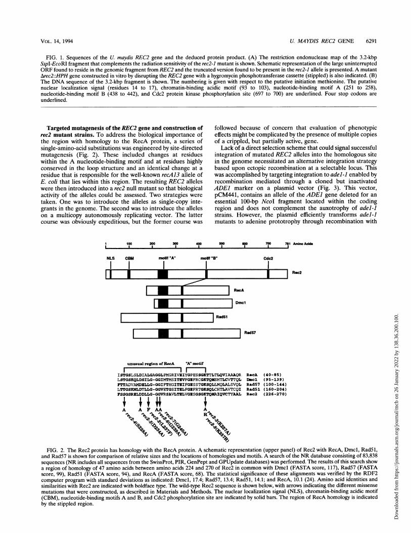
Targeted mutagenesis of the REC2 gene and construction ofrec2 mutant strains. To address the biological importance ofthe region with homology to the RecA protein, a series ofsingle-amino-acid substitutions was engineered by site-directedmutagenesis (Fig. 2). These included changes at residueswithin the A nucleotide-binding motif and at residues highlyconserved in the loop structure and an identical change at aresidue that is responsible for the well-known recA13 allele ofE. coli that lies within this region. The resulting REC2 alleleswere then introduced into a rec2 null mutant so that biologicalactivity of the alleles could be assessed. Two strategies weretaken. One was to introduce the alleles as single-copy inte-grants in the genome. The second was to introduce the alleleson a multicopy autonomously replicating vector. The lattercourse was obviously expeditious, but the former course was
followed because of concern that evaluation of phenotypiceffects might be complicated by the presence of multiple copiesof a crippled, but partially active, gene.Lack of a direct selection scheme that could signal successful
integration of mutated REC2 alleles into the homologous sitein the genome necessitated an alternative integration strategybased upon ectopic recombination at a selectable locus. Thiswas accomplished by targeting integration to adel-1 enabled byrecombination mediated through a cloned but inactivatedADE1 marker on a plasmid vector (Fig. 3). This vector,pCM441, contains an allele of the ADE1 gene deleted for anessential 100-bp NcoI fragment located within the codingregion and does not complement the auxotrophy of adel-Jstrains. However, the plasmid efficiently transforms adel-1mutants to adenine prototrophy through recombination with
I 100 200 300 400 soo 600 700 781a I I I I I I I I
NLS CBM mo 'A' mofif "B" Cdc2
l I I I IL-II I I I
d.I1.'Il
Amino Acids
Rec2
I RecA
I Dmcl
I Rad5l
I lI I Rad57
unusual region ofRecA "A" motif
ISTGSLSLDIALGAGGLPMGRIVEIYGPESSGKTTLTLQVIAAAQR RecA (40-85)LSTGSKQLDSILG-GGIMTMSITNVFGRFRCGKTQMSHTLCVTTQL Duc1 (95-139)FTTADVAMDELLG-QGIFTHGITNIFGSSTTGKSQLLMQLALSVQL Rad57 (100-144)LTTGSINLDTLLG-GGVETGSITILFGIFRTQKSQLCHTLAVTCQI Rad51 (160-204)FSSGSRELDDLZG-GGVRSAVLTRLVGNSQSGKTQMAIQVCTYAAL R-c2 (226-270)
41 AsiA A F AA i
J X~~~vvFIG. 2. The Rec2 protein has homology with the RecA protein. A schematic representation (upper panel) of Rec2 with RecA, Dmcl, Rad51,
and Rad57 is shown for comparison of relative sizes and the locations of homologies and motifs. A search of the NR database consisting of 83,838sequences (NR includes all sequences from the SwissProt, PIR, GenPept and GPUpdate databases) was performed. The results of this search showa region of homology of 47 amino acids between amino acids 224 and 270 of Rec2 in common with Dmcl (FASTA score, 117), Rad57 (FASTAscore, 99), Rad5l (FASTA score, 94), and RecA (FASTA score, 68). The statistical significance of these alignments was verified by the RDF2computer program with standard deviations as indicated: Dmcl, 17.4; Rad57, 13.4; Rad5l, 14.1; and RecA, 10.1 (24). Amino acid identities andsimilarities with Rec2 are indicated with boldface type. The wild-type Rec2 sequence is shown below, with arrows indicating the different missensemutations that were constructed, as described in Materials and Methods. The nuclear localization signal (NLS), chromatin-binding acidic motif(CBM), nucleotide-binding motifs A and B, and Cdc2 phosphorylation site are indicated by solid bars. The region of RecA homology is indicatedby the stippled region.
VOL. 14, 1994
E
Dow
nloa
ded
from
http
s://j
ourn
als.
asm
.org
/jour
nal/m
cb o
n 26
Jan
uary
202
2 by
138
.36.
200.
100.

6292 RUBIN ET AL.
pCM 441/REC2
Aadel REC2
x
adel-1
RB R
ADE1 REC2 adel-1
FIG. 3. Strategy for ectopic integration of REC2 alleles at adel-].Plasmid pCM463 (pCM441/REC2) contains a gap in the U. maydisADE] gene and a 3.2-kbp BamHI fragment which contains the REC2gene. Recombination by a single crossover event at the adel locus willresult in ectopic integration of the REC2 allele. This is verified bySouthern analysis after digestion of genomic DNA with EcoRI. Theadel-] lesion is represented by the solid bar, although its preciselocation is not known. Restriction endonuclease recognition sites are N(NcoI), R (EcoRI), and B (BamHI).
the homologous sequence that flanks the adel-l lesion. Sincethe plasmid does not replicate autonomously, only thoserecombination events that restore a functional chromosomalgene are selected. Recombinants can arise by a single-cross-over event, by gene conversion, by double-crossover events, orby multiple rounds of single-crossover events, but these can beeasily identified by Southern blot hybridization and the un-wanted classes can be eliminated. A single crossover betweenthe plasmid-borne and the genomic adel loci results in inte-gration of the plasmid, yielding a duplication in which thereconstructed ADE1 allele lies in tandem with the adel-]allele. Connecting the two alleles is the plasmid vector se-quence, which includes a unique BamHI site.
For construction of the desired strains, the mutagenizedversions of the REC2 gene were inserted into this uniqueBamHI site of plasmid pCM441. After transformation of aArec2::HPH adel-i strain to Ade+, transformants were ana-lyzed by Southern hybridization and strains for further analysiswere chosen with the REC2 allele of interest integratedbetween the two adel alleles (Fig. 4). The efficiency ofintegration was markedly reduced in the Arec2 mutant (seebelow), but it was possible to obtain the desired strains. All ofthe mutated REC2 strains constructed for this study containedsingle-copy integrants, as verified by Southern blot hybridiza-tion analysis. It should be noted that the sequence of RecAhomology in the mutated REC2 gene is present in the haploidstate in the strains constructed, since the resident rec2 allele,namely Arec2::HPH, is deleted for this sequence. Thus, anyobserved activity of the introduced rec2 allele in questionwould necessarily be an inherent feature of that allele andwould not arise through ectopic gene conversion directed bythe endogenous locus.DNA repair proficiency of the rec2 alleles. The mutants
generated were tested for DNA repair proficiency by exami-nation of sensitivity to killing by ultraviolet light (Fig. 4).Several were found to be as defective in repair as the rec2-1(originally isolated by Holliday) and Arec2::HPH null mutants.These included the rec2-3(K257A) mutant, in which the con-served lysine at position 257 in the Walker A-type nucleotide-binding motif is changed to alanine, but not the rec2-8(K257R)
0.1
0.01 .0 100 200 300
UV Dose (J/m2)FIG. 4. DNA repair proficiency of rec2 alleles. Cultures (10 ml) of
isogenic rec2 strains were grown to mid-log phase and harvested bycentrifugation. After washing, cells were plated on YEPS medium andirradiated with 254-nm UV light. Survivors were counted after 3 days.REC2 alleles are indicated as follows: closed squares, REC2; closedcircles, rec2-2; open circles, rec2-3; crosses, rec2-5; closed triangles,rec2-6; open triangles, Arec2::HPH. rec2-4, rec2-7, and rec2-8 were notdifferent from REC2 and are not shown.
mutant. Other strains with mutations in the RecA homologyregion with a null phenotype included the rec2-5(D234A) andrec2-6(G239A) mutants, although the mutant with the samechange in the adjacent residue, rec2-7(G240A), remainedcompletely proficient in repair. The mutant with the rec2-2(L237F) mutation, found also in the recA13 mutant, wasslightly less sensitive than the rec2 null control strain.
Recombination proficiency of the rec2 alleles. The recombi-nation proficiency of the different mutants was assessed with anassay that measures recombination of a cloned DNA fragmenton a plasmid with its homologous sequence in the genomeduring transformation. This assay utilizes the nonreplicatingplasmid pCM381, which carries an allele of the LEU1 genemissing an essential sequence (Fig. 5). This plasmid does notcomplement the leul-] mutant and cannot restore leucineprototrophy by illegitimate recombination, since it containsonly a fragment of the LEUI gene. Leucine prototrophy can berestored only by homologous recombination between thecrippled leul allele on the plasmid and the corresponding locusin the genome. Thus, transformation to leucine prototrophy isa direct measurement of homologous recombination and ofrecombination proficiency. When the frequency of recombina-tion was measured in a REC2 strain, there was a dose-dependent increase with increasing input DNA (Fig. 6). Abasal level of recombination was detected in the rec2-1 mutant,but this increased little in response to input DNA. To controlfor variation in DNA uptake and cellular competence, acomparison between the frequencies of Leu+ transformantsobtained with pCM381 and with pCM216, an autonomouslyreplicating plasmid bearing the fully complementing LEUJgene, was made. Transformation by the latter occurs at highefficiency and does not depend on recombination (10).
MC)L. CELL. BIOL.
Dow
nloa
ded
from
http
s://j
ourn
als.
asm
.org
/jour
nal/m
cb o
n 26
Jan
uary
202
2 by
138
.36.
200.
100.

U. MAYDIS REC2 GENE 6293
AppM 381
MJeul
xB H S H 8
*.I
leul-1
S HB H,,
AIeul-1
S HI B
LEU1
50Cl)b
40
3
0 30a)I..
co 204-'-
Cb
E 1000
00 2 4 6 8 10
OR
S H B
B
1 2 3 4
size (kbp)
13.0-12.0-
5.7
4.8-
FIG. 5. Homologous recombination assay. (A) Schematic repre-sentation of homologous recombination events between plasmidpCM381, which contains a noncomplementing fragment of the U.maydis LEUI gene deleted at the 3' end of 300 bp, and the leul-lmutant, which contains a lesion in the gene at the approximate positionindicated. Transformation to leucine prototrophy can take place bycrossing over, which results in integration of the plasmid to yield a
tandem repeat of leul sequences separated by vector sequence, or elseby replacement, which results in no change in the genomic DNAstructure. Restriction endonuclease recognition sites are B, BamHI; H,HindIlI; and S, SphI. All transformation experiments were performedwith pCM381 DNA cut to the linear form with SphI. (B) A represen-tational Southern blot illustrating the modes of homologous recombi-nation is shown. Genomic DNA (ca. 2 jig) extracted from U. maydisUCM5 transformed to leucine prototrophy with the 5.7-kbp pCM381plasmid was digested with BamHI and processed for Southern hybrid-ization analysis as described in Materials and Methods by use as a
probe of a 0.7-kbp NcoI fragment from the LEU1 gene that spans theSphI site. The endogenous leul locus resides on a 12.0-kbp BamHIfragment. Crossover events result in generation of fragments of 13.0and 4.8 kbp. Lanes: 1, untransformed control; 2, Leu+ transformantsfrom gene replacement; 3, Leu+ transformant from crossing over; 4,pCM381 plasmid DNA cut with SphI.
DNA (gg)FIG. 6. Homologous recombination during plasmid transformation
is defective in rec2. U. maydis strains UCM5 (REC2 leul-l) andUCM174 (rec2-1 leul-1) were transformed to leucine prototrophy withthe indicated amounts of pCM381 DNA cut to linear form with Sphl.Recombination frequencies were calculated by dividing the number ofLeu+ transformants obtained with the indicated amount of pCM381DNA by the number of Leu+ transformants per microgram of pCM216DNA. The latter determination was made with 30 ng of pCM216DNA, an amount within the linear range of response (10).
The mode of recombination was determined by Southernhybridization. Genomic DNA extracted from transformantswas digested with BamHI, which cleaves the plasmid once inthe vector sequence but does not cut within the endogenouschromosomal leul allele. The single BamHI fragment contain-ing the leul locus detected in the untransformed controldisappears and is replaced by two different fragments intransformants resulting from crossing over. In Leu+ transfor-mants arising as a result of gene conversion or double cross-overs, collectively termed replacements, this BamHI fragmentis not altered (Fig. 5). Of 20 Leu+ recombinants examinedafter transformation of the REC2 control, 7 resulted fromcrossing over and 13 resulted from gene replacement. Inrec2-1, the spectrum of events was not significantly different,although the overall frequency of recombination was reducedas mentioned above.When the rec2 site-directed mutants were assayed to test
their effects in the recombination assay, it was found that thestrains defective in DNA repair were also defective in homol-ogous recombination (Table 3). The range of difference infrequencies between the REC2 strains and the mutant strainswas about 40-fold in this particular assay. The site directedmutants could be grouped as either proficient or deficient, butno intermediate phenotype such as that seen in the UVsensitivity of rec2-2(L237F) mutants was observed. Some scat-ter in frequency was noted with the presumed rec2 nullmutants, due simply to the low number of Leu+ transformantsthat arose. For this reason, transformations were performedwith 10 jig of DNA, although with this amount the responsewas slightly out of the range of linearity (Fig. 6). Accordingly,there could be some inaccuracy and exaggeration of thedifference in recombination frequencies between wild-type andrec2 strains.None of the mutant alleles was found to interfere with the
T NIN ---41--
LEW
VOL. 14, 1994
B H
BH
Dow
nloa
ded
from
http
s://j
ourn
als.
asm
.org
/jour
nal/m
cb o
n 26
Jan
uary
202
2 by
138
.36.
200.
100.

6294 RUBIN ET AL.
TABLE 3. Recombination frequency of site-directed mutants
Allele Specific recombinationAllele ~~~~~~~~~~frequency' (10-3)REC2 .................................... 6.4Arec2::HPH.................................... 0.5rec2-2(L237F) .................................... 0.15rec2-3(K257A) .................................... 0.24rec2-4(S228A) ............ ........................ 11.0rec2-5(D234A) ............ ........................ 0.17rec2-6(G239A) ............ ........................ 0.16rec2-7(G240A) .................................... 6.0rec2-8(K257R) .................................... 4.3
a Transformation to Leu+ was carried out with 1 ,ug (for recombination-proficient strains) or 10 ,ug (for recombination-deficient strains) of pCM381DNA cut with SphI, except in the case of Arec2::HPH, for which only 1 ,ug wasused. Specific transformation frequency was evaluated as described in Materialsand Methods by parallel transformation with 30 ng of pCM216. Specificrecombination frequency = number of Leu+ transformants per microgram ofpCM381 DNA/number of Leu+ transformants per microgram of pCM216 DNA.
functioning of the REC2 gene. This was determined by intro-ducing the mutated genes on a multicopy autonomously rep-licating vector into a REC2 test strain. No decrease in DNArepair proficiency was observed with any rec2 allele.Recombination between plasmid and chromosomal se-
quences is not limited by the REC2 gene. It was of interest todetermine whether or not the REC2 gene might be ratelimiting in recombination during plasmid transformation. Toanswer this question, the frequency of recombination betweenthe plasmid and chromosomal leul alleles was measured forstrains harboring a multicopy vector expressing the REC2gene. This plasmid vector was pCM158, which had been shownpreviously to rescue the radiation sensitivity as well as thedefects in allelic recombination and meiosis of the rec2-1mutant (4). The level of the 2.7-kb REC2 message was about10-fold higher in the REC2 strain transformed with pCM158than in the strain transformed with the vector alone, asdetermined by Northern blot hybridization (Fig. 7). A compa-rable level of 2.7-kb REC2 message was present in the rec2-1strain transformed with pCM158. The 2.1-kb rec2-1 transcriptrecognized by the hybridization probe was also evident. Whenrecombination was measured, it was found that the frequencywas not any higher in cells with multiple copies of REC2 thanin cells with a single copy of REC2. It is concluded that neitherthe REC2 copy number nor the mRNA level limits recombi-nation between plasmid and chromosomal sequences.
DISCUSSION
Genetic studies on the rec2 mutant of U. maydis are consis-tent with the interpretation that the REC2 gene product playsa direct role in homologous recombination. The mutant isdefective in DNA repair and mitotic crossing over, shows littleradiation-induced allelic recombination, and is completelyblocked in meiosis. These are all properties to be expected fora mutant altered in a gene that plays a direct role in homolo-gous recombination (16). Furthermore, in studies on interchro-mosomal recombination between autonomously replicatingplasmids, it was deduced that the defect in recombinationobserved in the rec2 mutant was likely a result of a failure in astep comprising homologous pairing and strand exchange. Thetwo principal findings from the work reported here support theconclusion that REC2 plays a direct role in homologousrecombination. First, sequence analysis indicates that theREC2 gene product shares an important structural feature withthe E. coli RecA protein. Second, the REC2 gene governs
2.7 Kbb2.1 Kbb
Specific Recombination Frequency, 1 0
REC2 rec2-1
+ -80.12.5 8.2 10.1.5 4.0
FIG. 7. Homologous recombination in rec2 can be restored byREC2 on an ARS plasmid. Northern blot hybridization analysis wasperformed with 25 ,ug of total cellular RNA extracted from U. maydisUCM5 (REC2 leul-1) and UCM54 (rec2-1 leul-1) bearing either theshuttle vector pCM54(-) or pCM158(+), the vector containing theREC2 gene. The blot was probed with the 3.2-kbp SspI-EcoRIfragment spanning the REC2 gene (see Fig. 1). To control for slightdifferences in the amount of RNA loaded, the blot was reprobed witha 3.0-kbp EcoRI-HindIII fragment containing the LEUI gene (31).REC2 mRNA was overexpressed 7-fold in the REC2IpCM158 strainand 11-fold in the rec2-1/pCM158 strain (see Materials and Methods).Strains were transformed to Leu+ with pCM381 DNA cut with SphI. Ineach case, 1 ,ug of input DNA was used except for transformation intorec2-1IpCM54, for which 10 ,ug was used to ensure that a significantnumber of transformants were obtained. Variation in competence wascontrolled for by parallel transformation with pCM216 as described inMaterials and Methods. Specific recombination frequencies weredetermined as the number of Leu+ transformants per microgram ofpCM381 DNA divided by the number of Leu+ transformants permicrogram of pCM216 DNA. The results indicated are the averages oftwo separate experiments.
homologous recombination between plasmid and chromo-somal sequences during transformation.The region of homology with the RecA protein comprises a
stretch of 47 amino acid residues that spans the A motif of theATP-binding domain. It is evident from the crystal structure ofRecA that this stretch forms an unusual region of the proteinfeaturing tightly packed residues that are highly constrained byneighboring regions (35). The region includes residues in theloop connecting helix B and a-sheet 1 which, in turn, connectto a loop leading to the ATP-binding site A motif. This stretchextends away from the ATP-binding site towards the surface ofthe protein filament. The region is essential for biologicalactivity of RecA in recombination and for biochemical activityin homologous pairing reactions carried out in vitro. TherecA13 and recA56 mutants, resulting from missense mutationsin this region (L-51 to F and R-60 to C, respectively) aredeficient in recombinational activity as measured in vivo andlack strand exchange and DNA-dependent ATP hydrolyticactivity in vitro (19). The pair of glycine residues in the region,G-54 and G-55, contain unusual + and tj backbone torsionangles and are in a region with little space for a side chain. Thisentire region is also highly conserved in the yeast RecAhomologs Dmcl, Rad5l, and Rad57 (34).
This homologous region in the U. maydis Rec2 protein wasfound to be functionally sensitive to mutation. Site-directedchanges in the region which abolish DNA repair and recom-bination proficiency are in general coincident with residuesessential for the RecA family protein structure and function in
REC2 rec2-1
I
MOL. CELL. BIOL.
Dow
nloa
ded
from
http
s://j
ourn
als.
asm
.org
/jour
nal/m
cb o
n 26
Jan
uary
202
2 by
138
.36.
200.
100.

U. MAYDIS REC2 GENE 6295
accordance with a model for RecA-ATP interactions. In thestructural analysis of RecA and related yeast and bacterio-phage recombination proteins reported by Story et al. (34),residues T-42, D-48, G-54, and G-55 were of particular interestbecause of their evolutionary invariance and their clusteredlocation in the structurally unusual loop. From the crystalstructure of RecA protein, it is apparent that the hydroxylmoiety in the side chain of T-42 hydrogen bonds to the acidicgroup of D-48, that the G-54-G-55 peptide stacks on the D-48side chain, and that the backbone N-H of G-54 forms ahydrogen bond with the backbone carbonyl moiety of D-48.These prominent structural features prompted our choosingthe homologous residues in Rec2 protein for targeted mu-tagenesis, although the contribution of any of these fourresidues to the activity of RecA protein is not known. Theresults from the studies with Rec2 make it clear that theaspartate and the glycine residues in Rec2 corresponding toD-48 and G-54 of RecA are essential for Rec2 function,whereas the other two homologous and highly conservedresidues are not. One other notable amino acid change in Rec2in this region of conservation was L237F, corresponding to themissense mutation in RecA L51F, which gives rise to therecA13 allele. This mutation in Rec2 also cripples gene func-tion but does not completely abolish DNA repair capacity as isthe case in recA13.
Substitution of alanine for lysine within the nucleosidephosphate binding loop A motif eliminates recombination andrepair activity, indicating the likely importance of nucleosidetriphosphate hydrolysis in Rec2 function. An interesting dif-ference from RecA that is shared to some degree by both Rec2and Rad5l of S. cerevisiae (33) is the biological activityremaining in mutants containing arginine in place of lysine atthis position. The RecA K72R mutated protein is attenuated innucleoside triphosphate hydrolysis (28), but the mutant iscompletely defective in DNA repair and in the ability tosupport plaque formation of red- gam phage X (23). Insummary, the data gathered on site-directed mutagenesis ofRec2 are consistent with the hypothesis that Rec2 has afunctional RecA-like domain which is essential for activity inrecombination and repair.
In prokaryotes, RecA protein is highly conserved as amultifunctional enzyme dedicated to two very different pro-cesses, namely homologous recombination and proteolyticcleavage of repressors (see reference 19 for a review). Othermembers of the RecA family have maintained conservation inprotein structure but have diversified in biological function.For instance, the bacteriophage T4 UvsX protein is highlyconserved in structure in comparison with RecA protein but isfunctionally dedicated to DNA replication rather than recom-bination (7). Mechanistically, it lacks the ability to cleaverepressors (46) but possesses a homologous pairing function.In S. cerevisiae, it seems apparent that Dmcl, Rad5l, andRad57 could have evolved from an ancestral RecA, but thedistinct phenotypes and modes of expression indicate how thethree proteins have become diversified and specialized in theparticular recombinational processes needed in mitosis versusmeiosis. Rec2 bears structural similarity to RecA in a region ofthe protein that is essential for activity in genetic recombina-tion. Beyond this region, the structural resemblance to RecAand the other members of the RecA family ends, yet a functionfor Rec2 in recombination remains conserved. In recognitionof the similar and divergent structural and functional features,it might be supposed that the conserved structural element is asignature sequence for homologous recombination function.The reduction in homologous recombination between plas-
mid DNA and the genome in the rec2 mutant indicates that the
normal cellular functions operating in recombinational repairand meiosis in U. maydis also take part in the more artificialsituation of recombination after DNA transformation. Thisobservation could be relevant to the issue of altering thegenomes of higher organisms by external manipulations, sinceit is possible that insight into the control of gene targeting ineukaryotes might be realized from analysis of genes controllingrecombination in fungi. Several important findings are appar-ent from these experiments. Homologous recombination ofplasmid DNA with the genome is significantly decreased but isnot completely abolished in the rec2 mutant. This couldindicate that an alternative pathway for recombination remainsin operation in the absence of REC2 gene function. Neverthe-less, the spectrum of recombination events apparent in thecollection of infrequent transformants that did arise is similarto that in the wild type. Given the redundancy of RecA-likefunctions in yeast, if a second pathway is at work in U. maydis,it would seem more likely to result from a second RecA-likeactivity involved in promoting similar types of recombinationreactions than from a nonconservative process exemplified bythe single-strand annealing pathway (22). Perhaps more inter-esting from the perspective of utility for gene targeting is theobservation that no improvement in targeting frequency resultsafter an increase of the copy number of REC2. Thus, thesupply of RecA-like recombination function might not be ratelimiting in gene-targeting experiments, although the caveatthat no correlation between the level of REC2 mRNA andRec2 protein has been established as yet must be raised.Further analysis of the Rec2 protein will no doubt lead togreater knowledge of the mechanism of homologous pairing inrecombination.
ACKNOWLEDGMENTS
We are grateful to Scott Fotheringham and Robert Bauchwitz,formerly of this laboratory, for their generous and enthusiastic contri-butions in the preliminary analysis of this work, to Ying-zi Yang andLinda Chuang for isolating and subcloning the U. maydis ADEI gene,to Anthony Popowicz of the Rockefeller University computing facilityfor consultation, to Randall Story of Yale University for helpfuldiscussions, and to Lorraine Symington for comments on the manu-script.
This work was supported by grant GM42482 from the NationalInstitutes of Health.
REFERENCES1. Aboussekhra, A., R. Chanet, A. Adjiri, and F. Fabre. 1992.
Semidominant suppressors of Srs2 helicase mutations of Saccha-romyces cerevisiae map in the RAD51 gene, whose sequencepredicts a protein with similarities to procaryotic RecA proteins.Mol. Cell. Biol. 12:3224-3234.
2. Bairoch, A. 1991. PROSITE: a dictionary of sites and patterns inproteins. Nucleic Acids Res. 19:2241-2246.
3. Basile, G., M. Aker, and R. K. Mortimer. 1992. Nucleotidesequence and transcriptional regulation of the yeast recombina-tional repair gene RAD51. Mol. Cell. Biol. 12:3235-3246.
4. Bauchwitz, R., and W. K. Holloman. 1990. Isolation of the REC2gene controlling recombination in Ustilago maydis. Gene 96:285-288.
5. Bishop, D. K., D. Park, L. Xu, and N. Kleckner. 1992. DMCI: ameiosis-specific yeast homolog of E. coli recA required for recom-bination, synaptonemal complex formation, and cell cycle progres-sion. Cell 69:439-456.
6. Feinberg, A. P., and B. Vogelstein. 1983. A technique for radiola-beling DNA restriction endonuclease fragments to high specificactivity. Anal. Biochem. 132:6-13.
6a.Ferguson, D. O., and W. K. Holloman. Unpublished data.7. Formosa, T., and B. M. Alberts. 1986. DNA synthesis dependent
on genetic recombination: characterization of a reaction catalyzedby purified bacteriophage T4 proteins. Cell 47:793-806.
VOL. 14, 1994
Dow
nloa
ded
from
http
s://j
ourn
als.
asm
.org
/jour
nal/m
cb o
n 26
Jan
uary
202
2 by
138
.36.
200.
100.

6296 RUBIN ET AL.
8. Fotheringham, S., and W. K. Holloman. 1989. Cloning anddisruption of Ustilago maydis genes. Mol. Cell. Biol. 9:4052-4055.
9. Fotheringham, S., and W. K. Holloman. 1990. Pathways of trans-formation in Ustilago maydis determined by DNA conformation.Genetics 124:833-843.
10. Fotheringham, S., and W. K. Holloman. 1991. Extrachromosomalrecombination is deranged in the rec2 mutant of Ustilago maydis.Genetics 129:1053-1060.
11. Friedberg, E. C., W. Siede, and A. J. Cooper. 1991. Cellularresponses to DNA damage in yeast, p. 147-192. In J. R. Broach,J. R. Pringle, and E. W. Jones (ed.), The molecular biology of theyeast Saccharomyces. Genome dynamics, protein synthesis, andenergetics. Cold Spring Harbor Laboratory Press, Cold SpringHarbor, N.Y.
12. Grunstein, M., and D. Hogness. 1975. Colony hybridization: amethod for the isolation of cloned DNAs that contain a specificgene. Proc. Natl. Acad. Sci. USA 72:3961-3965.
13. Henikoff, S. 1984. Unidirectional digestion with exonuclease IIIcreates targeted breakpoints for DNA sequencing. Gene 28:351-359.
14. Ho, S. N., H. D. Hunt, R. M. Horton, J. K. Pullen, and L. R. Pease.1989. Site-directed mutagenesis by overlap extension using thepolymerase chain reaction. Gene 77:51-59.
15. Holliday, R. 1965. Radiation sensitive mutants of Ustilago maydis.Mutat. Res. 2:558-559.
16. Holliday, R. 1967. Altered recombination frequencies in radiationsensitive strains of Ustilago. Mutat. Res. 4:275-288.
17. Holliday, R., R. E. Halliwell, M. W. Evans, and V. Rowell. 1976.Genetic characterization of rec-1, a mutant of Ustilago maydisdefective in repair and recombination. Genet. Res. 27:413-453.
18. Kans, J., and R. Mortimer. 1991. Nucleotide sequence of theRAD57 gene of Saccharomyces cerevisiae. Gene 105:139-140.
19. Kowalczykowski, S. C. 1991. Biochemical and biological functionof Eschenichia coli recA protein: behavior of mutant recA proteins.Biochimie 73:289-309.
20. Kozak, M. 1984. Compilation and analysis of sequences upstreamfrom the translation start site in eukaryotic mRNAs. Nucleic AcidsRes. 12:857-872.
21. Leaper, S., M. A. Resnick, and R. Holliday. 1980. Repair ofdouble-strand breaks and lethal damage in DNA of Ustilagomaydis. Genet. Res. 35:291-307.
22. Lin, F.-L., K. Sperle, and N. Sternberg. 1984. Model for homolo-gous recombination during transfer of DNA into mouse L cells:role for DNA ends in the recombination process. Mol. Cell. Biol.4:1020-1034.
23. Logan, K. M., and K. L. Knight. 1993. Mutagenesis of the P-loopmotif in the ATP binding site of the RecA protein from Esche-nichia coli. J. Mol. Biol. 232:1048-1059.
24. Pearson, W. R., and D. J. Lipman. 1988. Improved tools forbiological sequence comparison. Proc. Natl. Acad. Sci. USA86:2444-2448.
25. Petes, T. D., R. E. Malone, and L. S. Symington. 1991. Recombi-nation in yeast, p. 407-522. In J. R. Broach, J. R. Pringle, andE. W. Jones (ed.), The molecular biology of the yeast Saccharo-myces. Genome dynamics, protein synthesis, and energetics. ColdSpring Harbor Laboratory Press, Cold Spring Harbor, N.Y.
26. Ptashne, M. 1988. How eukaryotic transcriptional activators work.Nature (London) 335:683-689.
27. Radding, C. M. 1989. Helical recA nucleoprotein filaments medi-
ate homologous pairing and strand exchange. Biochim. Biophys.Acta 1008:131-145.
28. Rehrauer, W. M., and S. C. Kowalczykowski. 1993. Alteration ofthe nucleoside triphosphate (NTP) catalytic domain within Esch-erichia coli recA protein attenuates NTP hydrolysis but not jointmolecule formation. J. Biol. Chem. 268:1292-1297.
29. Roberts, B. 1989. Nuclear location signal-mediated protein trans-port. Biochim. Biophys. Acta 1008:263-280.
30. Roca, A. I., and M. M. Cox. 1990. The recA protein: structure andfunction. Crit. Rev. Biochem. Mol. Biol. 25:415-455.
31. Rubin, B. P., D. S. Li, and W. Holloman. 1994. Sequence of theLEU1 gene of Ustilago maydis. Gene 140:61-65.
32. Shenoy, S., J.-K. Choi, S. Bagrodia, T. D. Copeland, J. M. Maller,and D. Shalloway. 1989. Purified maturation promoting factorphosphorylates pp6Oc-src at sites phosphorylated during fibroblastmitosis. Cell 57:763-774.
33. Shinohara, A., H. Ogawa, and T. Ogawa. 1992. Rad5l proteininvolved in repair and recombination in S. cerevisiae is a recA-likeprotein. Cell 69:457-470.
34. Story, R. M., D. K. Bishop, N. Kleckner, and T. A. Steitz. 1993.Structural relationship of bacterial recA proteins to recombinationproteins from bacteriophage T4 and yeast. Science 259:1892-1896.
35. Story, R. M., L. T. Weber, and T. A. Steitz. 1992. The structure ofthe E. coli recA protein monomer and polymer. Nature (London)355:318-325.
36. Tabor, S., and C. C. Richardson. 1987. DNA sequence analysiswith a modified bacteriophage T7 DNA polymerase. Proc. Natl.Acad. Sci. USA 84:4767-4771.
37. Thelen, M. P., K. Onel, and W. K. Holloman. 1994. The REC1gene of Ustilago maydis involved in the cellular response to DNAdamage encodes an exonuclease. J. Biol. Chem. 269:747-754.
38. Toneguzzo, R., S. Glynn, W. Levi, S. Mjolsness, and A. Hayday.1988. Use of a chemically modified T7 DNA polymerase formanual and automated sequencing of supercoiled DNA. BioTech-niques 6:460-469.
39. Tsang, T., V. Copeland, and G. T. Bowden. 1991. A set of cassettecloning vectors for rapid and versatile adaptation of restrictionfragments. BioTechniques 10:330.
40. Tsukuda, T., R. Bauchwitz, and W. K. Holloman. 1989. Isolation ofthe REC1 gene controlling recombination in Ustilago maydis.Gene 85:335-341.
41. Tsukuda, T., S. Carleton, S. Fotheringham, and W. K. Holloman.1988. Isolation and characterization of an autonomously replicat-ing sequence from Ustilago maydis. Mol. Cell. Biol. 8:3703-3709.
42. Unrau, P. 1975. The excision of pyrimidine dimers from the DNAof mutant and wild-type strains of Ustilago. Mutat. Res. 29:53-65.
43. Walker, J. E., M. Saraste, M. J. Runswick, and N. J. Gay. 1982.Distantly related sequences in the a- and ,-subunits of ATPsynthase, myosin, kinases and other ATP-requiring enzymes and acommon nucleotide binding fold. EMBO J. 1:945-951.
44. Wang, J., D. Holden, and S. A. Leong. 1988. Gene transfer systemfor the phytopathogenic fungus Ustilago maydis. Proc. Natl. Acad.Sci. USA 85:865-869.
45. Wen, L., J. Huang, B. H. Johnson, and G. R. Reeck 1989. Ahuman placenta cDNA clone that encodes nonhistone chromo-somal protein HMG-1. Nucleic Acids Res. 17:1197-1214.
46. Yonesaki, T., and T. Minagawa. 1985. T4 phage gene uvsX productcatalyzes homologous DNA pairing. EMBO J. 4:3321-3327.
MOL. CELL. BIOL.
Dow
nloa
ded
from
http
s://j
ourn
als.
asm
.org
/jour
nal/m
cb o
n 26
Jan
uary
202
2 by
138
.36.
200.
100.