structure-mediated excitation of air plasma and silicon
TRANSCRIPT
Research ArticleStructure-Mediated Excitation of Air Plasma and Silicon PlasmaExpansion in Femtosecond Laser Pulses Ablation
Qingsong Wang,1 Lan Jiang,1,⋆ Jingya Sun,1 Changji Pan,1 Weina Han,2 Guoyan Wang,1
Feifei Wang,1 Kaihu Zhang,3 Ming Li,4 and Yongfeng Lu5
1Laser Micro/Nano Fabrication Laboratory, School of Mechanical Engineering, Beijing Institute of Technology, Beijing 100081, China2Beijing Engineering Research Center of Applied Laser Technology, Institute of Laser Engineering, Beijing University of Technology,Beijing 100124, China3Beijing Spacecrafts, China Academy of Space Technology, Beijing 100094, China4State Key Laboratory of Transient Optics and Photonics, Xi’an Institute of Optics and Precision Mechanics,Chinese Academy of Sciences, Xi’an 710119, China5Department of Electrical and Computer Engineering, University of Nebraska-Lincoln, Lincoln, NE 68588-0511, USA
⋆Correspondence should be addressed to Lan Jiang; [email protected]
Received 6 June 2018; Accepted 5 November 2018; Published 9 December 2018
Copyright © 2018 Qingsong Wang et al. Exclusive Licensee Science and Technology Review Publishing House. Distributed undera Creative Commons Attribution License (CC BY 4.0).
Femtosecond laser-induced surface structures uponmultiple pulses irradiation are strongly correlatedwith the pulse number, whichin turn significantly affects successive laser-material interactions. By recording the dynamics of femtosecond laser ablation of siliconusing time-resolved shadowgraphy, here we present direct visualization of the excitation of air plasma induced by the reflectedlaser during the second pulse irradiation. The interaction of the air plasma and silicon plasma is found to enhance the shockwaveexpansion induced by silicon ablation in the longitudinal direction, showing anisotropic expansion dynamics in different directions.We further demonstrate the vanishing of air plasma as the pulse number increases because of the generation of a rough surfacewithout light focusing ability. In the scenario, the interaction of air plasma and silicon plasma disappears; the expansion of thesilicon plasma and shockwave restores its original characteristic that is dominated by the laser-material coupling. The results showthat the excitation of air plasma and the laser-material coupling involved in laser-induced plasma and shockwave expansion arestructure mediated and dependent on the pulse number, which is of fundamental importance for deep insight into the nature oflaser-material interactions during multiple pulses ablation.
1. Introduction
Femtosecond laser-induced plasma, as a critical processof complicated laser-material interactions, has attractedintense attention concerning the fundamental mechanismsof laser ablation and the practical applications inmicro/nanofabrication [1–3], nanoparticle synthesis [4, 5],thin film deposition [6, 7], and laser-induced breakdownspectroscopy [8, 9]. One remarkable characteristic offemtosecond laser-induced plasma is the absence of laser-plasma interactions because of the ultrashort pulse duration[10], making the behavior of the plasma differ significantlyfrom that induced by nanosecond laser ablation. To acquirethe plasma dynamics, several diagnostic techniques havebeen developed [11–14], among which time-resolved
shadowgraphy with femtosecond temporal resolution andmicrometer spatial resolution has been widely employed.In particular, shadowgraphy images of transient plasmastructure and material ejection provide insights into thermaland nonthermal laser ablation mechanisms [15–19]. Furtheranalysis of the time-dependent expansion of plasma andsubsequent shockwave based on the point explosiontheory provides an estimation of the energy conversion,as demonstrated in silicon ablation [20, 21]. In addition,shadowgraphy image also gives insight into the plasmadynamics generated under different ablation conditions,such as the variation of thickness of thermally grown oxidefilms [22, 23], air pressures [24], and in the case of excitationof air plasma at high laser intensity [24–26]. However, theaforementioned understanding of the plasma dynamics
AAASResearchVolume 2018, Article ID 5709748, 11 pageshttps://doi.org/10.1155/2018/5709748

2 Research
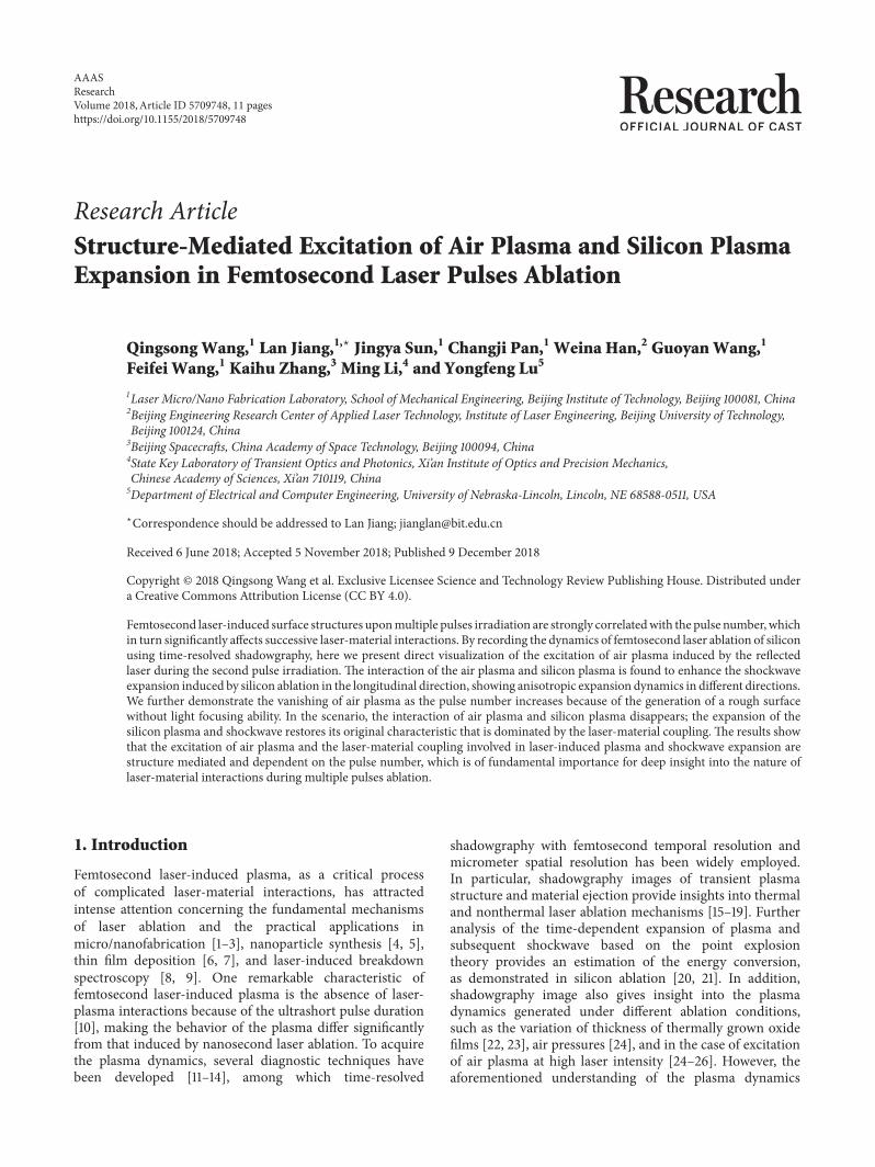
Probe beamSample
Objective
Mirror
FilterCCDProbe delay
Lens
(fs-ns)
Pump beam
2
Figure 1: Schematic of the pump-probe setup.
was mainly conducted with single-pulse irradiation, andthe plasma dynamics during multiple pulses laser ablationwas habitually ignored and still remains unclear. Recentstudies have indicated that the structures generated by priorpulses can interact with subsequent laser and reshape theintensity distribution, thereby affecting the ablation process[27–29]. Our previous study demonstrated the enhancementof plasma and shockwave expansion during femtosecondtwo-pulse ablation [30]. This phenomenon was theoreticallyinterpreted with laser-induced air breakdown enhanced bycrater-induced laser refocusing but was limited by the lackof direct experimental observation of the excitation of airfilaments. In addition, recent studies have suggested that themorphology of the laser-induced crater becomes rough asthe pulse number (N) increases [31–34], leading to the loss oflaser refocusing ability and can significantly alter the natureof the expansion of the laser-induced plasma and shockwave.
Herein, we investigate how laser-induced plasma andshockwave from silicon ablation vary as a function of incidentpulse number using time-resolved shadowgraphy.We presenta direct experimental visualization of the excitation of airplasma on femtosecond timescale induced by the reflectedpulse, which is mediated by the crater structure generatedby the previous pulse.The expansion of laser-induced plasmaand shockwave in the scenario is systematically analyzed.Theresults reveal two fundamental mechanisms that determinethe expansion of plasma and shockwave: the excitation ofair plasma with an anisotropic expansion effect and laser-material coupling with an isotropic expansion effect. Thedominant mechanism is determined by the morphology oflaser-induced crater, which is strongly dependent on the pulsenumber.
2. Results
2.1. Two Pulses Ablation. We first focus on the plasmaand shockwave evolution recorded during the first twopulses ablation at probe delays ranging from femtosecondsto nanoseconds with ultrafast pump-probe shadowgraphy(Figure 1). The pump pulse separation exceeded one secondand the laser fluence was set at a constant of 7.27 J/cm2.Figure 2 shows the transient shadowgraphs of the first twopulses ablation of silicon at probe delays on femtosecondtimescale. No transient phenomena were observed to emerge
and the shadowgraph remained unchanged on femtosec-ond timescale during the first pulse irradiation (Figures2(a)–2(d)). However, a pronounced narrow dark region wasclearly visible at a probe delay of 300 fs during the secondpulse irradiation (Figure 2(e)). The length of the dark regionextended with direction opposite to the incident direction ofthe pump pulse with the increase of probe delay (Figure 2(f)).For larger probe delays, the length of the dark region reachedsaturation;meanwhile, the overall shape and the transmissionof the dark region remained nearly unchanged (Figures2(g)–2(h)). The propagation velocity of the dark regionrecorded at probe delays from 300 fs to 400 fs was estimatedto be 2.91 × 10⁸ m/s, which is close to the velocity of lightin air. The distinctive features of the dark region indicate theexcitation of air plasma induced by the reflected beam duringthe second pulse irradiation. The laser-induced air plasmacan absorb the probe beam, thus resulting in the dark regionin the shadowgraphs.
In order to reveal the formationmechanism of air plasma,we have analyzed the ablation morphology induced by thefirst pulse. The scanning electron microscope (SEM) imageof the laser-induced structure formed on silicon upon single-pulse irradiation is shown in Figure 3(a). A smooth and sym-metric crater-shaped structure was generated on the siliconsurface with a rim formed around the crater. The diameterand depth of the laser-induced crater were approximately19.4 𝜇m and 0.27 𝜇m, respectively (Figure 3(b)). Details ofthe crater suggest that it can be considered as a concavemirror, which will reflect and focus the next incident pulse.Figure 3(d) shows the optical reflection image of the silicon-based crater array captured with an optical microscopysystem (Figure 3(c)); the crater was illuminated with a whitelight. On the focal plane of the crater, an array of brightfocal spots was visible in the crater center, which indicatesexcellent light focusing ability. Furthermore, the electric fielddistribution of the second pulse reflected by the preformedcrater was simulated with the finite difference time domainmethod; the focal spot of the incident pulse was set to beon the sample surface. For simplicity, we only conducteda two-dimensional simulation with the complex refractiveindex of silicon at 800 nm held constant at n = 3.692 +i0.0065 [35]. Figure 3(f) shows that a refocusing region wasgenerated above the silicon surface due to the focus effectof the crater as compared with the calculation result for theuntreated plane silicon surface (Figure 3(e)). Moreover, thecalculated electric field intensity of the refocused pulse canbe stronger than the incident field intensity. In fact, freeelectrons can be generated on the silicon surface via linearand nonlinear ionization by the rising edge of the pulseduring intense femtosecond laser irradiation, which resultsin a huge increase in transient reflectivity to significantlyreflect the latter part of the pulse according to the Drudemodel [36].Thus, the real field intensity of the refocused laseris much larger than the calculated intensity. The calculatedelectric field distribution is consistent with the morphologyof the air plasma observed in our experiment. The analysesdemonstrate the excellent light focusing ability of the craterinduced by the first pulse, which is able to focus the reflected

Research 3
(a)
(b)
(c)
(d)
(e)
(f)
(g)
(h)
Laser
Air Si50 mN = 1 N = 2
N = 1 N = 2
N = 1 N = 2
N = 1 N = 2
300 fs 300 fs
400 fs 400 fs
500 fs 500 fs
900 fs 900 fs
Air plasma
Figure 2: Time-resolved shadowgraphs of the laser ablation of silicon at probe delays on femtosecond timescale. (a–d) Shadowgraphs for thefirst pulse. (e–h) Shadowgraphs for the second pulse. The laser fluence is 7.27 J/cm2. The dashed line indicates the interface of silicon and air.The white arrow in (a) indicates the propagation direction of the incident laser.
(a)
(b)
(c) (d)
(e)
(f)
N = 1 10 m
100 m
White light
Beamsplitter
Lens CCD
ZSilicon with crater
Air Si
20 m
20 m
1.41.21.00.80.60.40.20.0
Er from untreated plane surface
Er from crater induced by first pulse
Er / E0
0.1
0.0
−0.1
−0.2
−0.3
z(
m)
0 10 20 30 40
x (m)
Figure 3: Characterization of the ablation morphology induced by the first pulse with a fluence of 7.27 J/cm2. (a) SEM image of the surfacestructure of silicon. (b) Atomic force microscope (AFM) profile of the cross-section of the structure in (a). (c) Sketch of the optical systemfor characterizing light focusing ability of the silicon-based crater array. (d) Focal spots image obtained with a CCD on the focal plane of thecrater. (e) Calculated electric field distribution of the reflected pulse (N = 1) from untreated plane silicon surface. (f) Calculated electric fielddistribution of the reflected pulse (N = 2) from the crater generated by the first pulse. The focal spot of the incident pulse in (e) and (f) wasset to be on the sample surface, indicated by right edge of the images.

4 Research
(a)
(b)
(c)
(d)
(e) (j)
(i)
(h)
(g)
(f)20 ps 20 ps
50 ps 50 ps
100 ps 100 ps
400 ps400 ps
800 ps 800 ps
N = 1 N = 2
N = 1 N = 2
N = 1 N = 2
N = 1 N = 2
N = 1 N = 2
50 m Air Si
Air plasma
Air shockwave
Figure 4: Time-resolved shadowgraphs of the plasma at probe delays on picosecond timescale. (a–e) Shadowgraphs for the first pulse. (f–j)Shadowgraphs for the second pulse. The laser fluence is 7.27 J/cm2. The dashed line indicates the interface of silicon and air.
pulse and generate a stronger laser intensity to excite airplasma during the second pulse irradiation.
The air plasma channel can affect the expansion of laser-induced plasma and the subsequent shockwave. Figure 4shows the transient shadowgraphs of the plasma inducedby the first two pulses ablation of silicon at probe delayson picosecond timescale. Similar to the observation onfemtosecond timescale, the air plasma channel was clearlyobserved on picosecond timescale during the second pulseirradiation (Figures 4(f)–4(j)). The air plasma channelremained constant at probe delay up to 100 ps (Figures4(f)–4(h)). When the probe delay exceeded 400 ps, theair plasma decayed gradually and expanded radially; thisinduced a cylinder-like shockwave (air shockwave for short)above the silicon surface (Figures 4(i) and 4(j)). The plasmainduced by laser ablation of silicon (silicon plasma) wasobserved at a probe delay of 20 ps (Figures 4(a) and 4(f)),which was two orders of magnitude later than the emergencetime of air plasma. Comparisons of Figures 4(a)–4(e) andFigures 4(f)–4(j) revealed no distinct differences between
the silicon plasma evolutions induced by the first two pulsesat probe delays on picosecond timescale. Furthermore, theexpansion distance of the silicon plasma was so small thatthe plasma front did not yet contact the air plasma channel,which enabled the plasma to retain its original expansioncharacteristics. With increasing probe delays, a shockwavewas formed and the shockwave front contacted with the airplasma channel, which significantly affects the expansion ofthe shockwave. Figure 5 shows the transient shadowgraphsof the shockwave induced by the first two pulses ablationof silicon at probe delays on nanosecond timescale; to bedistinguished from the shockwave induced by air plasma(air shockwave), the shockwave induced by laser-inducedsilicon plasma expansion was represented as silicon shock-wave for short. For the first pulse ablation, a small bulgeexisted on the shockwave front, which is attributed to theweak laser-induced air breakdown that cannot be detectedon femtosecond-to-picosecond timescale with the appliedtechnique. However, because of the generation of the evidentair shockwave with a lower density in the channel [24–26],

Research 5
(a)
(b)
(c)
(d)
(e)
(f)
(g)
(h)
50 mN = 1
N = 1
N = 1
N = 1 N = 2
N = 2
N = 2
N = 2Air Si
4 ns 4 ns
7 ns 7 ns
13 ns 13 ns
16 ns 16 ns
Air shockwave
Silicon shockwave(i)
(j)
(k)
160
140
120
100
80
60
40
20
0
Expa
nsio
n di
stan
ce (
m) Longitudinal (N = 1)
Longitudinal (N = 2)Radial (N = 1)Radial (N = 2)
N = 1
N = 2
50
40
30
20
10
0
Long
itudi
nal v
eloci
ty (k
m/s
)
0 2 4 6 8 10 12 14 16
Probe delay (ns)
Probe delay (ns)
100
10
Expa
nsio
n di
stan
ce (
m)
0.1 1 10
Slope (L1) = 0.57
Slope (L2) = 0.69
Slope (R1) = 0.42
Slope (R2) = 0.42
Longitudinal (N = 1)Longitudinal (N = 2)Radial (N = 1)Radial (N = 2)
Figure 5: Time-resolved plasma shockwave expansion at probe delays on nanosecond timescale. (a–d) Shadowgraphs for the first pulse. (e–h)Shadowgraphs for the second pulse. The dashed line indicates the interface of silicon and air. (i) Measurements of longitudinal and radialexpansion of the silicon shockwave as a function of time for the first two pulses ablation of silicon. (j) Calculated longitudinal velocities ofsilicon shockwave as a function of time for the first two pulses ablation of silicon. (k) Double logarithmic fitting of longitudinal and radialexpansion for the first two pulses ablation. The laser fluence is 7.27 J/cm2.
the front of the silicon shockwave induced by the secondpulse was distorted in the longitudinal direction when itcontacted the air plasma channel (Figures 5(e)–5(h)). Thus,the bulge on the front of the silicon shockwave was moredistinct compared with that induced by the first pulse.
Figure 5(i) shows the expansion distance of the siliconshockwave as a function of probe delay for the first twopulses ablation; the longitudinal expansion velocity obtainedby differentiating is presented in Figure 5(j). At 7 ns, thelongitudinal expansion distances were 66.5 𝜇m and 87.5𝜇m for the first and second pulse ablation, respectively;the corresponding velocities were 6.7 × 10³ m/s and 11.0 ×10³ m/s, respectively. The silicon shockwave was thereforeenhanced in the longitudinal direction during the secondpulse ablation, which is due to the acceleration effect ofthe laser-induced air plasma [24–26]. At larger probe delays(i.e., 16 ns), the longitudinal velocity difference for the firsttwo pulses ablation declined. This may be attributed to thelimitation of plasma length and the decay of the air plasma
channel. In general, the time-dependent expansion of thelaser-induced plasma and shockwave can be described by theSedov-Taylor solution [20]:
𝑅 = 𝜆(𝐸𝜌)1/(𝛽+2)
𝑡2/(𝛽+2), (1)
where R is the expansion distance, 𝜆 is a constant dependenton the specific heat capacity ratio and approximately equal tounity, E is the energy converted into the plasma state, 𝜌 is thedensity of the undisturbed air, t is the expansion time (probedelay), and 𝛽 describes the dimension of expansion (valuesof 1, 2, and 3 represent planar, cylindrical, and sphericalpropagation, respectively). Figure 5(k) shows the doublelogarithmic fitting of longitudinal and radial expansion atprobe delays larger than 1 ns for the first two pulses ablation.The slope in the radial direction remained constant at 0.42(𝛽 ∼ 3) for the first two pulses ablation, indicating thatthe radial expansion of the silicon shockwave was sphericaland not influenced by the air plasma. However, the slope

6 Research
(a1) (b1) (c1)
(a2) (b2) (c2)
(a3) (b3) (c3)
500 fs 500 fs 500 fs
800 ps 800 ps 800 ps
16 ns 16 ns 16 ns
N = 3 N = 4 N = 5
N = 3 N = 4 N = 5
N = 3 N = 4 N = 5
50 m Air Si
Air plasma
Air shockwave
Silicon shockwave
Figure 6: Shadowgraphs of the plasma shockwave at 500 fs, 800 ps, and 16 ns probe delays during multiple pulses ablation. (a1–a3)Shadowgraphs for the third pulse. (b1–b3) Shadowgraphs for the fourth pulse. (c1–c3) Shadowgraphs for the fifth pulse. The laser fluenceis 7.27 J/cm2. The dashed line indicates the interface of silicon and air.
in the longitudinal direction increased from 0.57 (𝛽 = 1.51)to 0.69 (𝛽 ∼ 1) for the first two pulses ablation, indicatingthat the longitudinal expansion of the silicon shockwave wasaltered to typically planar. These similarities and differencesin expansion characteristics suggest that the laser-inducedsilicon plasma and shockwave are significantly affected by theexcitation of air plasmaduring the secondpulse ablation.Andin the scenario, the expansion of laser-induced silicon plasmaand shockwave during second pulse ablation is similar to thatdemonstrated by previous works for the single femtosecondlaser ablation in the case of strong excitation of air plasma[24–26].
2.2. Multiple Pulses Ablation. Previous studies have investi-gated the evolution of multiple pulses ablation and demon-strated the transition in the ablation surface from smoothto rough [31, 37, 38]. To study the effect of structure evolu-tion on the plasma and shockwave dynamics, time-resolvedshadowgraphswere recorded duringmultiple pulses ablation.Figure 6 shows the typical shadowgraphs of the plasma andshockwave at probe delays on femtosecond, picosecond, andnanosecond timescales during the third, fourth, and fifthpulse ablation.During the third pulse ablation, air plasmawasalso observed at 500 fs probe delay (Figure 6(a1)). The lengthof the air plasma was shorter and positioned much closer to
the sample surface compared with the air plasma induced bythe second pulse, indicating that the crater induced by thesecond pulse had a smaller effective focal length. Afterwards,the air plasma made contact with the silicon plasma andenhanced the plasma and shockwave expansion (Figures6(a2) and 6(a3)), which is similar to the observations duringthe second pulse ablation. However, when the fourth pulsewas irradiated, the air plasma on femtosecond timescalefaded quickly (Figure 6(b1)) and could barely be detectedduring the fifth pulse ablation (Figure 6(c1)). No evident airshockwave was observed on picosecond timescale, and theenhancement of the silicon shockwave in the longitudinaldirection attenuated gradually. In particular, the morphol-ogy of the silicon shockwave induced by the fifth pulse(Figure 6(c3)) was similar to that induced by the first pulse(Figure 5(d)). The results indicate that the generation ofair plasma is pulse number-dependent. Figures 7(a) and7(b) depict the pulse number-dependent longitudinal andradial expansion of the silicon shockwave as a function oftime. With the increase of pulse number, the expansiondistance in the longitudinal direction increased rapidly tothe maximum value when the third pulse is irradiated andthen decreased gradually (Figures 7(a) and 7(c)). Althoughthe expansion distance decreased rapidly, it was still largerthan that induced by the first pulse. In addition, the expansion

Research 7
160
140
120
100
80
60
40
20
0
Long
itudi
nal d
istan
ce (
m)
0 2 4 6 8 10 12 14 16
Probe delay (ns)N = 1
N = 2
N = 3
N = 4
N = 5
(a)
0 2 4 6 8 10 12 14 16
Probe delay (ns)N = 1
N = 2
N = 3
N = 4
N = 5
60
50
40
30
20
10
Radi
al d
istan
ce (
m)
(b)
160
150
140
130
120
110
1000 2 4 6
Pulse numberLongitudinalRadial
62
61
60
59
58
57
56
Long
itudi
nal d
istan
ce (
m)
Radi
al d
istan
ce (
m)
(c)
Probe delay (ns)
Expa
nsio
n di
stanc
e (
m)
100
10011
Longitudinal direction (N = 3)Radial direction (N = 3)
Slope (L) = 0.64
Slope (R) = 0.42
(d)
Expa
nsio
n di
stan
ce (
m)
100
10
Probe delay (ns)011
Longitudinal direction (N = 4)Radial direction (N = 4)
Slope (L) = 0.53
Slope (R) = 0.43
(e)
Expa
nsio
n di
stan
ce (
m)
100
10
Probe delay (ns)011
Longitudinal direction (N = 5)Radial direction (N = 5)
Slope (L) = 0.52
Slope (R) = 0.43
(f)
Figure 7: Pulse number-dependent expansion distance of the silicon shockwave. (a–b) Pulse number-dependent longitudinal and radialexpansion of the silicon shockwave as a function of time. (c) Pulse number-dependent longitudinal and radial expansion of silicon shockwavesat a 16 ns probe delay. (d–f) Double logarithmic fitting of the longitudinal and radial expansion for the third, fourth, and fifth pulse ablation.
distance in the radial direction had a shaper increaseafter the fourth pulse irradiation (Figures 7(b) and 7(c)),indicating an increased energy deposition into the siliconplasma during multiple pulses ablation. These results signifyanother mechanism of shockwave enhancement in multiplepulses ablation. Furthermore, the expansion characteristics
of multiple pulses ablation were investigated based on theSedov-Taylor solution [20], as shown in Figures 7(d)–7(f).In the case of excitation of air plasma, the expansion of thesilicon shockwave in the longitudinal direction was alteredto a typically planar (one-dimensional) propagation and itwas basically restored to its original expansion dimension

8 Research
10 mN = 2
(a)
N = 3
(b)
N = 4
(c)
0.4
0.0
−0.4
−0.8
−1.2
−1.6
z (
m)
0 10 20 30 40
x (m)N = 2
N = 3
N = 4
(d)
Figure 8: Silicon structure evolution during multiple pulses ablation. (a-c) SEM images of silicon structures induced by the second, third,and fourth pulses. (d) AFM profiles of the structure induced by the second, third, and fourth pulses. The laser fluence is 7.27 J/cm2.
as the air plasma vanished. Simultaneously, the expansion ofthe silicon shockwave in the radial direction remained nearlyconstant at a spherical (three-dimensional) propagation forthe applied pulse number, showing no dependence on theexcitation and vanishing of air plasma.
To gain deeper insight into the pulse number-dependentplasma and shockwave evolution, the surface morphologywas examined. Figure 8 shows the silicon structures inducedby the second, third, and fourth pulses; the structure inducedby the first pulse is shown in Figures 3(a) and 3(b). Smoothcrater-shaped structures were generated during the first twopulses ablation. Thus, these structures could reflect andrefocus the next pulse, leading to the excitation of air plasmaduring the second and third pulse ablation and enhancementof the silicon shockwave expansion in the longitudinal direc-tion. However, after the third pulse irradiation, the siliconsurface became rough with some microbulges at the bottomof the crater (Figure 8(b)). This structure became rougherwhen the fourth pulse was irradiated; the periphery of therough crater was covered with dense nanostructures. Craterswith such micro/nanostructures fail to effectively focus thereflected laser during the next pulse irradiation, leading tothe vanishing of air plasma during the fourth and fifth pulseablation. In the scenario, the expansion of the silicon plasmaand shockwave is dominated by the laser-material coupling.
Silicon craters with micro/nanostructures can enhance laser-material coupling via antireflection effect and incubationeffect.The antireflection effect increases the absorption of theincident laser with the light trapping effect, surface plasmonpolaritons excitation, and effective medium effect [39–41];the incubation effect due to surface defects is beneficial tomaterial ablation [42–44]. The enhanced laser-material cou-pling can increase the energy deposition in the silicon plasmaand thus result in stronger plasma and shockwave expansionobserved before. Moreover, the enhancement by increasedlaser-material coupling is more isotropic in comparison withthe anisotropic expansion characteristic enhanced by airplasma.
3. Discussion
In summary, we have investigated the laser-induced plasmadynamics during multiple femtosecond laser pulses ablationof silicon, which was significant but always ignored inprevious studies. The structure-mediated excitation of airplasma was directly observed during multiple femtosec-ond laser pulses ablation. Furthermore, the expansion ofplasma and shockwave was systematically analyzed, revealingtwo fundamental mechanisms for the plasma and shock-wave expansion: the excitation of air plasma and laser-material coupling. These two mechanisms were found to be
Research 9
strongly dependent on the laser-induced surface structure.At small pulse number, smooth crater was generated byprior pulses, which can reflect and refocus the next pulse,inducing higher laser intensity above the sample surface.Thus, air plasma was excited and dominated the anisotropicexpansion of the silicon plasma and shockwave. At higherpulse number, the smooth crater became rough coveredwith micro/nanostructures, which was unable to refocus theincident pulse. In the scenario, the air plasma vanished andthe laser-material coupling was instead the core mechanismfor the expansion of the silicon plasma and shockwave withmore isotropic characteristics. Our findings indicate theunderlying mechanisms of laser-induced plasma dynamicsduring multiple pulses ablation, and they are of fundamentalimportance for deep insight into the nature of ultrafast laser-material interactions.
4. Materials and Methods
4.1. Femtosecond Pump-Probe Shadowgraphy. The ultrafastpump-probe shadowgraphy was performed with a commer-cial Ti: sapphire femtosecond laser regenerative amplifier(Spitfire, Spectral Physics), which delivers 50 fs pulses with acentral wavelength of 800 nm. Figure 1 shows the schematicof the pump-probe setup. During the experiment, the lasersystem was operated in external gated mode and a timecontroller was employed to trigger a single pulse.The emittedpulse was split into pump and probe pulses. The pumppulse was focused normally onto the silicon surface (opticalpolishing, orientation (100)) using a plano-convex lens (f =100 mm), generating a Gaussian intensity distribution with adiameter of approximately 35 𝜇m (1/e2-decay distance frompeak value). The laser fluence (peak fluence) herein was setat a constant of 7.27 J/cm2, which was much larger than theablation threshold of silicon.The probe pulse, after passing anoptical delay line (time uncertainty < 20 fs), was frequencydoubled with a BBO to 400 nm, and a dielectric coatingshort-pass filter (edge wavelength = 650 nm, Daheng Optics)was used to block the residual fundamental 800 nm pulse.The probe pulse was then directed to illuminate the interac-tion region along the direction perpendicular to the pumppulse and parallel to the sample surface. The transmittedprobe pulse was collected with an objective (20×, NA =0.45, Olympus) and directed onto a charge-coupled device(CCD). A 400 nm dielectric coating band-pass filter (fullwidth half maximum = 10 nm, Thorlabs) was added beforethe CCD to suppress the background illumination. Beforesilicon ablation experiment, the zero-delay was adjusted byprobing laser-induced air plasma at high laser fluence withthe sample being removed, which was set as the time the airplasma was detected at the focal spot. To study the ablationdynamics of multiple pulses irradiation, a series of shad-owgraphs of plasma and shockwave induced by successivepulses were recorded at probe delays ranging from fem-tosecond to nanosecond timescale. During multiple pulsesirradiation, the pump pulse separation exceeded one second.To improve the contrast of the obtained shadowgraphs,image background subtraction was performed by subtract-ing the background image from the obtained pump-probe
images [45]; the background image was recorded with thepump pulse being blocked. The appearance of the silicon(right area of the dashed line in the shadowgraphs) afterbackground subtraction looks different from the originalblack appearance.
4.2. Characterization of Surface Morphology. The ablationmorphology was characterized with a scanning electronmicroscope (XL30 S-FEG, FEI) and an atomic force micro-scope (Dimension edge, Bruker). The light focusing abilityof the silicon-based crater generated by the first pulse wascharacterized with an optical system shown in Figure 3(c).The reflected light of the crater was collected with a lens andimaged on a CCD.
Data Availability
All data needed to evaluate the conclusions in the paper arepresent in the paper. Additional data related to this papermaybe requested from the authors.
Conflicts of Interest
The authors declare that there are no conflicts of interestregarding the publication of this article.
Authors’ Contributions
Q. Wang, L. Jiang, and J. Sun conceived and designed theexperiments. Q. Wang, C. Pan, and F. Wang performed thepump-probe study of ablation of silicon and characterizationof the surface morphology. Q. Wang, W. Han, G. Wang,and K. Zhang analyzed the experimental results, and all theauthors contributed to the discussion of the results. Q.Wang,L. Jiang, and J. Sun wrote the paper with input from all theauthors, and all the authors reviewed the manuscript.
Acknowledgments
This research was supported by the National Key R&D Pro-gram of China (grant no. 2017YFB1104300) and the NationalNatural Science Foundation of China (grant nos. 91323301,11704028).
References
[1] L. Jiang, A. D. Wang, B. Li, T. H. Cui, and Y. F. Lu, “Electronsdynamics control by shaping femtosecond laser pulses inmicro/nanofabrication: modeling, method, measurement andapplication,” Light: Science & Applications, vol. 7, no. 2, p. 17134,2018.
[2] C. Kerse, H. Kalaycıoglu, P. Elahi et al., “Ablation-cooledmaterial removal with ultrafast bursts of pulses,” Nature, vol.537, no. 7618, pp. 84–88, 2016.
[3] K. Xu, C. Zhang, R. Zhou, R. Ji, and M. Hong, “Hybridmicro/nano-structure formation by angular laser texturing of Sisurface for surface enhanced Raman scattering,”Optics Express,vol. 24, no. 10, pp. 10352–10358, 2016.

10 Research
[4] B. Tan and K. Venkatakrishnan, “Synthesis of fibrous nanopar-ticle aggregates by femtosecond laser ablation in air,” OpticsExpress, vol. 17, no. 2, pp. 1064–1069, 2009.
[5] C. M. Rouleau, A. A. Puretzky, and D. B. Geohegan, “Slowingof femtosecond laser-generated nanoparticles in a backgroundgas,” Applied Physics Letters, vol. 105, no. 21, 2014.
[6] F. Bourquard, T. Tite, A.-S. Loir, C. Donnet, and F. Garrelie,“Control of the graphite femtosecond ablation plumekinetics bytemporal laser pulse shaping: Effects on pulsed laser depositionof diamond-like carbon,” The Journal of Physical Chemistry C,vol. 118, no. 8, pp. 4377–4385, 2014.
[7] D. Fischer, L. V. Meyer, M. Jansen, and K. Muller-Buschbaum,“Highly luminescent thin films of the dense framework3∞[EuIm2] with switchable transparency formed by scanningfemtosecond-pulse laser deposition,”Angewandte Chemie Inter-national Edition, vol. 53, no. 3, pp. 706–710, 2014.
[8] Z. Hou, Z. Wang, J. Liu, W. Ni, and Z. Li, “Signal qualityimprovement using cylindrical confinement for laser inducedbreakdown spectroscopy,” Optics Express, vol. 21, no. 13, pp.15974–15979, 2013.
[9] J. Penczak, R. Kupfer, I. Bar, and R. J. Gordon, “The role ofplasma shielding in collinear double-pulse femtosecond laser-induced breakdown spectroscopy,” Spectrochimica Acta Part B:Atomic Spectroscopy, vol. 97, pp. 34–41, 2014.
[10] A.Miloshevsky, S. S. Harilal, G.Miloshevsky, and A. Hassanein,“Dynamics of plasma expansion and shockwave formation infemtosecond laser-ablated aluminum plumes in argon gas atatmospheric pressures,” Physics of Plasmas, vol. 21, no. 4, 2014.
[11] C. T. Hebeisen, G. Sciaini, M. Harb, R. Ernstorfer, S. G. Kruglik,and R. J. D. Miller, “Direct visualization of charge distributionsduring femtosecond laser ablation of a Si (100) surface,” PhysicalReview B: Condensed Matter and Materials Physics, vol. 78, no.8, 2008.
[12] H. Hu, T. Liu, and H. Zhai, “Comparison of femtosecond laserablation of aluminum in water and in air by time-resolvedoptical diagnosis,” Optics Express, vol. 23, no. 2, pp. 628–635,2015.
[13] S. S. Harilal, P. K. Diwakar, M. P. Polek, and M. C. Phillips,“Morphological changes in ultrafast laser ablation plumes withvarying spot size,” Optics Express, vol. 23, no. 12, pp. 15608–15615, 2015.
[14] K. K. Anoop, S. S. Harilal, R. Philip, R. Bruzzese, and S.Amoruso, “Laser fluence dependence on emission dynamicsof ultrafast laser induced copper plasma,” Journal of AppliedPhysics, vol. 120, no. 18, 2016.
[15] N. Zhang, X. Zhu, J. Yang, X. Wang, and M. Wang, “Time-resolved shadowgraphs ofmaterial ejection in intense femtosec-ond laser ablation of aluminum,”Physical Review Letters, vol. 99,no. 16, 2007.
[16] H.Hu,X.Wang,H. Zhai,N. Zhang, andP.Wang, “Generation ofmultiple stress waves in silica glass in high fluence femtosecondlaser ablation,” Applied Physics Letters, vol. 97, no. 6, 2010.
[17] X. Zhao and Y. C. Shin, “Coulomb explosion and early plasmageneration during femtosecond laser ablation of silicon at highlaser fluence,” Journal of Physics D: Applied Physics, vol. 46, no.33, 2013.
[18] C. Kalupka, J. Finger, and M. Reininghaus, “Time-resolvedinvestigations of the non-thermal ablation process of graphiteinduced by femtosecond laser pulses,” Journal of Applied Physics,vol. 119, no. 15, 2016.
[19] G. Wang, Y. Yu, L. Jiang, X. Li, Q. Xie, and Y. Lu, “Cylindricalshockwave-induced compression mechanism in femtosecond
laser Bessel pulse micro-drilling of PMMA,” Applied PhysicsLetters, vol. 110, no. 16, 2017.
[20] L. I. Sedov, Similarity and Dimensional Methods in Mechanics,CRC Press, Boca Raton, FL, USA, 1993.
[21] T. Y. Choi and C. P. Grigoropoulos, “Plasma and ablationdynamics in ultrafast laser processing of crystalline silicon,”Journal of Applied Physics, vol. 92, no. 9, pp. 4918–4925, 2002.
[22] J. P. McDonald, J. A. Nees, and S. M. Yalisove, “Pump-probeimaging of femtosecond pulsed laser ablation of silicon withthermally grown oxide films,” Journal of Applied Physics, vol.102, no. 6, 2007.
[23] J. P. McDonald, V. R. Mistry, J. A. Nees, and S. M. Yalisove,“Time resolved dynamics of femtosecond laser ablation of Si(100) with Thin thermal oxide layers (20 - 1200 nm),”MaterialsResearch Society - Proceedings, vol. 929, 2006.
[24] Z. Wu, N. Zhang, M. Wang, and X. Zhu, “Femtosecond laserablation of silicon in air and vacuum,” Chinese Optics Letters,vol. 9, no. 9, pp. 093201–93204, 2011.
[25] M. Boueri, M. Baudelet, J. Yu, X. Mao, S. S. Mao, and R. Russo,“Early stage expansion and time-resolved spectral emission oflaser-induced plasma from polymer,” Applied Surface Science,vol. 255, no. 24, pp. 9566–9571, 2009.
[26] H. Zhang, F. Zhang, X. Du, G. Dong, and J. Qiu, “Influence oflaser-induced air breakdown on femtosecond laser ablation ofaluminum,” Optics Express, vol. 23, no. 2, pp. 1370–1376, 2015.
[27] L. S. Jiao, E. Y. K. Ng, H. Y. Zheng, and Y. L. Zhang, “Theoreticalstudy of pre-formed hole geometries on femtosecond pulseenergy distribution in laser drilling,” Optics Express, vol. 23, no.4, pp. 4927–4934, 2015.
[28] J. Zhang, R. Drevinskas, M. Beresna, and P. G. Kazansky,“Polarization sensitive anisotropic structuring of silicon byultrashort light pulses,” Applied Physics Letters, vol. 107, no. 4,2015.
[29] H. Zhang, J. Colombier, C. Li, N. Faure, G. Cheng, and R.Stoian, “Coherence in ultrafast laser-induced periodic surfacestructures,” Physical Review B: Condensed Matter and MaterialsPhysics, vol. 92, no. 17, 2015.
[30] Q. Wang, L. Jiang, J. Sun et al., “Enhancing the expansionof a plasma shockwave by crater-induced laser refocusing infemtosecond laser ablation of fused silica,” Photonics Research,vol. 5, no. 5, pp. 488–493, 2017.
[31] J. Bonse and J. Kruger, “Pulse number dependence of laser-induced periodic surface structures for femtosecond laserirradiation of silicon,” Journal of Applied Physics, vol. 108, no.3, 2010.
[32] F. Liang, R. Vallee, and S. L. Chin, “Physical evolution ofnanograting inscription on the surface of fused silica,” OpticalMaterials Express , vol. 2, no. 7, pp. 900–906, 2012.
[33] Y. Liu, Y. Brelet, Z. He et al., “Laser-induced periodic annularsurface structures on fused silica surface,” Applied PhysicsLetters, vol. 102, no. 25, 2013.
[34] S. He, J. J. J. Nivas, A. Vecchione, M. Hu, and S. Amoruso,“On the generation of grooves on crystalline silicon irradiatedby femtosecond laser pulses,” Optics Express, vol. 24, no. 4, pp.3238–3247, 2016.
[35] E. D. Palik, Handbook of Optical Constants of Solids, Academicpress, San Diego, CA, USA, 1998.
[36] L. Wang, B.-B. Xu, X.-W. Cao et al., “Competition betweensubwavelength and deep-subwavelength structures ablated byultrashort laser pulses,” Optica, vol. 4, no. 6, pp. 637–642, 2017.

Research 11
[37] H. Al-Khazraji and V. R. Bhardwaj, “Polarization dependentmicro-structuring of silicon with a femtosecond laser,” AppliedSurface Science, vol. 353, pp. 600–607, 2015.
[38] G. D. Tsibidis, C. Fotakis, and E. Stratakis, “From ripples tospikes: A hydrodynamical mechanism to interpret femtosecondlaser-induced self-assembled structures,” Physical Review B:Condensed Matter and Materials Physics, vol. 92, no. 4, 2015.
[39] A. Y. Vorobyev and C. Guo, “Antireflection effect of femtosec-ond laser-induced periodic surface structures on silicon,”OpticsExpress, vol. 19, no. 105, pp. A1031–A1036, 2011.
[40] J. Yang, F. Luo, T. S. Kao et al., “Design and fabricationof broadband ultralow reflectivity black Si surfaces by lasermicro/nanoprocessing,” Light: Science &Applications, vol. 3, no.7, p. e185, 2014.
[41] P. Fan, B. Bai, M. Zhong et al., “General strategy toward dual-scale-controlled metallic micro-nano hybrid structures withultralow reflectance,” ACS Nano, vol. 11, no. 7, pp. 7401–7408,2017.
[42] H. Varel, M. Wahmer, A. Rosenfeld, D. Ashkenasi, and E. E. B.Campbell, “Femtosecond laser ablation of sapphire: Time-of-flight analysis of ablation plume,” Applied Surface Science, vol.127-129, pp. 128–133, 1998.
[43] J. Bonse, S. Baudach, J. Kruger, W. Kautek, and M. Lenzner,“Femtosecond laser ablation of silicon-modification thresholdsand morphology,” Applied Physics A: Materials Science & Pro-cessing, vol. 74, no. 1, pp. 19–25, 2002.
[44] C. Gaudiuso, G. Giannuzzi, A. Volpe, P. M. Lugara, I. Choquet,and A. Ancona, “Incubation during laser ablation with burstsof femtosecond pulses with picosecond delays,” Optics Express,vol. 26, no. 4, pp. 3801–3813, 2018.
[45] Y. Yu, L. Jiang, Q. Cao, B. Xia, Q. Wang, and Y. Lu, “Pump-probe imaging of the fs-ps-ns dynamics during femtosecondlaser Bessel beam drilling in PMMA,” Optics Express, vol. 23,no. 25, pp. 32728–32735, 2015.