structure, gas chromatographic measurement, function suberin
TRANSCRIPT

Plant Physiol. (1974) 54, 116-121
Structure, Gas Chromatographic Measurement, and Function ofSuberin Synthesized by Potato Tuber Tissue Slices'
Received for publication January 29, 1974 and in revised form March 8, 1974
P. E. KOLATTUKUDY2 AND B. B. DEANDepartmenits of Agricultuiral Chemistry anid Horticultuire, Washin2gton2 State Uniiver.sity, Pullman, Washingtoni99163
ABSTRACT
The polymeric material (suberin) of the wound periderm ofpotato tuber slices was analyzed after depolymerization withLiAlH4 in tetrahydrofuran or BF3 in methanol with the use ofthin layer chromatography, chemical modification, and com-bined gas-liquid chromatography and mass spectrometry.Fatty acids (C10 to C26), fatty alcohols (C16 to C26), octadec-9-ene-1, 18-dioic acid, and 18-hydroxy-octadec-9-enoic acid wereidentified to be the major components. Based on the structuralinformation that the two bifunctional C1s molecules constituteda major portion of suberin, a gas chromatographic method ofmeasuring suberization was developed. This method consistedof hydrogenolysis of powdered tissue followed by thin layerchromatography and gas chromatographic measurement ofoctadecene-I , 18-diol as the trimethylsilyl ether. With this assayit was shown that the development of resistance to water lossby the tissue slices was directly proportional to the quantityof the bifunctional C1s molecules, thus providing evidence thata function of suberin is prevention of water loss.
Suberization is an important process because suberin plays akey role in preventing weight loss and decay, two of the majorproblems in the potato industry (4, 12). Suberization of potatotuber tissue has been studied by many investigators (7, 8, 11).Most of such studies on the so-called suberin layer have beenrestricted to microscopic examinations of stained tissues. Onthe basis of such staining techniques, it has been assumed thatsuberin is made of lipid materials. However, the exact chemicalnature of the material formed is not understood. Furthermore,studies on the suberization process have been limited becauseno reliable chemical methods of quantitation of suberizationhave been available. In this paper we report the results of struc-tural studies on the polymeric material of wound periderm ofpotato tuber tissue. Structural analysis by combined gas-liquidchromatography and mass spectrometry showed that the ali-phatic constituents of the suberin formed on the wound surfaceare very similar to those found in the natural skin of potatotuber. A gas chromatographic method for quantitating suber-
'This work was supported in part by National Science Founda-tion Grant GB-23081. Scientific Paper 4198, Project 2001, Agri-cultural Research Center, College of Agriculture, Washington StateUniversity, Pullman, Wash. 99163.
2 Author to whom inquiries should be made.
ization is also described. With this newly developed assay,suberization is shown to be correlated directly to the develop-ment of resistance to water loss from potato tuber tissue.
MATERIALS AND METHODS
Materials. Potato tubers (Russet Burbank) were grown at theOthello Experimental Farm of Washington State University.The tubers (1-2 kg) were washed free of soil and stored at 7 Cuntil used. Cylindrical sections of tissue (2 cm long, 1 cm di-ameter) were cut with a No. 5 cork borer; they were rinsedwith distilled water and placed on rubberized mesh in widemouth gallon jars, through which 0.6 liter/hr of air was passed.Generally the temperature was held at 20 C unless otherwisespecified, and at this temperature no bacterial growth was ob-served. Sources of LiAMH4, pectinase, cellulase, N,O-bis(tri-methylsilyl)acetamide were the same as those indicated earlier(6). OsO was from National Lead Co., New York.
Preparation of the Polymeric Material from the WoundPeriderm. After 7 days of suberization the tissue cylinders werecut in half with a razor blade, and the slices were treated over-night with 5 g/liter Aspergillus niger cellulase and 1 g/literfungal pectinase in 0.05 M acetate buffer, pH 4.0. All the in-ternal cells were removed by this treatment leaving behindsmall cuplike materials which were recovered. This residue wasground with a mortar and pestle, and the soluble lipids wereremoved by thorough extraction with a 2:1 mixture of chloro-form and methanol. The solid recovered was treated again withcellulase-pectinase as described above, and the solid recoveredby centrifugation or filtration was extracted thoroughly with a2: 1 mixture of chloroform and methanol. The solid was thenextracted with chloroform overnight with a Soxhlet extractorin order to remove any remaining soluble lipids.
Depolymerization by Hydrogenolysis. One gram of the solidwas refluxed with 2 g of LiAlH4 in 30 ml of tetrahydrofuranfor 24 to 48 hr. The excess LiAIH4 was decomposed by care-fully adding the reaction mixture dropwise into 75 ml of dis-tilled water with vigorous stirring. The mixture was acidified bythe addition of 5 to 10 ml of concentrated HCI. The lipid prod-ucts were extracted repeatedly with chloroform (4 x 50 ml).The chloroform extract was evaporated to dryness under re-duced pressure.
Depolymerization by Transesterification. One gram of thesolid was refluxed with 30 ml of 14% BF, in methanol for 24to 48 hr. After the addition of 50 ml of water, the productswere extracted repeatedly with chloroform (4 x 50 ml). Thechloroform extract was evaporated to dryness under reducedpressure.
Chromatography. Thin layer chromatography was done on0.5- or 1-mm layers of Silica Gel G (20 X 20 cm) activated
116
Dow
nloaded from https://academ
ic.oup.com/plphys/article/54/1/116/6074066 by guest on 27 N
ovember 2021

POTATO WOUND SUBERIN
overnight at 110 C. Components on the thin layer chromato-grams were visualized either by the dichromate-sulphuric acidcharring method, or by viewing the plate under UV light afterspraying it with a 0.1% ethanolic solution of 2',7'-dichloro-fluorescein. The components were recovered from the silica gelwith a 2: 1 mixture of chloroform and methanol. The develop-ing solvents are indicated elsewhere in this paper.Gas chromatography was performed with a coiled glass col-
umn (183 x 0.31 cm o.d.) packed with 5% OV-101 on 80 to100 mesh Gas Chrom Q. Part of the effluent of the gaschromatograph was passed into a Perkin Elmer-Hitachi RMU6D mass spectrometer with a Biemann separator interphase.Mass spectra were recorded at the apex of the gas chromato-graphic peaks with 70 ev ionizing voltage. In order to makesure that the peak did not represent an incompletely resolvedmixture, mass spectra were also recorded at either side of theapex.
Preparation of Derivatives. Trimethylsilyl ethers were pre-pared by heating the material with excess (0.25 ml) of N,O-bis(trimethylsilyl)acetamide at 90 to 100 C for 20 min. Excessreagent was removed with a stream of N2, and the product wasdissolved in a 2: 1 mixture of chloroform and methanol for gaschromatography. Introduction of a vic-diol function at thedouble bond was done by treatment of the olefin with a 0.1%solution of OsO in dioxane for 2 hr. The reaction mixture wasdecomposed with aqueous-methanolic Na,SO,; the resultingprecipitate was removed by centrifugation and washed oncewith methanol and centrifuged. Products were recovered byether extraction of combined supernatants.Gas Chromatographic Measurement of Suberin of the
Wound Periderm. About eight cylindrical tissue slices whichwere suberized for varying periods of time were freeze-driedand then ground in a Wiley mill followed by a finer grindingin a Wig-L-Bug Amalgamator (Crescent Dental ManufacturingCo.). One gram of the powder was subjected to hydrogenolysiswith LiAIH, for 48 hr, and the products were recovered as de-scribed in a previous section. The soluble lipid thus obtainedwas mixed with 80 ,ug of hexadecane-1, 16-diol as an internalstandard and was applied to 0.5-mm (20 X 20 cm) layers ofSilica Gel G. The chromatograms were developed in ethylether-hexane-methanol (20:5:1, v/v). The components werevisualized with 2',7'-dichlorofluorescein and recovered as indi-cated elsewhere. The recovered diol and alcohol fractions weretransferred into narrow-bottomed graduated centrifuge tubesand evaporated to dryness with a stream of N2, taking precau-tions to keep the solid in the narrow portion of the centrifugetube. To each tube, 0.3 ml of N,O-bis(trimethylsilyl)acetamidewas added and heated in an oil bath at 90 C for 20 min. Ex-cess reagent was evaporated with a stream of N2 and the prod-ucts were made up to 300 yl with a 2: 1 mixture of chloroformand methanol. Aliquots (generally 2 ,ul) were injected into aVarian gas chromatograph equipped with a flame ionization de-tector. The internal standard (C.8 diol) was used to quantitatethe C,, diol derived from the wound periderm, and the identityof the gas chromatographic peak of C1, diol was established byits mass spectrum. The alcohol fractions were also similarlyanalyzed.
Diffusion Resistance Measurements. The resistance of thetissue surface to water vapor loss (Rt) was calculated with thefollowing formula (10):
Pt= E-Pa
where pt = vapor density of the tissue in g/cm' = RHti,,,. XPr; RH,,s,,, is the relative humidity of the tissue which is 100
and pv was calculated using the formula Pv = 18e/RT, wheree = vapor pressure, T = temperature (OK), and R = the gasconstant. pa = vapor density of the air in g/cm' = RHa.r X p,where RHair is the relative humidity of the air. E = evapora-tion per unit surface area per unit time in g/ sec cm2. E wascalculated from the measured loss of weight of 20 tissuecylinders during a 3-hr exposure to the atmosphere. R. = re-sistance of the air to water vapor diffusion (sec/cm). When R,= 0, R = (pt - p)/E = the resistance of unsuberized tissue,and this was experimentally determined using freshly cut tissuecylinders.
RESULTS AND DISCUSSION
Identification of the Aliphatic Constituents of Potato WoundPeriderm. In order to determine the structure of the monomersof the polymeric material formed on the wound periderm, apreparation of periderm-enriched material was attempted. Af-ter 7 days of suberization the cylindrical tissue slices were cutinto halves to expose the internal, apparently nonsuberized, re-gion. Treatment of these tissue slices with pectinase and cellu-lase removed most of the internal part of the tissue, leavingbehind small cuplike structures, presumably enriched in thewound periderm material. After further treatment with thehydrolytic enzymes and thorough extraction of soluble lipids,20% of the dry weight of the original tissue slices was left asan insoluble material (suberin-enriched). Gas-liquid chroma-tography of the soluble lipids isolated from the hydrogenolysateof this insoluble material showed (Fig. 1) one major com-ponent (70%) and several smaller components. The mass spec-trum of the major component showed a molecular ion at mie428 and fragment ions at 413 (M+- 15), 397 (M+- 31), 338(M+- 90), and 323 (M+- 15 - 90). These ions are indicativeof octadecene-1, 1 8-diol. Confirming this structural assignment,a fairly intense doubly charged ion and the first isotope ionwere observed at mle 199 and 199.5, respectively (13). Themajor component of the hydrogenolysate of the natural skinwas also identified to be octadecene-l , 18-diol (5, 9), thus sug-
6
ca.
cr
8 4 ~~~~~0
0..
0
Time (min.)FIG. 1. Gas-liquid chromatogram of trimethylsilyl ethers of the
lipids obtained from the hydrogenolysate of a suberin-rich prepara-tion obtained from potato tuber cylinders suberized for 7 days.Gas-liquid chromatography was done as described under "Mate-rials and Methods" with 30 psi inlet pressure of He as carrier gaswith a column temperature of 252 C.
117Plant Physiol. Vol. 54, 1974
Dow
nloaded from https://academ
ic.oup.com/plphys/article/54/1/116/6074066 by guest on 27 N
ovember 2021
KOLATTUKUDY AND DEAN
gesting that the wound periderm material is similar to the nat-ural skin.
Components 1 and 2 were identified by their mass spectrumand retention times to be hexadecan-l-ol and a mixture of octa-decan-1-ol and octadecen-l-ol, respectively. Component 3 wassimilarly identified to be hexadecane-1, 16-diol. Component 4,which was not fully resolved from component 5, was tenta-tively identified as eicosan-1-ol by its mass spectrum, whilecomponent 5 could not be readily identified by its mass spec-trum. The minor component between components 6 and 7was found to be octadecan-1, 1 8-diol, while component 7 wasfound to be docosan-l-ol. The minor component 8 appears tobe octadecane triol, but definite structure assignment was notpossible. Components 9 and 10 were identified as tetracosanoland hexacosanol, respectively. Since most of the componentswere present only in very small quantities, further structuralstudies were not attempted.
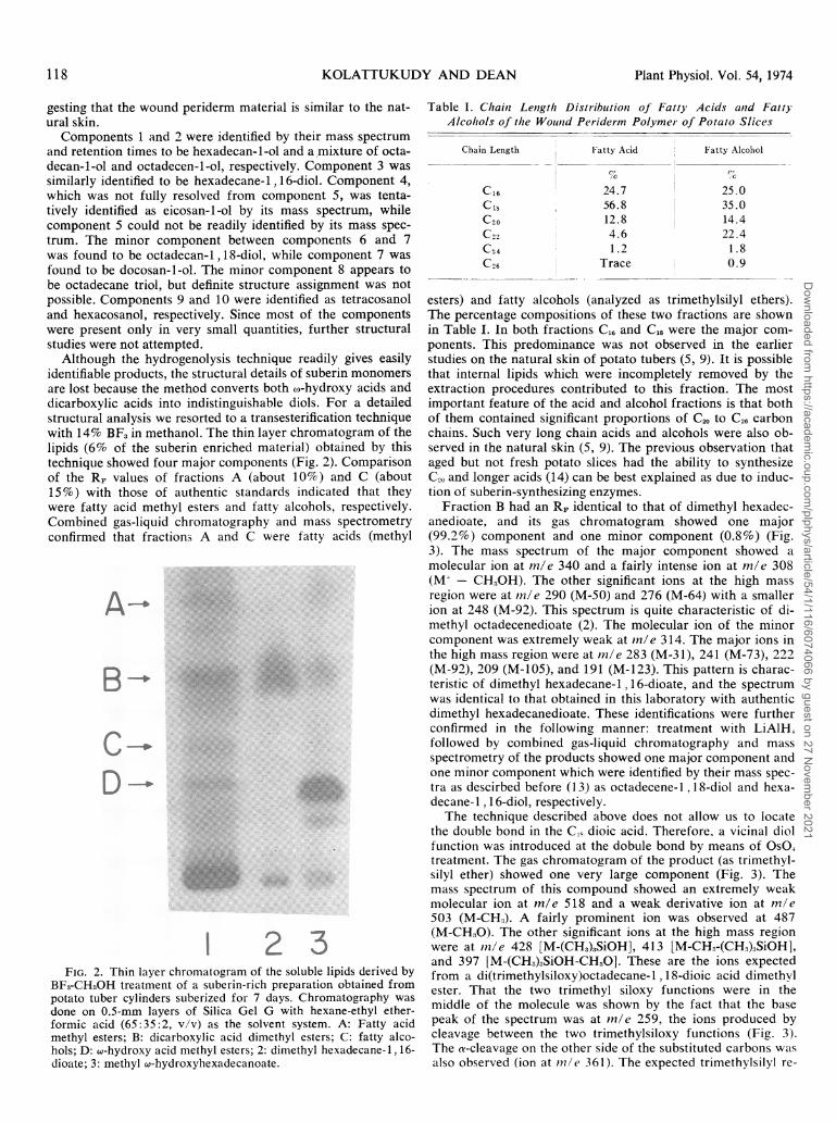
Although the hydrogenolysis technique readily gives easilyidentifiable products, the structural details of suberin monomersare lost because the method converts both o-hydroxy acids anddicarboxylic acids into indistinguishable diols. For a detailedstructural analysis we resorted to a transesterification techniquewith 14% BF, in methanol. The thin layer chromatogram of thelipids (6% of the suberin enriched material) obtained by thistechnique showed four major components (Fig. 2). Comparisonof the RF values of fractions A (about 10%) and C (about15%) with those of authentic standards indicated that theywere fatty acid methyl esters and fatty alcohols, respectively.Combined gas-liquid chromatography and mass spectrometryconfirmed that fractions A and C were fatty acids (methyl
... j.IM 23
FIG. 2. Thin layer chromatogram of the soluble lipids derived byBF3-CH30H treatment of a suberin-rich preparation obtained frompotato tuber cylinders suberized for 7 days. Chromatography wasdone on 0.5-mm layers of Silica Gel G with hexane-ethyl ether-formic acid (65:35:2, v/v) as the solvent system. A: Fatty acidmethyl esters; B: dicarboxylic acid dimethyl esters; C: fatty alco-hols; D: w-hydroxy acid methyl esters; 2: dimethyl hexadecane-1, 16-dioate; 3: methyl w-hydroxyhexadecanoate.
Table I. Chain Lenigth Distribuitioni of Fatty' Acids anld FattyAlcohols of the Wounid Peridernm Polyme- of Potato Slices
Chain Length Fatty Acid Fatty Alcohol
C16 24.7 25.0Cl0 56.8 35.0C20 12.8 14.41 A le 11 1 A
C24C26
4.61.2
Trace
22.41.80.9
esters) and fatty alcohols (analyzed as trimethylsilyl ethers).The percentage compositions of these two fractions are shownin Table I. In both fractions C16 and C18 were the major com-ponents. This predominance was not observed in the earlierstudies on the natural skin of potato tubers (5, 9). It is possiblethat internal lipids which were incompletely removed by theextraction procedures contributed to this fraction. The mostimportant feature of the acid and alcohol fractions is that bothof them contained significant proportions of C. to C26 carbonchains. Such very long chain acids and alcohols were also ob-served in the natural skin (5, 9). The previous observation thataged but not fresh potato slices had the ability to synthesizeC20 and longer acids (14) can be best explained as due to induc-tion of suberin-synthesizing enzymes.
Fraction B had an RF identical to that of dimethyl hexadec-anedioate, and its gas chromatogram showed one major(99.2%) component and one minor component (0.8%) (Fig.3). The mass spectrum of the major component showed amolecular ion at m/e 340 and a fairly intense ion at m/e 308(M+- CH,OH). The other significant ions at the high massregion were at in/e 290 (M-50) and 276 (M-64) with a smallerion at 248 (M-92). This spectrum is quite characteristic of di-methyl octadecenedioate (2). The molecular ion of the minorcomponent was extremely weak at in/e 314. The major ions inthe high mass region were at m/e 283 (M-31), 241 (M-73), 222(M-92), 209 (M-105), and 191 (M-123). This pattern is charac-teristic of dimethyl hexadecane-1 , 16-dioate, and the spectrumwas identical to that obtained in this laboratory with authenticdimethyl hexadecanedioate. These identifications were furtherconfirmed in the following manner: treatment with LiAlH,followed by combined gas-liquid chromatography and massspectrometry of the products showed one major component andone minor component which were identified by their mass spec-tra as descirbed before (13) as octadecene-l , 18-diol and hexa-decane-1 , 16-diol, respectively.The technique described above does not allow us to locate
the double bond in the C,0 dioic acid. Therefore, a vicinal diolfunction was introduced at the dobule bond by means of OsO1treatment. The gas chromatogram of the product (as trimethyl-silyl ether) showed one very large component (Fig. 3). Themass spectrum of this compound showed an extremely weakmolecular ion at m/e 518 and a weak derivative ion at m/e503 (M-CH2). A fairly prominent ion was observed at 487(M-CH3O). The other significant ions at the high mass regionwere at in/e 428 [M-(CHK) SiOH], 413 [M-CH,-(CH1)3SiOH],and 397 [M-(CHI)3SiOH-CH.O]. These are the ions expectedfrom a di(trimethylsiloxy)octadecane-1,18-dioic acid dimethylester. That the two trimethyl siloxy functions were in themiddle of the molecule was shown by the fact that the basepeak of the spectrum was at in/e 259, the ions produced bycleavage between the two trimethylsiloxy functions (Fig. 3).The a-cleavage on the other side of the substituted carbons wasalso observed (ion at ini/e 361). The expected trimethylsilyl re-
118 Plant Physiol. Vol. 54, 1974
Dow
nloaded from https://academ
ic.oup.com/plphys/article/54/1/116/6074066 by guest on 27 N
ovember 2021

POTATO WOUND SUBERIN
TIME (MIN) m/e
FIG. 3. Gas-liquid chromatogram of fraction B of Figure 2 (top left), partial mass spectrum of the major component (top right), the gas-liquid chromatogram of the trimethylsilyl ether of OS04 treatment product of fraction B (bottom left), and partial mass spectrum of the majorOs04 treatment product (bottom right). The column temperature and inlet pressure for the upper left chromatogram were 240 C and 23 p.s.i.,respectively. All attempts to detect longer dicarboxylic acids failed. The lower left chromatogram was obtained with 270 C column temperatureand 23 p.s.i. inlet pressure. The two minor components in this chromatogram were tentatively identified as dimethyl hexadecane-1, 16-dioate anddimethyl octadecane-1, 18-dioate.
arrangement ion was observed at mle 332 (3). Thus, it is clearthat the two trimethylsiloxy functions were at the C-9 and C-10positions and, therefore, the double bond in the original dioicacid was at the C-9 position.
Fraction D had an RF identical to that of authentic w-hy-droxy hexadecanoic acid methyl ester. The gas chromatogramof this fraction showed one major component and a few minorcomponents (Fig. 4). The mass spectrum of the major com-ponent showed a substantial molecular ion at mle 384. Theother major ions at the high mass region were at 369 (M-CH3),353 (M-CH30), and 337 (M-CH3-CH,OH). This pattern sug-
6 3TIME (MIN)
0 200
gests that this component is 1 8-trimethylsiloxy-octadecenoicacid methyl ester. The metastable ion representing the transi-tion [M-15] -- [M-47] was also observed. Confirming theidentification, LiA1H4 treatment followed by analysis of theproducts by combined gas chromatography and mass spectrom-etry showed that the major product was octadecene-1 , 1 8-diol.
In order to determine the position of the double bond, theo,-hydroxy acid fraction was treated with OsO which intro-duces a vic-diol function at the double bond. The gas chro-matogram of the products showed one major component (Fig.4). The mass spectrum of this compound showed significant
250 300 350 400m/e
FIG. 4. Gas-liquid chromatogram of the trimethyl silyl ether of fraction D (w-hydroxy acid methyl ester) of Figure 2 (upper left), partial massspectrum of the major component (upper right), gas-liquid chromatogram of the OS04 treatment product (as trimethylsilyl ether) of fraction Dof Figure 2 (lower left), and a partial mass spectrum of the major product of the OS04 treatment (lower right). The upper left chromatogramwas obtained with 240 C column temperature and 23 psi inlet pressure of He as carrier gas. Injection of larger quantities of fraction D revealedthe presence of small quantities of C20-C26 w-hydroxy acids, but they are not visible at the conditions used in this chromatogram. The lower leftchromatogram was obtained with a column temperature of 270 C and an inlet pressure of 23 p.s.i.
100-
Lii
_ (CH3)3SIOCH2(CH2)7CH=CH (CH2)7COOCH3C,)< 50mLL 1.337o (M-15-32) 369
(M-15)294 384 00CM-90) ~ ~ 53
(M) BSAF- (M9) 1(M-31)L L05L1LJ 259
z,303
3SIOCH C27H- CH (CH2COOCH,>_ (CH3)3SiO OSi(CH3)3r 50 213-2303-----259-3-2227
I213 332227
Plant Physiol. Vol. 54, 1974
Dow
nloaded from https://academ
ic.oup.com/plphys/article/54/1/116/6074066 by guest on 27 N
ovember 2021

KOLATTJUKUDY AND DEAN
ions at m/e 547 (M-CH,) and 531 (M-CHWO), indicating thatthe compound was tri(trimethylsiloxy) C18 acid methyl ester.The vic-diol function was clearly shown to be at the 9,10-position by the two dominant ions at m/e 259 and 303, whichwere produced by cleavage between C-9 and C-10. The ex-pected trimethylsilyl migration was also obvious by the pres-ence of a moderately strong ion at mle 332. These resultsclearly show that the original compound was o-hydroxy octa-dec-9-enoic acid. Small quantities of (,o-hydroxy C,0 acid, a)-hy-droxy C., acid, and w-hydroxy C." acid were also detected inthe w-hydroxy acid fraction by combined gas chromatographyand mass spectrometry.The results discussed thus far show that w-hydroxyoctadec-9-
enoic acid, octadec-9-ene-dioic acid, C,8-C., alcohols are themajor aliphatic components of the polymeric material con-tained in the wound periderm of potato tuber slices. Thesecomponents were also found to be the major components ofthe natural skin of potato tuber (5, 9). The relatively minorcomponents, such as 1 8-hydroxy-9, 1 0-epoxystearic acid and10,1 6-dihydroxypalmitic acids, identified in the natural skin ofpotato tubers (5) were not detected in the present investigationof the wound periderm. However, this difference might be at-tributed to the relative ease of detection of components presentin the large quantities of the natural skin to those present in therelatively small quantities of the polymer present in the woundperiderm. Since BF3-CH,OH treatment of crude material isknown to produce artifacts which remain in the origin, thismethod is not suitable for detection of minor components andquantitation.Gas Chromatographic Measurement of Suberization. Since
the cytochemical methods of measuring suberization are nei-ther direct nor quantitative, we used the structural informationdiscussed above to design a gas chromatographic method forquantitation of wound suberin. Since (w-hydroxy octadecenoicacid and octadec-9-ene-dioic acid are the major compoundswhich distinguish suberin from other cellular components, thequantity of these two compounds should be a reliable measureof suberization. Since hydrogenolysis of the polymer wouldconvert both these acids into octadecene-1, 18-diol, which canbe easily measured, we chose this technique. This compoundalso eliminated the necessity for measuring two different com-ponents, and it increased the sensitivity of the method. Hy-
LLuC/)
z00rC')
ulJ
0c-
()
LL
LI
TIME (MIN)FIG. 5. A typical gas-liquid chromatogram of the trimethylsilyl
ether of the diol fraction isolated by TLC from the hydrogenolysateof suberized (7 days) tissue slice used in the gas chromatographicassay for suberin. The C1, diol was added as a standard. In thisassay a coiled stainless steel column (l/8 in X 4 ft) packed with 5%
OV-1 on 80-100 mesh Gas Chrom Q was used with a column tem-perature of 230 C and an inlet pressure of 20 psi of N2 as the car-
rier gas.
Nl
C:
U-o(I)
cr1
3
Days Suberized
FIG. 6. Time course of development of suberin as measured bygas-liquid chromatography and the development of resistance todiffusion of water vapor. The suberin expressed as Ag/g representsonly the octadecene diol derived from w-hydroxy octadec-9-enoicacid and octadec-9-ene dioic acid. The reciprocal of diffusion re-sistance is plotted to show the inverse relationship between waterloss from the tissue and formation of the major aliphatic com-ponents of suberin.
drogenolysis of powdered dry tissue can be done either directlyor after extraction of soluble lipids with a Soxhlet extractor. Inthe former case, the large quantities of C,6 and C,8 alcohols,derived from the usual internal lipids, make it more difficult toquantitate the C,8 diol (derived from suberin) which is only arelatively minor component in the mixture. Therefore, prelimi-nary TLC is advisable, if not necessary. Since C,8 diol is only avery minor component of suberin, we chose to add C,, diolstandards prior to TLC. This procedure not only aided quanti-tation but also enabled us to visualize the diol fraction readilyon the thin layer chromatograms. Gas chromatography of thediol fraction recovered from such thin layer chromatograms(Fig. 5) showed two major components, which were identifiedby mass spectrometry to be octadecene-l ,18-diol and hexade-cane-l, 6-diol. The octadecene- 1, 1 8-diol which originated fromthe wound periderm was quantitated by comparison of the areaunder its gas chromatographic peak with the area under theCl6 diol peak obtained from the known quantity of the addedCi, diol.Time Course of Suberization and Development of Resistance
to Water Loss. A time course of development of suberin wasdetermined with the gas chromatographic assay discussedabove. There was very little of the suberin acids in the tissuefor 3 to 5 days. after which there was a dramatic increase (Fig.6). The quantity of these acids did not show a significantfurther increase after about 7 days. On the other hand, the lagperiod for the synthesis of the very long acids appeared to belonger, and their rate of increase leveled off later than that ofthe C,, suberin acids. The individual components showed some-what different time courses.
If suberin plays an important role in preventing water loss,the rate of formation of the major suberin acids might show acorrelation with the development of resistance to water loss.In order to investigate this possibility, the time course of de-velopment of resistance to water loss was determined as de-scribed under "Materials and Methods" (Fig. 6). The resultsclearly showed that up to 3 to 4 days the tissue slices had notdeveloped much resistance to water loss. After this time, therewas a dramatic increase in resistance, which reached a maxi-mum within 2 to 3 days and beyond which no further increasein resistance was observed. This pattern is identical to that ob-served for the formation of 1 8-hydroxyoctadec-9-enoic acid
120 Plant Physiol. Vol. 54, 1974
Dow
nloaded from https://academ
ic.oup.com/plphys/article/54/1/116/6074066 by guest on 27 N
ovember 2021

POTATO WOUND SUBERIN
and octadec-9-ene-18-dioic acid, the major components ofsuberin. The other components of suberin did not show such aclear correlation in that their levels increased even after theresistance to water loss leveled off and the lag period in theirdevelopment was somewhat longer than that observed for de-velopment of resistance to water loss (data not shown). Tem-perature dependence of development of water loss and forma-tion of the C18 aliphatic constituents of suberin providedfurther evidence that these constituents of suberin are involvedin preventing water loss. For example, suberization for 7 daysat 10 C did not result in the development of significant re-sistance to water loss, and gas chromatographic measurementshowed that little C18 diol was produced by hydrogenolysis ofthe tissue. Furthermore, under N2 no suberization was detectedas measured by the gas chromatographic technique or by thediffusion resistance technique. The effects of temperature andN2 reported here are in agreement with previous reports (7, 12).This experimental evidence strongly suggests that one role ofsuberin, or at least its major aliphatic constituents, is to protectthe tissue from water loss. One other important function wasrecently shown to be prevention of decay (4), but we have notas yet attempted to correlate this function to suberization asmeasured by gas-liquid chromatography. It is possible that thephenolic components of suberin prevent pathogen entry whilethe aliphatic constituents prevent water loss.The time course experiments suggest that the enzymes in-
volved in the suberization process might be induced during thefirst several days after wounding. From the structural studiesreported in this paper, it is suggested that an o-hydroxylatingenzyme and an o-hydroxy acid dehydrogenase, which wouldgive rise to the dicarboxylic acids, are the two key enzymesinvolved in the synthesis of the major suberin acids. An enzymewhich catalyzes the polymerization process must also be in-volved, as found earlier in cutin synthesis (1). It appears that asignal produced or activated by the wound triggers the induc-tion of these enzymes.
Acknowledgments-We thank Linda Brown for technical assistance, Dr.Rodney Croteau for a critical reading of the manuscript, and Drs. GaylonCampbell and Robert Kunkel for many helpful discussions.
LITERATURE CITED
1. CROTEAI, R. AND P. E. KOLA[TUKJUDY. 1973. Enzymatic synthesis of a hydroxyfatty acid polymer, cutin, by a particulate preparation from Vicia fabaepidermis. Biochem. Biophys. Res. Commun. 52: 863-869.
2. EGLINTON, G. AND D. H. HL-NN-EMA&N. 1968. Gas chromatographic-massspectrometric studies of long chain hydroxyacids. I. The constituentcutin acids of apple cuticle. Phytochemistry 7: 313-322.
3. EGLINTON, G., D. H. HUNNEMAN, AND A. MCCORMICa. 1968. Gas chroma-tographic-mass spectrometric studies of long chain hydroxyacids. III. Themass spectra of the methyl esters trimethylsilyl ethers of aliphatic hydroxy-acids. A facile method of double bond location. Org. Mass. Spec. 1: 593-611.
4. Fox, R. T. V., J. G. MANNERS, AND A. NIYERS. 1971. Ultrastructure of entryand spread of Erwinia carotovora var. atroseptica into potato tubers.
5. KOLATTXUKDY, P. E. AND V. P. AGRAWAL. 1974. Structure and compositionof the aliphatic components of potato tuber skin. Lipids. In press.
6. KOLATTUKUDY, P. E. AND T. J. WALTON. 1972. Structure and biosynthesisof the hydroxy fatty acids of cutin in Vicia faba leaves. Biochemistry 11:1897-1907.
7. LIPTON, W. J. 1967. Some effects of low oxygen atmospheres on potato tubers.Amer. Potato J. 44: 292-299.
8. PATTERSON, Ml. I. AND E. G. GRAY. 1972. The formation of wound peridermand the susceptibility of potato tubers to gangrene (Phoma exiqua) inrelation to rate of fertilizer application and time of planting. Potato Res.15: 1-11.
9. RODRIGUEZ-MIGUENxs, B. AND I. RIsAs-MARQmES. 1972. Investigacionesquimicas sobre el corcho de Solanum tuberosum L. (potata). Ann. R. Soc.Esp. Fis. Quim. 68: 303-308.
10. ROSE, C. W. 1969. In: R. Maxwell, ed., Agricultural Physics, Ed. 2. PergamonPress, London. p. 69.
11. SIMo.-DS, A. O., G. JOHNSON, AND L. A. SCHAAL. 1953. Comparative effectsof catechol, some related compounds and other chemicals on suberization ofcut potato tubers. Bot. Gaz. 114: 190-195.
12. SMITH, 0. 1968. In: Potatoes: Production, Storing and Processing. AV-IPublishing Co., Westport, Conn. p. 349.
13. WALTON-, T. J. AND P. E. KOLATTUJKTJDY. 1972. Determination of the structureof cutin monomers by a novel depolymerization procedure and combinedgas chromatography and mass spectrometry. Biochemistry 11: 1885-1897.
14. WILLEMOT, C. AsrD P. K. STUMPF. 1967. Fat metabolism in higher plants.XXXIV. Development of fatty acid synthetase as a function of proteinsynthesis in aging potato tuber slices. Plant Physiol. 42: 391-397.
Plant Physiol. Vol. 54, 1974
Dow
nloaded from https://academ
ic.oup.com/plphys/article/54/1/116/6074066 by guest on 27 N
ovember 2021