structure elucidation of sch 20562, a glucosidic cyclic
TRANSCRIPT

VOL. 52, NO. 4, APR. 1999 THE JOURNAL OF ANTIBIOTICS pp. 383 - 397
Structure Elucidation of Sch 20562, a Glucosidic Cyclic DehydropeptideLactone - the Major Component of W-10 Antifungal Antibiotic
Adriano Afonso,* Frank Hon and Ray Brambilla
Department of Chemistry, Schering-Plough Research Institute,2015 Galloping Hill Road, Kenilworth, New Jersey 07033, USA
(Received for publication November 25, 1998)
A novel bacterium designated as Aeromonas sp. W-10 produces the antibiotic W-10complexwhichcomprises of two major and several minorcomponents.Thetwo majorcomponents from this complex, Sch 20562 (1) and Sch 20561 (la), are of biological interestin view of their potent antifungal activity. The chemical degradation studies utilized forthe assignment of structure 1 for Sch 20562 are described here. Someof the noteworthydiversity of structural features in this glucosidic cyclic dehydrononapeptide lactone 1 are:
an TV-terminal (D)-jS-hydroxymyristyl unit, three D-amino acid units, two (£)-a-amino-
crotonyl units, and an O-a-D-glucosyl-TV-methyl-L-a/Zo-threonine unit. The structure deter-mination of 1 utilized the selective cleavage of the dehydropeptide units by ozonolysis toform fragments that were sequenced by mass spectrometry. The stereochemistry of theamino acid units were assigned by isolation of the free amino acids from the hydrolysates
of the fragments. The stereochemistry of the a-aminocrotonyl units and the glucosidic
Antibiotic W-10 complex is produced by a novelbacterium from the genus Aeromonas designated asAeromonas sp. W-10 NRRL B11053. The antibioticcomplex is comprised of several components includingSch 20561 and Sch 20562, the two major components inthis complex, which are of biological interest due to theirpotent antifungal activity against yeasts and derma-tophytes.1* We report here the chemical degradationstudies that led to the assignment of the structure and
stereochemistry 1 for Sch 20562 (Figure 1).2)
Results and Discussion
The physical and hydrolytic data for Sch 20562 aresummarized in Scheme 1.3) These initial data indicated
that Sch 20562 was a macrocyclic glucosidic dehydropep-tide lactone wherein the TV-terminal amine was acylatedwith a (D)-/?-hydroxymyristyl unit (D-Hma). Thus, the
amino acid composition and the formation of ammoniaand a-keto butyric acid (characterized as 3-ethyl-2-quinoxalinol 3)4) in the acid hydrolysate, the high
Fig. 1. Structure ofSch 20562 (1).

384 THE JOURNAL OF ANTIBIOTICS APR. 1999
Scheme 1. Physical data and hydrolysis products of 1,
extinction coefficient for the 240nmabsorption in theUVin base5) and the integration of the oleflnic methylsignal in the NMRsuggested that 1 was a dehydropeptidecontaining two a-aminocrotonyl units 9 (Aca). The facile
methanolysis of 1 to form a methyl ester 6, the
identification ofD-Hma (4a)6) in the base hydrolysis, and1 a-methyl glucoside 5 in the acid catalyzed methanolysis,together with the ninhydrin negative reaction supporteda lactonic glucosidic structure with a blocked yV-terminal
amine for the dehydropeptide. The presence of thedehydro units 9 was further confirmed by the formationof acetaldehyde (characterized as the dimedone adduct
8) in the ozonolysis of6.
Electrom impact (El) mass spectrometry of permeth-ylated 7V-acyl peptides is a convenient technique for the
sequencing of oligopeptides. Fragment ions derived fromthe El cleavage of the derivatized peptide at the peptide
bonds are characteristic since the charge is retained by
the N-terminal fragment.7) We utilized this technique forsequencing the peptides described here. Permethylation
of the acyclic methanolysis product 6 afforded 10 whichshowed the characteristic fragmentation pattern a~g in
the El mass spectrum thereby establishing a partial
sequence for the peptide 6 (Scheme 2). The fragment ionsbeyond g were not informative and hence the full
sequencing of 6 required smaller fragments derived fromthis peptide. Mild acid hydrolysis of 1 or 6 to generate
such fragments was found to form complexmixtures ofproducts arising from random peptide fragmentationsand this approach was not attractive. At this point, wemadeuse of an observation from the initial experimentin the ozonolysis of6 to 7 used for confirming the presenceof dehydropeptide units 9 (Scheme 1); TLC analysis ofthe ozonized product showed that 6 was converted intoa mixture of only three products which were easily
separable in view of their widely different polarities. Wereasoned that an intermediate oxalimide 12 formed in
the ozonolysis ofa dehydropeptide ll (Scheme 2), wouldundergo a selective methanolysis at the imide carbonylto afford the amide 13 and an TV-terminal methylox-alamide 14 as the two cleavage products wherein theoxalamide group in 14 and the nitrogen of the amide
group of 13 are derived from the dehydro unit.8) Thismethodology was utilized to obtain fragments from 6and from other intermediates derived from 1 that aredescribed here.Ozonolysis of the acyclic methyl ester 6 followed byreductive work-up with Me2Sand methanolysis of theintermediate oxalimide afforded, after chromatography,
exclusively the three fragments A~C (4c, 15, and 16).The crystalline fragment A was characterized as
D-j8-hydroxymyristamide 4c. Fragments B and C weresequenced by mass spectrometry of their permethylatedderivatives (Scheme 3). Fragment B (15) was a tripeptide

VOL. 52 NO. 4 THE JOURNAL OF ANTIBIOTICS 385
Scheme 2
Reagents: (a) MeOH/Et3N (b) DMSO/NaH/CH3I (c) i. O3/MeOH/-78°C, ii. Me2S (d) Et3N/rt.
containing the two Thr and the single Tyr units foundin 1, and an N-terminal methyloxalyl group derived fromone of the Aca units of 1. The tripeptide reacted with
diazomethane to form the methyl ether 15b, and onacetylation formed the triacetate 15c. Ammonolysis of
15 afforded the oxalamide 15a. The sequence in 15 wasestablished by permethylation of 15a to 17 which showed
the fragment ions a~c in the El mass spectrum, inagreement with the amino acid sequence shown.
Fragment C (16) was found to be a tetrapeptidecontaining the remaining residues identified in 1.Methanolysis of 16 under acidic conditions affordedmethyl glucoside, and amino acid analysis of its acid
hydrolysate showed, in addition to Glu and Gly, the
presence of Asp which was not found in 1. Tetrapeptide16 contained an N-terminal methyloxalyl group derivedfrom the second Aca unit in 1, and an A/-formyl-Asn-OCH3 as the carboxy terminal unit. The Af-formyl-Asnunit in the tetrapeptide arises from the ozonolysis of
the imidazole ring in the His unit originally present in 1
and 6.9) Acetylation of 16 afforded a tetraacetate 16b.Additionally, the *H NMRspectrum of 16 showed the
presence of an N-CH3signal at 3 2.90.This informationprompted us to use CD3Iin the permethylation of 16 inorder to differentiate the methyl group present in 16 fromthe methyl groups introduced by permethylation.
Ammonolysis of the two methyl esters and the TV-formylgroups of 16 formed the diamide 16a which was
permethylated using CD3I to afford 18. The El massspectrum of 18 showed the fragment ions a~d inagreement with the amino acid sequence shown. Thesequence confirmed the presence of an TV-methyl
threonine unit that was not evident in the preliminaryhydrolytic composition of 1 because TV-methyl-amino-acids are not detectable in conventional amino acidanalysis based on ninhydrin color yield detection.10)
In order to ascertain that the Asn residue in 16 (Hisresidue in 1 and 6) was indeed the terminal carboxy inthe acyclic peptide sequence 6 and also the carbonyl ofthe lactone in 1, the compoundwas aminolyzed to 6a

386 THE JOURNAL OF ANTIBIOTICS APR. 1999
Scheme 3
Reagents: (a) NH3/MeOH (b) CH2N2/Et2O (c) Ac2O/pyr (d) DMSO/NaH/CH3I (e) DMSO/NaH/CD3I(f) i. O3/MeOH/-78°C, ii. Me2S, iii. Et3N/rt (g) DMF/EtNH2.
with ethylamine thereby introducing the ethylamide
group in 6a as a tag for the carbonyl of the lactone.ll)Ozonolytic cleavage of 6a afforded the fragments 4a
and 15 as obtained previously from 6, and the newtetrapeptide fragment 16c containing the ethylamide
group. The permethylated product 18a derived from 16cshowed the fragment ions a~d 'm the El mass spectrum,in agreement with the amino acid sequence shown.The information provided by the partial sequence 10
in conjunction with the sequence overlapping12) of thefragments 4c, 15, 16, and 16c enabled us to assign thefull ndnapeptide sequence for the acyclic products 6 and
6a derived from 1 (Scheme 4).Wethen determined which of three possible hydroxylgroups was involved in the lactonic structure of 1. Thiswas accomplished by blocking the free hydroxyl groupsin 1 by preparing a per-tetrahydropyranyl derivative 19.Treatment of 19 with aqueous regenerated 1 thereby
ensuring that the integrity of the molecule was maintainedduring the THP formation. Ozonolysis of 19 followed
by reductive work-up and methanolysis afforded, afterchromatography, the THP derivative of D-jS-hydro-
xymyristamide 4d as a mixture of diastereoisomers, thetripeptide 20, and other uncharacterized products. The

VOL. 52 NO. 4 THE JOURNAL OF ANTIBIOTICS 387
Scheme 4
Reagents: (a) DHP/PTSA/DMF (b) 0.05n HC1 (c) i. O3/MeOH/-78°C, ii. Me2S, iii. Et3N/rt(d) DMSO/NaH/CH3I.
permethylation product 21 derived from 20 showed thefragment ions a~c in the El mass spectrum as expected
for the sequence shown. Additionally, the fragment ion
at m/e 216 identified the free hydroxyl group in 20 andthis result in conjunction with ethylamide functionality
in 6a also established that the hydroxyl group of thethreonine-2 and the carboxy of the terminal histidine-9form the lactonic bond in 1. The facile methanolysisobserved for 1 would be expected for a lactone derivedfrom a histidine unit. The information generated to this
point established the gross cyclic structure la for Sch20562 wherein the only unit with a defined stereo-
chemistry was the N-terminal Hmaresidue.
Stereochemistry of the Amino Acid UnitsThe approach used to define the absolute stereo-
chemistry of the amino acid units in la was based onthe isolation and characterization of the individual aminoacids formed by acid hydrolyses of the tri- andtetrapeptide fragments. Thus, acid hydrolysis of 15
(Scheme 5) afforded D-tyrosine (22) and threonine (23).However,proton nmr and optical rotation measurementsindicated that the threonine 23 isolated from 15 was a
1:1 mixture of L-threonine and D-a/fo-threonine and
hence tripeptide fragment required to -be modified so as topermit the isolation of the two threonine units separately.This was accomplished by destroying either one of thethreonine units by selective oxidation to allow the

388 THE JOURNAL OF ANTIBIOTICS APR. 1999
Scheme 5
Reagents (a) 6n HC1/11O°C (b) In HCl/70°C/20h (c) NaIO4/THF/H2O (d) 1. Ac2O/pyndme, n. 70%acetic acid-H2O (e) CrO3 - H2SO4 (Jones)/Me2CO.
isolation of the intact threonine unit. The TV-terminal
oxalate group of 15 was hydrolyzed under mild acidconditions and the vicinal amino alcohol of the resulting24 was oxidized with sodium periodate. The intermediate
glyoxylamide intermediate 25, without purification, wasthen hydrolyzed to afford the Thr-3 unit which was
characterized as D-allo-threonine (26).Tripeptide fragment (20) was found suitable for
accessing the Thr-2 unit. Thus the hydroxyl group of 20was protected as an acetate followed by removal of theTHPgroups under mild acidic conditions to afford 27.The hydroxyl group of the Thr-3 unit in 27 was oxidizedwith Jones reagent and the resulting mono-threoninederivative 28 was hydrolyzed to afford the Thr-2 unit
which was characterized as L-threonine (29). Minorchemical modifications of the tripeptide fragments 15and 20 prior to hydrolysis, thus served to define the
stereochemistry of amino acid units #2, 3, and 4 in 15,
6,and1.Hydrolysis of the tetrapeptide 16 afforded D-glutamicacid (30), glycine, and L-aspartic acid (31) arising fromamino acid units #6,7, and 9 (Scheme 6). The hydrolysatedid not contain TV-methyl threonine that is present in thetetrapeptide. We found that the glucosidated hydroxyl
in the substrate is prone to /^-elimination under mildacidic conditions leading to the formation of 2. De-glucosidation of 16 was hence required prior tohydrolysis in order to isolate the intact 7V-Me-Thr unit
for characterization. The glucose group in 16 wasoxidized with sodium periodate to afford the expectedglyoxal acetal intermediate 32 which was found to beless prone, relative to 16, to ^-elimination under mildacidic conditions. Removal of the acetal functionality
in 32 was effected under reductive conditions with

VOL. 52 NO. 4 THE JOURNAL OF ANTIBIOTICS 389
Scheme 6
Reagents: (a) i. 6n HC1/110°C/24h, ii. silica gel chromatography, iii. CuCO3/H2O/separation, iv.
(b) NaIO4/H2O (c) Zn dust/AcOH/H2O (d) i. 6n HC1/1 10°C/24h, ii. silica gel chromatography.
zinc-acetic acid to provide the unmasked hydroxy
compound 33. Compound33 was found to be stable to/^-elimination, and upon hydrolysis afforded the aminoacid unit #8 which was characterized as 7V-methyl-L-<2//<9-
threonine (34).13)
Stereochemistry of the Dehydro UnitsAssignment of the stereochemistry for 2-acylamino-
crotonates (N-acyl-Aca) using NMRspectroscopy hasbeen reported previously. Based on the XHchemical shiftsin CDC13of several 2-acylaminocrotonates, the isomerswith low field resonances for both the methyl doubletand the vinyl quartet were assigned the E configura-tion.14) However, we had observed an isomerization ofthe dehydro unit in the aminolysis of 1 to the ethylamide6a; the XH NMRspectrum of 6a in DMSO-d6showedthat the vinyl proton was shifted down field while theolefinic methyl was shifted up field relative to the
resonances in 1,ll) and this suggested that the chemical
shift positions and deshielding effects for these protonswere solvent dependent.
For direct comparisons of the chemical shifts in
Scheme 7
DMSO,we prepared the 7V-acetyl isomers 37 and 39 bythe ^-elimination of the 7V-acetyl-0-mesyl-threoninemethyl esters 36 and 38 (Scheme 7). It was found that

390 THE JOURNAL OF ANTIBIOTICS
Table 1. XH NMRdata for dehydro amino acid protons.
Compound Solvent Olefinic methyl Vinylic protonS (mult. /Hz) S (mult. /Hz)
Acetyl-Z-Aca-OCH3 (35)Acetyl-£-Aca-OCH3 (36)Sch 20562 (1)
DMSCM6 1.70
DMSCM6 1.82DMSO-4 1.78
Aeetyl-Z-Aca-OCH3 (35) CDC13Acetyl-£-Aca-OCH3 (36) CDC13
1.821.79
2.09
(d(d(d(d(d(d
/=7Hz) 6.48 (q, /=7Hz)
7=7Hz) 5.90(q,7=7Hz)J=7Hz) 5.80(q,7=7Hz)J=7Hz) 5.84(q,/-7Hz)
J
J
=7Hz)=1Hz)
6.78 (q, 7=7Hz)
7.02-(q, 7=7Hz)
Chemical shifts in ppm relative to
on decoupling experiments.
Table 2. Glucosidic 13C NMRchemical shifts(ppm).
Compound
1 -OMe-^-D-Glucopyranoside1 -OMe-^-D-Glucofuranoside1 -OMe-a-D-Glucopyranoside
1 -OMe-a-D-GlucofuranosideSch 20562
Q
c.
104.2 61.9-76.8104.2 64.2-78.8
100.1 61.7-72.5
110.0 64.7-82.3
94.9 No signal
>73.4
Chemicaal shifts in (DMSO-J6 25.2 MHz).
either of these substrates afforded a mixture of the olefins37 and 39 and these could arise by a competing inversionat C3, under the Et3N basic conditions used, followedby trans elimination of the inverted product. *H NMRdata in Table 1 showed that in DMSO-<i6, the vinylicproton in the Z isomer 37 is deshielded by the estercarbonyl by 0.58ppm relative to the resonance for theE isomer 39 and the methyl doublet in the latter isomeris deshielded by 0.1 ppm relative to the resonance for theZ isomer 37. The chemical shifts for these protons in Sch20562 (1) correlate with those in 39 and the Aca unitswere therefore assigned the E configuration.
Stereochemistry of the Glucosidic LinkageTo complete the structure elucidation of Sch 20562,
the stereochemistry of the glucosidic linkage at theN-MG-L-allo-Thr-8 unit remained to be assigned. Table
2 lists the 13C NMRchemical shifts of anomeric 1-methoxy-D-glucosides and 1. The chemical shift of theanomeric carbon and the absence of signals above 3 73.4
TMS (100MHz). Assignments are based
APR. 1999
Table 3. Glucosidic contribution to [M]D.
Compound
SCH 20562 (1)SCH 20561 (la)2)1633
1 -OMe-a-D-Glucoside
l -OMe-0-D-Glucoside
1, Calcd. as a-D-Glucoside1, Calcd. as ^-D-Glucoside16, Calcd. as a-D-Glucoside
[M]D (Solvent) A [M]D
-380° (DMSO) +228°-608° (DMSO)+68° (H2O) +282°-214° (H2O)+320° (DMSO)+306° (H2O)-70° (DMSO)-66° (H2O)-288° (DMSO)
-678° (DMSO)+92° (H,O)
16, Calcd. as ^-D-Glucoside -280° (H2O)
for C2-C6 in 1 was indicative of a a-D-glucopyranosidelinkage for glucose linked to the hydroxyl of theN-Me-L-allo-Thr-8.
A more definitive assignment was based on the
application of optical rotatory behavior of glycosides toconfigurational studies. It is well established incarbohydrate chemistry that the contribution of theglucosidic linkage to the molecular rotation, [M]D, of aglucosidic compound is characteristic for a-glucosidesand /?-glucosides.15) This contribution value can be
obtained from the difference, A [M]D, between the [M]D'sof the glucoside and its aglycone and the value is equalto the molecular rotation of the corresponding1-methyl-glucosides. Conversely, the calculated value
of the [M]Dof a glucoside corresponds to the sum ofthe [M]D's of the aglycone and the corresponding1 -methyl-glucosides.
VOL.52 NO.4 THE JOURNAL OF ANTIBIOTICS
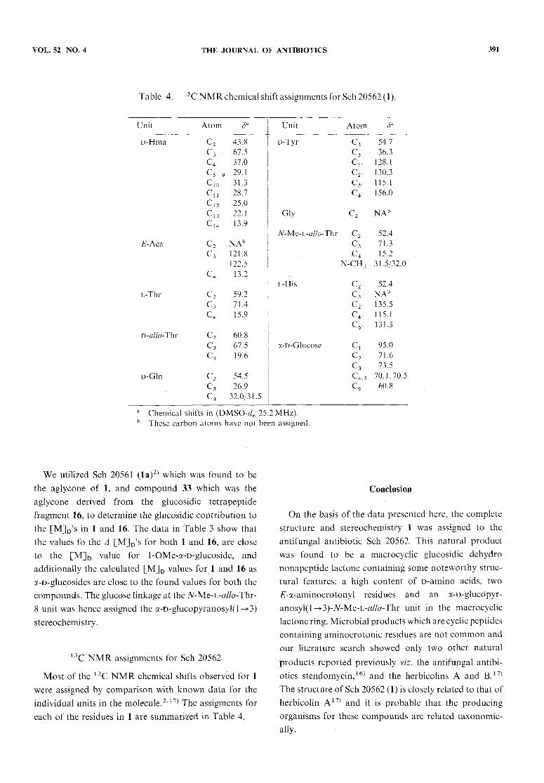
Table 4. 13CNMRchemicalshiftassignmentsforSch20562(1).
Unit Atom
D-Hma
£-Aca
C2 43.8
C3 67.5
C4 37.0
C5-9 29.1
C10 31.3
Cu 28.7
C12 25.0
C.,3 22.1C14 13.9
c2
c4NAb121.8122.5
13.2
L-ThrC2
c359.271.4
15.9
D-allo-Thr
D-Gln
C2 60.8
C3 67.5
C4 19.6
C2 54.5
C3 26.9
C4 32.0/31.5
Unit Atom
D-Tyr C2 54.7
C3 36.3
Cr 128.1
Cr 130.3
C3, 115.1
C4, 156.0
C2 NAb
N-Me-L-allo-Thr C2 52.4
C3 71.3
C4 15.2
N-CH3 31.5/32.0
c2c3c2c4,
52.4NAb135.5115.1
131.3
a-D-Glucose Ci 95.0
C2 71.6
C3 73.5
C45 70.1,70.5C6' 60.8
Chemical shifts in (DMSO-rf6 25.2 MHz).These carbon atoms have not been assigned.
We utilized Sch 20561 (la)2) which was found to bethe aglycone of 1, and compound 33 which was theaglycone derived from the glucosidic tetrapeptide
fragment 16, to determine the glucosidic contribution tothe [M]D's in 1 and 16. The data in Table 3 show thatthe values fo the A [M]D's for both 1 and 16, are close
to the [M]D value for 1-OMe-a-D-glucoside, and
additionally the calculated [M]D values for 1 and 16 asa-D-glucosides are close to the found values for both thecompounds. The glucose linkage at the N-MQ-L-allo-Thr-8 unit was hence assigned the a-D-glucopyranosyl(l ->3)stereochemistry.
13C NMRassignments for Sch 20562
Most of the 13C NMRchemical shifts observed for 1were assigned by comparison with known data for theindividual units in the molecule.2'17) The assigments foreach of the residues in 1 are summarized in Table 4.
391
Conclusion
On the basis of the data presented here, the completestructure and stereochemistry 1 was assigned to theantifungal antibiotic Sch 20562. This natural productwas found to be a macrocyclic glucosidic dehydro
nonapeptide lactone containing somenoteworthy struc-tural features: a high content of D-amino acids, two£-a-aminocrotonyl residues and an a-D-glucdpyr-
anosyl(1 ^3)-Af-Me-L-<2//<9-Thr unit in the macrocyclic
lactone ring. Microbial products which are cyclic peptidescontaining aminocrotonic residues are not commonandour literature search showed only two other natural
products reported previously viz. the antifungal antibi-otics stendomycin,16) and the herbicolins A and B.17)The structure ofSch 20562 (1) is closely related to that ofherbicolin A17) and it is probable that the producingorganisms for these compounds are related taxonomic-ally.

392 THE JOURNAL OF ANTIBIOTICS
In summary,wedescribe here our studies that led tothe structure elucidation of the antifungal antibiotic Sch20562 (1). Ozonolysis of the dehydropeptide units wasutilized to accomplish the selective cleavage of the peptideinto three fragments which were sequenced by massspectrometry. The stereochemistry of the amino acidunits was assigned by isolation of free amino acids fromthe hydrolysates of the fragments. Minor chemical
modifications of the fragments prior to hydrolyses werenecessary to allow the selective isolation of the individualthreonine units. The stereochemistry of the aminocroto-nic acid units and the glucosidic linkage were assignedby NMRspectroscopy, and molecular rotation datarespectively. Previously described structure elucidationsof the related dehydropeptides stendomycin,16) and theherbicolins A and B,17) were based on mild acidhydrolyses which afforded non-selective peptide cleav-
ages.
Experimental
General Procedures(a) Amino acid analyses were performed on total
hydrolysates (6n HC1/11O°C/18 hours) of the peptides(5mg) and are expressed as relative ratios, (b)Permethylations were performed by methodologydescribed previously7) as follows: A solution of the
peptide (10~20mg) in DMSO(0.2ml) was added withstirring at roomtemperature to a solution of meth-ylsulflnyl carbanion (1 ^2ml), freshly prepared fromDMSO (l~2ml) and sodium hydride (20~40mg)
at 80°C. After 5 minutes, Mel (0.1ml) was added andthe reaction was worked up after 1 hour by diluting withice/water, acidification to pH 4 with dilute acid andextraction with CH2C12. The major product from thepermethylation was isolated by TLC on silica-gel.
Sch 20562 (1)The fermentation of Aeromonas sp. W-10 and the
isolation of 1 from the W-10 antibiotic complex has beendescribed.1} Compound 1 was obtained as a white
amorphous powder from MeOH:mp 170~ 175°C; [a]D-60° (5% aq. pyridine, c=0.4);-IR (nujol) 1653,
1730cm-1; Amax (MeOH/OH~) 240nm (s 27000), 292nm(s 2370); XH NMR (DMSO-d6 100MHz) d 0.87 (t, 3H,J=l Hz),0.96-1.15 (ddd, 9HJ=l Hz), 1.25 (bs,20H),1.78, 1.82 (dd, 6H, 7=7 Hz), 2.98 (s, 3H), 5.80, 5.84(dq,2H, /=7.Hz), 6.61 (d, 2H, 7=8 Hz), 6.81 (s, 1H), 7.01(d, 2H,7-8 Hz), 7.62(s, 1H), 9.10(s, 1H), 9.40(s, 1H),
APR. 1999
9.64(s, 1H), ll.88 (s, 1H); MS (FAB)mjz 1357 (MH+);Amino acid analysis: His (1), Thr (2), Glu (1), Gly (1),
Tyr (1), NH3 (3).
3-Ethyl-2-quinoxalinol (3) from Acid Hydrolysis of 1A mixture of1 (0.1g) and 1n HC1 (10ml) was heated
at 110°C for 17 hours, cooled in ice and filtered on a
celite pad. o-Phenylenediamine (0.1 g) was added to theclear filtrate and heated on the steam bath for 10 minutes.The crystalline product was filtered and upon re-crystallization from methanol-water afforded 3 as fluffyneedles: (17mg); mp 198~200°C; XH NMR (CDC13,60MHz)3 1.40(t, 3H,7=8 Hz), 3.06(q, 2H, /=8 Hz);
identical (TLC, mp, NMR)with a reference sample.4)
D-/?-Hydroxymyristic Acid (4a) from Base Hydrolysisof1
A solution of1 (0.2g) in 15% NaOH (3ml) was heatedat 110°C in a teflon pressure tube for 42 hours, cooledto room temperature, acidified with dil HC1and extractedwith CH2C12. Evaporation of the extract followed bycrystallization from hexanes afforded 4a as colorlesscrystals: (20mg); mp 72~73°C; [a]D -15.8° (CHC13,
c=0.3). Reported6): mp 73~74°C; [a]D -16° (CHC13,
c=2). Anal Calcd for C14H28O3: C, 68.81; H, ll.55.
Found: C, 68.66; H, ll.64.
Treatment of 4a with ethereal CH2N2afforded methylD-/?-hydroxymyristate (4b) as an oil that was purified bysublimation to yield awax: mp 41°C; [a]D -8° (pyridine,c=43); XH NMR (CDC13, 60MHz) 3 0.89 (t, 3H), 1.28(bm, 18H), 1.40(m, 2H), 2.46(q, 1H,/=15,4 Hz), 2.51(q, 1H, /-15, 3Hz), 2.86 (bs, 1H), 3.75 (s, 3H), 4.01
(bm, 1H); MS (El) m/z 258 (M+).Ammonolysis of4b with methanolic ammonia afforded
D-/?-Hydroxymyristamide (4c) which was crystallizedfrom MeOH-Et2O as colorless needles: mp 110- 111°C;Md -4° (DMF, c-0.65); XH NMR (DMSO-J6
100MHz) 3 0.87 (bt, 3H), 2.14 (d, 2H, /= 6 Hz), 4.55(d, 1H, Z=4.5Hz), 6.78 (bs, 1H), 7.24 (bs, 1H); 13C
NMR (DMSO-d6 25MHz) 3 13.9, 22.1, 25.2, 28.8, 29.2,31.4, 37.0, 43.3, 67.6, 173.9; MS (El)m/z243 (M+). AnalCalcd for C14H29O2N: C, 69.09; H, 12.01, N, 5.76.
Found: C, 69.03; H, 12.15; N, 5.51.
1-Methyl-a-D-glucoside (5) from the Methanolysis of 1A solution of 1 (0.4g) in 6n methanolic HC1 (10ml)
was allowed to stand at room temperature for 20 hours,evaporated to dryness and the residue was chromato-graphed on silica-gel (15g) eluting with 5% MeOH-CH2C12.The anomeric mixture of methyl glucosides

VOL.52 NO.4 THE JOURNAL OF ANTIBIOTICS
(50 mg) thus isolated was peracetylated with Ac2O (1 ml)in pyridine (2ml) for 24 hours at room temperature,
evaporated to dryness and the product was chromato-graphed on silica-gel (4 g, 8% Me2CO-hexanes) to affordthe pure peracetates of the a-glucoside (47mg) and/?-glucoside (30mg). The a-glucoside tetraacetate was
dissolved in 75% methanol- ammonia (1.5ml) and after4hours the solution was evaporated, the residue wasdissolved in water followed by lyophilization and
crystallization from EtOH - Et2O to afford 1-methyl-a-D-glucoside 5 as needles: (21 mg) mp 166°C; [a]D + 158°(H2O, c=0.06); XH NMR (DMSO-J6-D2O 300MHz) 33.27 (s, 3H), 4.55 (d,lH, 7=3.6 Hz). Identical (TLC, mp,[a]D, NMR)with an authentic sample of 1-methyl-a-D-glucoside.
Acetaldehyde Dimedone Adduct (8) from Ozonolysisof1
A solution of 1 (0.3g) in MeOH(10ml) was ozonizedat -70°C followed by addition of Me2S (0.1ml). Thesolution was diluted with H2O (10ml) and filtered thruCelite and a solution ofdimedone (0.12g) in H2O (1 ml)was added to the clear filtrate. The solution was heatedon the steam bath and was then allowed to stand overnitein the refrigerator. The crystalline precipitate was filteredand recrystallized from MeOH-H2Oto afford 8 ascolorless crystals: (55mg) mp 141°C; XH NMR (CDC13,60MHz) 8 1.06 (s, 12H), 1.45 (d, 3H, /=8 Hz), 2.25 (s,8H), 4.10 (q, 1H, /=8 Hz); identical (TLC, mp, NMR)with an authentic sample of 8.
D-Hma-£-Aca-L-Thr-D-a//o-Thr-D-Tyr-£-Aca-D-Gln-
Gly-[a-D-glucopyranosyl (l ->3)-7V-Me-L-a//<9-Thr]-L-His-OCH3 (6)
A solution of 1 (1.0g) in MeOH(50ml) and Et3N(3.0ml) was stirred at room temperature for 24 hoursand then evaporated under reduced pressure. The residuewas dissolved in MeOHand diluted with Et2O. Theresulting suspension was filtered to afford 6 as a whiteamorphous solid: (0.95g) mp 138~145°C; [a]D -12°
(pyridine, c=0.7); Amax (MeOH/OH") 240nm (e 27600),292nm (e 3360); IR (nujol) 1653, 1739cm"1; *H NMR(CDC13, 60MHz) 8 0.80-1.15 (m, 12H), 1.83 (d, 6H,
7=7Hz), 3.64 (s, 3H), 5.76 (m, 2H), 6.67 (d, 2H,
/=8 Hz), 7.06 (d, 2H, J=S Hz), 7.62 (s, 1H), 9.14 (s,1H), 9.26 (s, 1H), 9.62 (s, 1H). Anal Calcd for
C64H100O22N12: C, 55.32; H, 7.25, N, 12.10. Found: C,54.83; H, 7.40; N, ll.90.
Permethylation of 6 afforded 10: HRMS(EI) (a) calcdfor C20H36O3N 338.26952 (b) C26H47O5N2 467.34849
393
(c) C32H58O7N3 596.42747 (d) C43H71O9N4 787.52210(e) C48H78O10N5 884.57486 (/) C56H92O12N7 1054
(LR), found 338.27016, 467.34954, 596.42947, 787.52380,
884.57178,1054.Ozonolysis of 6A stream of ozone was bubbled into a solution of 6
(1.0g) in MeOH(60ml) at -78°C until a blue colordeveloped followed which, excess O3 was removed witha stream ofN2 followed by the addition ofMe2S (2ml).The solution was stirred at roomtemperature until astarch-iodide test was negative, and then Et3N (1 ml) wasadded followed by evaporation under reduced pressure.The residue was chromatographed on silica-gel (80g).
Elution with 10% MeOH- CH2C12 afforded 4c as a whitecrystalline solid (0.17g). Further elution with the samesolvent afforded MeO-Oxalyl-L-Thr-D-a//6>-Thr-D-Tyr-
NH2(15) which was crystallized from acetone as granularcrystals: (0.20g) mp 130-135°C; [a]D +.9.5° (MeOH,c=0.67); XH NMR (DMSO-d6, 100MHz) 8 0.95-1.08(dd, 6H,/= 6Hz), 3.81 (s, 3H), 6.64 (d, 2H, J= 8Hz),
7.06 (d, 2H,J=8Hz), 7.95-8.35 (m, 3H), 9.10 (s, 1H).Amino acid analysis: Thr (2), Tyr (1), NH3 (1). AnalCalcd for C20H28O9N4: C, 51.27; H, 6.02, N, ll.96.
Found: C, 51.38; H, 6.ll;N, 1.16,Elution with 20% MeOH-CH2Cl2afforded MeO-
Oxalyl-D-Gln-Gly-[a-D-glucopyranosyl( l -åº 3)-Af-Me-L-tf//o-Thr]-y-iV-formyl-L-Asn-OCH3 (16) which was pre-cipitated from MeOH- Et2O as a white amorphous solid:(0.31 g) mp 128-134°C; [a]D +10.4° (MeOH, c=0.9S);IR (nujol) 1667, 1739cm"1; *H NMR (DMSO-d6-D2O,100.MHz). 8 0.95-1.10 (dt, 6H), 2.92 (s, 3H), 3.80 (s,
3H), 8.05 (s, 1H); Amino acid analysis Asp (1), Gly (1),Glu (1), NH3 (2). Anal Calcd for C27H42O17N6 H2O:C, 43.78; H, 5.99, N, ll.35. Found: C, 43.78; H, 6.00;
N,12.17.NH2-Oxalyl-L-Thr-D-q//6>-Thr-D-Tyr-NH2 (15a)
Methyl ester 15 (0.05g) was dissolved in 15%
NH3-MeOH(2ml) at room temperature and after 0.5hours was evaporated to dryness. The residue wascrystallized from H2O-Me2COto afford the amide 15as colorless prisms: mp 244-245°C; [a]D +7.5° (DMF,c=0.59); Amax (MeOH/OH") 245nm (e 9900), 295nm (s2000); IR (nujol) 1653, 1739cm"1; 1HNMR(DMSO-J6,100MHz) 80.98 (d, 3H, /=6Hz), 1.04(d, 3H, Z=6Hz),
6.64(d, 2H,J=S Hz), 7.05(d, 2H, /=8 Hz), 7.84-8.60(m, 5H), 9.08 (s, 1H). Anal Calcd for C19H27O8N5: C,50.32; H, 6.00, N, 15.45. Found: C, 50.01; H, 6.24; N,
15.07.

394 THE JOURNAL OF ANTIBIOTICS
Permethylation of 15a afforded 17: MS(EI) m/z (a)229 (b) 358 (c) 593.
MeO-Oxalyl-L-Thr-D-a//o-Thr-D-(O-methyl)-
Tyr-NH2(15b)
A solution of 15 (0.1 g) in MeOH(2ml) was treatedwith excess CH2N2-Et2O. The solution was stored at0°C for 8 hours and was then evaporated to dryness. Theproduct was purified by chromatography on two silicagel thick-layer plates (20 x 20 x 0.1 cm) using 15%MeOH/CH2C12as the developing solvent to afford 15b as a whitepowder: XH NMR (DMSO-d6, 100MHz) 5 1.02 (d, 3H,/=6Hz), 1.08 (d, 3H, 7=6Hz), 3.73 (s, 3H), 3.83 (s,
3H), 6.82 (d, 2H, 7=8Hz), 7.20 (d, 2H, 7=6Hz),
8.05~8.48 (m, 3H).
MeO-Oxalyl-(O-acetyl-L-Thr)-(O-acetyl-D-^//(9-Thr)-
(O-acetyl-D-Tyr)-NH2 (15c)
A solution of 15 (0.1 g) in pyridine (2.0ml) and Ac2O(1.0ml) was stored at 15°C for 24 hours and was thenevaporated under reduced pressure and azeotroped withbenzene. The crude product was dissolved in CH2C12followed by the slow addition of Et2O to afford 15c asa whithe amorphous solid: XHNMR(DMSO-d6, 100MHz) 5 1.08 (d, 3H, J=6Hz), 1.18 (d, 3H, /=6Hz),
1.94, 1.97,2.26(s,9H), 3.84(s, 3H), 7.00(d,2H,J=8Hz),7.28 (d, 2H, /-6Hz), 8.18-8.66(m, 3H).
MeO-Oxalyl-D-Gln-Gly-[tetra-acetyl-a-D-glucopyr-
anosyl(l -^3)-Ar-Me-L-(2//6>-Thr]-'y-Ar-formyl-L-Asn-OCH3 (16b)
A solution of16 (0.05 g) in pyridine (1.0ml) and Ac2O(0.5ml) was stored at 15°C for 24 hours and was thenevaporated under reduced pressure. The crude productwas purified by chromatography on two silica gelthick-layer plates (20x20x0.1cm) developed with
15%MeOH/CH2Cl2to afford 16b as a white amorphoussolid: 1H. NMR (CDC13, 100MHz) 3 1.05 (d, 3H,/=6Hz),'2.02 (s, 6H), 2.08 (s, 3H), 2.10 (s, 3H), 3.40 (s,
3H), 3.80 (s, 3H), 3.92 (s, 3H).
NH2-Oxalyl-D-Gln-Gly-[a-D-glucopyranosyl(l -> 3)-N-Me-L-tf//o-Thr]-L-Asn-NH2 (16a)
A solution of 16 (0.22g) in MeOH(20ml) was cooledin an ice-bath and saturated with NH3.The solution waskept at 10°C for 72 hours and was then evaporated todryness. The product was isolated by trituration withMe2COto afford 16a as a white amorphous solid: (0.2g)mp 122-132°C; [a]D +ll.5°(DMF,c=0.68); XHNMR(DMSO-d6, 100MHz) 8 1.08 (bd, 3H, /=6Hz), 2.95 (s,
APR. 1999
3H). Anal Calcd for C24H40O14N8 H2O: C, 42.23; H,6.20, N, 16.42. Found: C, 42.13; H, 6.18; N, 15.67.
Perdeuteriomethylation of 16a afforded 18: MS(EI)m/z (a) 285 (5) 359 (c) calcd for C30H22D30O12N5
704.5495, found 704.5493 (d) 920 (M+).
D-Hma-£'-Aca-L-Thr-D-«//(9-Thr-D-Tyr-£'-Aca-D-Gln-
Gly-[a-D-glucopyranosyl (l -^3)-Ar-Me-L-a//6>-Thr]-L-
His-NHEt (6a)
A solution of 1 (1.0g) in DMF (10ml) and EtNH2(0.25ml) was kept at 15°C for 5 days and was thenevaporated under reduced pressure. The product waspurified by chromatography on silica-gel (30g), usingCH2C12-MeOH-NH4OH-H2O(60:30:3:2v/v)asthe
eluting system to afford 6a as a white amorphoussolidfromMeOH-Et2O: (0.6g)mp 150-154°C; [a]D -13.2°(pyridine, c=0.6); Amax (MeOH/OH~) 240nm (e 25700),292nm (e 3380); IR (nujol) 1653cm-1; XH NMR
(DMSO-d6, 100MHz) 3 0.80-1.10 (m, 15H), 1.66, 1.80(dd, 6H), 6.36 (m, 1H), AnalCalcd for C65H103O21N13:C, 55.66; H, 7.40, N, 12.98. Found: C, 55.61; H, 7.56;
N,12.78.MeO-Oxalyl-D-Gln-Gly-[a-D-glucopyranosyl(l -»3)-
7V-Me-L-a//o-Thr] -7-7V-formyl-L-Asn-NHEt (16c)Ethyamide 15 (l.Og) in MeOH (60ml) was ozonized
at -78°C as described above for 6. The reaction productwas chromatographed on silica-gel (30g). Elution with10% MeOH-CH2Cl2afforded 4c and 15. Elution with20% MeOH-CH2C12 afforded 16c which was pre-cipitated from MeOH- Et2O as a white amorphous solid(0.25g): XHNMR (DMSO-</6, 100MHz) <5 1.05 (m, 6H),
2.95 (s, 3H), 3.80 (s, 3H). AnalCalcd for C28H45O16N7å H2O: C, 44.62; H, 6.29, N, 13.01. Found: C, 44.86; H,
6.27; N, 13.31.Permethylation of 16c afforded 18a: HRMS(ET) (a)
calcd for C^H^N, 257.11374 (b) C14H22O6N3
328.15086 (c) C29H49O13N4 661.32960 (d) C39H69-OI5N7 875.48513 found 257.11384, 328.15298,661.33034, and 875.48967 (M+).
MeO-Oxalyl-L-Thr-((9-tetrahydropyranyl-D-
a//o-Thr)-(6>-tetrahydropyranyl D-Tyr)-NH2 (20)A solution of1 (1.0 g) in DMF (6 ml) and dihydropyran
(20ml) containing p-TSA (0.1g) was heated in an oilbath at 115°C for 1 hour. The clear solution was cooledto 10°C, stirred for 10 minutes with KOAc(0.2g) andthen evaporated under reduced pressure. The residue wastriturated with Et2O, the solid was filtered, washed withwater and dried. The resulting per-THP 1 [IR (nujol)

VOL.52 NO.4 THE JOURNAL OF ANTIBIOTICS
1664, 1745cm"1] was dissolved in MeOH(60ml) andozonized at -78°C as described above for 6. After theaddition of Me2S, the solution was stirred until a
starch-iodide test was negative and then treated withEt3N (2ml). The reaction was allowed to stand at roomtemperature for 24 hours following which it was
evaporated under reduced pressure and the product waschromatographed on silica gel (30g). Elution with 2%MeOH- CH2C12 afforded D-/?-tetrahydropyranyloxy-myristamide (4d) as a diasteroisomeric mixture whichwas crystallized from hexane as colorless needles: (0.16 g)mp 65~74°C; [a]D -6.5° (MeOH, c=0.85); *H NMR(CDC13, 60MHz) S 0.90 (bt, 3H), 1.28 (s, 20H), 2.48 (m,2H). Anal Calcd for C19H37O3N 0.5H2O: C, 67.82; H,ll.38;N,4.16. Found: C67.72,H, ll.10,N4.48. Further
elution with the same solvent afforded 20 which wascrystallized from acetone-hexane as colorless prisms:(0.8g)mp 110- 117°C; [a]D +21.2° (MeOH, c=0.68);
Anal Calcd for Qc^On^ O^O: C, 55.80; H,7.02, N, 8.68. Found: C, 55.69; H, 7.13; N, 8.53.
Permethylation of 20 afforded 21: MS(EI) mjz (a) 216(6) 415 (c) 720 (M+).
D-Tyrosine (22) from Amino Acid Unit #4A solution of15 (0.3g) in 6n HC1 (10ml) was heated
at 110°C for 17 hours and was then evaporated underreduced pressure. The residue was chromatographed onsilica-gel eluting with CH2C12-MeOH-NH4OH-H2O(60: 30: 3 :2 v/v). Fractions containing threonine were
combined and crystallized from water - MeOHto afforda 1 : 1 mixture of L-threonine and D-a//o-threonine 23
(43mg): [a]D -19° (H2O, c=0.4); XH NMR (D2O,80MHz) S 1.24 (d, /=7Hz), 1.32 (d, 7=7Hz), 3.50 (d,
/=5Hz), 3.78 (d, /=4Hz). Fractions homogeneous in
tyrosine were evaporated, the residue was dissolved inwater (0.2ml) by adding NH4OH followed by acidifica-tion with AcOHand the solution was stored at 10°Covernight. The resulting crystalline product was filtered,washed with EtOH and dried to afford D-tyrosine 22(23mg): mp 280~285°'C; [a]D +8° (5n HC1, c=036);
identical (TLC, NMR,rotation) with an authentic sampleof D-tyrosine.
D-<2//<9-Threonine (26) from Amino Acid Unit #3A solution of15 (0.3g) in 1n HC1was heated at 70°C
for 20 hours and was then evaporated under reducedpressure. The resulting ninhydrin positive 24 was
dissolved in H2O (15ml), the pH of the solution wasadjusted to neutrality with NaHCO3followed by theaddition of NaIO4 (0.318g). After stirring for 3 hours,
395
ethylene glycol (0.092g) was added and after 0.5 hour
the mixture was evaporated to dryness. The residue wassuspended in MeOH and filtered. The filtrate wasevaporated, dissolved in H2O(10ml) and filtered thruAmberlite IR120 strongly acidic ion-exchange resin
(15ml). The eluate was evaporated to dryness to affordcrude 25 (0.28 g) which was hydrolyzed in 6n HC1 (10ml)at 110°C for 16 hours. The hydrolysate was chromato-graphed on silica-gel (5ml). Elution with CH2C12-
MeOH-NH4OH-H2O (60:30:3:2 v/v) yielded d-
tyrosine followed by the more polar fraction which afterrecrystallization from water-EtOH afforded D-allo-
threonine (26) as colorless needles (24mg): [a]D -25°
(5n HC1, c=0.35), -22.8° (H2O, c=0.47); XH NMR(D2O, 100MHz) 5 1.24 (d, 3H, J=lHz), 3.80 (d,
7=4 Hz); identical (TLC, NMR, rotation) with anauthentic sample of D-^//o-threonine.
L-Threonine (29) from Amino Acid Unit #2A solution of20 (0.2g) in pyridine (2.0ml) and Ac2O
(0.2ml) was allowed to stand at room temperature for24 hours and was then evaporated to dryness underreduced pressure. The residue was purified by chro-matography on silica-gel (5g, elution with 5%
MeOH/CH2Cl2) and the product was dissolved in 70%AcOH-H2O (10ml). After 3 hours at room temperaturethe solution was evaporated to dryness, the residue wasdissolved in acetone (5ml) and excess Jones reagent(0.3 ml) was added. The solution was then treated with/-PrOH (0.2ml), filtered thru a celite pad and the filtratewas evaporated to dryness. The residue was hydrolyzedin 6n HC1 (5ml) at 110°C for 16 hours. The hydrolysatewas chromatographed on silica gel (5g). Elution withCH2C12-MeOH-NH4OH-H2O (60:30:3 :2 v/v) yiel-
ded D-tyrosine followed by the more polar fraction whichafter recrystallization from water- EtOHafforded l-threonine (29) as colorless needles (5mg): [a]D -23°(5nHC1, c=0.24); *HNMR(D2O, 100MHz) 5 1.32 (d,
3H, /=7 Hz), 3.50 (d, J=5 Hz); identical (TLC, NMR,rotation) with an authentic sample of L-threonine.
D-Glutamic Acid (30), L-Aspartic Acid (34), and
Af-Methyl-L-<2//<9-threonine (34) from Amino Acid Units#6,'8 and 9
A solution of16 (0.5 g) in H2O (10ml) was stirred withNaIO4 (0.6g) at room temperature for 3 hours and wasthen quenched by the addition of ethylene glycol (0.1 g).After 1 hour, the pH of the solution was adjusted toneutrality with NaHCO3and the mixture was evaporatedto dryness. The crude 32 obtained by extracting the

396 THE JOURNAL OF ANTIBIOTICS
residue with MeOHwas dissolved in AcOH(10ml) andthe solution was then stirred with Zn dust (2g) at roomtemperature/1 hour followed by 100°C/10minutes. Themixture was then cooled, filtered and evaporated todryness. The resulting 33, was hydrolyzed in 6n HC1(20ml) at 110°C for 24 hours and the hydrolysate wasthen evaporated to dryness. Theresidue waschromato-graphed on silica-gel (30g). Elution with CH2C12-
MeOH-NH4OH-H2O (60 : 30 : 3 : 2v/v) afforded 92mg
of Af-methyl-L-tf/fo-threonine (34) which was crystallizedfrom MeOHas colorless needles: mp 247~254°C; [a]D+19.2° (5n HC1, c=0.25), +6° (H2O, c=Q.25); XHNMR (D2O 60MHz) 3 1.21 (d, 3H, /=7Hz), 2.76 (s,3H), 3.62 (d, 1H, /=4Hz), 4.30 (o, 1H, /=4, 7 Hz),
identical (TLC, NMR,rotation) with an authentic sampleof 7V-methyl-L-a//o-threonine.13) Subsequent fractions
afforded glycine. The column was then eluted with 20%NH4OH- MeOH(200 ml) and the eluate was evaporatedto dryness. The resulting residue was dissolved in H2O(5ml), CuCO3 (200mg) was added, the suspension washeated on a steam-bath for 0.5 hours and filtered hot.The dark blue filtrate was stored at 10°C overnite and theinsoluble ASP-copper salt was isolated by filtration. Thefiltrate containing the soluble Glu-copper salt was treatedwith excess H2S and filtered. The clear filtrate wasevaporated to dryness and the residue was recrystallizedfrom H2O-EtOH to afford D-glutamic acid 30 as
colorless crystals (15mg): [a]D -27° (5n HC1, c=0.45),identical (TLC, NMR,rotation) with an authentic sampleof D-glutamic acid. The insoluble Asp-copper salt wassuspended in water (2ml) and stirred with H2S at roomtemperature. The resulting black suspension was thenfiltered and the clear filtrate was evaporated to dryness.The residue was recrystallized from H2O - EtOH to affordL-aspartic acid (31) as colorless crystals (25mg): [a]D+23° (5 nHC1, c= 0.32), identical (TLC, NMR, rotation)with an authentic sample of L-aspartic acid.
Z-a-Aminocrotonic Acid Methyl Ester (37)A solution ofL-threonine (1.4g) in AcOH(30ml) and
H2O(3 ml) was saturated with HC1 gas and after 48 hoursat rt the solution was evaporated to dryness. The resultingO-acetyl-L-threonine was dissolved in NH4OH(15 ml) at0°C and after 48 hours at roomtemperature the solutionwas evaporated to dryness. The product was suspendedin MeOH(5ml) and treated with an excess CH2N2 inEt2O, filtered and the filtrate on evaporation afforded7V-acetyl-L-threonine methyl ester (0.5g) which was
dissolved in pyridine (10ml) and treated with mesylchloride (0.5 ml) at 0°C for 0.5 hour followed by 0.5 hour
APR. 1999
at room temperature. The reaction mixture was thenevaporated under reduced pressure, the residue waschromatographed on silica gel (15g). Elution with 1%MeOH-CH2C12 afforded 36 as an oil (0,3g) which wasdissolved in Me2CO(1 ml) containing Et3N (0.1 ml) andafter standing at room temperature for 24 hours the
solution was evaporated and the product was purifiedby chromatography on silica-gel (lOg). Elution with
CH2C12. afforded 39 (48mg) followed by 37 which wascrystallized from Et2O-hexane as colorless needles:
(0.15g)mp 52-55°C;Amax(MeOH)225nm(e 10,200); XHNMR (DMSO-d6, 100MHz) 8 1.70 (d, 3H, 7=7Hz),
1.95 (s, 3H), 3.68 (s, 3H), 6.48 (q, 1H,J=1Hz), 9.22(bs,
1H). Anal Calcd for C7H11O3N-0.3 H2O: C, 51.63; H,7.24, N, 8.41. Found: C, 51.72; H, 7.19; N, 8.62.
£-a-Aminocrotonic Acid Methyl Ester (39)A solution of TV-acetyl-L-a/Zothreonine methyl ester
(2.0 g), prepared from L-tf/fo-threonine using the proce-dure described for 37, in pyridine (20ml) was reacted
with mesyl chloride (1.3ml) at 0°C for 3 hours followedby addition of MeOH(0.6ml) and evaporation underreduced pressure. The crude 38 was dissolved in Me2CO(30ml) containing Et3N (3ml) and after standing at rtfor 24 hours the solution was evaporated and the productwas purified by chromatography on silica gel (60g).Elution with CH2C12 afforded 39 which was crystallizedfrom Et2O-hexane as colorless needles: (0.16g) mp
48~50°C; xmax (MeOH) 232nm (s 8600); XH NMR(DMSO-4, 100-MHz) 3 1.82 (d, 3H, ,7=7Hz), 1.96 (s,
3H), 3.75 (s, 3H), 5.90 (q, 1H, J=7 Hz), 9.55 (bs, 1H).AnalCalcd for C7H11O3N-0.6 H2O: C, 50.05; H, 7.32,N, 8.34. Found: C, 49.82; H, 7.09; N, 8.1-7. Subsequent
fractions afforded 37 (0.3 g).
Acknowledgements
The authors thank the Microbiology Department forproviding the antibiotic W-10 complex, Mr. Max
Kuegelman for the isolation and purification of the majorcomponents used in these investigations, Mr. PeterBartner for mass spectra, the Analytical ServicesDepartment for physical data, and Ms. E. Sharvordskayafor library research.
References and Notes
Taplin, D.; M. J. Weinstein, R. T. Testa, J. A.Marquez & M. G. Patel (Schering Corp.): AntibioticW-10 complex, Antibiotic 20561 and Antibiotic 20562as Antifungal Agents. U. S. Pat. 4,232,006, November
4,1980

VOL.52 NO.4 THE JOURNAL OF ANTIBIOTICS
Afonso, A.; F. Hon & R. Brambilla: Structureelucidation of Sch 20561, cyclic dehydropeptidelactone-a major component of W-10 antifungalantibiotic. J. Antibiotics 52: 398-406, 1999Abbreviations: Aca=a-aminocrotonic acid; Asp=Aspartic acid; Asn=Asparagine; Gin=glutamine;Glu=glutamic acid; Gly=glycine; His=histidine;
Hma = /^-hydroxymyristic acid; Thr = threonine; Tyr =Tyrosine
Morrison, D. C: Characterization of a-keto acids asquinoxalinols. J. Am. Chem. Soc. 76: 4483-4484,
1954
The single Tyr unit in 1 would account for an svalue= 10,000 at 240nm which indicated that otherchromophores like 9 contribute to the high observed
value for this absorptionIkawa, M.; J. B. Koepfli, S. G. Mudd & C.Niemann: An agent from E. coli causing hemorrhage.The component fatty acids of the phospholipid moiety.J. Am. Chem. Soc. 75: 1035-1038, 1953
(a) Thomas, D. W.; B. C. Das, S. D. Gero & E.Lederer: Mass spectrometry of permethylated peptide
derivatives. Bioch. Biophys. Res. Commun. 32:519-525, 1968. (b) Vilkas, E. & E. Lederer:
7V-Methylation de peptides par la methode dehakamori. Tetrahedron Lett. 26: 3089-3092, 1968.
(c) Williams, D. H.: Structural and sequencing studieson peptides by mass spectrometry. Pure & Appl.Chem. 50: 219-229, 1978
An analogous ozonolysis of an A^-a-pentenoic
beta-lactam has been reported for the deprotection ofthe TV-functionality. Cooper, R. D. G. & F. L. Jose:
Structural studies on penicillin derivatives. J. Am.Chem. Soc. 94: 1021-1022, 1972
A control experiment showed that ozonolysis ofA^-Ac-L-His followed by a) methanolysis and per-methylation afforded permethylated TV-Ac-Asn (m/e
397
230) or, b) hydrolysis with 6N HC1, afforded asparticacid
Additionally, it was determined later in these studiesthat the glucosyl-7V-Me-Thr unit in the peptide is prone
to ^-elimination to form 2 under acid hydrolysisconditions (see Scheme 6)
Aminolysis of the lactone 1 with EtNH2 was foundto proceed with a concomitant isomerization of the
dehydropeptide units. The 1H NMR oleflnic re-sonances at S 1.80 and 5.82 for 1 are shifted to S 1.70
and 6.40 respectively, in 6a. Minor side-products
resulting from conjugate addition of EtNH2 to thedehydropeptide were also identified
The TV-terminal methyloxalyl group of 15 and thenitrogen of the amide group of 4c are derived froman Aca unit linking these two fragments, and the same
functionalities in fragments 16 and 15 are in turnderived from the other Aca unit linking these latter
fragmentsBodansky, M.; G. G. Marconi & G. C. Colman: On
the Af-methyl-L-threonine residue in stendomycin. J.Antibiotics 21: 668-670, 1968For pertinent references see: Srinivasan, A.; R. W.
Stephenson & R. K. Olsen: Conversion of threoninederivatives to dehydroaminoamino acids by elimina-
tion of/?-chloro and /?-tosyl derivatives. J. Org. Chem.42: 2256-2260, 1977
Davidson, E. A.: In Carbohydrate Chemistry, pp.37-40, Holt, Rinehart and Winston, Inc., New York,
1967
Bodansky, M.; I. Izdebski & I. Muramatsu: TheStructure of the peptide antibiotic stendomycin. J. Am.Chem. Soc. 91: 2351-2358, 1969
Aydin, M.; N. Lucht, W. A. Konig, R. Lupp, G.Jung & G. Winkelmann: Structure elucidation ofpeptide antibiotics herbicolins A and B. Liebigs Ann.Chem.: 2285-2300, 1985