structure, development and functioning of conifers
Post on 21-Dec-2015
226 views
TRANSCRIPT

Structure, development and functioning of conifers

Zion Canyon
The Pinyon pine-Juniper community is an extreme, but
widely distributed type in SW USA
Conifers as principal tree species in dry regions

Pinyon pine and Juniper
Pinyon pine with its crooked trunk and reddish bark, is found in dry, rocky places at elevations of 4,000-8,000 ft where yearly precipitation is only 10-20 inches. Tap roots stretch down 40 or more feet into the soil while lateral roots stretch as far. Very slow growth rates: a 6-10 foot diameter tree, 10 feet tall will be 80-100 years old.
Utah juniper grows in dry, rocky or sandy locations in the high plateau country from 5000 to 9500 feet above sea level. It is the most common juniper of the Pinyon-juniper woodlands of the arid western intermountain basins. It is commonly 10 to 20 feet high with a maximum trunk diameter of 1 to 2 feet.
Pinus monophyllum Juniperus osteosperma

Many conifers are xerophytes
Xerophyte: a plant that can live where water supply is scanty or there is physiological drought
Xerophytes have adaptations of leaves, stems and/or roots.
Mesophyte: a plant that lives in places where the water supply is neither scanty nor abundant

Two features of conifers:
Morphology and anatomy of needles
Anatomy of wood
First, some basic nomenclature about patterns of plant growth and development

Three basic tissue systems that running through the plant
Fig. 31.6A
Three basic tissue systems that running through the plant
This is based on angiosperms – but conifers do have the same basic organization!

Tissues
A tissue is a cooperative unit of many similar cells that perform a specific function within a multicellular organism
Tissues usually have cells that are specialized for particular functions
For example the vascular tissue system conducts water and nutrients from roots to leaves through specialized cells for water conduction and conducts the products of photosynthesis, sugars, from leaves in different but equally specialized cells.

Unfortunately…… biologists are rather lax in their use of the word “tissue”.
Conducting tissue
Water from roots
Sugars from leaves
Xylem tissue
Phloem tissue
But the principle holds true that a tissue is a group of specialized cells, frequently of different types, performing a specific function.
Trachieds
The Secondary Phloem in Pinus has Sieve and Albuminous Cells and Parenchyma with dark contents.
Parenchyma

… photosynthesis?

Conifer needles (or fronds)
The site of photosynthesis
Exchange between the needle and the atmosphere of CO2 (into the needle) and water vapour (out of the needle).
Why is water loss inevitable?
Gaseous exchange takes place through a water film on the cells inside of the needle

StomataStomata have central openings surrounded by two photosynthetic guard cells. Usually stomata are open during the day and closed at night but can close if the leaf dehydrates. Guard cells change the shape of the opening by changing their own shape.
In Taxus caespitosa and other conifers stomata are arranged in rows
Epidermal cellGuard cellThick inner wall StomaChloroplast

Cross-section through a pine needle The xylem and phloem are surrounded by undifferentiated cells called transfusion tissue. This is surrounded by an endodermis, which typically controls passage of materials between conducting tissue and ground tissue
http://forest.wisc.edu/forestry415/INDEXFRAMES.HTM
Cuticle
EpidermisHypodermis
MesophyllStomaGuard cellXylemPhloemEndodermis
Transfusion tissue
Resin duct
Conifer needles are generally thicker and tougher than many angiosperm leaves in part because they have a layer of thick- walled cells, the hypodermis, below the epidermis. They also tend to have a thick cuticle.
The foliage of many but not all conifers also contains resin ducts. The lining cells secrete resin into the duct in response to leaf injury.
Cross-section through a pine needle

The needle is broader than that of the pine, but still has only one vascular bundle
Leaf cross section of Taxus (yew)
The mesophyll is differentiated into palisade and spongy layers
The endodermis is not so clearly developed as in the pine

This needle is still broader, yet contains only one vascular bundle
Leaf cross section of Podocarpus (a conifer)
Mesophyll is differentiated into palisade and spongy layers
Endodermis
Center of needle

How does wood form?
What is it that we are seeing when we look at tree rings?
Production of xylem and phloem tissues by the vascular cambium

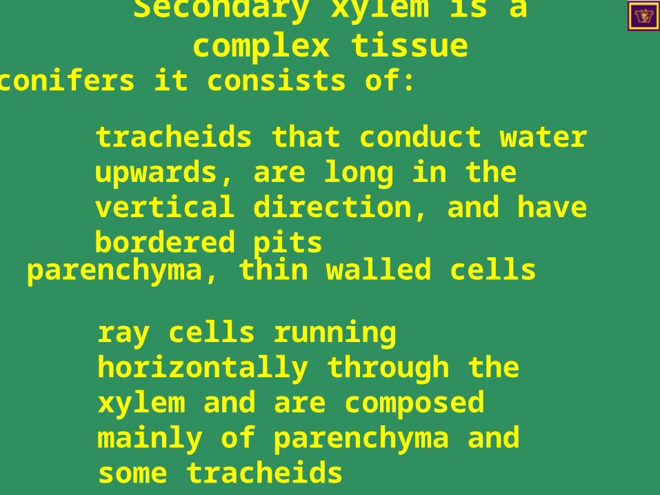
In conifers it consists of:
tracheids that conduct water upwards, are long in the vertical direction, and have bordered pits
parenchyma, thin walled cells
ray cells running horizontally through the xylem and are composed mainly of parenchyma and some tracheids
Secondary xylem is a complex tissue

Cross secction of a young pine stem

Cambium and secondary xylem of a conifer
Cambium
Rays
Ray initials
Tracheids with bordered pits
Late wood
Early wood
Parenchyma
Esau 1965
Cambium and secondary xylem of a conifer
Direction of growth
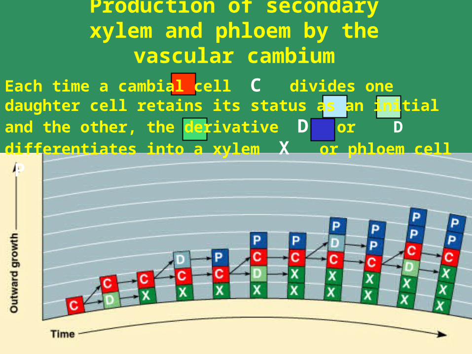
Production of secondary xylem and phloem by the vascular cambium
Each time a cambial cell C divides one daughter cell retains its
status as an initial and the other, the derivative D or D
differentiates into a xylem X or phloem cell P

http://www.uri.edu/artsci/bio/plant_anatomy/43.html
Tracheids and rays, pineRadial longitudinal section
Pits

Tracheids with bordered pits, pine
Tranverse longitudinal section

Bordered pits
Circular bordered pits of pine tracheids as seen in face view (left) and in side view (right).
http://www.uri.edu/artsci/bio/plant_anatomy/images.html
The torus at the center of the bordered pit moves and seals the pit when a tracheid aspirates

Xylary resin duct in Pinus
Transverse section
Ray
Epithelial cell
Tracheid

Sections you need to have read
7.2 31.5 31.6
Courses that deal with this topic
ESC 221 Dendrology and Autecology