structure and function of biotin-dependent...
TRANSCRIPT

REVIEW
Structure and function of biotin-dependent carboxylases
Liang Tong
Received: 22 May 2012 / Revised: 7 July 2012 / Accepted: 9 July 2012 / Published online: 7 August 2012
� Springer Basel AG 2012
Abstract Biotin-dependent carboxylases include acetyl-
CoA carboxylase (ACC), propionyl-CoA carboxylase
(PCC), 3-methylcrotonyl-CoA carboxylase (MCC), gera-
nyl-CoA carboxylase, pyruvate carboxylase (PC), and urea
carboxylase (UC). They contain biotin carboxylase (BC),
carboxyltransferase (CT), and biotin-carboxyl carrier pro-
tein components. These enzymes are widely distributed in
nature and have important functions in fatty acid metabo-
lism, amino acid metabolism, carbohydrate metabolism,
polyketide biosynthesis, urea utilization, and other cellular
processes. ACCs are also attractive targets for drug dis-
covery against type 2 diabetes, obesity, cancer, microbial
infections, and other diseases, and the plastid ACC of
grasses is the target of action of three classes of commer-
cial herbicides. Deficiencies in the activities of PCC, MCC,
or PC are linked to serious diseases in humans. Our
understanding of these enzymes has been greatly enhanced
over the past few years by the crystal structures of the
holoenzymes of PCC, MCC, PC, and UC. The structures
reveal unanticipated features in the architectures of the
holoenzymes, including the presence of previously unrec-
ognized domains, and provide a molecular basis for
understanding their catalytic mechanism as well as the
large collection of disease-causing mutations in PCC,
MCC, and PC. This review will summarize the recent
advances in our knowledge on the structure and function of
these important metabolic enzymes.
Keywords Fatty acid metabolism � Carbohydrate
metabolism � Amino acid metabolism �
Metabolic syndrome � Obesity � Diabetes � Cancer �Drug discovery � Antibiotics � Propionic acidemia �3-methylcrotonylglycinuria � Lactic acidemia
Abbreviations
ACC Acetyl-CoA carboxylase
BC Biotin carboxylase
BCCP Biotin carboxyl carrier protein
BT BC-CT interaction
CT Carboxyltransferase
GCC Geranyl-CoA carboxylase
GCD Glutaconyl-CoA decarboxylase
HCS Holocarboxylase synthase
MCC 3-methylcrotonyl-CoA carboxylase
MCG 3-methylcrotonylglycinuria
PA Propionic acidemia
PC Pyruvate carboxylase
PCC Propionyl-CoA carboxylase
PT PC tetramerization
UA Urea amidolyase
UC Urea carboxylase
YCC Acyl-CoA carboxylase (generic name)
Introduction
Biotin-dependent carboxylases are widely distributed in
nature and can be found in bacteria, archaea, fungi, algae,
plants, and animals. They have crucial roles in the metabo-
lism of fatty acids, amino acids, and carbohydrates [1–4]. In
some microorganisms, these enzymes also have important
functions in CO2 fixation [5, 6], methanol assimilation [7],
acetyl-CoA assimilation [8–10], 3-hydroxypropionate
assimilation [11], mycolic acid and methyl-branched fatty
L. Tong (&)
Department of Biological Sciences, Columbia University,
New York, NY 10027, USA
e-mail: [email protected]
Cell. Mol. Life Sci. (2013) 70:863–891
DOI 10.1007/s00018-012-1096-0 Cellular and Molecular Life Sciences
123

acid biosynthesis [12], polyketide biosynthesis [13],
metabolism of terpenoids [14], and the utilization of urea as a
nitrogen source [15, 16].
Biotin-dependent carboxylases have two distinct enzy-
matic activities and catalyze their reactions in two steps
[17, 18]. First, a biotin carboxylase (BC) component cat-
alyzes the MgATP-dependent carboxylation of the N10
atom of the biotin cofactor, using bicarbonate as the CO2
donor (Fig. 1a). Biotin is covalently linked through an
amide bond to a lysine side chain in the biotin carboxyl
carrier protein (BCCP) component. In the second step of
the reaction, a carboxyltransferase (CT) component cata-
lyzes the CO2 transfer from carboxybiotin to the acceptor
of the carboxyl group (referred to as the substrate here).
Most of the substrates are coenzyme A (CoA) esters of
organic acids, and the site of carboxylation is on the acarbon of a saturated acid (acetyl-CoA, propionyl-CoA) or
the c carbon of an a-b unsaturated acid (3-methylcrotonyl-
CoA, geranyl-CoA) (Fig. 1b). In addition, small com-
pounds can also serve directly as the substrate, such as
pyruvate and urea (Fig. 1b). Especially, the urea substrate
is unique in that carboxylation occurs on a nitrogen atom.
Depending on the organism and the enzyme, the BC,
BCCP, and CT components can be separate subunits or part
of a multi-domain protein (Fig. 2). In addition, while the BC
and BCCP components are conserved among these enzymes,
the sequence and structure of the CT component depend on
the chemical nature of the substrate. The CT components of
enzymes that carboxylate CoA esters share recognizable
sequence conservation because they recognize the CoA
segment (Fig. 2). On the other hand, the CT component that
carboxylates pyruvate or urea is entirely different.
Biotin must visit both the BC and CT active sites during
catalysis by biotin-dependent carboxylases. A swinging-
arm model had been the accepted mechanism for this
translocation. The connection between biotin and BCCP
contains eight methylene groups and ten rotatable single
bonds, and is likely to be rather flexible (Fig. 1a). When
fully extended, this flexible arm can approach a length of
*16 A (the distance from the N10 atom of biotin to the Caatom of the lysine). Therefore, it may be expected that
biotin can translocate by up to *30 A on this swinging
arm [19]. However, recent structures on the holoenzymes
of pyruvate carboxylase [20, 21], propionyl-CoA carbox-
ylase [22], 3-methylcrotonyl-CoA carboxylase [23], and
urea carboxylase [24] showed that the distance between the
BC and CT active sites ranges between 55 and 85 A
(Fig. 1a). Therefore, the swinging-arm model is not suffi-
cient for biotin to reach both active sites, and hence the
BCCP domain must also translocate during catalysis. This
is referred to as the ‘‘swinging-domain’’ model (Fig. 1a).
Besides biotin-dependent carboxylases, two other clas-
ses of biotin-dependent enzymes exist in nature. Biotin-
dependent decarboxylases couple the decarboxylation of
organic acids (possibly as CoA esters) to sodium ion
transport in anaerobes [25–30], while the biotin-dependent
transcarboxylase of Propionibacterium shermanii transfers
the carboxyl group from methylmalonyl-CoA to pyruvate
[31–33]. These enzymes are distinct from the biotin-
dependent carboxylases in that they lack the BC compo-
nent. They will not be specifically described further here.
Biotin-dependent carboxylases were first discovered
more than 50 years ago. They have been studied inten-
sively due to their important metabolic functions, and also
feature prominently in most biochemistry textbooks. Over
the past few years, there have been significant advances in
our understanding of these enzymes, especially from the
first structural information on several of the holoenzymes
[20–24]. This review summarizes our current knowledge
on the structure and function of biotin-dependent carbox-
ylases, with emphasis on recent studies (over the past
5 years). Space limitations unfortunately prevent detailed
Fig. 1 The biochemical activity of biotin-dependent carboxylases.
a Biotin is carboxylated in the active site of the biotin carboxylase
(BC) component, using bicarbonate as the CO2 donor with concom-
itant ATP hydrolysis. Biotin then translocates to the carboxyl-
transferase (CT) active site, where the CO2 is transferred to the
acceptor (substrate, acetyl-CoA is shown as an example). In the
swinging-arm model, biotin itself translocates between the BC and
CT active sites, while the biotin-carboxyl carrier protein (BCCP)
component remains stationary. The longest distance between the N10
of biotin and the Ca atom of the covalently linked lysine residue is
*16 A, giving the swinging arm a maximal reach of *30 A. This is
significantly shorter than the distances observed in the holoenzymes
so far, between 55 and 80 A. Therefore, the BCCP domain must also
translocate during catalysis, and this is known as the swinging-
domain model. b The substrates of biotin-dependent carboxylases.
The sites of carboxylation are indicated with the red arrows
864 L. Tong
123

descriptions of results from earlier studies or the citation of
those primary publications. These results are summarized
in the many reviews that have been published in the pre-
vious years, which are cited throughout this manuscript.
Classification of biotin-dependent carboxylases
Biotin-dependent carboxylases can be classified first based
on the identity of the substrate that becomes carboxylated.
This is dictated by the CT component, which can be highly
distinct among these enzymes (Fig. 2). These enzymes can
then be further classified by how the BC, BCCP, and CT
components are organized in them (Fig. 2). The different
components may exist as separate subunits, often in
bacteria, or they can be fused together into a large, multi-
domain enzyme in eukaryotes (Fig. 2). Various interme-
diates between these two extremes have also been observed
in nature (Fig. 2).
The largest collection of biotin-dependent carboxylases
uses CoA esters of (small) organic acids as the substrate;
hence they are acyl-CoA carboxylases (YCCs) in general.
These enzymes can have distinct substrate preferences,
such as acetyl-CoA carboxylase (ACC), propionyl-CoA
carboxylase (PCC), 3-methylcrotonyl-CoA carboxylase
(MCC), and geranyl-CoA carboxylase (GCC), although
some of them may have a wider collection of substrates, for
example enzymes that are active toward both acetyl- and
propionyl-CoA (ACC/PCC). In addition, some of these
enzymes can be identified based on genome sequences but
have not been studied in detail biochemically, and their
substrate preference is currently not known. They are
referred to generically as YCCs here. Acyl-CoA carboxy-
lases have also been referred to as ACCases [12], although
ACCs are sometimes called ACCases as well.
The CT components of the acyl-CoA carboxylases share
readily detectable amino acid sequence conservation,
because they all recognize CoA esters. It was generally
believed that these enzymes have the same organization of
their components. However, the recent structure of the
MCC holoenzyme indicates that there may be two distinct
lineages of these carboxylases [23]. Therefore, the acyl-
CoA carboxylases have been divided into two separate
collections, one including ACC, PCC, ACC/PCC, and most
of the other YCCs (families 1.1 through 1.7), while the
other consists of MCC and GCC (family 2.1) (Fig. 2). The
different families of biotin-dependent carboxylases are
described briefly next and in more detail in the later
sections.
Family 1.1 includes the bacterial ACC enzymes (Fig. 2).
They contain four subunits, with a BC subunit (*50 kD), a
BCCP subunit (*17 kD), and two subunits (a and b,
*33 kD each) for the CT activity. These enzymes are also
referred to as the multi-subunit ACCs [2]. The holoenzyme
has the stoichiometry (BC)2(BCCP)4(CTa, CTb)2, but it is
generally unstable and readily dissociates during purifica-
tion. These enzymes are also found in the chloroplasts of
many plants, reflecting the evolutionary origin of this
organelle.
Family 1.2 includes the ACC/PCC enzymes from
M. sedula [34], A. fulgidus, and other archaeal organisms,
but it appears to be absent in M. jannaschii. Compared to
the multi-subunit bacterial ACCs, the two CT subunits are
fused into a single protein (*60 kD), while the BC and CT
components remain as separate subunits. The holoenzyme
has the stoichiometry (BC)4(BCCP)4(CT)4.
Family 1.3 represents another possible way of fusing the
different components of the multi-subunit bacterial ACCs,
where the BC and BCCP subunits are fused together but
the CT subunits remain separate. An actual example of
such an enzyme has not been identified as yet, but its
existence may be expected.
Family 1.4 includes the ACC/PCC/acyl-CoA carboxy-
lases (YCCs) in S. coelicolor, M. tuberculosis, and other
organisms [12]. The BC and BCCP components are fused
into a single protein (a subunit, *65 kD), and the two
CT subunits are also fused (b subunit, *60 kD). The
Fig. 2 Classification of biotin-dependent carboxylases. These
enzymes are classified into four major collections and 13 different
families. Domains with sequence homology to each other are shown
with the same color. More detailed descriptions of the families can be
found in the text, and common examples of acyl-CoA carboxylase
family members include E. coli ACC (family 1.1), eukaryotic ACC
(family 1.7), PCC (family 1.5), and MCC (family 2.1). The proteins
are drawn to size, which is indicated with the scale bar at the bottom
(in number of residues)
Biotin-dependent carboxylases 865
123

holoenzyme has the stoichiometry a6b6 or a2b2 [12].
Another subunit (e) is required for some of these enzymes
to achieve maximal activity [12].
Family 1.5 includes the PCCs from various organisms.
The difference to family 1.4 is that the BC-BCCP fusion
(the a subunit, *73 kD) also contains a BT domain, which
mediates the interactions between the BC (in the a subunit)
and CT (the b subunit, *55 kD) domains [22]. This
domain is likely absent in family 1.4, as the linker between
BC and BCCP is too short to accommodate the residues of
the BT domain in that family. However, it is possible that
family 1.4 contains a modified version of the BT domain,
for example a structure similar to that of the PT domain in
pyruvate carboxylase (family 3.1) [21]. PCC holoenzyme is
a 750 kD a6b6 dodecamer.
Family 1.6 includes the acyl-CoA carboxylases from
P. aeruginosa and some other bacterial organisms. These
enzymes can be readily identified based on sequence
searches, but they have not been characterized biochemi-
cally (hence they are called YCCs here). All four subunits
of the multi-subunit bacterial ACCs are fused together in
these enzymes, giving rise to a multi-domain protein of
*120-kD molecular weight.
Family 1.7 includes the ACCs from most eukaryotic
organisms. These proteins can be thought of as being
made of three parts of equal lengths. The N-terminal one-
third of the proteins contains the BC, BCCP, and possibly
a BT domain, and the C-terminal one-third contains the
CT activity. The middle one-third is unique to eukaryotic
ACCs and has no other close sequence homologs in the
database. The structure and function of this part of
eukaryotic ACCs is currently not known. These enzymes
are generally referred to as the multi-domain ACCs, with
*250-kD molecular weight, and they function as 500-kD
homodimers and possibly higher oligomers. In light of the
multi-domain bacterial YCCs of family 1.6, it is probably
more appropriate to refer to family 1.7 as the multi-
domain eukaryotic ACCs. The multi-domain bacterial
YCCs lack the middle one-third of the multi-domain
eukaryotic ACCs, and they may lack the BT domain as
well (Fig. 2).
Family 2.1 includes the MCCs and GCCs from various
organisms. The overall domain organization of these
enzymes appears quite similar to that of PCCs (family 1.5).
However, the crystal structure of MCC indicates a large
difference in the architectures of the b subunit and the
holoenzyme [23], and therefore it has been placed into a
separate family. GCC is assigned to this family based on
sequence conservation and the similarity of its substrate to
that of MCC (Fig. 1b). These holoenzymes are also 750-kD
a6b6 dodecamers.
Besides the acyl-CoA carboxylases, two other major
collections of biotin-dependent carboxylases use small
organic compounds as substrates, specifically pyruvate and
urea (Fig. 1b). Family 3.1 includes the pyruvate carboxy-
lases (PCs) that are present in most organisms, from
bacteria to humans. It is a single-chain, multi-domain
enzyme of *130 kD and functions only as a 500-kD tet-
ramer. Besides the BC, CT, and BCCP domains, structural
studies have revealed the presence of another domain,
known as the PT (PC tetramerization) or the allosteric
domain, in these enzymes [20, 21].
Family 3.2 includes the two-subunit form of PCs. They
are found in some bacteria (such as P. aeruginosa) as well
as archaea (M. jannaschii). The a subunit contains the BC
component (*52 kD), and the b subunit contains CT and
BCCP (*65 kD). The stoichiometry of the holoenzyme is
a4b4. Whether the PT domain also exists in these enzymes
is currently not known.
Family 4.1 includes the urea carboxylase (UC) found in
yeast and some other fungal organisms. It is a single-chain,
multi-domain enzyme of *200 kD, with an allophanate
hydrolase (also known as the amidase) domain fused at the
N-terminus. The entire enzyme is known as the urea
amidolyase (UA). The CT component consists of four sub-
domains (A, B, C, D) and is distinct from that in acyl-CoA
carboxylase and PC [24]. UC functions as a monomer.
Family 4.2 includes the UCs found in many bacterial
organisms, some fungal species, and green algae. They are
different from family 4.1 in that they lack the allophanate
hydrolase domain, and hence they are somewhat smaller,
*130 kD.
Family 4.3 includes the UC found in P. aeruginosa and
possibly other bacterial organisms. It is a multi-subunit
form of the enzyme, with a BC subunit (*50 kD), BCCP
subunit (*10 kD), and two subunits for the CT activity
(*35 kD, each containing two domains of the multi-
domain form of the enzyme).
Overall, four major collections of biotin-dependent
carboxylases can be identified based on current biochemi-
cal, sequence, and structural information, which can be
further divided into 13 families. The availability of genome
sequences has enabled the identification of all biotin-
dependent enzymes in many organisms, which also pro-
vides insight into the evolution of these enzymes [35, 36].
An inventory of such enzymes can now be compiled for
these organisms (Table 1). E. coli has only one biotin-
dependent enzyme, a multi-subunit bacterial ACC (family
1.1, Fig. 2). The yeast S. cerevisiae has two multi-domain
eukaryotic ACCs (family 1.7), two PCs (family 3.1), and
one urea amidolyase (family 4.1), while most other fungal
species has only one ACC, one PC, and one UA. There are
five biotin-dependent carboxylases in humans, ACC1,
ACC2, PCC, MCC, and PC. The structure and function of
representative enzymes in these different families will be
described in more detail below.
866 L. Tong
123

Acetyl-CoA carboxylase (ACC)
Biological functions of ACC
ACC catalyzes the conversion of acetyl-CoA to malonyl-
CoA, which serves many functions [37]. It provides the
two-carbon building block for fatty acid biosynthesis in
most living organisms, and is also used for polyketide
biosynthesis in some organisms (Fig. 3). In mammals,
ACC1 (also known as ACCa) is cytoplasmic and catalyzes
the rate-limiting and committed step of long-chain fatty
acid biosynthesis in liver, adipose, and other lipogenic
tissues. In the yeast S. cerevisiae, the cytosolic ACC1 is
essential for viability. There is also a mitochondrial form of
the enzyme (known as HFA1), which is important for
growth on lactate or glycerol as carbon source and for fatty
acid (especially lipoic acid) biosynthesis in this organelle
[38–40]. However, this isoform is unique to S. cerevisiae
and is not present in other fungal organisms, although ACC
activity has been reported in the mitochondria of some
plants. Malonyl-CoA for fatty acid biosynthesis in the
mitochondria of other organisms may be produced by
malonyl-CoA synthetase [41, 42] and/or PCC working on
acetyl-CoA as the substrate.
Mammals carry a second isoform of ACC, ACC2 (also
known as ACCb), which is highly conserved with ACC1,
with 73 % amino acid sequence identity between human
ACC1 and ACC2. ACC2 is associated with the outer
mitochondrial membrane through a 140-residue segment at
the N-terminus that is absent in ACC1, the first 20 residues
of which are highly hydrophobic [43]. However, most of
the ACC2 protein faces the cytosol (Fig. 3). This isoform is
primarily expressed in heart and muscle tissues, as well as
liver. The malonyl-CoA product is a potent inhibitor of
carnitine palmitoyltransferase I (CPT-I), the crucial
enzyme for the transport of long-chain fatty acyl-CoAs into
the mitochondria for b-oxidation (Fig. 3) [44, 45]. There
are differences in the expression patterns of human and
rodent ACCs [46]. In addition, a form of ACC2 lacking the
N-terminal mitochondrial targeting segment is expressed at
high levels in white adipose tissue in humans and can
participate in de novo lipogenesis [47].
Single-nucleotide polymorphisms (SNPs) in ACC1 and
ACC2 are associated with hypertriglyceridemia and
hypercholesterolemia, respectively, in patients taking
antipsychotic drugs [48]. Polymorphisms in the promoter
or the coding region of the ACC1 gene is linked to fatty
acid composition in porcine meat [49], fatness traits in
Table 1 Inventory of biotin-dependent carboxylases in some common organisms
Organism Biotin-dependent carboxylase (family number)
Bacteria
Escherichia coli K-12 Multi-subunit ACC (1.1)
Pseudomonas aeruginosaPAO1
Multi-subunit ACC (1.1), multi-domain YCC (1.6), MCC (2.1), GCC (2.1), two-subunit PC (3.2), multi-subunit
UC (4.3)
Ruegeria pomeroyi DSS-3 Multi-subunit ACC (1.1), PCC (1.5), MCC (2.1), GCC (2.1), PC (3.1)
Cupriavidus metalliduransCH34
Multi-subunit ACC (1.1), multi-domain YCC (1.6, 2 copies), MCC (2.1), GCC (2.1), PC (3.1)
Deinococcus radiodurans R1 Multi-subunit ACC (1.1), multi-domain YCC (1.6), PCC (1.5), MCC (2.1)
Streptomyces coelicolorA3(2)
Two-subunit YCC (1.4), PCC (1.5, 2 copies), MCC (2.1, 2 copies), PC (3.1)
Staphylococcus aureus Multi-subunit ACC (1.1), PC (3.1), multi-subunit UC (4.3)
Bacillus subtilis Multi-subunit ACC (1.1), three-subunit YCC (1.4), PC (3.1)
Archaea
Methanocauldococcusjannaschii
Two-subunit PC (3.2)
Metallosphaera sedula Two subunit YCC (1.4)
Fungi
Saccharomyces cerevisiae Eukaryotic ACC (1.7, 2 copies), PC (3.1, 2 copies), UC (4.1)
Kluyveromyces lactis Eukaryotic ACC (1.7), PC (3.1), UC (4.1)
Schizosaccharomyces pombe Eukaryotic ACC (1.7), PC (3.1)
Animals
Xenopus laevis Eukaryotic ACC (1.7), PCC (1.5), MCC (2.1), PC (3.1)
Danio rerio Eukaryotic ACC (1.7), PCC (1.5), MCC (2.1), PC (3.1)
Mus musculus Eukaryotic ACC (1.7, 2 copies), PCC (1.5), MCC (2.1), PC (3.1)
Homo sapiens Eukaryotic ACC (1.7, 2 copies), PCC (1.5), MCC (2.1), PC (3.1)
Biotin-dependent carboxylases 867
123

chickens [50], fatty acid composition in beef [51], and milk
production traits in goats [52].
The activity of both ACC1 and ACC2 in mammals are
stimulated by citrate, inhibited by long-chain saturated
acyl-CoA, and inactivated by phosphorylation [53], espe-
cially AMP-activated protein kinase (AMPK, at Ser80 in
ACC1, Ser222 in ACC2) and cAMP-dependent protein
kinase (protein kinase A, at Ser1201 in ACC1). Steady-
state kinetic studies show that citrate increases the kcat
(threefold) and kcat/Km (tenfold) of the ATP and acetyl-
CoA substrates for human ACC2, with a Ka of *0.5 mM
[54]. In comparison, another report shows that human
ACC2 is activated 1,000-fold by citrate, while ACC1 is
activated only fourfold [55]. The data also suggest two
binding sites for citrate, a higher-affinity (Kd * 1 mM)
activating site and a lower-affinity (Ki * 30 mM) inhibi-
tory site. A cytosolic protein MIG12 (*20 kD) promotes
the polymerization and stimulation of ACC1 by citrate, and
is incorporated into the ACC1 polymer [56]. The activity
of MIG12 is negatively regulated by forming a complex
with another protein, Spot 14 (*17 kD) [57].
Human ACC1 interacts with BRCA1 in a phosphor-
ylation-dependent manner, which reduces the activity of
ACC1 [58, 59]. The tandem BRCT domains of BRCA1
recognize the phosphorylated Ser1263 residue of ACC1, in
a (pS)PTF motif, and the structure of this complex has been
reported [60]. The phosphorylation of Ser1263 is regulated
by the cell cycle, possibly through a proline-directed pro-
tein kinase such as the cyclin-dependent kinase (CDK).
In plants, plastid ACC (ACC1) produces malonyl-CoA
for the biosynthesis of long-chain fatty acids, while cyto-
plasmic ACC (ACC2) is important for secondary
metabolism, including the synthesis of very long-chain
fatty acids, flavonoids, cuticular waxes, and other com-
pounds, and for proper embryonic development [61–63].
Plastid ACC in dicots is a multi-subunit enzyme, similar to
the multi-subunit bacterial ACCs [64, 65]. On the other
hand, the plastid ACC in grasses is a single-chain, multi-
domain enzyme, similar to the multi-domain eukaryotic
ACCs and arising through gene duplication of the cytosolic
ACC. This multi-domain plastid ACC is the target of three
classes of herbicides (see below). The BCCP subunit of the
Fig. 3 Functions of biotin-
dependent carboxylases in
mammals. Reactions involving
the five biotin-dependent
carboxylases are shown in blue.
Selected intermediates of the
TCA cycle (green) are shown.
Glutamine can also be used for
anaplerosis, especially in some
cancer cells. Dashed arrowsindicate pathways with more
than one step. In other
organisms, the biotin-dependent
carboxylases have similar
functions (with the exception of
ACC2), and they are also
involved in additional cellular
processes
868 L. Tong
123

multi-subunit plastid ACC of Arabidopsis thaliana forms a
complex with the signaling protein PII, which reduces the
Vmax of ACC but does not affect its Km for acetyl-CoA
[66]. 2-oxoglutarate, pyruvate or oxaloacetate can disrupt
this inhibition. A second isoform of BCCP (BCCP2) in A.
thaliana plastids is expressed at much lower levels and
cannot rescue the lethal phenotype of BCCP1 null mutants
[67]. Two additional BCCP-like proteins lack the biotin-
ylation motif (MKM) but co-purify with the 1–2 MDa
ACC complex from A. thaliana chloroplasts [68].
ACC has important functions in other organisms as well.
In the apicomplexan pathogen Toxoplasma gondii, ACC1 in
the apicoplast is required for fatty acid biosynthesis [69]. In
Trypanosoma brucei, RNAi knockdown of ACC results in
reduced virulence of the pathogen, and lower fatty acid
elongation in procyclic forms [70]. Aedes aegypti mosquitos
with deficient ACC activity have reduced lipogenesis and
produce significantly fewer eggs [71]. Human cytomegalo-
virus (HCMV) upregulates the expression and activity of
ACC1 in infected cells, and inhibition of ACC1 attenuates
viral replication [72]. In archaea and some bacteria, ACC
(and PCC) activity is important for CO2 fixation (possibly
coupled with ammonia oxidation) [5, 6].
ACCs as drug discovery targets
The crucial roles of ACCs in fatty acid metabolism make
them attractive targets for drug discovery against a variety
of human diseases, including bacterial infections, fungal
infections, type 2 diabetes, cancer, artherosclerosis, and
others [2, 3, 73–77]. Besides microbial infections, many of
the other diseases are also manifestations of the metabolic
syndrome, which is linked to the current obesity epidemic
[78–80]. It has been projected that *50 % of the adult
population in the US will be obese by the year 2030 [81],
indicating a pressing need for new therapeutic agents and
modalities in this area.
The importance of ACCs as targets for drug discovery
against the metabolic syndrome was first validated by
observations on the ACC2 knockout mice [82–86]. These
mice have elevated fatty acid oxidation, increased energy
expenditure, reduced body fat and body weight, improved
insulin sensitivity, smaller heart size but with normal
function, and normal life span and fertility. The animals
have increased food intake (hyperphagia), due to reduced
malonyl-CoA levels in the brain [87, 88]. In comparison, a
null mutation in the ACC1 gene causes embryonic lethality
in mice [89]. ACC1 knockout in the liver in mice reduced
hepatic lipid accumulation but did not disturb glucose
homeostasis [90]. ACC1 knockout in the adipose tissues in
mice reduced lipid accumulation in these tissues, but also
caused prenatal growth retardation and impaired bone
development [91].
Knockdown of ACC1 and ACC2 expression with anti-
sense oligonucleotides confirms the beneficial effects of
inhibiting these enzymes [92]. Mice on a high-fat diet show
improved peripheral insulin sensitivity after treatment with
the potent ACC inhibitor soraphen A [93]. Downregulation
of ACC2 activity by the adipokine CTRP1, through
AMPK-mediated phosphorylation, leads to elevated fatty
acid oxidation and reduced adiposity in mice [94]. On the
other hand, overexpression of ACC2 is associated with
increased production of proinflammatory cytokines in a
human renal cell, which can be reversed by inhibition of
p38 MAP kinase [95]. This may be a mechanism for dia-
betic nephropathy development. An SNP in ACC2 is
associated with type 2 diabetic nephropathy and proteinuria
[96–98], while another SNP in ACC2 is linked to increased
risk for metabolic syndrome [99].
Two recent reports have failed to observe the beneficial
effects of ACC2 knock out or down regulation, as elevated
fatty acid oxidation did not change energy expenditure or
adiposity [100, 101]. Another study failed to demonstrate a
correlation between ACC2 phosphorylation (down regula-
tion) and fatty acid oxidation in skeletal muscle [102].
Some of these differences could be due to variations in the
genetic background of the mice or the strategies that were
used to create the knockout mice [86, 103, 104].
A large number of potent (nanomolar) inhibitors have
been reported against human ACCs [105–116]. Some of
these compounds have nearly equal activity against both
isoforms (isoform nonselective), while others are more
selective toward ACC2. They can reduce tissue malonyl-
CoA levels, inhibit fatty acid biosynthesis, enhance fatty
acid oxidation, reduce plasma triglyceride levels, improve
insulin sensitivity in cells or in rodent models. However,
long-term treatment with an inhibitor of both ACC1 and
ACC2 did not lead to sustained reduction in hepatic tri-
glyceride levels or body weight in rodent models, although
the compound was able to stimulate fatty acid oxidation
[117]. Long-term down-regulation of ACC1 activity in
b-cells leads to reduced glucose-stimulated insulin secre-
tion [118], indicating that ACC2 inhibition may be the
more desirable approach for diabetes therapy.
Recent studies suggest that ACCs are also attractive
targets for anti-cancer agents. ACC is over-expressed in
liver, prostate, breast, and other cancers [119–122], and
RNAi knockdown or chemical inhibition of ACC1 leads to
growth inhibition and apoptosis [111, 123–125] as well as
reduced tumor cell invasion [126]. AKR1B10 (aldo–keto
reductase family 1 B10) is overexpressed in some cancer
cells and can form a complex with ACC1 and stabilize its
cellular levels [127], consistent with the importance of
ACC1 in cancer. As discussed earlier, ACC1 activity is
downregulated through a phosphorylation-dependent
interaction with BRCA1 [58, 59].
Biotin-dependent carboxylases 869
123

Plastid multi-domain eukaryotic ACCs from grasses are
the targets of three classes of commercial herbicides, ary-
loxyphenoxypropionates (APPs or FOPs), cyclohexan-
ediones (CHDs or DIMs), and pinoxaden [128–131]. FOP
and DIM inhibitors have been used in the field for more
than 30 years, and resistance mutations have been reported
against them [132–136]. Pinoxaden was introduced in
2006, but resistance grasses were already present, likely
due to cross resistance to the FOPs and/or DIMs [137, 138].
Additional pinoxaden resistance mutations have also been
reported [136, 139]. A herbicide-resistant green foxtail
plant has higher fitness than the wild-type [140]. Dimeric
cyclohexanedione compounds also have activity against
the malaria parasite Plasmodium falciparum [141].
Potent inhibitors that target the BC activity of bacterial
ACCs have been developed, with potential use as antibi-
otics [142–146]. These compounds bind selectively to the
ATP binding site of BC, and the initial leads were identi-
fied by directed high-throughput screening, virtual
screening, and fragment-based approaches. For the CT
activity, the natural products moiramide and andrimid are
known inhibitors [146]. A single-site mutation (M203L) in
the b subunit of E. coli CT produces a fivefold resistance to
andrimid [147]. Components of cinnamon oil are also
inhibitors of CT activity, partly explaining the antibacterial
effect of cinnamon bark [148].
Structures of ACC BC component
The ACC holoenzymes have been difficult to study at the
structural level. The multi-subunit bacterial ACCs disso-
ciate readily during purification, while the multi-domain
eukaryotic ACCs are exceptionally large (*250-kD
monomers). On the other hand, using the divide-and-con-
quer approach, structural information has been obtained for
the BC, BCCP, and CT components of both bacterial and
eukaryotic ACCs. The structure of a bacterial BC subunit
(from E. coli ACC) was first reported in 1994 [149], and
the structure of a eukaryotic BC domain (from yeast ACC)
was first reported in 2004 [150]. Currently, a large number
of structures are available for bacterial and eukaryotic BC
components [143, 151–157].
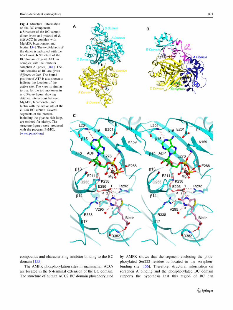
The BC structure contains three domains, A, B, and C
domains (Fig. 4a). Residues in domains A and C form the
active site. Domain B undergoes a large conformational
change to close over the active site during catalysis.
Eukaryotic BC domain contains several inserted segments,
especially at the N-terminus and between domains A and B
(the AB linker), which explains its larger size (*550 res-
idues) compared to bacterial BC subunits (*450 residues)
(Fig. 4b).
The structure of E. coli BC in a pentary complex with
the substrates MgADP, bicarbonate, and biotin provides
detailed insights in the catalysis by this enzyme (Fig. 4a)
[154]. The substrates are recognized by an intricate net-
work of ionic, hydrogen-bonding and van der Waals
interactions (Fig. 4c). One of the oxygen atoms of bicar-
bonate is poised to initiate the nucleophilic attack on ATP
to form the carboxyphosphate intermediate, and Glu296 is
the general base that extracts the proton from bicarbonate.
The orthophosphate (PO43–) derived from the decomposi-
tion of carboxyphosphate is the general base that extracts
the proton on the N10 atom of biotin [17], and Arg338
stabilizes the resulting enolate biotin intermediate for the
carboxylation reaction.
Mutation of residues in the BC active site (including
Glu296 and Arg338) confirms their importance for catal-
ysis. Mutation of Gly165 and/or Gly166 in the glycine-rich
loop in domain B and near the phosphates of ATP has no
effect on the kcat but leads to a 40-fold increase in Km for
ATP [158]. The catalytic activity of E. coli BC can be
inhibited by ATP at high concentrations (substrate inhibi-
tion) and other nucleotides [157]. A second ADP molecule
can be bound in the BC active site, with its phosphate
groups occupying the binding sites of bicarbonate and
biotin.
Escherichia coli BC is a dimer (Fig. 4a), and studies
suggest that catalysis at the two active sites of the dimer
may be coupled [159]. A chimeric mutant where one of the
active sites of the dimer is knocked out is essentially
inactive. However, each active site of the dimer is located
*25 A away from the dimer interface, with no contribu-
tions from residues in the other monomer (Fig. 4a). It is
postulated that there may be long-range communications
between the two active sites, through the dimer interface.
The two monomers may undergo alternating catalysis, with
one monomer binding substrates and catalyzing turnover
and the other releasing products. Half-site reactivity of the
BC subunit has also been proposed [152, 160], although no
cooperative behavior is observed from kinetic studies.
Mutations in the dimer interface of E. coli BC can
produce mutants that are monomeric in solution [161].
These mutants are still catalytically active, indicating that
dimerization is not absolutely required for BC catalysis in
vitro. On the other hand, dimerization of BC is required in
vivo, possibly for the assembly of the ACC holoenzyme
[162].
The polyketide natural product soraphen A is an allo-
steric inhibitor of eukaryotic ACC, binding to the BC
domain using the equivalent surface area as that for
dimerization of E. coli BC (Fig. 4b) [150]. Conformational
differences in this region of eukaryotic BC allow soraphen
A binding but disallow dimerization. The BC domain of
eukaryotic ACC is monomeric in solution and is catalyti-
cally inactive [163]. A BODIPY-labeled soraphen analog
has been developed and can be useful for screening for new
870 L. Tong
123
compounds and characterizing inhibitor binding to the BC
domain [155].
The AMPK phosphorylation sites in mammalian ACCs
are located in the N-terminal extension of the BC domain.
The structure of human ACC2 BC domain phosphorylated
by AMPK shows that the segment enclosing the phos-
phorylated Ser222 residue is located in the soraphen-
binding site [156]. Therefore, structural information on
soraphen A binding and the phosphorylated BC domain
supports the hypothesis that this region of BC can
Fig. 4 Structural information
on the BC component.
a Structure of the BC subunit
dimer (cyan and yellow) of E.coli ACC in complex with
MgADP, bicarbonate, and
biotin [154]. The twofold axis of
the dimer is indicated with the
black oval. b Structure of the
BC domain of yeast ACC in
complex with the inhibitor
soraphen A (green) [161]. The
sub-domains of BC are given
different colors. The bound
position of ATP is also shown to
indicate the location of the
active site. The view is similar
to that for the top monomer in
a. c Stereo figure showing
detailed interactions between
MgADP, bicarbonate, and
biotin with the active site of the
E. coli BC subunit. Several
segments of the protein,
including the glycine-rich loop,
are omitted for clarity. The
structure figures were produced
with the program PyMOL
(www.pymol.org)
Biotin-dependent carboxylases 871
123

allosterically regulate catalysis, although the exact molec-
ular mechanism for this long-range communication is
currently not known.
Structures of ACC BCCP component
The BCCP domain of E. coli ACC, including the ‘‘thumb’’
feature, is more flexible in the apo protein [164]. Upon
biotinylation, the thumb interacts with biotin [165], leading
to its stabilization as well as that of BCCP domain overall
[164, 166]. Solution structure of the BCCP domain of
human ACC2 is similar to that of BCCP subunit of E. coli
ACC [165], except that the human protein does not have
the thumb feature [167]. As a result, the covalently
attached biotin is flexible in human BCCP, while it inter-
acts with the thumb in E. coli BCCP. This thumb structure
interferes with biotinylation by the human holocarboxylase
synthase (HCS), though it has no effect on biotinylation by
the E. coli BirA enzyme [168]. In comparison, an engi-
neered E. coli BCCP lacking the thumb feature, and the
BCCP domain of human PCC (which also lacks the thumb
feature) can be readily biotinylated by HCS [168].
Structures of ACC CT component
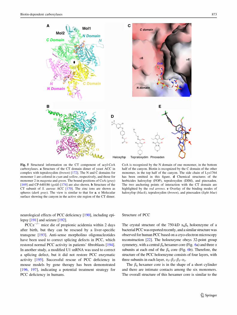
The structure of a eukaryotic CT domain (from yeast ACC)
was first reported in 2003 [169], and the structure of bac-
terial CT subunit was first reported in 2006 [170]. The
structure of the CT domain contains two sub-domains, N
and C domains (Fig. 5a), which are equivalent to the b and
a subunits of bacterial CT (Figs. 2, 5b). Each domain/
subunit has the crotonase fold (a b–b–a superhelix). The
active site of CT is located at the bottom of a ‘‘canyon’’ in
the interface of a dimer of the CT domains (Fig. 5c) or an
a2b2 heterotetramer of the bacterial CT subunits. There-
fore, CT must dimerize to be catalytically active.
Using the yeast CT domain as a surrogate since crystals
of the CT domain of grass plastid ACCs are not yet
available, the binding modes of all three classes of herbi-
cides, haloxyfop, tepraloxydim, and pinoxaden, have been
determined [171–173]. The compounds inhibit ACC by
competing against the binding of the acetyl-CoA substrate.
Despite their chemical diversity, the three compounds
share two common anchoring points of interactions with
the CT domain—a negatively charged oxygen atom that
likely mimics the oxyanion in the substrate during cataly-
sis, and a small hydrophobic group (methyl or ethyl) that is
probably located in the binding site for the acetyl group.
Each compound also establishes unique interactions with
the CT domain. Haloxyfop binding requires a large con-
formational change in the active site, in the dimer interface
of the CT domain, while tepraloxydim and especially
pinoxaden require much smaller changes. Most of the
herbicide resistance mutations are located in or near the
binding sites, consistent with their effects on herbicide
binding although the exact molecular mechanism is still not
fully understood.
Studies using the yeast CT domain also reveal that a
class of inhibitors of mammalian ACCs, as exemplified
by the CP-640186 compound, block the binding of
(carboxy)biotin to the CT-active site (Fig. 5a) [108, 174].
Structures of the human and bovine ACC2 CT domain
have been reported at 3.2 A and 2.4 A resolution, respec-
tively [112, 175]. In a different approach, nine mutations
are introduced into the active site of yeast ACC CT domain
to ‘‘humanize’’ this protein [176]. The resulting mutant
binds human ACC inhibitors with similar potency as the
human CT domain and produces crystals that diffract up to
2.4 A resolution.
A unique feature of bacterial CT is the presence of a
zinc finger in the b subunit (Fig. 5b) [170], which binds the
CT mRNA and inhibits its translation, and this inhibition
can be reversed by the substrate acetyl-CoA [177]. E. coli
CT also binds and is inhibited by single-stranded, double-
stranded, and hairpin DNA [178].
Propionyl-CoA carboxylase (PCC)
Biological functions of PCC
PCC catalyzes the conversion of propionyl-CoA to
D-methylmalonyl-CoA. In most organisms, it is crucial for
the catabolism of b-branched amino acids (Thr, Val, Ile)
and Met, cholesterol side chain, and fatty acids with an odd
number of carbon atoms (Fig. 3). Mammalian PCC is local-
ized in the mitochondrial matrix, and is activated by mono-
valent cations (K?, NH4?, Cs?) [179–181]. PCC activity is
also important for CO2 fixation in some archaeal organisms
[5, 6], methanol assimilation in Methylobacterium extor-
quens [7], acetyl-CoA assimilation in a-proteobacteria
[10], 3-hydroxypropionate assimilation in Rhodobacter
sphaeroides [11], mycolic acid and methyl-branched long-
chain fatty acid biosynthesis in M. tuberculosis [12], and
polyketide biosynthesis in Streptomyces and other organisms
[13].
Inherited deficiencies in PCC activity in humans are linked
to propionic acidemia (PA), with symptoms usually first
appearing during the neonatal period, including vomiting,
lethargy, ketoacidosis, delayed growth, cardiomyopathy,
mental retardation, and death in severe cases. A large number
of autosomal recessive mutations in both subunits of PCC
have been identified, including missense mutations, a few
nonsense mutations, insertions/deletions, and splicing muta-
tions [182–189]. PCC is expressed in the brain and may be
important for neurodevelopment, which may be related to the
872 L. Tong
123
neurological effects of PCC deficiency [190], including epi-
lepsy [191] and seizure [192].
PCCa-/- mice die of propionic acidemia within 2 days
after birth, but they can be rescued by a liver-specific
transgene [193]. Anti-sense morpholino oligonucleotides
have been used to correct splicing defects in PCC, which
restored normal PCC activity in patients’ fibroblasts [194].
In another study, a modified U1 snRNA was used to correct
a splicing defect, but it did not restore PCC enzymatic
activity [195]. Successful rescue of PCC deficiency in
mouse models by gene therapy has been demonstrated
[196, 197], indicating a potential treatment strategy for
PCC deficiency in humans.
Structure of PCC
The crystal structure of the 750-kD a6b6 holoenzyme of a
bacterial PCC was reported recently, and a similar structure was
observed for human PCC based on a cryo-electron microscopy
reconstruction [22]. The holoenzyme obeys 32-point group
symmetry, with a central b6 hexamer core (Fig. 6a) and three asubunits at each end of the b6 core (Fig. 6b). Therefore, the
structure of the PCC holoenzyme consists of four layers, with
three subunits in each layer, a3–b3–b3–a3.
The b6 hexamer core is in the shape of a short cylinder
and there are intimate contacts among the six monomers.
The overall structure of this hexamer core is similar to the
Fig. 5 Structural information on the CT component of acyl-CoA
carboxylases. a Structure of the CT domain dimer of yeast ACC in
complex with tepraloxydim (brown) [172]. The N and C domains for
monomer 1 are colored in cyan and yellow, respectively, and those for
monomer 2 in magenta and green. The bound positions of CoA (gray)
[169] and CP-640186 (gold) [174] are also shown. b Structure of the
CT subunit of S. aureus ACC [170]. The zinc ions are shown as
spheres (dark gray). The view is similar to that for a. c Molecular
surface showing the canyon in the active site region of the CT dimer.
CoA is recognized by the N domain of one monomer, in the bottom
half of the canyon. Biotin is recognized by the C domain of the other
monomer, in the top half of the canyon. The side chain of Lys1764
has been omitted in this figure. d Chemical structures of the
herbicides haloxyfop (FOP), tepraloxydim (DIM), and pinoxaden.
The two anchoring points of interaction with the CT domain are
highlighted by the red arrows. e Overlay of the binding modes of
haloxyfop (black), tepraloxydim (brown), and pinoxaden (light blue)
Biotin-dependent carboxylases 873
123

12S subunit of transcarboxylase [31] as well as the bsubunit of S. coelicolor and M. tuberculosis acyl-CoA
carboxylases [198–200]. In addition, the b2 dimer of PCC,
with one monomer from each layer of the b6 hexamer
(Fig. 6a), is similar to the dimer of the CT component of
ACC (Fig. 5a). The active site is located at the interface of
this dimer (Fig. 7a), at the bottom of a deep canyon
(Fig. 7b). In the PCC crystal, BCCP-biotin (from the asubunit) is bound in the CT active site, interacting with
residues in the C domain of one b subunit (Fig. 7b). The
propionyl-CoA substrate is recognized by the N domain of
the other subunit (Fig. 7b).
In comparison, the a subunits are arranged as mono-
mers, splayed far apart from each other, and there are
essentially no contacts among them in the PCC holoen-
zyme (Fig. 6b). This is in sharp contrast to the dimeric
organization of the BC subunit of bacterial ACCs (Fig. 4a)
and the BC domain of pyruvate carboxylase (see below).
While the overall structure of the BC domain of the PCC asubunit is similar to that of the bacterial BC subunit,
sequence, and conformational differences for those resi-
dues in the putative dimer interface in PCC precludes the
formation of a similar dimer.
There are few contacts between the BC domain in the asubunit and the b subunit (CT component) (Fig. 6a).
Instead, interactions between the a and b subunits in PCC
is mediated by a previously unrecognized domain, located
between the BC and BCCP domains in the primary
sequence of the a subunit (Fig. 2). It has been named the
BT domain [22], for its role in mediating BC-CT interac-
tions. The structure of this domain has a novel backbone
fold, with a long helix surrounded by an eight-stranded
anti-parallel b-barrel (Fig. 7c). A ‘‘hook’’, comprising the
C-terminal part of the helix and the loop connecting it to
the first strand of the b-barrel, has a central role in the
interactions between the a and b subunits in the PCC
holoenzyme.
The distance between the BC and CT active sites in the
PCC holoenzyme is *55 A (Fig. 7a). This cannot be
reached by biotin in the swinging-arm model. Therefore,
the BCCP domain (in addition to its attached biotin) must
translocate during PCC catalysis, hence the swinging-
domain model (Fig. 1a).
The structure of the PCC holoenzyme also provides a
framework for understanding the large collection of dis-
ease-causing mutations. The missense mutations are
distributed throughout the entire enzyme (Fig. 7a). Some
of them affect substrate binding and/or catalysis, while
others affect protein stability [201–207]. At the same time,
few of the mutations are located directly in the interface
between the subunits of the holoenzyme, consistent with
the extensive nature of the interface. Single-site mutations
in this interface may not be sufficient to disrupt the holo-
enzyme. In fact, five mutations in the hook region are
needed to disrupt the interactions between the a and bsubunits [22].
Fig. 6 Striking differences in
the overall architecture of the
holoenzymes of PCC and MCC.
a Crystal structure of the
bacterial PCC holoenzyme [22],
viewed down the twofold
symmetry axis within a b2
dimer. The domains are colored
as in Fig. 2. The four layers of
the structure are indicated.
b Structure of the PCC
holoenzyme, viewed down the
threefold symmetry axis.
c Crystal structure of the P.aeruginosa MCC holoenzyme
[23], viewed down the twofold
axis within a b2 dimer.
d Structure of the MCC
holoenzyme, viewed down the
threefold axis. e Structure of the
MCC holoenzyme, after a *60�rotation around the vertical axis
from c. The view is down the
twofold axis relating two b2
dimers. f Structure of the MCC
holoenzyme, after a *60�counterclockwise rotation from
panel D
874 L. Tong
123

3-Methylcrotonyl-CoA carboxylase (MCC)
Biological functions of MCC
MCC catalyzes the conversion of 3-methylcrotonyl-CoA to
3-methylglutaconyl-CoA (Fig. 1b). It is essential for the
catabolism of leucine and isovalerate in most living sys-
tems (Fig. 3) [61]. In Pseudomonas organisms, MCC is
required for the metabolism of acyclic terpenoids [14, 208–
212]. In Arabidopsis, MCC activity is required for seed
development and germination [213]. MCC expression level
is higher in male zebra finches, which may be important for
the development of their singing behavior [214].
MCC shares good sequence conservation with PCC and
its holoenzyme is also a 750-kD a6b6 dodecamer. On the
other hand, while PCC carboxylates the a carbon of the
substrate, MCC carboxylates the c carbon of an a–bunsaturated acid (Fig. 1b). MCC is located in the mito-
chondrial matrix in eukaryotes, and mutations in the
N-terminal mitochondrial targeting sequence of both sub-
units can affect their localization [215]. Human MCC has
been expressed and purified from the baculovirus system in
the active form [216]. It demonstrated hyperbolic kinetics
with ATP and 3-methylcrotonyl-CoA, while the Pseudo-
monas MCC showed sigmoidal kinetics toward ATP [212].
Deficiencies in MCC activity in humans are linked to
3-methylcrotonylglycinuria (MCG), which constitute one
of the most frequently observed inborn errors of metabo-
lism [217–226]. The clinical manifestations of MCG are
highly variable, from asymptomatic individuals to neonatal
onset, severe cases that can result in death. Neurological
symptoms such as psychomotor retardation, seizure and
coma have also been observed. Like PCC, a large number
of autosomal recessive mutations in both subunits of MCC
have been identified.
Structure of MCC
The structure of the P. aeruginosa MCC holoenzyme also
obeys 32-point group symmetry, with three subunits in
each of four layers (Fig. 6c) [23]. However, despite its
sequence conservation with PCC, the overall architecture
of the MCC holoenzyme is strikingly different from that of
PCC. This is especially apparent for the locations of the asubunit relative to the b subunit, the organization of the N
and C domains in the b subunit, and the structure and
placement of the BT domain. As a result of these differ-
ences, the overall shape of the MCC holoenzyme is highly
distinct from that of PCC (Fig. 6a, c).
Rather than being splayed away from each other as in
PCC, the three a subunits at each end of the central b6 core
of MCC are located close to each other, in fact showing
trimeric association (Fig. 6d). In addition, the BC domain
in the a subunit is located *20 A above the b subunit, and
there is no contact between them. The BT domain bridges
the BC domain and the b subunit. While the overall
structure of this domain is similar to that in PCC, there are
also important differences. Especially, the hook in the
MCC BT domain has a different conformation (Fig. 8a),
contributing to the different architecture of the MCC
holoenzyme compared to PCC. The Pseudomonas MCC
BT domain lacks the third strand of the eight-stranded
b-barrel (Fig. 8a), although this strand is likely present in
human and most other MCCs [23].
In the b subunit, the positions of its N and C domains in
MCC (Fig. 8b) are swapped relative to those in PCC (Fig. 8c).
Fig. 7 The active sites of PCC. a Relationship between the BC and
CT active sites (indicated with the asterisks) in the PCC holoenzyme.
The CT active site is located at the interface of a b2 dimer, with the bsubunit from the bottom layer colored in green. Sites of disease-
causing missense mutations are indicated with the spheres. The thirdasterisk indicates the other active site of the CT dimer. b Molecular
surface of the active site region of CT. The observed position of biotin
is shown (stick model in black). The position of CoA (gray) is
modeled based on that in the structure of the yeast ACC CT domain
[169]. c Structure of the BT domain of the bacterial PCC a subunit.
The hook region is labeled
Biotin-dependent carboxylases 875
123

This difference is not due to a swap of the two domains in the
primary sequences of the two b subunits. Instead, it is due to a
different connectivity between the N and C domains. In
essence, each layer of the b6 hexamer contains alternating N
and C domains of the three subunits. While PCC uses one way
of connecting neighboring N and C domains to make a bsubunit (Fig. 8c), MCC uses the alternative way, such that the
linker between the two domains runs in the opposite direction
as compared to PCC (Fig. 8b).
The N and C domains of the b subunits in PCC and
MCC have the same backbone fold. The pseudo symmetry
operation relating the two domains is a rotation of *60�along the threefold axis of the b6 hexamer. Therefore, the
b6 hexamers of MCC and PCC have pseudo sixfold sym-
metry. The swapping of the N and C domains in MCC
follows this pseudo symmetry, equivalent to a 60� rotation
around the threefold axis of the hexamer (Fig. 8b, c), and
therefore does not lead to a large change in the overall
shape of the hexamer. Such a rotation also places the
BCCP domain in the same position in the MCC holoen-
zyme compared to PCC (Fig. 6e, f).
The connectivity between the N and C domains
observed in MCC b subunit is the same as that in the asubunit of the Na?-transporting glutaconyl-CoA decar-
boxylase (GCDa) [27, 30]. However, GCDa is a dimer
only, and therefore the different connectivity leads to a
distinct organization of the active site in GCDa dimer
compared to the CT dimer of ACC (Fig. 5a). Like MCC
and GCC, GCDa is also active on the c carbon of an a-bunsaturated acid. It is possible that these three enzymes
Fig. 8 Structure of P. aeruginosa MCC. a Structure of the BT
domain of the P. aeruginosa MCC a subunit. The missing third strand
is indicated in red. b Structure of the b6 hexamer of MCC. The
subunit boundaries are indicated with the gray lines. The N (cyan) and
C (yellow) domains are labeled. The linker between the two domains
is shown in black, with the direction given by the red arrow. The
linkers from neighboring subunits come very close to each other at
one point (blue asterisk), and therefore only a small change is needed
to switch to the connectivity seen in the PCC b subunit. c Structure of
the b6 hexamer of PCC, shown in the same scheme as panel B. The
linker in PCC b runs in the opposite direction compared to MCC b.
d Relationship between the BC and CT active sites (indicated with the
asterisks) in the MCC holoenzyme. While the BT domain of the asubunit contacts one b2 dimer (b1 and b4), the BCCP domain of that asubunit is actually located in the active site of a different b2 dimer (b3
and b6). e Binding modes of biotin (black) and 3-methylcrotonyl-
CoA (gray) to the CT active site of MCC. The position of
3-methylcrotonyl-CoA is modeled based on that of CoA in MCC
[23] and crotonyl-CoA in GCDa [30]. The carbon atom to be
carboxylated is indicated with the red arrow. f Locations of disease-
causing missense mutations in the structure of MCC (indicated with
the spheres). Only the b1–b4 dimer is shown
876 L. Tong
123
share a common evolutionary origin, representing a second
lineage of biotin-dependent carboxylases that act on CoA
esters. Another common feature among MCC, GCDa and
likely GCC is the extended N-terminal segment of the bsubunit, which has extensive interactions with the C
domain of the same subunit (Fig. 8b). In comparison, the
N-terminal segment of PCC b subunit is much shorter
(Fig. 8c). These additional interactions may be the
molecular basis for the swapping of the positions of the N
and C domains [23].
The distance between the BC and CT active sites in the
MCC holoenzyme is *80 A (Fig. 8d), indicating that the
BCCP domain must translocate during catalysis (the
swinging-domain model). BCCP-biotin is located in the CT
active site in the MCC structures (Fig. 8d). Compared to
the structure of PCC, biotin is bound deeper into the CT
active site, consistent with its carboxylation of the c carbon
of the substrate. The N10 atom of biotin is *6 A from the ccarbon of the 3-methylcrotonyl-CoA substrate (Fig. 8e),
modeled based on the observed positions of CoA bound to
MCC [23] and crotonyl-CoA bound to GCDa [30]. A
conformational change for two helices in the CT active site
(a6 and a6A, Fig. 8e) is observed upon CoA binding in
MCC [23]. A similar conformational change is also
observed in GCDa [30].
The disease-causing mutations are distributed over the
entire structure of the holoenzyme (Fig. 8f). Some of them
are located in the BC or CT active site. For example, the
R385S mutation in the a subunit abolishes the stabilization
of the biotin enolate during BC catalysis. The A214T
mutation in the b subunit is centrally located between
biotin and 3-methylcrotonyl-CoA in the CT active site
(Fig. 8e) and may block binding of either or both sub-
strates. Some of the mutations may also disturb the
interaction between the BCCP or the BT domain and the bsubunit [23].
Geranyl-CoA carboxylase (GCC)
GCC is classified into the same family as MCC since its
site of carboxylation in the substrate is also on the c carbon
of an a-b unsaturated acid (Fig. 1b). The amino acid
sequences of bacterial GCC are highly conserved with
those of MCC, and the holoenzyme of GCC is likely also
an a6b6 dodecamer. At the same time, the geranyl group is
much larger than the 3-methylcrotonyl group, and therefore
the CT component (b subunit) of GCC is expected to have
a larger pocket in the active site to accommodate this
group. In fact, P. aeruginosa MCC cannot carboxylate
geranyl-CoA, while GCC has activity toward both geranyl-
CoA and 3-methylcrotonyl-CoA [212].
GCC is found in Pseudomonas organisms and several
other bacteria (Table 1) [14, 208–212, 227]. It is important
for the metabolism of the geranyl group and other acyclic
terpenes. In fact, some Pseudomonas organisms can use
these compounds as the sole carbon source. Through
reactions that are similar to b-oxidation, c-carboxygeranyl-
CoA is converted to 3-methylcrotonyl-CoA (releasing two
acetyl-CoAs and one acetate), and MCC facilitates the
further degradation of this compound.
GCC activity has been purified from maize leaves, and it
is probably also important for the metabolism of acyclic
terpenes in plants [61, 228]. The biotin-containing com-
ponent of this enzyme was found to have a molecular
weight of *122 kD, which is significantly larger than the
*75-kD a subunit of bacterial GCC (and MCC). Plant
GCCs may constitute another family of biotin-dependent
carboxylases, although a sequence for this protein is cur-
rently not available.
Other acyl-CoA carboxylases
Streptomyces, Mycobacterium, and Corynebacterium
organisms have a collection of a6b6 or a2b2 acyl-CoA
carboxylases (family 1.4, Fig. 2) [12, 229]. These enzymes
are different from PCC (family 1.5) in that they probably
lack the BT domain in the a subunit. In addition, they have
more diverse substrate preferences, being active toward
acetyl-, propionyl-, and/or butyryl-CoA [230, 231]. More-
over, some of these enzymes require the presence of a third
subunit, e subunit (*7 kD in S. coelicolor and *25 kD in
M. tuberculosis), for maximal activity, although the exact
stoichiometry of this subunit is currently not known [12,
232, 233]. These acyl-CoA carboxylases are important for
the biosynthesis of fatty acids, branched-chain fatty acids
and other compounds, such as mycolic acid [234, 235]. An
acyl-CoA carboxylase in Mycobacterium tuberculosis is
crucial for mycolic acid biosynthesis and pathogenesis
[236].
The structures of the b6 hexamer of these enzymes have
32 symmetry [198–200, 237], and are similar to that of
PCC [22] and the 12S subunit of transcarboxylase [31]. An
Asp residue near the CT active site may be important for
substrate preference for propionyl-CoA, while mutating
this residue to Ile changes the preference to acetyl-CoA
[198, 200]. On the other hand, an equivalent mutation in
PCC is not able to change the substrate preference of that
enzyme [22].
A Rhizobium etli acyl-CoA carboxylase has *15-fold
higher activity toward propionyl-CoA than acetyl-CoA,
and it is activated substantially by mono-valent cations,
K?, NH4? and Cs? [238].
Biotin-dependent carboxylases 877
123
Pyruvate carboxylase (PC)
Biological functions of PC
PC catalyzes the conversion of pyruvate to oxaloacetate
(Fig. 1b) and it is an important enzyme in intermediary
metabolism [4, 239–243]. In mammals, PC is localized in
the mitochondrial matrix and has crucial roles in gluco-
neogenesis in liver and kidney (being the first enzyme in
the gluconeogenesis pathway), lipogenesis and glycero-
neogenesis in adipocytes, and biosynthesis of the excitatory
neurotransmitter glutamate in astrocytes (Fig. 3). PC has
an important anaplerotic role, replenishing the intermedi-
ates of the tricarboxylic acid (TCA) cycle that have been
withdrawn for the biosynthesis of glucose, fatty acids,
amino acids and other molecules [244]. Tumor cells
depend on PC for anaplerosis in the absence of glutamine
(Fig. 3), and cells with high PC activity may be resistant to
inhibition of glutamine metabolism [245].
PC is important for glucose-induced insulin secretion by
pancreatic b-cells in rats [246], and knock-down of PC
activity by shRNA in rat insulinoma cells leads to impaired
insulin secretion [247]. However, PC protein level and
enzymatic activity are low in human pancreatic b-cells, and
these cells may use a different pathway for stimulating
insulin secretion [248].
Fungal PC is localized in the cytoplasm, in contrast to
other eukaryotes. S. cerevisiae is unique in that it carries
two PC enzymes, and the expression levels of the two
enzymes are regulated independently in response to dif-
ferent nutrients [4]. PC is required for the assembly,
import, and/or activation of some peroxisomal enzymes in
several fungi, including alcohol oxidase in Hansenula
polymorpha [249] and D-amino acid oxidase in Pichia
pastoris [250]. This function is mediated by the CT domain
of PC [251], but involves residues on the opposite face of
the CT domain from the active site and is independent of
its catalytic activity [252]. PC activity is important for
carbon metabolism and virulence of the human pathogen
Listeria monocytogenes [253].
PC is a single-chain, multi-domain enzyme in most
organisms (family 3.1, Fig. 2), and is active only as a
500-kD tetramer. In archaea and some bacteria, such as
P. aeruginosa [254], PC is in the two-chain form (family
3.2, Fig. 2), but it is also active as a tetramer of the two
chains. Acetyl-CoA stimulates single-chain form of PC, by
activating the BC activity, but it has no effect on the two-
chain form of PC [255, 256]. An ATP analog, MgTNP-ATP,
also activates PC activity and can allosterically compete with
acetyl-CoA [257]. On the other hand, aspartate, glutamate
and a-ketogluratate are allosteric, feedback inhibitors of the
CT activity of PC [258].
Deficiency in PC activity is a rare, autosomal recessive
metabolic disorder in humans, and has been associated with
three forms of clinical manifestations [259–264]. Patients
with form A PC deficiency have chronic, mild to moderate
lactic acidemia, psychomotor retardation and hypotonia,
and many patients die within a few years of birth. Form B
deficiency is associated with the most serious symptoms,
with severe lactic acidemia, hypoglycemia, hyperammo-
nemia, anorexia, convulsions, and the patients generally die
in the first months of life. Form C deficiency shows
occasional mild lactic acidemia and may also show mild
neurological symptoms. A collection of missense and
insertion/deletion mutations has been identified in patients
suffering from PC deficiency.
Structure of PC
Crystal structures of the full-length Rhizobium etli PC
(RePC) [20, 265], Staphyloccocus aures PC (SaPC) [21,
266], and human PC (HsPC) lacking only the BC domain
[21] are currently available. The overall structure of the
tetramer is in the shape of a square (or a diamond), with
approximate 222 symmetry for SaPC (Fig. 9a). The
structure consists of two layers, with two monomers in
each layer (Fig. 9b). BC and CT dimers are located at
alternate corners of the square, creating an extensive
interface between the two layers. On the other hand, there
are few contacts between the two monomers in the same
layer (Fig. 9a). The overall structures of the four mono-
mers in the tetramer are similar to each other, but there are
differences in the relative positions of their BC and CT
domains. This is especially true for RePC, where the two
monomers in one layer show large differences to those in
the other layer. As a result, the RePC tetramer shows
significant asymmetry between the two layers (Fig. 9c, d)
[20, 265].
Structural information on these PC enzymes reveals the
presence another domain, named as the PT (PC tetramer-
ization) [21] or allosteric [20] domain. The structure of this
domain contains a helix (formed by 30 residues between the
BC and CT domains, Fig. 2) surrounded by a highly twisted,
four-stranded anti-parallel b-sheet (formed by 60 residues
between the CT and BCCP domains) (Fig. 9e). In HsPC and
SaPC, the PT domain is important for tetramerization, hence
its name [21]. A PT domain in one layer interacts with a PT
domain in the other layer, and mutations in this PT–PT
interface can disrupt PC tetramerization and catalytic
activity. In contrast, the equivalent allosteric domain in
RePC does not appear to be important for tetramerization
(Fig. 9c) [20]. RePC lacking the BC domain is a dimer in
solution [265], while the same construct for HsPC is a tet-
ramer [21].
878 L. Tong
123

The structure of the PT domain shares remote similarity
to that of the BT domain in PCC and MCC. The eight-
stranded b-barrel in the BT domain (Fig. 7c) is replaced by
a four-stranded anti-parallel b-sheet in the PT domain
(Fig. 9e). However, while the helix is connected directly to
the b-barrel in the BT domain, the CT domain of PC is
inserted between the helix and the b-sheet in the PT
domain.
The structure of the CT domain of PC contains a tri-
osephosphate isomerase (TIM) barrel with a long
C-terminal extension, and has similarity to that of the 5S
(CT) subunit of transcarboxylase [32] and the CT domain
of oxaloacetate decarboxylase [29, 267]. In SaPC and
HsPC structures [21], one BCCP and its covalently
attached biotin is bound in the CT active site of the other
monomer in the same layer (Fig. 9b), providing direct
Fig. 9 Structure of PC. a Crystal structure of SaPC tetramer [21].
The domains of monomer 1 are colored as in Fig. 2, and the other
three monomers are in magenta, cyan and yellow. The BC and CT
active sites are indicated with the asterisks. The distance between the
exo site and the CT active site is also labeled (gray). b Structure of
SaPC tetramer, viewed from the bottom layer. c Crystal structure of
RePC tetramer [20]. d Structure of RePC tetramer, viewed from the
bottom layer. e Structure of the PT domain of SaPC [21]. f Structure
of the active site region of the CT domain, in complex with BCCP
(blue). The position of biotin in the SaPC structure is shown in black,
and that in the HsPC structure in gray [21]. g Molecular surface of the
binding site of CoA in SaPC [266]. h Detailed interactions between
CoA and the binding site in SaPC. i The activity of various acetyl-
CoA analogs in stimulating the catalysis by SaPC [266]
Biotin-dependent carboxylases 879
123

insight into the molecular mechanism of the CT reaction.
In contrast, the BCCP domains are in a non-productive
location and their attached biotins are disordered in the
RePC structure (Fig. 9c) [20].
In the CT active site, Thr908 is hydrogen-bonded to the
N10 atom of biotin (Fig. 9f), indicating that it may play an
important role in catalysis. The structure of the T908A
mutant of SaPC is essentially identical to that of the wild-
type enzyme, but the mutant is inactive [266]. Kinetic
studies with the equivalent mutant of RePC also confirm
the importance of this residue in the CT reaction [268]. The
disease-causing A610T and M743I mutations are located in
the active site of the CT domain, and are expected to dis-
rupt catalysis by interfering with the binding of biotin
(A610T mutation) [21, 266] or pyruvate (M743I mutation)
(Fig. 9f) [21, 32]. The importance of other residues in the
CT active site has also been assessed by mutagenesis
studies in SaPC [266] and RePC [269].
The structural information defines the molecular basis
for why PC is only active in the tetrameric form [20, 21].
BCCP-biotin is carboxylated in the BC domain of its own
monomer, but it then transfers the CO2 to pyruvate in the
CT domain of the other monomer in the same layer
(Fig. 9a). Therefore, both monomers in each layer are
required for catalysis, but their conformation is stabilized
only in the context of the entire tetramer because there are
few interactions between two monomers of the same layer.
The distance between the BC and CT active sites in PC
is *75 A (Fig. 9a), supporting the swinging-domain
model for catalysis (Fig. 1a). Binding of BCCP-biotin in
the CT active site is observed in SaPC and HsPC [21].
Recently, BCCP-biotin in a nonproductive binding mode in
the BC domain of RePC was reported [265].
The overall structures of the BC domain monomer and
dimer of PC are similar to those of the BC subunit of
E. coli ACC. Mutation of residues in RePC equivalent to
those important for substrate binding and catalysis in
E. coli BC [154] confirms their importance for PC catalysis
as well [270].
The activator CoA is bound at the interface of the BC
and PT domains of one monomer and the BC domain of
another monomer (Fig. 9g) [20, 266]. A change in the
organization of the dimer of the two BC domains is
observed upon CoA binding [266], consistent with the fact
that acetyl-CoA primarily activates the BC activity. The
phosphate groups of CoA interact with a collection of Arg
and Lys residues (Fig. 9h). Especially, Arg541 recognizes
the 30-phosphate group of CoA, which is critical for the
activating effect of CoA (Fig. 9i), and the R541C disease-
causing mutation abolishes acetyl-CoA activation [21]. In
SaPC, CoA binding made the four monomers of the PC
tetramer more similar to each other, essentially making the
tetramer more symmetric. The symmetrical structure of
SaPC is also supported by cryo-EM studies [266, 271]. In
contrast, a highly asymmetric tetramer is observed in the
RePC structure, and only two CoA molecules (the 30-phospho-ADP portion) are bound to the tetramer [20].
However, this asymmetry may be an inherent property of
RePC and does not depend on CoA binding, as the struc-
ture without CoA shows similar asymmetry [265].
In the SaPC structure, the other three biotin groups are
located in another binding site, at the interface between the
PT and CT domains of the other monomer in the same
layer, *20 A from the CT active site (Fig. 9a) [21]. This
exo site does not exist in HsPC due to sequence and
structural differences. The functional importance of this
binding site is currently unknown.
Urea carboxylase (UC)
Biological functions of UC
UC catalyzes the carboxylation of urea to produce allo-
phanate, which is then hydrolyzed to generate ammonia
and CO2 (Fig. 10a) [15, 16, 272]. Therefore, urea amid-
olyase (UA, family 4.1) can catalyze both steps of this
conversion, while a separate allophanate hydrolase activity
is needed for UC (families 4.2 and 4.3).
UC is found in some bacteria, fungi, and green algae, and
its activity is important for the utilization of urea as a
nitrogen source in these organisms [15, 16, 272]. A second
pathway for urea utilization is catalyzed by the nickel-
dependent enzyme urease, which can convert urea to
ammonia in a single step. Some of the fungal organisms
contain both pathways, although only the UA pathway is
present in S. cerevisiae. In fact, the UA form of this enzyme
is found primarily in fungal species. In Saccharomyces
kluyveri, UA is part of a pyrimidine degradation pathway,
converting the urea product of uracil degradation to
ammonia and enabling this organism to use uracil as the sole
nitrogen source [273]. This is a distinct pathway for uracil
catabolism, and it may also be present in other eukaryotes.
In Candida albicans, UA activity is important for the
virulence of this important human pathogen. The CO2
product of UA is a signal for hyphal switching [274], while
the ammonia product can be secreted to alkalinize the
extracellular environment, which also induces the yeast-
hyphal transition. The hyphal form is important for C.
albicans to escape phagocytosis by macrophages, thereby
evading the host immune system [275].
Structure of UC
The structure of the UC component of K. lactis urea
amidolyase was reported recently (Fig. 10b) [24]. In
880 L. Tong
123

contrast to the other biotin-dependent carboxylases, the UC
component is catalytically active as a monomer. The CT
domain of UC consists of four sub-domains, named A, B, C
and D (Fig. 10b), with many structural and sequence
homologs. Especially, domains A and B together are sim-
ilar to the KipA subunit, and domains C and D the KipI
subunit of the B. subtilis KipA-KipI complex [276, 277].
KipI binds and inhibits the autophosphorylation of the
histidine kinase KinA, thereby inhibiting sporulation. On
the other hand, KipA can sequester KipI and prevent it
from inhibiting KinA activation. CT and KipA-KipI have
*20–35 % sequence identity, but residues that are
important for CT catalysis are not conserved in KipA and
KipI. Therefore, the KipA-KipI complex is unlikely to
have CT activity. In addition, both domains B and D have
the cyclophilin fold, although they do not share any rec-
ognizable sequence homology with cyclophilin.
The active site of the CT domain is located in a cleft at
the interface between the B and D domains (Fig. 10c) [24].
The biotin head group is fully extended away from the
BCCP domain and interacts with highly conserved residues
in the active site. The urea substrate is recognized by
hydrogen-bonding interactions with the active site. Espe-
cially, one of the urea nitrogen atoms is directly hydrogen-
bonded to the N10 atom of biotin. Mutations of residues in
the active site region have detrimental effects on the
catalysis by UC.
The overall structure of the BC domain of UC is similar to
that of other BC components. However, the BC domain is
monomeric in UC, as the interface that mediates the dimer-
ization of the BC subunit of bacterial ACC is not conserved in
UC. The BC domain interacts with domains A and D of CT in
the UC holoenzyme. The distance between the BC and CT
active sites in UC is *60 A (Fig. 10b), indicating that the
BCCP domain must translocate during catalysis, consistent
with the swinging-domain model (Fig. 1).
Common features in the catalysis by biotin-dependent
carboxylases
The BC component has strong sequence conservation
among the biotin-dependent carboxylases. Insights into the
BC reaction are obtained from the structure of the E. coli
BC subunit in complex with substrates [154], which indi-
cates the crucial importance of two residues, Glu296 as a
general base to extract the proton from bicarbonate and
Arg338 to stabilize the biotin enolate oxyanion. The
Fig. 10 Structure of the urea
carboxylase (UC) domain of K.lactis urea amidolyase. a The
chemical reaction catalyzed by
UC, converting urea to
allophanate, which is then
hydrolyzed to ammonia and
bicarbonate. b Crystal structure
of K. lactis UC [24]. The
domains are labeled, and the
active site of BC is indicated
with the asterisk. c Interactions
of biotin and urea with the CT
active site of UC, located at the
interface between the B and D
domains
Biotin-dependent carboxylases 881
123

hydrolysis of ATP, via the formation of a carboxyphos-
phate intermediate, serves at least two functions: (1) the
dehydration of bicarbonate to CO2, which is a stronger
electrophile; (2) the production of orthophosphate (PO43-),
which is a strong base that is capable of extracting the
proton on the N10 atom of biotin [17]. This mechanism is
likely shared among all biotin-dependent carboxylases, and
Glu296 and Arg338 of E. coli BC are conserved among
these enzymes.
The CT components of the acyl-CoA carboxylases show
sequence and structural conservation. The acyl-CoA sub-
strate is recognized by a domain/subunit with the crotonase
fold, and it may be speculated that this part of the CT
component evolved from a primordial crotonase. This fold
is capable of binding CoA esters, and it also provides an
oxyanion hole, in the form of two main-chain amides, that
stabilizes the enolate oxyanion of the acyl group during
catalysis. Remarkably, the main-chain carbonyl of one of
the oxyanion hole residues recognizes the N6 amino group,
and the main-chain amide of the following residue is
hydrogen-bonded to the N1 atom of the adenine base of
CoA. Biotin is recognized by a separate domain/subunit,
but also with the crotonase fold. The equivalent oxyanion
hole in this domain/subunit stabilizes the biotin enolate
oxyanion during the CT reaction. It might be possible that
the biotin-binding domain/subunit of CT arose through
gene duplication of a crotonase enzyme and then evolved
its specificity toward biotin.
The CT components of PC and UC are entirely different
in sequence and structure from those of acyl-CoA car-
boxylases. However, they share the common feature of
providing two oxyanion holes to stabilize the intermediates
during the CT reaction. On the other hand, while acyl-CoA
carboxylases use primarily (carboxy)biotin for catalysis
[17, 169], side chains of conserved amino acids are
important for the CT reaction in PC and UC, for example
Thr908 in SaPC [21, 266, 268] and Lys1605 in KlUC [24].
Summary and perspective
Biotin-dependent carboxylases are crucial enzymes for
metabolism and many other cellular processes. They pro-
duce compounds that are important for the biosynthesis of
many molecules and/or help degrade potentially toxic
metabolic intermediates. ACCs are drug discovery targets
against type 2 diabetes, cancer, microbial infections, and
other diseases in humans. Three classes of herbicides
function by inhibiting the plastid ACC of grasses, dem-
onstrating the feasibility of small-molecule ACC
antagonists. On the other hand, deficiencies in PCC, MCC
and PC activity are linked to serious diseases in humans,
especially infants. Development of agents that can restore
the activity of the deficient enzyme, possibly through gene
therapy, will be of great benefits to these patients.
Structural information on the holoenzymes of biotin-
dependent carboxylases demonstrates the remarkable
degree of variability in these enzymes. They share con-
served domains, which have similar structures on their
own. However, the arrangement of these domains in the
holoenzyme monomers and especially the architecture of
the holoenzyme oligomers can be dramatically different,
for example the comparison between PCC and MCC or
between SaPC and RePC. These observations also serve as
a cautionary tale for the limitations of homology modeling
on such large, multi-domain systems.
The past few years have seen great progress in the
studies on biotin-dependent caboxylases, especially with
the first structural information on most of the holoenzymes
(PCC, MCC, PC, UC). At the same time, many important
questions remain to be answered. For example, structural
information on the holoenzymes of multi-subunit and
multi-domain ACCs is still not available. In addition, while
the recognition of BCCP by the CT component has been
observed in most of the holoenzyme structures, it is not
clear how BCCP binds to the BC component for catalysis.
Moreover, the molecular basis for substrate selectivity of
the acyl-CoA carboxylases is still poorly understood, and
presently one cannot readily determine the substrate pref-
erence(s) of a given acyl-CoA carboxylase based on a
sequence analysis of its CT component. While substitutions
of amino acid side chains are likely to play a major role in
determining substrate selectivity (for example the size and/
or shape of the acyl-CoA binding pocket), conformational
differences in the main chain of residues in the active site
may also be important and further structural information is
needed to illuminate such differences. Therefore, the
coming years should continue to be highly exciting in the
studies of biotin-dependent carboxylases.
Acknowledgments I thank Matthew Callaghan, Chi-Yuan Chou,
Daniel Floyd, Christine Huang, Yi Seul Kim, Jiang Li, Svetlana
Novoseletskaya, Kianoush Sadre-Bazzaz, Yang Shen, Timothy Tran,
Benjamin Tweel, Jia Wei, Song Xiang, Zhiru Yang, Linda Yu, and
Hailong Zhang for contributions to this project and for helpful dis-
cussions. I apologize for not being able to cite many of the earlier
publications due to space limitations. This research was supported in
part by a grant from the NIH to LT (DK067238).
References
1. Wakil SJ, Stoops JK, Joshi VC (1983) Fatty acid synthesis and
its regulation. Ann. Rev. Biochem. 52:537–579
2. Cronan JE Jr, Waldrop GL (2002) Multi-subunit acetyl-CoA
carboxylases. Prog Lipid Res 41:407–435
3. Tong L (2005) Acetyl-coenzyme A carboxylase: crucial meta-
bolic enzyme and attractive target for drug discovery. Cell Mol
Life Sci 62:1784–1803
882 L. Tong
123

4. Jitrapakdee S, St. Maurice M, Rayment I, Cleland WW, Wallace
JC, Attwood PV (2008) Structure, mechanism and regulation of
pyruvate carboxylase. Biochem J 413:369–387
5. Berg IA, Kockelkorn D, Ramos-Vera WH, Say RF, Zarzycki J,
Hugler M, Alber BE, Fuchs G (2010) Autotrophic carbon fixa-
tion in archaea. Nat Rev Microbiol 8:447–460
6. Pratscher J, Dumont MG, Conrad R (2011) Ammonia oxidation
coupled to CO2 fixation by archaea and bacteria in an agricul-
tural soil. Proc Natl Acad Sci USA 108:4170–4175
7. Smejkalova H, Erb TJ, Fuchs G (2010) Methanol assimilation in
Methylobacterium extorquens AM1: demonstration of all
enzymes and their regulation. PLoS One 5:e13001
8. Erb TJ, Berg IA, Brecht V, Muller M, Fuchs G, Alber BE (2007)
Synthesis of C5-dicarboxylic acids from C2-units involving
crotonyl-CoA carboxylase/reductase: the ethylmalonyl-CoA
pathway. Proc Natl Acad Sci USA 104:10631–10636
9. Khomyakova M, Bukmez O, Thomas LK, Erb TJ, Berg IA
(2011) A methylaspartate cycle in haloarchaea. Science
331:334–337
10. Alber BE (2011) Biotechnological potential of the ethylmalo-
nyl-CoA pathway. Appl Microbiol Biotechnol 89:17–25
11. Schneider K, Asao M, Carter MS, Alber BE (2012) Rhodobacter
sphaeroides uses a reductive route via propionyl coenzyme A to
assimilate 3-hydroxypropionate. J Bacteriol 194:225–232
12. Gago G, Diacovich L, Arabolaza A, Tsai S-C, Gramajo H
(2011) Fatty acid biosynthesis in actinomycetes. FEMS Micro-
biol Rev 35:475–497
13. Zhang H, Boghigian BA, Pfeifer BA (2010) Investigating the
role of native propionyl-CoA and methylmalonyl-CoA metab-
olism on heterologous polyketide production in Escherichiacoli. Biotechnol Bioeng 105:567–573
14. Forster-Fromme K, Jendrossek D (2010) Catabolism of citro-
nellol and related acyclic terpenoids in pseudomonads. Appl
Microbiol Biotechnol 87:859–869
15. Navarathna DHMLP, Harris SD, Roberts DD, Nickerson KW
(2010) Evolutionary aspects of urea utilization by fungi. FEMS
Yeast Res 10:209–213
16. Strope PK, Nickerson KW, Harris SD, Moriyama EN (2011)
Molecular evolution of urea amidolyase and urea carboxylase in
fungi. BMC Evol Biol 11:80
17. Knowles JR (1989) The mechanism of biotin-dependent
enzymes. Ann Rev Biochem 58:195–221
18. Attwood PV, Wallace JC (2002) Chemical and catalytic mech-
anisms of carboxyl transfer reactions in biotin-dependent
enzymes. Acc Chem Res 35:113–120
19. Perham RN (2000) Swinging arms and swinging domains in
multifunctional enzymes: catalytic machines for multistep
reactions. Ann Rev Biochem 69:961–1004
20. St. Maurice M, Reinhardt L, Surinya KH, Attwood PV, Wallace
JC, Cleland WW, Rayment I (2007) Domain architecture of
pyruvate carboxylase, a biotin-dependent multifunctional
enzyme. Science 317:1076–1079
21. Xiang S, Tong L (2008) Crystal structures of human and
Staphylococcus aureus pyruvate carboxylase and molecular
insights into the carboxyltransfer reaction. Nat Struct Mol Biol
15:295–302
22. Huang CS, Sadre-Bazzaz K, Shen Y, Deng B, Zhou ZH, Tong L
(2010) Crystal structure of the a6b6 holoenzyme of propionyl-
coenzyme A carboxylase. Nature 466:1001–1005
23. Huang CS, Ge P, Zhou ZH, Tong L (2012) An unanticipated
architecture of the 750-kDa a6b6 holoezyme of 3-methylcroto-
nyl-CoA carboxylase. Nature 481:219–223
24. Fan C, Chou C-Y, Tong L, Xiang S (2012) Crystal structure of
urea carboxylase provides insights into the carboxyltransfer
reaction. J Biol Chem 287:9389–9398
25. Benning MM, Haller T, Gerlt JA, Holden HM (2000) New
reactions in the crotonase superfamily: structure of methylmal-
onyl CoA decarboxylase from Escherichia coli. Biochem
39:4630–4639
26. Dimroth P, Jockel P, Schmid M (2001) Coupling mechanism of
the oxaloacetate decarboxylase Na? pump. Biochim Biophys
Acta 1505:1–14
27. Wendt KS, Schall I, Huber R, Buckel W, Jacob U (2003) Crystal
structure of the carboxyltransferase subunit of the bacterial
sodium ion pump glutaconyl-coenzyme A decarboxylase.
EMBO J 22:3493–3502
28. Boiangiu CD, Jayamani E, Brugel D, Herrmann G, Kim J, Forzi
L, Hedderich R, Vgenopoulou I, Pierik AJ, Steuber J, Buckel W
(2005) Sodium ion pumps and hydrogen production in glutamate
fermenting anaerobic bacteria. J Mol Microbiol Biotechnol
10:105–119
29. Studer R, Dahinden P, Wang W-W, Auchli Y, Li X-D, Dimroth
P (2007) Crystal structure of the carboxyltransferase domain of
the oxaloacetate decarboxylase Na? pump from Vibrio cholerae.
J Mol Biol 367:547–557
30. Kress D, Brugel D, Schall I, Linder D, Buckel W, Essen L-O
(2009) An asymmetric model for Na?-translocating glutaconyl-
CoA decarboxylase. J Biol Chem 284:28401–28409
31. Hall PR, Wang Y-F, Rivera-Hainaj RE, Zheng X, Pustai-Carey
M, Carey PR, Yee VC (2003) Transcarboxylase 12S crystal
structure: hexamer assembly and substrate binding to a multi-
enzyme core. EMBO J 22:2334–2347
32. Hall PR, Zheng R, Antony L, Pustai-Carey M, Carey PR, Yee
VC (2004) Transcarboxylase 5S structures: assembly and cata-
lytic mechanism of a multienzyme complex subunit. EMBO J
23:3621–3631
33. Carey PR, Sonnichsen FD, Yee VC (2004) Transcarboxylase:
one of nature’s early nanomachines. IUBMB Life 56:575–583
34. Hugler M, Krieger RS, Jahn M, Fuchs G (2003) Characterization
of acetyl-CoA/propionyl-CoA carboxylase in Metallosphaera
sedula. Eur J Biochem 270:736–744
35. Jordan IK, Henze K, Fedorova ND, Koonin EV, Galperin MY
(2003) Phylogenomic analysis of the Giardia intestinalis trans-
carboxylase reveals multiple instances of domain fusion and
fission in the evolution of biotin-dependent enzymes. J Mol
Microbiol Biotechnol 5:172–189
36. Lombard J, Moreira D (2011) Early evolution of the biotin-
dependent carboxylase family. BMC Evol Biol 11:232
37. Saggerson D (2008) Malonyl-CoA, a key signaling molecule in
mammalian cells. Annu Rev Nutr 28:253–272
38. Hoja U, Marthol S, Hofmann J, Stegner S, Schulz R, Meier S,
Greiner E, Schweizer E (2004) HFA1 encoding an organelle-
specific acetyl-CoA carboxylase controls mitochondrial fatty
acid synthesis in Saccharomyces cerevisiae. J Biol Chem
279:21779–21786
39. Tehlivets O, Scheuringer K, Kohlwein SD (2007) Fatty acid
synthesis and elongation in yeast. Biochim Biophys Acta
1771:255–270
40. Hiltunen JK, Chen Z, Haapalainen AM, Wierenga RK, Ka-
staniotis AJ (2010) Mitochondrial fatty acid synthesis—an
adopted set of enzymes making a pathway of major importance
for the cellular metabolism. Prog Lipid Res 49:27–45
41. Witkowski A, Thweatt J, Smith S (2011) Mammalian ACSF3
protein is a malonyl-CoA synthetase that supplies the chain
extender units for mitochondrial fatty acid synthesis. J Biol
Chem 286:33729–33736
42. Chen H, Kim HU, Weng H, Browse J (2011) Malonyl-CoA
synthetase, encoded by ACYL ACTIVATING ENZYME 13, is
essential for growth and development of Arabidopsis. Plant Cell
23:2247–2262
Biotin-dependent carboxylases 883
123

43. Abu-Elheiga L, Brinkley WR, Zhong L, Chirala SS, Woldeg-
iorgis G, Wakil SJ (2000) The subcellular localization of acetyl-
CoA carboxylase 2. Proc Natl Acad Sci USA 97:1444–1449
44. McGarry JD, Brown NF (1997) The mitochondrial carnitine
palmitoyl transferase system: from concept to molecular anal-
ysis. Eur J Biochem 244:1–14
45. Ramsay RR, Gandour RD, van der Leij FR (2001) Molecular
enzymology of carnitine transfer and transport. Biochim Bio-
phys Acta 1546:21–43
46. Kreuz S, Schoelch C, Thomas L, Rist W, Rippmann JF, Neu-
bauer H (2009) Acetyl-CoA carboxylases 1 and 2 show distinct
expression patterns in rats and humans and alterations in obesity
and diabetes. Diabetes Metab Res Rev 25:577–586
47. Castle JC, Hara Y, Raymond CK, Garrett-Engele P, Ohwaki K,
Kan Z, Kusunoki J, Johnson JM (2009) ACC2 is expressed at
high levels human white adipose and has an isoform with a
novel N-terminus. PLoS One 4:e4369
48. Diaz FJ, Meary A, Arranz MJ, Ruano G, Windermuth A, de
Leon J (2009) Acetyl-coenzyme A carboxylase alpha gene
variations may be associated with the direct effects of some
antipsychotics on triglyceride levels. Schizophr Res 115:
136–140
49. Gallardo D, Quintanilla R, Varona L, Diaz I, Ramirez O, Pena
RN, Amills M (2009) Polymorphism of the pig acetyl-coenzyme
A carboxylase alpha gene is associated with fatty acid compo-
sition in a Duroc commercial line. Anim Genet 40:410–417
50. Tian J, Wang S, Wang Q, Leng L, Hu X, Li H (2010) A single
nucleotide polymorphism of chicken acetyl-CoA carboxylase A
gene associated with fatness traits. Anim Biotechnol 21:42–50
51. Zhang S, Knight TJ, Reecy JM, Wheeler TL, Shackelford SD,
Cundiff LV, Beitz DC (2009) Associations of polymorphisms in
the promoter I of bovine acetyl-CoA carboxylase-alpha gene
with beef fatty acid composition. Anim Genet 41:417–420
52. Federica S, Francesco N, Giovanna DM, Carmela SM, Gennaro
C, Carmela T, Bianca M (2009) Identification of novel single
nucleotide polymorphisms in promoter III of the acetyl-CoA
carboxylase-alpha gene in goats affecting milk production traits.
J Heredity 100:386–389
53. Brownsey RW, Boone AN, Elliot JE, Kulpa JE, Lee WM (2006)
Regulation of acetyl-CoA carboxylase. Biochem Soc Trans
34:223–227
54. Kaushik VK, Kavana M, Volz JM, Weldon SC, Hanrahan S, Xu
J, Caplan SL, Hubbard BK (2009) Characterization of recom-
binant human acetyl-CoA carboxylase-2 steady-state kinetics.
Biochim Biophys Acta 1794:961–967
55. Locke GA, Cheng D, Witmer MR, Tamura JK, Haque T, Carney
RF, Rendina AR, Marcinkeviciene J (2008) Differential acti-
vation of recombinant human acetyl-CoA carboxylases 1 and 2
by citrate. Arch Biochem Biophys 475:72–79
56. Kim CW, Moon Y-A, Park SW, Cheng D, Kwon HJ, Horton JD
(2010) Induced polymerization of mammalian acetyl-CoA car-
boxylase by MIG12 provides a tertiary level of regulation of
fatty acid biosynthesis. Proc Natl Acad Sci USA 107:9626–9631
57. Colbert CL, Kim CW, Moon Y-A, Henry L, Palnitkar M,
McKean WB, Fitzgerald K, Deisenhofer J, Horton JD, Kwon HJ
(2010) Crystal structure of Spot 14, a modulator of fatty acid
synthesis. Proc Natl Acad Sci USA 107:18820–18825
58. Ray H, Moreau K, Dizin E, Callebaut I, Venezia ND (2006)
ACCA phosphopeptide recognition by the BRCT repeats of
BRCA1. J Mol Biol 359:973–982
59. Ray H, Suau F, Vincent A, Venezia ND (2009) Cell cycle
regulation of the BRCA1/acetyl-CoA-carboxylase complex.
Biochem Biophys Res Commun 378:615–619
60. Shen Y, Tong L (2008) Structural evidence for direct interac-
tions between the BRCT domains of human BRCA1 and a
phospho-peptide from human ACC1. Biochemical 47:5767–
5773
61. Nikolau BJ, Ohlrogge JB, Wurtele ES (2003) Plant biotin-con-
taining carboxylases. Arch Biochem Biophys 414:211–222
62. Chalupska D, Lee HY, Faris JD, Evrard A, Chalhoub B, Has-
elkorn R, Gornicki P (2008) Acc homoeoloci and the evolution
of wheat genomes. Proc Natl Acad Sci USA 105:9691–9696
63. Lu S, Zhao H, Parsons EP, Xu C, Kosma DK, Xu X, Chao D,
Lohrey G, Bangarusamy DK, Wang G, Bressan RA, Jenks MA
(2011) The glossyhead1 allele of ACC1 reveals a principal role
for multidomain acetyl-coenzyme A carboxylase in the bio-
synthesis of cuticular waxes by Arabidopsis. Plant Physiol
157:1079–1092
64. Li ZG, Yin WB, Guo H, Song LY, Chen YH, Guan RZ, Wang
JQ, Wang RRC, Hu ZM (2010) Genes encoding the alpha-car-
boxyltransferase subunit of acetyl-CoA carboxylase from
Brassica napus and parental species: cloning, expression pat-
terns, and evolution. Genome 53:360–370
65. Li ZG, Yin WB, Song LY, Chen YH, Guan RZ, Wang JQ, Wang
RRC, Hu ZM (2011) Genes encoding the biotin carboxylase
subunit of acetyl-CoA carboxylase from Brassica napus and
parental species: cloning, expression patterns, and evolution.
Genome 54:202–211
66. Bourrellier ABF, Valot B, Guillot A, Ambard-Bretteville F,
Vidal J, Hodges M (2010) Chloroplast acetyl-CoA carboxylase
activity is 2-oxoglutarate-regulated by interaction of PII with the
biotin carboxyl carrier subunit. Proc Natl Acad Sci USA
107:502–507
67. Li X, Ilarslan H, Brachova L, Qian HR, Li L, Che P, Wurtele
ES, Nikolau BJ (2011) Reverse-genetic analysis of the two
biotin-containing subunit genes of the heteromeric acetyl-
coenzyme A carboxylase in Arabidopsis indicates a unidirec-
tional functional redundancy. Plant Physiol 155:293–314
68. Olinares PDB, Ponnala L, van Wijk KJ (2010) Megadalton
complexes in the chroloplast stroma of Arabidopsis thaliana
characterized by size exclusion chromatography, mass spec-
trometry, and hierarchical clustering. Mol Cell Proteomics
9:1594–1615
69. Gornicki P (2003) Apicoplast fatty acid biosynthesis as a target
for medical intervention in apicoplexan parasites. Int J Parasitol
33:885–896
70. Vigueira PA, Paul KS (2011) Requirement for acetyl-CoA
carboxylase in Trypanosoma brucei is dependent upon the
growth environment. Mol Microbiol 80:117–132
71. Alabaster A, Isoe J, Zhou G, Lee A, Murphy A, Day WA,
Miesfeld RL (2011) Deficiencies in acetyl-CoA carboxylase and
fatty acid synthase 1 differentially affect eggshell formation and
blood meal digestion in Aedes aegypti. Insect Biochem Mol Biol
41:946–955
72. Spencer CM, Schafer XL, Moorman NJ, Munger J (2011)
Human cytomegalovirus induces the activity and expression
of acetyl-coenzyme A carboxylase, a fatty acid biosynthetic
enzyme whose inhibition attenuates viral replication. J Virol
85:5814–5824
73. Campbell JW, Cronan JE Jr (2001) Bacterial fatty acid bio-
synthesis: targets for antibacterial drug discovery. Ann Rev
Microbiol 55:305–332
74. Tong L, Harwood HJ Jr (2006) Acetyl-coenzyme A carboxy-
lases: versatile targets for drug discovery. J Cell Biochem
99:1476–1488
75. Wakil SJ, Abu-Elheiga LA (2009) Fatty acid metabolism: target
for metabolic syndrome. J Lipid Res 50:S138–S143
76. Schreurs M, Kuipers F, van der Leij FR (2010) Regulatory
enzymes of mitochondrial beta-oxidation as targets for treatment
of the metabolic syndrome. Obes Rev 11:380–388
884 L. Tong
123

77. Lenhard JM (2011) Lipogenic enzymes as therapeutic targets for
obesity and diabetes. Curr Pharm Des 17:325–331
78. Harwood HJ Jr (2004) Acetyl-CoA carboxylase inhibition for
the treatment of metabolic syndrome. Curr Opin Investig Drugs
5:283–289
79. Grundy SM (2006) Drug therapy of the metabolic syndrome:
minimizing the emerging crisis in polypharmacy. Nat Rev Drug
Discov 5:295–309
80. Reaven GM (2011) Insulin resistance: the link between obesity
and cardiovascular disease. Med Clin North Amer 95:875–892
81. Wang YC, McPherson K, Marsh T, Gortmaker SI, Brown M
(2011) Health and economic burden of the projected obesity
trends in the USA and the UK. Lancet 378:815–825
82. Abu-Elheiga L, Matzuk MM, Abo-Hashema KAH, Wakil SJ
(2001) Continuous fatty acid oxidation and reduced fat storage
in mice lacking acetyl-CoA carboxylase 2. Science 291:
2613–2616
83. Abu-Elheiga L, Oh W, Kordari P, Wakil SJ (2003) Acetyl-CoA
carboxylase 2 mutant mice are protected against obesity and
diabetes induced by high-fat/high-carbohydrate diets. Proc Natl
Acad Sci USA 100:10207–10212
84. Choi CS, Savage DB, Abu-Elheiga L, Liu Z-X, Kim S, Kulkarni
A, Distefano A, Hwang Y-J, Reznick RM, Codella R, Zhang D,
Cline GW, Wakil SJ, Shulman GI (2007) Continuous fat oxi-
dation in acetyl-CoA carboxylase 2 knockout mice increases
total energy expenditure, reduces fat mass, and improves insulin
sensitivity. Proc Natl Acad Sci USA 104:16480–16485
85. Essop MF, Camp HS, Choi CS, Sharma S, Fryer RM, Reinhart
GA, Guthrie PH, Bentebibel A, Gu Z, Shulman GI, Taegtmeyer
H, Wakil SJ, Abu-Elheiga L (2008) Reduced heart size and
increased myocardial fuel substrate oxidation in ACC2 mutant
mice. Am J Physiol Heart Cir Physiol 295:H256–H265
86. Abu-Elheiga L, Wu H, Gu Z, Bressler R, Wakil SJ (2012)
Acetyl-CoA carboxylase 2-/- protects mutant mice against fatty
liver under high-fat, high-carbohydrate dietary and de novo
lipogenic conditions. J Biol Chem 287:12578–12588
87. Lane MD, Wolfgang M, Cha SH, Dai Y (2008) Regulation of
food intake and energy expenditure by hypothalamic malonyl-
CoA. Int J Obesity 32:S49–S54
88. Fantino M (2011) Role of lipids in the control of food intake.
Curr Opin Clin Nutr Metab Care 14:138–144
89. Abu-Elheiga L, Matzuk MM, Kordari P, Oh W, Shaikenov T,
Gu Z, Wakil SJ (2005) Mutant mice lacking acetyl-CoA car-
boxylase 1 are embryonically lethal. Proc Natl Acad Sci USA
102:12011–12016
90. Mao J, Demayo FJ, Li H, Abu-Elheiga L, Gu Z, Shaikenov T,
Kordari P, Chirala SS, Heird WC, Wakil SJ (2006) Liver-spe-
cific deletion of acetyl-CoA carboxylase 1 reduces hepatic
triglyceride accumulation without affecting glucose homeosta-
sis. Proc Natl Acad Sci USA 103:8552–8557
91. Mao J, Yang T, Gu Z, Heird WC, Finegold MJ, Lee B, Wakil SJ
(2009) aP2-Cre-mediated inactivation of acetyl-CoA carboxyl-
ase 1 causes growth retardation and reduced lipid accumulation
in adipose tissues. Proc Natl Acad Sci USA 106:17576–17581
92. Savage DB, Choi CS, Samuel VT, Liu ZX, Zhang D, Wang A,
Zhang XM, Cline GW, Yu XX, Geisler JG, Bhanot S, Monia
BP, Shulman GI (2006) Reversal of diet-induced hepatic stea-
tosis and hepatic insulin resistance by antisense oligonucleotide
inhibitors of acetyl-CoA carboxylases 1 and 2. J Clin Investig
116:817–824
93. Schreurs M, van Dijk TH, Gerding A, Havinga R, Reijngoud DJ,
Kuipers F (2009) Soraphen, an inhibitor of the acetyl-CoA
carboxylase system, improves peripheral insulin sensitivity in
mice fed a high-fat diet. Diabetes Obesity Metab 11:987–991
94. Peterson JM, Aja S, Wei Z, Wong GW (2012) CTRP1 protein
enhances fatty acid oxidation via AMP-activated protein kinase
(AMPK) activation and acetyl-CoA carboxylase (ACC) inhibi-
tion. J Biol Chem 287:1576–1587
95. Kobayashi MA, Watada H, Kawamori R, Maeda S (2010)
Overexpression of acetyl-coenzyme A carboxylase beta increa-
ses proinflammatory cytokines in cultured human renal proximal
tubular epithelial cells. Clin Exp Nephrol 14:315–324
96. Tang SCW, Leung VTM, Chan LYY, Wong SSH, Chu DWS,
Leung JCK, Ho YW, Lai KN, Ma L, Elbein SC, Bowden DW,
Hicks PJ, Comeau ME, Langefeld CD, Freedman BI (2010) The
acetyl-coenzyme A carboxylase beta (ACACB) gene is associ-
ated with nephropathy in Chinese patients with type 2 diabetes.
Nephrol Dial Transplant 25:3931–3934
97. Maeda S, Kobayashi MA, Araki SI, Babazono T, Freedman BI,
Bostrom MA, Cooke JN, Toyoda M, Umezono T, Tarnow L,
Hansen T, Gaede P, Jorsal A, Ng DPK, Ikeda M, Yanagimoto T,
Tsunoda T, Unoki H, Kawai K, Imanishi M, Suzuki D, Shi HD,
Park KS, Kashiwagi A, Iwamoto Y, Kaku K, Kawamori R,
Parving HH, Bowden DW, Pedersen O, Nakamura Y (2010) A
single nucleotide polymorphism within the acetyl-coenzyme A
carboxylase beta gene is associated with proteinuria in patients
with type 2 diabetes. PLoS Genet 6:e1000842
98. Riancho JA, Vazquez L, Garcia-Perez MA, Sainz J, Olmos JM,
Hernandez JL, Perez-Lopez J, Amado JA, Zarrabeitia MT, Cano
A, Rodriguez-Rey JC (2011) Association of ACACB polymor-
phisms with obesity and diabetes. Mol Gen Metab 104:670–676
99. Phillips CM, Goumidi L, Bertrais S, Field MR, Cupples LA,
Ordovas JM, McMonagle J, Defoort C, Lovegrove JA, Drevon
CA, Blaak EE, Kiec-Wilk B, Riserus U, Lopez-Miranda J,
McManus R, Hercberg S, Lairon D, Planells R, Roche HM
(2010) ACC2 gene polymorphisms, metabolic syndrome, and
gene-nutrient interactions with dietary fat. J Lipid Res 51:
3500–3507
100. Hoehn KL, Turner N, Swarbrick MM, Wilks D, Preston E, Phua
Y, Joshi H, Furler SM, Larance M, Hegarty BD, Leslie SJ,
Pickford R, Hoy AJ, Kraegen EW, James DE, Cooney GJ (2010)
Acute or chronic upregulation of mitochondrial fatty acid oxi-
dation has no net effect on whole-body energy expenditure or
adiposity. Cell Metab 11:70–76
101. Olson DP, Pulinilkunnil T, Cline GW, Shulman GI, Lowell BB
(2010) Gene knockout of Acc2 has little effect on body weight,
fat mass, or food intake. Proc Natl Acad Sci USA 107(16):
7598–7603
102. Alkhateeb H, Holloway GP, Bonen A (2011) Skeletal muscle
fatty acid oxidation is not directly associated with AMPK or
ACC2 phosphorylation. Appl Physiol Nutr Metab 36:361–367
103. Hoehn KL, Turner N, Cooney GJ, James DE (2012) Phenotypic
discrepancies in acetyl-CoA carboxylase 2-deficient mice. J Biol
Chem 287:15801
104. Abu-Elheiga L, Wu H, Gu Z, Wakil SJ (2012) Reply to Hoehn
et al.: phenotypic discrepancies in acetyl-CoA carboxylase
2-deficient mice. J Biol Chem 287:15802
105. Harwood HJ Jr, Petras SF, Shelly LD, Zaccaro LM, Perry DA,
Makowski MR, Hargrove DM, Martin KA, Tracey WR, Chapman
JG, Magee WP, Dalvie DK, Soliman VF, Martin WH, Mularski
CJ, Eisenbeis SA (2003) Isozyme-nonselective N-substituted bi-
piperidylcarboxamide acetyl-CoA carboxylase inhibitors reduce
tissue malonyl-CoA concentrations, inhibit fatty acid synthesis,
and increase fatty acid oxidation in cultured cells and in experi-
mental animals. J Biol Chem 278:37099–37111
106. Corbett JW (2009) Review of recent acetyl-CoA carboxylase
inhibitor patents: mid-2007-2008. Expert Opin Ther Pat 19:
943–956
107. Jonas M, LaMarr WA, Ozbal C (2009) Mass spectrometry in
high-throughput screening: a case study on acetyl-coenzyme A
carboxylase using RapidFire-mass spectrometry (RF-MS).
Comb Chem High Throughput Screen 12:752–759
Biotin-dependent carboxylases 885
123
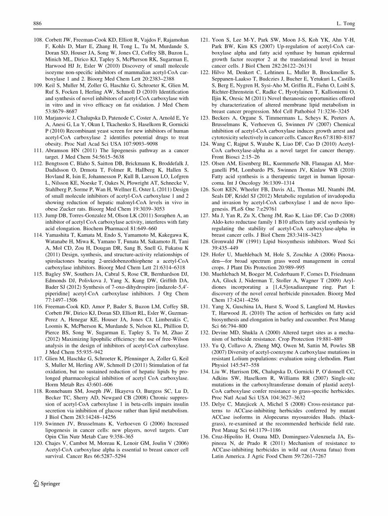
108. Corbett JW, Freeman-Cook KD, Elliott R, Vajdos F, Rajamohan
F, Kohls D, Marr E, Zhang H, Tong L, Tu M, Murdande S,
Doran SD, Houser JA, Song W, Jones CJ, Coffey SB, Buzon L,
Minich ML, Dirico KJ, Tapley S, McPherson RK, Sugarman E,
Harwood HJ Jr, Esler W (2010) Discovery of small molecule
isozyme non-specific inhibitors of mammalian acetyl-CoA car-
boxylase 1 and 2. Bioorg Med Chem Lett 20:2383–2388
109. Keil S, Muller M, Zoller G, Haschke G, Schroeter K, Glien M,
Ruf S, Focken I, Herling AW, Schmoll D (2010) Identification
and synthesis of novel inhibitors of acetyl-CoA carboxylase with
in vitro and in vivo efficacy on fat oxidation. J Med Chem
53:8679–8687
110. Marjanovic J, Chalupska D, Patenode C, Coster A, Arnold E, Ye
A, Anesi G, Lu Y, Okun I, Tkachenko S, Haselkorn R, Gornicki
P (2010) Recombinant yeast screen for new inhibitors of human
acetyl-CoA carboxylase 2 identifies potential drugs to treat
obesity. Proc Natl Acad Sci USA 107:9093–9098
111. Abramson HN (2011) The lipogenesis pathway as a cancer
target. J Med Chem 54:5615–5638
112. Bengtsson C, Blaho S, Saitton DB, Brickmann K, Broddefalk J,
Dadidsson O, Drmota T, Folmer R, Hallberg K, Hallen S,
Hovland R, Isin E, Johannesson P, Kull B, Larsson LO, Lofgren
L, Nilsson KE, Noeske T, Oakes N, Plowright AT, Schnecke V,
Stahlberg P, Sorme P, Wan H, Wellner E, Oster L (2011) Design
of small molecule inhibitors of acetyl-CoA carboxylase 1 and 2
showing reduction of hepatic malonyl-CoA levels in vivo in
obese Zucker rats. Bioorg Med Chem 19:3039–3053
113. Jump DB, Torres-Gonzalez M, Olson LK (2011) Soraphen A, an
inhibitor of acetyl CoA carboxylase activity, interferes with fatty
acid elongation. Biochem Pharmacol 81:649–660
114. Yamashita T, Kamata M, Endo S, Yamamoto M, Kakegawa K,
Watanabe H, Miwa K, Yamano T, Funata M, Sakamoto JI, Tani
A, Mol CD, Zou H, Dougan DR, Sang B, Snell G, Fukatsu K
(2011) Design, synthesis, and structure-activity relationships of
spirolactones bearing 2-ureidobenzothiophene a acetyl-CoA
carboxylase inhibitors. Bioorg Med Chem Lett 21:6314–6318
115. Bagley SW, Southers JA, Cabral S, Rose CR, Bernhardson DJ,
Edmonds DJ, Polivkova J, Yang X, Kung DW, Griffith DA,
Bader SJ (2012) Synthesis of 7-oxo-dihydrospiro [indazole-5,40-piperidine] acetyl-CoA carboxylase inhibitors. J Org Chem
77:1497–1506
116. Freeman-Cook KD, Amor P, Bader S, Buzon LM, Coffey SB,
Corbett JW, Dirico KJ, Doran SD, Elliott RL, Esler W, Guzman-
Perez A, Henegar KE, Houser JA, Jones CJ, Limberakis C,
Loomis K, McPherson K, Murdande S, Nelson KL, Phillion D,
Pierce BS, Song W, Sugarman E, Tapley S, Tu M, Zhao Z
(2012) Maximizing lipophilic efficiency: the use of free-Wilson
analysis in the design of inhibitors of acetyl-CoA carboxylase.
J Med Chem 55:935–942
117. Glien M, Haschke G, Schroeter K, Pfenninger A, Zoller G, Keil
S, Muller M, Herling AW, Schmoll D (2011) Stimulation of fat
oxidation, but no sustained reduction of hepatic lipids by pro-
longed pharmacological inhibition of acetyl CoA carboxylase.
Horm Metab Res 43:601–606
118. Ronnebaum SM, Joseph JW, Ilkayeva O, Burgess SC, Lu D,
Becker TC, Sherry AD, Newgard CB (2008) Chronic suppres-
sion of acetyl-CoA carboxylase 1 in beta-cells impairs insulin
secretion via inhibition of glucose rather than lipid metabolism.
J Biol Chem 283:14248–14256
119. Swinnen JV, Brusselmans K, Verhoeven G (2006) Increased
lipogenesis in cancer cells: new players, novel targets. Curr
Opin Clin Nutr Metab Care 9:358–365
120. Chajes V, Cambot M, Moreau K, Lenoir GM, Joulin V (2006)
Acetyl-CoA carboxylase alpha is essential to breast cancer cell
survival. Cancer Res 66:5287–5294
121. Yoon S, Lee M-Y, Park SW, Moon J-S, Koh YK, Ahn Y-H,
Park BW, Kim KS (2007) Up-regulation of acetyl-CoA car-
boxylase alpha and fatty acid synthase by human epidermal
growth factor receptor 2 at the translational level in breast
cancer cells. J Biol Chem 282:26122–26131
122. Hilvo M, Denkert C, Lehtinen L, Muller B, Brockmoller S,
Seppanen-Laakso T, Budczies J, Bucher E, Yetukuri L, Castillo
S, Berg E, Nygren H, Sysi-Aho M, Griffin JL, Fiehn O, Loibl S,
Richter-Ehrenstein C, Radke C, Hyotylainen T, Kallioniemi O,
Iljin K, Oresic M (2011) Novel theranostic opportunities offered
by characterization of altered membrane lipid metabolism in
breast cancer progression. Mol Cell Pathobiol 71:3236–3245
123. Beckers A, Organe S, Timmermans L, Scheys K, Peeters A,
Brusselmans K, Verhoeven G, Swinnen JV (2007) Chemical
inhibition of acetyl-CoA carboxylase induces growth arrest and
cytotoxicity selectively in cancer cells. Cancer Res 67:8180–8187
124. Wang C, Rajput S, Watabe K, Liao DF, Cao D (2010) Acetyl-
CoA carboxylase-alpha as a novel target for cancer therapy.
Front Biosci 2:15–26
125. Olsen AM, Eisenberg BL, Kuemmerle NB, Flanagan AJ, Mor-
ganelli PM, Lombardo PS, Swinnen JV, Kinlaw WB (2010)
Fatty acid synthesis is a therapeutic target in human liposar-
coma. Int J Oncology 36:1309–1314
126. Scott KEN, Wheeler FB, Davis AL, Thomas MJ, Ntambi JM,
Seals DF, Kridel SJ (2012) Metabolic regulation of invadopodia
and invasion by acetyl-CoA carboxylase 1 and de novo lipo-
genesis. PLoS One 7:e29761
127. Ma J, Yan R, Zu X, Cheng JM, Rao K, Liao DF, Cao D (2008)
Aldo-keto reductase family 1 B10 affects fatty acid synthesis by
regulating the stability of acetyl-CoA carboxylase-alpha in
breast cancer cells. J Biol Chem 283:3418–3423
128. Gronwald JW (1991) Lipid biosynthesis inhibitors. Weed Sci
39:435–449
129. Hofer U, Muehlebach M, Hole S, Zoschke A (2006) Pinoxa-
den—for broad spectrum grass weed management in cereal
crops. J Plant Dis Protection 20:989–995
130. Muehlebach M, Boeger M, Cederbaum F, Cornes D, Friedmann
AA, Glock J, Niderman T, Stoller A, Wagner T (2009) Aryl-
diones incorporating a [1,4,5]oxadiazepane ring. Part I:
discovery of the novel cereal herbicide pinoxaden. Bioorg Med
Chem 17:4241–4256
131. Yang X, Guschina IA, Hurst S, Wood S, Langford M, Hawkes
T, Harwood JL (2010) The action of herbicides on fatty acid
biosynthesis and elongation in barley and cucumber. Pest Manag
Sci 66:794–800
132. Devine MD, Shukla A (2000) Altered target sites as a mecha-
nism of herbicide resistance. Crop Protection 19:881–889
133. Yu Q, Collavo A, Zheng MQ, Owen M, Sattin M, Powles SB
(2007) Diversity of acetyl-coenzyme A carboxylase mutations in
resistant Lolium populations: evaluation using clethodim. Plant
Physiol 145:547–558
134. Liu W, Harrison DK, Chalupska D, Gornicki P, O’donnell CC,
Adkins SW, Haselkorn R, Williams RR (2007) Single-site
mutations in the carboxyltransferase domain of plastid acetyl-
CoA carboxylase confer resistance to grass-specific herbicides.
Proc Natl Acad Sci USA 104:3627–3632
135. Delye C, Matejicek A, Michel S (2008) Cross-resistance pat-
terns to ACCase-inhibiting herbicides conferred by mutant
ACCase isoforms in Alopecurus myosuroides Huds. (black-
grass), re-examined at the recommended herbicide field rate.
Pest Manag Sci 64:1179–1186
136. Cruz-Hipolito H, Osuna MD, Dominguez-Valenzuela JA, Es-
pinoza N, de Prado R (2011) Mechanism of resistance to
ACCase-inhibiting herbicides in wild oat (Avena fatua) from
Latin America. J Agric Food Chem 59:7261–7267
886 L. Tong
123
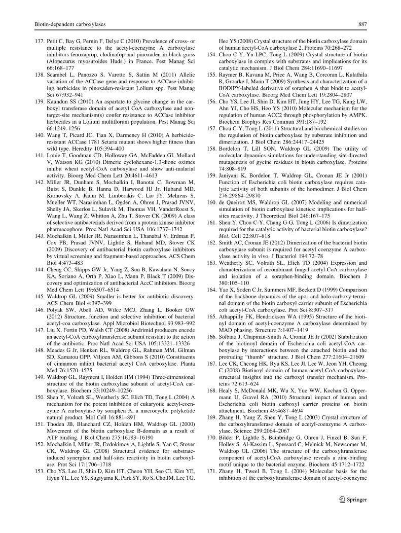
137. Petit C, Bay G, Pernin F, Delye C (2010) Prevalence of cross- or
multiple resistance to the acetyl-coenzyme A carboxylase
inhibitors fenoxaprop, clodinafop and pinoxaden in black-grass
(Alopecurus myosuroides Huds.) in France. Pest Manag Sci
66:168–177
138. Scarabel L, Panozzo S, Varotto S, Sattin M (2011) Allelic
variation of the ACCase gene and response to ACCase-inhibit-
ing herbicides in pinoxaden-resistant Lolium spp. Pest Manag
Sci 67:932–941
139. Kaundun SS (2010) An aspartate to glycine change in the car-
boxyl transferase domain of acetyl CoA carboxylase and non-
target-site mechanism(s) confer resistance to ACCase inhibitor
herbicides in a Lolium multiflorum population. Pest Manag Sci
66:1249–1256
140. Wang T, Picard JC, Tian X, Darmency H (2010) A herbicide-
resistant ACCase 1781 Setaria mutant shows higher fitness than
wild type. Heredity 105:394–400
141. Louie T, Goodman CD, Holloway GA, McFadden GI, Mollard
V, Watson KG (2010) Dimeric cyclohexane-1,3-dione oximes
inhibit wheat acetyl-CoA carboxylase and show anti-malarial
activity. Bioorg Med Chem Lett 20:4611–4613
142. Miller JR, Dunham S, Mochalkin I, Banotai C, Bowman M,
Buist S, Dunkle B, Hanna D, Harwood HJ Jr, Huband MD,
Karnovsky A, Kuhn M, Limberakis C, Liu JY, Mehrens S,
Mueller WT, Narasimhan L, Ogden A, Ohren J, Prasad JVNV,
Shelly JA, Skerlos L, Sulavik M, Thomas VH, VanderRoest S,
Wang L, Wang Z, Whitton A, Zhu T, Stover CK (2009) A class
of selective antibacterials derived from a protein kinase inhibitor
pharmacophore. Proc Natl Acad Sci USA 106:1737–1742
143. Mochalkin I, Miller JR, Narasimhan L, Thanabal V, Erdman P,
Cox PB, Prasad JVNV, Lightle S, Huband MD, Stover CK
(2009) Discovery of antibacterial biotin carboxylase inhibitors
by virtual screening and fragment-based approaches. ACS Chem
Biol 4:473–483
144. Cheng CC, Shipps GW Jr, Yang Z, Sun B, Kawahata N, Soucy
KA, Soriano A, Orth P, Xiao L, Mann P, Black T (2009) Dis-
covery and optimization of antibacterial AccC inhibitors. Bioorg
Med Chem Lett 19:6507–6514
145. Waldrop GL (2009) Smaller is better for antibiotic discovery.
ACS Chem Biol 4:397–399
146. Polyak SW, Abell AD, Wilce MCJ, Zhang L, Booker GW
(2012) Structure, function and selective inhibition of bacterial
acetyl-coa carboxylase. Appl Microbiol Biotechnol 93:983–992
147. Liu X, Fortin PD, Walsh CT (2008) Andrimid producers encode
an acetyl-CoA carboxyltransferase subunit resistant to the action
of the antibiotic. Proc Natl Acad Sci USA 105:13321–13326
148. Meades G Jr, Henken RL, Waldrop GL, Rahman MM, Gilman
SD, Kamatou GPP, Viljoen AM, Gibbons S (2010) Constituents
of cinnamon inhibit bacterial acetyl CoA carboxylase. Planta
Med 76:1570–1575
149. Waldrop GL, Rayment I, Holden HM (1994) Three-dimensional
structure of the biotin carboxylase subunit of acetyl-CoA car-
boxylase. Biochem 33:10249–10256
150. Shen Y, Volrath SL, Weatherly SC, Elich TD, Tong L (2004) A
mechanism for the potent inhibition of eukaryotic acetyl-coen-
zyme A carboxylase by soraphen A, a macrocyclic polyketide
natural product. Mol Cell 16:881–891
151. Thoden JB, Blanchard CZ, Holden HM, Waldrop GL (2000)
Movement of the biotin carboxylase B-domain as a result of
ATP binding. J Biol Chem 275:16183–16190
152. Mochalkin I, Miller JR, Evdokimov A, Lightle S, Yan C, Stover
CK, Waldrop GL (2008) Structural evidence for substrate-
induced synergism and half-sites reactivity in biotin carboxyl-
ase. Prot Sci 17:1706–1718
153. Cho YS, Lee JI, Shin D, Kim HT, Cheon YH, Seo CI, Kim YE,
Hyun YL, Lee YS, Sugiyama K, Park SY, Ro S, Cho JM, Lee TG,
Heo YS (2008) Crystal structure of the biotin carboxylase domain
of human acetyl-CoA carboxylase 2. Proteins 70:268–272
154. Chou C-Y, Yu LPC, Tong L (2009) Crystal structure of biotin
carboxylase in complex with substrates and implications for its
catalytic mechanism. J Biol Chem 284:11690–11697
155. Raymer B, Kavana M, Price A, Wang B, Corcoran L, Kulathila
R, Groarke J, Mann T (2009) Synthesis and characterization of a
BODIPY-labeled derivative of soraphen A that binds to acetyl-
CoA carboxylase. Bioorg Med Chem Lett 19:2804–2807
156. Cho YS, Lee JI, Shin D, Kim HT, Jung HY, Lee TG, Kang LW,
Ahn YJ, Cho HS, Heo YS (2010) Molecular mechanism for the
regulation of human ACC2 through phosphorylation by AMPK.
Biochem Biophys Res Commun 391:187–192
157. Chou C-Y, Tong L (2011) Structural and biochemical studies on
the regulation of biotin carboxylase by substrate inhibition and
dimerization. J Biol Chem 286:24417–24425
158. Bordelon T, Lill SON, Waldrop GL (2009) The utility of
molecular dynamics simulations for understanding site-directed
mutagenesis of gycine residues in biotin carboxylase. Proteins
74:808–819
159. Janiyani K, Bordelon T, Waldrop GL, Cronan JE Jr (2001)
Function of Escherichia coli biotin carboxylase requires cata-
lytic activity of both subunits of the homodimer. J Biol Chem
276:29864–29870
160. de Queiroz MS, Waldrop GL (2007) Modeling and numerical
simulation of biotin carboxylase kinetics: implications for half-
sites reactivity. J Theoretical Biol 246:167–175
161. Shen Y, Chou C-Y, Chang G-G, Tong L (2006) Is dimerization
required for the catalytic activity of bacterial biotin carboxylase?
Mol. Cell 22:807–818
162. Smith AC, Cronan JE (2012) Dimerization of the bacterial biotin
carboxylase subunit is required for acetyl coenzyme A carbox-
ylase activity in vivo. J Bacteriol 194:72–78
163. Weatherly SC, Volrath SL, Elich TD (2004) Expression and
characterization of recombinant fungal acetyl-CoA carboxylase
and isolation of a soraphen-binding domain. Biochem J
380:105–110
164. Yao X, Soden C Jr, Summers MF, Beckett D (1999) Comparison
of the backbone dynamics of the apo- and holo-carboxy-termi-
nal domain of the biotin carboxyl carrier subunit of Escherichia
coli acetyl-CoA carboxylase. Prot Sci 8:307–317
165. Athappilly FK, Hendrickson WA (1995) Structure of the bioti-
nyl domain of acetyl-coenzyme A carboxylase determined by
MAD phasing. Structure 3:1407–1419
166. Solbiati J, Chapman-Smith A, Cronan JE Jr (2002) Stabilization
of the biotinoyl domain of Escherichia coli acetyl-CoA car-
boxylase by interactions between the attached biotin and the
protruding ‘‘thumb’’ structure. J Biol Chem 277:21604–21609
167. Lee CK, Cheong HK, Ryu KS, Lee JI, Lee W, Jeon YH, Cheong
C (2008) Biotinoyl domain of human acetyl-CoA carboxylase:
structural insights into the carboxyl transfer mechanism. Pro-
teins 72:613–624
168. Healy S, McDonald MK, Wu X, Yue WW, Kochan G, Opper-
mann U, Gravel RA (2010) Structural impact of human and
Escherichia coli biotin carboxyl carrier proteins on biotin
attachment. Biochem 49:4687–4694
169. Zhang H, Yang Z, Shen Y, Tong L (2003) Crystal structure of
the carboxyltransferase domain of acetyl-coenzyme A carbox-
ylase. Science 299:2064–2067
170. Bilder P, Lightle S, Bainbridge G, Ohren J, Finzel B, Sun F,
Holley S, Al-Kassim L, Spessard C, Melnick M, Newcomer M,
Waldrop GL (2006) The structure of the carboxyltransferase
component of acetyl-CoA carboxylase reveals a zinc-binding
motif unique to the bacterial enzyme. Biochem 45:1712–1722
171. Zhang H, Tweel B, Tong L (2004) Molecular basis for the
inhibition of the carboxyltransferase domain of acetyl-coenzyme
Biotin-dependent carboxylases 887
123
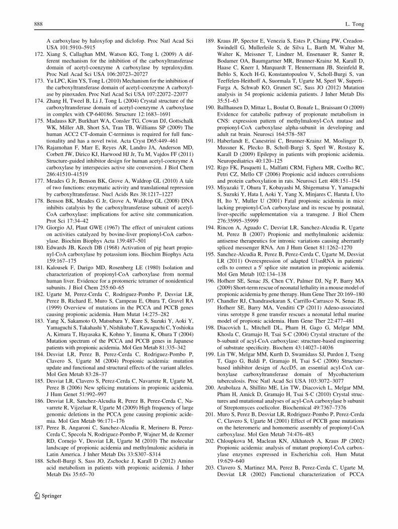
A carboxylase by haloxyfop and diclofop. Proc Natl Acad Sci
USA 101:5910–5915
172. Xiang S, Callaghan MM, Watson KG, Tong L (2009) A dif-
ferent mechanism for the inhibition of the carboxyltransferase
domain of acetyl-coenzyme A carboxylase by tepraloxydim.
Proc Natl Acad Sci USA 106:20723–20727
173. Yu LPC, Kim YS, Tong L (2010) Mechanism for the inhibition of
the carboxyltransferase domain of acetyl-coenzyme A carboxyl-
ase by pinoxaden. Proc Natl Acad Sci USA 107:22072–22077
174. Zhang H, Tweel B, Li J, Tong L (2004) Crystal structure of the
carboxyltransferase domain of acetyl-coenzyme A carboxylase
in complex with CP-640186. Structure 12:1683–1691
175. Madauss KP, Burkhart WA, Consler TG, Cowan DJ, Gottschalk
WK, Miller AB, Short SA, Tran TB, Williams SP (2009) The
human ACC2 CT-domain C-terminus is required for full func-
tionality and has a novel twist. Acta Cryst D65:449–461
176. Rajamohan F, Marr E, Reyes AR, Landro JA, Anderson MD,
Corbett JW, Dirico KJ, Harwood HJ Jr, Tu M, Vajdos FF (2011)
Structure-guided inhibitor design for human acetyl-coenzyme A
carboxylase by interspecies active site conversion. J Biol Chem
286:41510–41519
177. Meades G Jr, Benson BK, Grove A, Waldrop GL (2010) A tale
of two functions: enzymatic activity and translational repression
by carboxyltransferase. Nucl Acids Res 38:1217–1227
178. Benson BK, Meades G Jr, Grove A, Waldrop GL (2008) DNA
inhibits catalysis by the carboxyltransferase subunit of acetyl-
CoA carboxylase: implications for active site communication.
Prot Sci 17:34–42
179. Giorgio AJ, Plaut GWE (1967) The effect of univalent cations
on activities catalyzed by bovine-liver propionyl-CoA carbox-
ylase. Biochim Biophys Acta 139:487–501
180. Edwards JB, Keech DB (1968) Activation of pig heart propio-
nyl-CoA carboxylase by potassium ions. Biochim Biophys Acta
159:167–175
181. Kalousek F, Darigo MD, Rosenberg LE (1980) Isolation and
characterization of propionyl-CoA carboxylase from normal
human liver. Evidence for a protomeric tetramer of nonidentical
subunits. J Biol Chem 255:60–65
182. Ugarte M, Perez-Cerda C, Rodriguez-Pombo P, Desviat LR,
Perez B, Richard E, Muro S, Campeau E, Ohura T, Gravel RA
(1999) Overview of mutations in the PCCA and PCCB genes
causing propionic acidemia. Hum Mutat 14:275–282
183. Yang X, Sakamoto O, Matsubara Y, Kure S, Suzuki Y, Aoki Y,
Yamaguchi S, Takahashi Y, Nishikubo T, Kawaguchi C, Yoshioka
A, Kimura T, Hayasaka K, Kohno Y, Iinuma K, Ohura T (2004)
Mutation spectrum of the PCCA and PCCB genes in Japanese
patients with propionic acidemia. Mol Gen Metab 81:335–342
184. Desviat LR, Perez B, Perez-Cerda C, Rodriguez-Pombo P,
Clavero S, Ugarte M (2004) Propionic acidemia: mutation
update and functional and structural effects of the variant alleles.
Mol Gen Metab 83:28–37
185. Desviat LR, Clavero S, Perez-Cerda C, Navarrete R, Ugarte M,
Perez B (2006) New splicing mutations in propionic acidemia.
J Hum Genet 51:992–997
186. Desviat LR, Sanchez-Alcudia R, Perez B, Perez-Cerda C, Na-
varrete R, Vijzelaar R, Ugarte M (2009) High frequency of large
genomic deletions in the PCCA gene causing propionic acide-
mia. Mol Gen Metab 96:171–176
187. Perez B, Angaroni C, Sanchez-Alcudia R, Merinero B, Perez-
Cerda C, Specola N, Rodriguez-Pombo P, Wajner M, de Kremer
RD, Cornejo V, Desviat LR, Ugarte M (2010) The molecular
landscape of propionic acidemia and methylmalonic aciduria in
Latin America. J Inher Metab Dis 33:S307–S314
188. Scholl-Burgi S, Sass JO, Zschocke J, Karall D (2012) Amino
acid metabolism in patients with propionic acidemia. J Inher
Metab Dis 35:65–70
189. Kraus JP, Spector E, Venezia S, Estes P, Chiang PW, Creadon-
Swindell G, Mullerleile S, de Silva L, Barth M, Walter M,
Walter K, Meissner T, Lindner M, Ensenauer R, Santer R,
Bodamer OA, Baumgartner MR, Brunner-Krainz M, Karall D,
Haase C, Knerr I, Marquardt T, Hennermann JB, Steinfeld R,
Beblo S, Koch H-G, Konstantopoulou V, Scholl-Burgi S, van
Teeffelen-Heithoff A, Suormala T, Ugarte M, Sperl W, Superti-
Furga A, Schwab KO, Grunert SC, Sass JO (2012) Mutation
analysis in 54 propionic acidemia patients. J Inher Metab Dis
35:51–63
190. Ballhausen D, Mittaz L, Boulat O, Bonafe L, Braissant O (2009)
Evidence for catabolic pathway of propionate metabolism in
CNS: expression pattern of methylmalonyl-CoA mutase and
propionyl-CoA carboxylase alpha-subunit in developing and
adult rat brain. Neurosci 164:578–587
191. Haberlandt E, Canestrini C, Brunner-Krainz M, Moslinger D,
Mussner K, Plecko B, Scholl-Burgi S, Sperl W, Rostasy K,
Karall D (2009) Epilepsy in patients with propionic acidemia.
Neuropediatrics 40:120–125
192. Rigo FK, Pasquetti L, Malfatti CRM, Fighera MR, Coelho RC,
Petri CZ, Mello CF (2006) Propionic acid induces convulsions
and protein carboxylation in rats. Neurosci Lett 408:151–154
193. Miyazaki T, Ohura T, Kobayashi M, Shigematsu Y, Yamaguchi
S, Suzuki Y, Hata I, Aoki Y, Yang X, Minjares C, Haruta I, Uto
H, Ito Y, Muller U (2001) Fatal propionic acidemia in mice
lacking propionyl-CoA carboxylase and its rescue by postnatal,
liver-specific supplementation via a transgene. J Biol Chem
276:35995–35999
194. Rincon A, Aguado C, Desviat LR, Sanchez-Alcudia R, Ugarte
M, Perez B (2007) Propionic and methylmalonic acidemia:
antisense therapeutics for intronic variations causing aberrantly
spliced messenger RNA. Am J Hum Genet 81:1262–1270
195. Sanchez-Alcudia R, Perez B, Perez-Cerda C, Ugarte M, Desviat
LR (2011) Overexpression of adapted U1snRNA in patients’
cells to correct a 50 splice site mutation in propionic acidemia.
Mol Gen Metab 102:134–138
196. Hofherr SE, Senac JS, Chen CY, Palmer DJ, Ng P, Barry MA
(2009) Short-term rescue of neonatal lethality in a mouse model of
propionic acidemia by gene therapy. Hum Gene Ther 20:169–180
197. Chandler RJ, Chandrasekaran S, Carrillo-Carrasco N, Senac JS,
Hofherr SE, Barry MA, Venditti CP (2011) Adeno-associated
virus serotype 8 gene transfer rescues a neonatal lethal murine
model of propionic acidemia. Hum Gene Ther 22:477–481
198. Diacovich L, Mitchell DL, Pham H, Gago G, Melgar MM,
Khosla C, Gramajo H, Tsai S-C (2004) Crystal structure of the
b-subunit of acyl-CoA carboxylase: structure-based engineering
of substrate specificity. Biochem 43:14027–14036
199. Lin TW, Melgar MM, Kurth D, Swamidass SJ, Purdon J, Tseng
T, Gago G, Baldi P, Gramajo H, Tsai S-C (2006) Structure-
based inhibitor design of AccD5, an essential acyl-CoA car-
boxylase carboxyltransferase domain of Mycobacterium
tuberculosis. Proc Natl Acad Sci USA 103:3072–3077
200. Arabolaza A, Shillito ME, Lin TW, Diacovich L, Melgar MM,
Pham H, Amick D, Gramajo H, Tsai S-C (2010) Crystal struc-
tures and mutational analyses of acyl-CoA carboxylase b subunit
of Streptomyces coelicolor. Biochemical 49:7367–7376
201. Muro S, Perez B, Desviat LR, Rodriguez-Pombo P, Perez-Cerda
C, Clavero S, Ugarte M (2001) Effect of PCCB gene mutations
on the heteromeric and homomeric assembly of propionyl-CoA
carboxylase. Mol Gen Metab 74:476–483
202. Chloupkova M, Maclean KN, Alkhateeb A, Kraus JP (2002)
Propionic acidemia: analysis of mutant propionyl-CoA carbox-
ylase enzymes expressed in Escherichia coli. Hum Mutat
19:629–640
203. Clavero S, Martinez MA, Perez B, Perez-Cerda C, Ugarte M,
Desviat LR (2002) Functional characterization of PCCA
888 L. Tong
123

mutations causing propionic acidemia. Biochim Biophys Acta
1588:119–125
204. Perez-Cerda C, Clavero S, Perez B, Rodriguez-Pombo P, Des-
viat LR, Ugarte M (2003) Functional analysis of PCCB
mutations causing propionic acidemia based on expression
studies in deficient human skin fibroblasts. Biochim Biophys
Acta 1638:43–49
205. Sloane V, Waldrop GL (2004) Kinetic characterization of
mutations found in propionic acidemia and methylcrotonylgly-
cinuria. J Biol Chem 279:15772–15778
206. Jiang H, Rao KS, Yee VC, Kraus JP (2005) Characterization of
four variant forms of human propionyl-CoA carboxylase
expressed in Escherichia coli. J Biol Chem 280:27719–27727
207. Rodriguez-Pombo P, Perez-Cerda C, Perez B, Desviat LR,
Sanchez-Pulido L, Ugarte M (2005) Towards a model to explain
the intragenic complementation in the heteromultimeric pro-
tein propionyl-CoA carboxylase. Biochim Biophys Acta
1740:489–498
208. Diaz-Perez AL, Zavala-Hernandez AN, Cervantes C, Campos-
Garcia J (2004) The gny RDBHAL cluster is involved in acyclic
isoprenoid degradation in Pseudomonas aeruginosa. Appl
Environ Microbiol 70:5102–5110
209. Hoschle B, Gnau V, Jendrossek D (2005) Methylcrotonyl-CoA
and geranyl-CoA carboxylases are involved in leucine/isoval-
erate utilization (Liu) and acyclic terpene utilization (Atu), and
are encoded by liuB/liuD and atuC/atuF, in Pseudomonasaeruginosa. Microbiol 151:3649–3656
210. Forster-Fromme K, Hoschle B, Mack C, Bott M, Armbruster W,
Jendrossek D (2006) Identification of genes and proteins nec-
essary for catabolism of acyclic terpenes and leucine/isovalerate
in Pseudomonas aeruginosa. Appl Environmental Microbiol
72:4819–4828
211. Aguilar JA, Zavala AN, Diaz-Perez C, Cervantes C, Diaz-Perez
AL, Campos-Garcia J (2006) The atu and liu clusters are
involved in the catabolic pathways for acyclic monoterpenes and
leucine in Pseudomonas aeruginosa. Appl Environmental
Microbiol 72:2070–2079
212. Aguilar JA, Diaz-Perez C, Diaz-Perez AL, Rodriguez-Zavala JS,
Nikolau BJ, Campos-Garcia J (2008) Substrate specificity of the
3-methylcrotonyl coenzyme A (CoA) and geranyl-CoA car-
boxylases from Pseudomonas aeruginosa. J Bacteriol 190:
4888–4893
213. Ding G, Che P, Ilarslan H, Wurtele ES, Nikolau BJ (2012)
Genetic dissection of methylcrotonyl CoA carboxylase indicates
a complex role for mitochondrial leucine catabolism during seed
development and germination. Plant J 70(4):562–577
214. Tomaszycki ML, Peabody C, Replogle K, Clayton DF, Temp-
elman RJ, Wade J (2009) Sexual differentiation of the zebra
finch song system: potential roles for sex chromosome genes.
BMC Neurosci 10:24
215. Stadler SC, Polanetz R, Meier S, Mayerhofer PU, Herrmann JM,
Anslinger K, Roscher AA, Roschinger W, Holzinger A (2005)
Mitochondrial targeting signals and mature peptides of
3-methylcrotonyl-CoA carboxylase. Biochem Biophys Res
Commun 334:939–946
216. Chu C-H, Cheng D (2007) Expression, purification, character-
ization of human 3-methylcrotonyl-CoA carboxylase (MCCC).
Prot Expr Purif 53:421–427
217. Baumgartner MR, Almashanu S, Suormala T, Obie C, Cole RN,
Packman S, Baumgartner ER, Valle D (2001) The molecular
basis of human 3-methylcrotonyl-CoA carboxylase deficiency.
J Clin Investig 107:495–504
218. Gallardo ME, Desviat LR, Rodriguez JM, Esparza-Gordillo J,
Perez-Cerda C, Perez B, Rodriguez-Pombo P, Criado O, Sanz R,
Morton DH, Gibson KM, Le TP, Ribes A, Rodriguez de Cor-
doba S, Ugarte M, Penalva MA (2001) The molecular basis of
3-methylcrotonylglycinuria, a disorder of leucine metabolism.
Am J Hum Genet 68:334–346
219. Holzinger A, Roschinger W, Lagler F, Mayerhofer PU, Lichtner P,
Kattenfeld T, Thuy LP, Nyhan WL, Koch H-G, Muntau AC, Ro-
scher AA (2001) Cloning of the human MCCA and MCCB genes
and mutations therein reveal the molecular cause of 3-methylcro-
tonyl-CoA carboxylase deficiency. Human Mol Gen 10:1299–1306
220. Desviat LR, Perez-Cerda C, Perez B, Esparza-Gordillo J,
Rodriguez-Pombo P, Penalva MA, Rodriguez de Cordoba S,
Ugarte M (2003) Functional analysis of MCCA and MCCB
mutations causing methylcrotonylglycinuria. Mol Gen Metab
80:315–320
221. Stadler SC, Polanetz R, Maier EM, Heidenreich SC, Niederer B,
Mayerhofer PU, Lagler F, Koch H-G, Santer R, Fletcher JM,
Ranieri E, Das AM, Spiekerkotter U, Schwab KO, Potzsch S,
Marquardt I, Hennermann JB, Knerr I, Mercimek-Mahmutoglu
S, Kohlschmidt N, Liebl B, Fingerhut R, Olgemoller B, Muntau
AC, Roscher AA, Roschinger W (2006) Newborn screening for
3-methylcrotonyl-CoA carboxylase deficiency: population het-
erogeneity of MCCA and MCCB mutations and impact on risk
assessment. Hum Mutat 27:748–759
222. Uematsu M, Sakamoto O, Sugawara N, Kumagai N, Morimoto
T, Yamaguchi S, Hasegawa Y, Kobayashi H, Ihara K, Yoshino
M, Watanabe Y, Inokuchi T, Yokoyama T, Kiwaki K, Na-
kamura K, Endo F, Tsuchiya S, Ohura T (2007) Novel mutations
in five Japanese patients with 3-methylcrotonyl-CoA carboxyl-
ase deficiency. J Hum Genet 52:1040–1043
223. Stucki M, Suormala T, Fowler B, Valle D, Baumgartner MR
(2009) Cryptic exon activation by disruption of exon splice
enhancer. Novel mechanism causing 3-methylcrotonyl-CoA
carboxylase deficiency. J Biol Chem 284:28953–28957
224. Nguyen KV, Naviaux RK, Patra S, Barshop BA, Nyhan WL
(2011) Novel mutations in the human MCCA and MCCB gene
causing methylcrotonylglycinuria. Mol Gen Metab 102:218–221
225. Morscher RJ, Grunert SC, Burer C, Burda P, Suormala T,
Fowler B, Baumgartner MR (2012) A single mutation in
MCCC1 or MCCC2 as a potential cause of positive screening
for 3-methylcrotonyl-CoA carboxylase deficiency. Mol Gen
Metab 105:602–606
226. Jung CW, Lee BH, Kim JH, Kim GH, Lee J, Choi JH, Yoo HW
(2012) Uneventful clinical courses of Korean patients with
methylcrotonylglycinuria and their common mutations. J Hum
Genet 57:62–64
227. Forster-Fromme K, Jendrossek D (2010) AtuR is a repressor of
acyclic terpene utilization (Atu) gene cluster expression and
specifically binds two 13 bp inverted repeat sequences of the
atuA-atuR intergenic region. FEMS Microbiol Lett 308:166–174
228. Guan X, Diez T, Prasad TK, Nikolau BJ, Wurtele ES (1999)
Geranoyl-CoA carboxylase: a novel biotin-containing enzyme in
plants. Arch Biochem Biophys 362:12–21
229. Demirev AV, Khanal A, Hanh NPK, Nam KT, Nam DH (2011)
Biochemical characterization of propionyl-coenzyme A car-
boxylase complex of Streptomyces toxytricini. J Microbiol
49:407–412
230. Gago G, Kurth D, Diacovich L, Tsai S-C, Gramajo H (2006)
Biochemical and structural characterization of an essential acyl-
coenzyme A carboxylase from Mycobacterium tuberculosis.
J Bacteriol 188:477–486
231. Daniel J, Oh TJ, Lee CM, Kolattukudy PE (2007) AccD6, a
member of the Fas II locus, is a functional carboxyltransferase
subunit of the acyl-coenzyme A carboxylase in Mycobacterium
tuberculosis. J Bacteriol 189:911–917
232. Diacovich L, Peiru S, Kurth D, Rodriguez E, Podesta F, Khosla
C, Gramajo H (2002) Kinetic and structural analysis of a new
group of acyl-CoA carboxylases found in Streptomyces coeli-
color A3(2). J Biol Chem 277:31228–31236
Biotin-dependent carboxylases 889
123

233. Oh TJ, Daniel J, Kim HJ, Sirakova TD, Kolattukudy PE (2006)
Identification and characterization of Rv3281 as a novel subunit
of a biotin-dependent acyl-CoA carboxylase in Mycobacterium
tuberculosis H37Rv. J Biol Chem 281:3899–3908
234. Kurth DG, Gago GM, de la Iglesia A, Lyonnet BB, Lin TW,
Morbidoni HR, Tsai S-C, Gramajo H (2009) ACCase 6 is the
essential acetyl-CoA carboxylase involved in fatty acid and
mycolic acid biosynthesis in mycobacteria. Microbiol 155:
2664–2675
235. Gande R, Dover LG, Krumbach K, Besra GS, Sahm H, Oikawa
T, Eggeling L (2007) The two carboxylases of Corynebacterium
glutamicum essential for fatty acid and mycolic acid synthesis.
J Bacteriol 189:5257–5264
236. Pawelczyk J, Brzostek A, Kremer L, Dziadek B, Rumijowska-
Galewicz A, Fiolka M, Dziadek J (2011) AccD6, a key carb-
oxyltransferase essential for mycolic acid synthesis in
Mycobacterium tuberculosis, is dispensable in a nonpathogenic
strain. J Bacteriol 193:6960–6972
237. Holton SJ, King-Scott S, Eddine AN, Kaufmann SHE, Wil-
manns M (2006) Structural diversity in the six-fold redundant
set of acyl-CoA carboxyltransferases in Mycobacterium tuber-
culosis. FEBS Lett 580:6898–6902
238. Dunn MF, Araiza G, Mora J (2004) Biochemical characteriza-
tion of a Rhizobium etli monovalent cation-stimulated acyl-
coenzyme A carboxylase with a high substrate specficity con-
stant for propionyl-coenzyme A. Microbiol 150:399–406
239. Attwood PV (1995) The structure and the mechanism of action
of pyruvate carboxylase. Int J Biochem Cell Biol 27:231–249
240. Wallace JC, Jitrapakdee S, Chapman-Smith A (1998) Pyruvate
carboxylase. Int J Biochem Cell Biol 30:1–5
241. Jitrapakdee S, Wallace JC (1999) Structure, function and regu-
lation of pyruvate carboxylase. Biochem J 340:1–16
242. Jitrapakdee S, Vidal-Puig A, Wallace JC (2006) Anaplerotic
roles of pyruvate carboxylase in mammalian tissues. Cell Mol
Life Sci 63:843–854
243. Wallace JC (2010) My favorite pyruvate carboxylase. IUBMB
Life 62:535–538
244. Owen OE, Kalhan SC, Hanson RW (2002) The key role of
anaplerosis and cataplerosis for citric acid cycle function. J Biol
Chem 277:30409–30412
245. Cheng T, Sudderth J, Yang C, Mullen AR, Jin ES, Mates JM,
DeBerardinis RJ (2011) Pyruvate carboxylase is required for
glutamine-independent growth of tumor cells. Proc Natl Acad
Sci USA 108:8674–8679
246. Xu J, Han J, Long YS, Epstein PN, Liu YQ (2008) The role of
pyruvate carboxylase in insulin secretion and proliferation in rat
pancreatic beta cells. Diabetologia 51:2022–2030
247. Hasan NM, Longacre MJ, Stoker SW, Boonsaen T, Jitrapakdee
S, Kendrick MA, Wallace JC, MacDonald MJ (2008) Impaired
anaplerosis and insulin secretion in insulinoma cells caused by
small interfering RNA-mediated suppression of pyruvate car-
boxylase. J Biol Chem 283:28048–28059
248. MacDonald MJ, Longacre MJ, Stoker SW, Kendrick M, Thon-
pho A, Brown LJ, Hasan NM, Jitrapakdee S, Fukao T, Hanson
MS, Fernandez LA, Odorico J (2011) Differences between
human and rodent pancreatic islets. Low pyruvate carboxylase,
ATP citrate lyase, and pyruvate carboxylation and high glucose-
stimulated acetoacetate in human pancreatic islets. J Biol Chem
286:18383–18396
249. Ozimek P, van Dijk R, Latchev K, Gancedo C, Wang DY, van
der Klei IJ, Veenhuis M (2003) Pyruvate carboxylase is an
essential protein in the assembly of yeast peroximal oligomeric
alcohol oxidase. Mol Biol Cell 14:786–797
250. Klompmaker SH, Kilic A, Baerends RJ, Veenhuis M, van der
Klei IJ (2010) Activation of a peroximal Pichia pastoris
D-amino acid oxidase, which uses D-alanine as a preferred
substrate, depends on pyruvate carboxylase. FEMS Yeast Res
10:708–716
251. Ozimek PZ, Klompmaker SH, Visser N, Veenhuis M, van der
Klei IJ (2007) The transcarboxylase domain of pyruvate car-
boxylase is essential for assembly of the peroxisomal
flavoenzyme alcohol oxidase. FEMS Yeast Res 7:1082–1092
252. Huberts DHEW, Venselaar H, Vriend G, Veenhuis M, van der
Klei IJ (2010) The moonlighting function of pyruvate carbox-
ylase resides in the non-catalytic end of the TIM barrel. Biochim
Biophys Acta 1803:1038–1042
253. Schar J, Stoll R, Schauer K, Loeffler DIM, Eylert E, Joseph B,
Eisenreich W, Fuchs TM, Goebel W (2010) Pyruvate carbox-
ylase plays a crucial role in carbon metabolism of extra- and
intracellularly replicating Listeria monocytogenes. J Bacteriol
192:1774–1784
254. Lai H, Kraszewski JL, Purwantini E, Mukhopadhyay B (2006)
Identification of pyruvate carboxylase genes in Pseudomonas
aeruginosa PAO1 and development of a P. aeruginosa-based
overexpression system for a4- and a4b4-type pyruvate carbox-
ylases. Appl Environ Microbiol 72:7785–7792
255. Zeczycki TN, Menefee AL, Jitrapakdee S, Wallace JC, Attwood
PV, St. Maurice M, Cleland WW (2011) Activation and inhibition
of pyruvate carboxylase from Rhizobium etli. Biochemical
50:9694–9707
256. Adina-Zada A, Zeczycki TN, Attwood PV (2012) Regulation of
the structure and activity of pyruvate carboxylase by acetyl
CoA. Arch Biochem Biophys 519:118–130
257. Adina-Zada A, Hazra R, Sereeruk C, Jitrapakdee S, Zeczycki
TN, St. Maurice M, Cleland WW, Wallace JC, Attwood PV
(2011) Probing the allosteric activation of pyruvate carboxylase
using 20,30-O-(2,4,6-trinitrophenyl) adenosine 50-triphosphate as
a fluorescent mimic of the allosteric activator acetyl CoA. Arch
Biochem Biophys 509:117–126
258. Zeczycki TN, St. Maurice M, Attwood PV (2010) Inhibitors of
pyruvate carboxylase. Open Enzyme Inhib J 3:8–26
259. Carbone MA, MacKay N, Ling M, Cole DEC, Douglas C, Rigat
B, Feigenbaum A, Clarke JTR, Haworth JC, Greenberg CR,
Seargeant L, Robinson BH (1998) Amerindian pyruvate car-
boxylase deficiency is associated with two distinct missense
mutations. Am J Hum Genet 62:1312–1319
260. Wexler ID, Kerr DS, Du Y, Kaung MM, Stephenson W, Lusk
MM, Wappner RS, Higgins JJ (1998) Molecular characteriza-
tion of pyruvate carboxylase deficiency in two consanguineous
families. Pediatr Res 43:579–584
261. Robinson BH (2006) Lactic acidemia and mitochondrial disease.
Mol Gen Metab 89:3–13
262. Wang D, Yang H, de Braganca KC, Lu J, Shih LY, Briones P,
Lang T, de Vivo DC (2008) The molecular basis of pyruvate
carboxylase deficiency: mosaicism correlates with prolonged
survival. Mol Gen Metab 95:31–38
263. Monnot S, Serre V, Chadefaux-Vekemans B, Aupetit J, Romano
S, de Lonlay P, Rival JM, Munnich A, Steffann J, Bonnefont JP
(2009) Structural insights on pathogenic effects of novel muta-
tions causing pyruvate carboxylase deficiency. Hum Mutat
30:734–740
264. Marin-Valencia I, Roe CR, Pascual JM (2010) Pyruvate car-
boxylase deficiency: mechanisms, mimics and anaplerosis. Mol
Gen Metab 101:9–17
265. Lietzan AD, Menefee AL, Zeczycki TN, Kumar S, Attwood PV,
Wallace JC, Cleland WW, St. Maurice M (2011) Interaction
between the biotin carboxyl carrier domain and the biotin car-
boxylase domain in pyruvate carboxylase from Rhizobium etli.Biochemical 50:9708–9723
266. Yu LPC, Xiang S, Lasso G, Gil D, Valle M, Tong L (2009) A
symmetrical tetramer for S. aureus pyruvate carboxylase in
complex with coenzyme A. Structure 17:823–832
890 L. Tong
123

267. Balsera M, Buey RM, Li X-D (2011) Quaternary structure of the
oxaloacetate decarboxylase membrane complex and mechanistic
relationships to pyruvate carboxylase. J Biol Chem 286:
9457–9467
268. Zeczycki TN, St. Maurice M, Jitrapakdee S, Wallace JC, Att-
wood PV, Cleland WW (2009) Insight into the carboxyl
transferase domain mechanism of pyruvate carboxylase from
Rhizobium etli. Biochemical 48:4305–4313
269. Duangpan S, Jitrapakdee S, Adina-Zada A, Byrne L, Zeczycki
TN, St. Maurice M, Cleland WW, Wallace JC, Attwood PV
(2010) Probing the catalytic roles of Arg548 and Gln552 in the
carboxyl transferase domain of the Rhizobium etli pyruvate
carboxylase by site-directed mutagenesis. Biochemical
49:3296–3304
270. Zeczycki TN, Menefee AL, Adina-Zada A, Jitrapakdee S, Su-
rinya KH, Wallace JC, Attwood PV, St Maurice M, Cleland
WW (2011) Novel insights into the biotin carboxylase domain
reactions of pyruvate carboxylase from Rhizobium etli. Bio-
chemical 50:9724–9737
271. Lasso G, Yu LPC, Gil D, Xiang S, Tong L, Valle M (2010)
Cryo-EM analysis reveals new insights into the mechanism of
action of pyruvate carboxylase. Structure 18:1300–1310
272. Kanamori T, Kanou N, Atomi H, Imanaka T (2004) Enzymatic
characterization of a prokaryotic urea carboxylase. J Bacteriol
186:2532–2539
273. Andersen G, Bjornberg O, Polakova S, Pynyaha Y, Rasmussen
A, Moller K, Hofer A, Moritz T, Sandrini MPB, Merico A-M,
Compagno C, Akerlund HE, Gojkovic Z, Piskur J (2008) A
second pathway to degrade pyrimidine nucleic acid precursors
in eukaryotes. J Mol Biol 380:656–666
274. Ghosh S, Navarathna DHMLP, Roberts DD, Cooper JT, Atkin
AL, Petro TM, Nickerson KW (2009) Arginine-induced germ
tube formation in Candida albicans is essential for escape from
murine macrophage line RAW 264.7. Infection Immunity 77:
1596–1605
275. Collette JR, Lorenz MC (2011) Mechanisms of immune evasion
in fungal pathogens. Curr Opin Microbiol 14:668–675
276. Jacques DA, Langley DB, Hynson RMG, Whitten AE, Kwan A,
Guss JM, Trewhella J (2011) A novel structure of an antikinase
and its inhibitor. J Mol Biol 405:214–226
277. Jacques DA, Langley DB, Kuramitsu S, Yokoyama S, Trewhella
J, Guss JM (2011) The structure of TTHA0988 from Thermus
thermophilus, a KipI-KipA homolgue incorrectly annotated as
an allophanate hydrolase. Acta Cryst D67:105–111
Biotin-dependent carboxylases 891
123