structure and development of the cryptomonad periplast: a
TRANSCRIPT

Protoplasma (1994) 181: 106-122
Springer-Verlag 1994 Printed in Austria
Structure and development of the cryptomonad periplast:
S. J. Brett**, L. Perasso +, and R. Wetherbee*
School of Botany, University of Melbourne, Parkville, Victoria
Received March 30, 1994 Accepted April 30, 1994
a review
Summary. The structure and development of the complex periplast, or cell covering, of cryptomonads is reviewed. The periplast consists of the plasma membrane (PM) plus an associated surface periplast component (SPC) and cytoplasmic or inner periplast component (IPC). The structure of the SPC and IPC, and their association with the PM, varies considerably between genera. This review, which concentrates on cryptomonads with an IPC of discrete plates, dis- cusses relationships between periplast components and examines the development of this unique cell covering. Formation and growth of inner plates occurs throughout the cell cycle from specialized regions termed anamorphic zones. Crystalline surface plates, which comprise the SPC in many cryptomonad species, appear to form by self- assembly of disorganized subunits. In Komma caudata the subunits are composed of a high molecular weight glycoprotein that is pro- duced within the endomembrane system and deposited onto the cell surface within anamorphic zones. The self-assembly of subunits into highly ordered surface plates appears closely associated with devel- opmental changes in the underlying IPC and PM.
Keywords: Cryptophyceae; Cell wall; Periplast; Self-assembly; Freeze fracture-freeze etch.
Introduction
The c r y p t o m o n a d s are a unique g roup of phytof iagel -
lares, easily d is t inguished f rom o ther mic roa lgae by
their asymmetr ic shape. The dist inct ive appea rance of
these cells can be a t t r ibu ted , in par t , to the possess ion
o f a subapica l depress ion ( termed the ves t ibulum)
which m a y extend in ternal ly to form a gullet (Santore
1987; Hil l and Wetherbee 1990; Hill 1991 a, b) or p ro-
* Correspondence and reprints: School of Botany, University of Melbourne, Parkville, Vic 3052, Australia. ** Present address: Bigelow Laboratory for Ocean Sciences, W. Boothbay Harbor, Maine, U.S.A. + Present address: Botanisches Institut, Universit~it zu K61n, Fed- eral Republic of Germany.
gress a long the ventra l surface into a fur row ( M u n a w a r
and Bistr icki 1979, Klaveness 1981, 1985, Kugrens et al.
1986). The ves t ibu lum/fur row/gu l le t complex forms an
i m p o r t a n t surface feature o f all c r y p t o m o n a d cells, and
three m a j o r categor ies m a y be recognized within the
group (Kugrens and Lee 1991) (Fig. 1).
The character is t ic shape o f the c r y p t o m o n a d s is main-
t a ined by a complex s t ructure t e rmed the per iplast ,
which is present across mos t per iphera l regions o f the
cell bu t never extends into the vest ibulum, fur row or
A B C
Fig. 1 A-C. Organization of the vestibulum (F), furrow (F), and gullet (G) in the Cryptophyceae. A In Proteomonas, Falcomonas, Plagio- selmis, and Teleaulax a furrow progresses along the ventral surface from the vestibuhim. B Rhodomonas, Cryptomonas, Capylomonas, and Geminigera possess both furrow and gullet. C The vestibulum extends internally to form a gullet in Kornma, Chroomonas, Rhino- monas, Hemiselmis, Guillardia, and Storeatula
S. J. Brett et al.: Structure and development of the cryptomonad periplast 107
A PM
PM B
PM c
IPC
D PM
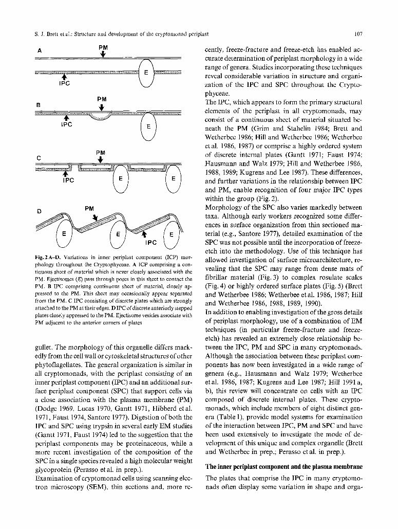
Fig. 2A-D. Variations in inner periplast component (ICP) mor- phology throughout the Cryptophyceae. A ICP comprising a con- tinuous sheet of material which is never closely associated with the PM. Ejectisomes (E) pass through pores in this sheet to contact the PM. B IPC comprising continuous sheet of material, closely ap- pressed to the PM. This sheet may occasionally appear separated from the PM. C IPC consisting of discrete plates which are strongly attached to the PM at their edges. D IPC of discrete anteriorly stepped plates closely appressed to the PM. Ejectisome vesicles associate with PM adjacent to the anterior corners of plates
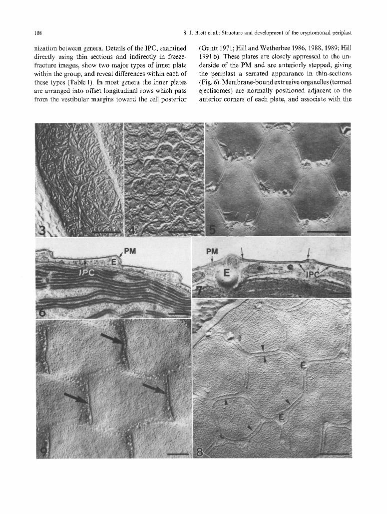
gullet. The morphology of this organelle differs mark- edly from the cell wall or cytoskeletal structures of other phytoflagellates. The general organization is similar in all cryptomonads, with the periplast consisting of an inner periplast component (IPC) and an additional sur- face periplast component (SPC) that support cells via a close association with the plasma membrane (PM) (Dodge 1969, Lucas 1970, Gantt 1971, Hibberd etal. 1971, Faust 1974, Santore 1977). Digestion of both the IPC and SPC using trypsin in several early EM studies (Gantt 1971, Faust 1974) led to the suggestion that the periplast components may be proteinaceous, while a more recent investigation of the composition of the SPC in a single species revealed a high molecular weight glycoprotein (Perasso et al. in prep.). Examination of cryptomonad cells using scanning elec- tron microscopy (SEM), thin sections and, more re-
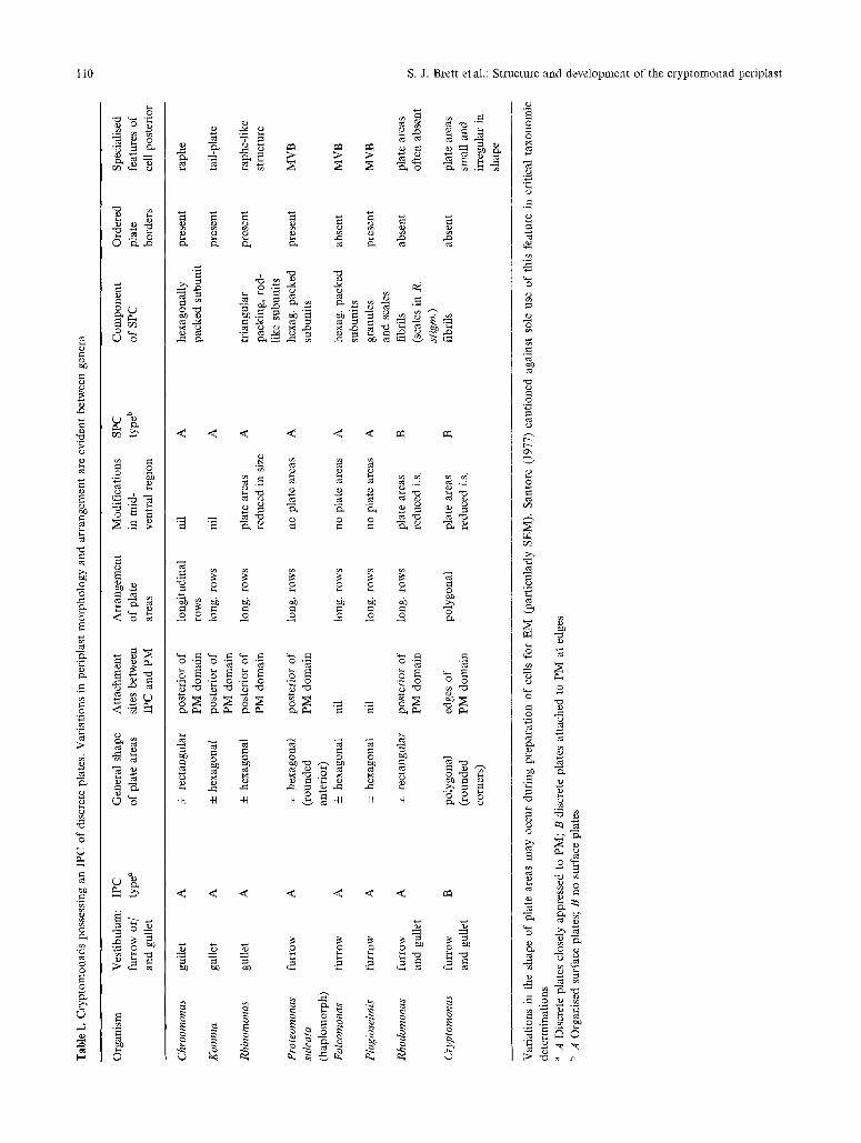
cently, freeze-fracture and freeze-etch has enabled ac- curate determination ofperiplast morphology in a wide range of genera. Studies incorporating these techniques reveal considerable variation in structure and organi- zation of the IPC and SPC throughout the Crypto- phyceae. The IPC, which appears to form the primary structural elements of the periplast in all cryptomonads, may consist of a continuous sheet of material situated be- neath the PM (Grim and Stahelin 1984; Brett and Wetherbee 1986; Hill and Wetherbee 1986; Wetherbee et al. 1986, 1987) or comprise a highly ordered system of discrete internal plates (Gantt 1971; Faust 1974; Hausmann and Walz 1979; Hill and Wetherbee 1986, 1988, 1989; Kugrens and Lee 1987). These differences, and further variations in the relationship between IPC and PM, enable recognition of four major IPC types within the group (Fig. 2). Morphology of the SPC also varies markedly between taxa. Although early workers recognized some differ- ences in surface organization from thin sectioned ma- terial (e.g., Santore 1977), detailed examination of the SPC was not possible until the incorporation of freeze- etch into the methodology. Use of this technique has allowed investigation of surface microarchitecture, re- vealing that the SPC may range from dense mats of fibrillar material (Fig. 3) to complex rosulate scales (Fig. 4) or highly ordered surface plates (Fig. 5) (Brett and Wetherbee 1986; Wetherbee etal. 1986, 1987; Hill and Wetherbee 1986, 1988, 1989, 1990). In addition to enabling investigation of the gross details of periplast morphology, use of a combination of EM techniques (in particular freeze-fracture and freeze- etch) has revealed an extremely close relationship be- tween the IPC, PM and SPC in many cryptomonads. Although the association between these periplast com- ponents has now been investigated in a wide range of genera (e.g., Hausmann and Walz 1979; Wetherbee etal. 1986, 1987; Kugrens and Lee 1987; Hill 1991 a, b), this review will concentrate on cells with an IPC composed of discrete internal plates. These crypto- monads, which include members of eight distinct gen- era (Table 1), provide model systems for examination of the interaction between IPC, PM and SPC and have been used extensively to investigate the mode of de- velopment of this unique and complex organelle (Brett and Wetherbee in prep.; Perasso et al. in prep.).
The inner periplast component and the plasma membrane
The plates that comprise the IPC in many cryptomo- nads often display some variation in shape and orga-
108 s.J. Brett et al.: Structure and development of the cryptomonad periplast
nization between genera. Details of the IPC, examined directly using thin sections and indirectly in freeze- fracture images, show two major types of inner plate within the group, and reveal differences within each of these types (Table 1). In most genera the inner plates are arranged into offset longitudinal rows which pass from the vestibular margins toward the cell posterior
(Gantt 1971; Hill and Wetherbee 1986, 1988, 1989; Hill 1991 b). These plates are closely appressed to the un- derside of the PM and are anteriorly stepped, giving the periplast a serrated appearance in thin-sections (Fig. 6). Membrane-bound extrusive organelles (termed ejectisomes) are normally positioned adjacent to the anterior corners of each plate, and associate with the
s. J. Brett et al.: Structure and development of the cryptomonad periplast 109
PM in these regions (Antia etal. 1973; Santore 1977, 1982, 1986, 1987; Meyer and Pienaar 1984; Erata and Chihara 1989). A markedly different inner plate organization is char- acteristic of the genus Cryptomonas. In thin sections the periplast has a flattened appearance, and the inner plates appear most intimately associated with the PM at their edges (Fig. 7). The inner plates and PM com- monly appear separate from one another in Crypto-
monas although this features may result from shrinkage during fixation for electron microscopy (Kugrens and Lee 1987; Kugrens pers. comm., Hill pers. comm.). In contrast to other genera, the inner plates of Crypto- monas are not aligned into rows, but instead exhibit a polygonal (generally hexagonal) arrangement (Hibberd etal. 1971; Faust 1974; Santore 1977, 1984, 1985; Brett and Wetherbee 1986; Kugrens and Lee 1987). Despite the variation in shape and morphology of inner plates, an extremely close relationship between the IPC and PM is evident in all genera. Freeze-fractures reveal that the PM is organized into discrete regions called domains, which are situated directly above inner plates (Figs. 8 and 9). The domains are densely packed with intra-membrane particles (IMPs), and may possess specialized rows of IMPs in regions where the IPC and PM are strongly attached (Hausmannn and Walz 1979; Brett and Wetherbee 1986; Hill and Wetherbee 1986, 1988; Kugrens and Lee 1987). The location and ar- rangement of attachment sites varies between genera. In Cryptomonas, particles are evident around the entire perimeter of each domain (Fig. 8), while in other genera ordered rows of IMPs are commonly observed along the posterior margins (Fig. 9).
In contrast to the ordered domains, regions of PM above the gaps between inner plates contain fewer, less ordered IMPs. These differences in the IMP organi- zation suggest that the inner plates may act as a cy- toskeletal template which directly influences the ar- rangement of IMPs within the PM.
The cell surface
The use of freeze-fracture/-etch preparation has also enabled examination of cell surface features and their relationship to underlying periplast components. In Cryptomonas and Rhodomonas, the SPC generally con- sists of elongate fibrils (or scales in Rhodomonas stig- matica) which form a dense mat across most of the cell surface (Brett and Wetherbee 1986, Hill and Wetherbee 1989). The arrangement of the SPC in these crypto- monads does not appear closely linked to the organi- zation of the underlying PM and IPC. In all other cryptomonads discussed in this review, however, an intimate relationship is evident between the SPC, PM and IPC. The SPC is composed of discrete plates sit- uated directly above ordered domains in the PM (e.g., Fig. 10). The details of surface plates vary markedly between genera, and often provide important criteria for the separation of taxa (Table 1). In Plagioselmis, the sur- face plates are composed of particulate material and granular scales (Fig. 11), those of Rhinomonas (Hill and Wetherbee 1988) are composed of rod-like subunits with a triangular arrangement (Figs. 12 and 13), while Komrna, Falcomonas, Chroomonas (Hill 1991 a), and Proteomonas sulcata (haplomorph) (Hill and Wether-
Figs. 3--5. Freeze-etch images of cryptomonad cell surfaces. Bars: Figs. 3 and 5, 0.5 ~tm; Fig. 4, 0.2 gm
Fig. 3. The SPC of Proteomonas sulcata (diplomorph), showing a dense mat of elongate fibrils
Fig. 4. Heptagonal scales form the SPC of Geminigera cryophila
Fig. 5. Ordered crystalline plates on the cell surface of Proteomonas sulcata (haplomorph)
Figs. 6 and 7. Thin sections through periplast. Bars: Fig. 6, 0.2 gm; Fig. 7, 0.5 gm
Fig. 6. The IPC of Proteomonas sulcata consists of discrete anteriorly stepped plates. Ejectisome vesicles (E) are located adjacent to the anterior corners of each inner plate
Fig. 7. IPC of Cryptomonas ovata has a flattened appearance in T.S. The discrete inner plates (IPC) appear intimately associated with the PM at their edges (arrows). Ejectisome vesicles (E) associate with the PM in the gaps between plates
Figs. 8 and 9. Freeze-fracture images of PM. Bars: Fig. 8, 0.5 gin; Fig. 9, 0.2 lam
Fig. 8. Polygonal arrangement of PM domains in Cryptomonas ovata. PM domains are densely packed with IMPs, and surrounded by a highly ordered row of particles (arrowheads). Ejectisome vesicles (E) are present adjacent to the corners of domains
Fig. 9. A distinct row of IMPs (arrows) defines the posterior margins of PM domains in Proteornonas sulcata (haplomorph). Regions of PM between adjacent domains contain fewer IMPs and have a pitted appearance
110 S.J. Brett et al.: Structure and development of the cryptomonad periplast
~o
~D
OD 0
~z
0
%
0
Z~
0
o
0
~o~
~ k
0:
�9
r!
< <
o
~ o . o o
o
~ ~ ~-~o ~ . ~
~ ~
o o o o o =
0 0 �9 0 0 ~D~
"o ~ ~ .~ .~ ' .~
< .< < < < < .<
0
O
..=
.=
'N
bD
0
C~
&
S. J. Brett et al.: Structure and development of the cryptomonad periplast 111
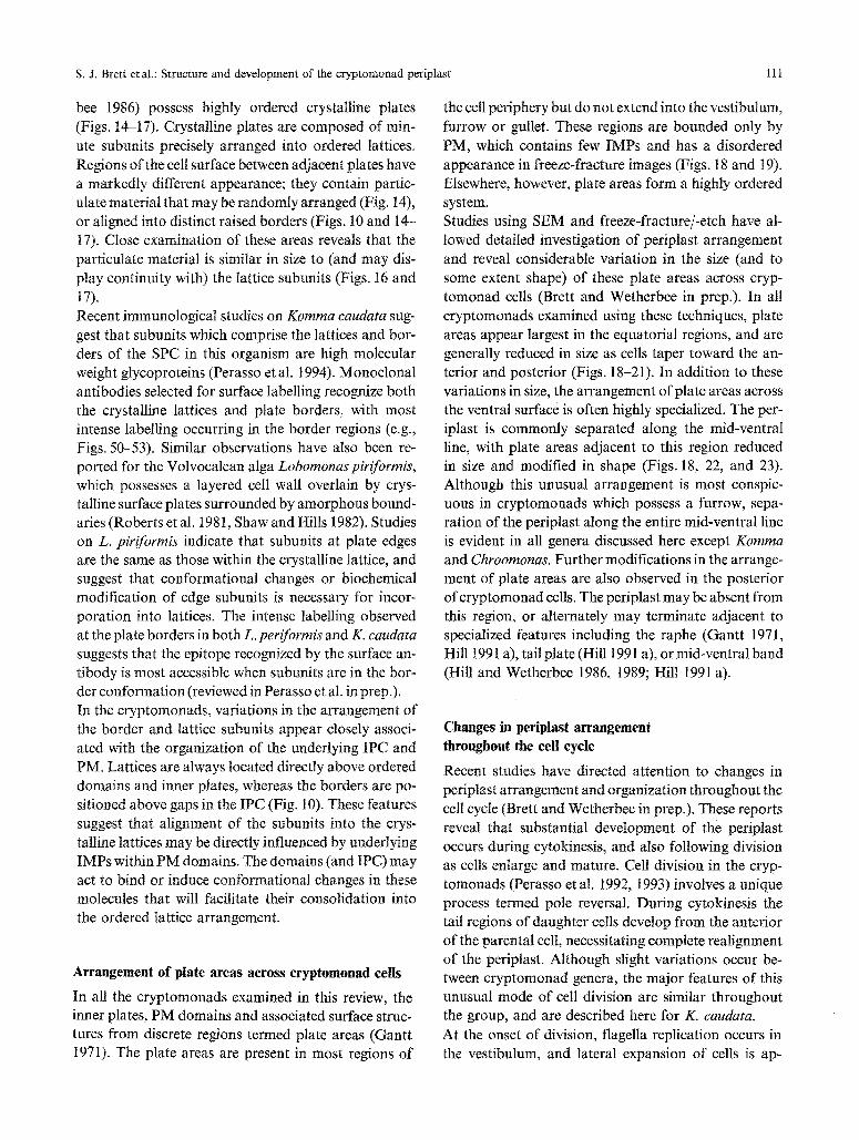
bee 1986) possess highly ordered crystalline plates (Figs. 14-17). Crystalline plates are composed of min- ute subunits precisely arranged into ordered lattices. Regions of the cell surface between adjacent plates have a markedly different appearance; they contain partic- ulate material that may be randomly arranged (Fig. 14), or aligned into distinct ~aised borders (Figs. 10 and 14- 17). Close examination of these areas reveals that the particulate material is similar in size to (and may dis- play continuity with) the lattice subunits (Figs. 16 and 17). Recent immunological studies on Komma caudata sug- gest that subunits which comprise the lattices and bor- ders of the SPC in this organism are high molecular weight glycoproteins (Perasso etal. 1994). Monoclonai antibodies selected for surface labelling recognize both the crystalline lattices and plate borders, with most intense labelling occurring in the border regions (e.g., Figs. 50-53). Similar observations have also been re- ported for the Volvocalean alga Lobomonas piriJbrmis, which possesses a layered cell wall overlain by crys- talline surface plates surrounded by amorphous bound- aries (Roberts et al. 1981, Shaw and Hills 1982). Studies on L. piriformis indicate that subunits at plate edges are the same as those within the crystalline lattice, and suggest that conformational changes or biochemical modification of edge subunits is necessary for incor- poration into lattices. The intense labelling observed at the plate borders in both L. periformis and K. caudata suggests that the epitope recognized by the surface an- tibody is most accessible when subunits are in the bor- der conformation (reviewed in Perasso et al. in prep.). In the cryptomonads, variations in the arrangement of the border and lattice subunits appear closely associ- ated with the organization of the underlying IPC and PM. Lattices are always located directly above ordered domains and inner plates, whereas the borders are po- sitioned above gaps in the IPC (Fig. 10). These features suggest that alignment of the subunits into the crys- talline lattices may be directly influenced by underlying IMPs within PM domains. The domains (and IPC) may act to bind or induce conformational changes in these molecules that will facilitate their consolidation into the ordered lattice arrangement.
Arrangement of plate areas across eryptomonad cells
In all the cryptomonads examined in this review, the inner plates, PM domains and associated surface struc- tures from discrete regions termed plate areas (Gantt 1971). The plate areas are present in most regions of
the cell periphery but do not extend into the vestibutum, furrow or gullet. These regions are bounded only by PM, which contains few IMPs and has a disordered appearance in freeze-fracture images (Figs. 18 and 19). Elsewhere, however, plate areas form a highly ordered system. Studies using SEM and freeze-fracture/-etch have al- lowed detailed investigation of periplast arrangement and reveal considerable variation in the size (and to some extent shape) of these plate areas across cryp- tomonad cells (Brett and Wetherbee in prep.). In all cryptomonads examined using these techniques, plate areas appear largest in the equatorial regions, and are generally reduced in size as cells taper toward the an- terior and posterior (Figs. 18-21). In addition to these variations in size, the arrangement of plate areas across the ventral surface is often highly specialized. The per- iplast is commonly separated along the mid-ventral line, with plate areas adjacent to this region reduced in size and modified in shape (Figs. 18, 22, and 23). Although this unusual arrangement is most conspic- uous in cryptomonads which possess a furrow, sepa- ration of the periplast along the entire mid-ventral line is evident in all genera discussed here except Komma and Chroomonas. Further modifications in the arrange- ment of plate areas are also observed in the posterior of cryptomonad cells. The periplast may be absent from this region, or alternately may terminate adjacent to specialized features including the raphe (Gantt 1971, Hill 1991 a), tail plate (Hill 1991 a), or mid-ventral band (Hill and Wetherbee 1986, 1989; Hill 1991 a).
Changes in periplast arrangement throughout the cell cycle
Recent studies have directed attention to changes in periplast arrangement and organization throughout the cell cycle (Brett and Wetherbee in prep.). These reports reveal that substantial development of the periplast occurs during cytokinesis, and also following division as cells enlarge and mature. Cell division in the cryp- tomonads (Perasso et al. 1992, 1993) involves a unique process termed pole reversal. During cytokinesis the tail regions of daughter cells develop from the anterior of the parental cell, necessitating complete realignment of the periplast. Although slight variations occur be- tween cryptomonad genera, the major features of this unusual mode of cell division are similar throughout the group, and are described here for K. caudata. At the onset of division, flagella replication occurs in the vestibulum, and lateral expansion of cells is ap-
112 S.J. Brett etat.: Structure and development of the cryptomonad periplast
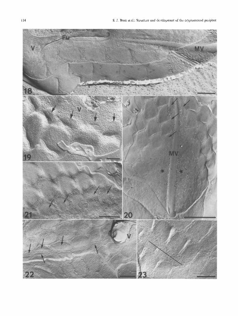
parent (Figs. 24 and 25). The periplast separates along the mid-ventral line progressing from the vestibulum toward the celt posterior. The edges of the periplast exposed during this process form specialized regions, termed anamorphic zones, which undergo develop- mental changes both during and after cytokinesis. As the ventral surface divides, these regions, as well as
additional anamorphic zones located around the ves- tibular margins, expand and realign (Figs. 26-28). Dis- tinct pointed structures form where the anamorphic zones converge, and eventually develop into the tail regions of daughter cells. The dorsal surface then di- vides, the forming tail regions migrate laterally (Figs. 29-31) and rotation of cell halves relative to one
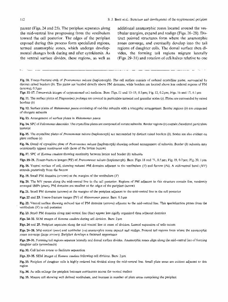
Fig. 10. Freeze-fractare/-etch of Proteomonas sulcata (haplomorph). The cell surface consists of ordered crystalline plates, surrounded by distinct raised borders (b). The plates are located directly above PM domains, while borders are located above less ordered regions of PM (arrows); 0,5 !am Figs. 11-17, Freeze-etch images of cryptomonad ceil surfaces, Bars: Figs. 11 and 13-15, 0.5 gm; Fig. 12, 0.2 gin; Figs. 16 and 17, 0,1 ~tm
Fig. 11. The surface plates of Plagioselmis p~olonga are covered in particulate material and granular scales (s). Plates are surrounded by raised borders (b)
Fig, 12. Surface plates of Rhinomonas pauca consisting of rod-like subunits with a triangular arrangement. Border regions (b) are composed of elongate subunits
Fig. 13. Arrangement of surface plates in Rhinomonas pauca
Fig. 14. SPC of Falcomonas daucoides. The crystalline plates are composed of minute subunits. Border regions (b) contain disordered particulate material
Fig. 15. The crystalline plates of Proteomonas sulcata (haplomorph) are surrounded by distinct raised borders (b), Scales are also evident on plate surfaces (s)
Fig. 16. Detail of crystalline plate of Proteomonas sulcata (haplomorph) showing ordered arrangement of subunits. Border (b) subunits may occasionally appear continuous with those of the lattice (arrow)
Fig. 17. SPC of Komma caudata showing continuity between lattice and border (b) subunits
Figs, 18-21, Freeze-fracture images (PF) of Proteomonas sulcata (haplomorph). Bars: Figs. 18 and 21, 0.5 gin; Fig. 19, 0.2 gin; Fig. 20, 1 pm
Fig. 18. Ventral surface of cell, showing reduced PM domains adjacent to the vestibulum (V) and furrow (Fu). A mid-ventral band (MV) extends posteriorly from the furrow
Fig. 19, Small PM domains (arrows) at the margins of the vestibulum (V)
Fig. 20. The MV passes along the mid-ventral line to the cell posterior. Regions of PM adjacent to this structure contain few, randomly arranged IMPs (stars), PM domains are smallest at the edges of the periplast (arrow)
Fig. 21. Small PM domains (arrows) at the margins of the periplast adjacent to the mid-ventral line in the celt posterior
Figs. 22 and 23. Freeze-fracture images (PF) of Rhinornonas pauca. Bars: 0.5 gm
Fig. 22. Ventral surface showing reduced size of PM domains (arrows) adjacent to the mid-ventral line. This specialization passes from the vestibulum (V) to cell posterior
Fig. 23. Small PM domains along mid-ventral line (line) appear less rigidly organized than adjacent domains
Figs. 24-32, SEM images of Komma caudata during cell division. Bars: 2 ~tm
Figs. 24 and 25. Periplast separates along the mid-ventral line at onset of division. Lateral expansion of cells occurs
Figs. 26-28. Mid-ventral (mva) and vestibular (va) anamorphic zones expand and reali~a, Pointed tail regions form where the anamorphic zones converge (large arrows). Periplast develops a flattened appearance
Figs. 29-31. Forming tail regions separate laterally and dorsal surface divides. Anamorphic zones align along the mid-ventral line of forming daughter cells (arrowheads)
Fig. 32. Cell halves rotate to facilitate separation
Figs. 33-35. SEM images of Komma caudata following cell division. Bars: 2 pm
Fig. 33. Periplast of daughter cells is highIy ordered but divided along the mid-ventral line. Small plate areas are evident adjacent to this region
Fig.34. As cells enlarge the periptast becomes continuous across the ventral surface
Fig. 35. Mature cell showing well defined vestibultma, and increase in number of plate areas comprising the periptast

S. J. Brett et al.: Structure and development of the cryptomonad periplast 113
114 S.J. Brett et at.: Structure and development of tl~e cryptomonad perip[ast
S. J. Brett et al.: Structure and development of the cryptomonad periplast 115
116 S.J. Brett et al.: Structure and development of the cryptomonad periplast
another facilitates separation (Fig. 32). Further rea- lignment of the anamorphic zones occurs, and these eventually position on either side of the mid-ventral line of developing daughter cells (Figs, 32 and 33). The unusual mode of cell division in the cryptomonads, involving reorientation of daughter cells relative to the parental cell, necessitates major changes in the periplast in many cryptomonads (Brett and Wetherbee 1994 a). In cells with anteriorly stepped plate areas, the periplast persists, but develops a flattened appearance during cell division (Figs. 27 32). The plate areas remain pre- cisely aligned throughout this process, and after cy- tokinesis adopt a polarity consistent with the newly formed daughter cell. Although the process of periplast reorientation is no~ fully understood, detailed exami- nation of cells using thin sections and freeze-fracture/ -etch suggests that realignment may be facilitated by relocation of the at tachment sites between inner plates and the PM (Brett and Wetherbee in prep.). Following cell division, daughter cells are substantially smaller than their parental counterparts, the vestibular margins are indistinct and the periplast is always di- vided along the mid-ventral line (Fig. 33). As cells en- large and mature the vestibulum/furrow/gullet complex becomes more clearly defined, and changes in periplast organization occur. In K o m m a (Figs. 34 and 35) and C h r o o m o n a s the periplast becomes continuous across the ventral surface as the gullet develops~ although in all other organisms the penplast remains separated along the mid-ventral line throughout the cell cycle (Figs. 36 39).
The elongation and lateral expansion of cells that oc- curs after division is associated with an increase in the number of plate areas comprising the periplast (Figs. 33-39). The periplast remains precisely aligned throughout this process, suggesting that growth occurs in an orderly manner by addition of new plate areas to anamorphic zones (Brett and Wetherbee in prep.). In most genera elongation results from addition of new plate areas to anamorphic zones around the vestibulum and in the posterior, while lateral expansion is facili- tated by growth and addition from the mid-ventral line (Figs. 36-39). Growth from these anamorphic zones appears to enable highly ordered expansion of cryp- tomonads throughout the entire cell cycle. The absence of mid-ventral specializations in K o m m a (Figs. 34 and 35) suggests, however, that lateral expansion in this genus may occur by the enlargement of existing plates in the ventral regions of the cell.
Formation and development of the inner plates
The general changes in the arrangement of plate areas revealed in the SEM studies provided a basis for more detailed investigation of periplast development. Sub- sequent work (which indirectly examined the formation and growth of the inner periplast plates using freeze fracture images) revealed variations in the size, orga- nization and alignment of PM domains within ana- morphic zones (Brett and Wetherbee in prep.). Despite differences in the morphology of the inner plates be- tween genera, similar features were observed in all cryp- tomonads examined.
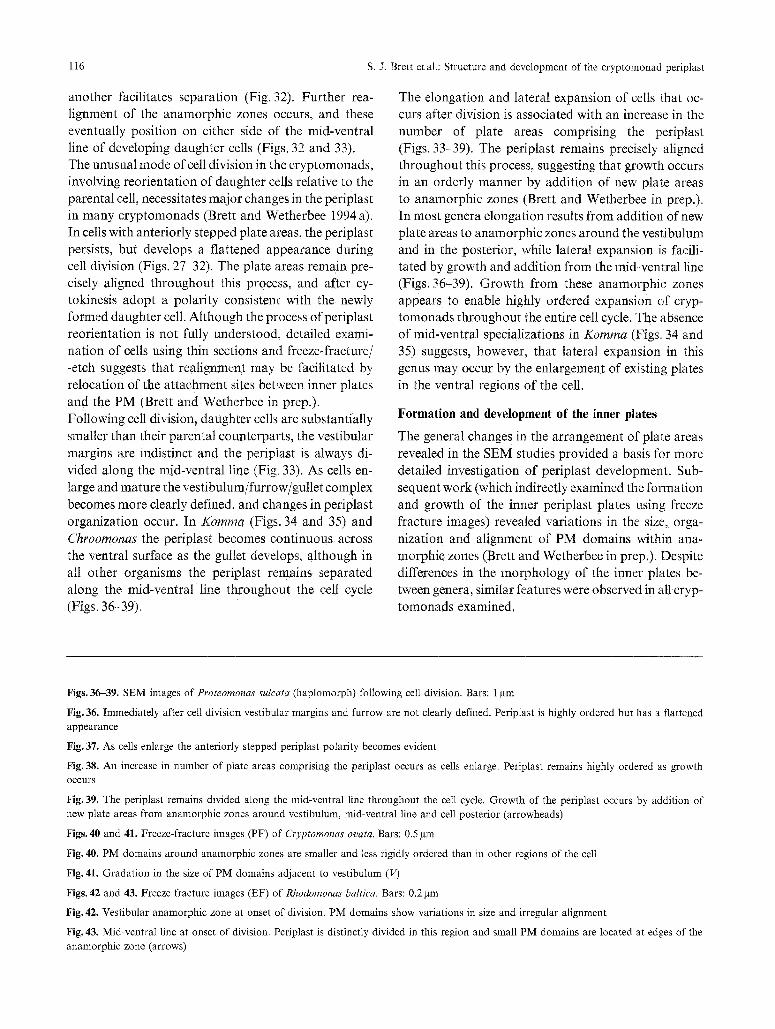
Figs. 36-39. SEM images of Proteomonas sulcata (haplomorph) following cell division. Bars: 1 gm
Fig. 36. Immediately after cell division vestibular margins and furrow are not clearly defined. Periplast is highly ordered but has a flattened appearance
Fig. 37. As cells enlarge the anteriorly stepped periplast polarity becomes evident
Fig. 38. An increase in number of plate areas comprising the periplast occurs as cells enlarge. Periplast remains highly ordered as growth occurs
Fig. 39. The periplast remains divided along the mid-ventral line throughout the cell cycle. Growth of the periplast occurs by addition of new plate areas from anamorphic zones around vestibulum, mid-ventral line and cell posterior (arrowheads)
Figs. 40 and 41. Freeze-fracture images (PF) of Cryptomonas ovata. Bars: 0.5 gm
Fig. 40. PM domains around anamorphic zones are smaller and less rigidly ordered than in other regions of the cell
Fig. 41. Gradation in the size of PM domains adjacent to vestibulum (V)
Figs. 42 and 43. Freeze-fracture images (EF) of Rhodomonas baltica. Bars: 0.2 lam
Fig. 42. Vestibular anamorphic zone at onset of division. PM domains show variations in size and irregular alignment
Fig. 43. Mid-ventral line at onset of division. Periplast is distinctly divided in this region and small PM domains are located at edges of the anamorphic zone (arrows)

s. J. Brett et al.: Structure and development of the cryptomonad periplast t 17
The PM domains within anamorphic zones range over a continuum from minute accumulations or circular aggregates of IMPs to larger, more rigidly ordered re- gions densely packed with IMPs and possessing spe-
icalized attachment sites. Although most conspicuous in dividing and newly formed cells (Figs. 40-43), similar variations are evident adjacent to anamorphic zones throughout the cell cycle (Figs. 18-23). The distinct
118 S.J. Brett et al.: Structure and development of the cryptomonad periplast
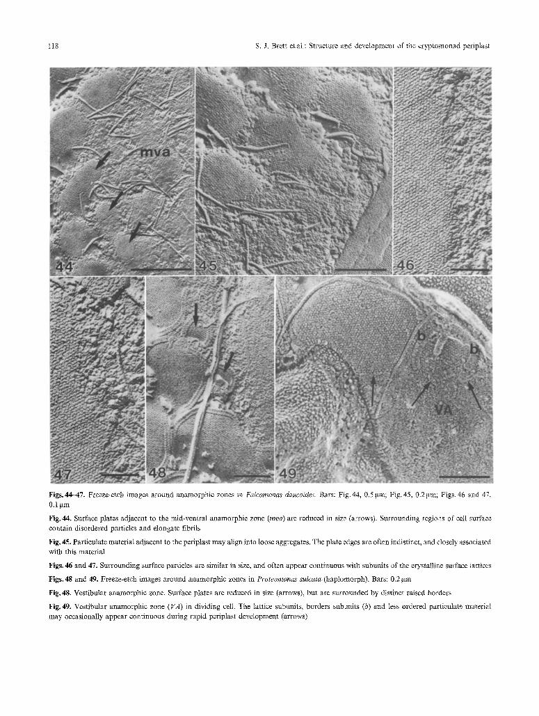
Figs.44-47. Freeze-etch images around anamorphic zones in Falcomonas daucoides. Bars: Fig. 44, 0.5 gm; Fig. 45, 0.2 gin; Figs. 46 and 47, 0.1 ~tm
Fig. 44. Surface plates adjacent to the mid-ventral anamorphic zone (mva) are reduced in size (arrows). Surrounding regions of cell surface contain disordered particles and elongate fibrils
Fig. 45. Particulate material adjacent to the periplast may align into loose aggregates. The plate edges are often indistinct, and closely associated with this material
Figs. 46 and 47. Surrounding surface particles are similar in size, and often appear continuous with subunits of the crystalline surface lattices
Figs. 48 and 49. Freeze-etch images around anamorphic zones in Proteomonas sulcata (haplomorph). Bars: 0.2 gm
Fig. 48. Vestibular anamorphic zone. Surface plates are reduced in size (arrows), but are surrounded by distinct raised borders
Fig. 49. Vestibular anamorphic zone (VA) in dividing cell. The lattice subuults, borders subunits (b) and less ordered particulate material may occasionally appear continuous during rapid periplast development (arrows)
s. J. Brett et al.: Structure and development of the cryptomonad periplast 119
gradation in the size of domains within these regions indicates that growth of the perfplast does not occur by addition of large, fully formed inner plates at the edges of the IPC. The features observed suggest, in- stead, that the inner plates form de novo and undergo considerable enlargement within these specialized ana- morphic zones. Variations in the spacing and arrange- ment of PM domains in developing cells (Figs. 40, 42, and 43) imply that the inner plates develop indepen- dently, and are capable of some movement relative to one another as they align within the highly ordered periplast.
Development of crystalline surface plates
The imimate relationship between IPC, PM and SPC in the cryptomonads suggests that the development of crystalline surface plates may be closely linked to the formation and growth of the inner plates within ana- morphic zones. The unique SPC of Falcomonas dau- coides, comprising crystalline surface plates which lack distinct borders (Hill 1991 a), provides a model system for investigation of features associated with lattice de- velopment (Brett and Wetherbee in prep.). Examina- tion of this organism reveals that surface plates within anamorphic zones are commonly reduced in size, and surrounding areas of the cell surface are covered with disordered particulate material (Figs. 44 and 45). The surface particles are similar in size to lattice subunits, and may be aligned into loose aggregates or closely associated with the edges of periplast plates (Figs. 45- 47). Plate margins are often indistinct and surface par- ticles adjacent to these regions may appear continuous with, and are often indistinguishable from, lattice sub- units. The close relationship between the particulate material and lattice subunits suggests that development of the crystalline surface may occur by accumulation and in- corporation of less ordered precursors from surround- ing regions of the cell surface. After formation of these crystalline plates, enlargement and growth appears fa- cilitated by addition of subunits to the lattice edges. Detailed studies of bacteria (for reviews, see Sleytr and Messner 1983, Messner and Sleytr 1992) and L. piri- formis (Roberts etal. 1981, Shaw and Hills 1982) de- scribe a similar mode of lattice development and sug- gest that conformational changes occur to precursor molecules during addition. As stated above, the close spatial relationship between the organization of the IPC, PM and SPC in the cryptomonads suggests that the formation and enlargement of surface plates may
be intimately associated with developmental changes in the IPC and PM. Growth of the inner plates and related expansion of PM domains, may induce changes in the precursor molecules which enable incorporation into the highly ordered lattice. Although the relatively simple arrangement of the SPC in 17. daucoides provides an ideal system for exami- nation of lattice assembly, the crystalline surface plates in other genera are generally surrounded by distinct raised borders (Figs. 48 and 49) (Hill and Wetherbee t986, Hill 1991 a). Consequently, growth of these lat- tices during self-assembly does not appear to occur by direct addition of disordered precursors from sur- rounding regions of the cell surface. The continuity between borders and lattices in these organisms (Figs. 16, 17, and 49) suggests, instead, that subunits may be rearranged. For example, subunits previously within a border may subsequently become incorporated into the highly ordered surface plates during devel- opment (Brett and Wetherbee in prep.). The close re- lationship between the organization of the SPC and underlying periplast components indicates that incor- poration of border subunits into the lattices during self- assembly may be closely associated with the growth of inner plates and PM domains. The immunological work of Perasso et al. (in prep.) also suggests similarities between the components com- prising the lattices, borders and less ordered regions of the cell surface. These studies indicate that the subunits which form the SPC are produced in the Golgi ap- paratus and secreted through the endomembrane sys- tem for deployment at the edges of the periplast (Figs. 50-53). Examination of K. caudata through the cell cycle reveals that deposition of these macromolecules occurs predominantly within anamo1~hic zones, where the majority of lattice formation occurs.
Conclusions
The complexity of the cryptomonad periplast has ne- cessitated use of a wide range of techniques to accu- rately determine morphology of the IPC, PM and SPC. Although general features of periplast structure and arrangement may be determined using thin sections and SEM, the use of freeze-fracture/-etch has greatly en- hanced the understanding of this unique organelle. Studies incorporating these techniques reveal consid- erable diversity throughout the Cryptophyceae and provide important characters for taxonomic distinc- tions within the group. The detailed information about PM and surface microm'chitecture available from these
120 S, J. Brett et al.: Structure and development of the cryptomonad peripJast

s. J. Brett eta1.: Structure and development of the cryptomonad periplast 121
reports has also enabled examinat ion o f the complex
relationships between components comprising the peri-
plast.
The cells discussed in this review all possess an IPC of
discrete plates, and have proven ideal systems for in- vestigation o f the association between IPC, PM, and
SPC. In all these organisms the PM is ordered into
discrete domains which are situated directly above the
inner periplast plates. In contrast , regions o f the PM
not supported by the IPC generally contain fewer IMPs
and appear less ordered. The I P C - P M association sug-
gests that the inner plates are cytoskeletal, and may
influence the ar rangement o f IMPs within the PM.
Organizat ion o f the crystalline surface plates found in
many c ryp tomonad genera also appears closely asso-
ciated with that o f the underlying components . The
location o f these plates directly above ordered PM do-
mains suggests that the PM, and ultimately the IPC, may influence the al ignment o f subunits within surface
lattices.
Development o f bo th the IPC and SPC occurs f rom specialized anamorph ic zones th roughout the cell cycle.
Unlike the wall components o f m a n y microalgal groups, fully formed inner and surface plates are not
pre-packaged within cytoplasmic vesicles for later ad- dition to the periplast. Instead the inner periplast plates
appear to form de novo and undergo considerable en-
largement within anamorphic zones. Growth of the
crystalline surface lattices, which occurs by addit ion
and incorpora t ion o f less ordered precursors f rom sur- rounding surface areas, may be closely associated with
this process. The intimate relationship between IPC,
PM, and SPC suggests that growth of inner plates and
PM domains may result in modificat ions which enable
incorpora t ion o f precursor molecules into the highly
organized surface lattices. The precise mode of for-
mat ion may be more fully unders tood as further details o f periplast chemistry are revealed.
Acknowledgements
RW thanks the Australian Research Council for financial assistance. SJB and LP were supported by Commonwealth Postgraduate Schol-
arships, the University of Melbourne. We thank Dr. Paul Kugrens for Fig. 1 and Dr. David R. A. Hill for valuable discussions and for reviewing the manuscript.
References
Antia N J, Kalley JP, McDonald J, Bisalputra T (1973) Ultrastructure of the marine cryptomonad Chroomonas salina cultured under conditions ofphotoautotrophy and glycerol-heterotrophy. J Pro- tozool 20:377-385
Brett S J, Wetherbee R (1986) A comparative study of periplast struc- ture in Cryptomonas cryophila and C. ovata (Cryptophyceae). Protoplasma 131:23-31
Dodge JD (1969) The ultrastructure of Chroomonas mesostigmatica Butcher (Cryptophyceae). Arch Microbiol 69:266-280
Erata M, Chihara M (1989) Re-examination of Pyrenomonas and Rhodomonas (Class Cryptophyceae) through ultrastructural sur- vey of red pigmented cryptomonads. Bot Mag Tokyo 102: 429- 443
Faust MA (1974) Structure of the periplast of Cryptomonas ovata var. palustris. J Phycol 10:121-124
Gantt E (1971) Micromorphology of the periplast of Chroomonas sp. (Cryptophyceae). J Phycol 7:177-184
Grim JN, Staehelin LA (1984) The ejectisomes of the flagellate Chi- lomonas paramecium: visualization by freeze-fracture and iso- lation techniques. J Protozool 31:259-267
Hausmann K, Walz B (1979) Periplaststruktur und Organisation der Plasmamembrane von Rhodomonas spec. (Cryptophyceae). Pro- toplasma 101:349-354
Hibberd DJ, Greenwood AD, Griffiths HB (1971) Observations on the ultrastructure of the flagella and periplast in the Crypto- phyceae. Br Phycol J 6:61-72
Hill DRA (1991 a) Chroomonas and other blue-green cryptomonads. J Phycol 27:133-145
- (1991 b) A revised circumscription of Cryptomonas (Cryptophy- ceae) based on examination of Australian strains. Phycologia 30: 179-188
- Wetherbee R (1986) Proteomonas sulcata gen. et sp.nov. (Cryp- tophyceae), a cryptomonad with two morphologically distinct and alternating forms. Phycologia 25:521-543
(1988) The structure and taxonomy ofRhinomonaspauca gen. et sp.nov. (Cryptophyceae). Phycologia 27:355-365
(1989) A reappraisal of the genus Rhodomonas (Cryptophy- ceae). Can J Bot 28:143-158
(1990) Guillardia theta gen. et sep.nov. (Cryptophyceae). Can J Bot 68:1873-1876
Klaveness D (1981) Rhodomonas lacustris (Pascher & Ruttner) Ja- vornicky (Cryptomonadida): ultrastructure of the vegetative cell. J Protozool 28:83-90
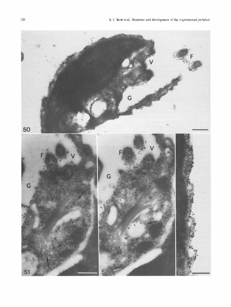
Figs. 50-53. MAb immunogold labelling of Komma caudata. Bars: Figs. 50-52, 1 gm; Fig. 53, 0.5 gm
Fig. 50. Labelling of an interphase cell, labelling is observed over the entire surface, most obvious at the surface plate edges, and also over the vestibular (V), gullet (G), and flagellar (F) surfaces
Figs. 51 and 52. Labelling of non-consecutive serial sections through a prophase cell, showing labelling about the GA and vesicles associated with the GA (arrows)
Fig. 53. High magnification of labelling of the periplast
122 S.J. Brett et al.: Structure and development of the cryptomonad periplast
Klaveness D (1985) Ciassical and modern criteria for determining species of Cryptophyceae. Bull Plankton Soc Jap 32:111-123
Kugrens P, Lee RE, Andersen RA (1986) Cell form and surface patterns in Chroomonas and Cryptomonas cells (Cryptophyta) as revealed by scanning electron microscopy. J Phycol 22:512-522
Kugrens P, Lee RE (1987) An ultrastructural survey of cryptomonad periplast using quick-freezing freeze fracture techniques. J Phycol 23:365-376
- - (1991) Organization ofcryptomonads. In: Patterson D J, Lar- sen J (eds) The biology of free-living heterotrophic flagellates. Clarendon Press, Oxford, pp219-233
Lucas IAN (1970) Observations on the fine structure of the Cryp- tophyceae. 1. The genus Cryptornonas. J Phycol 6:30-38
Messner P, Sleytr UB (1992) Crystalline bacterial cell-surface layers. Adv Microbiol Physiol 33:213-275
Meyer SR, Pienaar RN (1984) The microanatomy of Chroomonas
africana sp.nov. (Cryptophyceae). S Afr Tydskr Plank 3: 306- 319
Mnawar M, Bistricki T (1979) Scanning electron microscopy of some nanoplankton cryptomonads. Scanning Electron Microsc 3: 247- 252
Perasso L, Hill DRA, Wetherbee R (1992) Transformation and de- velopment of the flagellar apparatus of Cryptomonas ovata (Cryptophyceae) during cell division. Protoplasma 170:53-67
- Brett S J, Wetherbee R (1993) Pole reversal during cytokinesis in the Cryptophyceae. Protoplasma 174:19-24
Roberts K, Shaw PJ, Hills GJ (1981) High-resolution electron mi- croscopy of glycoproteins: the crystalline cell wall of Lobomonas. J Cell Sci 51:295-321
Santore UJ (1977) Scanning electron microscopy and comparative micromorphology of the periplast of Hemiselmis rufeseens, Chroomonas sp., Chroomonas salina and members of the genus Cryptomonas (Cryptophyceae). Br Phycol J 12:255-270
- (1982) Comparative ultrastructure of two members of the Cryp- tophyceae assigned to the genus Chroomonas - with comments on their taxonomy. Arch Protistenk 125:5-29
- (1984) Some aspects of taxonomy in the Cryptophyeeae. New Phytol 98:627-646
- (1985) A cytological survey of the genus Cryptomonas (Cryp- tophyceae) with comments on its taxonomy. Arch Protistenk 130:1 52
- (1986) The ultrastructure of Phyrenomonas heteromorpha comb. nov. (Cryptophyceae). Bot Mar 29:75-82
- (1987) A cytological survey of the genus Chroomonas - with comments on the taxonomy of this natural group of the Cryp- tophyceae. Arch Protistenk 134:83-114
Shaw P J, Hills GJ (1982) Three-dimensional structure of a ceil wall glycoprotein. J Mol Biol 162:459-471
Sleytr UB, Messner P (1983) Crystalline surface layers on bacteria. Annu Rev Microbiol 37:311-339
Wetherbee R, Hill DRA, McFadden GI (1986) Periplast structure of the cryptomonad flagellate Hemiselmis brunnescens. Proto- plasma 131:11-22
- - Brett SJ (1987) The structure of the periplast components and their association with plasma membrane in a cryptomonad flagellate. Can J Bot 65:1019-1026