structuralstudiesandproteinengineeringofinositol ... ·...
TRANSCRIPT

Structural Studies and Protein Engineering of InositolPhosphate Multikinase*□S
Received for publication, April 9, 2012, and in revised form, August 3, 2012 Published, JBC Papers in Press, August 15, 2012, DOI 10.1074/jbc.M112.365031
Stuart Endo-Streeter, Man-Kin Marco Tsui, Audrey R. Odom1, Jeremy Block, and John D. York2
From the Departments of Pharmacology and Cancer Biology, and of Biochemistry, Howard Hughes Medical Institute, DukeUniversity Medical Center, Durham, North Carolina 27710
Background: IPMK is a key enzyme in signaling required for cellular adaptation and organismal development.Results:We report the structure of IPMK, use structure-based enzyme design and perform complementation analysis inmodelsystems.Conclusion:We define a basis for substrate selectivity and report that 6-kinase activity is important for signaling.Significance: Determination of the IPMK structure enabled rewiring of signaling in organisms.
Inositol phosphates (IPs) regulate vital processes in eukary-otes, and their production downstream of phospholipase Cactivation is controlled through anetworkof evolutionarily con-served kinases and phosphatases. Inositol phosphate multiki-nase (IPMK, also called Ipk2 and Arg82) accounts for phos-phorylation of IP3 to IP5, as well as production of several otherIPmolecules.Here,we report the structure ofArabidopsis thali-ana IPMK� at 2.9 Å and find it is similar to the yeast ho-molog Ipk2, despite 17% sequence identity, as well as the activesite architecture of human IP3 3-kinase. Structural comparisonand substratemodelingwere used to identify a putative basis forIPMK selectivity. To test this model, we re-engineered bindingsite residues predicted to have restricted substrate specificity.Using steady-state kinetics and in vivometabolic labeling stud-ies in modified yeast strains, we observed that K117W andK117W:K121W mutants exhibited nearly normal 6-kinasefunction but harbored significantly reduced 3-kinase activity.These mutants complemented conditional nutritional growthdefects observed in ipmk null yeast and, remarkably, suppressedlethality observed in ipmknull flies.Ourdata are consistentwiththe hypothesis that IPMK 6-kinase activity and production ofIns(1,4,5,6)P4 are critical for cellular signaling. Overall, ourstudies provide new insights into the structure and function ofIPMK and utilize a synthetic biological approach to redesigninositol phosphate signaling pathways.
Inositol phosphates (IPs)3 are a large class of distinct chemicalcodes that regulate various intracellular processes. Classically, IP-mediated signaling begins with the production of IPs in response
to cleavage of phosphoinositides by phosphatidylinositol-specificphospholipaseC (reviewed inRef. 1).Additional IP species such asinositol tetrakisphosphate (IP4) and inositol pentakisphosphate(IP5) are generated by subsequent series of phosphorylation anddephosphorylation (2–5). Multiple IP species have been linked toprocesses, including metabolic regulation and nutrient selection,stress responses, nuclear mRNA export, apoptosis, RNA editing,and organism development (6, 7).Inositol phosphate multikinase (IPMK) is among the en-
zymes central to the production of IP species downstream ofphospholipase C activation. For clarity, we use IPMK through-out this manuscript, as it most accurately reflects the “promis-cuous” 6-/3-/5-kinase functions of the enzyme. As a historicalnote, IPMK was first designated ArgRIII (later called Arg82)based on a yeast genetic screen designed to identify the compo-nents of a nutrient response pathway in which arginine servedas the sole nitrogen source (8–11). ArgRIII was then renamedIpk2 based on the discovery that it functioned as an inositolphosphate kinase (12, 13), and this nomenclature was also usedin referring to the plant gene orthologs when they were firstidentified and characterized as AtIpk2� and AtIpk2� in Arabi-dopsis (14, 15). IPMK has now been cloned from a variety ofspecies and has been shown to function as a 6/3/5-kinase thatcatalyzes the phosphorylation of at least seven different IP spe-cies. Its loss of function in a variety of organisms is critical forsurvival (reviewed in Refs. 5 and 6).To gain insights into the basis of IPMK substrate promiscu-
ity, we initiated a structural approach of the plant enzyme,which possesses three kinase activities capable of phosphory-lating the D-6, D-3, and D-5 ring positions of a variety of IPsubstrates (14). We obtained Arabidopsis thaliana IPMK�crystals, complete data sets of native and seleno-methioninederivatives, and calculated an electron densitymap from exper-imental phases. Concomitant to our studies, the yeast IPMKcrys-tal structurewas published (16). From these studies and a numberof subsequent biological and protein engineering experiments, wenow report the crystal structure of A. thaliana IPMK� at 2.9 Å, abasis of IP substrate binding, the kinetic characterization of thewild-type enzyme and selective K117W and K121W mutants,and the yeast and fly IP profiles andphenotypes of theK121WandK117W/K121W mutants. Collectively, our data build upon and
* This work was supported by the Howard Hughes Medical Institute andNational Institutes of Health Grant RO1 HL-55672 (both to J. D. Y.).
□S This article contains supplemental Figs. S1–S3.1 Present address: Dept. of Pediatrics, WA University School of Medicine, St.
Louis, MO 63110.2 To whom correspondence should be addressed: Dept. of Pharmacology and
Cancer Biology, Duke University Medical Center, DUMC Box 3813, Durham,NC 27710. Tel.: 919-681-6414; Fax: 919-668-0991; E-mail: [email protected].
3 The abbreviations used are: IP, inositol phosphate; IP4, tetrakisphosphate;IP5, inositol pentakisphosphate; IPMK, inositol phosphate multikinase;FOM, figure of merit; SAD, single anomalous dispersion; CSM, completesynthetic medium.
THE JOURNAL OF BIOLOGICAL CHEMISTRY VOL. 287, NO. 42, pp. 35360 –35369, October 12, 2012© 2012 by The American Society for Biochemistry and Molecular Biology, Inc. Published in the U.S.A.
35360 JOURNAL OF BIOLOGICAL CHEMISTRY VOLUME 287 • NUMBER 42 • OCTOBER 12, 2012
by guest on October 29, 2018
http://ww
w.jbc.org/
Dow
nloaded from

extend insightsderived fromtheyeast crystal structureand, for thefirst time, provide a re-engineering of IPMK to be substrate-selec-tive. We exploit these mutants to address important biologicalquestions related to IPMK function, and our data are consistentwith a role for 6-kinase activity in the regulation of cellular andorganismal signaling.
EXPERIMENTAL PROCEDURES
Plasmid Constructs—Full-length and N-terminal truncated(residues 16–286; �16) A. thaliana IPMK were amplified fromthe pGEX-KG construct (14) with the restriction sites NdeI orPsiI (N terminus or �16 truncation) and BamHI (C terminus)and cloned into the 6xHis-tagged vector pET-15b. The 16–286construct was designed based on alignment with the Saccharo-myces cerevisiae IPMK sequence when crystals of full-lengthA. thaliana IPMK were found to be twinned. The 5�-sense full-lengthprimerwas5�-CATAGTGGACATATGCAGCTCAAAGTC CCT GAA CAT CAG-3� (restriction sites are underlined).The �16–286 was 5�-CAG TAT TAT AAA GAC GGG AAGCCT GGT CCT CTC-3�. The 3�-antisense primer was 5�-TACGGA TCC TCA TCA CTA AGA ATC TGC AGA CTC ATCTGC-3�. The constructs were transformed into XL1-blue chemi-cally competent cells and plasmids extracted from transformantsand sequenced at the Duke University DNA sequencing facility.Generation of Altered Specificity or Kinase-dead Constructs—
PCRmutagenesis based on the Stratagene QuikChange kit wasused to generate the mutants K100A, K117W, K121W, andK117W:K121W in full-length A. thaliana IPMK in pET-15b.Lys-100 is part of the canonical inositol kinase PxxxDxKxGmotif, whereas Lys-117 and Lys-121 were identified from theA. thaliana IPMK structure as potential sites for generating IPsubstrate-selective mutants.Expression and Purification of Recombinant A. thaliana
IPMK—Constructs were transformed into BL21(DE3) (Invitro-gen) cells and protein expression was induced with 1 mM iso-propyl-�-D-thiogalactopyranoside at 30 °C for 3 h (seleno-me-thionine labeled) or overnight (native). Seleno-methioninelabeling was performed by the methionine inhibition process(17), and complete incorporation was confirmed by mass spec-trometry (supplemental Fig. S1). Cells were suspended in lysisbuffer (50mMTris-HCl (pH 7.5), 500mMKCl, 5 mM imidazole,5 mM �-mercaptoethanol, and 1 tablet “Complete Mini” prote-ase inhibitor (RocheApplied Science) per 10 g of cell pellet) andlysed by passing three times through an M110L microfluidizer(Microfluidics). The soluble fraction was loaded onto a nickel-nitrilotriacetic acid Superflow column (Qiagen), washed, resus-pended in one column volume of buffer, and digested withhuman thrombin (Hematologic Technologies). The resin wasrepacked and run over a nickel-nitrilotriacetic acid plus benz-amidine-Sepharose 4 Fast Flow (GE Healthcare) column toremove any low-affinity proteins, and the thrombin and frac-tions were collected. Fractions with protein were concentratedand runover a Superdex 200HiLoad 16/60 gel filtration column(Pharmacia). Final purity was confirmed by SDS-PAGE gels,concentrated to 10 mg/ml, aliquoted, and stored at �80 °C.Crystallization and Data Collection—Native �16 AtIPK2�
crystallized in 2.0–2.15 M ammonium sulfate and 0.1 M Tris-HCl, pH 8.5, at 16–20mg/ml. Full-length SeMetAtIPK2� crys-
tallized in 2.10M ammonium sulfate and 0.1MTris-HCl, pH7.0,at 18 mg/ml. All crystals were grown by hanging-drop vapordiffusion usingHampton trays. Crystals were cryo-protected bysuccessive transfer into 30% glycerol and saturated ammoniumsulfate at the crystallization pH in 5% glycerol increments. Theywere equilibrated overnight in the final protectant and thenflash-frozen with liquid nitrogen. Data were collected at theAdvanced Photon Source, Argonne National Laboratory(Argonne, IL), beamlines 22ID (SeMet) and 22BM (native) onMAR300 or MAR225 CCD detectors, respectively. The datawere processed with XDS (18).Structure Determination and Model Refinement—The
SHELXC/D/E suite (19) was used to solve the structure by sin-gle-wavelength anomalous dispersion. Experimental synthesismaps were calculated to 3.10 Å for both enantiomers in spacegroups P3221 and P3121. Comparison clearly revealed the cor-rect hand and space group (P3221). The initial phases wereimproved by solvent flattening and density modification withCNS (version 1.1) (20) using the 2.9 Å �16 AtIPK2� nativeamplitudes. A polyalanine search model was generated inPyMOL (21) using the structures of S. cerevisiae IPMK andHomo sapiens IP3K (Protein Data Bank codes 2IEW, 1TZD,and 2AQX) (16, 22–24). Molrep (25) was used to fit the searchmodel into the experimental density and provide a frameworkto build the remainingmodel.Model buildingwas performed initerative rounds using O (26), Coot (27), and KiNG (28). Theinitial poly-Amodel was refined by simulated annealing in CNS(20), and subsequent refinement rounds were carried out withRefmac5 (25). Translation-libration-screw refinement (29) wasapplied after completion of the backbone. Experimental densitywas observed equivalent to the �1 strand of S. cerevisiae IPK2and IP3K but could not be satisfactorily modeled. Structurevalidation was performed using MolProbity (30). Data andrefinement statistics are reported in Table 1.Design of Substrate-selective A. thaliana IPMKMutants—To
test the substrate selectivity motif and design substrate-selec-tive mutants for in vivo signal probing, constructs weredesigned to block specific phosphate-binding pockets. Thestructures of A. thaliana IPMK, ADP-bound S. cerevisiaeIPMK (Protein Data Bank code 2IF8), and substrate-boundH. sapiens IP3K (Protein Data Bank code 1W2C) were overlaidwith LSQMAN (31), and the coordinates of Ins(1,4,5)P3 fromH. sapiens IP3K were transferred into the structure of A. thali-ana IPMK. Using PyMOL (21), a model IP binding library wasgenerated for IP6 (from the structure of ADAR2, Protein DataBank code 1ZY7) in the 6-/3-/5-kinase reactions while conservingthe original ring orientation. Phosphate groups were deleted asnecessary to model the appropriate IP3 or IP4 species. Phosphatepockets were defined as the volume of space occupied by an equa-torial phosphate group. Residues within 5 Å of the putative �- or�-phosphate pockets were identified as potential candidates formutation. Lys-117 andLys-121weremutated in silicousingKiNG(28, 30) to residues able to occupy the corresponding phosphatepockets. Candidates were identified by selecting rotamer confor-mations without protein main-chain steric conflicts.Substrate-selective Mutant Product Identification—Radiola-
beled assays were performed to test the selectivity and identifythe product of the mutant IPMKs. Ins(1,4,5)P3 was phosphory-
Redesigning IPMK
OCTOBER 12, 2012 • VOLUME 287 • NUMBER 42 JOURNAL OF BIOLOGICAL CHEMISTRY 35361
by guest on October 29, 2018
http://ww
w.jbc.org/
Dow
nloaded from
lated by the K121W or K117W/K121W mutants in the pres-ence of [�-32P]ATP, and the productswere separated viaHPLC.Products were identified by treatment of [32P]IP4 with 5-ptase(selective for Ins(1,3,4,5)P4 but not Ins(1,4,5,6)P4) in the pres-ence of [3H]Ins(1,3,4,5)P4 and separated with a 4.6 � 125 mmPartisphere SAX anion exchange column over a 10mM to 1.7 M
NH4H2PO4 gradient, whereas 3H and 32P activity was observed.Generation and Purification of [32P]IP4 Products—[32P]-
Ins(1,4,5,6)P4 and [32P]Ins(1,3,4,5)P4 were generated asfollows. 10 �M Ins(1,4,5)P3 and ATP with trace [�-32P]ATPwere treated with either K121W A. thaliana IPMK to gener-ate Ins(1,4,5,6)P4 or D. melanogaster IP3K to generateIns(1,3,4,5)P4. The reactions were performed in 50 mM Hepes,pH6.6, 50mMKCl, 10�MATP, 1mMMgCl2, at 30 °Cusing 1�gof IPMK or IP3K and terminated by heat inactivation. [32P]-IP4products were purified over a 0.2 mM HCl to 0.5 M HCl, 2 mM
EDTA, 30-min gradient on a Partisphere SAX column, and1-ml fractions were collected. Fractions containing [32P]IP4were identified, and activity was quantitated by scintillationcounting with ScintiSafe Econo2 (Fisher Chemicals) scintilla-tion fluid in a Beckman LS6500 multipurpose scintillationcounter. Samples were neutralized by the addition of 5 M KOH,until pH 7.0 was reached and brought to 50 mM Hepes, pH 7.0,aliquoted, and stored at �20 °C.Inositol Phosphate Kinase Assays—All unlabeled IPs were
purchased from Cell Signals, Inc. Tritiated IPs were purchasedfrom American Radiolabeled Chemicals. Inositol phosphatesteady-state kinetic reactions were performed in 50 mMHepes,pH 6.6, 50 mM KCl, 1.5 mM ATP, 5 mM MgCl2, and 0.1 mg/mlbovine serum albumin at 30 °C and 9.8–1500 �M Ins(1,4,5)P3,Ins(1,4,5,6)P4, or Ins(1,3,4,5)P4, with trace amounts of thematching species of [3H]IP3 or [32P]IP4. Reactions were haltedwith 2 �l of 2.5 M HCl and separated by HPLC (3H) or TLC(32P). 3H traces were quantitated with the Karat32TM program,and 32P TLC plates were quantitated with the ImageQuantTMprogram.All reactionswere performed in at least triplicate, anddata were analyzed with the Graphpad PrismTM program.Yeast Strains and Media—Yeast strains (12, 14, 32) were
grown in complete minimal media with the appropriate aminoacids and 2% sugar. Yeast lines were transformed using the lith-ium acetate method (33).Yeast Construct Generation—Full-length A. thaliana IPMK
was PCR-amplified from the pET-15 constructwith primers forSalI and EcoRI N- and C-terminal sites. It was then ligated intopRS426 Cup1 and transformed into XL1-blue cells. An HA tagwas included at the C terminus. Plasmids were sequenced andpoint mutants were generated, as with the pET-15b constructs.Metabolic Labeling—Labeling and analysis were performed
as described previously (34). Strains were inoculated from sin-gle colonies into 3 ml of CSM-selective media, containing 20�Ci/ml [3H]D-myo-inositol and 50–150 �M CuSO4 and grownto saturation at 30 °C and 250 rpm. The cultures were har-vested by centrifugation, and soluble inositol phosphates wereextracted. Organic and aqueous phases were separated by cen-trifugation, and soluble IPs in the aqueous phase were resolvedby HPLC on a 4.6 � 125 mm Partisphere SAX anion exchangecolumn. Individual IP isomers were assigned on the basis ofco-elution with IP standards.
Western Blots—Yeast extract blots were performed usingmouse anti �-actin (mAbcam 8224) or �-HA (primary) andgoat anti-mouse (Odyssey no. 926-32210) and quantitated onan Odyssey Li-Cor thermal imager running software (version2.1).Plate Growth Assays—Single colonies were picked from
streaks on selective media, suspended in sterile water, andwater bath sonicated for 3 � 5 s. 5 �l of 10-fold serial dilutionswere spotted on CSM-Ura�Dex, Glu-Ura�Dex, or Orn-Ura�Dex agar media and incubated at 30 °C.Generation of A. thaliana IPMK Transgenic D. melanogaster
Lines—Wild-type and K121W A. thaliana IPMK in pET-15bwere used as the templates to generate BamHI-XbaI fragmentsligated into enhanced GFP and pUAST. The resultant p-ele-ment plasmids were injected into W1118 embryos, and theresultant transgenic lines were crossed with the ipk220B-3 alleleon chromosome II.Measurement of D. melanogaster Wing Morphology—Wings
from 6-day-old adult males were dissected and mounted inglycerol. Pictures of the mounted wings were taken using aNikon dissecting microscope and camera at 10� magnifica-tion. The relative sizes of thewingsweremeasured by theMeta-morph program.
RESULTS
Crystallization and Structure Determination—The struc-tures of full-length and 16–286 (�16) A. thaliana IPMK weresolved in space group P3221 by single wavelength anomalousdispersion at 3.1 Å, and the structure of �16 A. thaliana IPMKrefined to 2.9Åwith anR/Rfree of 23.64/24.61. Each asymmetricunit contains two copies of A. thaliana IPMK, mirroring itselution as a dimer in gel chromatography (data not shown). 215of 286 residues are modeled in each monomer, consisting ofresidues 41–70, 81–175, 183–230, and 238–279. Anomalousdiffraction density was observed for five of six seleno-methio-nine residues, the remaining residue in the disordered N termi-nus. 95.7/99.0% of the residues were in the favored/allowedregions of the Ramachandran plot. The solvent content wasabnormally high at 76.5%withVm� 5.22 (35), but experimentalmaps reveal no density for additional molecules, and none canbe modeled within the constraints of the asymmetric unit.Additional statistics are presented in Table 1.Structural Description—A. thaliana IPMK has a similar sec-
ondary and tertiary structure to other members of the ATPgrasp fold family (36, 37). It consists of eight �-helices and 10�-strands organized around a central �-sheet, backed byslightly off-parallel�-helices organized into three domains (Fig.1A). The N- and C-terminal subdomains are ��� groupings,whereas the inositol binding domain consists of two �-helicesand connecting loop regions. To facilitate comparisons, wehave followed the secondary structure assignment as used byHolms and Jogl (16) for the yeast structure (supplemental Fig.S2). The larger C-terminal subdomainmakes up themajority ofthe structure of A. thaliana IPMK. Residues 94–102 and 132–277 are organized into five anti-parallel�-strands in a 3-2-1-4-5order, flanked by helix �6 and backed by helices �7, �8, �10,and two smaller anti-parallel�-strands. Yeast helices�5 and�9are not observed in the plant structure. �5 is part of an insert in
Redesigning IPMK
35362 JOURNAL OF BIOLOGICAL CHEMISTRY VOLUME 287 • NUMBER 42 • OCTOBER 12, 2012
by guest on October 29, 2018
http://ww
w.jbc.org/
Dow
nloaded from

the yeast sequence, whereas �9 is in the residues 231–237 dis-ordered region. Similarly, the �6 strand in the same insert ashelix�5 is not present. Thismay be an insert unique to S. cerevi-siae IPMK as it is not present inH. sapiens IP3K, which has thesame structure in this region as A. thaliana IPMK (Figs. 2 and3A). The inositol binding domain is inserted between strands�4 and �5. Helices �6, �7, and �8 connect �6 and �7, whereasthe small sheet strands �8 and �11 are between �8 and �9, and�10 and �10, respectively. Packing between non-crystallo-graphic copies is minimal, limited to parts of the N-terminaldomain (residues 54–58 and 68), with a few residues in theC-terminal domain (195–196, 203, 274, 277). Crystallographicpacking is much more extensive, with contacts throughout the�-helix-packed side of the central �-sheet in a back-to-backarrangement, in which half of the helices interact with onemonomer and half with the other monomer. A second set ofcontacts exist at the edge of the central �-sheet, where the �6strand of chain A packs anti-parallel to the �6 strand of thechain B symmetry mate. The two sets of symmetry contacts areon nearly orthogonal axes to each other; this may explain theability of theA. thaliana IPMK crystals to grow to nearly 1-mmdimensions despite their high (75%) solvent content. Large sol-vent channels are evident when symmetry mates are generatedbut are not large enough to fit a third monomer.The Structural Core of A. thaliana IPMK Is Homologous to
Those of S. cerevisiae IPMK and H. sapiens IP3K—To examinethe structural basis of substrate selectivity, the models ofS. cerevisiae IPMK and H. sapiens IP3K were overlaid ontoA. thaliana IPMK. As expected, the cores of each protein werenearly identical to one another (root mean square deviation �1.067 and 1.118 Å, Fig. 1C). Similarly, the active sites of all threeproteins were well conserved, with the majority of conservedresidues in either the central �-sheet or secondary structureelements that support it. The structure of the IP-bindingdomain was highly conserved between both IPMK species anddiverges from H. sapiens IP3K, with the additional IP-bindingdomain inserts (Fig. 1C), which provide IP3K with its greater
substrate selectivity (23, 24). Outside of the conserved core,there were several differences, concentrated in the externalloops and species-specific inserts, and in the expanded inositolbinding domain of IP3K.Comparison of All Known Substrate and Non-substrate IP
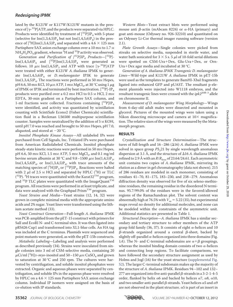
Species Suggests a Steric-basedBindingMotif—To identify an IPsubstrate binding motif, we compared all known substrate andnon-substrate IP species based on a common ring orientationand target hydroxyl position. Two phosphate groups were con-served in all substrate species and were identified as those withthemost contacts with the IP3K substrate-bound structure (4�-and 5�-phosphates). Orientated with the target hydroxylpointed down and away from the plane of view, these phos-phates are the first two located clock-wise from the targethydroxyl. No IP species with a 2�-phosphate group that wasadjacent to the target hydroxyl and pointed in the same direc-tion relative to the plane of the inositol ring was a substrate. Aphosphate was also present for all substrates except Ins(4,5)P2at one of the first two positions immediately counter-clockwisefrom the target hydroxyl. Three IP4 substrates of A. thalianaIPMK and a non-substrate species are shown in Fig. 2A, thecommonmotif in Fig. 2B. The presence of a phosphate oppositethe target hydroxyl does not appear to play any role and waspredicted to be solvent-exposed or interact with residues ableto adopt accommodating conformations such as lysine. Duringthe course of this work, Chang andMajerus (38) independentlyproposed a similar theory for H. sapiens IPMK. Their modeladdresses the 5-/3-kinase activities of human IPMK and doesnot include a role for the 2�-hydroxyl and does not account forthe 6-kinase activity of the yeast or plant homologues, which isseverely reduced in the human form.Putative Binding Modes of Ins(1,4,5)P3, Ins(1,4,5,6)P4,
Ins(1,3,4,5)P4, and Ins(1,3,4,6)P4—All known substrate andnon-substrate IP species were modeled into the active site ofA. thaliana IPMK, following the proposed motif. As expected,the A. thaliana IPMK structure was able to accommodate allsubstrate species, and no non-substrate species except
TABLE 1Crystallographic data, phasing, and refinement statistics
Data collection and phasing 16-286 SeMet
Space group P3221 P3221Unit cell a � b � 130.8 Å, c � 129.9 Å; � � � � 90°, � � 120° a � b � 130.2 Å, c � 129.4 Å; � � � � 90°, � � 120°Wavelength (Å) 1.000 0.97166Resolution (Å) 46-2.9 50-3.1Unique reflections 28,738 44,459Completeness (%) 100 (100)a 100 (100)Rmerge
b 8.5 (52.4) 9.2 (52.6)FOM (SAD) 1.31
RefinementResolution range (Å) 46–2.9 (2.975–2.90)No. of reflections 314,752 (43,621)R (%) 23.64 (31.5)Rfree (%) 24.61 (37.0)r.m.s.d.cBond length (Å) 0.015Bond angle 1.496°B factor (Å2) main chain bonds 0.917B factor (Å2) side chain bonds 2.725Protein atoms, number 3444Ligand atoms, number 30Residues in allowed �-� region 99.5%
a Data in parentheses are for highest resolution shells.b Rsym � �hkl�(N/(N � 1))�i�Ii(hkl) � I(hkl)�/�iIi(hkl).c r.m.s.d., root mean square deviation.
Redesigning IPMK
OCTOBER 12, 2012 • VOLUME 287 • NUMBER 42 JOURNAL OF BIOLOGICAL CHEMISTRY 35363
by guest on October 29, 2018
http://ww
w.jbc.org/
Dow
nloaded from

Ins(1,4)P2 and Ins(1,3,4)P3. These species lacked one of the twophosphate groups conserved in all substrate species. Asexpected, the ring positions that did not appear to require or
exclude phosphate groups proved to be either solvent-exposedor had potential favorable interactions with residues Lys-117,Lys-121, Lys-153, or Arg-156. All possessed highly flexible side
Redesigning IPMK
35364 JOURNAL OF BIOLOGICAL CHEMISTRY VOLUME 287 • NUMBER 42 • OCTOBER 12, 2012
by guest on October 29, 2018
http://ww
w.jbc.org/
Dow
nloaded from
chainswithminimal adjacent packing, presumably allowing theconformational flexibility required to tolerate the presenceor absence of an IP phosphate group. IP species with a2�-phosphate group were modeled into this presumed activesite and were found to have extensive steric clashes. Fig. 2Cshows inositol modeled in the proposed 6-kinase orientationin the active site of A. thaliana IPMK, with each phosphatepocket designated �-� located clockwise from the kinase-target hydroxyl .Substrate-selective Design and Product Identification—To
test the substrate-selective motif, mutants were designed thatwere predicted to exclude specific classes of IP species frombinding. Mutant A. thaliana IPMK constructs were treatedwith [�-32P]ATP and Ins(1,4,5)P3. TheK117Wmutantwas pre-dicted to partially inhibit both the 6�-phosphorylation ofIns(1,3,4,5)P4 and the 3�-phosphorylation of Ins(1,4,5,6)P4 byblocking the �-pocket, presumably occupied by a (different)phosphate for both IP4 species. Full inhibitionwas not expecteddue to the existence of a clash-relieving near-rotamer. K121Wwas predicted to severely inhibit the 3�-phosphorylation ofIns(1,4,5,6)P4 but have little or lesser effect on the 6�-phos-
phorylation of Ins(1,3,4,5)P4, as the blocked �-pocketwould notbe occupied by a phosphate for the latter IP4 species (Fig. 2D).Treatment of Ins(1,4,5)P3 with [�-32P]ATP and K117Wresulted in IP4 and IP5 formation, with reduced IP5 levels com-pared with wild-type, whereas K121W generated IP4 and no ortrace IP5 (data not shown). Time courses of phosphorylation of[3H]IP3 by wild-type and K121W confirmed the 32P-labeledobservations (Fig. 3A). Treatment of the purified [32P]IP4 prod-uct fromK121Wwith 5-ptase identified it as Ins(1,4,5,6)P4 (Fig.3B), as the internal control [3H]Ins(1,3,4,5)P4 was de-phosphorylated to Ins(1,3,4)P3; the [32P]IP4 product was notand eluted at a different time point, matching the control[3H]Ins(1,4,5,6)P4 (data not shown).K117W and K121W Mutants Possess Different Substrate
Profiles—Michaelis-Menten steady-state kinetics experimentswere performed to determine the changes inmutant specificity.The kinetic parameters Km and Vmax were measured for thefollowing reactions for wild-type IPMK and both selectivemutants: Ins(1,4,5)P3 3 Ins(1,4,5,6)P4 (6-kinase), Ins(1,4,5,6)P4 3 Ins(1,3,4,5,6)P5 (3-kinase), and Ins(1,3,4,5)P4 3Ins(1,3,4,5,6)P5 (6-kinase) (Fig. 3E and Table 2). The following
FIGURE 1. Structure and comparison of A. thaliana IPMK. A, stereo schematic of secondary structure. B, representative stereo density of core of A. thalianaIPMK and anomalous peaks. Single coefficient-weighted density (calculated by FFT in CCP4 4. 6.1) is shown at 1.5 (blue), and anomalous density is shown at12 around three of the five seleno-methionine residues used to solve the structure. C, stereo overlay of A. thaliana IPMK (blue), S. cerevisiae IPMK (green), andH. sapiens IP3K (yellow) structures. D, active site comparison of A. thaliana IPMK and H. sapiens IP3K with Ins(1,4,5)P3. Stereo view of the active sites of A. thalianaIPMK and H. sapiens IP3K with AMP-PnP from H. sapiens IP3K and Ins(1,4,5)P3 modeled in the proposed 6-kinase orientation. Note the poor structural conser-vation on the opposite side of the inositol ring and much more extensive structure and contacts of H. sapiens IP3K.
FIGURE 2. Inositol phosphate species, binding motif, and A. thaliana IPMK selective designs. A, substrate and non-substrate IP species. Three A. thaliana IPMK IPsubstrate species, one for each kinase activity, and one non-substrate species are shown. All are orientated to preserve the position of the target hydroxyl (circled) andring orientation. B, proposed IP substrate motif. Also shown are proposed rules for IP substrate selectivity by A. thaliana IPMK. Two phosphate groups are absolutelyrequired (boldface type), the ring orientation must be maintained, one of two phosphate groups is required (arrows), and an axial phosphate cannot be tolerated if itoccupies the first position clockwise of the target hydroxyl. C, phosphate binding pockets. Inositol in the active site of A. thaliana IPMK is shown conserving ring-carbonorientation. The phosphate pockets are designated �-� clockwise from the kinase target hydroxyl . Surrounding residues are omitted for clarity. D, proposed bindingof IP4 species with K117W and K121W mutants. Ins(1,3,4,5)P4 and Ins(1,4,5,6)P4 are modeled with either K117W or K121W mutant models. K117W is predicted to havesteric clashes with both IP4 species, whereas K121W is predicted to clash with only Ins(1,4,5,6)P4.
Redesigning IPMK
OCTOBER 12, 2012 • VOLUME 287 • NUMBER 42 JOURNAL OF BIOLOGICAL CHEMISTRY 35365
by guest on October 29, 2018
http://ww
w.jbc.org/
Dow
nloaded from

pattern in kcat/Km was predicted based on the number ofblocked sites for each combination of mutant and IP4 species,the presence of adjacent phosphate groups that would restrictthe ability of the blocked phosphate to adopt a relieving confor-mation, and the possibility that alternate residue conforma-tions existed to reduce clash:WT�K121W� IP3 �K117W�Ins(1,3,4,5)P4 � K121W � Ins(1,3,4,5)P4 � K117W �Ins(1,4,5,6)P4 � K121W � Ins(1,4,5,6)P4. This pattern wasobserved, supporting the inositol selectivity hypothesis. If lossof activity were due to a general inhibition of IP binding such assteric exclusion of the inositol ring, similar losses of activitywould have been expected for all IP species for a given mutant.The conservation of the 1�- and 4�-phosphates in two con-served pockets proposed by Ongusaha (39) and reviewed byIrvine (3) is not consistent with the single active site nor thepattern of activity loss.
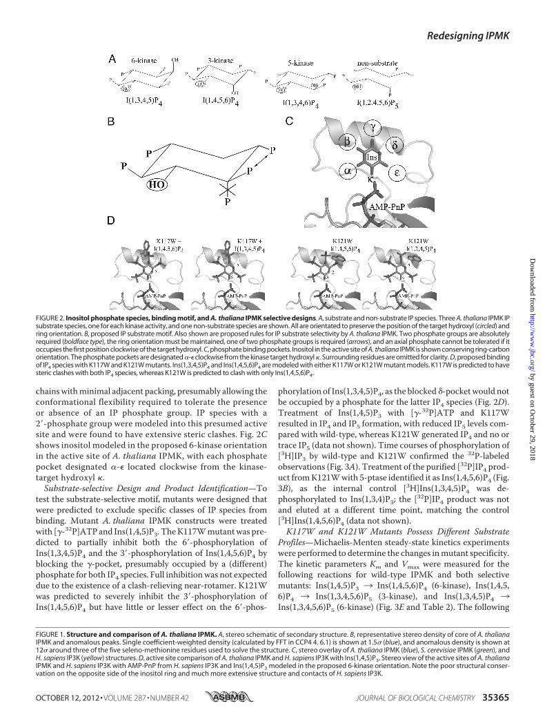
In Vivo Different Inositol Phosphate Populations Are Alteredby K121W and K117W/K121W—To determine whetherchanges in the in vitro selectivity of themutants affected in vivoIP production, the inositol profiles of ipk2� S. cerevisiae withvarious A. thaliana IPMK constructs were measured by HPLC(Fig. 4A). In addition to the K121W and K117W/K121W dou-ble-mutants, empty vector, wild-type, and D247A kinase-deadconstructswere also used. Similar levels of Ins, IP1, and IP2weremeasured for all constructs. IP3 levels were highest in the con-trol and kinase-dead constructs, and progressively less in dou-ble mutant, K121W, and wild-type (negligible). Slightly ele-vated levels of IP3 observed inD247Amutant as comparedwithvector were not found to be statistically significant. IP4 levelswere negligible in all constructs except the K121W andK117W/K121Wmutants, approaching the control and kinase-dead IP3 levels. IP6 (produced from IP5 by IPK1(40)) levels wereequivalent in K121W and wild-type but greatly reduced in thedouble mutant. The IP4 to IP6 ratios changed dramatically withthe different selective mutants; although essentially, no IP4 ispresent with wild-type IPMK, levels of IP4 and IP6 are equiva-lent in K121W, and IP4 is 6-fold higher or greater than IP6 inK117W/K121W.Mutants Have Different Rescue of Nitrogen Growth Defects
ThanWild-type—Rescue of the ipmk� ornithine growth defectwas compared between wild-type A. thaliana IPMK and theselective constructs (Fig. 4C). Yeast, plant, and fly IPMK havepreviously been shown to fully rescue growth with ornithine asthe sole source of nitrogen (12, 14). Growth on CSM and glu-tamate media was unaffected by the deletion of IPMK but waslost when plated onto media containing ornithine as the solenitrogen source. Both IPMK mutants were able to fully rescue
FIGURE 3. IP production, identity, and kinetics. A, production of IP4 and IP5 over time. Time courses of generation of IP4 and IP5 by wild-type and K121WA. thaliana IPMK from [3H]Ins(1,4,5)P3. Note the simultaneous appearance of IP4 and IP5 in the wild-type reactions, but no IP5 signal for the K121W reactions,even after 60 min. B, identification of the K121W IP4 product as Ins(1,4,5,6)P4. Samples containing [32P]IP4 produced by K121W and [3H]Ins(1,3,4,5)P4 as aninternal standard treated or not treated with 5-ptas and separated by HPLC, and radioactivity was counted on 3H and 32P channels simultaneously. 5-Ptaseproduct is observed in only for [3H]IP4. C, representative TLC plate of [32P]IP4 assay. D, representative HPLC trace of [3H]Ins(1,4,5)P3 assay. E, Michaelis-Mentensteady-state kinetic curves for Ins(1,4,5)P3 and Ins(1,4,5,6)P4 with wild-type and K121W A. thaliana IPMK.
TABLE 2Kinetics
IP Species Wild-type K117W K121W
I(1,4,5)P3Vmax (�mol/min/mg) 8.96 NPa 3.52Km (�M) 162.2 50.05kcat/Km (s�1 M�1) 2.97 � 104 3.78 � 104
I(1,3,4,5)P4Vmax (�mol/min/mg) 6.06 6.11 6.61Km (�M) 30.82 568 490kcat/Km (s�1 M�1) 1.06 � 105 5.79 � 103 7.27 � 103
I(1,4,5,6)P4Vmax (�mol/min/mg) 11.04 0.96 0.39Km (�M) 74.97 254 216kcat/Km (s�1 M�1) 7.91 � 104 2.04 � 103 9.73 � 102
a NP, not performed.
Redesigning IPMK
35366 JOURNAL OF BIOLOGICAL CHEMISTRY VOLUME 287 • NUMBER 42 • OCTOBER 12, 2012
by guest on October 29, 2018
http://ww
w.jbc.org/
Dow
nloaded from
growth on ornithine, with similar colony number and size aswild-type A. thaliana IPMK, whereas the catalytic knock-outD247A mutant failed to rescue.Nitrogen Metabolism Control Does Not Appear to Require a
Polyaspartate Region—It has been proposed that the polyaspar-tate region of S. cerevisiae IPMK, rather than the kinase func-tionality, is responsible for rescue of ornithine growth (41).Although kinase-deadmutants of yeast IPMKwere reported topartially rescue growth on ornithine media, we observed thatgrowth could also be rescued by plant and fly IPMK. Not onlydoes A. thaliana IPMK clearly lack the polyaspartate insert orany sequence similarity to it (supplemental Fig. S3A), but thestructure reveals that short of a complete unfolding of strands�9 and �10, there is no way a similar conformation could beadopted. Although residues 231–236 are disordered, any pos-sible loop would be far too small to imitate the size of the28-residue disordered region in yeast that includes the poly-aspartate insert. Furthermore, the polyaspartate region in yeastbegins pointed in the opposite direction of the beginning of thedisordered region in plant (supplemental Fig. S3B). With onlysix residues, the plant disordered region would be unable toimitate even the residues of the yeast insert that are observable,much less the disordered region.
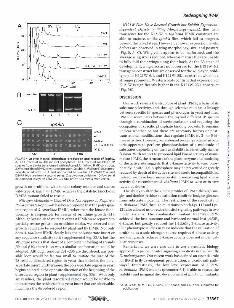
K121W Flies Have Rescued Growth but Exhibit Expression-dependent Defects in Wing Morphology—ipmk� flies withtransgenes for the K121W A. thaliana IPMK construct areable to mature, unlike ipmk� flies, which fail to progressbeyond the larval stage. However, at lower expression levels,defects are observed in wing morphology, size, and posture(Fig. 5, A–C). Wing veins appear to be malformed, and theaverage wing size is reduced, whereas mature flies are unableto fully fold their wings along their back. At the L3 stage ofdevelopment, wing discs are not observed for the K121W-6.1transgene construct but are observed for the wild-type, wild-type plus K121W-6.1, and K121W-25.1 construct, which is astronger promoter.Western blots confirm that expression ofK121W is significantly higher in the K121W-25.1 construct(Fig. 5E).
DISCUSSION
Our work reveals the structure of plant IPMK, a basis of itssubstrate selectivity, and, through selective mutants, a linkagebetween specific IP species and phenotypes in yeast and flies.IPMK discriminates between the myriad different IP speciesthrough a combination of steric exclusion and requiring theoccupation of specific phosphate binding pockets. It remainsunclear whether or not there are accessory factors or post-translational modifications that regulate IPMK 6-, 3-, or 5-ki-nase activities.However, recombinant protein produced in bac-teria appears to perform phosphorylation of a multitude ofsubstrates depending on their availability in kinetically similarfashion.With respect to proposed lipid kinase activity of mam-malian IPMK, the structure of the plant enzyme and modelingof the active site suggests that 3-kinase activity toward phos-phatidylinositol 4,5-bisphosphate may be prevented or greatlyreduced by depth of the active site and steric incompatibilities.Indeed, we have been unsuccessful in measuring lipid kinaseactivity for recombinant A. thaliana IPMK in vitro or in vivo(data not shown).The ability to alter the kinetic profiles of IPMK through sin-
gle- and double-residue substitution confirms insights gleanedfrom substrate modeling. The restriction of the specificity ofA. thaliana IPMK through mutations to both Lys-117 and Lys-121 also allowed us to rewire inositol signaling pathways in twomodel systems. The combination mutant K117W/K121Wachieved the best outcome and harbored normal Ins(1,4,5)P36-kinase, but greatly reduced Ins(1,4,5,6)P4 3-kinase activity.Our phenotypic studies in yeast indicate that the utilization ofornithine as a sole nitrogen source requires 6-kinase activityand that greatly reduced 3-kinase activity does not impair cel-lular responses.Remarkably, we were also able to use a synthetic biology
approach to probe inositol signaling specificity in the fruit flyD. melanogaster.Our recent work has defined an essential rolefor IPMK in fly development, proliferation, and cell death path-ways.4 Interestingly, the low level expression of K121WA. thaliana IPMK mutant (promoter 6.1) is able to rescue theviability and imaginal disc development of ipmk null mutants;
4 A. M. Seeds, M. M. Tsui, C. Sunu, E. P. Spana, and J. D. York, submitted forpublication.
FIGURE 4. In vivo inositol phosphate production and rescue of ipmk�.A, HPLC traces of soluble inositol phosphates. HPLC traces of soluble [3H]IPspecies from ipmk� transformed with indicated A. thaliana IPMK constructs.B, Western blot of IPMK construct expression. Soluble A. thaliana IPMK expres-sion detected with �-HA and normalized to �-actin. K117W/K121W andD247A blots are from a second series. C, growth on ornithine. 10-Fold serialdilution spot assays on CSM-Ura, Glu-Ura, or Orn-Ura media. Vect, vector.
Redesigning IPMK
OCTOBER 12, 2012 • VOLUME 287 • NUMBER 42 JOURNAL OF BIOLOGICAL CHEMISTRY 35367
by guest on October 29, 2018
http://ww
w.jbc.org/
Dow
nloaded from
however, we noticed that proper wing patterning and positionin adult flies remained perturbed, whereas high copy expres-sion of the mutant (promoter 25.1) fully rescued viability andwing development. Our in vitro kinetics and yeast in vivomet-abolic labeling of the K121W mutant indicates IP33 IP4 pro-duction is normal, but conversion to IP5 is reduced but notabolished. We favor a model in which 6-kinase activity is criti-cal; however, we cannot rule out that the residual 3-kinase, andpossible untested 5-kinase, activities also contribute to theIMPK function in fly development. Further optimization ofselective IPMK designs and testing of the K117W/K121Wmutant in flies may help resolve these hypotheses in the flysystem.Our structure also provided suggestions that non-catalytic
regions of IPMK may participate in cellular signaling andregulation. Of interest, the polyaspartate loop of S. cerevisiaeIPMK, which was disordered in the crystal structure, was notpresent in A. thaliana IPMK. We also found that the loop ofsix residues between the �9 and �10 strands was disordered.Given that the plant protein does not harbor a polyaspartateregion, the fact that the protein is fully capable of restoringgrowth with ornithine as the sole nitrogen source contra-dicts previous reports that the polyaspartate region isrequired for transcriptional responses mediated by IMPK(41). The fact that the rescue of growth by the plant enzymedepends on catalysis further emphasizes a role for IPMK and
production of Ins(1,4,5,6)P4 in regulating transcriptionalresponses through the ArgR-Mcm1 complex. We also notethat recent reports of the structural basis for Ins(1,4,5,6)P4 inbinding to and regulating the histone deacetylase/co-repres-sor complex (HDAC3-NCOR2) provides an exciting newtarget for inositol signaling through IPMK (42).Overall, our study provides insights into both the structure
and biology of IPMK through a synthetic biology approach.Insights into substrate binding, kinase specificity and non-cat-alytic regions of IMPKhave been used to answer unique biolog-ical questions in two model organisms, budding yeast and fruitflies. These have helped resolve relevance of signaling mole-cules and provide a foundation for future studies aimed atunderstanding signaling specificity and biology.
Acknowledgments—We thank the members of the York laboratory forhelpful discussions, in particular Dr. D. Eric Dollins for discussionsduring phase determination, model building, and kinetic assays; Dr.Jim Otto and Peter Fridy for HPLC analysis, and Ace Hatch andTrang Pham for protocols and advice. We thank Drs. Jane and DavidRichardson and Dr. Bruce R. Donald (R01-GM-078031) for helpfulsupport. We finally thank the x-ray crystallographic structure facilityat Duke University and the Advanced Photon Source at ArgonneNational Laboratories and Advanced Light Source (Berkeley) wherediffraction data were collected.
FIGURE 5. Fly complementation analysis and wing morphology. A and B, representative wings and size distributions. Mean size � S.D., n � 19. C andD, wing postures and eggs laid by indicated genotypes. WT, WT � K121W, ipmk� � K121W-6.1, and ipmk� � K121W-25.1. E, Western blot of GFP-A. thaliana IPMK expression. Expression of various GFP-A. thaliana IPMK transgenes in D. melanogaster L3 larvae using �-GFP and �-actin as loadingcontrols.
Redesigning IPMK
35368 JOURNAL OF BIOLOGICAL CHEMISTRY VOLUME 287 • NUMBER 42 • OCTOBER 12, 2012
by guest on October 29, 2018
http://ww
w.jbc.org/
Dow
nloaded from
REFERENCES1. Drin, G., and Scarlata, S. (2007) Stimulation of phospholipase C� bymem-
brane interactions, interdomain movement, and G protein binding–howmany ways can you activate an enzyme? Cell Signal. 19, 1383–1392
2. Gonzales, M. L., and Anderson, R. A. (2006) Nuclear phosphoinositidekinases and inositol phospholipids. J. Cell Biochem. 97, 252–260
3. Irvine, R. F. (2005) Inositide evolution–towards turtle domination?J. Physiol. 566, 295–300
4. Blazer-Yost, B. L., and Nofziger, C. (2005) Phosphoinositide lipid secondmessengers: New paradigms for transepithelial signal transduction.Pflugers Arch. 450, 75–82
5. Hatch, A. J., and York, J. D. (2010) SnapShot: Inositol phosphates. Cell143, 1030–1030.e1
6. York, J. D. (2006) Regulation of nuclear processes by inositol polyphos-phates. Biochim. Biophys. Acta 1761, 552–559
7. Seeds, A. M., Bastidas, R. J., and York, J. D. (2005) Molecular definition ofa novel inositol polyphosphate metabolic pathway initiated by inositol1,4,5-trisphosphate 3-kinase activity in Saccharomyces cerevisiae. J. Biol.Chem. 280, 27654–27661
8. Bechet, J., Greenson, M., andWiame, J. M. (1970)Mutations affecting therepressibility of arginine biosynthetic enzymes in Saccharomyces cerevi-siae. Eur. J. Biochem. 12, 31–39
9. Dubois, E., and Messenguy, F. (1994) Pleiotropic function of ArgRIIIp(Arg82p), one of the regulators of arginine metabolism in Saccharomycescerevisiae. Role in expression of cell-type-specific genes.Mol. Gen. Genet.243, 315–324
10. Messenguy, F., Dubois, E., and Boonchird, C. (1991) Determination of theDNA-binding sequences of ARGR proteins to arginine anabolic and cat-abolic promoters.Mol. Cell Biol. 11, 2852–2863
11. Dubois, E., andMessenguy, F. (1985) Isolation and characterization of theyeast ARGRII gene involved in regulating both anabolism and catabolismof arginine.Mol. Gen. Genet. 198, 283–289
12. Odom, A. R., Stahlberg, A., Wente, S. R., and York, J. D. (2000) A role fornuclear inositol 1,4,5-trisphosphate kinase in transcriptional control. Sci-ence 287, 2026–2029
13. York, J. D., Odom, A. R., Murphy, R., Ives, E. B., andWente, S. R. (1999) Aphospholipase C-dependent inositol polyphosphate kinase pathway re-quired for efficient messenger RNA export. Science 285, 96–100
14. Stevenson-Paulik, J., Odom, A. R., and York, J. D. (2002) Molecular andbiochemical characterization of two plant inositol polyphosphate 6-/3-/5-kinases. J. Biol. Chem. 277, 42711–42718
15. Stevenson-Paulik, J., Bastidas, R. J., Chiou, S. T., Frye, R. A., and York, J. D.(2005) Generation of phytate-free seeds in Arabidopsis through disrup-tion of inositol polyphosphate kinases. Proc. Natl. Acad. Sci. U.S.A. 102,12612–12617
16. Holmes, W., and Jogl, G. (2006) Crystal structure of inositol phosphatemultikinase 2 and implications for substrate specificity. J. Biol. Chem. 281,38109–38116
17. Van Duyne, G. D., Standaert, R. F., Karplus, P. A., Schreiber, S. L., andClardy, J. (1993) XDS atomic structures of the human immunophilinFKBP-12 complexes with FK506 and rapamycin. J. Mol. Biol. 229,105–124
18. Kabsch, W. (1993) XDS automatic processing of rotation diffraction datafrom crystals of initially unknown symmetry and cell constants. J. Appl.Cryst. 26, 795–800
19. Sheldrick, G. M. (1997) SHELX97 Manual, University of Gottingen,Germany
20. Brünger, A. T., Adams, P. D., Clore, G. M., DeLano, W. L., Gros, P.,Grosse-Kunstleve, R.W., Jiang, J. S., Kuszewski, J., Nilges,M., Pannu,N. S.,Read, R. J., Rice, L. M., Simonson, T., andWarren, G. L. (1998) Crystallog-raphy &NMR system: A new software suite formacromolecular structuredetermination. Acta Crystallogr. D Biol. Crystallogr. 54, 905–921
21. DeLano, W. L. (2010) The PyMOL Molecular Graphics System, version1.3r1, Schrödinger, LLC, New York
22. Chamberlain, P. P., Sandberg, M. L., Sauer, K., Cooke, M. P., Lesley, S. A.,and Spraggon, G. (2005) Structural insights into enzyme regulation forinositol 1,4,5-trisphosphate 3-kinase B. Biochemistry 44, 14486–14493
23. Miller, G. J., and Hurley, J. H. (2004) Crystal structure of the catalytic coreof inositol 1,4,5-trisphosphate 3-kinase.Mol. Cell 15, 703–711
24. González, B., Schell,M. J., Letcher, A. J., Veprintsev, D. B., Irvine, R. F., andWilliams, R. L. (2004) Structure of a human inositol 1,4,5-trisphosphate3-kinase: Substrate binding reveals why it is not a phosphoinositide 3-ki-nase.Mol. Cell 15, 689–701
25. Collaborative Computational Project, Number 4 (1994) The CCP4 suite:programs for protein crystallography.ActaCrystallogr. DBiol. Crystallogr.50, 760–763
26. Jones, T. A., Zou, J. Y., Cowan, S. W., and Kjeldgaard. (1991) Improvedmethods for building protein models in electron density maps and thelocation of errors in these models. Acta Crystallogr. A 47, 110–119
27. Emsley, P., and Cowtan, K. (2004) Coot: Model-building tools for molec-ular graphics. Acta Crystallogr. D Biol. Crystallogr. 60, 2126–2132
28. Lovell, S. C., Davis, I.W., Arendall,W. B., 3rd, de Bakker, P. I.,Word, J.M.,Prisant, M. G., Richardson, J. S., and Richardson, D. C. (2003) Structurevalidation by C� geometry: �, �, and C� deviation. Proteins 50, 437–450
29. Winn, M. D., Isupov, M. N., and Murshudov, G. N. (2001) Use of TLSparameters to model anisotropic displacements in macromolecular re-finement. Acta Crystallogr. D Biol. Crystallogr. 57, 122–133
30. Davis, I.W.,Murray, L.W., Richardson, J. S., and Richardson, D. C. (2004)MOLPROBITY: Structure validation and all-atom contact analysis fornucleic acids and their complexes. Nucleic Acids Res. 32,W615–619
31. Kleywegt, G. J. (1996) Use of non-crystallographic symmetry in proteinstructure refinement. Acta Crystallogr. D Biol. Crystallogr. 52, 842–857
32. Seeds, A. M., Sandquist, J. C., Spana, E. P., and York, J. D. (2004) Amolec-ular basis for inositol polyphosphate synthesis in Drosophila melano-gaster. J. Biol. Chem. 279, 47222–47232
33. Ito, H., Fukuda, Y., Murata, K., and Kimura, A. (1983) Transformation ofintact yeast cells treated with alkali cations. J. Bacteriol. 153, 163–168
34. Stolz, L. E., Huynh, C. V., Thorner, J., and York, J. D. (1998) Identificationand characterization of an essential family of inositol polyphosphate5-phosphatases (INP51, INP52, and INP53 gene products) in the yeastSaccharomyces cerevisiae. Genetics 148, 1715–1729
35. Matthews, B.W. (1968) Solvent content of protein crystals. J.Mol. Biol.33,491–497
36. Murzin, A. G. (1996) Structural classification of proteins: New superfami-lies. Curr. Opin. Struct. Biol. 6, 386–394
37. Fan, C., Moews, P. C., Shi, Y., Walsh, C. T., and Knox, J. R. (1995) Acommon fold for peptide synthetases cleaving ATP to ADP: Glutathionesynthetase and D-alanine:D-alanine ligase of Escherichia coli. Proc. Natl.Acad. Sci. U.S.A. 92, 1172–1176
38. Chang, S. C., and Majerus, P. W. (2006) Inositol polyphosphate multiki-nase regulates inositol 1,4,5,6-tetrakisphosphate. Biochem. Biophys. Res.Commun. 339, 209–216
39. Ongusaha, P. P., Hughes, P. J., Davey, J., andMichell, R. H. (1998) Inositolhexakisphosphate in Schizosaccharomyces pombe: Synthesis fromIns(1,4,5)P3 and osmotic regulation. Biochem. J. 335, 671–679
40. Ives, E. B., Nichols, J., Wente, S. R., and York, J. D. (2000) Biochemical andfunctional characterization of inositol 1,3,4,5,6-pentakisphosphate 2-ki-nases. J. Biol. Chem. 275, 36575–36583
41. Dubois, E., Dewaste, V., Erneux, C., and Messenguy, F. (2000) Inositolpolyphosphate kinase activity of Arg82/ArgRIII is not required for theregulation of the arginine metabolism in yeast. FEBS Lett. 486, 300–304
42. Watson, P. J., Fairall, L., Santos, G.M., and Schwabe, J.W. (2012) Structureof HDAC3 bound to co-repressor and inositol tetraphosphate. Nature481, 335–340
Redesigning IPMK
OCTOBER 12, 2012 • VOLUME 287 • NUMBER 42 JOURNAL OF BIOLOGICAL CHEMISTRY 35369
by guest on October 29, 2018
http://ww
w.jbc.org/
Dow
nloaded from

D. YorkStuart Endo-Streeter, Man-Kin Marco Tsui, Audrey R. Odom, Jeremy Block and John
Structural Studies and Protein Engineering of Inositol Phosphate Multikinase
doi: 10.1074/jbc.M112.365031 originally published online August 15, 20122012, 287:35360-35369.J. Biol. Chem.
10.1074/jbc.M112.365031Access the most updated version of this article at doi:
Alerts:
When a correction for this article is posted•
When this article is cited•
to choose from all of JBC's e-mail alertsClick here
Supplemental material:
http://www.jbc.org/content/suppl/2012/08/15/M112.365031.DC1
http://www.jbc.org/content/287/42/35360.full.html#ref-list-1
This article cites 40 references, 12 of which can be accessed free at
by guest on October 29, 2018
http://ww
w.jbc.org/
Dow
nloaded from