structural models of the nicotinic acetylcholine receptor and its
TRANSCRIPT
SCIENTIFIC REPORT
Structural models of the nicotinicacetylcholine receptor and itstoxin-binding sites
H. R. Guy
C,
DEFENSE NUCLEAR AGENCY
. ARMED FORCES RADIOBIOLOGY RESEARCH INSTITUTEie.I- BETHESDA, MARYLAND 20814
APPROVED FOR PUBLIC RELEASE; DISTRIBUTION UNLIMITED- I•
4
BestAvailable
Copy
Al
REVIEWED AND APPROVED
DAVID R. LIVENGOOD, Ph.D. PAUL E. TYLER, M.D.Chairman CAPT, MC, USNPhysiology Department Director
I
I 9Q
Research was conducted i-c-'rdilg to the principles enunciated in the"Guide for the Care and Use of Laboratory Animals,," prepared by theInstitute of Laboratory Animal Resources, National Research Council.
UINC LASSIFIEDSECURITY CLASSIFICATION OF THIS PAGE (When 1).t. Fw-c-dl
REPORT DOCUMENTATION PAGE RELAD 1NsTrRucTIONS
I. REPORT NUMBER 12 GOVT ACCESSION NO. 3 RECIPIENT'S CATALOG NUMBER
AFRRI SR81-30II''
4. TITLE (an~d Sub(l,CNI 5 TP FRP R EIDC V:
STRUCTURAL MODELS OF THE NICOTINICACETYLCHOLINE RECEPTOR ANO ITS TOXIN-BINDING SITES 6 PERFORMING 010. REPORT NuM8ER
7. AUTH-OR~s) B. CONTRACT OR GRANT NUMBERS
H. R., Guy
9 PERFORMING OOGA'NIZATION N 'ME AN' DDGRESS 10 PROGRAM ELEMENT PROJEC- TAS,'C
Armned Forces Radiobiology Research Institute (AERRI) AE OKUý UBR
Defense Nuclear ,A\gency NWED QAXMBethesda, Maryland 20814 __rJ_____ hJ 00032
I I CONTROLLING3 OFFILCE NAME AND ADDRESS '2PE-PORT DATF
Director November 1981Defense Nuclear Agency (DNA) '3 NUMBER OF PAGES
Washington., DC 20305 3314 MONITORING AGENCY NAME 8 ADOýESS'11 differen rrI,.-: C'.1fr~IIrrp Ott.,- IS SE'_URI'ý Cu ASS ',,t (h,,;s-r
UNCLASSIFIED
15_-CFC__ASSIPICAT[N DOWN~eRADING
16. DISTRIBUjTION STA-EMENT 1,! :hi.R11
Approved for public release; distribution unlimited.
17. DISTRIBUTION STATEMENT 'of ýhe rr.Sstracf er,'c-f. .'r-,,,k 26. f different Iro,ý ReporI) V
18. SUPPLEMENTARY NOTESI
Published in Cellular and Molecular Neurobiologgy 1: 231-258, 1981.
19. KEY WORDS (Continure on reverse .i,ii if ner.sesry and idenIty, by block numaber)
acetylcholine receptor; molecular model; protein structure; shake neurotoxin; cholinergicagonists; cholinergic antagonists.
20. ABSTRACT (Continue on reverse side /I necessary and identifv 5
y block numeber)
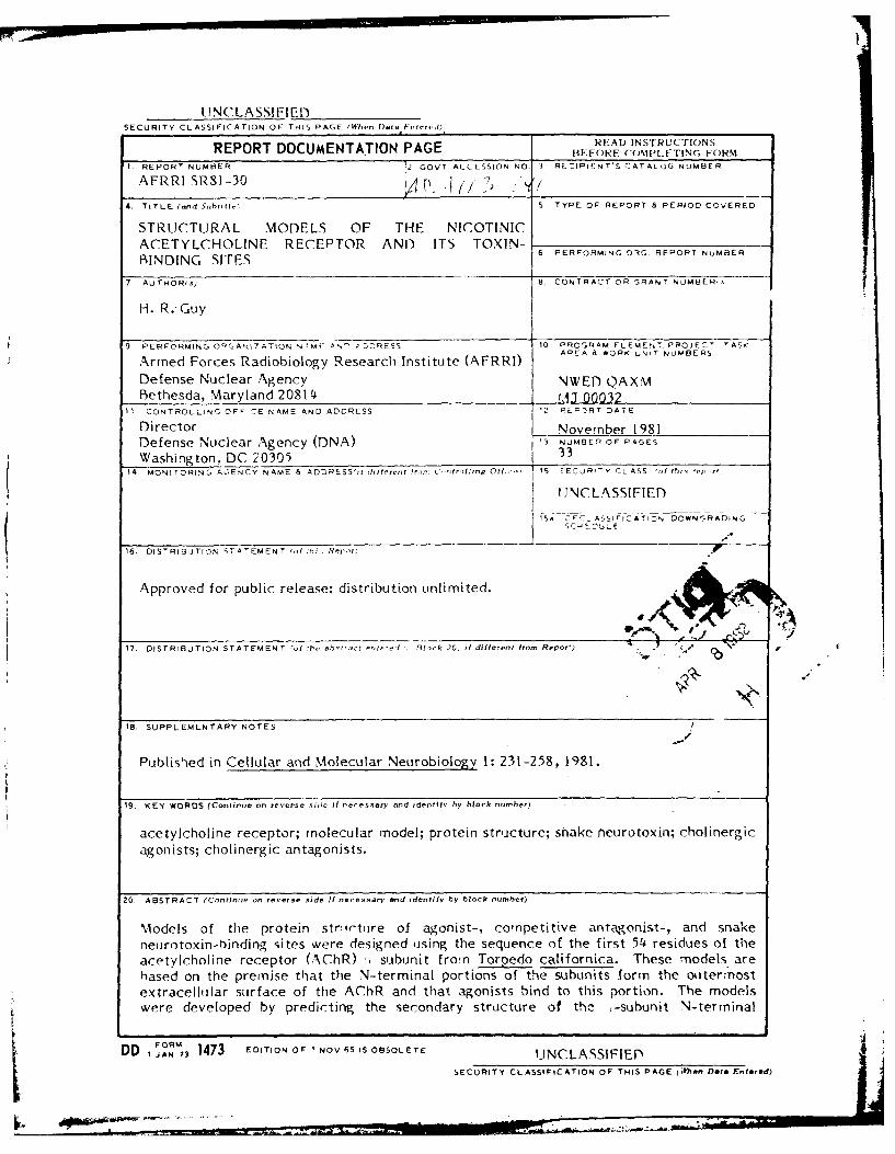
Models of the protein str~ictture of agonist-, competitive antagonist-, and snakeneIurotoxin-rbindina sites were designed using the sequence of the first 54 residues of theacetylcholine receptor (AChR) ý.subunit fro'n Torpedo californica. These models arehased on the premise that the N-terminal portions of the subunits form the otitermnostextracelllllar surface of the AChR and that agonists bind to this portion. The modelswere developed by predicting the secondary structure of the ;-subunit N-terminal
DD I AN7 1473 EDITION OF I NOVA65 IS OBSOLETE UNCLASSIFIEDSECURITY CLASSIFICATION OF THIS PAGE (Whlen Date Fettered)
UNCLASSIFIEDSECURITY CLASSIFICATION OF THIS PAGE(Whelm Date Entered)
20. ABSTRACT (continued)
segment from its sequence, then using these predictions to fold the segment into tertiarystructures that should bind snake neurotoxins, agonists, and antagonists. Possible gatingmechanisms and quaternary structures are suggested by the proposed tertiary structuresof the subunits. Experiments are suggested to test aspects of the models.
A&I
D ITIC USl [
L m.anaouacod EjustifiatlOn
DOTIO DitiAtO I
COPY elfb1tCv'
Dist
UNCLASSIFIED-SECURITY CL ASSIFICATIC.19 OF - '
(ellular and Aloh'c lar Vo'ur,hi,,o'. 1 ,d I No 3. 1 Q/
Structural Models of the NicotinicAcetylcholine Receptor and ItsToxin-Binding Sites'
H. Robert Guy 2.3
Received November 21, 1980:accepted March 20, 198/
Models of the protein structure oj agonist-. competitive antagonist-, and snake neurotoxin-binding siteswere designed using the sequence of the first 54 residues ojfthe acetylcholine receptor (AChRJ a subunitfrom Torpedo californica. These models are based on the premise that the N-terminal portions of thesubunits jorm the outermost extracellular surface of the AC/hR and that agonists bind to this portion. Themodels were developed by predicting the secondary strucutre of the ar-subunit N-terminal segment from itssequence, then using these predictions to fold the segment into tertiary structures that should bind snakeneurotoxins. agonists, and antagonists.. Possible gating mechanisms and quaternar" structures aresuggested by the proposed tertiary structures of the subunits. Experiments are suggested to test aspects ofthe models.
KEY WORDS: acetylcholine receptor; molecular model; protein structure: snake neurotoxin:cholinergic agonists; cholinergic antagonists.
INTRODUCTION
The electrical activity of nerves and muscles is regulated by a variety of voltage- andtransmitter-activated proteins that form ion channels through the cellular membranes.A major unresolved problem in neurobiology is the molecular structure of thesechannels and their associated receptors. It is generally recognized that a preciseknowledge of these structures would be invaluable in understanding the functionalmechanisms of the nervous system and the ways that various drugs and toxins alter
Supported by Armed Forces Radiobiology Research Institute. Defense Nuclear Agency, under ResearchWork Unit MJ 00032. The views presented in this paper are those of the author. No endorsement by theDefense Nuclear Agency has been given or should be inferred.
2 Physiology Department, Armed Forces Radiobiology Research Institute. Bethesda, Maryland 20014.'Present address: Building 36, Room 2A29, Division of Molecular Biophysics, NINCDS, National
Institutes of Health, Bethesda. Maryland 20205.
231
0272-4340/81 /0900-021 I S03 00/( W 1991 Plenum Pu.bh,,h, ('.gr, ,,"-.
232 G(u.
these mechanisms. Unfortunately. there are two severe impediments to determiningmembrane channel structures: t I ) most membrane channels are ditlicult to isolate insufficient quantities and purity to determine their amino acid sequences, and (2) it isdifficult to crystallize membrane proteins in the manner required for X-ray diffractionanalysis. Nicotinic acetylcholine receptors and their associated channels (AChRcomplexes) have been studied extensively. Because of the abundance oi" ,ChRcomplexes in the electric organs of electric rays and eels, considerable progress ii-.been made in isolating them and analyzing their structures. The sequence of the first54 residues of each of the subunits from Torpedo calijbrnica AChR complexes hasbeen determined recently (Raftery el al.. 1980). Since X-ray data of AChR complexcrystal may not be available in the forseeable future, alternative approaches may haveto be used to analyze the data that are available.
The models presented here were developed by using the sequence of theN-terminal portions of the t subunit to design structures that should be energeticallxstable and that should bind the potent snake neurotoxins and the most potent of thesmall competitive inhibitors: alloferin. The models are consistent with the availablestructural data for the AChR complex and suggest mechanisms by which activation
occurs. Both models are intended as working hypotheses that can be used to designexperiments to eliminate the ambiguities of the model. The models also serve as amethod of integrating the various data on the structure and function of the AChRcomplex into a coherent picture.
EXPERIMENTAL DATA
Structural Data
Before proposing detailed structures for the N-terminal segments, it is necessary
to describe the data and a nonspecific model of the entire AChR structure on which
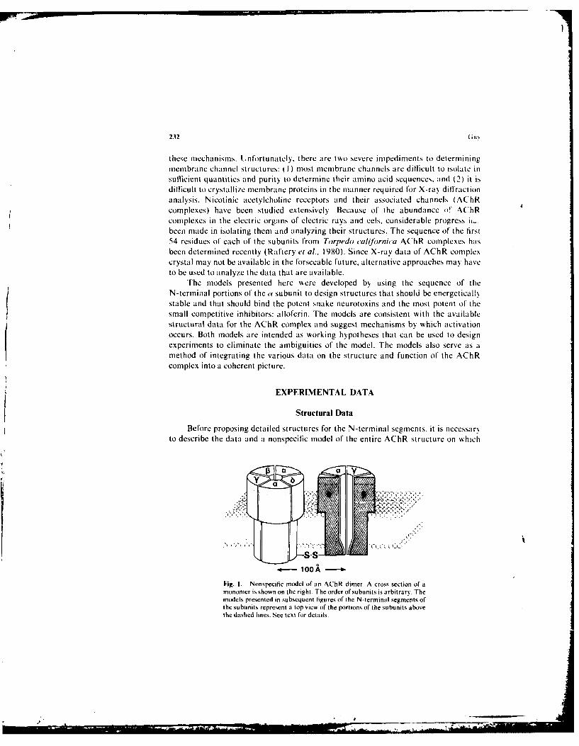
Fig. I. Nonspecific model of an AChR dimer. A cross section of amonomer is shown on the right. The order of subunits is arbitrar). Themodels presented in subsequent figures of the N-terminal segments ofthe subunits represent a top view of the portions of the subunits abovethe dashed lines. See text for details.
S1
A,(i Reccptor S•ructurc 233
the detailed models are based. A schematicized drawing of the nonspecific model isshown in Fig. I. Most of the structural data described here were obtained from AChRcomplexes of the electric organ of Torpedo californica. These AChR complexes arecomprised of four types of subunits, called ar, d. '1, and 5 with apparent molecularweights of about 40,000, 50,000, 60,000, and 65,000, respectively (for review, seeHeidmann and Changeux, 1978: Raftery el al.. 1979). The stoichiometry of thesubunits is a,1,-yh (Raftery et al.. 1980). These subunits apparently comprise both theagonist receptor and the channel since functional ACh-activated channels have beenobtained in preparations in which these were the only subunits present in significantquantities (Gonzalez-Ros et al.. 1980: Moore et al.. 1979: Nelson et al.. 1980:Schindler and Quast, 1980). Studies with cell-free synthesis of AChR subunitssuggest that each of the subunits spans the membrane and that the N-terminal endsare on the extracellular side of the membrane (Anderson and Blobel, 1980).
Electron micrographs of membrane fragments that are enriched in AChRcomplexes indicate the presence of asymmetric "rosette" or "'doughnut"-shapedstructures that are about 85 A in diameter and have a center pit or hole of about 20-Adiameter (see Fig. 7) (Ross et al.. 1977: Zingsheim et al., 1980). The length of theprotein perpendicular to the membrane is about 110 A• with a 50-A segmentextending from the extracellular membrane surface and a 15-A segment extendingfrom the intracellular surface (Ross el al., 1977). A calculation of the volume of therosette structure indicates that its molecular weight is about 255,000 (Klymkowskyand Stroud, 1979). This and the stoichiometries described above indicate that eachrosette is comprised of two a, one J3, one -y, and one 6 subunits. In Torpedo californica.the AChR complexes appear to form dimers which are joined by a disulfide bridgebetween two 6 subunits (Raftery et al.. 1979). This disullide bridge appears to be nearthe C-terminal end of the 6 subunits (Oswald et al.. 1980) and is thus probably on theinside of the cell.
X-ray diffraction studies of AChR-rich membrane fragments suggest thepresence of two large structures: one with a repeat distance of 5.2 A and a length of 80A that probably corresponds to a helices oriented perpendicularly to the membrane.and one with a repeat distance of 6.3 A and a length of 90 A that may be due to a 3structure (Ross el al.. 1977). Circular dichroism and infrared spectroscopy studies ofisolated AChR complexes from various preparations suggest that they are comprisedof 34% a helices and 29%7 J3 sheets ( Moore el al.. 1974). However, the isolation processmay have altered the conformations. Sequences of the N-terminal portions and thetotal amino acid compositions of all the subunits are very similar (Raftery et al.. 1980:Vandlen et al.. 1979; Lindstrom et al.. 1979). These compositional similarities suggestthat the subunits evolved from the same protein and that their overall structures aresimilar.
The data described in this section support a model in which the AChR complex iscomprised of five structurally similar subunits (two ay, one 0, one 'y, and one 6) thatstack next to each other so that a channel forms between the subunits when the AChRis in the open conformation. The N-terminal portions for which the sequence is knownprobably form part of the extracellular domain that binds agonists and competitiveinhibitors.
234 Gu%
Binding of Agonists and Antagonists
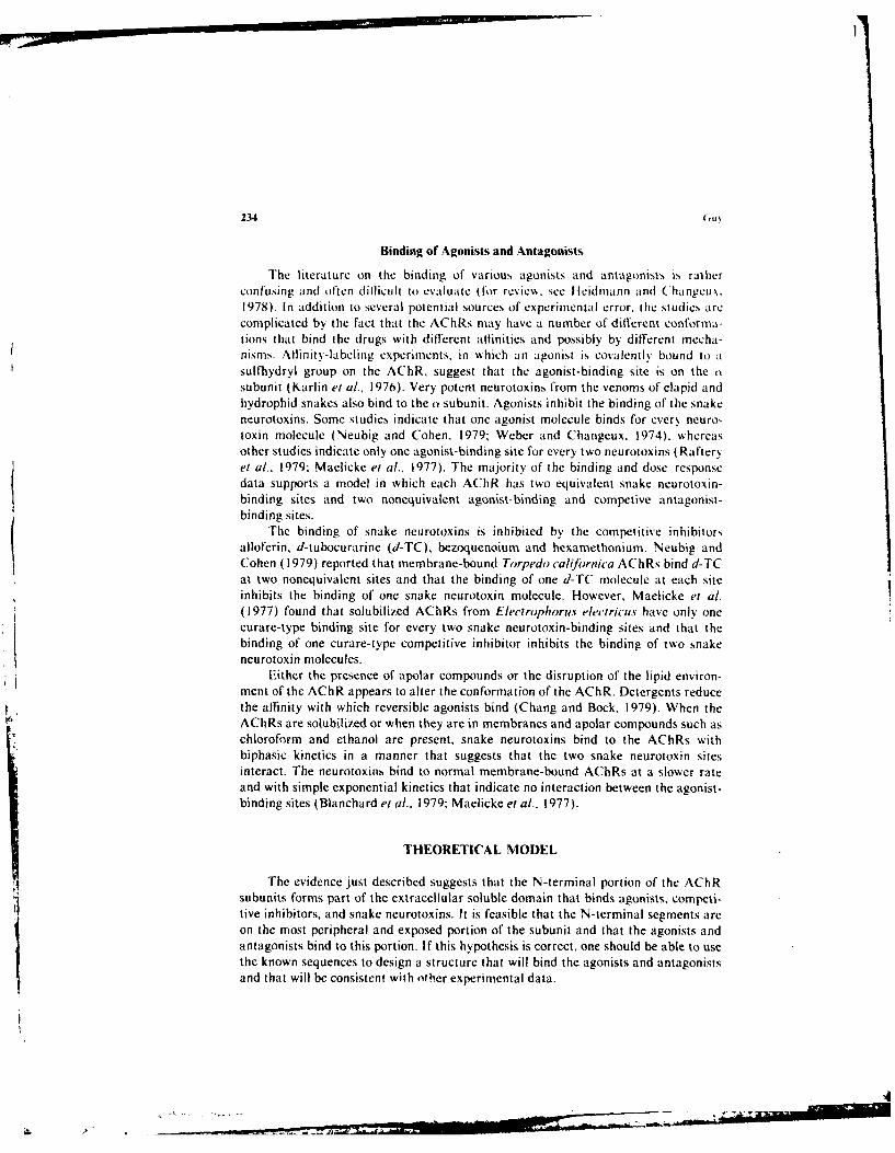
The literature on the binding of various agonists and antagonists is ratherconfusing and often ditlicult to evaluate (for review, see Ilcidmann and (hangcux.1978). In addition to several potential sources of experimental error, the studies arecomplicated by the fact that the AChRs may have a number of different conforma-tions that bind the drugs with different aflinities and possibly by different mecha-nisms. AllinitN-labeling experiments, in which an agonist is covalently bound to asulfhydryl group on the AChR, suggest that the agonist-binding site is on the osubunit (Karlin et al.. 1976). Very potent neurotoxins from the venoms of elapid andhydrophid snakes also bind to the a subunit. Agonists inhibit the binding of the snakeneurotoxins. Some studies indicate that one agonist molecule binds for every neuro-toxin molecule (Neubig and Cohen. 1979: Weber and Changeux. 1974). whereasother studies indicate only one agonist-binding site for every two neurotoxins (Rafteryel al., 1979: Maelicke et al.. 1977). The majority of the binding and dose-responsedata supports a model in which each AChR has two equivalent snake neurotoxin-binding sites and two nonequivalent agonist-binding and competive antagonist-binding sites.
The binding of snake neurotoxins is inhibited by the competitive inhibitorsalloferin, d-tubocurarine (d-TC), bezoquenoium and hexamethonium. Neubig andCohen (1979) reported that membrane-bound Torpedo califoirnica AChRs bind d-TCat two nonequivalent sites and that the binding of one d-TC molecule at each siteinhibits the binding of one snake neurotoxin molecule. However, Maelicke et al.(1977) found that solubilized AChRs from Electrophorus electricus have only onecurare-type binding site for every two snake neurotoxin-binding sites and that thebinding of one curare-type competitive inhibitor inhibits the binding of two snakeneurotoxin molecules.
Either the presence of apolar compounds or the disruption of the lipid environ-ment of the AChR appears to alter the conformation of the AChR. Detergents reducethe affinity with which reversible agonists bind (Chang and Bock. 1979). When theAChRs are solubilized or when they are in membranes and apolar compounds such aschloroform and ethanol are present, snake neurotoxins bind to the AChRs withbiphasic kinetics in a manner that suggests that the two snake neurotoxin sitesinteract. The neurotoxins bind to normal membrane-bound AChRs at a slower rateand with simple exponential kinetics that indicate no interaction between the agonist-binding sites (Blanchard et al.. 1979; Maelicke et al., 1977).
THEORETICAL MODEL
The evidence just described suggests that the N-terminal portion of the AChRsubunits forms part of the extracellular soluble domain that binds agonists. competi-tive inhibitors, and snake neurotoxins. It is feasible that the N-terminal segments areon the most peripheral and exposed portion of the subunit and that the agonists andantagonists bind to this portion. If this hypothesis is correct, one should be able to usethe known sequences to design a structure that will bind the agonists and antagonistsand that will be consistent with other experimental data.
.\( h Receptor Structure 235
I I 114 1127 1 4 114 119
t IllI18 1/ 105
LIM f'''/V ^/ ^ . I V..,:..\ ,
1.17 114 1 4 1,.57 1 19 104 1 15 1301131301 14 90 114
MODEL A ',.Q \J i , ,, /-V..AVV \/ VV/X/. VW "V' ,/\.v/'1l11)
,SEQUENCE SEHEIRLVANLL ENYNKV I RPVEHHTHFVO ITVGLOL I OL ISVOEVNO I VET NV
1.13 116 112 1 ,21 22 111
C & F 9 Vi9 tj' Q /-\ /-,'"v 'vVA'\\, .I., .I., '* ', A.
1.16 1.89) 10.90 1.11
LIM U )QQQQ /\/\VV,1.13 1.23 1.131.57 104 82 1 1 30 99 1 30 96 .97 88
MODEL 9.v&_i3_0 OW % Oi VVV ,V V'JA'V\AO VVVISEQUENCE SVMEDTLILSVLF .ETYNPKVRPAOTVGDKVTVRVGLTLTNLL I LNEK IEEMRTNV
1.21 1.20 1.17 121 1.15 1.26 1.19 1.17
01.13:) 1.17 1.11 1.17
LI.19 1 21 .221-57 1.08 104 1 .10 991.30098 144 105
" i ] : " (1.62) " ...
ISEQUENCE ENEEGRLIEKLL. G0YOKR I IPAKT LOH I I OVTLKLTLTNL IS LNEMEEALTTNV
1.13 11.l0i 1.14 1.30 !A2 1l.1
1.91.308 1.3 .04.
,. 1 6 , . 1s .4 1, .1 , 1 .5 7 ,. 7 .9 0 1 .0 9 , . 8 6 • , • 1 .0 3 , o 1 .0 6
0 SEQUENCE :VNEEERL INDLL I VNKYNKHV RPVKHNNEVVN I ALS LTLSNL I S L KETOETLTSNV
5 10 15 20 25 30 35 40 45 50 55
S9L aHELIX
\JV\ •SHEET
0 TURN
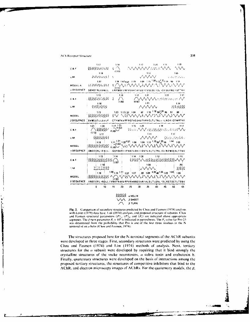
Fig. 2. Comparison of secondary structures predicted by Chou and Fasmen (1978) analysiswith Levitt (1979) data base, Lim (1974) analysis, and proposed structure of subunits. Chouand Fasmen structural parameters (P)_, (P)•, and (P,) are indicated above appropriatesegments. The l-turn parameter P, x 10' is indicated in parentheses. The P, value for Pro-23was determined from the probability that Pro is one of the first three residues in the Nterminal of an o helix (Chou and F:asmcn. 1974).
The structures proposed here for the N-terminal segments of the AChR subunitswere developed in three stages. First, secondary structures were predicted by using theChou and Fasmen (1974) and Lim (1974) methods of analysis. Next, tertiarystructures for the a subunit were developed by requiring that it bind strongly thecrystalline structures of the snake neurotoxins, a cobra toxin and erabutoxin b.Finally, quaternary structures were developed on the basis of interactions among theproposed tertiary structures, the structures of competitive inhibitors that bind to theAChR, and electron microscopy images of AChRs. For the quaternary models, the j,
WWI
9.®
!B
0
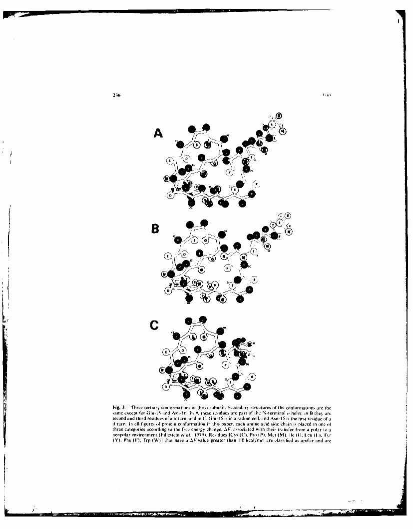
Fig. 3. Three tertiary conformations of the w subunit. Secondary structures of the conformations are thesame except for (Glu-1 5 and Asn-16. In A these residues are part of the N-tcrminal ' helix; in B the aresecond and third residues of a j turn; and in C. Glu- 15 is in a radom coil. and Asnh 15 is the first residue of aJ1 turn. In all figures of protein conformation in this paper, each amino acid side chain is placed in one ofthree categories according to the free energy change. AF. associated with their transfer from a Polar to anonpolar environment (idlestein e at.. 1979). Residues [Cys (C). Pro (P). Met (M). Ile (I). I.cu (LA. T~r(Y), Phe (F). Trp (W)j that have a AF value greater [han 1.0 kcal/mol are classilied as apolar and are
I....
V i It Rc,:clitr Ni ru.. iirc 23'7
,anid io subunits we~rc given tertiarý backbone structures, similar ito that of one of thleOk-sulbunit models. IDitlerent tertiary aind quaternart, structures %%ere considered in iinattempt lto account f'or thle conformnational changes during actit ation and descnsrtI/.I-tion of' the A'lhls.
Secondari and lertiarii Structure%
The m~ost conlimOnly obser~ ed stiuctures in proteins are ithelices. I sheets, and .1turns. Seseral methods have been developed (Oir using the -sequence of' a protein itopredict s"hich residues %%ill be in each of these ty pe conl'ormations (for rev ies . seeChou and I-asmen. I 978: Sternberg and Thornton. 1 978 I.
Iii thle Chou anrd I a sninen anal sis. each residue has at contform atiop a pa ra mete r.1P. f or each of the three secondary struictures. A P value greater than 1 .0 indicates thaithe residue occurs in the particular structure more l'requentl\ than in a total proteincomposition. An ithelix is predicted f'or si\ or more residue,, %~hen the average iconfoirmational parameter. 1) srae than 1.03. anda I sheet is. predicted w hen
(P) 1.5. iltur ispre i-ted I'O a etr pepideitenits (P. - 1. and th
facor ase ontheprouctof he 'rcuen\ o'ocurrnceofec residue in eachof thle four positions, Of the 41 turn, is greater than 0.-5 x10 4. The predictions. of1 theChou and Iisiiien analksis and thle values of the conformational parameter f'or the
four A~hR subunits are show4n in I ig. 2. The dlata base on \x hich the method dependshits been enlarged recentl\ ( I-e~itt, 1979). The enlarged data base is used here.
An alternative approach of' predicting it and 3 segrient,, is to analxie thedistribution of' polar and apolar residue,, w~ithin the sequence. Soluble proteins, tend tohave polar groups on the exterior of each doniain and apolar groups buried in theh~drophobic core. I(,sing thie principle. Lim ( 1974) developed a method of' predictingthe o-helix and 4l-sheet segments. The predictions of this method are shov. in Fig. 2The (v helices and Al sheets of' the A~hR subunits predicted bi, this anal'sis should beoin the surf'ace of' the subunit. -Iflie o helices predicted bx the ILini anailxsrs aireaniphipathic: i.e.. the hydrophobic side chains are clustered on one side of the helices.These hydrophobic residues should be in contact w~ith the hs~drophobic core of thesubunit. The predicted 3 sheet formed by residues 31 to 39 in each subunit has athydrophobic residue in every other position. so that one side of the 3 sheet has allhydrophobic residues. These residues should face the h'tdrophobic core of the protein.
it Subun it. Three possible tertiary structures for the o subunit are show~n inF~ig. 3. The differences amiong the secondary structures oft the three conformationsinvolve only residue Glu- IS and Asn- 16. In conformation A these residues are the lasttwo residues of' the N-terminal (Y helix. x\ hereas in conformation B the\ are in thesecond and third positions tif a 4l turn. The transition of these residues from the 3 turnto the tY helix moves the (t helix about 2.2 A closer to the rest of the subunit. low~ers thetv helix slightly, and changes its orientation slightly. In conformation C' the N-terminalty helix hits hinged about the (ilu- I5 and Asn- 16 residues so that the ( helix is oriented
represented bý titled circles. Residues ISer (S). Asn (i i. n (Q). (;I, (6,). Thrl. ( T), litt 1). Alta I \)I thaiihave itAI'valtue beirkeen 0I.1 and 0i S lical/mnol are classified it, indifferent and represenied bs shadedcirctes. Residues, Asp 0i)). (itu 01. 1 Ns (K). Arg (R~l thai have At vatlues more negalive ihan 2kcat/mot are charged and represented kts open circles The N~Ierrtninat amine groups are indicated b.% a
23N(u
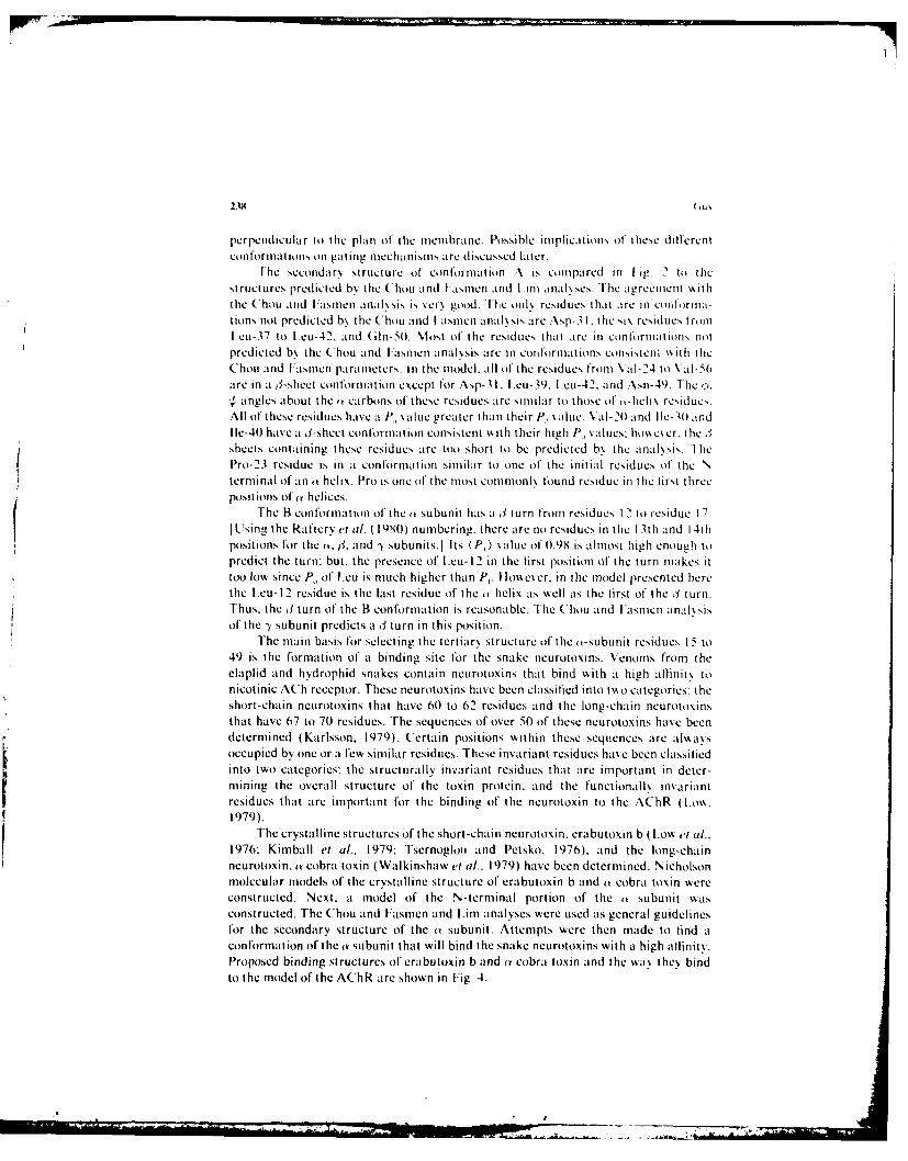
perpendicular to the plan of the membrane. Possible implications of these dillerentconformations on gating mechanisms, are discussed later.
[he sccondar, structure of conformation A is comtpared in I ig. 2 t, thestructures predicted b, the (hou and I aslen and Lint anal,.scs. The agreemernt v iththe Chou and [asmen analxsis is very good. lhc on11 residues that are in coti lrma-tions not predicted b\ the Chou and I asmen analysis are .sp-31. the si\ residues fromIeu-37 to [cu-42. and (iln-SO. Most of the residues that are in conlormation, notpredicted b, the Chou and I:asncn i anal~sis are in conformations consistetn' ith theChou and I~asmen parameters. in the model. all of the residues from \al-2 4 to \ al-56are in a s3-sheet conformation except for Asp-3 1. I.eu-39. L.eu-42. and Asn-49. The ,ý angles about the t carbons of these residues are similar to those of ,-helix residues.All of these residues have a P, value greater than their P., alue. \%al-(20 and I Ie-30 andlie-40 have a il-sheet conformation consistent ),% ith their high P., values: htm c er. tihe ,Isheets containing these residues are too short to be predicted b\ the anal•sis. ThePro-23 residue is in a conformation similar to one of the initial residues of the Nterminal of an (k helix. Pro is one of the most cotnmonl\ found residue in the first threeposilions of a helices.
"The B conformation of the a subunit has a 3 turn from residues I 2 to icsiduc 17.Iusing the Raftery et a/. ( 1080) numbering, there are no residues in the 13th and l4thpositions for the a. r. and ") subunits.] Its (P,) value of 0.98 is almost high enough to
predict the turn: but, the presence of l.eu- 12 in the tirst position of the turn makes ittoo low since P, of I.eu is much higher than P. lhoever, in the model presented herethe L.eu- 12 residue is the last residue of the o helix as well as the first of the 3 turn.Thus, the ý3 turn of the B conformation is reasonable. The Chou and FasmcIn analssisof' the y subunit predicts a ý1 turn in this position.
The main basis for selecting the tertiary structure of the o-subunit residues I5 to49 is the formation of a binding site for the snake neurotoxins. Venoms from theelaplid and hydrophid snakes contain neurotoxins that bind with a high atlinit% tonicotinic ACh receptor. These neurotoxins have been classified into two categories: theshort-chain neurotoxins that have 60 to 62 residues and the long-chain neurotoxinsthat have 67 to 70 residues. The sequences of over 50 of these neurotoxins have beendetermined (Karlsson, 1979). Certain positions within these sequences are alwa\soccupied by one or a few similar residues. These invariant residues have been classiliedinto two categories: the structurally invariant residues that are important in deter-mining the overall structure of the toxin protein, and the functionall\ invariantresidues that are important for the binding of' the neurotoxin to the AChR (I.on.1979).
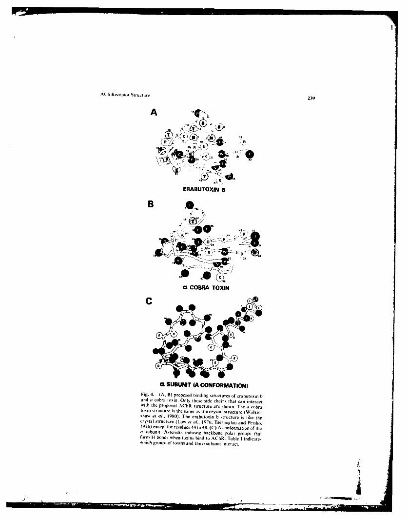
The crystalline structures of the short-chain neurotoxin, erabutoxin b ( Low a,[ al..1976: Kimball ei al., 1979: Tsernogloi and Petsko, 1976), and the long-chainneurotoxin, Y cobra toxin (Walkinshaw et al.. 1979) have been determined. Nicholsonmolecular models of the crystalline structure of erabutoxin b and (a cobra toxin wereconstructed. Next. a model of the N-terminal portion of the a subunit watsconstructed. The Chou and Fasmen and L.im analyses were used as general guidelinesfor the secondary structure of the a subunit. Attempts were then made to find aconformation of the (Y subunit that will bind the snake neurotoxins with a high allinity.Proposed binding structures of erabutoxin b and ay cobra toxin and the wa. they bindto the model of the AChR are shown in Fig. 4.
A(?h Receptor Structure 239
A
• R't (j
ERABUTOXIN B
B
' D
~e .9U COBRA TOXIN
K
C SUBUNIT (A CONFORMATION)
Fig. 4. (A. B) proposed binding structures of erabutoxin band (v cobra toxin. Only those side chains that can interactwith the proposed AChR structure are shown. The (a cobratoxin structure is the same as the crystal structure (Walkin-shaw et al., 1980). The erabutoxin b structure is like thecrystal structure (Low et al.. 1976; Tsernoglou and Petsko.1976) except for residues 44 to 48. (C) A conformation of the(v subunit. Asterisks indicate backbone polar groups thatform H bonds when toxins bind to AChR. Table I indicateswhich groups of toxins and the tv subunit interact.
-'P •
240 6u)
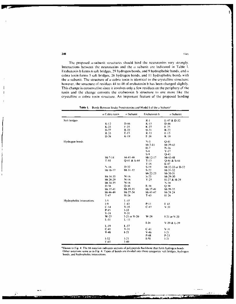
The proposed ,a-subunit structures should bind the neurotoxins very strongly.Interactions between the neurotoxins and the a subunit are indicated in Table I.Erabutoxin b forms 6 salt bridges, 29 hydrogen bonds, and 9 hydrophobic bonds, and acobra toxin forms 5 salt bridges, 26 hydrogen bonds, and I I hydrophobic bonds withthe av subunit. The structure of av cobra toxin is identical to the crystalline structure:however, the structure of residues 44 to 48 of erabutoxin b has been changed slightly.This change is conservative since it involves only a few residues on the periphery of thetoxin and the change converts the erabutoxin b structure to one more like thecrystalline at cobra toxin structure. An important feature of the proposed binding
Table I. Bonds Between Snake Neutrotoxins and Model I of the a Subunit"
a Cobra toxin (Y Subunit Erabutoxin b a Subunit
Salt bridges R-I E-47 & D-32K-12 D-46 K-15 D-46K-23 E-25 K-27 E-25D-27 R-22 D-31 R-22R-33 E-15 R-33 F-15D-38 K-19 E-38 K-19
Hydrogen bonds N-5 Q-41
bb 7-I1 bb 39-43
H-7 N-16S-8 Y-17s-9 Q-41
bb 7-14 bb 41-48 bb 12-17 bb 43-48T-10 Q-41 & S-44 T-13 Q-41 & S-44
T-16 E-47N-16 D-32 S-19 bb 32-33 or D-32bb 16-17 bb 31-32 S-22 bb 31-32
bb 22-23 bb 30-31bb 34-35 N-16 S-23 bb 29-30bb 28-29 N-16 Y-25 H-27 & H-29bb 34-35 N-16 N-16D-38 Q-38 E-38 Q-38bb 37-41 bb 39-35 bb 37-41 bb 39-35bb 46-49 bb 27-24 bb 43-47 bb 28-24T-47 H-26 T-45 H-26
Hydrophobic interactions I-5 L-371-9 1 43 P-Il 1-43C-14 V-35 C-17 V-31P-15 1-33V-19 V-31W-25 1-21 or V-20 W-29 1-21 or V-20L-31 L II
1-36 V-20 & L-39L-39 L-37C-41 V-31 C-41 V-31V-48 1-21 V-46 1-21
P-48 P-231-52 1-21 1-50 1-21F-65 1-40
'Shown in Fig. 4. The bb notation indicates sections of polypeptide backbone that form hydrogen bonds.Other notations same as in Fig. 4. Types of bonds are divided into three categories: salt bridges, hydrogenbonds, and hydrophobic interactions.
) -. >-.o., .
A~h Receptor Structure 241
scheme is the hydrogen bonds formed between the backbones of the neurotoxins andAChR. Most of these bonds involve the formation of extended j sheets. The postulatethat the backbone polar groups of the J3 sheets of the neurotoxins bind to backbonegroups on j3 segments of the a subunit is very helpful in designing the AChR modelsince the positions of the backbone groups are much more constrained than those ofthe side chains.
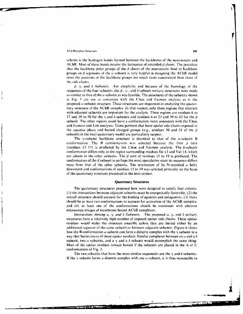
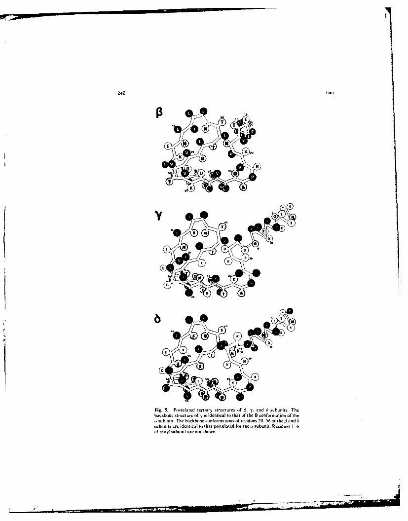
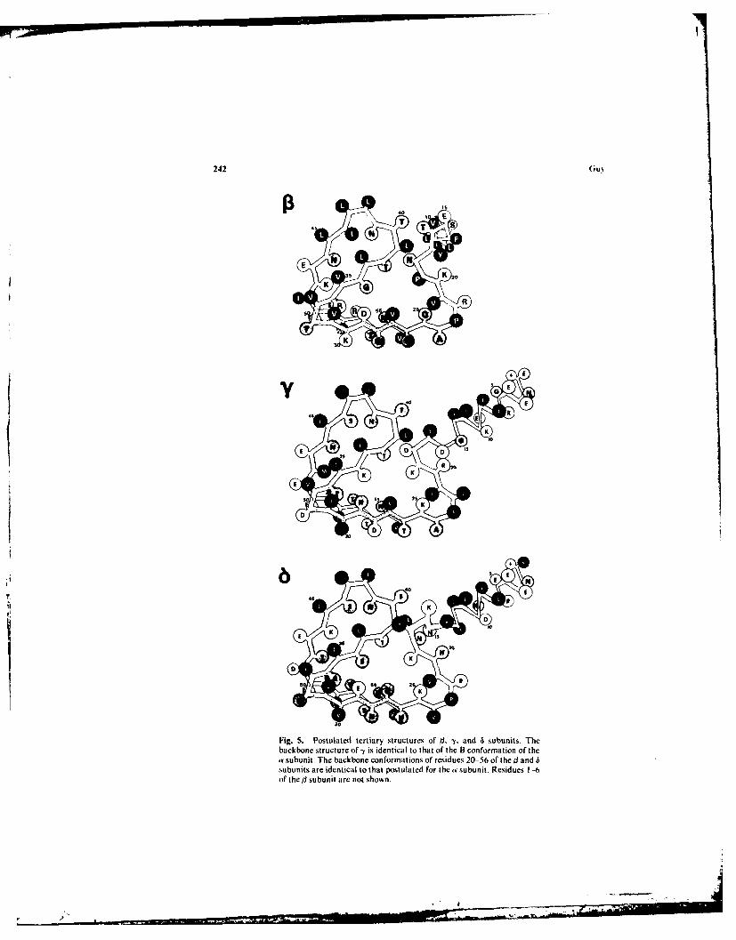
, y,. anid 6 Subunits. For simplicity and because of the homology of thesequences of the four s&bunits, the il-, -y-, and 6-subunit tertiary structures were madeas similar to that of the a subunit as was feasible. The structures of the subunits shownin Fig. 5 are not as consistent with the Chou and Fasmen analysis as is theproposed a-subunit structure. These structures are important in analyzing the quater-nary structure of the AChR complex. In that respect, only those regions that interactwith adjacent subunits are important for the analysis. These regions are residues 6 to17 and 39 to 50 for the -y and 6 subunits and residues 6 to 22 and 39 to 42 for the /subunit. The other regions could have a conformation more consistent with the Chouand Fasmen and Lim analyses. Those portions that have apolar side chains exposed tothe aqueous phase and buried charged groups (e.g., residues 50 and 51 of the 3subunit) in the final quaternary model are particularly suspect.
The -y-subunit backbone structure is identical to that of the ar-subunit Bconformation. The B conformation was selected because the first 1 turn(residues 12-17) is predicted by the Chou and Fasmen analysis. The 6-subunitconformation differs only in the region surrounding residues lle-13 and Val-14, whichare absent in the other subunits. The 0 turn of residues 15 to 18 is predicted. Theconformation of the tl subunit is perhaps the most speculative since its sequence differsmost from that of the other subunits. The orientation of its N-terminal a helixdownward and conformations of residues 15 to 19 was selected primarily on the basisof the quaternary structure presented in the next section.
Quaternary Structures
The quaternary structures proposed here were designed to satisfy four criteria:(I ) the interactions between adjacent subunits must be energetically favorable. (2) theoverall structure should account for the binding of agonists and antagonists, (3) thereshould be at least two conformations to account for activation of the AChR complex,and (4) at least one of the conformations should be consistent with electronmicroscopy images of membrane-bound AChR complexes.
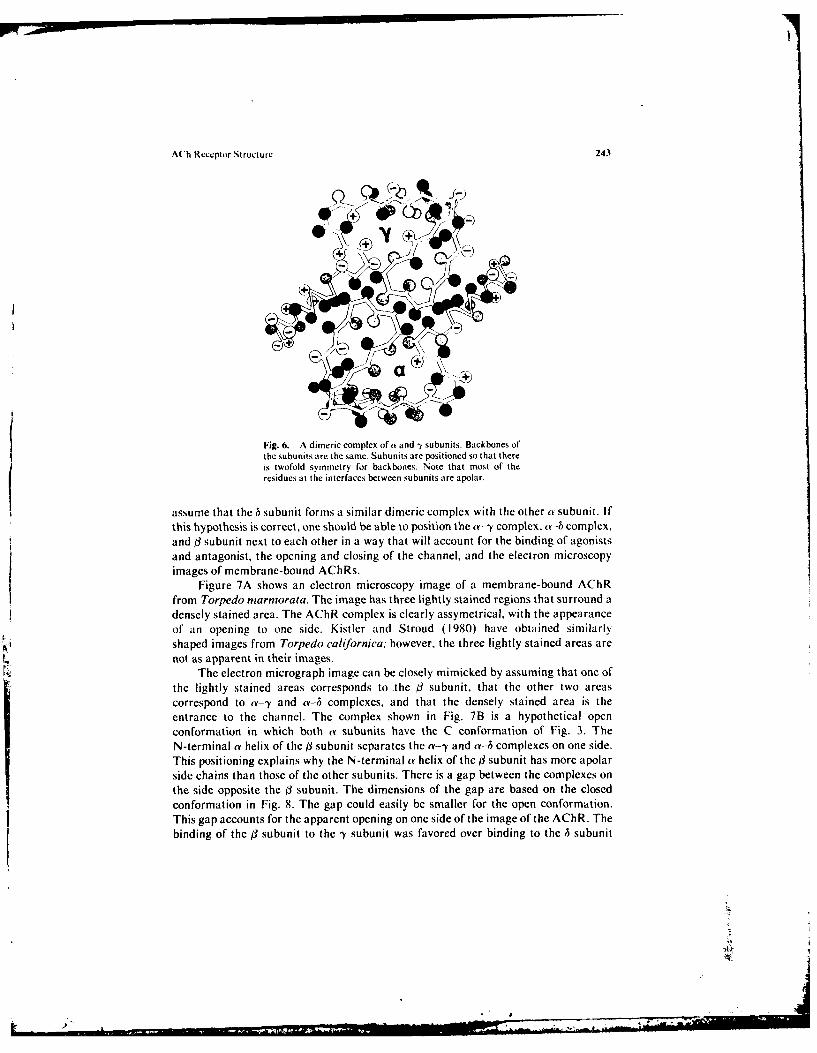
Interactions Among a, -y. and b Subunits. The proposed a, -Y, and 6 tertiarystructures have a relatively high number of exposed apolar side chains. These apolarresidues would make the structure unstable unless they are buried either by anadditional segment of the same subunit or between adjacent subunits. Figure 6 showshow the B-conformation a subunit can form a dimeric complex with the -Y subunit in away that buries many of these apolar residues. Similar complexes between an a and a hsubunit, two a subunits, and a -y and a 6 subunit would accomplish the same thing.Most of the apolar residues remain buried if the subunits are placed in the A or Cconformations of Fig. 3.
The two subunits that have the most similar sequencies are the 7 and 6 subunits.If the y subunit forms a dimeric complex with one a subunit, it is thus reasonable to
7"'"
242 Guv.
(3 ,o ,
4 (T )
E u36K 0
K3A
+, E
9 T.
K K
2 K
D D A
-t0
Fig. 5. Postulated tertiary structures of B, -y. and 6 subunits. Thebackbone structure of -y is identical to that of the B conformation of thea• subunit. The backbone conformations of residues 20- 56 of the ] andsubunits are identi~cal to that postulated for the a subunit. Residues 1 --of the/] subunit arc not shown.
)
242 (;u.
40
4 T TI~
E *K
K K
R6
2
K
a•sbnt Tebcbn cnomtos frsde 1.-6 f the•ad
40 E
45 f
K K
KK
,t A-D
)
ACh Receptor Structure 243
- .
Fig. 6. A dimeric complex of a and -y subunits. Backbones of
the subunits are the same. Subunits are positioned so that thereis twofold symmetry for backbones. Note that most of theresidues at the interfaces between subunits are apolar.
assume that the h subunit forms a similar dimeric complex with the other a subunit. Ifthis hypothesis is correct, one should be able to position the a-y complex. a-h complex.and 3 subunit next to each other in a way that will account for the binding of agonistsand antagonist, the opening and closing of the channel, and the electron microscopyimages of membrane-bound AChRs.
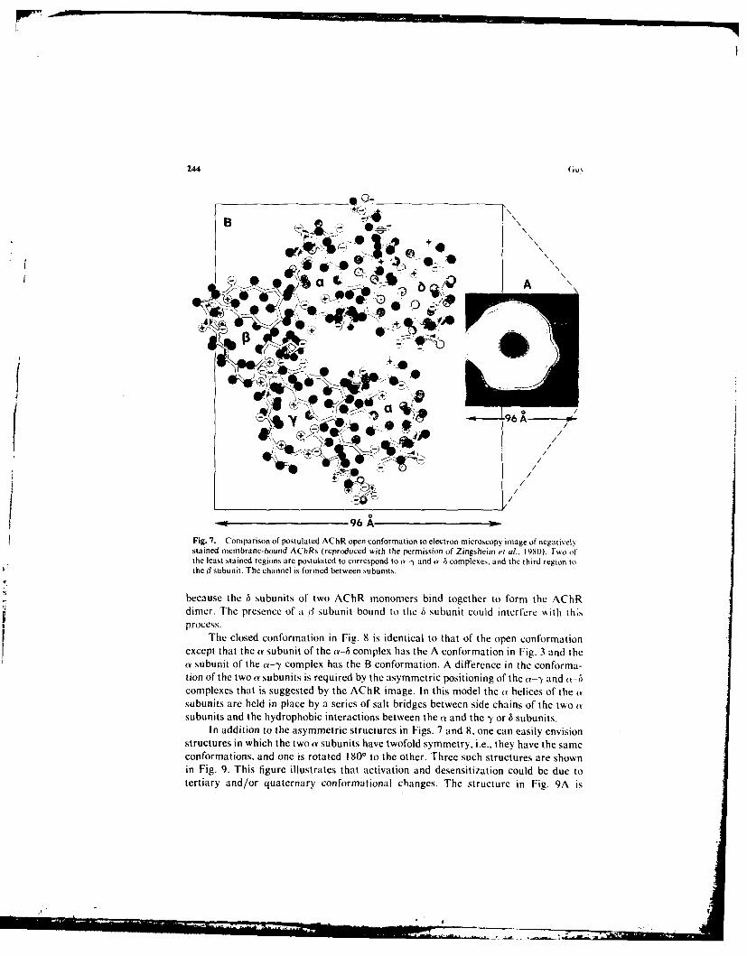
Figure 7A shows an electron microscopy image of a membrane-bound AChRfrom Torpedo marmorata. The image has three lightly stained regions that surround adensely stained area. The AChR complex is clearly assymetrical, with the appearanceof an opening to one side. Kistler and Stroud (1980) have obtained similarlyshaped images from Torpedo californica; however, the three lightly stained areas arenot as apparent in their images.
The electron micrograph image can be closely mimicked by assuming that one ofthe lightly stained areas corresponds to the (3 subunit, that the other two areascorrespond to a--y and a-h complexes, and that the densely stained area is theentrance to the channel. The complex shown in Fig. 7B is a hypothetical openconformation in which both ty subunits have the C conformation of Fig. 3. TheN-terminal a helix of the 3 subunit separates the a-y and av-i complexes on one side.This positioning explains why the N-terminal aY helix of the j subunit has more apolarside chains than those of the other subunits. There is a gap between the complexes onthe side opposite the / subunit. The dimensions of the gap are based on the closedconformation in Fig. 8. The gap could easily be smaller for the open conformation.This gap accounts for the apparent opening on one side of the image of the AChR. Thebinding of the 1i subunit to the -y subunit was favored over binding to the bS subunit
244 (ius
+
~rL~0
.e9 A-
+/A' + 4-_ ,
9 A
Fig. 7. Comparison of postulated AChR open conformation to electron microscopy image of negativelystained membrane-bound AChRs (reproduced with the permission of Zingsheim et al., 1 980). Two ofthe least stained regions are postulated to correspond to Y- -t~ and a- 6 complexes, and the third region to
the J3 subunit. The channel is formed between subunits.
because the 6 subunits of two AChR monomers bind together to form the AChRdimer. The presence of a ý3 subunit bound to the h subunit could interfere kkith thisprocess.
The closed conformation in Fig. 8 is identical to that of the open conformationexettattea subunit of the c--y complex has the A conformation. differenceain thecofraexet htvh subunit of the c-t3 complex has the A conformation. infFig.nceand thecofration of the two a subunits is required by the asymmetric positioning of the V--y and ak-hcomplexes that is suggested by the AChR image. In this model the ay helices of the (tsubunits are held in place by a series of salt bridges between side chains of the two (Ysubunits and the hydrophobic interactions between the (v and the -y or i5 subunits.
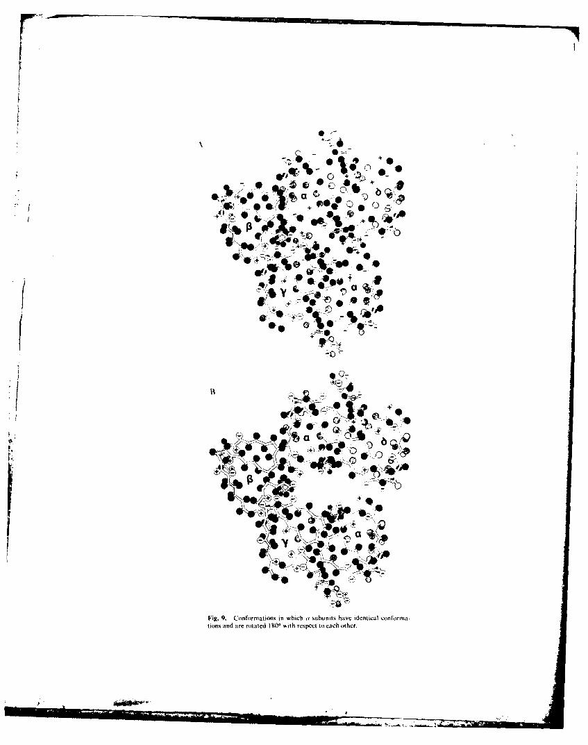
In addition to the asymmetric structures in Figs. 7 and 8, one can easily envisionstructures in which the two a subunits have twofold symmetry. i.e.. they have the sameconformations, and one is rotated 180' to the other. Three such structures are shownin Fig. 9. This figure illustrates that activation and desensitization could be due totertiary and/or quaternary conformational changes. The structure in Fig. 9A is
A(h Receptor Struct'urc 245
++
S - ,Y%-" I, o+
++ 4
0S.,,+-b
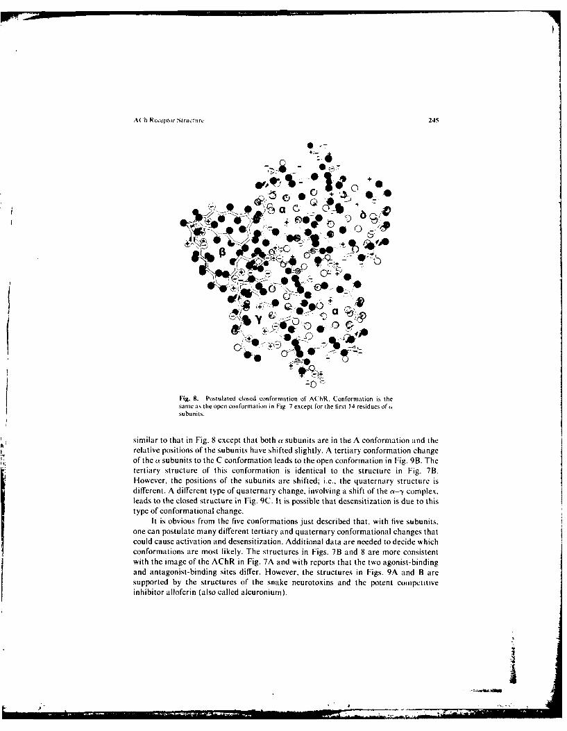
Fig. S, Postulated closed conformation of AChR. Conformation is thesame as the open conformation in Fig. 7 except ror the first 14 residues of osubunits.
similar to that in Fig. 8 except that both a• subunits are in the A conformation and therelative positions of the subunits have shifted slightly. A tertiary conformation changeof the ar subunits to the C conformation leads to the open conformation in Fig. 9B. Thetertiary structure of this conformation is identical to the structure in Fig. 7B.However, the positions of the subunits are shifted; i.e.. the quaternary structure isdifferent. A different type of quaternary change, involving a shift of the a•-- complex.leads to the closed structure in Fig. 9C. It is possible that desensitization is due to thistype of conformational change.
It is obvious from the five conformations just described that, with five subunits.one can postulate many different tertiary and quaternary conformational changes thatcould cause activation and desensitization. Additional data are needed to decide whichconformations are most likely. The structures in Figs. 7B and 8 are more consistentwith the image of the AChR in Fig. 7A and with reports that the two agonist-bindingand antagonist-binding sites differ. However, the structures in Figs. 9A and B aresupported by the structures of the snake neurotoxins and the potent cotipetitveinhibitor alloferin (also called alcuronium).
4
)i
-: %
r!
20-1 S+i +Ot" V
;0 #-
4V~, Qj *
+*
+2i
Fig. 9. Conformations in which (" subunits have identical conforma-tions and are rotated I 0 with respect to each other.
- -
%( "h Rcccptor Structure 247
ISO-
Swo
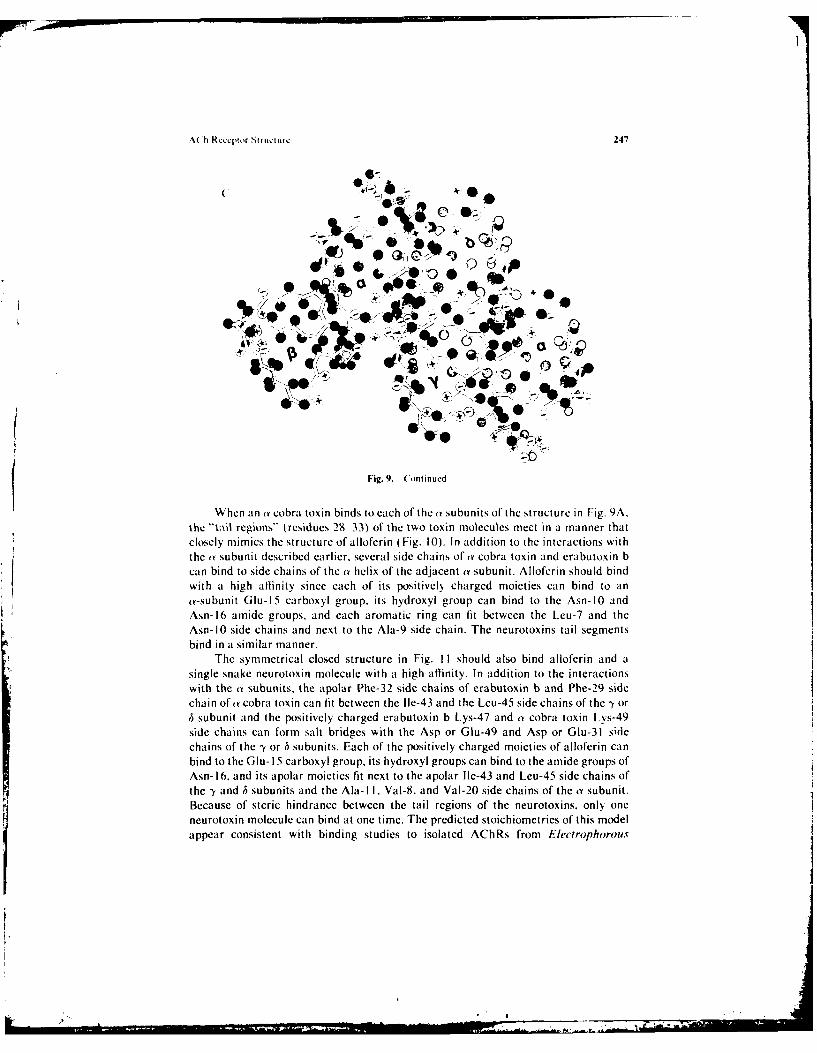
Fig. 9. Continuedi
When an a cobra toxin binds to each of the a subunits of the structure in Fig. 9A.the -It;il regions," trcsidues 29 33) of the two toxin molecules meet in a manner thnatclosely mimics the structure of alloferin ( Fig. 10). In addition to the interactions with
the ty subunit described earlier, several side chains of (Y cobra toxin and erabutoxin bcan bind to side chains of the a• helix of the adjacent a subunit. Alloferin should bindwith a high affinity since each of its positively charged moieties can bind to an(Y-subunit Glu- 15 carboxyl group. its hydroxyl group can bind to the Asn- 10 andAsh-16 amide groups, and each aromatic ring can fit between the Leu-7 and theAsn-1O side chains and next to the Ala-9 side chain. The neurotoxins tail segments
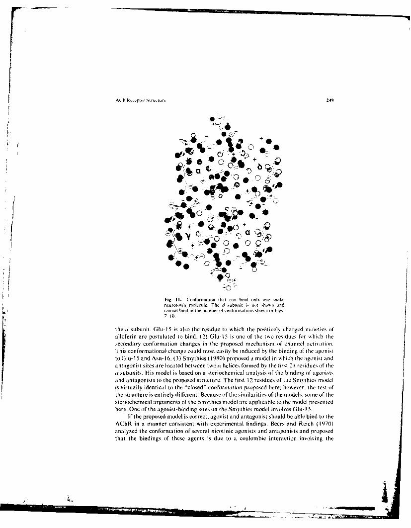
bind in a similar manner.The symmetrical closed structure in Fig. I1I should also bind alloferin and a
single snake neurotoxin molecule with a high affinity. In addition to the interactionswith the (w subunits, the apolar Phe-32 side chains of erabutoxin b and Phe-29 sidechain of t cobra toxin can fit between the Ile-43 and the Leu-45 side chains of the ', orh subunit and the positively charged erabutoxin b Lys-47 and a cobra toxin Lys-49side chains can form salt bridges with the Asp or Glu-49 and Asp or Glu-31 sidechains of the -y or ,5 subunits. Each of the positively charged moieties of alloferin canbind to the Glu- 15 carboxyl group, its hydroxyl groups can bind to the amide groups ofAsh- 16. and its apolar moieties fit next to the apolar Ile-43 and Leu-45 side chains ofthe -y and ,5 subunits and the Ala- 11, Val-8, and Val-20 side chains of the a subunit.Because of steric hindrance between the tail regions of the neurotoxins, only oneneurotoxin molecule can bind at one time. The predicted stoichiometries of this modelappear consistent with binding studies to isolated AChRs from Electrophorous
2.8) (iu
248 Ou%
ALLOFERIN
I7
a COBRA TOXIN TAILS
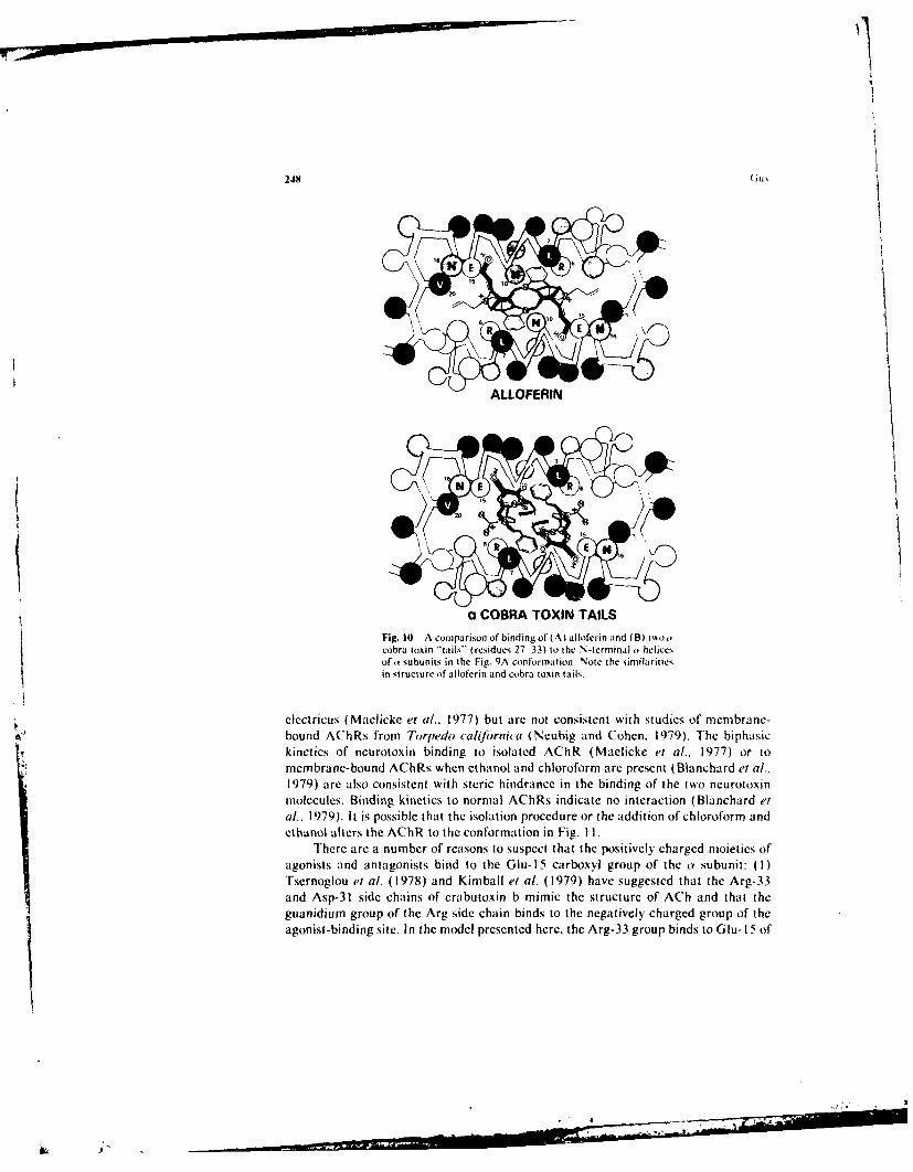
Fig. 10 A contparison of bindingof (A) alloferin and (B) t~o•cobra toxin "'tails" (residues 27 33) to the N-terminal r• helices.of o subunits in the Fir.. 9A conformation. Note the similaritiesin struetture of atloferin and cobra toxin tailS.
•. clectricus (Maclicke et al., 1977) but are not consistent with studies of membrane-•_• bound AChRs from Torpedo califrrnhia (Neubig and Cohen. 1979). The biphasic•r kinetics of neurotoxin binding to isolated AChR (Maelicke et a!.. 1977) or to• mcenbrane-bound AChRs when ethanol and chloroform are present (Blanchard et a!..
1979) are also consistent with steric hindrance in the binding of the two neurotoxinmolecules. Bintding kinetics to normal AChRs indicate no interaction (Blanchard eta!.. 1979). It is possible that the isolation procedure or the addition of chloroform andethanol alters the AChiR to the conformation in Fig. 11I.
There arc a number of reasons to suspect that the positively charged moieties ofagonists and antagonists bind to the Glu-l15 carboxyl group of the n• subunit: (I)Tsernoglou el a!. (1978) and Kimball et a!. (1979) have suggested that the Arg-33and Asp-31 side chains of erabutoxin b mimic the structure of ACh and that theguanidium group of the Arg side chain binds to the negatively charged group of theagonist-binding site. In the model presented here, the Arg-33 group binds to Glu- I5 of
A(h Rcceptir Structure 249
f+-- A-
, q%---l'
* ~+-Q
wor e•-'* 0we0*'
-Y _
+ (Dca
Fig. 11. Conformation thtcnbind )nl% one snakeneurotoxin molecule+ The 3 subunit is not h,)%kn andcannot bind in the manner of conformations showýn in I ig,ý7 1 t.
the tv subunit. Glu- 15 is also the residue to which the positively charged moieties ofalloferin arc postulated to bind. (2) (ilu-l S is one of the two residues for which thesecondary conformation changes in the proposed mechanism of channel activation.
1 dngofoheagois
'I his conformational change could most easily be indueed by the bind f ato Glu-1 5 and Asn-uo6. (3) Smythics (1980) proposed a model in which the agonist andantagonist sites are located between two an helices formed bn the first 21 residues of thea subunits. His model is based on a steriochemdical analysis of the binding of agonistiand antagonists to the proposed structure. The first 1 2 residues of Lte Sm•thics modelis virtually identical to the "closed" conformation proposed here: however, the rest of
"the strueture is entirely different. Because of the similarities of the models, some of thesteriochemical arguments of th ies model are applicable to the model presentedhere. One of the agonist-binding sites on the Smythies model involves Glu-2 5.
If the proposed model is correct, agonist and antagonist should be able bind to theAChR in a manner consistent with experimental findings. Beers and Reich (m1970)analyzed the conformation of several nicotinic agonists and antagonists and proposedthat the bindings of these agents is due to a coulombic interaction involving the
_ _ _ _ _ _ _ _ _ _ __e s oretaons adanaons _ _ud ea ebn tAh
A.4 hR, in-, a mnnr onsstntwitepeimetafndigsBersan Rich(170
2141
poiKtk% elk charged .ilk_ lainmnimiui moie!'. and at hsdrogen botnd it) an acceptor groiupon the agents that is formned Si) A f'rom the center oil the charL'e. Allf of thc altinists
(ACh. nicot)ine, and c~ tisine) and antagonists I trimectaphan. 1-cr\ thr(odinc. andstrwchniine) considered b\ Beer,, and Reich can bind to; the (i lu-I 5, cairbo)\\I group oilthe post ulated clo)sed and o~pen conforta tions in a mainoer that A imt~s their accep-t. rgroup ito form a hxdrogen bond it h the guanidium groupof \rv-b lir Arp-2 ) r %%oththe amide group ofl Asn- 1( or Asn- lb. L~sing these groups. one can co~nceisc e ofsc~ eraI\&aos the agonists and antagonist could bind. I or the clo~sed cilonforation in I ig S.one of' the ot subunits is in the A conf'ormation and the other is in the H conformnation.The positivels charged moiets of' ACh can bind ito the (ilu- IS carbo\\ grioup ofl Aconflormation site so that the ox~ gens of' the AC h ester linkage foml h~ drogen bonds\&ilh the Asn- l6 and Asni- 10 amide groups and the end mieth~l group set,, next to theI[eu-IlI side chains. or it can bind so that the os'tgens bind to the Arg-6 or Arg-22guanidiuni groups and to the Asn- lb amiido: group and the end metho.I group fits nextto the Val-20fside cha in. ACh could bind to the sa me I s~o site~s of hc H conftmkrmi;i ti tn.but the binding \Aould be dilferent, 1 he simultaneous binding of the ("~o ACh oxx\gens,
to the .'sn- 10 and Asn- lb amnide groups is not as f'aorable -since the amide grttu ps arcf'arther apart. The binding of' the oxxgens to the Nsn- lb and Arg-22 side chains nmabe more f'aorable. In all of' the conflormiatiions there %kill be sonmc c u Itmnbi cinteraction bct\,een the positively charged moietx of ACh and the (lu-2 cairbttxyg-roup.
Fran agonist to activate the ACh R. it must bind \k ith a higher aflinit\ to) theopen than to the closed conflormiation. ACh can bind tto the tt-subunit C conf~ormiationol' the open A~hR bi binding ito the same groups. %%ith the exception of' *rg-6. as itbinds in the closed conf'ormation. 'The t~oo o\\gens can bind simiultaneouss it)t theNsn-10) and .Xsn-Ib amnide groups or ito the Arg-22 and Asn-lb groups. The spacingbet\%een the groups and thus the binding sould be different. Perhaps more importantis the renmnoal of' the Are-b side chain f'rom the vicinito, of' the cation bindinv site. Iiithe open conf'ormation the ACh binding sites have a net negative charge due itoGlIu- IS, Arg-22 of the to subunit. (iu-47 of the I~ or h subunit, and, f'or one of the sites.(;Iu- 15 of the il subunit. The closed conf'ormiation has a neutral charge if one considersonly the (ilu- 15, Arg-22. (;lu-2. and Arg-6 side chains. Wkhen lHis-2 is ch-arged. thesite is inore po~sitive than negative.
D~ivalent agonists and antagonists also can bind in a number of %%i\,, A possiblemechanism by %khich alloferin binds to the Glu- IS side chains of' the tmo it subunitshas already been described. This mechanism is ntot appropriate f'or the binding ofd-tubocurarine (d-Tc to menibrane-bound iorpeedo cali firnica ACh Rs since it binds,to t\AO nonequivalent sites and the binding to one site inhibits the binding of' kink onesnake neurotoxin molecule (Neubig and Cohen. 1979). More likel\ mechanisms f'ord-Tc binding are to the closed conformations (ilu- I5 and (ilu-25 of' the same subunitor to (ilu- I5 and (iu-4 of the adjaciaLb.uauuits. Divalent agonists, such as sucein\ I-choline and decamethonium, maN bind ito the ai-subunit (;lu- 15 of the open conf~orma-tion and to (iu-47 of the I~ or h subunit or to Glu- 15 of the ý1 subunit.
Othe'r Con"f,o~rmnation.s. Because of' the many degrees of freedom in foldingpolypeptidc chains and in positioning subunits next to each other, it is unlikel\ that
V h Rccepor Structure 251
E.
E R L
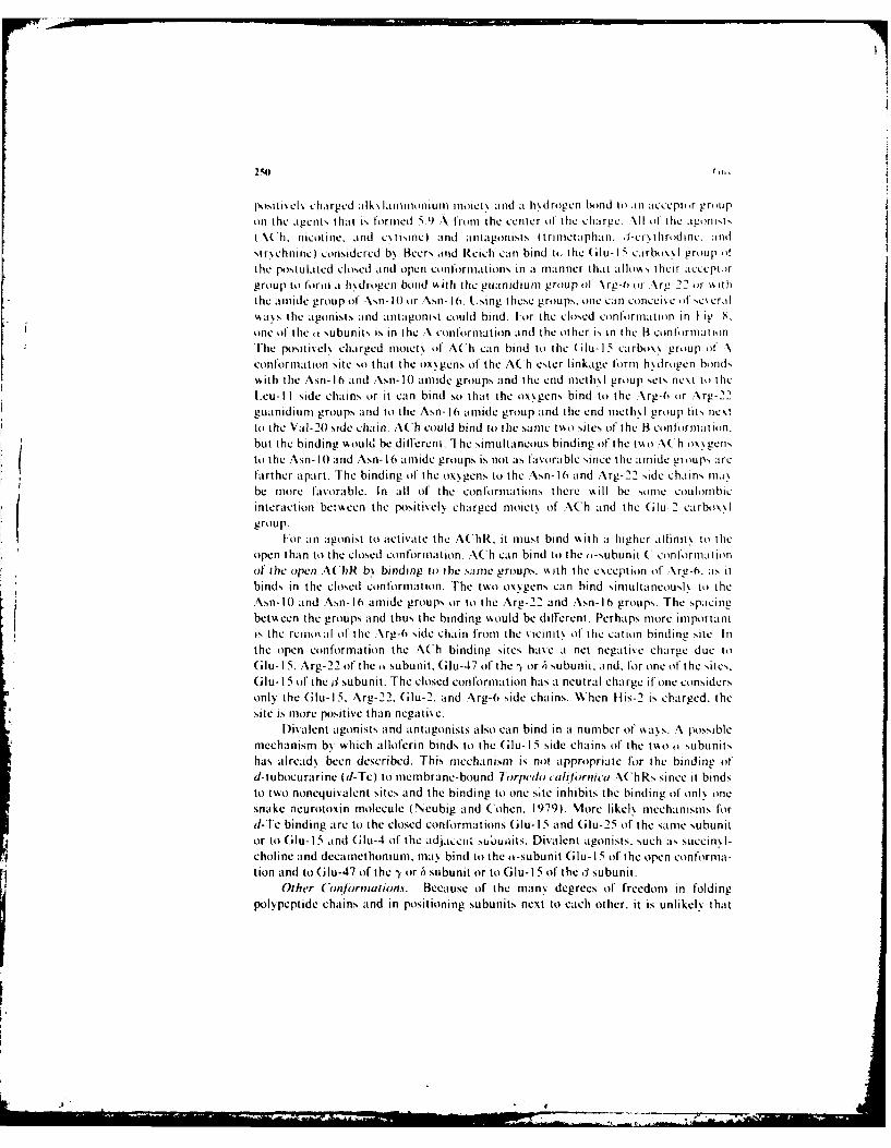
Fiig. 12. 'lIod l l -l,, -,tjbunii Iertlurr \ uructur.cS.c tls, hur
everN aspect of Ihe proposed mnodelI, is correct. The Ieast ambiguous p~irt of' the modeI,is, the tertiary, structure of' the portion (residues 1 5 to 49) of' the o subunit that bind to
the snake neurotoxins. Even portions of this segment are not certain: e.g.. there areother %'a\s of folding the -sectiions 23 36 and 39 56 that %, ill bind the toxin a, %%ell asthe model presented here.
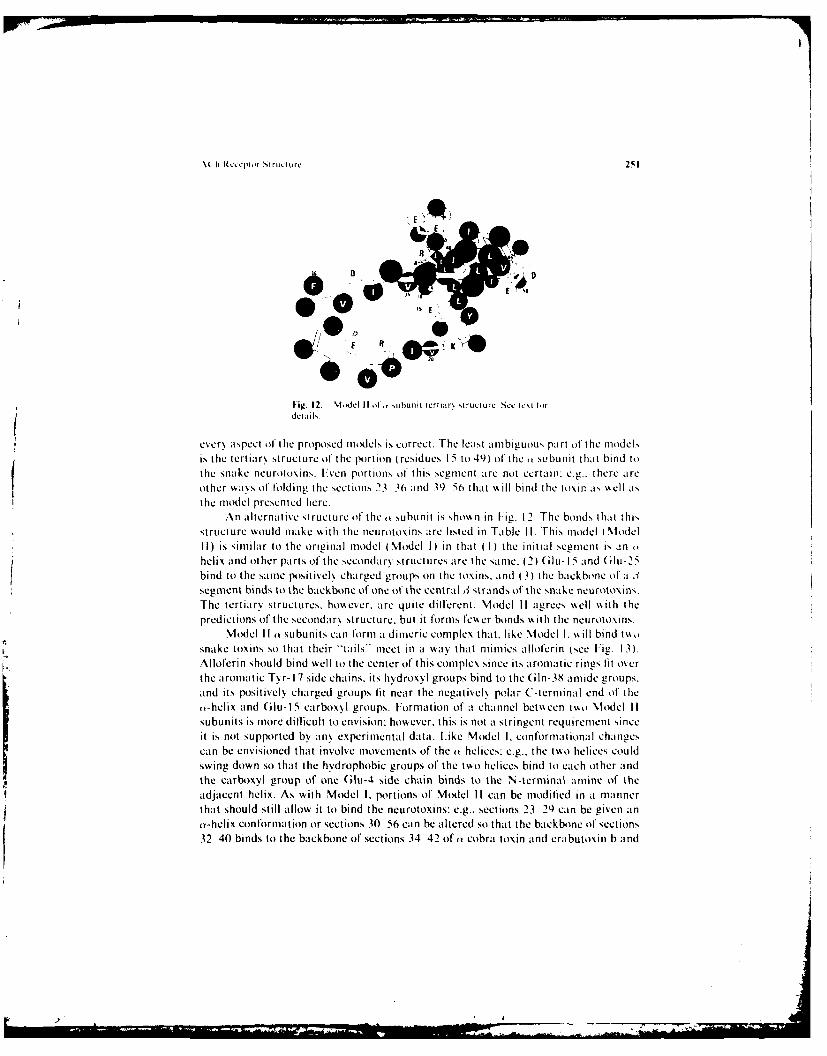
An alternative structure of the k subunit is sho\, n in Fig. 12. The bond,, that thi,structure would make , ith the neurotoxins are listed in Table II. This model \ModelII) is similar to the original model (Model I) in that (I) the initial segment is anhelix and other parts of the secondar,, structures are the sarne. (2) (Glu- 15 and (ilu-25bind to the same positivelh charged groups on the toxins, and (3) the backbone of a 3:segment binds to the backbone of one of the central 3 strands of the snake neurotoxins.The tertiarv structures, however, are quite different. Model II agrees \well \%ith thepredictions of the secondar', structure. but it forms fco~er bonds \• ith the neurotoxins.
Model II o subunits can form a dimcric complex that. like Model I, %%ill bind t\%,,
snake toxins so that their -tails- meet in a way that mimics alloferin (see Fig. 13).Alloferin should bind well to the center of this complex since its aromatic rings lit overthe aromatic Tyr- 17 side chains, its hydroxyl groups bind to the (iln-38 amide groups.and its positively charged groups lit near the negativel, polar C-terminal end of theo-helix and Glu-1 5 carboxyl groups. Formation of a channel bet\ een tmo Model IIsubunits is more dillicult to envision: however, this is not a stringent requirement sinceit is not supported by any experimental data. Like Model I. conformational changescan be envisioned that involve movements of the o helices: e.g.. the to helices couldswing down so that the hydrophobic groups of the two helices bind to each other andthe carboxyl group of one (;lu-4 side chain binds to the N-terminal amine of theadjacent helix. As with Model I. portions of Model II can be modilied in a mannerthat should still allow it to bind the neurotoxins: e.g., sections 23 29 can be given anaY-helix conformation or sections 30 56 can be altered so that the backbone of sections32 40 binds to the backbone of sections 34 42 of an cobra toxin and erabutoxin b and
- - .• , .. . . . ... .. .... ,.- -• ... • •-... . . I II I - •, . . "-
252 (iu.
able II, BIonds Between Snake %Ic routomn and nhjdul I II ,t ihe-Stubunit"
n Cobra toxin i, Subunit [rabutoxin b Subunit
Salt bridges K-12 I)-32 K-15 1D-32K-23 [-25 K-27 [ -25I)-27 K-19 D-31 K-19R-33 F--15 R-33 F-15-R-36 E- I5 [-38 R-22D-38 R--22
II'drogen bonds Y-21 T-28K-23 T-2h K-27 T-25bb 30-33 bb 37-40S-31 Q-38 bb 32-33 Q-38bb 34-42 bb 37-29 bb 34-43 bb 37-28
Q-9 T-34
bb 44-47 bb 27-24T-47 H-26
|lldrophobic interactions W-25 1-21 W-25 1-21F--28 Y-17 F-32 Y-171-32 1-12 & L-39 1-36 V-35C-41 & C-14 F-30 C-41 & C-17 F-30
P-50 P-23
"Shown in Fig. 12. Notation is the same as in Table I.
the backbone of sections 49 -55 binds to sections 8- 14 of v cobra toxin and sectionsI- 17oferabuxton b.
Finding that the same sequence can be placed in quite different tertiarystructures that are consistent with the secondary structures and that should stronglybind the neurotoxins indicates the limitations of the approach. It also stresses theimportance of obtaining better structural data and developing better methods ofpredicting the structure.
• ~-*O 0 ,¢+li+ 77
Fig. 13. A dimeric complex of two Model II y subunits. Alloferinwould bind in the center between the two subunits. See text fordetails
) .j
254 GuN
the free sulfhydryls without altering the binding of the toxin. It may be possible to usethis method to bind together two neurotoxin molecules. From the model, one canpredict the connecting chain lengths that should be required to allow both toxinmolecules to bind to the various conformations. When both toxin molecules bind, thetotal number of bound toxins should be only half the value obtained when only onemolecule binds, and the binding allinity should be higher. Alternatively, the distancebetween the tails of bound neurotoxins may be determined by attaching to thesulfhydryl group fluorescent or spin probes that interact with each other.
f Many features of the models described here were developed by constructingNicholson or CPK molecular models of the proposed structure and examining possibleways that the toxins or drugs could interact with various sites. This method appearssatisfactory for large molecules, such as the snake neurotoxins. that bind very stronglyand have many points of interaction. It is less satisfactory for small molecules like theagonists because of the small number of interactions, the degrees of freedom that onehas in positioning the protein side chains and altering the conformations of the agents.and the nonquantitative nature of the approach. Assuming that the positively chargedgroup of agonists and antagonists binds to Glu-1 5 of the a subunit, it is still dilicult touse this approach to determine which of the possible conformations is most likely to becorrect. Showing that agonists could bind to a given conformation is a necessarycondition, but it is certainly not sullicient. For this type of analysis to be convincing, amore quantitative approach is needed that can evaluate the binding constants of aseries of drugs to each conformation. Several groups are attempting to develop systemsto evaluate interactions between drugs and their receptors (for review, see Gund ei al..1980): however, it is not apparent that these approaches are sufficiently quantitativeto correctly predict which of the possible models is most likely to be correct. In spite ofthe uncertainties, the models predict the positions of various groups on the receptorfairly well. It thus may be feasible either to systhesize new compounds or to modifyexisting compounds that should bind to specitic sites on the AChR with a high affinitY.If the agents bind covalently, it may be possible to identify the subunit and residue towhich they bind.
Covalently bound aflinity-labeling agents and cross-linking agents could helpdetermine which subunits are next to each other. Raftery et al. (1979) have shownthat a photolabeling agent that is covalently bound to a sulfhydryl on the tail portionof a-bungarotoxin will bind covalently to the h-subunit. This result is consistent %% iththe model presented here, although it is not apparent why the -y subunit is not labeledalso.
One of the main purposes of this discussion is to emphasize the importance of thedetermination of the entire sequence of all of the subunits. Although Raftery et al.(1980) have made a good start, there is difficulty in isolating large quantities of theprotein and in analyzing the sequence of large, insoluble proteins. The recentidentification of the AChR messenger RNA suggest.; an alternative approach in whichthe complementary corresponding DNA is synthesized and the DNA is thensequenced (Mendez el al., 1980). Thus the prospects of eventually obtaining the entiresequence are fairly good.
Obviously, the determination of more of the sequence of the subunits would behelpful in analyzing more of the AChR structure and in eliminating some of the
.5(h Rcceptor Structure 253
IDISCUSSION
The main linding of this paper is that the polypeptide chains of residues I5 to 49of the a subunit can be folded so that it should bind the snake neur',toxins with a highatlinity. The implications of these structures on aggregation of the AChR subunits,the agonist and antagonist binding, AChR gating, and overall structure of thesubunits were examined. These implications are secondary and more speculative thanthe tertiary structure of the a subunits. It is important to recognize that the modelspresented here are working hypotheses and that it is unlikely that every aspect of themodels is correct. The main purpose of the models is to aid in the design ofexperiments that will test aspects of the models.
Before a great deal of time and energy is spent on testing precise details of themodels, it is prudent to test more general features. One of the most crucial tests of themodel is to determine whether the N -terminal segments are part of the extracellularsoluble domains and, if so, whether they form the postulated binding sites. In anumber of experiments, side chains near the cholinergic or neurotoxin-binding siteshave been covalently labeled. By analyzing the sequence of labeled AChRs, it may bepossible to identify the portions that comprise the receptor-binding sites. These types
of experiments could be facilitated by enzymatically cleaving the subunits intoidentiliable peptides and then determining which peptides contain the labels and/orwhether any of the peptides will bind the drugs and toxin. An initial step in thisdirection has already been taken. Trypsin treatment of isolated AChRs can be used toseparate the a subunit into two domains: a 27,000-dalton soluble domain that bindsneurotoxins, agonists, and competitive inhibitors (Bartfeld and Fuchs, 1979). and amembrane domain that can be selectively labeled with [5-' 2'I]iodonaphthyl-l-azidewhen the AChR complex is in the membrane (Tarrab-Hazdi et al.. 1980). If themodel proposed here is correct, the soluble domain shoild have the same N-terminalsequence as the entire subunit.
An alternative approach of determining whether the N-terminal segments formthe receptor-binding sites is to isolate or synthesize the N-terminal segments. makeantibodies to these segments, and determine whether the antibodies bind to theextracellular AChR domains and, if so, whether they inhibit the binding of theneurotoxins, agonists, or antagonists to the AChR. Also, if antibodies are found thatinhibit the binding of these agents to AChRs, their binding and that of agonists andantagonists to the N-terminal segments could be analyzed.
If it is shown that the N-terminal segments form the cholinergic binding sites.one must determine whether any of the conformations suggested here are correct. If itcan be shown that the N-terminal segments or some other segment still has thestructural integrity to bind neurotoxins and/or cholinergic agents, then structuralanalysis of these segments could be informative. It is probable that the solubledomains of AChRs can be crystallized more easily than the entire subunit. However,the crystalline structure of portions of the subunits must be interpreted with carebecause of possible differences in the conformations.
The proposed mechanism of binding of the snake neurotoxins may be testable.Raftery el al. (1979) have found that the disullide bond of the tail portion ofaY-bungarotoxin can be reduced and that a molecule can be covalently bound to one of
)
A(h Receptor Structurc 255
ambiguities of the model proposed here. Aflinity-labeling experiments indicate thatreduction of a disullide bond on the AChR leads to the exposure of an n-subunitsulfhydryl group that is near the cholinergic binding site (Karlin. 1969: I)amle andKarlin, 1980). Thus, one hopes that determination of more of the sequence N terminalwill reveal a sulfhydryl group that can be positioned near the proposed agonist-bindingsite without altering the model conformations. It is quite feasible that this would bepossible for only one of the alternative conformations described here.
The model presented here has not dealt seriously with conformation of thetransmniembrane domain other than to suggest that the channel is formed between thesubunits. However, transmembrane domains may be more appropriate for the model-building approach than are soluble domains. There are theoretical reasons to believethat the hydrophobic environment of the lipid phase will impose considerably moreorder (i.e., more n-helix and W-sheet structures and more regular packing of thesestructures) than is generally observed in soluble proteins (Kennedy, 1978). Thishypothesis is supported by the very regular structurL of bacteriorhodopsin (Unp.in andtlenderson, 1975: Engleman el al.. 1980). Because the protein is in a membrane,
labeling agents can be used to identify residues and segments that are in contact withthe lipid phase, the extracellular aqueous phase. and the intracellular aqueous phase,and residues that become exposed when the channel opens. Labeling agents have
already been used to show that portions of the o-subunit are in contact with themembrane lipid (Tarrab-Hazdai et al.. 1980) and that the t5 subunit of Torpedomarmorata is near and/or comprises the local anesthetic-binding site (Saitoh el al..1980). In designing a model of the transmembrane protein structure, one can also usethe structure of the putative channel-blocking drugs. the size and nature of the variouscations that will and will not pass through the channel, and the voltage dependence ofactivation and desensitization kinetics. If membrane fragments can be isolated thathave present only the transmembrane portions, then X-ray diffraction, electronmicroscopy, or other techniques that give structural information could be informative.Additional constraints on the model would be suggested by similarity of the atninoacid compositions and sequences of the transmnembrane portions to those of gapjunction channels or other channels for which the protein structure is better delined.
This paper is intended to indicate the role that model building may play indetermining the structure of the AChR complex. With current progress in analyzingAChR complexes, data should be available soon to allow the design of models lessspeculative than those presented here. Progress is also being made in the developmentof methods to predict the secondary and tertiary structure of proteins from theirsequences (Cohen et al., 1980), in modeling structures of other membrane proteins(Engelman et al.. 1980: Guy, 1980), and in predicting more precisely how drugs bindto proteins (Gund et al., 1980). Thus it may be possible to predict the structure of theAChR complex without having precise X-ray diffraction data.
Biochemical and structural analysis of the nicotine AChR complex is madepossible primarily because of its abundance in the electric organs of rays and eels.Most postsynaptic receptors and channels are not as easily analyzed. It is quite likely,however, that most postsynaptic receptor complexes evolved from the same proteinand that they have similar structural features. This concept is supported by the findingthat several postsynaptic channels that are activated by different transmilters are
256 G(us
blocked by the same drugs (Carpenter et al., 1977). Determination of the structure ofthe nicotonic AChR complex may thus be important in the design of molecular modelsof other postsynaptic channels. For example, by changing side chains on the generalbackbone structures of portions of the AChR, one may be able to design molecularmodels of other types of receptors and channels that account for differences among thepharmacology, gating kinetics, and ion selectivities of the channels.
SUMMARY
(I) The N-terminal sequence of the first 54 residues of the four subunits thatcomprise the acetylcholine receptor (AChR) complex of Torpedo calilornica weredetermined recently (Raftery et al., 1980). The aim of this paper is to examine thehypothesis that these segments form the binding sites for the cholinergic snakeneurotoxins, agonists, and competitive inhibitors.
(2) Nicholson molecular models were constructed of the structures of erabutoxinb, (Y-cobra toxin, various agonists and antagonists, and the N-terminal segments of theAChR subunits. The conformations of the AChR subunits were influenced by theoriesthat predict the secondary structure from the sequence, the requirement that thestructures bind the agonists and antagonists with the appropriate stoichiometries, therequirement that most of the apolar side chains be buried in the interior of thestructure, and the dimensions and spatial arrangements of the AChR indicated byelectron microscopy studies.
(3) Subunit conformations were found that were consistent with the hypothesisthat the cholinergic binding sites are formed by the N-terminal segments of the (ksubunit. The models suggest mechanisms by which opening of the channels istriggered and the AChR complex desensitizes. For some portions where the structureis less certain, a number of alternative conformations are suggested.
(4) The proposed models serve as excellent working hypotheses for the design ofexperiments to examine the AChR structure. By testing the hypotheses, by obtainingmore structural data on the AChR. -ind by improving the methods of analyzing theAChR structure, it may be possible to determine the structure and functionalmechanisms of the AChR without having precise X-ray crystallographic data.
REFERENCES
Anderson, D. J., and Blobel, G. (1980). In vitro synthesis and membrane integration of the subunits of
Torpedo acetylcholine receptor. Neurosci. Abstr. 6:209.Barrantes, F. J. (1974). The nicotinic cholinergic receptor: Different compositions evidenced b% statistical
analysis. Biochern. Biophyvs. Res. Comrnun. 62:407 -414.Bartfeld, D., and Fuchs. S. (1979). Active acetylcholine receptor fragment obtained by tryptic digestion of
acetylcholine receptor from Torpedo californica. Biochemn. BiophYs. Res. CoInnoun. 89:512 -519.Beers, W. H.. and Reich E. (1970). Structure and activity of acetylcholine. Nature 228:917-922.Blanchard. S. G.. Quast, U., Reed, K., Lee. T.. Schimerlik, M. I., Vandlen. R.. Claudio, T.. Strader. C. D.,
Moore, Fl. -P. H.. and Raftery, M. A. (1979). Interaction of [2"l]-Y-bungarotoxin with acetylcholinereceptor from Torpedo californica. Biochemnistrv 18:1875 1883,
Carpenter, D. 0., Swann, .. W., and Yarowsky, P. .1. (1977). Effect of curare on responses to differentputative neurotransmitters in Aplvsia neurons. J. Neurohiol. 8:119 132.
......... . . .. . . .. .. .• .• • • . .. . .. I I - '• , . . - , -_ • t •-
A~h Receptor Structure 257
Chang, If. W., and Bock. F. 1979). Structural stabilization of isolated acetl% choline receptor: Specificinteraction with phospholipids. Biocherni~sirv 18:172 179.
Chou. P. Y.. and Fasnicn. G. D.. ( 1974). Conforittation parameters for amino acids in helical. ý1-shcct. andrandom coil regions calculated from proteins. Bitocltenosirt 13:2 It 245.
Chou, P. Y., and Fasinen. (i. 0. ( 1978). Empirical predictions of protein conformation. A-nna. Rev.Hioc hent. 47:251I 276.
Cohen, F. E., Sternberg. M. J1. F.. and Taylor. W. R. ( 1980). Analysis and prediction of protein 4-shcetstructures by a combinatorial approach. Natutre 285:378 382.
Damle. V. N.,.and Karlin. A. ( 1980). Effects of agonists and antagonists on the reactivity of the binding sitedisulfide in acetylcholine receptor from -Torpedo calijornica. Biochemistrr 19:3924 393 2.
Edlestein, C_. Kezdy, F. J.. Scanu. A. M., and Shen, B. W. (1979). Apolipoproteins and the structuralorganization of plasma lipoproteins: Human plasma high density lipoproteirt-3- J. Lipid Res. 2:143153.
Engelman, D. M.. Henderson, R., McLachlan, A. D_. and Wallace, B. A. ( 1980). Path of the polypcpttde inbacter ,hodopsin. Proc. Nail. Acad. Sci. UISA 77:2023-.2027.
Feltz. A., and Trautmann. A. ( 1980). interaction between nerve-released aeetNlcholine and bath appliedagonists at the frog end-plate. J. Pht- siol. 299:533 552.
Gonzalei-Ross. J. MI., Parasehos. A., and Martinez-Carrion. M. ( 1980). Reconstitution of functionalmembrane-bound acetylcholine receptor from isolated Torpedo californica receptor protein and
f Gund. P., Andose. J. D., Rhodes, J. B.. and Smith, G. M. (1980). Three-dimensional molecular modeling
anddru deig. Siene.208J1425 143f.
Guy It R.(190).Thecoied oilina beta barrel: A model of the porin-lipoprotein complex of E. cohi outer( Heidmann, T., and Changeux, J. -P. (1978). Structural and functional properties of the acetslcholinereceptor protein in its purified and membrane-bound states. Anna. Rev,. Biachen,. 47:317-.357.
Henderson. D., FibI ., Hand Weber. K. (11979). Structure and biochemistry of mouse hepatic gap junctions.J. Mol. Biol. 132:193 218.
Karlin, A. ( 1969). Chemical modification of the active site of the ACh receptor. J. Gen. Ph~tsiol. 54:245.Karlin, A., Weill. C.. McNamee, M., and Valderrama. R. (1976). Facets of the structures of acetylcholine
receptors from Electrophtorus and Torpedo. Cold Spring Harbor S -vntp. Quant. Biol. 40:203-2 10./ Karlsson. F. (1979). Chemnistry offproteins in snake venoms. Hand. Exsp. Pharniacol. 52:159 204.Kennedy. S. J. (1978). Structures of membrane proteins J. Afernbr. Biol. 42:265 -279.Kimball. M. R., Sato, A., Richardson. J. S.. Rosen. L. S. and Low. B. W. (1979). Molecular conformation
of erabutoxin b: Atomic coordinates at 2.5 A resolution. Richetern. Riophir-v. Res. Comnmun. 88:953959.
Klymkowsky. M. W., and Stroud, R. NI. (1979). Immunospecilic identification and three-dimensionalstructure of a membrane-bound acetylcholine receptor from Torpedo californica. J. Mol. Bio!.128:319-334.
Kistler. J.. and Stroud, R. M, ( 19h81). Proc. Nail. Aad. Sci. USA. 78:3678.3682.Levitt. MI. (1979). Conformational preferences of amino acids in globular proteins. Biochr-mistrY 17:4277-
4284.L.im, V. 1. ( 1974). Algorithms for prediction of o-helical and di-structural regions in globular proteins. J.
Mat. Biol. 88:873. 894.Lindstrom, J.. Merdie, J.. and Yogeeswaran. G. 1 1979). Biochemical properties of acetylcholine receptor
subunits from Torpedo cahifijrnica. Biachemfisirr 18:4465 -4470.Low, B. W. ( 1979). Three-dimensional structure of erabutoxin b, prototype structure of the snake venom
postsynaptic neurotoxins: Consideration of structure and function description of the reactive site. InAdvance~s in ytiopharmacologi- (Ceccarelli. B.. and Clementi. F.. Eds.). Raven Press. New York. pp.141-147.
Low, B. W.,.Preston, H. S.. Sato,., ARosen, L..S., Searly, J. E.. Rudko. A. D., and Richardson, J. S. (1976).Three dimensional structure of crabutoxin b neurotoxic Protein: Inhibitor of acetylcholine receptor.Prot-. Nail. Acad. Sci. UISA 73:2991 2994.
Maclicke. A., Fulpius. B. W., Kieft. R. P.. and Reich. E. ( 1977). Acetylcholine receptor: Responses to drugbinding. J. Rio). (hemn. 25:4811 4830.
Mendez. B.. Valenzucla. P.. Martial, 3. A.. and Baxter. J. D. (1980). Cell-free synthesis of acetylcholinereceptor polypeptides. Science 209:695 697.
Moore, H. P. H.. Hartig. P. R., and Raftery. MI. A. (1979). Correlation of polypeptidle composition withfunctional events in acetylcholine receptor-enriched membrane from Torpedo californica. Proc. N'ail.Acad. Sc,. LISA 76:626,S 6269.
ds-C.2
258 (ins
Moore, W. M., Hlolladay, I.. A.. Puett. 1),. and Brady, R. N. (1974). On the conformation of theacetylcholine receptor protein fromt Jorpc'do nobiliwicj. FhBS Lett. 45:145 149.
Nelson. N.. Anholt. R., L~indstrom. J., and Montil, MI. 11980). Reconstitution of* purified acetylcholinereceptors with functional ion channels. in planar lipid bilayers. Prot- all/ atd.Nci. I.NA 77:30573061.
Neubig, R. R., and Cohen. J. B. (1979). Equilibriumn binding of 11111 tubocurarine and [rilj acet "lcholincby Torpedlo postsynaptic membranes: Stoichiomectry and ligand interactions. Bio, heoinitrry 18:i4645475.
Neubig. R. R., and Cohen, J. B. ( 19811). Permeability control by cholinergic receptors in io~rpcdopostsynaptic membranes: Agonist dose-response relations measured at second and millisecond tines.Biochennstri- 9:2770 2770.
Neubig. R. R., Krodel. F. K.. Bovd. N. D).. and Cohen. J. B. ( 1979). Acetylcholine and local anestheticbinding to Torpedo nicotinic postsynaptic menibranes after removal of nonreceptor peptides. Pr...NatlI. At-ad. Set. USA1 76:69(1 694.
Oswald. R. F.. Wennogle. L.. P.. Saitoh. T., and Changeux, J. P. ( 198(11. Structural aspects of' the 660H(1dalton subunit of the Torpedo ,,arniorata electroplaque acetylcholine receptor analsc'ed by limitedproteolytic digestion . Neurosci. Abs ir. 8:252.
Raftery. M., Blanchard. S.. Elliott, J.. llartig. P.. Moore, H. -P.. Quast. U.. Schitnerlik. MI.. Witzemann.V.. and Wu, W. ((19791. Properties of Torpedo calilornita acetylcholine receptors. A-dv. (rYtopltarnia-cot. 3:159 182.
Raftery. M. A., Hunkapiller. M.W.. Strader, C. D.. and Hood. L. E. 11980) . Acety lcholine receptor:Comnplex of- hontologous subunitS, ientie. 2118:1454 14,;6.
Ross. M. J., Klymkowsky. M. W., Agard. 1). A.. and Stroud. R. MI. (19771. Structural studies of a
mem brane-bound acetylcholine receptor from Torpedo californica. J. Mo?. Bio?. 116:635 659.Saitob, T_. Oswald. R., Wennogle. L. P.. and Changeux. 3.-P. ( 1980). Conditions for the selective label ing
of the 6600 dalton chain of the acetylcholine receptor by the covalent non-competitive blocker5-azido-j'llj trimethisoquin. FE85 Left. 116:30-36.
Schindler. H.. and Quast. U. (1980)). Eunctional acetylcholine receptor fromt Torpedo Piarnforata in planarmembranes. Proc. Natl. Acad. Sol. USA 77:3052 3056.
Smythies, J. R. (1980). Stcrochcmical evidence that the N-terminal 20 residue .segment of the Yc-subunitdimner of the acetylcholine receptor protein contains the acetylcholine binding site. Fed. Proc. 39:1997.
Sternberg. M. J. E.. and Thornton. J. M. ((1978). Prediction of protein structure from amino acid sequence.Nature. 271:15 20.
Tarrab-Hazdai, R., Bercovici. T.. Gioldfarb. V.. and Gitler. C. ( 1980). Identification of the acety lcholinereceptor subunit in the lipid bilayer of Torpedo electric organ excitable membranes. J. Biol. (hen,.
Tsernoglou. D_. Petsko. G. A. ( 1976). The crystal structure of a post-synaptic neurotoxin from sea snake at2.2 A resolution. FEBS Lett. 68:1-4.
Tsernoglu. D., Petsko. G. A.. and Hudson. R. A. (1978). Structure and function of snake venomcurarimimetic neurotoxins. Mlol. P/tarntucol. 14:7(1) 716.
Unwin, P. N. T.. and Henderson. R. ( 1975). Moleeular structure determination bv electron microscopy ofunstained crystalline specimens. J. Mol. Biol. 94:425 -440.
Vandlen. R. I... Wu., W. C. S_. F~isenaeh. J. C.. and Raftery. M. A. ( 1979). Studies of the composition ofpurified Torpedo cahifirnica acetylcholine receptor and of its subunits. Biochemtist rt 18:1845 1854.
Walkinshaw. M. D., Saenger. W., and Maelieke. A. ((980). Three-dimensional structure of the "long"neurotoxin from cobra venom. Proc. Natl. Acad. Sci. USA 77:2400- 2404.
Weber, M.. and Changeux. J. P. ( 1974). Binding of Naja nigricolis [3HI1-cc-toxin to membrane fragmentsfrom Electrophortus and Torpedo-electric organs. 11. Effects of eholinergic agonists and antagonists onthe binding of tritiated aY-neurotoxin. Mot.Pharntacol. 10- 15-34.
Zingsheim. H. P.. Neugebauer. D. -C.. Barrantes. F. J.. and Frank. J. (1980). Structural details ofmembrane-bound acetylcholine receptor from Torpedo marotorata. Proc% Nat/. A-cad. Soi. t5.-A77:952 956.