structural heart disease workship - cases...one study has reported 10 year survival at 59% in those...
TRANSCRIPT

AAIM 2015 Structural Heart Disease Workshop Notes
Case Notes
for
Structural Heart Disease Workshop
Presented by
Michael Clark, MD
Senior Vice President and Chief Medical Officer SwissRe
and
Valerie R. Kaufman, MD, FACC, DBIM Vice President and Medical Director
Reinsurance Group of America
AAIM Annual Meeting 2015 Broadmoor, Colorado Springs, Colorado

AAIM 2015 Structural Heart Disease Workshop Notes - 1 -
Case 1 Notes: Diastolic Dysfunction
Myocardial ischemia is known to impact systolic ventricular function, but the availability of enhanced echocardiographic tools has shown that ischemia affects diastolic function as well. Heart failure affects over 5 million Americans, with Mayo Clinic data suggesting that 47% of CHF patients present with diastolic failure (heart failure with preserved ejection fraction (HFPEF)). Diastolic dysfunction, defined as impaired ventricular relaxation and filling, is often present in the absence of heart failure. Chronic hypertension, diabetes, pulmonary disease and valvular heart disease have all been associated with cardiac diastolic abnormalities. Management is directed at these underlying conditions, and can include beta-blockers, calcium channel antagonists, angiotensin enzyme inhibitors, as well as diuretics. Echocardiographic investigation is directed at identifying both impaired relaxation and restricted ventricular filling: Impaired relaxation: IVRT – intraventricular relaxation time. A measure of increased ventricular wall stiffness. Left atrial (LA) pressure/volume - ventricular stiffness is reflected back to the atrium as diastolic dysfunction becomes more severe. Restricted ventricular filling: E/A ratio; E/E' ratio: measures using conventional and tissue Doppler echocardiography that indicate the increasing reliance on atrial contraction ("A") over passive filling ("E") to support diastolic ventricular filling. Classification of the severity of diastolic dysfunction considers both relaxation and filling parameters: Grade I ("abnormal relaxation") is the mildest form and may at times be related to aging. This is seen as an increase in the E/A ratio. Grade II indicates more severe diastolic abnormality and includes increased left atrial pressure. Due to the increased atrial pressure, the E/A ratio may actually return to normal ("pseudo-normalization"). Grade III (or "Class III") relates to more severe Grade II abnormalities that, however, show improvement with the Valsalva maneuver, suggesting that there is still some reversibility of cardiac dysfunction. Grade IV (or "Class IV") denotes "severe restrictive diastolic dysfunction" that is not reversible.

AAIM 2015 Structural Heart Disease Workshop Notes - 2 -
B-type natriuretic peptides: Elevated levels of BNP and NT-proBNP have been associated with adverse outcomes in both systolic and diastolic heart failure. Failure of elevated BNP levels to improve after therapy has also been found to have adverse prognostic significance as well.
Mortality (from Halley et al)
Survival with BNP monitored treatment (from Maeda et al)

AAIM 2015 Structural Heart Disease Workshop Notes - 3 -
References for Diastolic Dysfunction Sharma K, Kass D. Heart failure with preserved ejection fraction. Mechanisms, clinical features, and treatment. Circulation Research 2014; 115:79 Owan, TE, et al. Trends in prevalence and outcome of heart failure with preserved ejection fraction. N Eng J Med 2006; 355:251. Zile M and Brutsaert D. New concepts in diastolic dysfunction and diastolic heart failure: Part I. Circulation 2002;105;1387. Halley CM et al. Mortality rate in patients with diastolic dysfunction and normal systolic function. Arch Int Med (Now JAMA Internal Medicine) 2011; 171:1082. Maeda K et al. High levels of plasma brain natriuretic peptide and interleukin-6 after optimized treatment for heart failure are independent risk factors for morbidity and mortality in patient with heart failure. J Am Coll Cardiol 2000; 36:1587.

AAIM 2015 Structural Heart Disease Workshop Notes - 4 -
Case 2 Notes: Hypertrophic Cardiomyopathy (HCM) Background
• Characterized by thickened, nondilated ventricle (usually involving the left, may also involve the right) in the absence of another condition capable of producing these changes, such as hypertension or aortic stenosis.
• HCM phenotype occurs in about 0.2% of the general US population (about 1 in 500)
• HCM genotype likely is more common than the phenotype
• HCM is a global disease
• Under-recognized in woman and minorities
• Most common cause of sudden death in young athletes in the US Genetics
• Complex, genetically-mediated disease
• Caused by mutations of genes encoding for different components of the sarcomere (myofilaments), along with influence of modifier genes and environmental factors
• Currently mutations involving at least 8 genes have been identified o More than 1500 individual mutations o Most of these are “private” – unique to individual families
• Generally inherited as autosomal dominant trait
• Variable, but high penetrance and inconsistent genotype/phenotype relationships
• Commercial genetic testing is readily available for many mutations
• Genetic testing cannot identify “benign” or “malignant” mutations, therefore cannot reliably predict outcome in individual patients or who may benefit from ICD therapy
Diagnosis
• Ventricular hypertrophy, usually left o Typically asymmetric o Any location, any pattern, diffuse or segmental o Maximum wall thickness usually greater than 1.5 cm
• Left ventricle nondilated and hyperdynamic
• No other cardiac disease such as hypertension or aortic stenosis to explain the degree of hypertrophy
• May have an outflow tract gradient (but not always)
• EKG abnormal in > 90% cases
• EKG changes and diastolic dysfunction may precede development of hypertrophy
• Hypertrophy is often not evident until adolescence, and in some cases, does not manifest until much later – middle or even older age
• Cardiac MRI having an increased role in diagnosis and risk stratification o Involvement of anterior free wall is common; this area is hard to see on echo o 10% of patients have hypertrophy in only 1 or 2 segments, which could be
missed on echo o Hypertrophy may not be extensive and may not involve contiguous sections o Can identify other structural changes in those who have positive genetic tests o Also helpful in distinguishing HCM from athletic heart syndrome o Can identify myocardial fibrosis (late gadolinium enhancements)
• Those with a positive genetic test for a familial mutation without echocardiographic evidence of hypertrophy may have
o Diastolic dysfunction

AAIM 2015 Structural Heart Disease Workshop Notes - 5 -
o Abnormal EKG o Increased collagen synthesis o Other pathophysiologic changes
Outcome
• About 1% per year mortality for adults with no markers of high risk; higher in those with high risk markers (perhaps as high as 3% per year or higher)
• High risk markers o Prior cardiac arrest or spontaneous sustained ventricular tachycardia o Family history of premature HCM-related sudden cardiac death o Unexplained syncope o Multiple runs of nonsustained ventricular tachycardia on Holter o Abnormally low or decrease in BP with exercise, especially in those < age 50 o Extreme LVH with maximum wall thickness ≥ 3.0 cm o LV outflow tract gradient ≥ 30 mm Hg o Participation in strenuous physical activities or sports o Multiple mutations (double or compound heterozygotes)
• Only 3% of those who die suddenly due to HCM have none of these high risk markers
• Mortality in children, once thought to be as high as 3-6% per year, is now felt to be perhaps as low as 2% per year
• Presence of 1 high risk marker may warrant consideration for ICD placement (some experts require 2 or more high risk markers)
• Presence of 3 or more high risk markers – one source reports mortality of 5% per year
• History of aborted sudden cardiac death – mortality risk may be as high as 10% per year
• Outcome of myectomy: longer period of study, and appears to have lower acute and chronic complication rate than alcohol septal ablation. When successful in reducing outflow tract gradient, appears to reduce mortality to the “low risk”, 1% per year or less category. Often results in permanent LBBB on EKG
• Outcome of alcohol septal ablation: shorter period of study, and appears to have somewhat higher acute and chronic complication rate than myectomy. However, when successful in reducing outflow tract gradient without significant complications, appears to reduce mortality to the “low risk” (1% per year or less) category. Often results in permanent LBBB.
• ICDs appear to be effective in aborting some potentially lethal arrhythmias, however there can be complications (bleeding, infection, lead fracture, device failure, inappropriate shocks, etc)
Recommended clinical follow up and screening
• For those with HCM: annual history and physical, echocardiogram, stress test and Holter monitor
• Screening for first degree relatives of those with HCM o Ages 12-18: annual history and physical, echocardiogram and EKG o Age > 18 with normal echo: rescreen every 5 years
• Screening for those known to be genotype positive, but have no left ventricular hypertrophy – same as for first degree relatives

AAIM 2015 Structural Heart Disease Workshop Notes - 6 -
Discussion Questions: Hypertrophic Cardiomyopathy
1. What is known about the long-term mortality in hypertrophic cardiomyopathy? Over the last 25 years, as this condition has been widely studied, the estimated mortality rate has decreased from a high of 4-5% per year, to most recently as low as 0.5% per year in adults with no markers of high risk. This decline has been due to better awareness, screening and more accurate diagnosis of those less severely affected as well as improvements in disease management.
1. Are there markers of higher or lower risk? Yes. The strongest predictor of future arrhythmia risk is aborted sudden death. Other markers of high risk (for both arrhythmia and heart failure) include: family history of premature death due to HCM, unexplained syncope, nonsustained VT on Holter, extreme wall thickness > 30 mm, abnormal BP response to exercise (failure to rise or drop), extensive fibrosis (late gadolinium enhancements on MRI), left ventricular outflow tract gradient ≥ 30 mm Hg and participation in strenuous physical activity including sports.
2. Is apical hypertrophic cardiomyopathy different from other varieties of hypertrophic cardiomyopathy?
Hypertrophy occurring primarily in the apex of the left ventricle is one of many variations of hypertrophic cardiomyopathy. The location(s) of the hypertrophy has not been shown to correlate well with risk.
3. What is the significance of the cardiac MRI findings? The MRI findings confirm the diagnosis by verifying wall thickness of 1.9 cm. In addition, it identifies a significant amount of fibrosis, which may be a marker of increased risk for arrhythmias.

AAIM 2015 Structural Heart Disease Workshop Notes - 7 -
References for Hypertrophic Cardiomyopathy
Maron BJ et al. Hypertrophic cardiomyopathy present and future, with translation into contemporary cardiovascular medicine. J Am Coll Cardiol 2014;64:83-99. Gersh BJ, Maron BJ et al. 2011 ACCF/AHA guideline for the diagnosis and treatment of hypertrophic cardiomyopathy: a report of the American College of Cardiology Foundation/American Heart Association Task Force on Practice Guidelines. J Am Coll Cardiol 2011;58:e212-60. Semsarian C et al. New perspectives on the prevalence of hypertrophic cardiomyopathy. J Am Coll Cardiol 2015;65:1249-1254. Maron BJ et al. Hypertrophic cardiomyopathy in adulthood associated with low cardiovascular mortality with contemporary management strategies. J Am Coll Cardiol 2015;65:1915-1928. Maron BJ et al. The 50-year history, controversy and clinical implications of left ventricular outflow tract obstruction in hypertrophic cardiomyopathy. J Am Coll Cardiol 2009;54:191-200. Green JJ et al. Prognostic value of late gadolinium enhancement in clinical outcomes for hypertrophic cardiomyopathy. J Am Coll Cardiol Img 2012;5:370-377. Vriesendorp PA et al. Long-term outcomes after medical and invasive treatment in patients with hypertrophic cardiomyopathy. J Am Coll Cardiol HF 2014;2:630-636.

AAIM 2015 Structural Heart Disease Workshop Notes - 8 -
Case 3 Notes: Dilated Cardiomyopathy Dilated cardiomyopathy refers to a number of conditions, both familial and acquired, that produce significant left ventricular dilatation and systolic dysfunction. Inherited forms of the disease include autosomal dominant and X-linked patterns as well as mitochondrial and metabolic genetically-associated impairments. Alcohol overuse, viruses, endocrine disorders, hemochromatosis, chemotherapy, and nutritional deficiencies are associated with acquired forms of dilated cardiomyopathy. One subset of patients demonstrate what has been termed "mildly dilated congestive cardiomyopathy" and is often (~50%) familial. Despite only minimal LV dilatation and no demonstrable fibrosis, LV function is severely reduced and the prognosis is similar to other forms of dilated cardiomyopathy. The diagnosis of dilated cardiomyopathy usually rests on echocardiographic demonstration of a dilated left ventricle (LV) and reduced LV function (low EF). CAT scanning and MRI can, at times, add useful diagnostic information – infiltration of muscle by iron or fat, extent of fibrosis, as well as right ventricular anatomy and function. Interestingly, exercise tolerance in patients with dilated cardiomyopathy correlates poorly with LV ejection fraction. Rather, exercise capacity is related to other variables such as the presence of CAD, LBBB on ECG, and severity of diastolic dysfunction. Exercise capacity in cardiomyopathy (from Duncan et al)

AAIM 2015 Structural Heart Disease Workshop Notes - 9 -
Therapy in dilated cardiomyopathy includes beta-blockade, angiotensin converting enzyme inhibitors, and diuretics. Several high-risk factors have been identified that influence the decision with regard to the use of the implantable defibrillator (AICD). Among them are the severity and reversibility of the ventricular dysfunction, the response to medical therapy, and the occurrence of high-risk arrhythmias or aborted sudden death. Mortality in dilated cardiomyopathy is primarily influenced by the underlying etiology. One study has reported 10 year survival at 59% in those cases classified as "idiopathic". Mortality in dilated cardiomyopathy (Felker et al)

AAIM 2015 Structural Heart Disease Workshop Notes - 10 -
Class I recommendations for AICD implantation (adapted from 2012 AHA guidelines)

AAIM 2015 Structural Heart Disease Workshop Notes - 11 -
References for Dilated Cardiomyopathy
Elliot P et al. Classification of the cardiomyopathies: a position statement from the European society of cardiology working group on myocardial and pericardial diseases. Eur Heart J 2008; 29: 270. Felker GM et al. Underlying causes and long-term survival in patients with initially unexplained cardiomyopathy. N Eng J Med 2000; 342:1077. Duncan AM et al. Limitation of exercise tolerance in chronic heart failure: distinct effects of left bundle-branch block and CAD. J Am Coll Cardiol 2004; 43: 1524. 2012 ACCF/AHA/HRS Focused update incorporated into the ACCF/AHA/HRS 2008 guidelines for device-based therapy of cardiac rhythm abnormalities. J Am Coll Cardiol 2013; 61:e6. Kusumoto FM et al. HRS/ACC/AHA expert consensus statement on the use of implantable cardioverter-defibrillator therapy in patients who are not included or well represented in clinical trials. Circulation 2014; 130:94.

AAIM 2015 Structural Heart Disease Workshop Notes - 12 -
Case 4 Discussion: Athlete’s Heart Syndrome
“Athlete’s heart is generally regarded as a benign increase in cardiac mass, with specific circulatory and cardiac morphological alterations, that represents a physiological adaptation to systematic training.” Maron and Pelliccia, 2006, reference # 1 Important caveats:
� Data is limited on important subsets of athletes, including women, adolescents, blacks and other minorities
� The information that follows pertains to highly trained athletes only. The extent to which it applies to a more modestly trained athlete, such as a participant in a youth sports program, is unknown.
Background
• About 50% of trained athletes develop some evidence of cardiac remodeling. The pattern and magnitude varies with respect to the nature of the training
• Endurance training = dynamic = isotonic = aerobic o Examples include long distance running and swimming o Tendency to develop LV dilatation
• Strength training = static = isometric = power = anaerobic o Examples include wrestling, weight-lifting and throwing heavy objects o Tendency to develop increased LV wall thickness
• Most sports/activities are a mix of both static and dynamic training
• Changes in LV size and wall thickness seen in athlete’s heart syndrome (AHS) are relatively modest (usually no more than 10-20% increase), but are statistically significant
o Greatest increases are seen in rowing, cross-country skiing, cycling and swimming
Echocardiographic findings in AHS
• Left ventricular end diastolic dimension < 6.0 cm in 86% of athletes (and ≤ 6.2 cm in about 95%)
• Left ventricular wall thickness (maximum) , 1.3 cm in 98% of athletes
• Left atrial dimension < 4.5 cm in 98% of athletes
• Hypertrophy, when present, is usually eccentric (chamber size normal or slightly enlarged)
• Normal systolic function
• Normal diastolic function EKG findings in AHS
• Abnormal EKG present in 40% of trained athletes o 2x more common in men than in women o More frequent in endurance sports
• Most common changes: early repolarization, increased QRS voltage, diffuse T-wave inversions and deep Q waves
• Very abnormal an bizarre changes are seen in up to 15% of elite athletes
• Arrhythmias due to high vagal tone are common o Sinus bradycardia o Junctional rhythm

AAIM 2015 Structural Heart Disease Workshop Notes - 13 -
o First degree AV block o Wenckebach AV block (Mobitz type 1)
• Ventricular arrhythmias, including nonsustained ventricular tachycardia Cessation of training
• Morphologic changes of AHS will decrease within a few weeks
• May be helpful to differentiate AHS from cardiomyopathies Long term outcome
• Currently, no evidence that AHS leads to progressive disease, cardiovascular disability or sudden cardiac death
• Some studies are raising concerns o Pellicia, NEJM 2008: “bizarre” EKG changes may be associated with
increased risk o LaGerche, JACC Img 2009: morphologic changes may not always resolve
once athletic activity stops. Not known if this is a marker for increased risk.
Differentiating Athlete’s Heart Syndrome from Cardiomyopathy
AHS
HCM
DCM
Systematic physical training
Yes Yes or No (usually not endurance)
Yes or No
Family history
No Usually Yes Usually No
LV end diastolic dimension
≤ 6.2 cm Usually small, often < 4.5 cm
> 5.7 cm
LV wall thickness, maximum
< 1.3 cm Usually > 1.5 cm Usually normal or thin
Pattern of hypertrophy
Eccentric Asymmetric, segmental or
unusual
Left atrial size
≤ 4.5 cm > 4.0 cm > 4.0 cm
Systolic function
Normal Normal or High Low
Diastolic function
Normal Abnormal Abnormal
EKG
Often abnormal (Beware of major, bizarre
changes)
Usually abnormal, LVH
Often abnormal
Arrhythmias
Due to high vagal tone
Ventricular, AF Ventricular, AF
MRI findings
No segmental hypertrophy
Segmental hypertrophy, fibrosis
Fibrosis
Response to training cessation
Changes improve No change No change
AHS = Athlete’s Heart Syndrome HCM = Hypertrophic Cardiomyopathy DCM = Dilated Cardiomyopathy
AAIM 2015 Structural Heart Disease Workshop Notes - 14 -
Discussion Questions: Athlete’s Heart Syndrome
1. How is athlete’s heart syndrome defined? Athlete’s heart syndrome (AHS) refers to a spectrum of morphological and functional changes of the heart generally considered to be physiologic adaptations in response to structured, regular, vigorous physical activity. The nature of these changes varies according to the type of physical activity. In most cases, the structural changes are relatively minor, though statistically significant. More extreme changes must be differentiated from cardiac pathology, such as hypertrophic cardiomyopathy, dilated cardiomyopathy, arrhythmogenic right ventricular cardiomyopathy and ventricular noncompaction syndrome. Traditionally felt to be completely physiologic, some studies in the literature recently are starting to question whether the AHS is always completely benign, or whether there could be increased risk longterm in some situations.
2. Are you comfortable that this is likely athlete’s heart syndrome? Why or why
not?
In favor of AHS diagnosis
Factors that could point either way
Not in favor of AHS diagnosis
• History of being highly trained
• Physically demanding job
• History of atrial fibrillation
• Borderline low LVEF
• Possible dilatation of right atrium and ventricle
• Doesn’t work out regularly all the time
• Left ventricular internal dimension > 6.2 cm
• Left atrium > 4.5 cm
• LBBB
There are a number of factors of concern that raise questions about the diagnosis. o First, we do not know that this individual continues to be physically active to
the point that remodeling of the heart would be expected o The left ventricle and left atrium are much bigger than usual limits set for AHS o The LBBB is worrisome
3. What is known about the long-term outcome of athlete’s heart syndrome?
It is generally felt that true AHS is a physiologic response to regular strenuous exercise, and that there are no adverse long-term sequelae. Some studies are raising some questions about this – more research will be needed to sort out definitively.
4. Are there factors in this case of concern? See answer to question #2
5. Would you like to have additional information? If so, what? A lot of questions come to mind:
o How “athletic” is this individual currently?

AAIM 2015 Structural Heart Disease Workshop Notes - 15 -
o What does his current echocardiogram look like? Are the left sided chambers still enlarged to the degree previously? Are they better or worse? What is his left ventricular systolic function currently?
o What about diastolic function – is it normal or not? o Is there any family history of cardiomyopathy?
6. If proposed insured was 6’9” (206 cm) and 250 lbs (114 kg), would you view risk
differently? No. While bigger individuals may have slightly bigger hearts, the vast majority fall within the standard “normal” ranges. There are a few studies looking specifically at “large” professional athletes ( including NFL and NBA players). Again, the vast majority (>95%) fall within the established ranges expected in AHS. Most data on AHS has been obtained in male athletes. Female athletes have smaller hearts to start with changes due to AHS would be expected to stay well within the AHS normal range.

AAIM 2015 Structural Heart Disease Workshop Notes - 16 -
References for Athlete’s Heart Syndrome
Maron BJ and Pelliccia A. The heart of trained athletes: cardiac remodeling and the risk of sports, including sudden cardiac death. Circulation 2006;114:1633-1644. Pelliccia A et al. Outcomes in athletes with marked ECG repolarization abnormalities. N Engl J Med 2008;358:152-161. LaGerche A et al. Athlete’s heart: the potential for multimodality imaging to address the critical remaining questions. J Am Coll Cardiol Img 2009;2:350-363. Maron BJ and Zipes DP. 36th Bethesda Conference: Eligibility recommendations for competitive athletes with cardiovascular abnormalities. J Am Coll Cardiol 2005;45:1312-1375. Pelliccia A et al. Bethesda Conference #36 and the European Society of Cardiology Consensus Recommendations revisited: a comparison of the US and European criteria for eligibility and disqualification of competitive athletes with cardiovascular abnormalities. J Am Coll Cardiol 2008;52:1990-1996. Iskander A et al. Left atrium size in elite athletes. J Am Coll Cardiol Img 2015;8:753-762. Baggish AL and Wood MJ. Athlete’s heart and cardiovascular care of the athlete: scientific and clinical update. Circulation 2011;123:2723-2735.

AAIM 2015 Structural Heart Disease Workshop Notes - 17 -
Case 5 Notes: Peripartum Cardiomyopathy
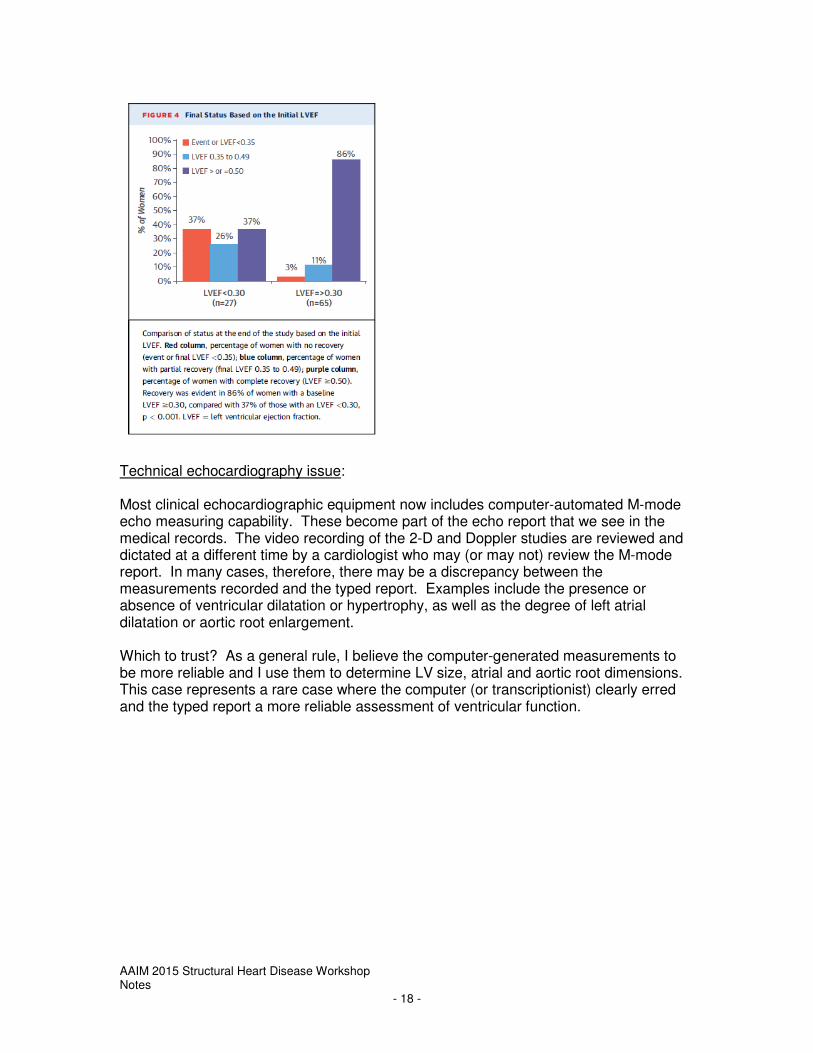
Although peripartum cardiomyopathy (or pregnancy-associated cardiomyopathy) is classified within the spectrum of the dilated cardiomyopathies, it is considered a distinct form of heart failure. It is rare, with reported incidence in the USA of 1 case in 2500-4000 live births. The clinical presentation, usually mild, may deteriorate to heart failure and circulatory collapse within a few days or, alternatively, there may be rapid improvement to normal within a few weeks. Until recently thought to be non-genetic and non-familial, recent family studies have identified a link between cases of peripartum cardiomyopathy and often asymptomatic family members with early mild dilated cardiomyopathy. Clinical criteria for the diagnosis include: development during the last month of pregnancy or within 5 months post-partum; decreased left ventricular function (ejection fraction <45%); no apparent heart disease prior to the diagnosis, and; all other causes for cardiac failure excluded. Mortality estimates in peripartum cardiomyopathy are influenced by the population, region, and time period studied. One US study in 2006 reported in-hospital mortality rates of 1.36% and overall mortality of 2.05% from 1991 – 1997, a significant improvement in mortality from that previously reported. Prognosis for return of cardiac function in peripartum cardiomyopathy is dependent on the severity of illness on presentation. In one study, univariate analysis identified LVEF (left ventricular ejection fraction) as the most significant prognostic variable. However, race (Afro-Americans' prognosis worse than Caucasians') and LVEDD (left ventricular end-diastolic diameter) > 6.0 cm were the only variables that remained significant on multivariate analysis.
AAIM 2015 Structural Heart Disease Workshop Notes - 18 -
Technical echocardiography issue: Most clinical echocardiographic equipment now includes computer-automated M-mode echo measuring capability. These become part of the echo report that we see in the medical records. The video recording of the 2-D and Doppler studies are reviewed and dictated at a different time by a cardiologist who may (or may not) review the M-mode report. In many cases, therefore, there may be a discrepancy between the measurements recorded and the typed report. Examples include the presence or absence of ventricular dilatation or hypertrophy, as well as the degree of left atrial dilatation or aortic root enlargement. Which to trust? As a general rule, I believe the computer-generated measurements to be more reliable and I use them to determine LV size, atrial and aortic root dimensions. This case represents a rare case where the computer (or transcriptionist) clearly erred and the typed report a more reliable assessment of ventricular function.

AAIM 2015 Structural Heart Disease Workshop Notes - 19 -
References for Peripartum Cardiomyopathy
Sliwa K et al. Current state of knowledge on aetiology, diagnosis, management, and therapy of peripartum cardiomyopathy: a position statement from the Heart Failure Association of the European Cardiology Society Working Group on peripartum cardiomyopathy. Eur J HF 2010; 12: 767. Van Spaendonk-Zwartz KY et al. Peripartum cardiomyopathy as a part of familial dilated cardiomyopathy. Circulation 2010; 121:2169. McNamara DM et al. Clinical outcomes for peripartum cardiomyopathy in North America. J Am Coll Cardiol 2015; 66:905.

AAIM 2015 Structural Heart Disease Workshop Notes - 20 -
Case 6 Notes: Mitral Regurgitation
• Must distinguish between “primary” (degenerative) mitral regurgitation (MR) and “secondary” (functional).
• In secondary (functional) MR, the valve is usually normal however its geometry is disrupted due to severe left ventricular dilatation and/or dysfunction.
• Will not be discussing secondary MR, as underlying condition is usually associated with very high extra mortality risk, and rarely would be considered insurable
• Primary MR is caused by pathology of 1 or more components of the mitral valve apparatus (leaflets, chordae tendineae, papillary muscles, annulus)
• Most common cause of primary (degenerative) MR in developed countries is mitral valve prolapse (MVP)
• A “normal” LVEF in primary MR is approximately 70%. The onset of LV dysfunction is inferred when the LVEF declines toward 60% or when the end-systolic diameter of the LV is ≥ 4.0 cm
• 2008 American College of Cardiology (ACC) and American Heart Association (AHA) guidelines used the following factors to define severity of MR:
2008 ACC/AHA Criteria for Severity of Mitral Regurgitation
Mild Moderate Severe
Qualitative
Angiographic grade 1+ 2+ 3 - 4+
Color Doppler jet area
Small, central jet (< 4 cm
2 or < 20% LA area)
Signs of MR > mild, but no criteria for
severe
Large central jet (area
> 40% LA area) OR
Wall-impinging jet of any size
OR swirling in LA
Doppler vena contracta width
< 0.3 cm 0.3 – 0.69 cm ≥ 0.70 cm
Quantitative
Regurgitant volume < 30 ml/beat 30 – 59 ml/beat ≥ 60 ml/beat
Regurgitant fraction < 30% 30 – 49% ≥ 50%
Effective regurgitant orifice area
< 0.20 cm2 0.20 – 0.39 cm
2 ≥ 0.40 cm
2
Additional Criteria
Left atrial size Enlarged
Left ventricular size Enlarged
Bonow RO et al. 2008 Focused Update Incorporated into the ACC/AHA 2006 Guidelines for the Management of Patients with Valvular Heart Disease. JACC 2008:52.
• The updated 2014 ACC guidelines now use a “staging” system for assessing the severity of MR (see next page).

AAIM 2015 Structural Heart Disease Workshop Notes
2014 ACC/AHA Guidelines: Stages of Primary Mitral Regurgitation Grade Definition Valve Anatomy Valve Hemodynamics Hemodynamic
Consequences Symptoms
A
At risk of MR
• Mild MVP with normal coaptation
• Mild valve thickening and leaflet restriction
• No MR jet or small central jet area <20% LA on Doppler
• Small vena contracta <0.3 cm
• None • None
B
Progressive MR
• Severe MVP with normal coaptation
• Rheumatic valve changes with leaflet restriction and loss of central coaptation
• Prior infective endocarditis
• Central jet MR 20-40% LA or late systolic eccentric jet MR
• Vena contracta <0.7 cm
• Regurgitant volume <60 ml
• Regurgitant fraction <50%
• Effective regurgitant orifice <0.40 cm
2
• Angiographic grade 1-2+
• Mild left atrial enlargement
• No LV enlargement
• Normal pulmonary artery pressure
• None
C
Asymptomatic severe MR
• Severe MVP with loss of coaptation or flail leaflet
• Rheumatic valve changes with leaflet restriction and loss of central coaptation
• Prior infective endocarditis
• Thickening of leaflets with radiation heart disease
• Central jet MR >40% LA or holosystolic eccentric jet MR
• Vena contracta ≥0.7 cm
• Regurgitant volume ≥60 ml
• Regurgitant fraction ≥50%
• Effective regurgitant orifice ≥0.40 cm
2
• Angiographic grade 3-4+
• Moderate or severe LA enlargement
• LV enlargement
• Pulmonary hypertension may be present at rest or with exercise
• C1: LVEF>60% and LVESD < 4.0 cm
• C2: LVEF ≤ 60% and LVESD ≥ 4.0 cm
• None
D
Symptomatic severe MR
• Severe MVP with loss of coaptation or flail leaflet
• Rheumatic valve changes with leaflet restriction and loss of central coaptation
• Prior infective endocarditis
• Thickening of leaflets with radiation heart disease
• Central jet MR >40% LA or holosystolic eccentric jet MR
• Vena contracta ≥0.7 cm
• Regurgitant volume ≥60 ml
• Regurgitant fraction ≥50%
• Effective regurgitant orifice ≥0.40 cm
2
• Angiographic grade 3-4+
• Moderate or severe LA enlargement
• LV enlargement
• Pulmonary hypertension present
• Decreased exercise tolerance
• Exertional dyspnea
Nishimura RA et al. 2014 AHA/ACC guideline for the management of patients with valvular heart disease: a report of the American College of Cardiology/ American Heart Association Task Force on Practice Guidelines. J Am Coll Cardiol 2014;63:e99.

AAIM 2015 Structural Heart Disease Workshop Notes
• Important Doppler definitions: o Vena contracta – the narrowest diameter of the regurgitant jet. The wider the
vena contracta, the worse the MR o Regurgitant volume (RV) - the volume in ml of regurgitant blood per heart
beat o Regurgitant fraction (RF) – the percentage of regurgitant volume compared
to the total flow across the valve o Effective regurgitant orifice (ERO) – the area of the valve opening through
which blood regurgitates into the left atrium during systole o Velocity of MR jet – the velocity of the MR jet is higher when the opening
through which it flows is smaller. A decreasing jet velocity is suggestive of worsening MR
o Jet area – the proportion of the area of the left atrium occupied by the regurgitant jet
• Triggers for surgical consideration o LVEF ≤ 60% o LVESD ≥ 4.0 cm o Development of symptoms (decreased exercise tolerance, exertional dyspnea) o Pulmonary artery pressure approaching 50 mm Hg o Possibly, rising BNP
• Future considerations o 3-D echo o Strain imaging (Doppler technique) o Cardiac MR
• Recommended follow up (physical exam and transthoracic echocardiogram) for those with primary MR:
o Stages A and B – “periodically” to evaluate for changes in MR severity � Mild MR: every 3-5 years unless clinical change � Moderate MR: every 1-2 years unless clinical change
o Stage C1 – follow up 1-2 times/year � Trigger for surgery reached in those with Stage C1 MR at rate of
8%/year o Any stage – at any time if symptoms develop or signs change

AAIM 2015 Structural Heart Disease Workshop Notes - 23 -
Discussion Questions: Mitral regurgitation 1. What factors in this case, both clinical and from the echo report, can be used to help
assess the severity of the mitral regurgitation?
Factor How Does it Help?
Clinical Murmur Timing and character consistent with
MR? Symptoms Presence of symptoms such as
dyspnea suggest advanced disease HTN/BP control HTN is a common cause of MR
Echo
Mitral valve appearance If normal, why would it leak? Mitral annular calcification Distortion of annulus can disrupt
valve function Left atrial size Increased LA consistent with chronic
MR Left ventricular size Dilated LV can cause MR due to
disruption of mitral apparatus, or if caused by MR, suggests advanced disease
LV wall thickness LVH can cause MR through diastolic dysfunction, LA dilatation, distortion of mitral apparatus
LV systolic function Low normal or low EF with MR suggests advanced disease
Qualitative description “Eyeball” approach, may identify flow but not its significance
Eccentric jet Suggests significant MR RV systolic pressure Elevated RVSP suggests moderate
to severe MR What other information might be helpful?
• Has the murmur changed over the past few years?
• Has functional capacity changed over the last few years?
• Is there any history of palpitations, syncope or near-syncope? Dyspnea on exertion?
• Any prior echos or more recent evaluation?
• End systolic dimension of the left ventricle?
• Any more quantitative measures of MR, such as vena contracta, regurgitant volume, effective regurgitant orifice, regurgitant fraction
• Assessment of mitral regurgitation by Doppler echocardiography:
Qualitative/Semiquantitative Quantitative
− Mild, moderate, severe
− 1+, 2+, 3+, 4+
− Vena contracta width
− Pulmonary vein flow reversal
− Regurgitant volume
− Regurgitant fraction
− Effective regurgitant orifice (ERO)

AAIM 2015 Structural Heart Disease Workshop Notes - 24 -
2. How do you view the ejection fraction of 52%? Is it normal and favorable?
Although this is a low normal value for those with a normal heart, it is significantly abnormal in the setting of primary mitral regurgitation. It is a marker that the left ventricle is starting to fail in response to a chronic volume overload state.
3. What is the significance of the NTproBNP?
This value is above normal for a 62 year old man. It suggests significant wall stress on the ventricular myocardium – another marker that this is in fact “significant” mitral regurgitation, and likely to be associated with some degree of extra mortality and morbidity risk. NTproBNP (N-terminal proB-type natriuretic peptide)
o ProBNP released by myocardial cells in response to wall stress and cleaved into physiologically active BNP and the inactive fragment NTproBNP
o NTproBNP used for insurance screening because it is stable in transit and doesn’t require use of special collection tubes
o Expected range varies by age, sex and renal function o BNP and NTproBNP have different ranges! o Increase in NTproBNP suggests underlying cardiac issue and is associated
with an increased risk of heart failure, cardiovascular morbidity and mortality and all-cause mortality.
o Increases have been associated with ischemia, atrial fibrillation, valvular disease, left ventricular hypertrophy and cardiomyopathy in addition to heart failure
4. Does the severity of mitral regurgitation correlate with mortality risk?
It is difficult to accurately determine the severity of MR – this factor hinders the assessment of associated mortality risk. The best measures of severity are the quantitative Doppler measurements - however we rarely see these on echo reports. Data correlating different degrees of MR with mortality are scarce. Most studies look at severity in terms of when to refer for surgical intervention rather than in terms of pure mortality risk. MR, especially when the mitral valve apparatus is structurally abnormal, is known to progress, but it is difficult to predict the rate. The few available natural history studies (most quite old) indicate that those with severe, asymptomatic MR with normal LV function (stage C1) are likely to develop symptoms or LV dysfunction warranting operation over a 6-10 year period (rate of about 8%/year) The Enriquez-Sarano article (see references) is the best paper I found that looks at mortality rather than timing for surgery. It uses the risk categories shown above. The length of follow up is short, only 5 years, however the study did report significant differences in survival in asymptomatic MR patients, according to increasing effective

AAIM 2015 Structural Heart Disease Workshop Notes - 25 -
regurgitant orifice (ERO). Regurgitant volume was less predictive, and other measures such as qualitative grading and jet area were predictive on univariate but not multivariate analysis.
5-Year Survival Risk Ratio for Death from any cause
ERO < 20 mm2 91% 1 ERO 20 – 39 mm2 66% 2.58 ERO ≥ 40 mm2 58% 2.90 Per 10-mm2 increment in ERO
1.20
Enriquez-Sarano M et al. NEJM 2005;352(9):875.
5. Is there likely to be excess mortality associated with the findings in this case? What
about morbidity?
This appears to be very significant MR – with at least 1 potential trigger for surgical consideration. Qualitatively, the MR is described as “moderately severe”, and the jet is eccentric. The RVSP is elevated, though not approaching 50. The LV is at the upper limits of normal in size at end diastole and we unfortunately don’t have the end-systolic measurement. The EF, however, is quite low in the setting of MR – this is the single most concerning factor. In addition, the NTproBNP is elevated. And this is based on echo data from 4 years ago. There is a high likelihood of progression to even worse MR and LV dysfunction at this point. In addition, there is risk for arrhythmias (atrial fibrillation and ventricular) and thromboembolic events as the left atrium enlarges.
6. What is the likelihood that this person will need surgical intervention? Repair or
replacement? Would it matter from an insurance risk perspective?
We have incomplete data, however there are several red flags. It seems quite likely that this person could need surgical intervention – probably in the near future Determining the optimal timing for surgery is very important. At some point, changes in left ventricular function, the pulmonary vasculature and cardiac rhythm become irreversible, and surgery may not help (and in some cases, may worsen) LV function and symptomatology. Important when considering surgical intervention:
o Those with severe MR may have no symptoms, even during vigorous exercise
o Repair is preferred over replacement because � Lower operative mortality rate � LV function is better preserved due to conservation of the integrity
of the mitral valve apparatus � Repair avoids the risks inherent to prosthetic valves, including
thromboembolism (2-2.5%/patient-year) or anticoagulant-induced

AAIM 2015 Structural Heart Disease Workshop Notes - 26 -
hemorrhage (2%/patient-year) for mechanical valves and structural deterioration (only a 78% freedom from reoperation at 15 years) of bioprosthetic valves
o Successful repair has better long-term outcome (reduced mortality and morbidity) than replacement
o Mitral valve replacement with preservation of chordal structures is more likely to have better symptomatic and functional outcome (than when chordal structures are removed)
o Mitral valve surgery has less favorable outcome than aortic valve surgery o Long-term survival after mitral valve repair for primary MR (DiBardino, J
Thorac Cardiovasc Surg 2010;139:76-84) � 10 year survival 79% � 20-year survival 62% � 30-year survival 52%
7. What is the recommended clinical follow up for mitral regurgitation, and how is that
relevant for insurance risk selection?
• Recommended follow up (physical exam and transthoracic echocardiogram) for those with primary MR:
o Stages A and B – “periodically” to evaluate for changes in MR severity � Mild MR: every 3-5 years unless clinical change � Moderate MR: every 1-2 years unless clinical change
o Stage C1 – follow up 1-2 times/year � Trigger for surgery reached in those with Stage C1 MR at rate of
8%/year o Any stage – at any time if symptoms develop or signs change
It is helpful to be familiar with these guidelines when assessing the quality of information available to you in underwriting files. In this case, for example, the echo is 4 years old and shows stage C2 MR. For stage C1 MR, the clinical guidelines suggest that the echo should have been repeated at least 1-2 times/year. You may feel there is enough information in the file to make an underwriting recommendation, but it would not be unreasonable to postpone the case pending clinically appropriate follow up.

AAIM 2015 Structural Heart Disease Workshop Notes - 27 -
References for Mitral Regurgitation
Bonow RO et al. 2008 focused update incorporated into the ACC/AHA 2006 guidelines for the management of patients with valvular heart disease: a report of the American College of Cardiology/American Heart Association Task Force on Practice Guidelines (Writing Committee to Revise the 1998 Guidelines for the Management of Valvular Heart Disease). J Am Coll Cardiol 2008:52:e1-e142.
Nishimura RA et al. 2014 AHA/ACC guideline for the management of patients with valvular heart disease: a report of the American College of Cardiology/ American Heart Association Task Force on Practice Guidelines. J Am Coll Cardiol 2014;63:e57-e185 (this document has over 900 references and covers all forms of valvular heart disease – an excellent starting point for a review)
Enriquez-Sarano M et al. Quantitative determinants of the outcome of asymptomatic mitral regurgitation. N Engl J Med 2005;352(9):875-883. DiBardino DJ et al. Four decades of experience with mitral valve repair: analysis of differential indications, technical evolution, and long-term outcome. J Thorac Cardiovasc Surg 2010;139:76-84. Maganti K et al. Valvular heart disease: diagnosis and management. Mayo Clin Proc 2010;85(5):483-500. (An excellent overview of all types of valvular heart disease)

AAIM 2015 Structural Heart Disease Workshop Notes - 28 -
Case 7 Notes: Ventricular Noncompaction
In 1984, Drs. Engberding and Bender from Germany published a case report of a rare congenital anomaly found by echocardiography in an adult. The anomaly, "persistence of myocardial sinusoids", had previously been seen only in infants with multiple congenital defects. Their patient presented with exertional dyspnea and palpitations, and was found to have left bundle block on ECG. Echocardiography showed marked left ventricular enlargement and wall thickening. "Huge sinusoids" were seen and confirmed on cardiac catheterization where increased cardiac chamber pressures confirmed the diagnosis of congestive heart failure. Since that report, ventricular noncompaction has been recognized as a rare but identifiable congenital abnormality. In these cases, the usual embryonic condensation of myocardial fibers and trabecular spaces is incomplete to a varying degree. Several modes of inheritance have been documented, including autosomal dominant, X-linked, and mitochondrial familial patterns. Within familial occurrences, phenotypic expression also varies between 18% and 50%. Recent reports suggest that ventricular noncompaction is not solely a genetic disease but could be acquired. Patients with other neuromuscular or mitochondrial disorders have been reported to have developed noncompaction after prior echocardiogram studies had been normal. Perhaps more interestingly, ventricular noncompaction has been shown to have developed de novo with intensive athletic training, trauma, or myocardial ischemia. Until fairly recently, most ventricular noncompaction cases presented with otherwise unexplained heart failure. With better awareness and screening, however, noncompaction is now being diagnosed in apparently asymptomatic adults and children. The classic presentation, though, is one of systolic heart failure (up to 60% of patients), with myocardial hypo-perfusion the presumed mechanism. Diastolic dysfunction is almost always present as the abnormal trabeculations restrict ventricular filling. Most have arrhythmias, and systemic emboli are not uncommon. Treatment protocols, for the most part, mimic those of dilated cardiomyopathy, including automatic defibrillation implantation and cardiac transplantation. Survival Early studies reported a very high mortality rate (58% 5 year survival). More recent reports, presumably reflecting better awareness and broader inclusion of cases, have been less ominous, with 4 year survival reported at 97%. Survival is, at least in part, related to symptoms at presentation, with one center describing 100% survival in those asymptomatic at diagnosis. Technical issues: Echocardiography and magnetic resonance are both useful in the diagnosis of ventricular noncompaction. Thickened endocardium with prominent trabeculae and deep recesses are seen well with both modalities. Universally accepted criteria to describe the severity of ventricular noncompaction, defined as the noncompaction/compaction ratio or the total noncompacted myocardial mass index, remains to be established.

AAIM 2015 Structural Heart Disease Workshop Notes - 29 -
The echocardiogram has excellent resolution compared to other imaging modalities, but there is significant intra-observer and inter-observer variation that needs to be considered when looking at serial studies. Ventricular chamber measurements may vary as much as 0.5 cm in the absence of technical difficulties, and considerably more when there are technical challenges.
Technique Spatial resolution Inter-observer variation
Magnetic Resonance with flow mapping
1.5 - 3 mm 5-16 %
CAT scanning 2 – 4 mm 8-10 %
Echocardiography 2 – 4 mm 3-10 %

AAIM 2015 Structural Heart Disease Workshop Notes - 30 -
References for Ventricular Noncompaction
Engberding R, Bender F. Identification of a rare congenital anomaly of the myocardium by two-dimensional echocardiography: persistence of isolated myocardial sinusoids. Am J Cardiol 1984; 53:1733. Hussein A et al. Isolated noncompaction of the left ventricle in adults. J Am Coll Cardiol 2015; 66:578. Weissman NJ, Adelmann GA. Cardiac Imaging Secrets. Hanley and Belfus. Philadelphia, PA 19106. 2004. Singh K, et al. Intra- and Inter-observer variability in the measurements of abdominal aortic and common iliac artery diameter in Computed Tomography. The Tromso study. J Vasc Endovasc Surg 2003; 25: 399. Bogaert, J et al. Clinical Cardiac MRI. Springer Science and Business Media. New York. 2012.

AAIM 2015 Structural Heart Disease Workshop Notes - 31 -
Case 8 Notes: Takotsubo Cardiomyopathy
• “Broken heart syndrome” is also known as apical ballooning syndrome, stress-induced cardiomyopathy, or takotsubo cardiomyopathy (TTC).
• First described in Japan in 1990
• Takotsubo means “octopus pot”. Describes the characteristic appearance of the left ventricle, with ballooning of the left ventricular apex during systole
• Is an acute heart failure syndrome that typically presents with chest pain and/or dyspnea and must be distinguished from acute coronary syndrome
• Accounts for 1-2% of all admissions for presumed acute coronary syndrome; incidence has been estimated at about 5.2 per 100,000 for US women and 0.6 per 100,000 for US men (reference #3)
• Etiology may be related to stress-related catecholamine excess
• Mayo Clinic diagnostic criteria (must meet all 4): o Transient abnormality of the left ventricular wall motion beyond the territory of a
single coronary artery distribution o Absence of obstructive coronary artery disease or angiographic evidence of an
acute plaque rupture o New EKG abnormalities or increased troponin o Absence of pheochromocytoma and myocarditis
• In western countries, most commonly affects older women. (In Japan, more often affects men)
• Is commonly preceded by an emotional or physical trigger
• Originally felt to be fairly benign; more recent studies suggesting some long term risk. Reference #1 reported 5.6% per year all-cause mortality rate (some of which may be due to comorbidities) and 1.8% per year recurrence rate over 10 year follow up.
Discussion Questions
1. What is “broken heart syndrome”? Broken heart syndrome (also known as takotsubo or stress-induced cardiomyopathy or the apical ballooning syndrome) is an acute heart failure syndrome associated with significant acute mortality and morbidity. It is characterized by transient reduction in left ventricular ejection fraction and a dyskinetic ventricular apex. It is often triggered by a stressful physical or psychological event. It is thought to be related to abnormal microvascular responses to high levels of circulating catecholamines. Takotsubo cardiomyopathy may resolve with no or minimal residua, however there is a risk of recurrence and it is not clear whether long-term mortality is normal or increased. .
2. How is it different from acute coronary syndrome? Takotsubo cardiomyopathy (TTC) is not caused by coronary artery plaque rupture or obstructive coronary artery disease. While cardiac troponins are commonly elevated in TTC, the value is usually less than that seen in acute coronary syndrome (ACS). BNP values are often higher in TTC than in ACS. Creatine kinase (CK) is usually not elevated in TTC.

AAIM 2015 Structural Heart Disease Workshop Notes - 32 -
3. What is the long-term outlook for TTC? There is considerable mortality and morbidity in the acute phase of this condition. Men are twice as likely as women to have major adverse events (recurrent TTC, MI, stroke, TIA or death). Older age and the presence of an emotional trigger predict better outcomes. Long term, reference #1 suggests a 5.6% per year risk of death from any cause and a 1.8% per year risk of recurrence.
References for Takotsubo Cardiomyopathy
Templin C et al. Clinical features and outcomes of takotsubo (stress) cardiomyopathy. N Engl J Med 2015;373(10):929-938. Parodi G et al. Natural history of tako-tsubo cardiomyopathy. Chest 2011;139(4):887-892 Deshmukh A et al. Prevalence of takotsubo cardiomyopathy in the United States. Am Heart J 2012;164:66-71.