structural basis of the regulatory mechanism of the plant ... · structural basis of the regulatory...
TRANSCRIPT

Structural basis of the regulatory mechanism of theplant CIPK family of protein kinases controllingion homeostasis and abiotic stressAntonio Chaves-Sanjuana, Maria Jose Sanchez-Barrenaa, Juana Maria Gonzalez-Rubioa, Maria Morenoa, Paula Ragelb,Marta Jimeneza, Jose M. Pardob, Martin Martinez-Ripolla, Francisco J. Quinterob, and Armando Alberta,1
aDepartamento de Cristalografía y Biología Estructural, Instituto de Química Física “Rocasolano,” Consejo Superior de Investigaciones Científicas,Madrid E-28006, Spain; and bDepartamento de Biotecnología Vegetal, Instituto de Recursos Naturales y Agrobiología de Sevilla, Consejo Superior deInvestigaciones Científicas, Sevilla E-41012, Spain
Edited by Natasha V. Raikhel, University of California, Riverside, CA, and approved September 15, 2014 (received for review May 8, 2014)
Plant cells have developed specific protective molecular machineryagainst environmental stresses. The family of CBL-interactingprotein kinases (CIPK) and their interacting activators, the calciumsensors calcineurin B-like (CBLs), work together to decode calciumsignals elicited by stress situations. Themolecular basis of biologicalactivation of CIPKs relies on the calcium-dependent interaction ofa self-inhibitoryNAFmotifwith a particular CBL, the phosphorylationof the activation loop by upstream kinases, and the subsequentphosphorylation of the CBL by the CIPK. We present the crystalstructures of the NAF-truncated and pseudophosphorylated kinasedomains of CIPK23 and CIPK24/SOS2. In addition, we providebiochemical data showing that although CIPK23 is intrinsicallyinactive and requires an external stimulation, CIPK24/SOS2 displaysbasal activity. This data correlateswell with the observed conforma-tion of the respective activation loops: Although the loopofCIPK23 isfolded into a well-ordered structure that blocks the active site accessto substrates, the loop of CIPK24/SOS2 protrudes out of the activesite and allows catalysis. These structures together with biochemi-cal and biophysical data show that CIPK kinase activity necessarilyrequires the coordinated releases of the activation loop from theactive site and of the NAF motif from the nucleotide-binding site.Taken all together, we postulate the basis for a conserved calcium-dependent NAF-mediated regulation of CIPKs and a variable regu-lation by upstream kinases.
signaling | ion transport | abiotic stress
Cell perception of extracellular stimuli is followed by a tran-sient variation in cytosolic calcium concentration. Plants
have evolved to produce the specific molecular machinery tointerpret this primary information and to transmit this signal tothe components that organize the cell response (1–4). The plantfamily of serine/threonine protein kinases PKS or CIPKs (here-inafter CIPKs) and their activators, the calcium-binding proteinsSCaBPs or CBLs (hereinafter CBLs) (5, 6) function together indecoding calcium signals caused by different environmental stim-uli. Available data suggest a mechanism in which calcium mediatesthe formation of stable CIPK–CBL complexes that regulate thephosphorylation state and activity of various ion transporters in-volved in the maintenance of cell ion homeostasis and abioticstress responses in plants. Among them, the Arabidopsis thalianaCIPK24/SOS2-CBL4/SOS3 complex activates the Na+/H+ anti-porter SOS1 to maintain intracellular levels of the toxic Na+ lowunder salt stress (7–9), the CIPK11–CBL2 pair regulates the plasmamembrane H+-ATPase AHA2 to control the transmembrane pHgradient (10), the CIPK23–CBL1/9 (11, 12) regulates the activity ofthe K+ transporter AKT1 to increase the plant K+ uptake capabilityunder limiting K+ supply conditions (12, 13), and CIPK23–CBL1mediates nitrate sensing and uptake by phosphorylation of thenitrate transporter CHL1 (14). Together these findings show thatunderstanding the molecular mechanisms underling CIPKs func-
tion provides opportunities to increase plant tolerance to abioticstress and to improve plants for human benefit.CIPKs and CBLs contain discrete structural modules that are
involved in the calcium-dependent regulation of the activity ofthe system and ensure the colocalization of the CIPK–CBLinteracting pairs with their substrates at particular sites withinthe cell (15–17). CIPKs include an N-terminal kinase catalyticdomain followed by a characteristic self-inhibitory motif knownas FISL or NAF motif (hereinafter NAF, Pfam no. PF03822)(1, 6) and a protein phosphatase 2C binding domain designatedas PPI (11, 18, 19). The NAF motif directly interacts with thecatalytic domain and inhibits the kinase activity. The calcium-dependent interaction of CBLs with the NAF motif relieves theself-inhibition and activates the CIPKs (5, 6, 19, 20). The calciumbinding to CBLs is mediated by four EF hand-like calciumbinding motives. In addition, several CBLs are myristoylated and/orpalmitoylated. These modifications are essential for recruiting theirinteracting CIPK partner to the plasma or vacuolar membrane (17,21–23), and they may also be involved in the interaction of theCIPK–CBL complexes with their substrates (24). In addition, thephosphorylation of a conserved serine residue at the C terminus ofCBLs by its interacting CIPK is required for activation of trans-porter substrates. It has been proposed that this process may sta-bilize the CIPK–CBL complex and trigger conformational changes
Significance
The transport of ions through the plant cell membrane estab-lishes the key physicochemical parameters for cell function.Stress situations such as those created by soil salinity or lowpotassium conditions alter the ion transport across the mem-brane producing dramatic changes in the cell turgor, the mem-brane potential, and the intracellular pH and concentrations oftoxic cations such as sodium and lithium. As a consequence,fundamental metabolic routes are inhibited. The CIPK family of26 protein kinases regulates the function of several ion trans-porters at the cell membrane to restore ion homeostasis understress situations. Our analyses provide an explanation on howthe CIPKs are differentially activated to coordinate the adequatecell response to a particular stress.
Author contributions: A.C.-S., J.M.G.-R., M.M., P.R., M.J., and A.A. performed research;M.J.S.-B., J.M.P., M.M.-R., F.J.Q., and A.A. designed research; A.C.-S., M.J.S.-B., J.M.G.-R.,M.M., P.R., J.M.P., M.M.-R., F.J.Q., and A.A. analyzed data; and A.A. wrote the paper.
The authors declare no conflict of interest.
This article is a PNAS Direct Submission.
Data deposition: The atomic coordinates have been deposited in the Protein Data Bank(PDB), www.pdb.org (PDB ID codes 4CZT, 4CZU, and 4D28).1To whom correspondence should be addressed. Email: [email protected].
This article contains supporting information online at www.pnas.org/lookup/suppl/doi:10.1073/pnas.1407610111/-/DCSupplemental.
E4532–E4541 | PNAS | Published online October 6, 2014 www.pnas.org/cgi/doi/10.1073/pnas.1407610111
to the binary complex that enhance its specificity toward targetproteins (13, 25).Like many other kinases, CIPKs are also regulated by the
phosphorylation of the activation loop by upstream kinases. Thisloop undergoes large conformational changes upon phosphory-lation, allowing the entrance and the stabilization of substrates atthe kinase active site (26). The activation loop of the CIPKs con-tains three conserved Tyr, Thr, or Ser residues. For some membersof the family, the mutation of one of these residues to Asp mimicsphosphorylation and produces the activation of the kinase, partlyovercoming the effect of the self-inhibitory NAF motif. In fact,these phosphorylation-mimicking mutations and the deletion of theinhibitory domain produce a synergistic effect on the CIPK activity(6, 27–29). Transgenic plants expressing these CIPK24/SOS2 mu-tant proteins show improved salt tolerance (30).The kinase self-phosphorylation is another regulatory mecha-
nism used by CIPKs. CIPK24/SOS2 is able to self-phosphorylate,and the autophosphorylation is important for its activity (31). Al-though the default state of CIPKs is inactive, some degree ofautophosphorylation activity has been observed even for dephos-phorylated and CBL-unbound CIPKs, which suggests that someCIPKs display basal activity (6). Indeed, it has been shown that thegeneral regulatory factor 14-3-3 proteins (32) interact withCIPK24/SOS2 and repress its basal kinase activity when plants aregrown in the absence of salt stress (33).The crystal structure of the binary complex of Ca2+-CBL4/
SOS3 with the C-terminal regulatory moiety of CIPK24/SOS2revealed the molecular mechanism underlying CBL-mediatedactivation of the CIPKs. The structure showed that the CIPK24/SOS2 self-inhibitory NAF motif is bound to CBL4/SOS3 and,consequently, it is not accessible to the kinase domain (19, 20).However, whether the CBL-unbound NAF blocks the active siteor inhibits the enzyme by an allosteric mechanism is not known.To determine the molecular and structural basis for the CIPKsautoinhibition by the NAF and the activation by upstream kinases,we solved the structures of CIPK23 and CIPK24/SOS2. Our datashow that inactivation of the kinases relies on the blockage ofthe active site by the NAF motif and the activation loop, whichconstitutes the basis for the conserved NAF-mediated self-inhibition of the CIPKs.
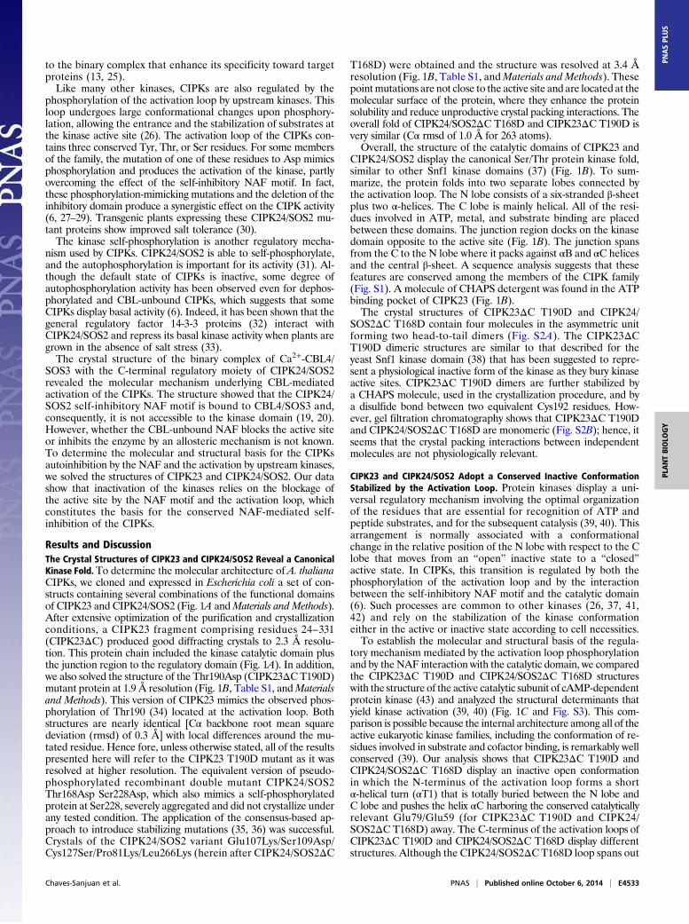
Results and DiscussionThe Crystal Structures of CIPK23 and CIPK24/SOS2 Reveal a CanonicalKinase Fold.To determine the molecular architecture of A. thalianaCIPKs, we cloned and expressed in Escherichia coli a set of con-structs containing several combinations of the functional domainsof CIPK23 and CIPK24/SOS2 (Fig. 1A andMaterials and Methods).After extensive optimization of the purification and crystallizationconditions, a CIPK23 fragment comprising residues 24–331(CIPK23ΔC) produced good diffracting crystals to 2.3 Å resolu-tion. This protein chain included the kinase catalytic domain plusthe junction region to the regulatory domain (Fig. 1A). In addition,we also solved the structure of the Thr190Asp (CIPK23ΔC T190D)mutant protein at 1.9 Å resolution (Fig. 1B, Table S1, andMaterialsand Methods). This version of CIPK23 mimics the observed phos-phorylation of Thr190 (34) located at the activation loop. Bothstructures are nearly identical [Cα backbone root mean squaredeviation (rmsd) of 0.3 Å] with local differences around the mu-tated residue. Hence fore, unless otherwise stated, all of the resultspresented here will refer to the CIPK23 T190D mutant as it wasresolved at higher resolution. The equivalent version of pseudo-phosphorylated recombinant double mutant CIPK24/SOS2Thr168Asp Ser228Asp, which also mimics a self-phosphorylatedprotein at Ser228, severely aggregated and did not crystallize underany tested condition. The application of the consensus-based ap-proach to introduce stabilizing mutations (35, 36) was successful.Crystals of the CIPK24/SOS2 variant Glu107Lys/Ser109Asp/Cys127Ser/Pro81Lys/Leu266Lys (herein after CIPK24/SOS2ΔC
T168D) were obtained and the structure was resolved at 3.4 Åresolution (Fig. 1B, Table S1, andMaterials and Methods). Thesepointmutations are not close to the active site and are located at themolecular surface of the protein, where they enhance the proteinsolubility and reduce unproductive crystal packing interactions. Theoverall fold of CIPK24/SOS2ΔC T168D and CIPK23ΔC T190D isvery similar (Cα rmsd of 1.0 Å for 263 atoms).Overall, the structure of the catalytic domains of CIPK23 and
CIPK24/SOS2 display the canonical Ser/Thr protein kinase fold,similar to other Snf1 kinase domains (37) (Fig. 1B). To sum-marize, the protein folds into two separate lobes connected bythe activation loop. The N lobe consists of a six-stranded β-sheetplus two α-helices. The C lobe is mainly helical. All of the resi-dues involved in ATP, metal, and substrate binding are placedbetween these domains. The junction region docks on the kinasedomain opposite to the active site (Fig. 1B). The junction spansfrom the C to the N lobe where it packs against αB and αC helicesand the central β-sheet. A sequence analysis suggests that thesefeatures are conserved among the members of the CIPK family(Fig. S1). A molecule of CHAPS detergent was found in the ATPbinding pocket of CIPK23 (Fig. 1B).The crystal structures of CIPK23ΔC T190D and CIPK24/
SOS2ΔC T168D contain four molecules in the asymmetric unitforming two head-to-tail dimers (Fig. S2A). The CIPK23ΔCT190D dimeric structures are similar to that described for theyeast Snf1 kinase domain (38) that has been suggested to repre-sent a physiological inactive form of the kinase as they bury kinaseactive sites. CIPK23ΔC T190D dimers are further stabilized bya CHAPS molecule, used in the crystallization procedure, and bya disulfide bond between two equivalent Cys192 residues. How-ever, gel filtration chromatography shows that CIPK23ΔC T190Dand CIPK24/SOS2ΔC T168D are monomeric (Fig. S2B); hence, itseems that the crystal packing interactions between independentmolecules are not physiologically relevant.
CIPK23 and CIPK24/SOS2 Adopt a Conserved Inactive ConformationStabilized by the Activation Loop. Protein kinases display a uni-versal regulatory mechanism involving the optimal organizationof the residues that are essential for recognition of ATP andpeptide substrates, and for the subsequent catalysis (39, 40). Thisarrangement is normally associated with a conformationalchange in the relative position of the N lobe with respect to the Clobe that moves from an “open” inactive state to a “closed”active state. In CIPKs, this transition is regulated by both thephosphorylation of the activation loop and by the interactionbetween the self-inhibitory NAF motif and the catalytic domain(6). Such processes are common to other kinases (26, 37, 41,42) and rely on the stabilization of the kinase conformationeither in the active or inactive state according to cell necessities.To establish the molecular and structural basis of the regula-
tory mechanism mediated by the activation loop phosphorylationand by the NAF interaction with the catalytic domain, we comparedthe CIPK23ΔC T190D and CIPK24/SOS2ΔC T168D structureswith the structure of the active catalytic subunit of cAMP-dependentprotein kinase (43) and analyzed the structural determinants thatyield kinase activation (39, 40) (Fig. 1C and Fig. S3). This com-parison is possible because the internal architecture among all of theactive eukaryotic kinase families, including the conformation of re-sidues involved in substrate and cofactor binding, is remarkably wellconserved (39). Our analysis shows that CIPK23ΔC T190D andCIPK24/SOS2ΔC T168D display an inactive open conformationin which the N-terminus of the activation loop forms a shortα-helical turn (αT1) that is totally buried between the N lobe andC lobe and pushes the helix αC harboring the conserved catalyticallyrelevant Glu79/Glu59 (for CIPK23ΔC T190D and CIPK24/SOS2ΔC T168D) away. The C-terminus of the activation loops ofCIPK23ΔC T190D and CIPK24/SOS2ΔC T168D display differentstructures. Although the CIPK24/SOS2ΔC T168D loop spans out
Chaves-Sanjuan et al. PNAS | Published online October 6, 2014 | E4533
PLANTBIOLO
GY
PNASPL
US

of the active site, the CIPK23ΔC T190D loop contains anotherwell-ordered α-helical turn (αT2) followed by a long loop thatblocks the access of ATP and peptide substrate to the active siteand drives the nucleotide binding loop (the G loop) apart (Fig.1B). The observed inactive conformations of CIPK23ΔCT190D and CIPK24/SOS2ΔC T168D are surprising because theconstructions used for protein crystallization lack the NAF self-inhibitory motif and include the T190D mutation (T168D forCIPK24/SOS2), mimicking a phosphorylated state of the activation
loop that leads to hyperactive forms of CIPK24/SOS2 and someother CIPK proteins (6, 27). The fact that CIPK23ΔC T190D andCIPK24/SOS2ΔC T168D display largely identical overall con-formations, although we solved them in two different space groupswith different crystal packing contacts, indicates that their confor-mations are unlikely to be affected by crystal packing.To determine the biological significance of our structural ob-
servations, we performed kinase activity assays comparing theCIPK23ΔC T190D and CIPK24/SOS2ΔC T168D constructs lacking
Fig. 1. The crystal structures of CIPK23 and CIPK24/SOS2. (A) Domain structure of CIPKs. (B) A ribbon representation of the crystal structures of CIPK23ΔC T190D(Left) and CIPK24/SOS2ΔC T168D (Right). The key structural features are labeled and highlighted in different colors. (C) The ribbon representation of the active sitesection of the CIPK23ΔC T190D, CIPK24/SOS2ΔC T168D (semitransparent overlaid on the left), and the related active kinase PKA (Right) (PDB ID code: 1ATP). Thecatalytically relevant residues and the ATP are displayed in a stick representation; CIPK23 and CIPK24/SOS2 residues are displayed in white and cyan, respectively.
E4534 | www.pnas.org/cgi/doi/10.1073/pnas.1407610111 Chaves-Sanjuan et al.
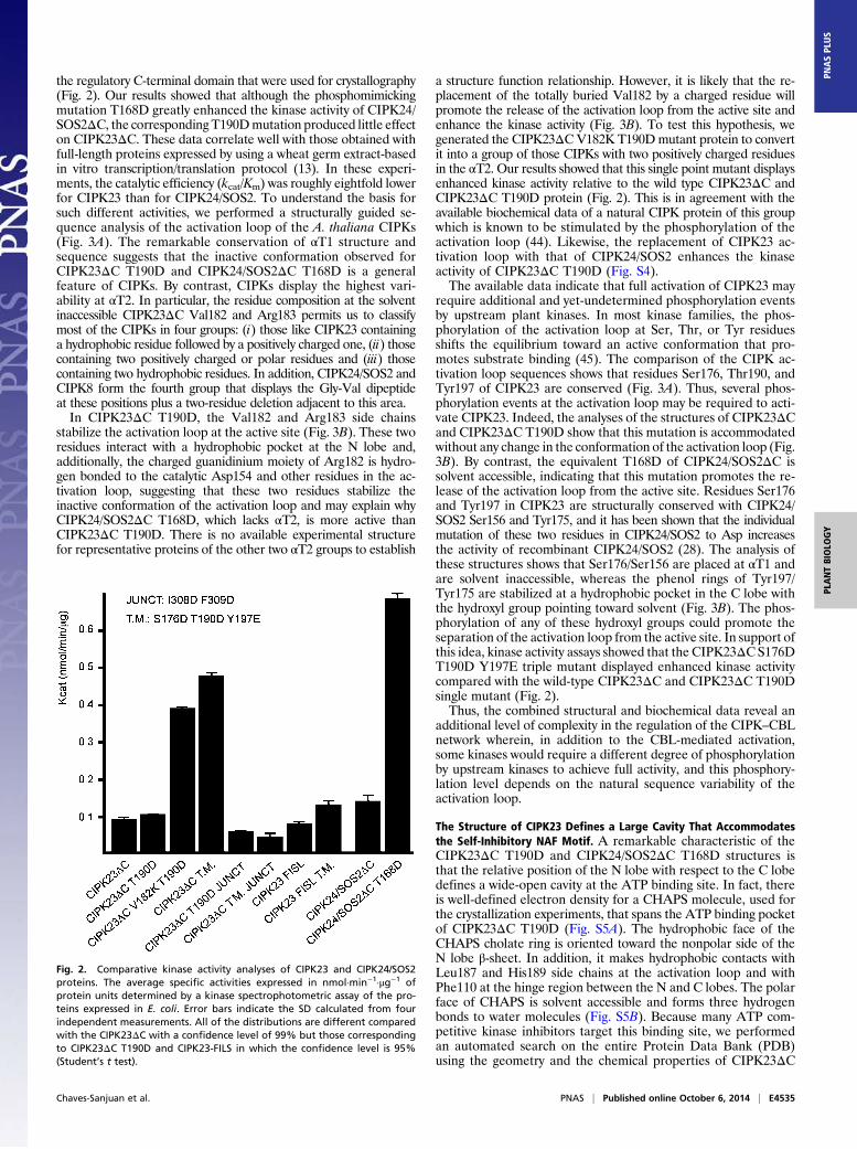
the regulatory C-terminal domain that were used for crystallography(Fig. 2). Our results showed that although the phosphomimickingmutation T168D greatly enhanced the kinase activity of CIPK24/SOS2ΔC, the correspondingT190Dmutation produced little effecton CIPK23ΔC. These data correlate well with those obtained withfull-length proteins expressed by using a wheat germ extract-basedin vitro transcription/translation protocol (13). In these experi-ments, the catalytic efficiency (kcat/Km) was roughly eightfold lowerfor CIPK23 than for CIPK24/SOS2. To understand the basis forsuch different activities, we performed a structurally guided se-quence analysis of the activation loop of the A. thaliana CIPKs(Fig. 3A). The remarkable conservation of αT1 structure andsequence suggests that the inactive conformation observed forCIPK23ΔC T190D and CIPK24/SOS2ΔC T168D is a generalfeature of CIPKs. By contrast, CIPKs display the highest vari-ability at αT2. In particular, the residue composition at the solventinaccessible CIPK23ΔC Val182 and Arg183 permits us to classifymost of the CIPKs in four groups: (i) those like CIPK23 containinga hydrophobic residue followed by a positively charged one, (ii) thosecontaining two positively charged or polar residues and (iii) thosecontaining two hydrophobic residues. In addition, CIPK24/SOS2 andCIPK8 form the fourth group that displays the Gly-Val dipeptideat these positions plus a two-residue deletion adjacent to this area.In CIPK23ΔC T190D, the Val182 and Arg183 side chains
stabilize the activation loop at the active site (Fig. 3B). These tworesidues interact with a hydrophobic pocket at the N lobe and,additionally, the charged guanidinium moiety of Arg182 is hydro-gen bonded to the catalytic Asp154 and other residues in the ac-tivation loop, suggesting that these two residues stabilize theinactive conformation of the activation loop and may explain whyCIPK24/SOS2ΔC T168D, which lacks αT2, is more active thanCIPK23ΔC T190D. There is no available experimental structurefor representative proteins of the other two αT2 groups to establish
a structure function relationship. However, it is likely that the re-placement of the totally buried Val182 by a charged residue willpromote the release of the activation loop from the active site andenhance the kinase activity (Fig. 3B). To test this hypothesis, wegenerated the CIPK23ΔCV182K T190Dmutant protein to convertit into a group of those CIPKs with two positively charged residuesin the αT2. Our results showed that this single point mutant displaysenhanced kinase activity relative to the wild type CIPK23ΔC andCIPK23ΔC T190D protein (Fig. 2). This is in agreement with theavailable biochemical data of a natural CIPK protein of this groupwhich is known to be stimulated by the phosphorylation of theactivation loop (44). Likewise, the replacement of CIPK23 ac-tivation loop with that of CIPK24/SOS2 enhances the kinaseactivity of CIPK23ΔC T190D (Fig. S4).The available data indicate that full activation of CIPK23 may
require additional and yet-undetermined phosphorylation eventsby upstream plant kinases. In most kinase families, the phos-phorylation of the activation loop at Ser, Thr, or Tyr residuesshifts the equilibrium toward an active conformation that pro-motes substrate binding (45). The comparison of the CIPK ac-tivation loop sequences shows that residues Ser176, Thr190, andTyr197 of CIPK23 are conserved (Fig. 3A). Thus, several phos-phorylation events at the activation loop may be required to acti-vate CIPK23. Indeed, the analyses of the structures of CIPK23ΔCand CIPK23ΔC T190D show that this mutation is accommodatedwithout any change in the conformation of the activation loop (Fig.3B). By contrast, the equivalent T168D of CIPK24/SOS2ΔC issolvent accessible, indicating that this mutation promotes the re-lease of the activation loop from the active site. Residues Ser176and Tyr197 in CIPK23 are structurally conserved with CIPK24/SOS2 Ser156 and Tyr175, and it has been shown that the individualmutation of these two residues in CIPK24/SOS2 to Asp increasesthe activity of recombinant CIPK24/SOS2 (28). The analysis ofthese structures shows that Ser176/Ser156 are placed at αT1 andare solvent inaccessible, whereas the phenol rings of Tyr197/Tyr175 are stabilized at a hydrophobic pocket in the C lobe withthe hydroxyl group pointing toward solvent (Fig. 3B). The phos-phorylation of any of these hydroxyl groups could promote theseparation of the activation loop from the active site. In support ofthis idea, kinase activity assays showed that the CIPK23ΔC S176DT190D Y197E triple mutant displayed enhanced kinase activitycompared with the wild-type CIPK23ΔC and CIPK23ΔC T190Dsingle mutant (Fig. 2).Thus, the combined structural and biochemical data reveal an
additional level of complexity in the regulation of the CIPK–CBLnetwork wherein, in addition to the CBL-mediated activation,some kinases would require a different degree of phosphorylationby upstream kinases to achieve full activity, and this phosphory-lation level depends on the natural sequence variability of theactivation loop.
The Structure of CIPK23 Defines a Large Cavity That Accommodatesthe Self-Inhibitory NAF Motif. A remarkable characteristic of theCIPK23ΔC T190D and CIPK24/SOS2ΔC T168D structures isthat the relative position of the N lobe with respect to the C lobedefines a wide-open cavity at the ATP binding site. In fact, thereis well-defined electron density for a CHAPS molecule, used forthe crystallization experiments, that spans the ATP binding pocketof CIPK23ΔC T190D (Fig. S5A). The hydrophobic face of theCHAPS cholate ring is oriented toward the nonpolar side of theN lobe β-sheet. In addition, it makes hydrophobic contacts withLeu187 and His189 side chains at the activation loop and withPhe110 at the hinge region between the N and C lobes. The polarface of CHAPS is solvent accessible and forms three hydrogenbonds to water molecules (Fig. S5B). Because many ATP com-petitive kinase inhibitors target this binding site, we performedan automated search on the entire Protein Data Bank (PDB)using the geometry and the chemical properties of CIPK23ΔC
Fig. 2. Comparative kinase activity analyses of CIPK23 and CIPK24/SOS2proteins. The average specific activities expressed in nmol·min−1·μg−1 ofprotein units determined by a kinase spectrophotometric assay of the pro-teins expressed in E. coli. Error bars indicate the SD calculated from fourindependent measurements. All of the distributions are different comparedwith the CIPK23ΔC with a confidence level of 99% but those correspondingto CIPK23ΔC T190D and CIPK23-FILS in which the confidence level is 95%(Student’s t test).
Chaves-Sanjuan et al. PNAS | Published online October 6, 2014 | E4535
PLANTBIOLO
GY
PNASPL
US

T190D cavity as search criteria (Relibase+; ref. 46). Our analysisreveals that, in contrast with the chemical nature of CHAPS, theinhibitory molecules bound to this site consist of planar aromaticrings that mimic the adenine ring of ATP. In addition, the studyshows that the CIPK23 cavity is wider than other occupied cav-ities (Fig. S5C). This observation, together with the fact thata similar cavity is present in CIPK24/SOS2 (Fig. S5D), show thatCHAPS binding is indicative of the hydrophobic nature and ofthe characteristic enlarged size of the ATP binding site of CIPKswith respect to the size and shape of other inactive kinase mole-cules. In addition, the fact that CIPK23ΔC T190D activation loopis mainly stabilized by contacts with the kinase domain suggeststhat CHAPS binding does not affect the conformation of theactivation loop, although local effects in the vicinity of Leu-187cannot be discarded (Fig. S5E).The fact that the kinase activation via phosphorylation of the
activation loop and via CBL binding are synergic processes in-dicates that the release of both the activation loop and the NAFmotif from the catalytic domain are coupled (6, 27–29). A plau-sible model to account for these observations would imply that theinactive kinase bears two different but interacting sites for NAFand activation loop. Thus, we explored the possibility that theATP binding pocket of CIPK23 could accommodate the NAFself-inhibitory domain, blocking ATP entrance and stabilizinga closed conformation of the activation loop, thereby hindering thesubstrate entrance. The crystal structures of the regulatory moiety ofCIPK24/SOS2 and CIPK14 in complex with their interacting CBLsrevealed that the NAF motif consists of two amphipathic helicesconnected by a variable loop (19, 47) (Fig. 4A). Interestingly, thevisual and automatic analyses of the CIPK23ΔC T190D andCIPK24/SOS2ΔC T168D molecular surfaces using CAVER (48)reveal that the ATP cavity spans linearly, wrapping the kinase do-main around the hinge region between the N and C lobes (Fig. 4A).This cavity is wide enough to accommodate the two amphipathichelical segments of the NAF motif and it is connected to the ac-tivation loop, thus suggesting that it constitutes the CIPKs self-inhibitory binding pocket. Remarkably, the cavity overlaps wellwith the calmodulin binding helix of the regulatory domain of thecalcium/calmodulin-dependent protein kinase I (49) (Fig. S6).To corroborate this hypothesis, we first checked whether the
self-inhibitory NAF extension inhibits the kinase activity. The un-phosphorylated CIPK23-NAF protein expressed in E. coli (com-prising residues 24–349) displayed slightly lower activity thanCIPK23ΔC and CIPK23ΔC T190D. This effect is more noticeablewhen comparing the highly active CIPK23ΔC S176D T190DY197E triple mutant with the corresponding CIPK23-NAF S176DT190D Y197E enlarged protein (Fig. 2). Nevertheless, it is in agree-ment with the biochemical data on CIPK24/SOS2 (6), CIPK20 (44),and CIPK8 (27) in which amoderated reduction of the kinase activityof the full-length active CIPK compared with a mutated versionlacking the NAF was observed. Second, we checked whether theNAF motif folds into a α-helix in the absence of CBL by com-paring and analyzing the circular dichroism spectra of CIPK23ΔCand CIPK23-NAF. This spectroscopic technique provides a directrelationship between the α-helical content of a protein and thespectral signal intensity at 195, 208, and 222 nm (50). Our datashowed that CIPK23-NAF has a higher helical content thanCIPK23ΔC (Fig. 4B). Using DichroWeb, a server to analyze cir-cular dichroism data using several algorithms, this increase wasestimated to be approximately 5–8% in accordance the expectedincrease if the NAF folds as a α-helix (51). In addition, we testedwhether CHAPS and the NAF compete for the same site at theCIPK23 catalytic domain. Hence, we monitored the thermal
Fig. 3. The activation loop. (A) Sequence alignment of the activation loopof CIPKs together with a schematic representation of CIPK23ΔC T190Dand CIPK24/SOS2ΔC T168D secondary structures. Sequences are groupedaccording to their sequence similarity. Residues are colored according totheir conservation (77). The arrows highlight the totally conserved and po-tentially phosphorylable residues. The rectangular box highlights the resi-dues at αT2 that serve to classify the CIPKs into four groups. (B) A detailedview of CIPK23ΔC T190D showing the structural environment of the con-served Ser176, Thr190, and Tyr197. The view highlights the role of Val182and Arg183 in the stabilization of the loop at the active site. Inset shows
a comparison of CIPK23ΔC (white sticks) and CIPK23ΔC T190D (cyan sticks)structures showing that the point mutation is accommodated withouta significant change in the loop conformation.
E4536 | www.pnas.org/cgi/doi/10.1073/pnas.1407610111 Chaves-Sanjuan et al.

denaturation of CIPK23ΔC and CIPK23-NAF at increasingconcentrations of CHAPS by circular dichroism spectroscopy.Our results showed that CHAPS was able to bind CIPK23ΔC andCIPK23-NAF because it induced a change in the thermal de-naturation temperature (Tm) of these proteins. However, analysisof the variation of Tm with the concentration of the ligand showsthat Tm variations occur at lower CHAPS concentration forCIPK23ΔC than forCIPK23-NAF. These data show that CIPK23ΔCdisplays higher affinity for CHAPS than CIPK23-NAF and indicatethat the NAFmotif and CHAPS bind to the same site in the catalyticdomain of CIPK23 (Fig. 4C).
Thus, the joined structural and biochemical data suggest amodel in which the NAF segment folds as a α-helix bound to theinterface between the N and C lobes, blocking ATP access to theactive site and hindering the conformational change required forkinase activation. This regulatory mechanism strongly resemblesthe inhibition by the C-terminal pseudosubstrate segment ob-served in the CaMKI, another calcium calmodulin-dependentprotein kinase (49). In this case, an inhibitory helical segment,equivalent to the NAF, competes with ATP for the same site andinduces a pronounced distortion of the kinase structure towardan inactive conformation. Like CBLs and CIPKs, the calcium-dependent interaction of calmodulin with the CaMKI kinasereleases the pseudosubstrate from the active site and acti-vates the kinase.
The Junction Domain of CIPK23 Is Involved in the Stabilization of theCatalytically Relevant α-C helix. The characterization of the func-tional domains of CIPK24/SOS2 revealed that the junction re-gion placed between the catalytic and the self-inhibitory NAFmotif is required for kinase activity (6, 30, 33). In addition, it hasbeen shown that the phosphorylation by an uncharacterized ki-nase of Ser294 at this junction region enhances the interactionwith the general regulatory protein 14-3-3 and produces re-pression of CIPK24/SOS2 basal activity (33). Despite the highsequence variability of this region among CIPKs (Fig. S7A), weinvestigated whether the structures of CIPK23ΔC T190D andCIPK24/SOS2ΔC T168D could unveil the structural basis forthis additional level of regulation.The crystallographic analysis reveals that the junction domain
connects the C lobe and the N lobe through a long unstructuredlinker followed by a α-helix turn and a β-strand parallel to thecentral β-sheet (Fig. 1B). Interestingly, the CIPK23ΔC T190Dresidues Ile308 and Phe309 (Val287 and Phe288 in CIPK24/SOS2ΔC T168D) at the end of the α-helix turn, αT5 (αK inCIPK24/SOS2), are buried in a conserved hydrophobic surfacepocket placed at the N lobe between αB and αC (Fig. 5). Theinteraction of a regulatory segment on a structurally equivalent hy-drophobic motif is a common feature of several kinases and involvesthe stabilization of the helix αC to facilitate its optimal alignment forcatalysis (42) or to provide the minimal structural scaffold necessaryfor basal activity as observed for the Arabidopsis Snf1-relatedSnRK2.6/OST1 (52, 53). Moreover, in some cases such as theACG kinases (54), this regulation mechanism is triggered by thephosphorylation of a conserved Ser on the regulatory segment.Accordingly, recombinant unphosphorylated CIPK24/SOS2 dis-plays some basal constitutive activity and the C-terminal trunca-tion of SOS2/CIPK24 just before the Val287 (equivalent to Ile308residue in CIPK23), but not after, produces an inactive kinase anda salt-sensitive phenotype in planta (6, 30). Thus, it seems that atleast for CIPK24/SOS2, the interaction of these two hydrophobicresidues with N lobe is sufficient for basal activity and is necessaryfor activation of the kinase. Moreover, it appears clear thatphosphorylation of Ser294 and 14-3-3 binding would counteractthis basal level of activation. To investigate the importance of Ile308and Phe309 in CIPK23, we mutated them to Asp and performedkinase activity assays. Our results show that the I308D F309D mu-tated versions of theCIPK23ΔCand of the highly active CIPK23ΔCS176D T190D Y197E triple mutant do not display kinase activity(Fig. 2), corroborating the role of these two residues in catalyticstabilization of the kinases. The sequence analysis of the CIPKfamily of proteins reveals that this hydrophobicmotif is conserved in10 of 26 members of the family (Fig. S7A), suggesting an importantrole in CIPKs activity, although its precise role remains to beinvestigated.The presence of a structurally equivalent linker between the N
and C lobes like the one observed in the structures of CIPK23ΔCT190D and CIPK24/SOS2ΔC T168D is another recurrent themein kinase structures. Remarkably, Zap-70 kinase autoinhibition
Fig. 4. (A) Ribbon and schematic representations of the CIPK24/SOS2 reg-ulatory domain structures in complex with CBL4/SOS3 (PDB ID code: 2EHB)(Left) and CIPK23ΔC T190D together with a surface representation of thecavity connecting the ATP binding site and the hinge region between N andC lobes (Right). The cavity is wide enough to accommodate the two am-phipathic helices forming the NAF motif. (B) Comparison of the far-UV CDspectra of CIPK23ΔC and CIPK23-NAF; the arrows indicate the characteristicα-helical maximum at 195, and minima at 208 and 222 nm. (C) Comparisonof the thermal stability profiles monitored by CD for CIPK23ΔC (Left)and CIPK23-NAF (Right) at increasing CHAPS concentration.
Chaves-Sanjuan et al. PNAS | Published online October 6, 2014 | E4537
PLANTBIOLO
GY
PNASPL
US

relies on the blockage of the transition from an “open” inactivestate to a “closed” active state by an equivalent linker connectingN and C lobes (55). In CIPKs, the N to C lobe linker seems to beflexible because it forms few intramolecular contacts with thecatalytic domain and has high crystallographic temperature fac-tors (Fig. S7B). Thus, it does not seem to be hindering the kinaseactivation process.
The Regulatory Mode of CIPKs. The structural data presented hereshow that the activation loop and the self-inhibitory pocket arespatially connected, providing a working model in which theactivation loop at the active site and the NAF segment at theself-inhibitory pocket are mutually stabilized. In this scenario,the CIPK activation necessarily implies the simultaneous releaseof these two elements from the catalytic domain. The release canbe effectively achieved by the calcium-dependent CBL in-teraction, or through phosphorylation of the activation loop byan upstream kinase, or both, thereby explaining the additiveeffects on activity achieved by mimicking phosphorylation by pu-tative upstream kinases and deletion of the NAF domain (Fig. 6).Moreover, the reported basal kinase activity for some CIPKsindicates that these processes need not always be concurrent foractivation. In accordance with the biochemical data, the activity ofa particular CIPK will depend on its ability to stabilize these activemolecular species and/or on the balance between them and theinactive form of the kinase. Consequently, the maximum activitywill be observed for CBL-bound and phosphorylated CIPKs, be-cause the active form will be fully stabilized. The required phos-phorylation of the C-terminal region of CBLs by CIPKs to achievefully functional activation of the CBL–CIPKs complexes may becentral to understand why the moderate NAF motif-mediatedstimulation of the in vitro CIPK23 (Fig. 2) is absolutely required forin vivo activation of AKT1 K+ channel (13). This data additionallysupports the idea that CBL phosphorylation may affect the struc-ture of the CBL-CIPKs complexes and consequently, their activity.Besides, an additional layer of CIPK regulation comes from thecofactor preference of Mn2+ compared with Mg2+ (13, 27, 28).The fact that one of the metal ion binding sites of CBL4/SOS3displays higher affinity for Mn2+ than for Ca2+ suggests that CBLscould act as a carrier for this cofactor or, alternatively, couldbuffer the availability of free Mn2+ to prevent a constitutive ac-tivation of the CIPKs (56).The available structural and biochemical data support a model
wherein CIPK23 is intrinsically inactive and requires the calciumdependent CBL1/9 binding and/or the phosphorylation by up-stream kinases for activation, while CIPK24/SOS2 does not. Thisdifferential behavior may be related to the physiology roles ofthese proteins. To our knowledge, CIPK23 targets solely the K+
channel AKT1 and the nitrate transporter CHL1 (11, 14). Bycontrast, in addition to SOS1, CIPK24/SOS2 is involved in theregulation of a number of antiporters and H+-ATPases (57–59)and it has been reported to participate in signaling pathwaysregulating flowering time and redox metabolism (60, 61). Con-sequently, compared with CIPK23, CIPK24/SOS2 will require awider range of activation states and a more specialized regula-tion. In contrast to CIPK23, which is intrinsically inactive andshows an absolute requirement for external stimulation, CIPK24/SOS2 displays basal activity that is finely regulated by phos-phorylation of Ser294 and 14-3-3 binding, thus adding a furtherlayer of biochemical regulation (33).In conclusion, the analysis of the CIPK structure reported here
provides the basis for comprehending the regulatory mechanismof a major signaling network in plant response to environmentalcues. Understanding this mechanism is central to enhance cropspecies production by augmenting the limited capacities of plantsto cope with environmental stresses.
Materials and MethodsGene Cloning and Site-Directed Mutagenesis. The sequences of the primersused in the cloning and the site-directed mutagenesis of all constructs arelisted in Table S2. The kinase domain of CIPK23 (CIPK23ΔC; residues 24–331)was cloned between NdeI and NotI restriction sites of the pGEX4T2 ex-pression vector (GE Healthcare) that had been previously modified to con-tain an NdeI restriction site between EcoRI and SmaI (forward primer 23Nand reverse primer 23C). CIPK23ΔC T190D mutant was generated by PCRusing pGEX4T2-CIPK23 as a template (forward primer 23T190D5 and reverseprimer 23T190D3) and CIPK23ΔC V182K T190D mutant using CIPK23ΔC T190Das a template (forward primer 23V182K5 and reverse primer 23V182K3).CIPK23 S176D T190D Y197Ewas generated successively by PCR using CIPK23ΔCT190D as a template, and the resulted constructs were used as a template forthe following ones (forward primers 23S176D5 and 23Y197E5 and reverseprimers 23S176D3and23Y197E3). CIPK23ΔCT190D I308D F309DandCIPK23ΔCS176D T190D Y197E I308D F309D were prepared by using pGEX4T2-CIPK23ΔCT190D and pGEX4T2-CIPK23ΔC S176D T190DY197E as a template, respectively,(forward primer 23I308DF309D5 and reverse primer 23I308DF309D3) andCIPK23-SOS2-like mutant was generated by using pGEX4T2-CIPK23ΔC as atemplate (forward primer 23SOS2like5 and reverse primer 23SOS2like3).
Fig. 5. A section of the CIPK23ΔC T190D structure showing the interactionbetween Ile308 and Phe309 with the hydrophobic pocket at the N lobe ofCIPK23. A cartoon representation of the kinase is displayed together with asemitransparent surface.
Fig. 6. A working model for CIPKs regulation. CIPK is schematically repre-sented with the N lobe and the C lobe depicted in green and blue, re-spectively, the junction as a purple line, the activation loop in red, andthe NAF motif as a cyan rectangle. Activation of CIPKs implies necessarily therelease of both the activation loop and the self-inhibitory NAF motif. Thecalcium-dependent CBL interaction and/or phosphorylation of the activationloop set the equilibrium between the inactive and the active forms. Theactivity of CIPKs depends on the balance between active and inactive forms.Some CIPKs (for instance CIPK24/SOS2) display basal activity. The junctionregion may be responsible for the stabilization of a CBL unbound and un-phosphorylated CIPK form with basal activity. Once CIPK is active, the fullyfunctional activation is achieved by the phosphorylation of the CBL.
E4538 | www.pnas.org/cgi/doi/10.1073/pnas.1407610111 Chaves-Sanjuan et al.
The kinase domain of CIPK23 (residues 1–331) was amplified by PCR usingthe primers 23N2 and 23C and cloned as SmaI/NotI fragment in the yeastexpression vector pEG(KT) (62). To subclone the mutant allele CIP23-SOS2-like in pEG(KT), a 0.69-kb BglII/NotI fragment encoding amino acids Lys103to Val331 of CIPK23 was obtained from the corresponding pGEX4-T2construct and ligated in the pEG(KT)-CIPK23 digested with the samerestriction enzymes.
The kinase domain of CIPK24/SOS2 (CIPK24/SOS2ΔC residues 7–308) wascloned between BamHI and EcoRI restriction sites of the pGEX4T2 expressionvector (GE Healthcare) (forward primer 24N and the reverse primer 24C).CIPK24/SOS2 mutants were generated successively by PCR using this con-struct as the first template, and the resulted constructs were used as a tem-plate for the following ones. CIPK24/SOS2ΔC T168D (forward primer 24T168D5and reverse primer 24T168D3), S228D (forward primer 24S228D5 and reverseprimer 24S228D3), E107K and S109D (forward primer 24E107KS109D5 and re-verse primer 24E107KS109D3), C127S (forward primer 24C127S5 and reverseprimer 24C127S3), P81K (forward primer 24P81K5 and reverse primer 24P81K3),and L266K (forward primer 24L266K5 and reverse primer 24L266K3). All se-quences were confirmed by plasmid DNA sequencing.
CIPK23-NAF (CIPK23; residues 24–352) was cloned between BamHI andHindIII restriction sites of the multiple cloning sites I of pETDuet-1 expressionvector (Novagen) (forward primer 23FN and reverse primer 23FC), previouslymodified to contain CBL1 (residues 1–213) between NdeI and KpnI re-striction sites of multiple cloning site II (forward primer CBL1N and reverseprimer CBL1C). CIPK23-NAF S176D T190D Y197E was generated successivelyby PCR using CIPK23-NAF as a template, and the resulted constructs were usedas a template for the following ones (forward primers 23S176D5, 23T190D5,and 23Y197E5 and reverse primers 23S176D3, 23T190D3, and 23Y197E3).
Protein Preparation, Crystallization, and Data Collection. Recombinant pro-teins were expressed in Rosetta 2 (DE3) pLysS cells (Stratagene) by inductionat OD600 = 0.7 with 0.3 mM isopropyl-β-D-thiogalactoside for 16 h at 16 °C.Following isolation by centrifugation, the CIPK23ΔC and CIPK23ΔC T190Dbacterial pellets were then resuspended in a 150 mM NaCl, 20 mM Hepes pH7.0, and 0.5 mM Tris(2-carboxyethyl)phosphine (TCEP) buffer, whereas theCIPK24/SOS2ΔC T168D bacterial pellet was resuspended in a 20 mM NaCl, 20mM Tris·HCl pH 8.5, and 5 mM DTT buffer. Cells were lysed by sonication andpurified by using glutathione Sepharose 4B (GE Healthcare) following over-night thrombin cleavage for CIPK23 and an overnight preScission cleavagefor CIPK24/SOS2ΔC T168D. The protein was dialyzed overnight in 20 mMNaCl, 20 mM Tris·HCl pH 9.0, and 5 mM DTT. Proteins were further purified bygel filtration chromatography on a Hi Load 16/60 Superdex 200 prep gradecolumn (GE Healthcare). Chromatography yields single peaks, which corre-spond to the monomeric proteins. CIPK23ΔC, CIPK23ΔC T190D, and CIPK24/SOS2ΔC T168D constructs were concentrated to 14.0 mg·ml−1, 14.0 mg·ml−1,and 5.0 mg·ml−1, respectively, using an Amicon Ultra 10 K centrifugal filterdevice (Millipore). The sample purity was determined by SDS/PAGE.
Initial crystallization screens were set up as sitting-drop vapor-diffusionexperiments. We used an Innovadine crystallization robot and crystallizationkits from Qiagen, Hampton Research, and Jena Bioscience. Initial screeningsof CIPK23ΔC lead to spherulites in many conditions. We selected conditionnumber 4 (0.1 M Tris·HCl pH 8.0 and 2.0 M ammonium sulfate) from CrystalScreen HT (Hampton Research) as a starting point for crystal optimization.CIPK23ΔC spherulites were screened against a set of detergents (DetergentScreen HT; Hampton Research). Needle-shaped crystals grew after 3 d usingthe under oil microbatch method. The final crystallization condition wasobtained by mixing 1 μL of 3.5 M ammonium sulfate, 0.1 M Hepes pH 7.5,16 mM CHAPS, and 1 μL of protein preparation. These crystals were cry-oprotected by adding a saturated solution of ammonium sulfate to thecrystallization drop until saturation. Identical crystallization protocol wasfollowed for CIPK23ΔC T190D. Initial screenings of CIPK24/SOS2ΔC T168Dlead to thin plates in condition number 47 (30% wt/vol PEG 4000, 0.1 MTris·HCl pH 8.5, and 0.2 M magnesium chloride) from JBScreen Classic: 1, 2, 3,and 4 (Jena Bioscience). CIPK24/SOS2ΔC T168D crystallyzation was furtheroptimized by screening against a matrix of pH (7.0, 8.2, 8.5, 8.8, and 9.0)versus PEG 4K concentration (12–30%) using the under oil microbatchmethod. Thicker plates appeared between pH 5.8 and 9.0 and PEG 4K con-centration between 20 and 24%. In-well dehydrated crystals were used forcrystal structure determination.
Crystals from CIPK23ΔC, CIPK23ΔC T190D, and CIPK24/SOS2ΔC T168Dwere mounted in fiber loops and then flash cooled in liquid nitrogen. X-raydiffraction data from CIPK23ΔC and T190D mutant were collected at theEuropean Syncrotron Radiation Facility beamlines ID14-4 and ID23-2, re-
spectively. CIPK24/SOS2ΔC T168D diffraction data were collected at thePetra-III synchrotron, Hamburg, beamline P13.
CIPK23ΔC, CIPK23ΔC T190D, and CIPK24/SOS2ΔC T168D diffraction data-sets were processed by using MOSFLM (63) and XDS (64) respectively, andscaled using SCALA (65) from the CCP4 package (Collaborative ComputationalProject, Number 4, 1994). A summary of the data collection statistics is givenin Table S1.
Structure Determination. The X-ray structures of CIPK23ΔC and CIPK23ΔCT190D were solved by molecular replacement with the program MolRep (66)using the coordinates of a kinase domain (PDB ID code: 3H4J) (37). Theelectron density map calculated by using these phases was good enough tomanually build and refine the structure of CIPK23ΔC T190D. Several cycles ofrestrained refinement with PHENIX (67) and REFMAC5 (68) and iterativemodel building with COOT (69) were carried out. The X-ray structure ofCIPK24/SOS2ΔC T168D was solved by molecular replacement with the pro-gram MolRep (66) using the coordinates of a kinase domain of CIPK23. Sev-eral cycles of Jelly-body and restrained refinement with REFMAC5 (68) anditerative model building with COOT (69) were carried out. Calculations wereperformed by using CCP4 programs (70). The stereochemistry of the modelswas verified with MolProbity (71). Ribbon figures were produced by usingPyMOL (72). The refinement statistics are summarized in Table S1.
In Vitro Kinase Assays. Kinetic studies of kinases were conducted by couplingreaction method as described (73, 74). In brief, 3 μM kinase was added to areaction mixture containing 1 mM MgCl2, 100 mM phosphoenolpyruvate(Sigma), 0.28 mM NADH (Calbiochem), 10–20 units of pyruvate kinase and20–30 units of lactate dehydrogenase (Sigma), 1 mg/mL histone (Sigma), and0.5 mM ATP (Sigma). The reaction was performed in a 96-well flat-bottomplate (Sarstedt). The NADH depletion, which is coupled to the rate of thekinase reaction, was followed by measuring the decrease in absorbance at340 nm for 40 min at 22 °C in a Synergy HT BioTek microplate spectropho-tometer. Blank reactions without enzyme were carried out for each enzyme.Four replicas were collected for each reaction mixture, and the average ac-tivities were quantified.
[γ-32P] ATP Kinase Assays. For radioactive in vitro phosphorylation assays,wild-type and mutant CIPK23ΔC and GST-CIPK23-SOS2–like proteins werepurified from yeast cells as described (62). Cells were collected by centri-fugation and lysed with glass beads in PBS buffer (10 mM Na2HPO4, 2 mMKH2PO4, 2.7 mM KCl, and 137 mM NaCl, pH 7.4). Recombinant proteinswere affinity-purified on glutathione-Sepharose (GE Healthcare). Myelin basicprotein (Sigma) (∼1 μg) was subjected to phosphorylation by recombinantCIPK23 protein kinases (∼100 ng) in 30 μL of buffer (66.7 mM Tris·HCl, pH 8,100 mM NaCl, 5 mM MnSO4, 0.5 mM CaCl2, and 2 mM DTT). Reactionswere started by adding ATP (10 μM with 20 μCi of [γ-32P]ATP), which wasincubated at 30 °C for 45 min, and stopped with 10 μL of 4× SDS/PAGEsample buffer. Proteins were separated by SDS/PAGE by using a 12% (wt/vol)acrylamide gel. Radioactivity was detected on the dry gels by using BiomaxLight-1 (Kodak) films.
Circular Dichroism Spectra. Protein samples were prepared in a buffer con-taining 20mMsodiumphosphate at pH8.2 and0.5mMTCEP tooptimize signal-to-noise ratio. Special attention was paid to the instrument calibration (50).Circular dichroism spectra were recorded in a JASCO J-810 spectropolarimeter.Far-UV spectra were recorded in a 0.02 cm-pathlength quartz cell at a proteinconcentration of 0.7 mg·ml−1 (20 μM). The spectra were analyzed by usingCONTIN (75) included in the DICROWEB web page (76). Thermal denaturationexperiments were carried out in a 0.1 cm-pathlength quartz cell at a proteinconcentration of 0.2 mg·ml−1 (5.6 μM) and monitored at wavelength of 220 nmby increasing the temperature from 30 °C to 70 °C at 50 °C·h−1.
ACKNOWLEDGMENTS. A.A. thanks Dr. Douglas Vinson Laurents for criticalreading of the manuscript, and the European Syncrotron Radiation Facility(beamlines ID14-4 and ID23-2) and PETRAIII (beamline P13, BIOSTRUCTX_3100.5)for the access to the synchrotron radiation source. This work was funded byMinisterio de Economía y Competitividad Grants BFU2011-25384 and CSD2006-00015 (to A.A.), BIO2011-28184-C02-02 (toM.J.S.-B.), and BIO2012-36533 (to F.J.Q.),which was cofinanced by the European Regional Development Fund, andComunidad de Madrid Grant S2010/BMD-2457 (to A.A.). A.C.-S. is supportedby a Formación de Personal Investigador Predoctoral Fellowship, and M.J.S.-B.is supported by Ramón y Cajal Contract RYC-2008-03449 from MINECO.
Chaves-Sanjuan et al. PNAS | Published online October 6, 2014 | E4539
PLANTBIOLO
GY
PNASPL
US

1. Albrecht V, Ritz O, Linder S, Harter K, Kudla J (2001) The NAF domain defines a novel protein-protein interaction module conserved in Ca2+-regulated kinases. EMBO J 20(5):1051–1063.
2. Shi J, et al. (1999) Novel protein kinases associated with calcineurin B-like calciumsensors in Arabidopsis. Plant Cell 11(12):2393–2405.
3. Batistic O, Kudla J (2012) Analysis of calcium signaling pathways in plants. BiochimBiophys Acta 1820(8):1283–1293.
4. Steinhorst L, Kudla J (2013) Calcium and reactive oxygen species rule the waves ofsignaling. Plant Physiol 163(2):471–485.
5. Luan S, Kudla J, Rodriguez-Concepcion M, Yalovsky S, GruissemW (2002) Calmodulinsand calcineurin B-like proteins: Calcium sensors for specific signal response coupling inplants. Plant Cell 14(Suppl):S389–S400.
6. Guo Y, Halfter U, Ishitani M, Zhu JK (2001) Molecular characterization of functionaldomains in the protein kinase SOS2 that is required for plant salt tolerance. Plant Cell13(6):1383–1400.
7. Qiu QS, Guo Y, Dietrich MA, Schumaker KS, Zhu JK (2002) Regulation of SOS1,a plasma membrane Na+/H+ exchanger in Arabidopsis thaliana, by SOS2 and SOS3.Proc Natl Acad Sci USA 99(12):8436–8441.
8. Quintero FJ, et al. (2011) Activation of the plasma membrane Na/H antiporter Salt-Overly-Sensitive 1 (SOS1) by phosphorylation of an auto-inhibitory C-terminal do-main. Proc Natl Acad Sci USA 108(6):2611–2616.
9. Núñez-Ramírez R, et al. (2012) Structural insights on the plant salt-overly-sensitive 1(SOS1) Na(+)/H(+) antiporter. J Mol Biol 424(5):283–294.
10. Fuglsang AT, et al. (2007) Arabidopsis protein kinase PKS5 inhibits the plasmamembrane H+ -ATPase by preventing interaction with 14-3-3 protein. Plant Cell 19(5):1617–1634.
11. Lee SC, et al. (2007) A protein phosphorylation/dephosphorylation network regulatesa plant potassium channel. Proc Natl Acad Sci USA 104(40):15959–15964.
12. Xu J, et al. (2006) A protein kinase, interacting with two calcineurin B-like proteins,regulates K+ transporter AKT1 in Arabidopsis. Cell 125(7):1347–1360.
13. Hashimoto K, et al. (2012) Phosphorylation of calcineurin B-like (CBL) calciumsensor proteins by their CBL-interacting protein kinases (CIPKs) is required for fullactivity of CBL-CIPK complexes toward their target proteins. J Biol Chem 287(11):7956–7968.
14. Ho CH, Lin SH, Hu HC, Tsay YF (2009) CHL1 functions as a nitrate sensor in plants. Cell138(6):1184–1194.
15. Kim BG, et al. (2007) The calcium sensor CBL10 mediates salt tolerance by regulatingion homeostasis in Arabidopsis. Plant J 52(3):473–484.
16. Quan R, et al. (2007) SCABP8/CBL10, a putative calcium sensor, interacts with theprotein kinase SOS2 to protect Arabidopsis shoots from salt stress. Plant Cell 19(4):1415–1431.
17. Ishitani M, et al. (2000) SOS3 function in plant salt tolerance requires N-myristoylationand calcium binding. Plant Cell 12(9):1667–1678.
18. Ohta M, Guo Y, Halfter U, Zhu JK (2003) A novel domain in the protein kinase SOS2mediates interaction with the protein phosphatase 2C ABI2. Proc Natl Acad Sci USA100(20):11771–11776.
19. Sánchez-Barrena MJ, et al. (2007) The structure of the C-terminal domain of theprotein kinase AtSOS2 bound to the calcium sensor AtSOS3. Mol Cell 26(3):427–435.
20. Sánchez-Barrena MJ, Martínez-Ripoll M, Albert A (2013) Structural biology of a majorsignaling network that regulates plant abiotic stress: The CBL-CIPK mediated path-way. Int J Mol Sci 14(3):5734–5749.
21. Batistic O, et al. (2012) S-acylation-dependent association of the calcium sensor CBL2with the vacuolar membrane is essential for proper abscisic acid responses. Cell Res22(7):1155–1168.
22. Quintero FJ, Ohta M, Shi H, Zhu JK, Pardo JM (2002) Reconstitution in yeast of theArabidopsis SOS signaling pathway for Na+ homeostasis. Proc Natl Acad Sci USA99(13):9061–9066.
23. Eckert C, et al. (2014) The vacuolar calcium sensors CBL2 and CBL3 affect seed size andembryonic development in Arabidopsis thaliana. Plant J 78(1):146–156.
24. Held K, et al. (2011) Calcium-dependent modulation and plasma membrane targetingof the AKT2 potassium channel by the CBL4/CIPK6 calcium sensor/protein kinasecomplex. Cell Res 21(7):1116–1130.
25. Lin H, et al. (2009) Phosphorylation of SOS3-LIKE CALCIUM BINDING PROTEIN8 bySOS2 protein kinase stabilizes their protein complex and regulates salt tolerance inArabidopsis. Plant Cell 21(5):1607–1619.
26. Nolen B, Taylor S, Ghosh G (2004) Regulation of protein kinases; controlling activitythrough activation segment conformation. Mol Cell 15(5):661–675.
27. Gong D, Gong Z, Guo Y, Chen X, Zhu JK (2002) Biochemical and functional charac-terization of PKS11, a novel Arabidopsis protein kinase. J Biol Chem 277(31):28340–28350.
28. Gong D, Guo Y, Jagendorf AT, Zhu JK (2002) Biochemical characterization of theArabidopsis protein kinase SOS2 that functions in salt tolerance. Plant Physiol 130(1):256–264.
29. Gao P, Kolenovsky A, Cui Y, Cutler AJ, Tsang EW (2012) Expression, purification andanalysis of an Arabidopsis recombinant CBL-interacting protein kinase3 (CIPK3) andits constitutively active form. Protein Expr Purif 86(1):45–52.
30. Gong D, Guo Y, Schumaker KS, Zhu JK (2004) The SOS3 family of calcium sensors andSOS2 family of protein kinases in Arabidopsis. Plant Physiol 134(3):919–926.
31. Fujii H, Zhu JK (2009) An autophosphorylation site of the protein kinase SOS2 is im-portant for salt tolerance in Arabidopsis. Mol Plant 2(1):183–190.
32. Oecking C, Jaspert N (2009) Plant 14-3-3 proteins catch up with their mammalianorthologs. Curr Opin Plant Biol 12(6):760–765.
33. Zhou H, et al. (2014) Inhibition of the Arabidopsis salt overly sensitive pathway by14-3-3 proteins. Plant Cell 26(3):1166–1182.
34. Zhang H, et al. (2013) Quantitative phosphoproteomics after auxin-stimulated lateralroot induction identifies an SNX1 protein phosphorylation site required for growth.Mol Cell Proteomics 12(5):1158–1169.
35. Steipe B (2004) Consensus-based engineering of protein stability: From intrabodies tothermostable enzymes. Methods Enzymol 388:176–186.
36. Mesa-Torres N, et al. (2014) The consensus-based approach for gene/enzyme re-placement therapies and crystallization strategies: The case of human alanine-glyoxylate aminotransferase. Biochem J 462(3):453–463.
37. Chen L, et al. (2009) Structural insight into the autoinhibition mechanism of AMP-activated protein kinase. Nature 459(7250):1146–1149.
38. Nayak V, et al. (2006) Structure and dimerization of the kinase domain from yeastSnf1, a member of the Snf1/AMPK protein family. Structure 14(3):477–485.
39. Taylor SS, Kornev AP (2011) Protein kinases: Evolution of dynamic regulatory proteins.Trends Biochem Sci 36(2):65–77.
40. Meharena HS, et al. (2013) Deciphering the structural basis of eukaryotic proteinkinase regulation. PLoS Biol 11(10):e1001680.
41. Adams JA (2003) Activation loop phosphorylation and catalysis in protein kinases: Isthere functional evidence for the autoinhibitor model? Biochemistry 42(3):601–607.
42. Jura N, et al. (2011) Catalytic control in the EGF receptor and its connection to generalkinase regulatory mechanisms. Mol Cell 42(1):9–22.
43. Zheng J, et al. (1993) 2.2 A refined crystal structure of the catalytic subunit of cAMP-dependent protein kinase complexed with MnATP and a peptide inhibitor. ActaCrystallogr D Biol Crystallogr 49(Pt 3):362–365.
44. Gong D, Zhang C, Chen X, Gong Z, Zhu JK (2002) Constitutive activation and trans-genic evaluation of the function of an arabidopsis PKS protein kinase. J Biol Chem277(44):42088–42096.
45. Johnson LN, Noble ME, Owen DJ (1996) Active and inactive protein kinases: Structuralbasis for regulation. Cell 85(2):149–158.
46. Hendlich M, Bergner A, Günther J, Klebe G (2003) Relibase: Design and developmentof a database for comprehensive analysis of protein-ligand interactions. J Mol Biol326(2):607–620.
47. Akaboshi M, et al. (2008) The crystal structure of plant-specific calcium-binding pro-tein AtCBL2 in complex with the regulatory domain of AtCIPK14. J Mol Biol 377(1):246–257.
48. Chovancova E, et al. (2012) CAVER 3.0: A tool for the analysis of transport pathways indynamic protein structures. PLOS Comput Biol 8(10):e1002708.
49. Goldberg J, Nairn AC, Kuriyan J (1996) Structural basis for the autoinhibition of cal-cium/calmodulin-dependent protein kinase I. Cell 84(6):875–887.
50. Kelly SM, Jess TJ, Price NC (2005) How to study proteins by circular dichroism. BiochimBiophys Acta 1751(2):119–139.
51. Whitmore L, Wallace BA (2008) Protein secondary structure analyses from circulardichroism spectroscopy: Methods and reference databases. Biopolymers 89(5):392–400.
52. Yunta C, Martínez-Ripoll M, Zhu JK, Albert A (2011) The structure of Arabidopsisthaliana OST1 provides insights into the kinase regulation mechanism in response toosmotic stress. J Mol Biol 414(1):135–144.
53. Ng LM, et al. (2011) Structural basis for basal activity and autoactivation of abscisicacid (ABA) signaling SnRK2 kinases. Proc Natl Acad Sci USA 108(52):21259–21264.
54. Gold MG, Barford D, Komander D (2006) Lining the pockets of kinases and phos-phatases. Curr Opin Struct Biol 16(6):693–701.
55. Deindl S, et al. (2007) Structural basis for the inhibition of tyrosine kinase activity ofZAP-70. Cell 129(4):735–746.
56. Sánchez-Barrena MJ, Martínez-Ripoll M, Zhu JK, Albert A (2005) The structure of theArabidopsis thaliana SOS3: Molecular mechanism of sensing calcium for salt stressresponse. J Mol Biol 345(5):1253–1264.
57. Batelli G, et al. (2007) SOS2 promotes salt tolerance in part by interacting with thevacuolar H+-ATPase and upregulating its transport activity. Mol Cell Biol 27(22):7781–7790.
58. Cheng NH, Pittman JK, Zhu JK, Hirschi KD (2004) The protein kinase SOS2 activates theArabidopsis H(+)/Ca(2+) antiporter CAX1 to integrate calcium transport and salttolerance. J Biol Chem 279(4):2922–2926.
59. Qiu QS, et al. (2004) Regulation of vacuolar Na+/H+ exchange in Arabidopsis thalianaby the salt-overly-sensitive (SOS) pathway. J Biol Chem 279(1):207–215.
60. Kim WY, et al. (2013) Release of SOS2 kinase from sequestration with GIGANTEAdetermines salt tolerance in Arabidopsis. Nat Commun 4:1352.
61. Verslues PE, et al. (2007) Interaction of SOS2 with nucleoside diphosphate kinase 2and catalases reveals a point of connection between salt stress and H2O2 signaling inArabidopsis thaliana. Mol Cell Biol 27(22):7771–7780.
62. Mitchell DA, Marshall TK, Deschenes RJ (1993) Vectors for the inducible over-expression of glutathione S-transferase fusion proteins in yeast. Yeast 9(7):715–722.
63. Battye TG, Kontogiannis L, Johnson O, Powell HR, Leslie AG (2011) iMOSFLM: A newgraphical interface for diffraction-image processing with MOSFLM. Acta CrystallogrD Biol Crystallogr 67(Pt 4):271–281.
64. Kabsch W (2010) Xds. Acta Crystallogr D Biol Crystallogr 66(Pt 2):125–132.65. Evans P (2006) Scaling and assessment of data quality. Acta Crystallogr D Biol Crys-
tallogr 62(Pt 1):72–82.66. Vagin AA, Isupov MN (2001) Spherically averaged phased translation function and its
application to the search for molecules and fragments in electron-density maps. ActaCrystallogr D Biol Crystallogr 57(Pt 10):1451–1456.
67. Adams PD, et al. (2010) PHENIX: A comprehensive Python-based system for macro-molecular structure solution. Acta Crystallogr D Biol Crystallogr 66(Pt 2):213–221.
68. Murshudov GN, et al. (2011) REFMAC5 for the refinement of macromolecular crystalstructures. Acta Crystallogr D Biol Crystallogr 67(Pt 4):355–367.
E4540 | www.pnas.org/cgi/doi/10.1073/pnas.1407610111 Chaves-Sanjuan et al.

69. Emsley P, Cowtan K (2004) Coot: Model-building tools for molecular graphics. ActaCrystallogr D Biol Crystallogr 60(Pt 12 Pt 1):2126–2132.
70. Anonymous; Collaborative Computational Project, Number 4 (1994) The CCP4 suite: Pro-grams for protein crystallography. Acta Crystallogr D Biol Crystallogr 50(Pt 5):760–763.
71. Chen VB, et al. (2010) MolProbity: All-atom structure validation for macromolecularcrystallography. Acta Crystallogr D Biol Crystallogr 66(Pt 1):12–21.
72. Schrödinger LLC (2013) The PyMOL molecular graphics system (Schrödinger, LLC, NewYork), Version 1.3.
73. Lietha D, et al. (2007) Structural basis for the autoinhibition of focal adhesion kinase.Cell 129(6):1177–1187.
74. LaFevre-Bernt M, et al. (1998) Intramolecular regulatory interactions in the Src familykinase Hck probed by mutagenesis of a conserved tryptophan residue. J Biol Chem273(48):32129–32134.
75. Lees JG, Miles AJ, Wien F, Wallace BA (2006) A reference database for circular dichroismspectroscopy covering fold and secondary structure space. Bioinformatics 22(16):1955–1962.
76. Lobley A, Whitmore L, Wallace BA (2002) DICHROWEB: An interactive website for theanalysis of protein secondary structure from circular dichroism spectra. Bioinformatics18(1):211–212.
77. Gouet P, Courcelle E, Stuart DI, Métoz F (1999) ESPript: Analysis of multiple sequencealignments in PostScript. Bioinformatics 15(4):305–308.
Chaves-Sanjuan et al. PNAS | Published online October 6, 2014 | E4541
PLANTBIOLO
GY
PNASPL
US