structural basis of inhibition of mycobacterium ... · structural basis of inhibition of...
TRANSCRIPT

Structural basis of inhibition of Mycobacteriumtuberculosis DprE1 by benzothiazinone inhibitorsSarah M. Batta, Talat Jabeena, Veemal Bhowrutha, Lee Quilla, Peter A. Lunda, Lothar Eggelingb, Luke J. Alderwicka,1,Klaus Fütterera,1, and Gurdyal S. Besraa,1
aSchool of Biosciences, University of Birmingham, Edgbaston, Birmingham B15 2TT, United Kingdom; and bInstitut für Biotechnologie I, ForschungszentrumJülich, D-52428 Jülich, Germany
Edited* by Michael B. Brenner, Brigham and Women’s Hospital and Harvard Medical School, Boston, MA, and approved June 4, 2012 (received for reviewApril 4, 2012)
Resistance against currently used antitubercular therapeutics in-creasingly undermines efforts to contain the worldwide tubercu-losis (TB) epidemic. Recently, benzothiazinone (BTZ) inhibitorshave shown nanomolar potency against both drug-susceptibleand multidrug-resistant strains of the tubercle bacillus. However,their proposed mode of action is lacking structural evidence. Wereport here the crystal structure of the BTZ target, FAD-containingoxidoreductaseMycobacterium tuberculosis DprE1, which is essen-tial for viability. Different crystal forms of ligand-free DprE1 revealconsiderable levels of structural flexibility of two surface loopsthat seem to govern accessibility of the active site. Structures ofcomplexes with the BTZ-derived nitroso derivative CT325 revealthe mode of inhibitor binding, which includes a covalent link toconserved Cys387, and reveal a trifluoromethyl group as a secondkey determinant of interaction with the enzyme. Surprisingly, wefind that a noncovalent complex was formed between DprE1 andCT319, which is structurally identical to CT325 except for an inertnitro group replacing the reactive nitroso group. This demon-strates that binding of BTZ-class inhibitors to DprE1 is not strictlydependent on formation of the covalent link to Cys387. On thebasis of the structural and activity data, we propose that the com-plex of DrpE1 bound to CT325 is a representative of the BTZ-targetcomplex. These results mark a significant step forward in the char-acterization of a key TB drug target.
mycobacterial cell wall | X-ray crystallography | multi drug resistance |arabinan | decaprenylphosphoryl-D-arabinose
For the past several decades, treatment of active tuberculosis(TB) infections has relied on a relatively small set of che-
motherapeutic agents, including the widely used front-linedrugs isoniazid, ethambutol, rifampicin, and pyrazinamide (1).The lack of novel inhibitors of adequate efficacy has favoredthe emergence of multidrug-resistant (MDR) and extensivedrug-resistant (XDR) strains of the tubercle bacillus, drasticallyincreasing the cost and duration of treatment and threateningto undermine World Health Organization-led efforts to containthe global TB pandemic (2, 3). Responding to the urgent needfor new antibiotics, partnerships between academic institutions,research charities, and industry have been able to feed a modestpipeline of drug leads, with two candidates currently in phaseIII clinical trials (4). However, more recently nitro-benzothia-zinones (BTZs) have emerged as a promising class of inhibitors,effective against both drug-susceptible and MDR/XDR strainsof Mycobacterium tuberculosis at significantly lower minimuminhibitory concentrations (MICs) than either isoniazid or ri-fampicin, in combination with reduced toxicity (5, 6). The mostpotent compound in this series to date, BTZ043 (Scheme 1A),has an MIC of 1 ng/mL against M. tuberculosis, 20-fold less thanthat of isoniazid (5). A structurally related set of compounds,dinitrobenzamides (DNBs), which includes N-(2-(4-methox-yphenoxy) ethyl)-3,5-dinitrobenzamide (DNB1; Scheme 1A),have likewise shown potency against XDR strains combinedwith low toxicity (7).
BTZ and related compounds inhibit the conversion of decap-renylphosphoryl-β-D-ribose (DPR) to decaprenylphosphoryl-β-D-arabinofuranose (DPA; Scheme 1B), a precursor of mycobacterialcell wall arabinan. This two-step epimerization reaction is cata-lyzed by the joint or successive action of the FAD-containingoxidoreductase DprE1 (Rv3790) and the NADH-dependent re-ductase DprE2 (Rv3791) (Scheme 1B) (8). DPA is the sole knowndonor substrate for a series of membrane-embedded arabinosyl-transferases, including the ethambutol targets EmbC, EmbA, andEmbB (9). Essentiality of DPA supply and lack of alternativesynthetic pathways position DprE1, which is highly conserved inmycobacteria (Fig. S1A), and DprE2 at a critical intersection ofcell wall biosynthesis, a notion confirmed by transposon muta-genesis (10, 11). This situation has led some to speak of DprE1 asa magic drug target (1).BTZ and DNB class inhibitors both contain a central benzene
ring carrying a nitro group at position 3 (Scheme 1A). Inhibitionof DprE1 by BTZ/DNB inhibitors has been shown to requireconversion of the nitro to a nitroso group, proposed to forma semimercaptal linkage with a conserved cysteine in the activesite of DprE1 (Cys387 in M. tubercuclosis) (12, 13). There hasbeen speculation that BTZ and DNB inhibitors require activa-tion by a separate nitroreductase (14), but more recently it hasbeen proposed that DprE1 itself is able to activate BTZ ina substrate-dependent fashion, prompting their characterizationas suicide inhibitors (13).Efforts to structurally characterize M. tuberculosis DprE1 and
its interaction with inhibitors were hampered by the failure toobtain sufficient amounts of soluble recombinant protein. Wehave been able to overcome this roadblock, facilitating crystal-lization and structure determination of this target. Herein, wereport the crystal structure of M. tuberculosis DprE1, both inthe ligand-free form and bound to the BTZ-derived inhibitorsCT325 and CT319 (12).
ResultsCoexpression with Mycobacterial Chaperonin Yields SolubleRecombinant M. tuberculosis DprE1. Exploring coexpression withchaperones we found that small amounts of soluble DprE1 couldbe obtained using the Escherichia coli Tuner strain (Fig. S2A).
Author contributions: S.M.B., L.J.A., K.F., and G.S.B. designed research; S.M.B., T.J., L.Q.,L.J.A., and K.F. performed research; V.B., P.A.L., and L.E. contributed new reagents/ana-lytic tools; S.M.B. and K.F. analyzed data; and K.F. and G.S.B. wrote the paper.
The authors declare no conflict of interest.
*This Direct Submission article had a prearranged editor.
Freely available online through the PNAS open access option.
Data deposition: The atomic coordinates and structure factors reported in this paper havebeen deposited in the Protein Data Bank, www.pdb.org (PDB ID codes 4FDN, 4FDO, 4FDP,4FEH, and 4FF6).1To whom correspondence may be addressed. E-mail: [email protected], [email protected], or [email protected].
This article contains supporting information online at www.pnas.org/lookup/suppl/doi:10.1073/pnas.1205735109/-/DCSupplemental.
11354–11359 | PNAS | July 10, 2012 | vol. 109 | no. 28 www.pnas.org/cgi/doi/10.1073/pnas.1205735109
Dow
nloa
ded
by g
uest
on
May
18,
202
0
The amount of DprE1 was consistently less than that ofchaperonin E. coli GroEL, and attempts to separate the twoproducts by chromatography were not successful. CoexpressingDprE1 with the mycobacterial GroEL homolog Cpn60.1 andthe cochaperonin E. coli GroES (Fig. S2B) gave similarly dis-appointing results. However, coexpression with M. tuberculosisCpn60.2 and E. coliGroES led to a significant increase of solubleDprE1 that could be readily separated from the chaperonecomponent by Ni-NTA and ion exchange chromatography,yielding ∼5 mg of pure DprE1 from a 2-L culture (Fig. S2 C andD).Although no FAD was added to cell lysates or purification buf-fers, concentrated and crystallized DprE1 showed the charac-teristic intense yellow color of a FAD-containing protein in theoxidized state.
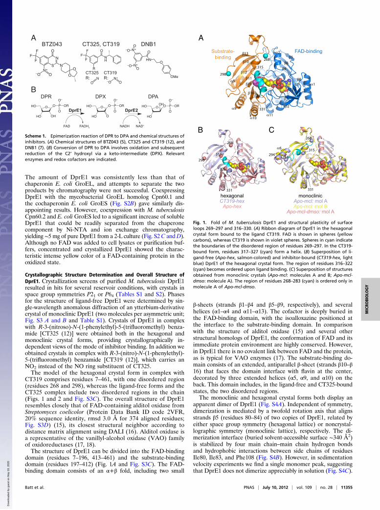
Crystallographic Structure Determination and Overall Structure ofDprE1. Crystallization screens of purified M. tuberculosis DprE1resulted in hits for several reservoir conditions, with crystals inspace group symmetries P21 or P64 (Tables S1 and S2). Phasesfor the structure of ligand-free DprE1 were determined by sin-gle-wavelength anomalous diffraction of an ytterbium-derivativecrystal of monoclinic DprE1 (two molecules per asymmetric unit;Fig. S3 A and B and Table S1). Crystals of DprE1 in complexwith R-3-(nitroso)-N-(1-phenylethyl)-5-(trifluoromethyl) benza-mide [CT325 (12)] were obtained both in the hexagonal andmonoclinic crystal forms, providing crystallographically in-dependent views of the mode of inhibitor binding. In addition weobtained crystals in complex with R-3-(nitro)-N-(1-phenylethyl)-5-(trifluoromethyl) benzamide [CT319 (12)], which carries anNO2 instead of the NO ring substituent of CT325.The model of the hexagonal crystal form in complex with
CT319 comprises residues 7–461, with one disordered region(residues 268 and 298), whereas the ligand-free forms and theCT325 complex include two disordered regions in the chain(Figs. 1 and 2 and Fig. S3C). The overall structure of DprE1resembles closely that of FAD-containing alditol oxidase fromStreptomyces coelicolor (Protein Data Bank ID code 2VFR,20% sequence identity, rmsd 3.0 Å for 374 aligned residues;Fig. S3D) (15), its closest structural neighbor according todistance matrix alignment using DALI (16). Alditol oxidase isa representative of the vanillyl-alcohol oxidase (VAO) familyof oxidoreductases (17, 18).The structure of DprE1 can be divided into the FAD-binding
domain (residues 7–196, 413–461) and the substrate-bindingdomain (residues 197–412) (Fig. 1A and Fig. S3C). The FAD-binding domain consists of an α+β fold, including two small
β-sheets (strands β1–β4 and β5–β9, respectively), and severalhelices (α1–α4 and α11–α13). The cofactor is deeply buried inthe FAD-binding domain, with the isoalloxazine positioned atthe interface to the substrate-binding domain. In comparisonwith the structure of alditol oxidase (15) and several otherstructural homologs of DprE1, the conformation of FAD and itsimmediate protein environment are highly conserved. However,in DprE1 there is no covalent link between FAD and the protein,as is typical for VAO enzymes (17). The substrate-binding do-main consists of an extended, antiparallel β-sheet (strands β10–β16) that faces the domain interface with flavin at the center,decorated by three extended helices (α5, α9, and α10) on theback. This domain includes, in the ligand-free and CT325-boundstates, the two disordered regions.The monoclinic and hexagonal crystal forms both display an
apparent dimer of DprE1 (Fig. S4A). Independent of symmetry,dimerization is mediated by a twofold rotation axis that alignsstrands β5 (residues 80–84) of two copies of DprE1, related byeither space group symmetry (hexagonal lattice) or noncrystal-lographic symmetry (monoclinic lattice), respectively. The di-merization interface (buried solvent-accessible surface ∼340 Å2)is stabilized by four main chain–main chain hydrogen bondsand hydrophobic interactions between side chains of residuesIle80, Ile83, and Phe108 (Fig. S4B). However, in sedimentationvelocity experiments we find a single monomer peak, suggestingthat DprE1 does not dimerize appreciably in solution (Fig. S4C).
A
B
Scheme 1. Epimerization reaction of DPR to DPA and chemical structures ofinhibitors. (A) Chemical structures of BTZ043 (5), CT325 and CT319 (12), andDNB1 (7). (B) Conversion of DPR to DPA involves oxidation and subsequentreduction of the C2’ hydroxyl via a keto-intermediate (DPX). Relevantenzymes and redox cofactors are indicated.
1
2
3 4
56
9
78
10
11
12
13
1416
1
2
10
9
5
11
A
B C
298
268
298315
268
323
15
315
331
331 331
284
268
315294
hexagonalCT319-hex
Apo-hex
monoclinicApo-mcl: mol AApo-mcl: mol B
Apo-mcl-dmso: mol A
FAD-bindingSubstrate-binding
6
Fig. 1. Fold of M. tuberculosis DprE1 and structural plasticity of surfaceloops 269–297 and 316–330. (A) Ribbon diagram of DprE1 in the hexagonalcrystal form bound to the ligand CT319. FAD is shown in spheres (yellowcarbons), whereas CT319 is shown in violet spheres. Spheres in cyan indicatethe boundaries of the disordered region of residues 269–297. In the CT319-bound form, residues 317–327 (cyan) form a helix. (B) Superposition of li-gand-free (Apo-hex, salmon-colored) and inhibitor-bound (CT319-hex, lightblue) DprE1 of the hexagonal crystal form. The region of residues 316–322(cyan) becomes ordered upon ligand binding. (C) Superposition of structuresobtained from monoclinic crystals (Apo-mcl: molecules A and B; Apo-mcl-dmso: molecule A). The region of residues 268–283 (cyan) is ordered only inmolecule A of Apo-mcl-dmso.
Batt et al. PNAS | July 10, 2012 | vol. 109 | no. 28 | 11355
MICRO
BIOLO
GY
Dow
nloa
ded
by g
uest
on
May
18,
202
0

Structural Flexibility of DprE1. Electron density of both crystalforms obtained for ligand-free DprE1 reveal disorder of twosurface loops in the substrate-binding domain. As a consequenceof the disorder, the active site is wide open in the ligand-freeform of the enzyme (Fig. 2A). At a maximum, residues 269–297and 316–330 are lacking electron density, but the degree ofdisorder or extent of the apparent gaps varies between themonoclinic and hexagonal crystal forms. In the hexagonal formof ligand-free DprE1, the first region is disordered betweenresidues 269 and 297, and the second region lacks density forresidues 316–322 (Fig. 1B). In the monoclinic crystal form, thedensity gap in the second region is considerably larger, affectingresidues 316–330 (Fig. 1C). In contrast, the density gap in thefirst region is smaller than in hexagonal crystals but varies be-tween the two crystallographically distinct molecules of mono-clinic DprE1: molecule A is lacking density for residues 269–283,whereas in molecule B this gap extends from 269 to 293 (Fig.1C). As a result, helix α6 (residues 292–298) only forms fully inmolecule A of monoclinic DprE1, whereas it appears as a two-turn fragment in molecule B (Fig. 1C) and is altogether absentfrom the structural model of hexagonal DprE1 (Fig. 1B).However, there are two conditions under which we observe
ordering of region one or region two, but not both simulta-neously. In the density for crystal Apo-mcl-dmso, the chain canbe completely traced between residues 268 and 284 for mole-cule A (trace in cyan in Fig. 1C) but not for molecule B. Thepeptide chain of this region is vaulting over the β-sheet of thesubstrate-binding domain. Contacts with a symmetry-relatedcopy of molecule A in the center of this loop help stabilize thisparticular conformation. Complete ordering of the second re-gion (residues 316–330) is observed in hexagonal crystal of theDprE1:CT319 complex, where residues 316–322 become or-dered when CT319 is bound (trace in cyan in Fig. 1B, redsurface in Fig. 2B, and Fig. S5 A and B). As result, residues317–327 form a three-turn helix (Fig. 1A, trace in cyan), al-though it does not maintain strict α-helical geometry. Unit cellparameters and crystal symmetry are unchanged between theligand-free and the CT325- and CT319-bound forms of hex-agonal DprE1, despite different reservoir conditions (TableS2). We, therefore, attribute ordering of residues 316–322 tobinding of CT319, the position of which in the active site differssubtly but distinctly from that of CT325 (Fig. S5C).
Structures of the Complexes with BTZ Derivatives CT325 and CT319.Although fluorescence-based ligand-binding experiments (Fig.S6A) demonstrated binding of the inhibitor BTZ043 (5) toDprE1, crystals grown in the presence of BTZ043 did not re-veal a bound ligand. Trefzer et al. (13) had suggested thatformation of the covalent complex between BTZ043 andDprE1 requires prior conversion of the nitro group to thenitroso form. To circumvent the need for inhibitor activation,
we synthesized the nitroso-benzothiazinone CT325 (12) forcocrystallization experiments.We obtained crystals of the DprE1:CT325 complex in both
the hexagonal and monoclinic crystal forms (CT325-hex3 andCT325-mcl; Table S1) and recorded diffraction data to reso-lutions of 2.4 and 2.6 Å (CuKα), respectively. A σA-weightedFo-Fc difference density map, calculated with model phasesbefore including the ligand in the model, displays prominentdensity between flavin and Cys387, the residue postulated to becritical for BTZ-mediated inhibition of DprE1 activity andBTZ-dependent cessation of mycobacterial growth (5) (Fig. 3A and B). We observe bridging density between this cysteineand the density enveloping the inhibitor model, evidence forthe formation of a covalent bond between CT325 and Cys387in both crystal forms. However, the conformation of the
A
298
268 268
298315
323
B315
323
Fig. 2. Molecular surfaces of ligand-free and inhibitor-bound DprE1 (hex-agonal form). (A) Ligand-free DprE1 (Apo-hex), with FAD shown as spheres(yellow carbons). The boundaries of the disordered regions are marked incyan, and the corresponding residue number is indicated. (B) CT319-boundDprE1 (CT319-hex), where ordering of residues 316–322 (red surface) leadsto shielding of the active site from solvent, except for an access channel tothe re side of flavin (arrow).
A
B
C
Fig. 3. Mode of binding of the nitroso derivative CT325 in the active siteof DprE1. (A) Stick model of FAD (yellow carbons), CT325 (blue carbons),and selected protein residues (gray carbon) situated within a 4-Å cutoffdistance from the inhibitor. The σA-weighted FO-FC density map was cal-culated on the basis of model phases before incorporation of CT325 in thestructural model and is contoured at 3.0σ above the mean. The structurewas refined including a covalent link between Cys387 and CT325. (B)Analogous view to A, showing the active site of molecule A in monocliniccrystal CT325-mcl. The σA-weighted FO-FC density map is contoured at 2.0σabove the mean. (C ) Schematic representation of CT325-protein contactsin the active site of DprE1. Thick, hashed lines indicate noncovalentinteractions (van der Waals, hydrophobic), with interatomic distancesgiven in units of Å, whereas dashed lines indicate hydrogen bonds. Forgraphical simplicity, only contacts of 4 Å or shorter have been included,and Gln336 has been omitted from the view.
11356 | www.pnas.org/cgi/doi/10.1073/pnas.1205735109 Batt et al.
Dow
nloa
ded
by g
uest
on
May
18,
202
0

phenylethyl group remained undefined in the CT325 com-plexes. While attempting to obtain diffraction data of theDprE1:CT325 complex, we noticed that for some crystals thebridging density between CT325 and Cys387 was notably ab-sent, but the whole of the inhibitor molecule was defined byelectron density, including the terminal phenylethyl group. Wesuspected that the reactive nitroso group was unstable and mayhave reverted to the more stable NO2 state before binding to theprotein, preventing formation of the covalent link to Cy387. Totest this assumption we generated crystals of CT319-boundDprE1, which led to a noncovalent complex and ordering of theloop region encompassing residues 316–322 over the active site(Fig. S5A). As a result of the ordered state of residues 316–330,the active site is shielded from bulk solvent (Fig. 2B), whereas inthe crystals of the DprE1:CT325 complex, the 316–330 loopregion remains in the disordered state.The consistent density features obtained from crystals in dif-
ferent crystal forms allow one to unequivocally position CT325 inthe active site. The inhibitor adopts an extended conformation,running parallel to the isoalloxazine. The trifluoromethyl moietypacks against the backbone of residues 132–134, forming van derWaals interactions with Gly133 and Lys134 (contact distancesbetween 3.3 Å and 3.9 Å; Fig. 3C and Fig. S7A), with additionalvan der Waals contacts to the side chains of His132, Ser228, andLys367. The central benzamide group primarily interacts withthe flavin (contact distances between 3.0 Å and 3.5 Å), withGly117 (3.2 Å), and with the side chain of Val365 (shortestcontact 3.7 Å). In addition the NO group forms a strong H-bond(2.4 Å) with the amide of Asn385. Compared with the ligand-free and CT319-bound state, the side chain of Cys387 is rotatedby 180° about the Cα–Cβ bond, facilitating the covalent bond tothe nitroso group, consistent with a previous prediction (12). Theterminal phenylethyl group is disordered in the CT325-boundcomplex but well ordered for CT319, in which case the benzenering occupies a hydrophobic cavity formed by Leu363 andVal365 in strand β16, Trp230 in strand β11, and by Leu317 andPhe320 in the “316–322” loop (Fig. S7B). In the absence of loopordering, the cavity applies fewer constraints on the orientationof the phenyl ring, and it is plausible that the terminal benzenering can assume variable orientations.
CT325 Inhibits Epimerization of DPR to DPA. Although CT319-de-pendent inhibition of DprE1 had been previously demonstratedfor the Mycobacterium smegmatis ortholog (12), activity datawere not available to us for CT325. Incubating 14C-labeleddecaprenylphosphoryl-β-D-ribose (14C-DPR) with purified M.tuberculosis DprE1 and DprE2 and analyzing reaction productsby TLC, we observe almost complete conversion of DPR to DPA(Fig. 4A). When incubated with DprE1 alone, the DPR band isreduced in intensity, and a distinct band appears below the DPRband, representing the keto-intermediate DPX (Fig. 4 A and Band Scheme 1A). No DPA is generated under these conditions.Adding CT325 to the reaction mixture represses DPX formationin a dose-dependent fashion (Fig. 4B), confirming inhibition ofDprE1 activity by this inhibitor. Adding CT325 to a reactionmixture containing both DprE1 and DprE2 represses conversionto DPA (Fig. 4A). Furthermore, growth of the nonpathogenicsurrogate organisms M. smegmatis and Mycobacterium bovis ba-cillus Calmette–Guérin are inhibited by CT325, with EC50 valuesof 10.4 and 4.6 μg/mL, respectively (Fig. 4C).In agreement with previous studies in which activity of
BTZ043 was assessed against M. smegmatis DrpE1 (5, 12, 13),we find that BTZ043 inhibits DPR to DPX conversion ofM. tuberculosis DprE1 (Fig. 4A). Thus, we are able to show thatthat CT325 replicates the activity of BTZ043, in terms of in-hibition of conversion of DPR to DPX (Fig. 4A) and in terms ofwhole-cell activity (Fig. 4C), albeit with significantly lower po-tency than BTZ043.
DiscussionThe recent controversy surrounding the report of a “totally drugresistant” strain of M. tuberculosis in India (19) illustrates thehigh stakes involved in the effort to control the TB pandemic, inparticular because this burden is carried primarily by resource-limited health care systems in the developing world. Thus, thediscovery of benzothiazinones and dinitrobenzamides as novelclasses of potent inhibitors, effective against both drug-suscep-tible and MDR/XDR forms of TB, and the identification of theirtarget—DprE1—are significant steps forward.Epimerization of DPR is the final step of DPA synthesis,
which involves the conversion of glucose to ribose-5-phosphatevia the pentose phosphate pathway, followed by synthesis ofphosphoribose-diphosphate (pRpp), nucleophilic replacementof pyrophosphate by decaprenyl-phosphate, and removal of the5′-phosphate to yield DPR (20). Although not firmly established, itis likely that after attachment of the decaprenyl group, the DPAprecursor compounds are anchored into the cell membrane. Thisraises the question of how DprE1 gains access to substrate.DprE1 may need to be able to partially or wholly sequester thedecaprenyl moiety of the substrate. The wide-open active site ofligand-free DprE1 and the two flexible loops contrast with thenarrow, funnel-like access channel to the active site in the knownstructures of ligand-free VAO enzymes, for instance in alditoloxidase (Fig. S7C). Therefore, it seems plausible to invoke a linkbetween flexibility of these loops and DPR binding.
Fig. 4. Assays probing inhibition of DprE1 and whole-cell activity of CT325.(A) 14C-labeled DPR was incubated with DprE1 and DprE2, and epimerizationof DPR to DPA monitored by TLC. (B) As in A, but 14C-labeled substrate wasincubated with DprE1 alone, and conversion to the keto-intermediate DPXwas monitored. (C) CT325-induced inhibition of growth in batch cultures ofM. bovis bacillus Calmette–Guérin and M. smegmatis, monitored for 10 dand 40 h, respectively, by measuring absorbance at 600 nm. SDs are derivedfrom two independent experiments.
Batt et al. PNAS | July 10, 2012 | vol. 109 | no. 28 | 11357
MICRO
BIOLO
GY
Dow
nloa
ded
by g
uest
on
May
18,
202
0

We propose that the structure of CT325-bound DprE1 isa representative of the activated BTZ-target complex. Thestructure and activity data demonstrate that CT325 binds toDprE1 and that this interaction interferes with the conversion ofDPR to DPX in a dose-dependent fashion (Fig. 4B). We havenot shown explicitly that inhibition of DprE1 by CT325 com-promises cell wall arabinan, but the suppression of DPR to DPAconversion (Fig. 4A) and CT325’s whole-cell activity againstM. smegmatis and M. bovis bacillus Calmette–Guérin (Fig. 4C)provides indirect evidence for such an effect.Covalent binding of BTZ043 to DprE1 has been shown to be
dependent on Cys387: substitutions by Gly or Ser at this se-quence position resulted in resistance to BTZ or DNB inhib-itors (5, 12). The Fo-Fc difference electron density of theinhibitor-bound structure of hexagonal and monoclinic DprE1unequivocally indicate a covalent link between CT325 andCys387 (Fig. 3 A and B), and the shape of the bridging densityis consistent with a semimercaptal linkage. Density for thetrifluoromethyl group (8.7σ in CT325-mcl, 6.6 σ in CT325-hex3), the electron-withdrawing ring substituent, is seen con-sistently in all CT325 complex structures we obtained. Thus,the trifluoromethyl seems to be a second key determinant ofCT325 binding, which together with the covalent bond and thespatial constraints imposed by the active site unequivocallyposition the ligand on the si side of flavin. The terminal phe-nylethyl moiety is only defined in the noncovalent complex withCT319. In the latter complex, ligand binding is accompanied byordering of the second surface loop (residues 316–322), whichcloses the active site and provides additional contacts to sta-bilize the phenylethyl in a defined conformation (Figs. S5A andS7B). Apart from the covalent link to Cys387, van der Waalsinteractions dominate the network of noncovalent contactsbetween enzyme and CT325 (Fig. 3C).Given the key structural features shared between CT325 and
(activated) BTZ043, it is plausible to predict that binding of thelatter rests on the same key interactions—the covalent bond toCys387 and the van der Waals interactions of the trifluoromethylwith the backbone of residues 132–134. Evidence from hexago-nal crystals grown in the presence of (nominally) 3 mM BTZ043is consistent with such reasoning. In a 2.5-Å density map, cal-culated with CuKα diffraction data (BTZ-hex; Table S1 and Fig.S6B), we observe distinct density peaks at the site of the tri-fluoromethyl group (5.5σ in the Fo-Fc difference map) and be-tween the NO2 group and Cys387 (6.2σ), indicating thealternative rotamer conformation of Cys387 that would facilitatethe semimercaptal. Although we cannot be certain that thesedensity features are due to a bound BTZ molecule, they alignwith those of the CT325 complex and are consistent with theobserved saturation binding behavior of BTZ043 (Fig. S6A).Overall, our structural study of DprE1 complexed with CT325
will provide a framework for the rational design of new BTZ-basedinhibitors targeting M. tuberculosis, whereas the complex withCT319 indicates that a covalent link with DprE1 may be dispens-able, provided that contacts with the disordered loop 316–322 andother parts of the active site can be exploited to enhance affinity.
MethodsOverexpression and Purification of DprE1. DprE1 (Rv3790) was coexpressedwith chaperones from E. coli (GroES) and M. tuberculosis (CPN60.2) in E. coliBL21 (DE3). Liquid cultures of E. coli, harboring pET28a-Rv3790 and pTrc-60.2-GroES, were grown at 37 °C in LB broth (Difco) supplemented withkanamycin (50 μg/mL) and ampicillin (100 μg/mL). At OD600nm 0.4–0.6 thetemperature was reduced to 16 °C, and protein expression of Rv3790 andchaperones was induced by 0.5 mM isopropylthiogalactoside, followed byincubation overnight (16 °C). Cells were harvested, washed with 0.85% sa-line, and stored at −20 °C. Frozen cell pellets were thawed on ice andresuspended in 30 mL of 50 mM NaH2PO4 (pH 8), 300 mM NaCl, and 10 mMimidazole (buffer A), supplemented with EDTA-free protease inhibitormixture (Roche). The suspension was sonicated (Sonicator Ultrasonic Liquid
Processor XL; Misonix) on ice for a total time of 10 min (20-s pulses, 40-s cooling).The lysate was centrifuged (27,000 × g, 40 min, 4 °C), and the supernatant passedthrough a preequilibrated (buffer A) 1-mL Ni2+-charged His-Trap column (GEHealthcare). The column was washed with 50 mL of buffer A plus 20 mMimidazole, and the protein was eluted with a 50–300 mM gradient of im-idazole. Fractions containing protein were dialyzed against 20 mM Tris·HCl(pH 8.5), 10 mM NaCl, and 10% (vol/vol) glycerol (buffer B). After dialysis, theprotein was loaded on a 1-mL QHP ion exchange column (Amersham),washed with 10 mL of buffer B with 50 mM NaCl, and eluted with a gradientof 20-mM NaCl steps from 100 mM to 200 mM, in buffer B. Fractions con-taining pure protein were dialyzed overnight against buffer B and concen-trated to 35 mg/mL, by ultrafiltration (10-kDa cutoff; Amicon Ultra).
Crystallization and Structure Determination. Crystals of DprE1 were grown bysitting drop vapor diffusion in 96-well plates (Swissci), aided by a Mosquito(TTP Labtech) liquid handling robot. Conditions producing diffracting crystalsof DprE1 are listed in Table S2. Crystals appeared after 1–3 d and could bemounted from the drop and frozen in liquid nitrogen without additionalcryoprotection. The heavy atom derivative was obtained by immersingmonoclinic crystals for 12 min in mother liquor supplemented with 50 mMytterbium acetate, before mounting and flash freezing. Diffraction datawere processed using XDS, XSCALE (21) (Table S1). Single-wavelengthanomalous diffraction data from the Yb-derivative crystal (Table S1) alloweddetermination of the heavy atom positions [SHELXD (22)]. Heavy atom siteswere refined and phases calculated [to 2.0 Å; SHARP, SOLOMON (23, 24)](Fig. S2A). Autobuilding [ARPWARP (25)] produced fragments of the twomolecules in the crystallographic asymmetric unit, sufficient to derive thenoncrystallographic symmetry operator and enabling subsequent non-crystallographic symmetry averaging [DM (26)] (Fig. S3B). Placing the initialmodel into the unit cell of the hexagonal crystal form [PHASER (27), Apo-hex; Table S1] helped to complete the model of DprE1 through manualbuilding and refinement [COOT (28), REFMAC5 (26), PHENIX.REFINE (29)].Structures of the inhibitor-bound enzyme were solved by molecular re-placement [PHASER (27)], with model building and refinement according tostandard protocols. Difference maps indicating the presence of CT325 or CT319were calculated according to protein coordinates alone, before incorporationof the ligand coordinates in the structural model. Figures were prepared usingPyMol (www.pymol.org), adopting the Corey-Pauling-Koltun (CPK) coloringscheme: O, red; N, blue; S, sulfur; and C, as indicated in figure legends.
Activity Assays. The activity of DprE1, in the presence and absence of DprE2,was assayed using radiolabeled 14C-DPR substrate, prepared as previouslydescribed (30). Reaction mixtures contained 50 μg of (each) enzyme, 1 mMFAD, 1mMATP, 1 mMNAD, and 1mMNADP, in 50mMMops (pH 7.9), 10 mMMgCl2. To probe inhibition, enzymes were incubated with inhibitors (CT325at 2-mM final concentration, or BTZ043 at a final concentration of 0.1 mMin 3.13% dimethyl sulfoxide) for 30 min at 30 °C, followed by addition of2,000 cpm of 14C-DPR in 5.5 μL of 1% IGEPAL. Reactions were allowed toproceed for a further 90 min and then quenched with 350 μL of CHCl3:CH3OH(2:1, vol/vol) and 55 μL H2O. The bottom layer of the biphase was drieddown, resuspended in 10 μL of CHCl3:CH3OH (2:1, vol/vol), and analyzed.Samples were spotted on to a high-performance aluminum-backed TLC plate(Merck) and separated in CHCl3:CH3OH:1M CH3COONH4: conc. NH4OH:H2O(180:140:9:9:23) (vol/vol). Bands were visualized using a phosphor imagingscreen (Fuji) and a phosphor imager (Bio-Rad). CT325 was dissolved in di-methyl sulfoxide to a concentration of 2 mg/mL and added to 2 mL of trypticsoy broth in culture tubes to a final concentration between 0 and 100 μg/mLAfter the addition of 10 μL of a cell culture of stationary phaseM. smegmatismc2155 or M. bovis bacillus Calmette–Guérin, the cultures were incubatedat 37 °C for 40 h (M. smegmatis) or 10 d (M. bovis bacillus Calmette–Guérin)with shaking. Culture growth was monitored by measuring absorbance at600 nm (OD600) and the experiment was performed in duplicate.
Deposition of Coordinates and Structure Factors. Coordinates and structurefactors of DprE1 in ligand-free and CT325-bound forms have been depositedin the Protein Data Bank (www.pdb.org).
ACKNOWLEDGMENTS. We thank Martin Day for help with recording oneof the X-ray diffraction datasets, and Andy Lovering for comments on themanuscript. This work used a crystal imaging system obtained through theBirmingham Science City Translational Medicine Clinical Research andInfrastructure Trials Platform. This work was supported by Grant 084923/B/08/Z from the Wellcome Trust. G.S.B. recieves support from a PersonalResearch Chair from Mr. James Bardrick, and a Royal Society WolfsonResearch Merit Award.
11358 | www.pnas.org/cgi/doi/10.1073/pnas.1205735109 Batt et al.
Dow
nloa
ded
by g
uest
on
May
18,
202
0

1. Manina G, Pasca MR, Buroni S, De Rossi E, Riccardi G (2010) Decaprenylphosphoryl-β-D-ribose 2′-epimerase from Mycobacterium tuberculosis is a magic drug target. CurrMed Chem 17:3099–3108.
2. Liu CH, et al. (2011) Characteristics and treatment outcomes of patients with MDR andXDR tuberculosis in a TB referral hospital in Beijing: A 13-year experience. PLoS One 6:e19399.
3. Gandhi NR, et al. (2010) Multidrug-resistant and extensively drug-resistant tubercu-losis: A threat to global control of tuberculosis. Lancet 375:1830–1843.
4. Zumla A, Hafner R, Lienhardt C, Hoelscher M, Nunn A (2012) Advancing the de-velopment of tuberculosis therapy. Nat Rev Drug Discov 11:171–172.
5. Makarov V, et al. (2009) Benzothiazinones kill Mycobacterium tuberculosis byblocking arabinan synthesis. Science 324:801–804.
6. Pasca MR, et al. (2010) Clinical isolates of Mycobacterium tuberculosis in four Euro-pean hospitals are uniformly susceptible to benzothiazinones. Antimicrob AgentsChemother 54:1616–1618.
7. Christophe T, et al. (2009) High content screening identifies decaprenyl-phosphor-ibose 2′ epimerase as a target for intracellular antimycobacterial inhibitors. PLoSPathog 5:e1000645.
8. Mikusová K, et al. (2005) Decaprenylphosphoryl arabinofuranose, the donor of the D-arabinofuranosyl residues of mycobacterial arabinan, is formed via a two-step epi-merization of decaprenylphosphoryl ribose. J Bacteriol 187:8020–8025.
9. Alderwick LJ, Birch HL, Mishra AK, Eggeling L, Besra GS (2007) Structure, function andbiosynthesis of the Mycobacterium tuberculosis cell wall: Arabinogalactan and lip-oarabinomannan assembly with a view to discovering new drug targets. Biochem SocTrans 35:1325–1328.
10. Sassetti CM, Boyd DH, Rubin EJ (2001) Comprehensive identification of conditionallyessential genes in mycobacteria. Proc Natl Acad Sci USA 98:12712–12717.
11. Sassetti CM, Boyd DH, Rubin EJ (2003) Genes required for mycobacterial growth de-fined by high density mutagenesis. Mol Microbiol 48:77–84.
12. Trefzer C, et al. (2010) Benzothiazinones: Prodrugs that covalently modify the de-caprenylphosphoryl-β-D-ribose 2′-epimerase DprE1 of Mycobacterium tuberculosis.J Am Chem Soc 132:13663–13665.
13. Trefzer C, et al. (2012) Benzothiazinones are suicide inhibitors of mycobacterial de-caprenylphosphoryl-β-D-ribofuranose 2′-oxidase DprE1. J Am Chem Soc 134:912–915.
14. Manina G, et al. (2010) Biological and structural characterization of the Mycobacte-rium smegmatis nitroreductase NfnB, and its role in benzothiazinone resistance. MolMicrobiol 77:1172–1185.
15. Forneris F, et al. (2008) Structural analysis of the catalytic mechanism and stereo-selectivity in Streptomyces coelicolor alditol oxidase. Biochemistry 47:978–985.
16. Holm L, Rosenström P (2010) Dali server: Conservation mapping in 3D. Nucleic AcidsRes 38(Web Server issue):W545-9.
17. Leferink NG, Heuts DP, Fraaije MW, van Berkel WJ (2008) The growing VAO flavo-protein family. Arch Biochem Biophys 474:292–301.
18. Mattevi A, et al. (1997) Crystal structures and inhibitor binding in the octameric fla-voenzyme vanillyl-alcohol oxidase: The shape of the active-site cavity controls sub-strate specificity. Structure 5:907–920.
19. Udwadia ZF, Amale RA, Ajbani KK, Rodrigues C (2012) Totally drug-resistant tuber-culosis in India. Clin Infect Dis 54:579–581.
20. Alderwick LJ, et al. (2011) Biochemical characterization of the Mycobacterium tu-berculosis phosphoribosyl-1-pyrophosphate synthetase. Glycobiology 21:410–425.
21. Kabsch W (2010) Integration, scaling, space-group assignment and post-refinement.Acta Crystallogr D Biol Crystallogr 66:133–144.
22. Sheldrick GM (2008) A short history of SHELX. Acta Crystallogr A 64:112–122.23. Schiltz M, et al. (2004) Phasing in the presence of severe site-specific radiation
damage through dose-dependent modelling of heavy atoms. Acta Crystallogr D BiolCrystallogr 60:1024–1031.
24. Abrahams JP, Leslie AG (1996) Methods used in the structure determination of bovinemitochondrial F1 ATPase. Acta Crystallogr D Biol Crystallogr 52:30–42.
25. Langer G, Cohen SX, Lamzin VS, Perrakis A (2008) Automated macromolecular modelbuilding for X-ray crystallography using ARP/wARP version 7. Nat Protoc 3:1171–1179.
26. Collaborative Computational Project, Number 4 (1994) The CCP4 suite: Programs forprotein crystallography. Acta Crystallogr D Biol Crystallogr 50:760–763.
27. McCoy AJ, et al. (2007) Phaser crystallographic software. J Appl Cryst 40:658–674.28. Emsley P, Lohkamp B, Scott WG, Cowtan K (2010) Features and development of Coot.
Acta Crystallogr D Biol Crystallogr 66:486–501.29. Adams PD, et al. (2010) PHENIX: A comprehensive Python-based system for macro-
molecular structure solution. Acta Crystallogr D Biol Crystallogr 66:213–221.30. Scherman MS, et al. (1996) Polyprenylphosphate-pentoses in mycobacteria are syn-
thesized from 5-phosphoribose pyrophosphate. J Biol Chem 271:29652–29658.
Batt et al. PNAS | July 10, 2012 | vol. 109 | no. 28 | 11359
MICRO
BIOLO
GY
Dow
nloa
ded
by g
uest
on
May
18,
202
0