structural and vibrational studies (ft-ir, ft-raman) of ...structural and vibrational studies...
TRANSCRIPT

Structural and Vibrational studies (FT-IR, FT-Raman) of Voglibose using DFT calculation
R. Solaichamya, J. Karpagama* aDepartment of Physics (Engg.), Annamalai University, Annamalainagar-608 002, Tamil Nadu,
India.
*Corresponding author
E-mail address: [email protected]
Keywords: FT-IR; FT-Raman; NBO; UV-Vis; NMR; TED.
ABSTRACT. In the present study, we report on the Molecular structure and infrared (IR) and FT-
Raman studies of Voglibose (VGB) as well as by calculations based on the density functional
theory (DFT) approach; utilizing B3LYP/6-31G(d,p) basis set. The targeted interpretation of the
vibrational spectra intended to the basis of calculated potential energy distribution matrix (PED)
utilizing VEDA4 program. Stability of the molecule arising from hyperconjugative interactions and
charge delocalization was studied using natural bond orbital (NBO) analysis. The results show that
change in electron density in the σ∗ and π∗ antibonding orbitals and E2 energies confirm the
occurrence of intramolecular charge transfer within the molecule. The UV-Visible and NMR
spectral analysis were reported by using TD-DFT and gauge GIAO approach respectively and their
chemical shifts related to TMS were compared. The lowering of HOMO and LUMO energy gap
appears to be the cause for its enhanced charge transfer interactions. Besides, molecular
electrostatic potential (MEP) analysis was reported. Due to different potent biological properties,
the molecular docking results are also reported.
1. INTRODUCTION
Voglibose is a new and potent in-hibitor of -glucosidases and is used for the treatment of
diabetes mellitus. Voglibose is chemically known as 3,4-Dideoxy-4-[2-hydroxy-1-(hydroxyl
methyl) ethyl]amino-2-c-(hydroxymethyl)-D-epinositol has attracted considerable interests due to
its wide range of therapeutic and pharma-cological properties, including its excellent inhibitory
activity against α-glucosidase and its action against hyperglycemia and various disorders caused by
hyper-glycemia [1]. Voglibose obtained from organic synthesis processes is similar to structurally
related carbohydrates found naturally [2, 3] and has the empirical formula C10H21NO7. For the
treatment of diabetes. It is specifically used for lowering postprandial blood glucose levels thereby
reducing the risk of macrovascular complications.
Recently, Iwamoto et al reported [4] Efficacy and safety of vildagliptin and voglibose in
Japanese patients with type 2 diabetes: a 12-week, randomized, double-blind, active-controlled
study. Mallikarjuna Rao et al [1] presented RP-HPLC method development and validation for
estimation of Voglibose in bulk and tablet dosage forms. Determination of voglibose in
pharmaceutical formulations by high performance liquid chromatography using refractive index
detection given by Karunanidhi Lakshmi et al [5]. Hong Zhang et al [2] reported 1H and
13C NMR
analysis of Voglibose and its derivatives. A Comparative Study of Acarbose and Voglibose on
Postprandial Hyperglycemia and serum lipids in Type 2 Diabetic patients reported by P. Revathi et
al [6].
With the guide of above seen written works, it is clear that there is no quantum mechanical
study on this VGB molecule which has propelled to do a definite quantum mechanical investigation
for comprehension the vibrational modes, UV-Visible, chemical shifts, HOMO-LUMO, MEP. In
this commitment, the structural and vibrational investigations of a basic VGB molecule was
introduced and talked about. In the present work we want to focus on vibrational spectrum of the
title compound inclusive its interpretation based on the theoretical spectrum calculated by means of
International Letters of Chemistry, Physics and Astronomy Online: 2016-02-15ISSN: 2299-3843, Vol. 64, pp 45-62doi:10.18052/www.scipress.com/ILCPA.64.452016 SciPress Ltd, Switzerland
SciPress applies the CC-BY 4.0 license to works we publish: https://creativecommons.org/licenses/by/4.0/

density functional theory (DFT). The redistribution of electron density (ED) in various bonding,
antibonding orbitals and E(2)
energies have been calculated by natural bond orbital (NBO) analysis
to give clear evidence of stabilization originating from the hyperconjugation of various
intramolecular interactions.
2. MATERIALS AND METHODS
2.1. FT-IR, FT-Raman and UV–Vis analysis
The compound Voglibose was purchased from Aldrich chemicals, USA and used as such to
record the FT-IR and FT-Raman, UV spectra. Bruker IFS 66 V spectrometer was used to record the
FT-IR spectrum by KBr pellet method on a equipped with a Globar source, Ge/KBr beam splitter,
and a TGS detector in the range of 4000–400 cm-1
. The spectral resolution was 2 cm-1
. The FT-
Raman spectrum was obtained on a Bruker RFS 100/s, Germany and the excitation of the spectrum
is with the emission of Nd:YAG laser with a wavelength of 1064 nm, maximal power 150 mW.
Cary 500 UV-VIS-NIR spectrometer was used to record the UV absorption spectra associated with
Voglibose were examined with the range 200-800 nm. The UV pattern is usually acknowledged
from the 10-5
molar solution connected with VGB, dissolved with ethanol solvent.
3. COMPUTATIONAL DETAILS
Calculations of the title compound were carried out with the Gaussian 03W program [7]
using B3LYP/6-31G(d,p) basis set to predict the molecular structure and vibrational wave numbers
and a scaling factor of 0.9608 is used for obtaining a considerably better agreement with
experimental data [8,9]. The assignments of the calculated wave numbers are aided by the
animation option of the VEDA4 [10] program. The atomic charges, distribution of electron density
(ED) in various bonding and antibonding orbitals and stabilization energies, E(2)
have been
calculated by natural bond orbital (NBO) analysis were performed using NBO 3.1 program [11] as
implemented in the Gaussian 03W [7] package at the DFT/B3LYP level of calculation. UV–Visible
spectra, electronic transitions, vertical excitation energies and oscillator strengths were computed
with the time-dependent DFT method with 6-31G(d,p) basis set in gas phase and using ethanol as
solvent. The 1H and
13C NMR isotropic shielding were calculated with the GIAO method [12] using
the optimized parameters obtained from B3LYP/6-31G(d,p) method.
3.1. Prediction of Raman intensities
The Raman activities (SRa) calculated with Gaussian 03W program [7] converted to relative
Raman intensities (IRa) using the following relationship derived from the intensity theory of Raman
scattering [13,14]
Where, ν0 is the laser exciting wavenumber in cm-1
(in this work, we have used the excitation
wavenumber ν0 = 9398.5 cm-1
, which corresponds to the wavelength of 1064 nm of a Nd-YAG
laser), νi the vibrational wavenumber of the ith
normal mode (cm-1
) while Si is the Raman scattering
activity of the normal mode νi [15].
4. RESULTS AND DISCUSSIONS
4.1. Conformational stability
In order to describe conformational flexibility of the title molecule, the energy profile as a
function of N13–C15–C16–O17 torsion angle was achieved with B3LYP/6-31G(d,p) level of
calculation as shown in Fig. 1. All the geometrical parameters were simultaneously relaxed during
the calculations while the N13–C15–C16–O17 torsional angle was varied in steps of 10 , 20 , 30 ... 360 .The energy values obtained from the scan output show that, the structure has a minimum
energy (-974.609 Hartree), when the dihedral angle N13–C15–C16–O17 is 0 or 360 (global
minimum) and -974.994 Hartree (local minimum) when the dihedral angle N13–C15–C16–O17 is
)]/exp(1[
)( 4
kthcvv
SvvfI
ii
iioi
46 ILCPA Volume 64

160 . Therefore, in the present work we have focused on the most stable form of VGB molecule to
clarify molecular structure and assignments of vibrational spectra.
Fig. 1. Dihedral angle-relative energy curves of the Voglibose by B3LYP/6-31G (d,p) level of
theory
4.2. Molecular geometry
The optimized geometrical parameters such as bond length, bond angles and dihedral angles
calculated by B3LYP/6-31G(d,p) level of calculation using Gaussian 03W program package. To the
best of our knowledge, the experimental data on geometric structure of VGB is not available in the
literature. Therefore, the theoretical results of VGB have been compared together with closely
related molecule 1-Cyclohexylmethoxymethyl-5-[2-hydroxy-1-(hydroxymethyl) ethylamino]
cyclohexane-1, 2, 3, 4-tetraol [16] as given in Table 1. The C-C bond length of the cyclohexane
ring varies from 1.534 Å-1.565 Å. Due to the O-H group substitution on the C1, C2, C3 and C4th
position of the cyclohexane ring, the C-C bond lengths are not same for example C1-C2=1.565 Å,
C1-C6=1.536 Å, C2-C3=1.544 Å, C3-C4=1.538 Å calculated by DFT method. The C-O bond
length on the cyclohexane ring varies from 1.416 Å -1.429 Å by DFT method is good agreement
with experimental value 1.429 Å -1.439 Å. The N13-H32 bond length is 1019 Å calculated by DFT
method. N-C bond lengths are C5-N13=1.481 Å / 1.480 Å, N13-C15=1.480 Å /1.467 Å calculated
by DFT/XRD respectively, this result shows good agreement between Theoretical and experimental
values. The C-H bond lengths of ethyl alcohol (CH2OH) group is C11-H29=1.100 Å, C11-
H30=1.094 Å and C14-H33=1.092 Å, C14-H34=1.096 Å calculated by DFT method.
As shown in Fig. 2, the molecular structure of title compound contains one six-membered
ring this ring (from C-1 to C-6) adopt chair conformations. The cyclohexane ring is disordered, with
three of the C atoms distributed on two sites with approximately equal occupancy. In addition, one
of the hydroxymethyl groups attached to C1 is disordered over the positions. The bond angle at
point on the substitution is C2-C1-C6=110.9 /109.0 ° calculated by DFT/XRD respectively. The
unit –N13–C15–C16–O17- connected with C5 by the way of an equatorial bond, and the angles of
N13-C5-H22 show 110.9 ° (DFT), C6-C5-N13 show 112.0 ° (DFT), 110.9 ° (XRD) and C4-C5-
N13 show 106.2 ° (DFT), 108.9 ° (XRD). The N13-C15–C16 was like a bridge that aligned with
cyclohexane ring and CH2OH.
Dihedral angles of cyclohexane part are found as C1-C2-C3-C4=-50.92 °, C2-C3-C4-
C5=53.78 °, C3-C4-C5-C6=-54.98 ° and C4-C5-C6-C1=54.89 °. In case of twist form, the N-atom
with the attached carbon 5 was considered are twisted about N13-C5-C6-C1=173.04 ° and C3-C4-
C5-N13=-176.63 °. Meanwhile in the molecule, hydrogen bonded N13–H32...O10 appeared in the
crystal with a bond length 2.803 (2) Å and bond angle 117.3° [17]. From the theoretical values, we
found the idea most of our optimized bond lengths are slightly larger than experimental values due
(C)
-50 0 50 100 150 200 250 300 350 400
-975.5
-975.0
-974.5
-974.0
-973.5
-973.0
-972.5
-972.0
-971.5
Rel
ativ
e en
ergy
(H
artr
ee)
Dihedral angle (º)
T(N13-C15-C16-O17)
International Letters of Chemistry, Physics and Astronomy Vol. 64 47
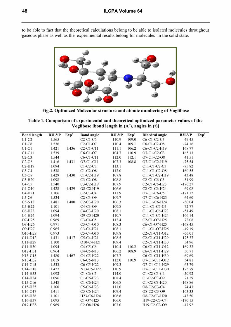
to be able to fact that the theoretical calculations belong to be able to isolated molecules throughout
gaseous phase as well as the experimental results belong for molecules in the solid state.
Fig.2. Optimized Molecular structure and atomic numbering of Voglibose
Table 1. Comparison of experimental and theoretical optimized parameter values of the
Voglibose [bond length in (A˚), angles in (◦)]
Bond length B3LYP Expa Bond angle B3LYP Exp
a Dihedral angle B3LYP Exp
a
C1-C2 1.565 C2-C1-C6 110.9 109.0 C6-C1-C2-C3 49.45
C1-C6 1.536 C2-C1-O7 110.4 109.1 C6-C1-C2-O8 -74.16
C1-O7 1.421 1.436 C2-C1-C11 111.1 106.2 C6-C1-C2-H19 168.77
C1-C11 1.539 C6-C1-O7 104.7 110.9 O7-C1-C2-C3 165.13
C2-C3 1.544 C6-C1-C11 112.0 112.1 O7-C1-C2-O8 41.51
C2-O8 1.416 1.431 O7-C1-C11 107.3 108.8 O7-C1-C2-H19 -75.54
C2-H19 1.094 C1-C2-C3 113.1 C11-C1-C2-C3 -75.82
C3-C4 1.538 C1-C2-O8 112.0 C11-C1-C2-O8 160.55
C3-O9 1.429 1.430 C1-C2-H19 107.8 C11-C1-C2-H19 43.48
C3-H20 1.098 C3-C2-O8 108.8 C2-C1-C6-C5 -51.99
C4-C5 1.540 C3-C2-H19 107.9 C2-C1-C6-H23 -176.27
C4-O10 1.428 1.429 O8-C2-H19 106.6 C2-C1-C6-H24 69.08
C4-H21 1.106 C2-C3-C4 111.9 O7-C1-C6-C5 -171.12
C5-C6 1.534 C2-C3-O9 109.7 O7-C1-C6-H23 64.60
C5-N13 1.481 1.480 C2-C3-H20 106.3 O7-C1-C6-H24 -50.04
C5-H22 1.101 C4-C3-O9 109.8 C11-C1-C6-C5 72.77
C6-H23 1.094 C4-C3-H20 108.1 C11-C1-C6-H23 -51.49
C6-H24 1.094 O9-C3-H20 110.7 C11-C1-C6-H24 -166.14
O7-H25 0.969 C3-C4-C5 112.4 C2-C1-O7-H25 72.08
O8-H26 0.971 C3-C4-O10 108.3 C6-C1-O7-H25 -168.45
O9-H27 0.965 C3-C4-H21 108.1 C11-C1-O7-H25 -49.19
O10-H28 0.973 C5-C4-O10 109.8 C2-C1-C11-O12 -66.01
C11-O12 1.431 1.417 C5-C4-H21 108.5 C2-C1-C11-H29 175.37
C11-H29 1.100 O10-C4-H21 109.4 C2-C1-C11-H30 54.96
C11-H30 1.094 C4-C5-C6 110.4 110.2 C6-C1-C11-O12 169.32
O12-H31 0.964 C4-C5-N13 106.2 108.9 C6-C1-C11-H29 50.71
N13-C15 1.480 1.467 C4-C5-H22 107.7 C6-C1-C11-H30 -69.69
N13-H32 1.019 C6-C5-N13 112.0 110.9 O7-C1-C11-O12 54.81
C14-C15 1.533 C6-C5-H22 109.3 O7-C1-C11-H29 -63.79
C14-O18 1.427 N13-C5-H22 110.9 O7-C1-C11-H30 175.79
C14-H33 1.092 C1-C6-C5 114.0 C1-C2-C3-C4 -50.92
C14-H34 1.096 C1-C6-H23 108.4 C1-C2-C3-O9 71.29
C15-C16 1.548 C1-C6-H24 106.8 C1-C2-C3-H20 -168.86
C15-H35 1.100 C5-C6-H23 111.0 O8-C2-C3-C4 74.43
C16-O17 1.411 C5-C6-H24 109.4 O8-C2-C3-O9 -163.33
C16-H36 1.101 H23-C6-H24 106.6 O8-C2-C3-H20 -43.50
C16-H37 1.095 C1-O7-H25 106.0 H19-C2-C3-C4 -170.15
O17-H38 0.969 C2-O8-H26 107.0 H19-C2-C3-O9 -47.92
48 ILCPA Volume 64

O18-H39 0.968 C3-O9-H27 107.9 H19-C2-C3-H20 71.90
C4-O10-H28 104.5 C1-C2-O8-H26 79.87
C1-C11-O12 106.1 C3-C2-O8-H26 -46.11
C1-C11-H29 108.0 H19-C2-O8-H26 -162.36
C1-C11-H30 111.6 C2-C3-C4-C5 53.78
O12-C11-H29 110.5 C2-C3-C4-O10 -67.79
O12-C11-H30 110.9 C2-C3-C4-H21 173.64
H29-C11-H30 109.4 O9-C3-C4-C5 -68.40
C11-O12-H31 108.6 O9-C3-C4-O10 170.01
C5-N13-C15 117.3 115.1 O9-C3-C4-H21 51.44
C5-N13-H32 106.5 H20-C3-C4-C5 170.61
C15-N13-H32 107.9 H20-C3-C4-O10 49.03
C15-C14-O18 111.3 H20-C3-C4-H21 -69.52
C15-C14-H33 109.3 C2-C3-O9-H27 163.46
C15-C14-H34 110.5 H20-C3-O9-H27 46.37
O18-C14-H33 106.1 C3-C4-C5-C6 -54.98
O18-C14-H34 111.1 C3-C4-C5-N13 -176.63
H33-C14-H34 108.0 C3-C4-C5-H22 64.37
N13-C15-C14 111.0 O10-C4-C5-C6 65.72
N13-C15-C16 107.9 O10-C4-C5-N13 -55.92
N13-C15-H35 110.9 O10-C4-C5-H22 -174.91
C14-C15-C16 109.9 H21-C4-C5-C6 -174.57
C14-C15-H35 108.2 H21-C4-C5-N13 63.77
C16-C15-H35 108.7 H21-C4-C5-H22 -55.21
C15-C16-O17 113.5 C3-C4-O10-H28 158.01
C15-C16-H36 108.0 C5-C4-O10-H28 34.83
C15-C16-H37 109.5 H21-C4-O10-H28 -84.29
O17-C16-H36 110.9 C4-C5-C6-C1 54.89
O17-C16-H37 107.0 C4-C5-C6-H23 177.74
H36-C16-H37 107.4 C4-C5-C6-H24 -64.74
C16-O17-H38 105.6 N13-C5-C6-C1 173.04
C14-O18-H39 105.9 N13-C5-C6-H23 -64.10
N13-C5-C6-H24 53.41
H22-C5-C6-C1 -63.49
H22-C5-C6-H23 59.35
H22-C5-C6-H24 176.86
C4-C5-N13-C15 -177.98
C4-C5-N13-H32 -56.91
C6-C5-N13-C15 61.40
C6-C5-N13-H32 -177.53
N13-C15-C16-H37 -79.02
C14-C15-C16-O17 40.10
C14-C15-C16-H36 -83.48
C14-C15-C16-H37 159.73
H35-C15-C16-O17 -78.21
H35-C15-C16-H36 158.19
H35-C15-C16-H37 41.41
C15-C16-O17-H38 -72.23
H36-C16-O17-H38 49.73
H37-C16-O17-H38 166.68
4.3. Vibrational assignments The Voglibose molecule consists of 39 atoms, which undergoes 111 normal modes of
vibrations. The molecule belongs to C1 point group symmetry. On the assumption of C1 group of
symmetry, the 111 fundamental vibrations of the molecule can be distributed as 38 stretching, 75
in-plane bending and 26 out-of plane bending modes. The calculated wavenumbers, observed IR,
Raman bands and assignments are given in Table 2 and their experimental and simulated spectra of
IR and Raman are plotted in the Fig. 3 and Fig. 4, respectively.
International Letters of Chemistry, Physics and Astronomy Vol. 64 49

4.3.1. Cyclohexane ring vibrations
The C-C stretching vibration of the Cyclohexane ring observed the FTIR band at 1070, 892
cm-1
and FT-Raman band at 698 cm-1
and the computed scaled wavenumbers at 1397, 1070, 895,
698 cm-1
by DFT method. These modes are good agreement with literature [18,19]. Guirgis et
al.,2012 [20] assigned C-C-C in-plane bending vibration at 450, 349, 354 cm-1
by DFT method. In
our present work C-C-C in-plane bending vibration observed at 937, 426, 421 cm-1
in FTIR
spectrum and 440 cm-1
in FT-Raman spectrum and theoretically predicted wavenumbers at 939,
444 and 416 cm-1
by DFT method, which is good agreement with Cyclohexane derivatives
[18,19,20].
The heteroaromatic structure shows the presence of C–H stretching vibration in the region
3100–3000 cm-1
which is the characteristics region for the ready identification of C–H stretching
vibration [21]. In our present work C-H symmetric and anti symmetric stretching vibrations in ring
CH2 identified at 2988 and 2976 cm-1
by DFT calculation. The ring CH2 anti symmetric stretching
vibration observed at 2979 cm-1
in FT-Raman spectrum. The computed wavenumbers at 2970,
2948, 2917 and 2816 cm-1
by DFT method have been identified as C2-H19, C5-H22, C3-H20 and
C4-H21 stretching vibrations respectively. The FTIR band at 1436 and 1233 cm-1
have been
identified as scissoring and twisting type CH2 inplane bending vibration in ring respectively and
computed wavenumbers at 1449 and 1216 cm-1
for this mode. The C–H in-plane bending
frequencies occur in the region 1300–1000 cm-1
. The out-of-plane bending vibrations are strongly
coupled vibration and occur in the region 1000–750 cm-1
[22]. In our present work C-C-H in-plane
bending vibrations observed at 1263 cm-1
in FT-Raman spectrum and the predicted wavenumbers at
1356, 1287, 1286 and 1260cm-1
by DFT method. Mode.nos 78 and 93 has been identified as CH
out-of plane bending vibration, which is good agreement with expected values [22].
4.3.2. O-H vibrations Hydrogen bonding alerts the frequencies of the stretching and bending vibrations. The O–H
stretching bands move to lower frequencies usually with increase intensity and band broadening in
the hydrogen bonded species. Hydrogen bonding present in five or six member ring system would
reduce the O–H stretching bands to 3550 to 3200 cm-1
region [23]. Our title molecule contains six
O-H groups, so we expect six O-H stretching vibrations. From the Table 2 which is evident that the
mode no: 1-7 has been identified as O-H stretching vibrations, these are pure modes and the PED
exactly contributes to 100%. The O–H in-plane-bending vibration in phenol, in general, lies in the
region 1250 to 1150 cm-1
. In our present work, C-O-H in-plane bending vibrations observed at 1070
cm-1
in FT-IR spectrum and 1411, 1377 cm-1
in FT-Raman spectrum. The calculated wavenumbers
at 1415, 1382, 1374, 1371, 1360, 1179, 1168 and 1156 cm-1
by DFT method has been identified as
C-O-H in-plane bending vibrations. The OH out-of-plane deformation vibration for phenol lies in
the 290 to 320 cm−1
region for free OH [21]. In our work 4 OH groups directly attached to the
Cyclohexane ring. The computed wavenumbers at 463, 438, 279 and 234 cm-1
by DFT method has
been identified as the O-H out-of plane bending vibrations. Mode no: 63, 66, 68, 70 and 73 has been
identified as the O-H out-of plane bending vibrations of the CH2OH group.
4.3.3. Methylene group vibrations
For the assignments of CH2 group frequencies, basically six fundamentals vibration can be
associated to each CH2 group namely, CH2 symmetric stretch, antisymmetric stretch, scissoring and
rocking modes, which belong to polarized in-plane vibrations . In addition to that, wagging and
twisting mode of CH2 group would be expected to be depolarized for out-of-plane bending
vibration. The asymmetric CH2 stretching vibration generally observed in the region 3000–2900
cm-1
, while the CH2 symmetric stretch will appear between 2900 cm-1
and 2800 cm-1
[24]. For title
molecule CH2 anti symmetric and symmetric stretching vibrations observed at 2979, 2889 cm-1
and
2895 cm-1
in FT-Raman and FT-IR spectrum respectively. The computed wavenumbers at 3001,
2976 and 2963 cm-1
and 2988, 2888, 2882 and 2876 cm-1
are assigned as CH2 anti symmetric and
symmetric stretching vibrations. The CH2 scissoring vibrations appear normally in the region 1490–
50 ILCPA Volume 64

1435 cm-1
as medium intense bands [25]. In our present investigation FT-Raman band at 1460 cm-1
and computed wavenumbers at 1465, 1464 and 1457 cm-1
have been identified as CH2 scissoring
vibrations. Absorption of hydrocarbons due to CH2 twisting and wagging vibration is observed in
the 1350-1150 cm-1
region [26]. For title molecule the FT-IR band at 1233 cm-1
and 1216 cm-1
by
DFT calculation gives the CH2 twisting vibrations. Mode no: 53 have been identified as the CH2
Rocking mode.
4.3.4. N-H and C-N vibrations
The N-H stretching vibrations generally give rise to bands at 3500–3400 cm-1
[27]. For title
molecule N-H stretching vibration observed at 3254 cm-1
in FT-Raman spectrum and computed at
3334 cm-1
by DFT method. The observed FT-Raman band at 3254 cm-1
and calculated scaled
wavenumber at 3334 is cm-1
is red shifted by 146 cm-1
and 66 cm-1
respectively. The reason for this
long deviation is due to presence of the N13–H32...O10 intra molecular interaction. This red
shifting is got enhanced by the reduction in the N-H bond values occurring in the hydrogen bonding
interactions. The CNH vibration where N and H atoms move in the same direction relative to the
carbon atom gives rise to a weaker band [25] near 1250 cm-1
. In our present study the theoretically
predicted wave number at 1458 cm-1
gives the CNH in-plane bending vibration.
Silverstein et al., [26] assigned the C-N stretching absorption in the region 1382 to 1286 cm-
1 for aromatic amines. For title molecule C-N stretching vibration observed at 1134 cm
-1 in FT-IR
spectrum and 1136 and 1086 cm-1
in FT-Raman spectrum. The calculated wave numbers at 1135
and 1087 cm-1
has been identified as C-N stretching vibration, which is good agreement with
experimental values. The calculated scaled wavenumber at 505 cm-1
by DFT method gives CNC in-
plane bending vibrations. The observed FT-Raman band at 377 cm-1
and computed wave number at
379 cm-1
by DFT method has been identified as CCNC out-of plane bending vibration.
4.3.5. C-O vibrations
The C–O stretching vibration in phenol occurs as a strongest band in the region 1300 to
1200 cm-1
[28]. For title molecule C-O stretching vibration observed at 1026 cm-1
in FT-IR
spectrum and 1047 cm-1
in FT-Raman spectrum. The calculated wavenumbers by DFT method at
1100, 1063, 1057, 1049, 1045, 1032 and 1008 cm-1
are assigned C-O stretching vibrations. The
observed FT-Raman band at 630 cm-1
and theoretically predicted wavenumbers at 649, 629, 333,
331, 316 and 234 cm-1
are identified as C-C-O in-plane bending vibrations. The C-C-C-O out-of
plane bending vibration observed at 397 cm-1
in FT-Raman band and calculated wavenumbers at
398, 339, and 251 cm-1
by DFT calculation. Mode nos: 105-109 has been identified as C-C-C-O
torsional modes.
International Letters of Chemistry, Physics and Astronomy Vol. 64 51

Fig. 3. Comparison of experimental and theoretical B3LYP/6-31G(d,p) FT-IR spectrum
for Voglibose
4000 3500 3000 2500 2000 1500 1000 500
20
30
40
50
60
70
80
90
100
110
3110
2895
1701
1658
1548
1477
1436 1
233 1185
1134
1026
850
757
666
617 498
421
Tra
nsm
issi
on (
%)
Wavenumber (cm-1)
Experimental
4000 3500 3000 2500 2000 1500 1000 500
100
80
60
40
20
0
138
254
377
415
615
6467
61
830
938
1022
1191
1284
13762
813
2882
2967
3582
3628
3697
IR I
nte
nsi
ty (
arb.u
nit
s)
Wavenumber (cm-1
)
B3LYP/6-31G(d,p)
52 ILCPA Volume 64

Fig.4. Comparison of experimental and theoretical B3LYP/6-31G(d,p) FT-Raman spectrum
for Voglibose
3500 3000 2500 2000 1500 1000 500
0.00
0.02
0.04
0.06
0.08
0.10
0.12
0.14
3254
2890
2737
2672
2124
1752 1
464
1341
1264
1137 1
086
1034
876
851
778
699
629
560
475
357
192 89R
aman
inte
nsi
ty (
arb.u
nit
s)
Wavenumber (cm-1
)
Experimental
3500 3000 2500 2000 1500 1000 500
-0.5
0.0
0.5
1.0
1.5
2.0
2.5
3.0
3.5
4.0
38
77
131
254
331
400
415
484
576
630
684
761
792
853
892
945
1061
1168
1214
1253
13681460
28132
882
2967
3336
3536
35823
628
3697
Ram
an i
nte
nsi
ty (
arb.u
nit
s)
Wavenumber (cm-1
)
B3LYP/6-31G(d,p)
International Letters of Chemistry, Physics and Astronomy Vol. 64 53

Table 2. Comparison of the experimental and calculated vibrational spectra and proposed
assignments of Voglibose
Mode
No
Experimental wave
numbers/cm-1
Theoretical wave numbers/cm
−1
Vibrational assignments with PED
(≥10%) FT-IR
FT-
Raman
B3LYP/6-31G(d,p)
Unscaled scaled IIRa IRa
b
1 3846 3695 44.46 3.09 νO12H31 (100)
2 3816 3666 33.39 2.82 νO9H27(100)
3 3784 3636 31.97 1.67 νO18H39(100)
4 3779 3631 51.69 2.45 νO7H25(100)
5 3775 3627 49.99 1.87 νO17H38(100)
6 3730 3584 72.26 1.33 νO18H39(100)
7 3684 3540 90.59 2.65 νO8H26(100)
8 3254 3470 3334 4.93 2.95 νN13H32(98)
9 3123 3001 37.79 3.89 νC14H33(96) anti.sym CH2
10 3110 2988 32.98 2.82 νC6H23(64)sym in ring CH2
11 2979 3097 2976 33.48 3.00 νC6H24(32)ant.sym in ring CH2
12 3091 2970 34.62 4.93 νC2H19(93)
13 3084 2963 45.7 4.22 νC11H29(68)ant.sym in CH2
14 2951 3068 2948 40.31 5.38 νC5H22(47) in ring
15 3055 2935 49.46 3.39 νC5H22(46) in ring
16 2912 3036 2917 48.4 5.54 νC3H20(73) in ring
17 2895 2889 3006 2888 49.19 2.89 νC16H36(91) sym in CH2
18 3000 2882 54.89 4.62 νC11H9(95)sym in CH2
19 2993 2876 44.4 3.63 νC14H34(91)sym in CH2
20 2985 2868 47.69 4.85 νC15H35(93)
21 2931 2816 71.28 5.33 νC4H21(93) in ring
22 1525 1465 18.85 4.93 δsciH36C16H37(56)
23 1524 1464 22.43 5.48 δsciH33C14H34(56)
24 1518 1458 29.11 3.61 δH32N13C15(63)
25 1460 1516 1457 20.99 7.64 δsciH36C16H37(56)
26 1436 1508 1449 17.22 5.98 δsciH23C6H24(84) in Ring+ γC11C1O12H30(42)
27 1411 1473 1415 58.17 4.35 δH28O10C4(55)+ γC4C5O10H21(28)
28 1458 1401 21.16 4.65 δH26O8C2(20)+ γC3C4O9H20(10)
29 1454 1397 52.62 4.47 νC2C3(11) in ring+ δH27O9C3(14)
30 1438 1382 46.56 3.83 δH25O7C1(21)+ γC14H33C15H34(20)
31 1377 1430 1374 43.12 2.51 δH38O17C16(32)+γC16C15O17H36(11)+
τH37C16C15C14(14)
32 1428 1372 52.23 7.79 δH36C16O17(41)
33 1419 1363 45.79 4.09 δH38O17C16(20)
34 1416 1360 35.84 4.04 δH27C3O9(45)
35 1411 1356 33.92 4.11 δH22C5C6(24)
36 1399 1344 39.13 4.85 δH29C11O12(35)
37 1329 1334 1388 1334 27.87 7.85 δH20C3O9(37)+ γC15C16N13H35(30)
38 1368 1314 40.48 3.71 δH25O7C1(29)+ γC5C6N13H22(36)
39 1358 1305 51.23 3.73 δH25O7C1(29)+ γC5C6N13H22(36)
40 1339 1287 29.93 7.98 δH21C4C5(37)
41 1338 1286 48.2 6.00 δH22C5C6(11)+ γC5C6N13H22(10)+ τH24C6C5C4(13)
42 1326 1274 35.09 6.99 τC2H19O8H26(40)
43 1263 1311 1260 37.6 6.66 δH22C5C6(13)
44 1307 1256 17.37 9.09 δH29C11H30(43)
45 1301 1250 20.21 11.09 γC4C5O10H21(14)
46 1233 1266 1216 32.67 9.10 δtwistH29C11H30(33)+ δtwistH23C6H24(43) in ring
47 1244 1195 51.12 5.88 δH19C2C3(20)+ γC4C5O10H21(11)
48 1185 1236 1188 47.36 7.95 δH23C6C1(15)+ γC3C4O9H20(16)
49 1227 1179 15.77 4.51 δH39O18C14(15)
50 1216 1168 33.03 7.59 δH33C14O18(39)
51 1203 1156 48.55 8.98 δH31C11O12(59)
52 1134 1136 1181 1135 10.62 9.19 νN13C5(11)
53 1169 1123 25.65 7.53 δrockH29C11H30(59)
54 1145 1100 37.97 4.75 νO10C4(11)
54 ILCPA Volume 64
55 1087 1131 1087 59.4 6.92 νN13C5(23)
56 1070 1114 1070 42.99 7.10 νC2C3(16)+ δH31O12C11(11)
57 1106 1063 69.69 11.34 νO17C16(61)
58 1100 1057 56.01 10.12 νO7C1(14)
59 1092 1049 61.85 6.56 νO12C11(13)
60 1047 1088 1045 75.34 4.86 νO9C3(46)
61 1075 1033 23.11 5.88 νO8C2(30)
62 1026 1074 1032 100 4.33 νO8C2(21)
63 1063 1021 97.92 5.42 νO18C14(16)+ γC14C15O18H33
64 1049 1008 39.34 3.56 νO12C11(16)
65 978 1024 984 26.31 6.52 νO10C4(21)
66 950 986 947 19.54 10.86 γC16C15O17H36(28)
67 937 977 939 31.25 7.71 δC2C3C4(11)
68 914 949 912 25 7.79 νN13C15(10)+ δC2C3C4(20)+ γC14C15O18H33(27)
69 892 931 895 37.29 12.21 νC4C5(12)
70 874 902 867 17.85 7.14 νO7C1(30)+ γC11C1O12H30(16)
71 850 850 885 850 29.51 13.00 νC14C15(10)+ γN13C5C15H32(10)
72 861 827 37.33 10.10 γN13C5C15H32(30) +νC14C15(40)
73 776 823 791 18.39 12.63 νC14C15(38)+ γN13C5C15H32(39)+ γC16C15O17H36(10)
74 757 789 758 44.32 11.32 νC1C11(22)
75 698 710 682 22.84 15.04 νC4C5(20)
76 675 649 46.35 8.51 δC1C11O12(21)
77 630 655 629 46.57 20.00 δC1C11O12(31)
78 617 641 616 94.83 10.36 γH23C6C5C4(12)
79 634 609 33.63 14.57 τH28O10C4C3(51)
81 553 571 549 35.45 18.29 τH39O18C14C15(21)
82 539 518 23.93 7.77 δC16C15N13(31)
83 526 505 24.88 9.01 δC5N13C15(10)+ τH39O18C14C15(11)
84 480 475 499 479 13.98 16.75 γC11C2C6C1(11)
85 482 463 31.8 8.22 γH28O10C4C5(11)
86 446 440 462 444 65.76 20.07 δC2C1C6(11)
87 456 438 34.99 14.46 γH25O7C1C6(11)
88 421 433 416 62.42 24.67 δC3C4C5(13)
89 397 414 398 20.04 16.66 γC1C6C11O7(21)
90 377 394 379 40.8 15.22 δC14C15N13(19)+ γC16C14N13C15(16)
91 353 339 30.23 15.53 δC1C2O8(31)+ τH31O12C11C1(73)+ γC1C6C11O7(11)
92 345 331 15.18 18.14 δC6C1O7(19)
93 329 316 5 8.16 δC5C4O10(14)+ γH23C6C5C4(10)
94 308 296 24.68 18.69 δC2C3O9(19)+ γC11C2C6C1(20)
95 290 279 16.21 15.52 γ H27O9C3C2(58)
96 257 271 260 15.2 20.30 τC15C14O18H39(46)
97 261 251 74.46 40.47 γC4C3C5O10(10)
98 244 234 45.48 33.89 δC15C14O18(16)+ γ H26O8C3C2(48)
99 235 226 11.65 15.78 γC11C2C6C1(17)
100 227 218 20.99 16.53 δC6C1C11(28)
101 208 200 18.72 22.95 δC2C3O9(13)+ τC3C2C1C6(12)
102 178 182 175 12.38 11.85 τC1C2C3O9(35)
103 157 151 54.09 34.87 τO31C1C11O12(55)
104 140 135 64.73 100.00 δC5N13C15(20)
105 123 118 12.2 21.49 τC16C15C14O18
106 98 94 13.97 24.33 τC14C15C16O38(45)
107 95 91 15.09 17.58 τC14C15C18O39(49)
108 86 83 80 3.16 21.65 τC14C15C16O17(38)
109 75 72 8.34 46.95 τC6C1C11O12(55)
110 42 40 14.13 46.18 δC5N13C15(15)+τC5N13C15C14(44)
111 40 38 6.65 48.75 τC6C5N13C15(57)
ν-stretching; δ-in-plane-bending; γ-out-of-plane bending; τ-torsion; w-weak; s-strong; vs-very
strong; vw-very weak; m-medium. aIIR-IR Intensity (Kmmol
−1);
bIRa-Raman intensity (Arb units) (intensity normalized to 100%).
International Letters of Chemistry, Physics and Astronomy Vol. 64 55

ij
jiF
iij qEE
2),(
2
4.4. NBO analysis
In the NBO analysis [29], all possible interactions between ‘‘filled’’ (donor) Lewis-type
NBOs and ‘‘empty’’ (acceptor) non-Lewis NBOs were investigated. The energies of these
interactions were calculated second-order perturbation theory. These interactions (or energetic
stabilizations) are named as ‘delocalization’ corrections to the zeroth-order natural Lewis structure.
For each donor NBO (i) and acceptor NBO (j); the stabilization energy E associated with i→j
delocalization, is explicitly estimated by the following equation:
where qi is the donor orbital occupancy, are i and j diagonal elements and F(i,j) is the off
diagonal NBO Fock matrix element.
The interactions σ (C2-H19) → σ*(C1-C6) and σ*(C3-C4) having the stabilization energy is
3.08, 3.27 KJ mol-1
and σ(C3-H20) → σ*(C1-C2) having the stabilization energy is 3.24 KJ mol-1
are responsible for conjugation of respective σ-bonds in Cyclohexane ring (Table 3). The
intramolecular interaction is formed by the orbital overlap between LP(1)N13→ π*(O10-H28) bond
orbital, which results intramolecular charge transfer causing stabilization energy 4.86 KJ mol-1
of
the system. The energy contribution of LP(2)O17→π*(C15-C16), LP(2)O18→ π*(C15-H35),
LP(2)O10→π*(C4-H21) are 9.07, 6.38, 7.65 KJ mol-1
, respectively, and hence there is a possibility
for delocalization of lone pair (LP) of electrons between O17 and C15-C16 and O10 and C4-H21.
Table 3. Second order Perturbation theory analysis of Fock Matrix in NBO basis
forVoglibose.
Donor (i) ED (i)(e) Acceptor(j) ED (j)(e) E
(2)a KJ
mol-1
E(j)-E(i)b
a.u
F(i,j)c
a.u
σ (C2-H19) 1.973 σ* (C1-C6) 0.029 3.08 0.87 0.046
σ* (C3-C4) 0.041 3.27 0.85 0.047
σ (C3-H20) 1.978 σ* (C1-C2) 0.070 3.24 0.86 0.048
LP(2)O7 1.947 π*(C1-C2) 0.070 8.65 0.63 0.066
LP(1)O7 1.976 RY*(1)C1 0.008 4.29 1.52 0.072
LP(2)O7 1.947 π*(C1-C11) 0.043 4.22 0.64 0.046
LP(1)O8 1.977 RY*(1)C2 0.007 3.36 1.38 0.061
LP(2)O8 1.946 π*(C1-C2) 0.070 9.09 0.62 0.067
LP(2)O8 1.946 π*(C2-C3) 0.015 3.86 0.63 0.044
LP(2)O9 1.957 π*(C3-C4) 0.042 7.40 0.67 0.063
LP(2)O9 1.957 π*(C3-H20) 0.026 3.72 0.76 0.048
LP(1)O10 1.974 RY*(1)C4 0.007 3.13 1.49 0.061
LP(1)O10 1.974 π*(C4-C5) 0.038 3.97 0.92 0.054
LP(2)O10 1.941 π*(C4-H21) 0.034 7.65 0.77 0.069
LP(2)O10 1.941 π*(O8-H26) 0.028 7.14 0.84 0.069
LP(2)O12 1.965 π*(C11-H29) 0.026 5.22 0.75 0.056
LP(2)O12 1.965 π*(C11-30) 0.027 5.50 0.77 0.058
LP(1)N13 1.908 π*(C5-H22) 0.032 6.31 0.76 0.063
LP(1)N13 1.908 π*(O10-H28) 0.027 4.86 0.76 0.055
LP(1)N13 1.908 π*(C14-C15) 0.040 3.46 0.69 0.044
LP(1)N13 1.908 π*(C15-H35) 0.028 4.21 0.76 0.051
LP(2)O17 1.948 π*(C15-C16) 0.039 9.07 0.65 0.069
LP(2)O17 1.948 π*(C16-H36) 0.028 4.43 0.72 0.051
LP(2)O18 1.954 π*(C14-H34) 0.023 4.83 0.67 0.051
LP(2)O18 1.954 π*(C15-H35) 0.028 6.38 0.77 0.063
56 ILCPA Volume 64

4. 5. Electronic properties
4.5.1. UV-Visible spectral analysis
The experimental UV–Visible electronic absorption maxima (λmax) of Voglibose recorded
in ethanol together with the theoretical results involving the vertical excitation energies, oscillator
strength (f) and wavelength at maximum absorption calculated at B3LYP/ 6-31G(d,p) basis set in
gas phase and in ethanol solvent are given in Table 4. The experimental UV-Vis spectrum of the
title compound is shown in Fig. 5. Due to the Frank–Condon principle, the maximum absorption
peak (λmax) in an UV–visible spectrum corresponds to vertical excitation. The B3LYP/6-31G(d,p)
calculations (in ethanol) predict two intense electronic transitions at 7.2622 eV (270.73 nm) with an
oscillator strength f = 0.0014 and other one 7.5971 eV (234.44 nm) with an oscillator strength f =
0.0103, which are in good agreement with the measured experimental data (261 and 227 nm).
Fig. 5. UV-Visible spectrum (Ethanol) of Voglibose
Table 4. The experimental and computed absorption wavelength λ (nm), excitation
energies E (eV), absorbance and oscillator strengths (f) of Voglibose in Ethanol solution
and gas phase
Experimental TD-DFT/B3LYP/6-31G(d,p)
Ethanol Ethanol Gas
λ(nm) Abs. λ(nm) E(eV) f(a.u) λ(nm) E(eV) f(a.u)
261 2.9868 270.73 7.2622 0.0163 268.84 6.9325 0.0014
227 3.0363 223.20 7.5971 0.0006 234.44 7.1077 0.0103
- - 160.52 7.7240 0.0404 170.09 7.2894 0.0026
4.5.2. HOMO-LUMO Analysis
The highest occupied molecular orbital (HOMO) is the orbital that primarily acts as an
electron donor and the lowest unoccupied molecular orbital (LUMO) is the orbital that largely acts
as the electron acceptor. The frontier orbital energy gap helps characterize the chemical reactivity
200 300 400 500 600 700 800
0.0
0.5
1.0
1.5
2.0
2.5
3.0
3.5
227
261
Abso
rpti
on
Wave length (nm)
Experimental
International Letters of Chemistry, Physics and Astronomy Vol. 64 57

and kinetic stability of the molecule. According to Fig. 6, the HOMO is spread heavily over the
Cyclohexane ring region and LUMO is spread over the entire molecule. For title molecule the
calculated HOMO energy is -6.3460 eV and LUMO energy is 1.1263 eV and the HOMO-LUMO
energy gap is 5.2179 eV.
Fig.6. The atomic orbital compositions of the frontier molecular orbital for Voglibose
4.5.3. Molecular Electrostatic Potential (MEP) Analysis
In order to grasp the molecular interactions, the molecular electrostatic potentials (MEPs)
are used. Recently, the MEPs have been used for interpreting and predicting relative reactivities
sites for eletrophilic and nucleophilic attack, investigation of biological recognition, hydrogen
bonding interactions, studies of molecular cluster, crystal behavior, correlation and prediction of a
wide range of macroscopic properties [30]. The MEP diagram (front and back view) of the
voglibose molecule is shown in Fig. 7. The color scheme for the MEP surface will be partially
negative charge or maybe red-electron rich; partially positive charge or maybe blue-electron
deficient; yellow slightly electron packed region; light blue-slightly electron deficient region,
Additionally, green color parts represent also regions of zero potential respectively. For the title
molecule yellow color represents the electron packed region which is mostly cover the oxygen
atoms and also the positive region is actually over the NH group. Green color represents the zero
potential regions mostly over the all protons.
Fig.7. a) Front view b) back view of Molecular electrostatic Potential map (MEP) for
Voglibose
a b
58 ILCPA Volume 64

4.6. NMR spectral Analysis
NMR spectroscopy has proved to be an exceptional tool to elucidate structure and molecular
conformation. The ‘‘gauge-independent atomic orbital’’ (GIAO) method [31] has proven to be quite
accepted and accurate, in particular when applied in the context of highly correlated abinitio
methods. The 1H and
13C theoretical and experimental [2] (water solvent) chemical shifts, isotropic
shielding constants and the assignments of voglibose are also given in the Table 5. Taking into
account that the range of 13
C NMR chemical shift for analogous organic molecules usually is >100
ppm [32]. The chemical shift of C1,C2,C3,C4 carbon peaks in the rings attached to oxygen atom are
observed from 17.10 ppm to 76.28 ppm are calculated from 69.54 ppm to 70.59 ppm at B3LYP/6-
31G(d,p) level of theory. For title molecule 13
C chemical values of these carbon atoms are down
shifted from the expected values. The reason for the down shift is due to the substitution of oxygen
and carbon atoms for C1 atom and oxygen and hydrogen atoms for C2, C3, and C4 atom
respectively. Due to the double proton substitution C6 atom get down shifted, the observed /
calculated value is 29.34 ppm/28.96 ppm. Similarly, due to electro negative oxygen atom
substitution causes C11, C14 and C16 atoms gets down shielding shown in Table 5.
Hydrogen attached or nearby electron donating atom or group increases the shielding and
moves the resonance towards to a lower frequency. The chemical shifts obtained and calculated for
the hydrogen atoms of methyl groups are quite low. All values are ≤3 ppm [33] due to shielding
effect. In our present study ethylene group chemical shift values of protons H23 and H24 is
1.36/1.01, 1.89/1.48 (Experimental/calculated). Due to electro negative oxygen bonding causes
H25-H28 have the low chemical shift values, which are calculated from 2.29-2.86 ppm by DFT
method.
Table 5. Table predicted 1H and
13C NMR isotropic chemical shifts
(with respect to TMS, all values in ppm) for Vogliboise
13
C NMR 1H NMR
Atom position Experimental B3LYP Atom position Experimental B3LYP
C1 74.10 69.54 H19 3.67 3.69
C2 65.19 66.38 H20 4.01
C3 72.07 67.65 H21 3.48 3.38
C4 76.28 70.59 H22 2.82
C5 - 50.59 H23 1.36 1.01
C6 29.34 28.96 H24 1.89 1.48
C11 62.25 63.67 H25 2.29
C14 58.51 58.55 H26 2.70 2.68
C15 54.46 52.83 H27 0.69
C16 56.51 56.89 H28 2.86
H29 3.23 3.21
H30 4.19
H31 0.44
H32 -0.29
H33 3.56 3.57
H34 3.25 3.31
H35 2.61
H36 3.17
H37 3.35 3.39
H38 2.61
H39 3.17
International Letters of Chemistry, Physics and Astronomy Vol. 64 59

5. Conclusion
The optimized molecular structure, vibrational frequencies and corresponding vibrational
assignments of Voglibose have been calculated using B3LYP level with 6-31G(d,p) basis set.
Considering that experimental and the theoretical studies are performed in different phase, it can be
said that there is a good agreement between the experimental and theoretical data. The reduction of
N-H stretching wavenumber is attributed to the N-H...O hydrogen bonding interactions. NBO
analysis clearly explains the formation of weak H bonded interaction between the LP(1)N13 and
π*(O10-H28) antibonding orbitals and charge transfer causing stabilization of energy 4.86 KJmol-1
the system. The energies of important MOs and the max of the compound were also evaluated from
TD-DFT method. The 13
C and 1H NMR chemical shifts calculated by B3LYP/6-31G(d,p) are closer
to the experimental values. Moreover, frontier molecular orbitals and molecular electrostatic
potential were visualized. Electronic transition and energy band gap of the title molecule were
investigated and interpreted.
References
[1] N.Mallikarjuna Rao, Konda Ravi Kuma, J. Bagyalakshmi, T.K. Ravi, Ramakotaiah Mogili.,
“RP-HPLC method development and validation for estimation of Voglibose in bulk and
tablet dosage forms” Int. J. Res. Pharm. Sci. Vol-1, Issue-2, (2010), 190-194.
[2] Zhang, C.R. Sun, O. Ishurd, Y.J. Pan, L.S. Ding, “Determination of the structures of four
new isomeric cyclitols” Carbo-hydr. Res. 339 (2004) 2027-2030.
[3] X. Chen, Y. Zheng, Y.Shen, Curr. “Voglibose (Basen®, AO-128), one of the most
important α-glucosidase inhibitors”, Med. Chem. 13 (2006) 109-116.
[4] Y. Iwamoto, A. Kashiwagi, N. Yamada, S. Terao, N. Mimori, M. Suzuki & H. Tachibana,
“Efficacy and safety of vildagliptin and voglibose in Japanese patients with type 2 diabetes:
a 12-week, randomized, double-blind, active-controlled study”, Diabetes, Obesity and
Metabolism 12, (2010), 700–708.
[5] Karunanidhi Lakshmi and Tirumala Rajesh, “Determination of voglibose in pharmaceutical
formulations by high performance liquid chromatography using refractive index detection”
European Journal of Chemistry 1 (4) (2010) 262‐265.
[6] P. Revathi, T. Jeyaseelan Senthinath, K. Prakash Shyam, A Comparative Study of Acarbose
and Voglibose on Postprandial Hyperglycemia and serum lipids in Type 2 Diabetic patients”
Int J Med Res. 1(2), (2011) 121-129.
[7] Gaussian 03 program, (Gaussian Inc, Wallingford CT), 2004.
[8] National Institute of Standards and Technology. Vibrational Frequency Scaling Factors on
the Web. <http://srdata.nist.gov/cccbdb/vsf.asp> (accessed 24.09.07).
[9] S.M. Islam, S.D. Huelin, M. Dawe, R.A. Poirier, “Comparison of the Standard 6-31G and
Binning-Curtiss Basis Sets for Third Row Elements”, J. Chem. Theory Comput. 4 (2008)
86–100.
[10] M.H. Jamroz "Vibrational Enegy Distribution Analysis”, VEDA 4 Computer Program”,
Poland, (2004).
[11] E.D. Glendening, A.E. Reed, J.E. Carpenter, F. Weinhold, “NBO Version 3.1”, TCI,
University of Wisconsin, Madison, (1998).
[12] R.Ditchfield, “Molecular orbital theory of magnetic shielding and magnetic susceptibility”,
J. Chem. Phys. 56 (1972) 5688–5691.
60 ILCPA Volume 64

[13] G. Kereztury, in: J.M. Chalmers, P.R. Griffith (Eds.), “Raman Spectroscopy: Theory, in
Hand book of Vibrational Spectroscopy”, vol. 1, John Wiley & Sons Ltd, New York,
(2002).
[14] L.E. Sutton, “Tables of Interatomic Distances, Chemical Society”, London, 1958.
[15] K. Govindarasu, E. Kavitha, “Vibrational spectra, molecular structure, NBO, NMR, UV,
first order hyperpolarizability, analysis of (S)-(−)-N-(5-Nitro-2-pyridyl) alaninol by Density
functional theory” Spectro.chim Acta A 127 (2014) 498–510.
[16] Hong Zhang, Wei-Fen Li, Kui-Wu Wang and Yuan Jiang Pan, “1
Cyclohexylmethoxymethyl-5-[2-hydroxy-1-(hydroxymethyl) ethylamino] cyclohexane-1, 2,
3, 4-tetraol” Acta Cryst. (2004). E60, o299-o300.
[17] J.F. Chiang, S.H. Bauer, “Molecular structure of cyclohexene”, J. Am. Chem. Soc. 91 (8)
(1969) 1898–1901.
[18] N.H. Andersen , C. J. Nielsen , P.Klaeboe , G.A. Guirgis , J. S. Overby ,S.M. Askarian,
“Infrared and Raman spectra, DFT-calculations and spectral assignments of 1,3,5-
trisilacyclohexane” J. Mol. Struct. 1076 (2014) 419–425.
[19] G.A. Guirgis, H.W. Dukes, J.K. Wyatt, C.J. Nielsen, A. Horn ,V. Aleksa P.Klaeboe
“Vibrational spectra, quantum chemical calculations and spectral assignments of 1,1-
difluoro-1-silacyclohexane” Spectro chim. Acta A 136 (2015) 51–57.
[20] G.A. Guirgis, J.K. Wyatt, C.J. Nielsen, A. Horn ,V. Aleksa P.Klaeboe,’ “Infrared and
Raman spectra, DFT-calculations and spectral assignments of silacyclohexane” J. Mol.
Struct. 1023 (2012) 189–196.
[21] G.Varsanyi, “Assignments for Vibrational Spectra of Seven Hundred Benzene Derivatives”,
vol. 1–2, Academic Kiaclo, Budapet, (1973).
[22] M. Govindarajan, S. Periyandy, K. Carthigayen, “FT-IR and FT-Raman spectra, thermo
dynamical behavior, HOMO and LUMO, UV, NLO properties, computed frequency
estimation analysis and electronic structure calculations on α-bromotoluene” Spectrochim.
Acta 97 (2012) 411–422.
[23] V. Krishnakumar, M. Kumar, N. Prabavathi, R. Mathammal, “Molecular structure,
spectroscopic studies (FTIR, FT-Raman and NMR) and HOMO–LUMO analysis of 6-
chloro-o-cresol and 4-chloro-3-methyl phenol by density functional theory” Spectrochim.
Acta A97 (2012) 144–154.
[24] D. Sajan, J. Binoy, B. Pradeep, K.V. Krishnan, V.B. Kartha, I.H. Joe, V.S. Jayakumar,
“NIR-FT Raman and infrared spectra and ab initio computations of glycinium oxalate”
Spectrochim. Acta A60 (2004) 173.
[25] N.B.Colthup, L.H.Daly, S.E.Wiberly, “Introduction to Infrared and Raman Spectroscopy”,
3rd ed., Academic Press, Boston (1990).
[26] R.M.Silverstein, G.C. Bassler, T.C.Morril, “Spectrometric Identification of Organic
Compounds”, ed. 5, John Wiley and Sons, Inc., Singapore (1991).
[27] K. Govindarasu, E. Kavitha, “Structural, vibrational spectroscopic studies and quantum
chemicalcalculations of n-(2,4-dinitrophenyl)-L-alanine methyl ester by density functional
theory” J. Mol. Struct. 1088 (2015) 70–84.
[28] M. Jag, “Organic Spectroscopy-Principles and Application’’, second ed., Narosa Publishing
House, New Delhi, (2001).
[29] A.E. Reed, L.A. Curtiss, F. Weinhold, "Intermolecular Interactions from a Natural Bond
Orbital, Donor-Acceptor Viewpoint," Chem. Rev. 88 (1988) 899–926.
International Letters of Chemistry, Physics and Astronomy Vol. 64 61

[30] J.S. Murray, K.Sen, “Molecular Electrostatic Potential Concepts and Applications, Elsevier
Science” B.V, Amsterdaam, The Netherlands (1996).
[31] P.V.R. Schleyer, N.L. Allinger, T. Clark, J. Gasteiger, P.A. Kolmann, H.F. Schaefer, P.R.
Schreiner, “The Encyclopedia of Computational Chemistry”, John Wiley and Sons,
Chichester, (1998).
[32] H.O. Kalinowski, S. Berger, S. Braun, “Carbon-13 NMR Spectroscopy”, John Wiley &
Sons, Chichester, (1988).
[33] F.A. Cotton, C.W. Wilkinson, “Advanced Inorganic Chemistry”, 3rd ed., Interscience
publisher, New York, (1972).
62 ILCPA Volume 64