structural and theoretical studies of complexes of …
TRANSCRIPT

STRUCTURAL AND THEORETICAL STUDIES OF
COMPLEXES OF URANYL(VI) AND PALLADIUM(II)
IONS HAVING SOFT AND HARD DONOR LIGANDS
By
DEBASISH DAS
CHEM01201104007
Bhabha Atomic Research Centre,
Mumbai 400085, India
A thesis submitted to the
Board of studies in Chemical Sciences
in partial fulfillment of the requirements
for the degree of
Doctor of Philosophy of
Homi Bhabha National Institute
March, 2018


List of Publications Journal 1. Steric effects on uranyl complexation: synthetic, structural, and theoretical studies of
carbamoyl pyrazole compounds of the uranyl(VI) ion; Debasish Das, Shanmugaperumal
Kannan, Dilip K. Maity, Michael G. B. Drew, Inorg. Chem. 51 (2012) 4869 4876.
2. Steric effects in pyrazole palladium(II) compounds: synthetic, structural, and theoretical
studies of a highly distorted octahedral palladium(II) pyrazole compound; Debasish Das,
Bal Govind Vats, Shanmugaperumal Kannan, Dilip K. Maity, Michael G.B. Drew,
Polyhedron 54 (2013) 104 109.
3. Synthetic and structural studies of piperidine carboxamide uranyl ion complexes; D. Das,
B.G. Vats, S. Kannan, Mukesh Kumar, M.K. Sureshkumar, Polyhedron 81 (2014) 39 44.
4. Selective recognition of uranyl ions from bulk of thorium(IV) and lanthanide(III) ions by
tetraalkyl urea: a combined experimental and quantum chemical study; Bal Govind Vats,
Debasish Das, Biswajit Sadhu, S. Kannan, I. C. Pius, D. M. Noronha, Mahesh
Sundararajan, Mukesh Kumar, Dalton Trans. 45 (2016) 10319-10325.
5. Coordination diversity in palladium(II) picolinamide ligand complexes: structural and
quantum chemical studies; Debasish Das, Amsaveni Muruganantham, S. Kannan,
Mukesh Kumar, Mahesh Sundararajan, M.K. Sureshkumar, J. Coord. Chem. 70 (2017)
1548-1553.
Conferences/Symposium 1. Synthesis and structural studies of N-oxo picolinamide based ligands with uranyl and
lanthanide nitrates - An evaluation of CONO based ligands; Debasish Das, S. Kannan,
Mukesh Kumar, M.K. Sureshkumar, J.S. Yadav, P.M. Gandhi; 6th DAE-BRNS
Interdisciplinary Symposium on Materials Chemistry (ISMC 2016), Page No. 248.
2. Synthesis and structural diversity in some U(VI) and Pd(II) complexes derived from
picolinamide ligands with different alkyl groups; Debasish Das, S. Kannan, Mukesh
Kumar, M.K. Sureshkumar, J.S. Yadav, P.M. Gandhi; 6th DAE-BRNS Interdisciplinary
Symposium on Materials Chemistry (ISMC 2016), Page No. 250.

Acknowledgments
I wish to express my sincere gratitude to my guide Prof. S. Kannan for his continuous
guidance, encouragement and constant supervision during the progress of my research work.
I am highly indebted to him for introducing me to experimental aspects of coordination
chemistry of lanthanides and actinides. I feel highly privileged to work under his invaluable
guidance.
I am grateful to my co-guide Prof. Sunil K. Ghosh for his continuous support of my work
and providing guidelines for synthesizing new organic ligands. He also supported to get the
NMR spectra of my synthesized compounds from Bio Organic Division.
I would also like to thank members of the doctoral committee: Prof. D. Das (Chairman),
Prof. V. K. Jain (Member) and Prof. P. K. Mohapatra (Member), for their valuable
advice, encouragement and insightful comments, valuable suggestions and extensive
discussions on my research work.
I would like to express my sincere gratitude to Dr. M. K. Sureshkumar for supporting me in
the research work and giving time to time suggestion to improve myself. Moreover during
my PhD period, I got excellent cooperation and support from all senior and junior colleagues
in Plutonium Plant.
I specially thank Dr. Raghunath Chowdhury, Dr. Liladhar Kumbhare, Dr. Rekha Singh,
Shri Trilochan Gadly, Shri Adish Tyagi, Shri Manojkumar Pal and Dr. Nisha Kushwah
for recording the NMR spectra of my compounds at various stages of my PhD programme. I
also want to thank Dr. R.K. Singhal for recording the FTIR spectra of my compounds
whenever required.
I am specially thankful to my collaborators Dr. Mukesh Kumar, Dr. Dilip K. Maity, Dr.
Mahesh Sunderarajan for sharing their expert skill in my research work.


Table of Contents
Synopsis i-xvii
Chapter 1: General introduction to chemistry of uranyl(VI), palladium(II) and lanthanide(III) ions and methods and materials used in present investigation
1
1.1 Introduction 2
1.2 Uranium chemistry 4
1.2.1 An overview 4
1.2.2 Chemical behaviour 5
1.2.3 Scope of synthetic uranium chemistry 6
1.2.4 Extraction and Purification of Uranium 7
1.2.5 Uranium Isotope Separation 7
1.3 Compounds of uranium 7
1.3.1 Uranium halides 7
1.3.1.1 Uranium(III) halides 8
1.3.1.2 Uranium (IV) halides 8
1.3.1.3 Uranium(V) halides 9
1.3.1.4 Uranium(VI) halides 10
1.3.2 Uranium Oxides 10
1.3.3 Oxyhalides 11
1.3.4 Uranyl Chemistry 11
1.3.4.1 Uranyl complexes 11
1.3.4.2 Molecular orbital description of uranyl complexes 13
1.3.4.3 Absorption and emission spectra 14
1.3.4.4 Coordination numbers and geometries in uranyl complexes 14
1.3.4.5 Some Other Complexes 15
1.3.4.6 Uranyl nitrate and its complexes; their role in processing nuclear waste 16
1.3.4.7 Uranyl beta-diketonates 16
1.3.5 Complexes of the uranium(IV) 17
1.3.5.1 Nitrate complexes 17
1.3.5.2 Halide complexes 17

1.3.5.3 Thiocyanates 18
1.4. Palladium 19
1.4.1 General introduction 19
1.4.2 Oxidation states 20
1.4.3 Compounds of palladium 21
1.4.4 Molecular orbital description of palladium(II) square planar complexes 23
1.5 Structural studies on the compounds of lanthanides relevant to separation process 24
1.5.1 Nitrate compounds 24
1.5.2 Monodentate neutral ligand lanthanide nitrate compounds 25
1.5.3 Bidentate neutral ligand lanthanide nitrate compounds 25
1.5.4 Tridentate neutral ligand lanthanide nitrate compounds 26
1.6 Materials and methods 26
1.6.1 Glassware 26
1.6.2 Solvents and Chemicals 27
1.7 Analytical techniques 27
1.7.1 Infrared Spectroscopy 27
1.7.2 Nuclear magnetic spectroscopy 27
1.7.3 X-Ray Crystallography 28
1.7.4 Total Reflection X-Ray Fluorescence analysis 28
1.7.5 Electrospray ionization mass spectrometric analysis 29
1.8 Scope of the present work 29
1.9 References 30
Chapter 2: Synthesis, structural and separation studies of piperidine carboxamide and tetraalkyl urea with uranyl ions
36
2.1 Introduction 37
2.2 Experimental 38
2.2.1 General considerations 38
2.2.2 Synthesis of the ligands 38
2.2.2.1 Piperidine carboxamide ligands 38
2.2.2.1.1 Synthesis of C5H10NCON(CH3)2 38

2.2.2.1.2 Synthesis of C5H10NCON(C2H5)2 38
2.2.2.1.3 Synthesis of C5H10NCON(iC3H9)2 38
2.2.2.2 Tetraalkyl urea ligands 39
2.2.2.2.1 Synthesis of C4H10NCON(iC4H9)2 39
2.2.2.2.2 Synthesis of C4H10NCON(n-C8H17)2 39
2.2.3 Synthesis of the compounds 40
2.2.3.1 Synthesis of [UO2(NO3)2{C5H10NCON(CH3)2}2] 40
2.2.3.2 Synthesis of [UO2(NO3)2{C5H10NCON(C2H5)2}2] 40
2.2.3.3 Synthesis of [UO2(NO3)2{C5H10NCON(iC3H7)2}2] 41
2.2.3.4 Synthesis of [UO2(C6H5COCHCOC6H5)2{C5H10NCON(CH3)2}] 41
2.2.3.5 Synthesis of [UO2(C6H5COCHCOC6H5)2{C5H10NCON(C2H5)2}] 41
2.2.3.6 Synthesis of [UO2(C6H5COCHCOC6H5)2{C5H10NCON(iC3H7)2}] 42
2.2.3.7 Synthesis of [UO2(C4H3SCOCHCOCF3)2{C5H10NCON(CH3)2}] 42
2.2.3.8 Synthesis of [UO2(C4H3SCOCHCOCF3)2{C5H10NCON(C2H5)2}] 43
2.2.3.9 Synthesis of [UO2(C4H3SCOCHCOCF3)2{C5H10NCON(iC3H7)2}] 43
2.2.3.10 Synthesis of [UO2Cl2{C5H10NCON(iC3H7)2}2] 44
2.2.3.11 Synthesis of [UO2Br2{C5H10NCON(iC3H7)2}2] 44
2.2.3.12 Synthesis of [UO2(NO3)2{C4H10NCON(iC4H9)2}2] 44
2.2.3.13 Synthesis of [UO2Cl2{C4H10NCON(iC4H9)2}2] 45
2.2.3.14 Synthesis of [UO2Br2{C4H10NCON(iC4H9)2}2] 45
2.2.4 Solvent extraction studies 45
2.2.5 Separation studies 46
2.2.6 X-ray diffraction studies of compounds 3, 9, 10, 11, 12, 13 and 14 47
2.2.7 Theoretical study 49
2.3 Results and discussion 52
2.3.1 Complexation study of piperidine carboxamide ligands with the uranyl nitrate 52
2.3.2 Molecular structure of compound 3 55
2.3.3 -diketonates) 56
2.3.4 Molecular structure of compound 9 59
2.3.5 Complexation study of piperidine carboxamide ligands with the uranyl dihalides 60
2.3.6 Molecular structure of compounds 10 and 11 62
2.3.7 Complexation study of N,N-diethyl-N ,N -di-isobutyl urea with the uranyl nitrate 63
2.3.8 Molecular structure of compound 12 66
2.3.9 Complexation study of N,N-diethyl-N ,N -di-isobutyl urea with the uranyl dihalides 67
2.3.10 Molecular structure of compounds 13 and 14 70
2.3.11 Comparison of ligand (L1-L4) donor strength in the uranyl complexes by
comparing the uranyl stretching frequency in the vibrational spectroscopy
71
2.3.12 Precipitation studies of U(VI) ions from the bulk of Th(IV) and Ln(III) ions 73
2.3.13 Extraction studies of U(VI) and Pu(IV) with C4H10NCON(C8H17)2 from nitric acid 75
2.3.14 Theoretical studies 76
2.4 Conclusions 78
2.5 References 79
Chapter 3: Synthesis, structural and coordination studies of N-oxo pyridine 2-carboxamide ligands with uranyl(VI) and lanthanide (III) ions
83
3.1 Introduction 84
3.2 Experimental 85
3.2.1 Synthesis of N-oxo pyridine 2-carboxamide ligands 85
3.2.1.1 Synthesis of C5H4NOCON(iC3H7)2 85
3.2.1.2 Synthesis of C5H4NOCON(iC4H9)2 86
3.2.1.3 Synthesis of C5H4NOCONH(tC4H9) 86
3.2.2 Synthesis of uranyl complexes of N-oxo pyridine 2-carboxamide ligands 87
3.2.2.1 Synthesis of [UO2(NO3)2{C5H4NOCON(iC3H7)2}] 87
3.2.2.2 Synthesis of [UO2(NO3)2{C5H4NOCON(iC4H9)2}] 87
3.2.2.3 Synthesis of [UO2(NO3)2{C5H4NOCONH(tC4H9)}] 87
3.2.3 Synthesis of lanthanide complexes of N-oxo pyridine 2-carboxamide ligands 88
3.2.3.1 Lanthanum complexes 88
3.2.3.1.1 Synthesis of [La(NO3)3(H2O){C5H4NOCON(iC3H7)2}2] 88
3.2.3.1.2 Synthesis of [La(NO3)3(H2O){C5H4NOCON(iC4H9)2}2] 88
3.2.3.2 Samarium complexes 88
3.2.3.2.1 Synthesis of [Sm(NO3)2(H2O){C5H4NOCON(iC3H7)2}2] 88
3.2.3.2.2 Synthesis of [Sm(NO3)2(H2O){C5H4NOCON(iC4H9)2}2] 89

3.2.3.3 Europium complexes 89
3.2.3.3.1 Synthesis of [Eu(NO3)3(H2O){C5H4NOCON(iC3H7)2}2] 89
3.2.3.3.2 Synthesis of [Eu(NO3)3(H2O){C5H4NOCON(iC4H9)2}2] 89
3.2.4 X-ray crystallography 90
3.3 Results and discussion 90
3.3.1 Complexation study of N-oxo pyridine 2-carboxamide ligands with uranyl nitrate 90
3.3.2 Molecular structure of compound 3 93
3.3.3 Comparison of ligand (L1-L3) donor strength in the uranyl complexes by
comparing the uranyl stretching frequency in the vibrational spectroscopy
95
3.3.4 Complexation study of N-oxo pyridine 2-carboxamide ligands with the
lanthanide nitrates
96
3.3.5 Molecular structure of compound 8 99
3.4 Conclusions 100
3.5 References 101
Chapter 4: Synthesis, structural and theoretical studies of carbamoyl pyrazole compounds of the uranyl(VI) and Pd(II) ions
104
4.1 Introduction 105
4.2 Experimental 106
4.2.1 Synthesis of the carbamoyl pyrazole ligands 106
4.2.1.1 Synthesis of C3H3N2CON(CH3)2 106
4.2.1.2 Synthesis of C3H3N2CON(C2H5)2 106
4.2.1.3 Synthesis of C3H3N2CON(iC3H7)2 107
4.2.1.4 Synthesis of C5H7N2CON(CH3)2 107
4.2.1.5 Synthesis of C5H7N2CON(C2H5)2 107
4.2.1.6 Synthesis of C5H7N2CON(iC3H7)2 107
4.2.2 Synthesis of uranyl complexes of carbamoyl pyrazole ligands 108
4.2.2.1 Synthesis of [UO2(NO3)2{C3H3N2CON(CH3)2}] 108
4.2.2.2 Synthesis of [UO2(NO3)2{C3H3N2CON(C2H5)2}] 108
4.2.2.3 Synthesis of [UO2(NO3)2{C3H3N2CON(iC3H7)2}] 108
4.2.2.4 Synthesis of [UO2(NO3)2(H2O)2{C5H7N2CON(CH3)2}2] 109
4.2.2.5 Synthesis of [UO2(NO3)2(H2O)2{C5H7N2CON(C2H5)2}2] 109

4.2.2.6 Synthesis of [UO2(NO3)2(H2O)2{C5H7N2CON(iC3H7)2}2] 109
4.2.2.7 Synthesis of [UO2(C6H5COCHCOC6H5)2{C3H3N2CON(CH3)2}] 109
4.2.2.8 Synthesis of [UO2(C6H5COCHCOC6H5)2{C3H3N2CON(C2H5)2}] 110
4.2.3 Synthesis of palladium complexes of carbamoyl pyrazole ligands 110
4.2.3.1 Synthesis of [PdCl2{C3H3N2CON(CH3)2}2] 110
4.2.3.2 Synthesis of [PdCl2{C5H7N2CON(C2H5)2}2] 111
4.2.3.3 Synthesis of [PdCl2{C3H3N2CON(iC3H7)2}2] 111
4.2.3.4 Synthesis of [PdCl2{C5H7N2CON(CH3)2}2] 111
4.2.3.5 Synthesis of [PdCl2{C5H7N2CON(C2H5)2}2] 111
4.2.3.6 Synthesis of [PdCl2{C5H7N2CON(iC3H7)2}2] 112
4.2.4 X-ray crystallography 113
4.2.5 Theoretical calculations 114
4.3 Results and discussion 115
4.3.1. Synthesis of carbamoylpyrazole and carbamoyl 3,5-dimethyl pyrazole ligands 115
4.3.2 Synthesis and structural studies of uranyl complexes 117
4.3.2.1 Synthesis and complexation studies of carbamoyl pyrazole ligands with uranyl nitrate 117
4.3.2.2 Molecular structure of compound 2 120
4.3.2.3 Synthesis and complexation studies of carbamoyl 3,5-dimethyl pyrazole ligands
with uranyl nitrate
121
4.3.2.4 Molecular structure of compound 5 123
4.3.2.5 Synthesis and complexation studies of carbamoyl pyrazole ligands with uranyl
bis(dibenzoylmethanate)
124
4.3.2.6 Molecular structure of compound 8 126
4.3.2.7 Comparison of ligand (L1-L6) donor strength in the uranyl complexes by
comparing the uranyl stretching frequency in the vibrational spectroscopy
127
4.3.2.8 Theoretical study 128
4.3.3 Synthesis and structural studies of palladium complexes 131
4.3.3.1 Synthesis and complexation studies of carbamoyl pyrazole and carbamoyl 3,5-
dimethyl pyrazole ligands with palladium chloride
131
4.3.3.2 Molecular structure of compound 10 133
4.3.3.3 Molecular structure of compound 13 134

4.3.3.4 Theoretical studies 136
4.4 Conclusions 139
4.5 References 140
Chapter 5: Synthesis, structural and theoretical Studies of picolinamide complexes with palladium(II) and uranyl (VI) ions
143
5.1 Introduction 144
5.2 Experimental 145
5.2.1 Synthesis of pyridine 2-carboxamide ligands 145
5.2.1.1 Synthesis of C5H4NCON(iC3H7)2 145
5.2.1.2 Synthesis of C5H4NCON(iC4H9)2 146
5.2.1.3 Synthesis of C5H4NCONH(tC4H9) 146
5.2.2 Synthesis of uranyl complexes of pyridine 2-carboxamide ligands 146
5.2.2.1 Synthesis of [UO2(NO3)2{C5H4NCON(iC3H7)2}] 146
5.2.2.2 Synthesis of [UO2(NO3)2{C5H4NCON(iC4H9)2}] 147
5.2.2.3 Synthesis of [UO2(NO3)2{C5H4NCONH(tC4H9)}] 147
5.2.3 Synthesis of palladium complexes of pyridine 2-carboxamide ligands 147
5.2.3.1 Synthesis of [PdCl2{C5H4NCON(iC3H7)2}2] 147
5.2.3.2 Synthesis of [PdCl2{C5H4NCON(iC4H9)2}2] 148
5.2.3.3 Synthesis of [PdCl2{C5H4NCONH(tC4H9)}] 148
5.2.4 X-ray crystallography 148
5.3 Results and discussion 149
5.3.1. Synthesis and characterization of N-substituted pyridine 2-carboxamide ligands 149
5.3.2 Synthesis and structural studies of uranyl complexes 151
5.3.2.1 Synthesis and complexation studies of pyridine 2-carboxamide ligands with
uranyl nitrate
151
5.3.2.2 Molecular structure of compound 1 153
5.3.2.2 Comparison of ligand (L1-L3) donor strength in the uranyl complexes by
comparing the uranyl stretching frequency in the vibrational spectroscopy
155
5.3.3 Synthesis and structural studies of palladium complexes 155
5.3.3.1 Synthesis and complexation studies of pyridine 2-carboxamide ligands with
palladium chloride
155

5.3.3.2 Molecular Structure of compound 4 157
5.3.3.3 Molecular Structure of compound 6 158
5.3.3.4 Computational studies 159
5.4 Conclusions 161
5.5 References 161

SYNOPSIS
There is always a high demand for suitable solvents which can be used to separate particular
metal ion or group of metal ions according to the need. As nuclear industries are concern,
separation of actinides from high level liquid waste is the important step which not only helps
to close the nuclear fuel cycle but also take part in environmental remediation [1-2]. To
design and synthesize suitable solvents for metal ions, the molecular level understanding of
the metal ligand interaction is a prerequisite factor. The coordination chemistry is the basic
tool for solving this factor.
The selective separation of uranium-233 from the mixture of thorium and other fission
products is an important step in the advanced water reactor (AHWR) technology of the Indian
Atomic Energy programme [3-4]. Many new extractants have been studied for the selective
recognition and separation of uranyl ion from various media in recent years [5-7]. In past, we
have carried out the systematic structural studies on isobutyramide uranyl complexes and
explained the stability and selectivity on the basis of strong bonding between the amide
oxygen and uranyl group [7(a)]. Inspired by the results obtained, we studied the coordination
and separation studies of few urea based ligands with uranyl nitrate, chloride and bromide by
expecting even more strong bonding due to increased electron density on the O-atom of the
ligands by mesomeric effects of two N-atoms placed both side of C=O group.
Steric effect is the common phenomenon observed in the uranium complexes and this is one
of the controlling factors for metal ligand stoichiometry, geometry and stability. Uranium
forms stable complexes with ligands consist of hard donor atoms like O, N, etc, which often
form five or six membered cyclic chelates. From the previous studies, it is observed that five
membered chelates are sterically more controlled than the other types [8].
Separation of palladium from high active liquid waste is getting recent attention due to many
applications like catalysis, photoluminescence, medicine etc. Till date a number of extractants

containing hard and/or soft donor atoms were tested for their efficacy for the separation and
recovery of palladium from simulated and actual high level liquid waste [9-13]. Although
these studies produced some satisfactory results, all of them have their own limitations such
as selectivity, use of non-ideal diluents, non suitable acidity range, hydrolytic and radiolytic
stability of the solvents etc. This demand more research on finding suitable solvent which can
satisfy all characteristics of an ideal solvent. With this in mind, we have explored the
complex chemistry pyrazole and picolinamide based ligands with palladium (II) ion.
N-oxide based ligands are expected to form strong complexes with lanthanides well as
actinides due to highly polar nature of the N-O bond in these ligands. Till date various N-
oxide based ligands were studied for their complexation reaction with uranyl and lanthanide
ions in the solid state [14-16]. But their application in the solvent extraction processes
especially in the back end of the nuclear fuel cycle is very limited [17]. In the solid state, it is
observed that, for multifunctional N-oxide ligands, there is various mode of bonding with the
metal ions. In few cases, the N-oxide group takes part in the bonding with the metal ions
while in other cases, the ligand is bonded through other functional group and N-oxide group
is free, often attached with the solvent molecules or form intra/inter molecular hydrogen
bonds. In view of the above observation, we report herein the synthesis of N-oxo
picolinamide ligands and study their coordination chemistry with lanthanides and actinides
which can help to explore the use of this type of ligands for the separation studies.
The thesis consists of five chapters and the details are given below.
Chapter 1
Introduction
This chapter deals with a brief introduction to coordination chemistry of uranium, lanthanide
and palladium ions and the factors affect the coordination number and geometry around the
metal ions. It also deals with different types of ligands used in the different stages of the
nuclear fuel cycle and the structural studies of uranyl and lanthanide ions with these ligands
with latest literature information. A brief discussion on the characterization techniques like
infrared spectroscopy (IR), nuclear magnetic resonance (NMR) and single crystal X-ray
diffraction (XRD) employed in this work is also presented. Finally, the scope of the present
work is discussed.
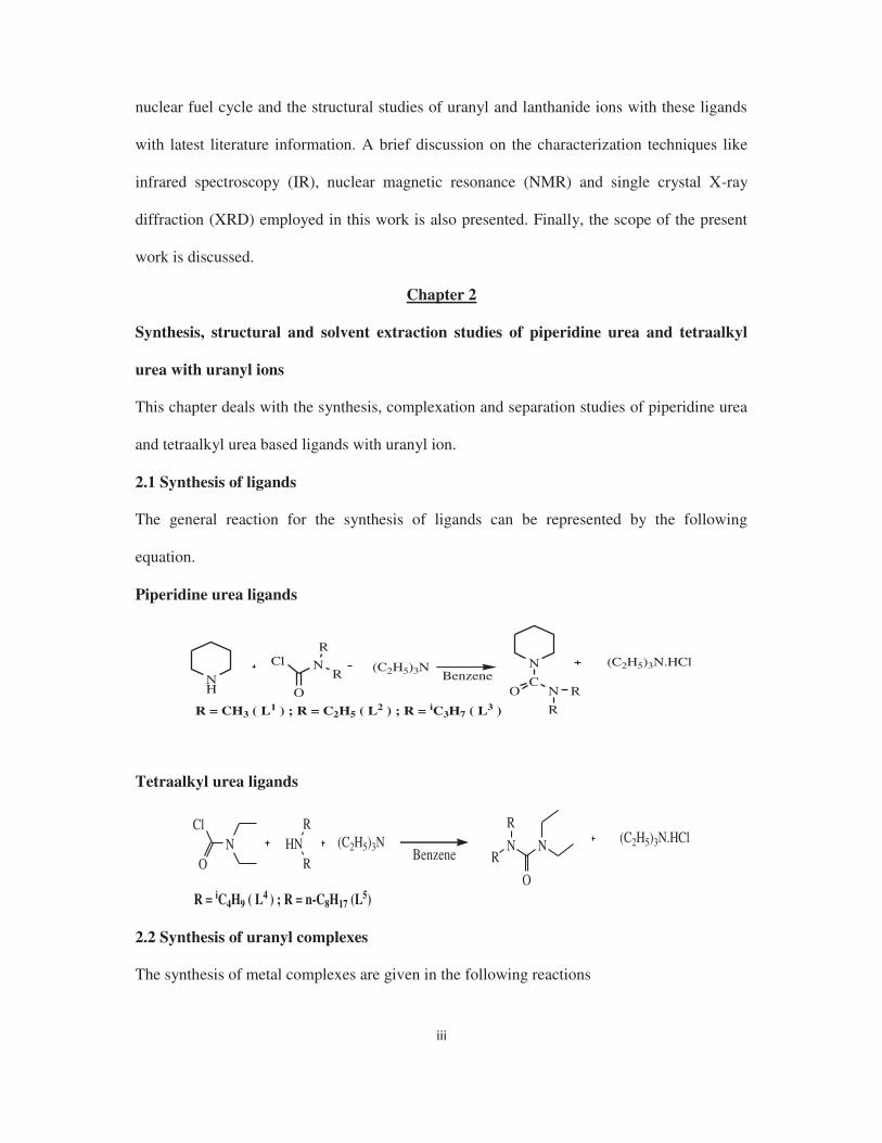
Chapter 2
Synthesis, structural and solvent extraction studies of piperidine urea and tetraalkyl
urea with uranyl ions
This chapter deals with the synthesis, complexation and separation studies of piperidine urea
and tetraalkyl urea based ligands with uranyl ion.
2.1 Synthesis of ligands
The general reaction for the synthesis of ligands can be represented by the following
equation.
Piperidine urea ligands
(C2H5)3N (C2H5)3N.HClBenzene
R = CH3 ( L1 ) ; R = C2H5 ( L2 ) ; R = iC3H7 ( L3 )
NH
Cl N
O
R
RN
CNO R
R
Tetraalkyl urea ligands
NCl
OHN
R
RNN
O
R
R(C2H5)3N (C2H5)3N.HCl
Benzene
R = iC4H9 ( L4 ) ; R = n-C8H17 (L5)
2.2 Synthesis of uranyl complexes
The synthesis of metal complexes are given in the following reactions

Piperidine urea ligand complexes
2L [UO2(NO3)2L2] (L = L1, L2, L3)
[UO2(DBM)2.2H2O] L [UO2(DBM)2.L] (L = L1, L2, L3)
(DBM = Dibenzoyl methanate)
[UO2(TTA)2.2H2O] L (L = L1, L2, L3)[UO2(TTA)2.L]
(TTA = Thenoyl trifluroacetonate)
[UO2X2, nH2O] 2L UO2X2L2(L = L1, L2, L3)
(X = Cl, Br)
[UO2(NO3)2.6H2O]
Tetraalkyl urea ligand complexes
2L [UO2(NO3)2L2] (L = L4)
[UO2X2. nH2O] 2L UO2X2L2 (L = L4)
(X = Cl, Br)
[UO2(NO3)2. 6H2O]
2.3 Results and discussion
The ligands L1-L4 and their compounds of uranyl nitrate, uranyl halide (halide = chloride,
bromide) and uranyl - -diketonate = dibenzoylmethanate,
thenoyltrifluoroacetonate) were characterized by CHN analysis, IR and NMR techniques. The
IR spectra of all complexes indicate that the carbamoyl group is bonded to the uranyl group
directly. The 1H NMR spectra of the compounds confirm the bonding between the carbamoyl
oxygen and uranyl group in solution. The structures of L3 ligand with uranyl nitrate, uranyl
chloride, uranyl bromide and uranyl bis(thenoyltrifluoroacetonate) are shown in Figures 1, 2,
3 and 4 respectively and with L4 with uranyl nitrate, chloride and bromide are shown in 5, 6
and 7 respectively.
Fig. 1. Molecular structure of [UO2(NO3)2{C5H10NCON(iC3H7)2}2]
Fig. 2. Molecular structure of [UO2Cl2{C5H10NCON(iC3H7)2}2]

Fig. 3. Molecular structure of [UO2Br2{C5H10NCON(iC3H7)2}2]
Fig. 4. Molecular structure of [UO2(C4H3SCOCHCOCF3)2{C5H10NCON(iC3H7)2}]
The figures show that both the ligands are bonded to the uranyl group in monodentate fashion
through the urea oxygen atom. The geometry of the uranyl nitrate, uranyl halide and uranyl
bis(thenoyltrifluoroacetonate) complexes with the ligands are hexagonal bi-pyramidal,
octahedral and pentagonal bipyramidal respectively.
Fig. 5. Molecular structure of [UO2(NO3)2{C4H10NCON(iC4H9)2}2]
Fig. 6. Molecular structure of [UO2Cl2{C4H10NCON(iC4H9)2}2]
Fig. 7. Molecular structure of [UO2Br2{C4H10NCON(iC4H9)2}2]
Fig. 8. The DM values of UO22+ and Pu(IV)
at different nitric acid concentration

When the ligand L4 is layered over a 3M HNO3 solution of uranyl nitrates containing large
excess of thorium(IV), lanthanum(III), samarium(III) and europium(III) nitrates, selectively
precipitate yellow crystalline solid of [UO2(NO3)2·2L4]. Moreover solvent extraction studies
of ligand L5 in dodecane with uranyl(VI) and plutonium(IV) ions from a nitric acid medium
show that uranyl ion is selectively extracted over plutonium(IV) ion (Fig. 8).
Based on electronic structure calculations at the DFT level of theory, we report the
preferential binding to uranyl compared to Th is modulated by both steric and electronic
factors.
2.4 Conclusions
The coordination chemistry of piperidine urea and tetra alkyl urea ligands with uranyl nitrate
and uranyl halides reveal that the ligands act as monodentate ligands and bond through the
C=O groups to uranyl ion. Density functional theory study revealed that the steric effect plays
a crucial role for the uranyl selectivity by the ligands.
Chapter 3
Synthesis, structural and coordination studies of N-oxo picolinamide complexes with
uranyl and lanthanide ions
This chapter deals with the synthesis and structural studies of N-oxo picolinamide complexes
of uranyl nitrate and lanthanide nitrates.
3.1 Synthesis of ligands
The ligands were synthesized according to following reaction.
Dry CH2Cl2
R = iC3H7, R' = iC3H7: L1 ; R = iC4H9, R' = iC4H9: L2 ; R = tC4H9, R' = H: L3
N
O
NR
R'
COOOH
Cl
N
O
NR
R'
O
COOH
Cl

3.2 Synthesis of metal complexes
The metal complexes were synthesized by following reactions.
L [UO2(NO3)2L]
(L = L1 :1), (L = L2:2), (L = L3 :3)
2L
[UO2(NO3)2. 6H2O]
[Ln(NO3)3. 6H2O] [Ln(NO3)3.H2O.L2]
For Ln = La and (L = L1 :4), (L = L2 :5), (L = L3 :6)
For Ln = Sm and (L = L1 :7), (L = L2 :8), (L = L3 :9)
For Ln = Eu and (L = L1 :10), (L = L2 :11), (L = L3 :12)
3.3 Results and discussion
All ligands and their compounds with uranyl nitrate and lanthanide nitrates (where,
lanthanide = lanthanum, samarium, europium) were characterized by elemental analysis
followed by IR and NMR spectroscopic techniques. IR spectra of the compounds indicate the
bonding of amide and N-oxo groups of the ligands with uranyl ions in complexes 1-3, where
as only the N-O groups of the ligands are bonded with the metal ions in complexes 4-12. The
1H NMR spectra of all the complexes show that the coordination of the ligands to the metal
ions persists in solution. The structures of the two compounds [UO2(NO3)2
{C5H4NCONH(tC4H9)}] (3) and [Eu(NO3)3{C5H4NOCON(iC3H7)2}2.H2O] (10) were
characterized by single crystal XRD and are shown in the Fig. 9-10.
Fig. 9. Molecular structure of [UO2(NO3)2{C5H4NOCONH(tC4H9)}]
Fig. 10. Molecular structure of [Eu(NO3)3(H2O){C5H4NOCON(iC3H7)2}2]
The molecular structure of (1) and (3) show that the uranium atom is surrounded by eight
oxygen atoms in a hexagonal bi-pyramidal geometry. The ligands act as bidentate chelating

ligand and bond through both N-oxo group and amide oxygen atoms to uranyl group. The
molecular structure of (10) shows that the ligand is bonded with europium ion through N-oxo
group in a monodentate mode and Eu(III) ion is surrounded by nine oxygen atoms.
3.4 Conclusions
Structure of N-oxo picolinamide with uranyl nitrate shows bidentate chelating mode of
bonding, where as it shows monodentate mode of bonding with lanthanide ions through N-
oxo group only.
Chapter 4
Synthesis, structural and theoretical studies of carbamoyl pyrazole compounds of the
uranyl(VI) and Pd(II) ions
This chapter deals with the synthesis, coordination, structural and theoretical studies of
carbamoyl pyrazole ligands with uranyl and Pd(II) ions.
4.1 Synthesis of ligands
The general reaction for the synthesis of ligands can be represented by the following
equation.
+ (C2H5)3N + (C2H5)3N.HClBenzene
NN
R
R HCl N
OR'
R'
NN
R
RO
NR'
R'
R = H, R' = Me: (L1); R = H, R' = Et: (L2); R = H, R' = iPr: (L3); R = Me, R' = Me: (L4); R =
Me, R' = Et: (L5); R = Me, R' = iPr: (L6)
4.2 Synthesis of the metal complexes
4.2.1 Synthesis of complexes with uranyl ion
These ligands have been used for the synthesis of uranyl complexes and reactions are given
below:

L [UO2(NO3)2L]
(L = L1 :1), (L = L2 :2), (L = L3 :3)
2L [UO2(NO3)2(H2O)2(L)2]
(L = L4 :4), (L = L5 :5), (L = L6 :6)
[UO2(DBM)2.2H2O] L [UO2(DBM)2.L]
(L = L1 :7), (L = L2 :8)(DBM = Dibenzoylmethanate)
[UO2(DBM)2.2H2O] L (L = L4, L5, L6)No Reaction
[UO2(NO3)2.6H2O]
[UO2(NO3)2.6H2O]
4.2.2 Synthesis of complexes with palladium(II) ion
These ligands have been used for the synthesis of palladium(II) complexes and reactions are
given below:
PdCl2 2LCH3CN
[PdCl2L2]
L = L1 (9); L = L2 (10); L = L3 (11); L = L4 (12); L = L5 (13); L = L6 (14)
4.3 Results and discussion
All ligands and their compounds of uranyl nitrate and uranyl dibenzoylmethanate were
characterized by elemental analysis followed by IR and NMR spectroscopic techniques. IR
spectra of 1-3 indicate the strong bonding between carbamoyl groups of the ligands with
uranyl ion. On the other hand, IR spectra of 4-6 signify that the ligands are uncoordinated in
the complexes. The observed 1H NMR spectra of 1-3 and 7 8 show that the pyrazolyl
protons is deshielded w.r.t to free ligands, indicating that the bonding persist in solution also.
The molecular structures of three compounds [UO2(NO3)2{C3H3N2CON(C2H5)2}] (2),
[UO2(NO3)2(H2O)2{C5H7N2CON(C2H5)2}2] (5) and
[UO2(C6H5COCHCOC6H5)2{C3H3N2CON(C2H5)2}] (8) are characterized by single crystal
XRD and shown in the Fig. 11, Fig. 12 and Fig. 13, which confirm the spectral observations.

Fig. 11 Molecular Structure of [UO2(NO3)2{C3H3N2CON(C2H5)2}]
Fig. 12 Molecular Structure of [UO2(NO3)2(H2O)2{C5H7N2CON(C2H5)2}2]
Fig. 13 Molecular Structure of [UO2(C6H5COCHCOC6H5)2{C3H3N2CON(C2H5)2}]
The structure of 2 shows that the uranium atom is surrounded by one nitrogen and seven
oxygen atoms in a hexagonal bipyramidal geometry. On the other hand, the structure of 5
consists of centrosymmetric [UO2(NO3)2·2H2O] groups, bridged by carbamoyl dimethyl
pyrazole ligand via O H···O and O H···N hydrogen bonds. The structure shows that there is
no direct bonding between ligand and the uranyl ion and the uranium(VI) ion are surrounded
by eight oxygen atoms to give hexagonal bipyramidal geometry. In compound 8, the pyrazole
ligand acts as a monodentate ligand and is bonded through the carbamoyl oxygen atom to the
uranyl group.
The CHN analyses revealed that the ratio of metal to ligand is 1:2 in all the palladium
compounds 9-14. The IR spectra of 9-14 signify that the carbamoyl group is un-coordinated
in all the compounds and the ligands are bonded through the pyrazole nitrogen atom to the
metal centres. The 1H NMR spectra of the compounds show that the pyrazole protons are
deshielded with respect to the free ligands proving further evidence for the coordination of

the ligand to the metal ion. The structures for compounds 10 (Fig. 14) and 13 (Fig. 15)
confirm our interpretation of the spectral data.
Theoretical studies revealed that the steric effect play an important role in deciding the
bonding of pyrazole based ligands with uranyl nitrate and palladium (II) chloride.
Fig. 14. Molecular Structure of [PdCl2{C5H7N2CON(C2H5)2}2]
Fig. 15. Molecular Structure of [PdCl2{C5H7N2CON(C2H5)2}2]
4.4 Conclusions
Pyrazole based ligands form 1:1 complex with uranyl nitrate and uranyl
bis(dibenzoylmethanate), and dimethyl pyrazole based ligands form second sphere complexes
with uranyl nitrate with metal to ligand ratio of 1:2. Both pyrazole and dimethyl pyrazole
ligands form 1:2 complexes with palladium(II) chloride and bond through the pyrazolyl
nitrogen atom only. Theoretical study explains the steric effect plays the important role
behind the structural difference between carbamoyl pyrazole and dimethyl carbamoyl
pyrazole complexes with uranyl and palladium ion.
Chapter 5
Synthesis, structural and theoretical studies of picolinamide complexes with palladium
and uranyl ions
This chapter deals with the synthesis and structural studies of picolinamide complexes of
palladium chloride and uranyl nitrate.

5.1 Synthesis of ligands
The ligands were synthesized according to following reaction.
SOCl2 HCl SO2
Et3N (C2H5)3N.HCl
R = iC3H7, R' = iC3H7 : L1 ; R = iC4H9, R' = iC4H9: L2 ; R = tC4H9, R' = H: L3
Dry CH2Cl2
N COOH N COCl
N
O
NR
R'N COClHN
R'
R
5.2 Synthesis of metal complexes
The metal complexes were synthesized by following reactions.
L [UO2(NO3)2L]
(L = L1 : 1), (L = L2 : 2), (L = L3 : 3)
PdCl2 2LPdCl2L2
PdCl2L
(L = L1: 4), (L = L2: 5)
(L = L3: 6)CH3CN
[UO2(NO3)2. 6H2O]
5.3 Results and discussion
All ligands and their compounds with uranyl nitrate and palladium chloride were
characterized by elemental analysis followed by IR and NMR spectroscopic techniques. IR
spectra of 1-3 show that absorption frequency for the C=O is 34-55 cm-1 lower as compared
to that of free ligands, indicating a direct bonding between the amide group and uranyl ion.
On the other hand, while the IR spectra of 4-5 show that absorption frequency for the C=O is
unchanged and is red shifted by 56 cm-1 for compound 6. This indicates that in complex 6 the
C=O is bonded with the metal ion and is free in 4-5. The 1H NMR spectra of all compounds
of uranium and palladium show that the pyridyl protons are deshielded with respect to the
free ligands proving further evidence for the coordination of the ligands to the metal ion. The
structures of compounds [UO2(NO3)2{C5H4NCON(iC3H7)2}] (1),
[PdCl2{C5H4NCON(iC3H7)2}2] (4) and [PdCl2{C5H4NCONH(tC4H9)}] (6) were characterized
by single crystal XRD and are shown in the Figures 16-18.

Fig. 16. Molecular structure of [UO2(NO3)2{C5H4NCON(iC3H7)2}]
Fig. 17. Molecular structure of [PdCl2{C5H4NCON(iC3H7)2}2]
Fig. 18. Molecular structure of [PdCl2{C5H4NCONH(tC4H9)}]
The molecular structure of (1) shows that the uranium atom is surrounded by seven oxygen
atoms and one nitrogen atom in a hexagonal bi-pyramidal geometry. The molecular structure
of (4) shows that two ligands are bonded with palladium in a monodentate mode and Pd(II)
ion is surrounded by two nitrogen and two chlorine atoms in a trans square planar geometry.
On the other hand, the molecular structure of (6) shows that the ligand is bonded with
palladium in a bidentate mode and Pd(II) ion is surrounded by one nitrogen, one oxygen and
two chlorine atoms in a cis square planar geometry. DFT study indicates that steric hindrance
between 3-pyridyl H atom and N-substituted alkyl groups inside the ligands are the reason
behind this kind of coordination diversity in palladium complexes.
Theoretical studies revealed that the steric effect is the reason behind the coordination
diversity observed in picolinamide complexes with palladium (II) chloride.

5.4 Conclusions
The structures of picolinamide compounds show that the ligands show coordination diversiry
with the two selected metal ions. DFT studies clearly revealed that the steric effects play an
important role for deciding the coordination mode of the ligands.
Overall conclusions
Study of basic coordination chemistry of organic ligands having different functionalities with
the specific metal ion can provide the ideas about the stability, bonding, stoichiometry of the
complexes formed due to their mutual interaction. These ideas could be very helpful to
design suitable molecules by synthetic organic chemists and their deployment in the field of
separation science.
We have synthesized total five types of mono and bidentate ligands with different
functionality and studied their coordination chemistry with uranyl(VI), palladium(II) and few
lanthanide(III) ions, viz. La3+, Sm3+, Eu3+ in order to understand their mode of bonding,
structural variation, stoichiometry etc. It helps us to make a layout of best solvent system for
the separation of different metal ions especially in the field of back end of nuclear fuel cycle.
While both the monodentate ligands, piperidine carboxamide and tetraalkyl urea form
1:2 complexes with uranyl nitrate and uranyl dihalides, they form 1:1 complexes with uranyl
-diketonates. They form very strong complexes with the uranyl compounds which is
confirmed by the short U-O(amide) bond length observed in X-ray crystallography and lower
vibrational frequency of both C=Ostr and U=Oasy bonds observed in IR spectroscopy. N,N-
di- n-Octyl tetraalkyl urea showed very high selectivity towards 233U from the simulated
AHWR fuel dissolver solution.
In case of, N-Oxo picolinamide complexes of uranyl and lanthanide (La, Sm, Eu)
nitrates variation in bonding and structure was observed in the solid state. While the ligands

act as bidentate chelating ligands with uranyl nitrate, they act as monodentate ligands with
lanthanide nitrates through the O(N-Oxo)-metal bonding. The metal:ligand stoichiometry in
the uranyl nitrate and lanthanide nitrates complexes is found to be 1:1 and 1:2 respectively.
-
diketonates and act as bidentate chelating and monodentate ligand respectively. On the other
hand, carbamoyl 1,3-dimethyl pyrazole ligands form second sphere complexes with uranyl
-diketonates. This type of variation in
reactivity of these two ligand systems was successfully explained (by density functional
theory calculation) on the basis of steric effect provided by the later when approaches
towards the identical metal system. Both the type of ligands form 1:2 complexes with
palladium chloride and bonded through pyrazoyl N-atom. Here they act as monodentate
ligands.
Pyridine carboxamide ligands form stable complexes with uranyl nitrate and act as
bidentate chelating ligands. The metal:ligand stoichiometry of these complexes was found to
be 1:1. But it was observed that with variation in the alkyl group, the ligands show structural
variety with palladium chloride. While the isopropyl and isobutyl based ligands form 1:2
complexes and act as monodentate ligands, the mono substituted tertiary butyl based ligand
form 1:1 complex and act as bidentate chelating ligand.
From the above observation it can be concluded that with changing the electronic and
steric property of the ligands the reactivity and bonding mode of these ligands can be tuned.
The donor strength of the ligands is the combination of basicity of the donor atoms and steric
hindrance provided by the ligands towards the metal system.
Future perspectives
Nuclear waste management is the most important and challenging task in the nuclear fuel
cycle, not only to recover the valuables from the waste even to protect the environment as

well. Nuclear waste solution after dissolution of spent fuel comprises of a varieties of metal
ions, like alkali and alkaline earth metal ions, transition metal ions even lanthanides and
actinides. Solution chemistry of theses metal ions are different and they present in various
oxidation states, especially transition metal ions. Speciation data of these metal ions in 3-4 M
HNO3 medium is not fully known till date. So, it is really very difficult to achieve selectivity
of their separation from complex mixture solution like high level liquid waste (HLLW). We
got partial achievement to selectivity separate 233U from simulated AHWR high level liquid
waste using one of our synthesized ligands. This may show a new direction for the
development of thoria based fuel cycle.
In future there is huge scope to develop suitable solvent system for separation of other
metal ions from various waste streams. Apart from their high donor strength and favourable
steric property, they should also have other suitable properties to get a global acceptance like,
viscosity, radiolytic and hydrolytic stability, solubility in commonly used diluents etc. For
this doctoral work, we have synthesized some simple mono and bidentate ligands with
preferably amide functional group and studied their coordination chemistry with uranyl,
palladium and lanthanide compounds. However complex molecules with more number of
functional groups could be synthesized and tested for separating rather difficult and
intractable metal ion system.
References
1. K.L. Nash, J.C. Braley, Challenges for Actinide Separations in Advanced Nuclear Fuel
Cycles, ACS Symposium Series, 1046 (2010) 19 (Chapter 3).
2. Dominique Warin, IOP Conf. Series: Materials Science and Engineering 9 (2010) 012063.
3. S. Kant, Int. J. Nucl. Ener. Sci. Tech. 1 (2005) 204.
4. R. K. Sinha, A. Kakodkar, Nucl. Eng. Des. 236 (2006) 683.
5. (a) G. Szigethy, K.N. Raymond, Chem. Eur. J. 17 (2011) 1818 1827. (b) C. Ni, D.K.
Shuh, K.N. Raymond, Chem. Commun. 47 (2011) 6392 6394. (c) G. Szigethy, K.N.

Raymond, Inorg. Chem. 49 (2010) 6755 6767. (d) G. Szigethy, K.N. Raymond, J. Am.
Chem. Soc. 133 (2011) 7942 7956. (e) A.E.V. Gorden, J. Xu, K.N. Raymond, P. Durbin,
Chem. Rev. 103 (2003) 4207 4282. (f) C. Drouza, V. Gramlich, M.P. Sigalas, I.
Pashalidis, A.D. Keramidas, Inorg. Chem. 43 (2004) 8336 8345.
6. A.C. Sather, O.B. Berryman, J. Jr. Rebek, J. Am. Chem. Soc. 132 (2010) 13572 13574.
7. (a) S. Kannan, S.B. Deb, J.S. Gamare, M.G.B. Drew, Polyhedron 27 (2008) 2557 2562.
(b) P.N. Pathak, D.R. Prabhu, R.B. Ruikar, V.K. Manchanda, Solvent Extr. Ion Exch. 20
(2002) 293 311.
8. (a) R.D. Hancock, A.E. Martell, Chem. Rev. 89 (1989) 1875 1914. (b) A.E. Martell, R.D.
Hancock, Metal complexes in aqueous solution; Plenum Press: New York, 1996.
9. Z. Zhu, Y. Sasaki, H. Suzuki, S. Suzuki, T. Kimura, Anal. Chim. Acta 527 (2004) 163.
10. Y. Sasaki, Y. Morita, Y. Kitatsuji, T. Kimura, Solv. Extr. Ion Exch. 28 (2010) 335.
11. N.T. Hung, M. Watanabe, T. Kimura, Solv. Extr. Ion. Exch. 25 (2007) 407.
12. E.A. Mezhov, A.V. Kuchmunov, V.V. Druzhenkov, Radiochemistry 44 (2002) 135.
13. A. Dakshinamoorthy, P.S. Dhami, P.W. Naik, N.L. Dudwadkar, S.K. Munshi, P.K. Dey,
V. Venugopal, Desalination 232 (2008) 26.
14. Bal Govind Vats, Jayashree S. Gamare, S. Kannan, I.C. Pius, D.M. Noronha, M. Kumar,
Inorg. Chim. Acta 467 (2017) 1 6.
15. Sylvie Pailloux, Iris Binyamin, Lorraine M. Deck, Benjamin P. Hay, Eileen N. Duesler,
Lev N. Zakharov, W. Scott Kassel, Arnold L. Rheingold, Robert T. Pain, Polyhedron 28
(2009) 3979 3984.
16. Hai-Jun Zhang, Ru-Hu Gou, Lan Yan, Ru-Dong Yang, Spectrochim. Acta, Part A 66
(2007) 289 294.
17. M. Ejaz, Sep. Sci. 10 (1975) 425-446.

Abbreviations
An = Actinide
AHWR = Advanced heavy water reactor
BE = Binding energy
Bipy = Bipyridyl
CCD = Charge-coupled device
CMP = Carbamoyl methyl phosphonates
CMPO = Carbamoyl methyl phosphine oxides
CMSO = Carbamoyl methyl sulfoxide
DBA = Dibenzylidineacetone
DBM = Dibenzoylmethanate
DCM = Dichloromethane
Dcpe = Dicyclohexylphosphino ethane
DEHPA = Di-(2-ethylhexyl) phosphoric acid
DFT = Density functional theory
DIAMEX = Diamide Extraction
DIDPA = Di-isodecyl phosphoric acid
DMDBTDMA = Dimethyl-dibutyl-tetradecylmalonamide
-dimethyl- -dioctylhexylethoxymalonamide
DMF = Dimethylformamide
Dmpz = Dimethyl pyrazole
DMSO = Dimethyl sulfoxide
Dppe = Diphenylphosphinomethane
DNPPA = Di-nonylphenyl phosphoric acid
DOPPA = Di-octylphenyl phosphoric acid

DU = Depleted uranium
EDA = Energy decomposition analysis
EDXRF = Energy dispersive X-Ray Fluorescence
ESIMS = Elctrospray ionization mass spectrometry
FMO = Frontier molecular orbital
FTIR = Fourier transform infrared
HDEHP = Di-(2-ethylhexyl) phosphoric acid
Ln = Lanthanide
MPA = Mülliken population analysis
NLMO = Natural localized molecular orbital
NMR = Nuclear magnetic resonance
ORTEP = Oak Ridge Thermal Ellipsoid Plot
PCM = Polarizable continuum model
PGM = Platinum group metals
PUREX = Plutonium uranium extraction
Pz = Pyrazole
RE = Rare earth
RI = Resolution of Identity
SC-ECP = small-core effective core potential
Tacn = 1,4,7-triazacyclononane
TBP= Tri-butyl phosphate
THF = Tetrahydrofuran
TRUEX = Transuranic extraction
Ttcn = 1,4,7-trithiacyclononane
TTA = Theonyltrifluoroacetonate

TXRF = Total Reflection X-Ray Fluorescence
UV = Ultra violet
XRD = X-ray diffraction
ZORA = Zeroth Order Regular Approximation

Table of Contents
Synopsis i-xvii
Chapter 1: General introduction to chemistry of uranyl(VI), palladium(II) and lanthanide(III) ions and methods and materials used in present investigation
1
1.1 Introduction 2
1.2 Uranium chemistry 4
1.2.1 An overview 4
1.2.2 Chemical behaviour 5
1.2.3 Scope of synthetic uranium chemistry 6
1.2.4 Extraction and Purification of Uranium 7
1.2.5 Uranium Isotope Separation 7
1.3 Compounds of uranium 7
1.3.1 Uranium halides 7
1.3.1.1 Uranium(III) halides 8
1.3.1.2 Uranium (IV) halides 8
1.3.1.3 Uranium(V) halides 9
1.3.1.4 Uranium(VI) halides 10
1.3.2 Uranium Oxides 10
1.3.3 Oxyhalides 11
1.3.4 Uranyl Chemistry 11
1.3.4.1 Uranyl complexes 11
1.3.4.2 Molecular orbital description of uranyl complexes 13
1.3.4.3 Absorption and emission spectra 14
1.3.4.4 Coordination numbers and geometries in uranyl complexes 14
1.3.4.5 Some Other Complexes 15
1.3.4.6 Uranyl nitrate and its complexes; their role in processing nuclear waste 16
1.3.4.7 Uranyl beta-diketonates 16
1.3.5 Complexes of the uranium(IV) 17
1.3.5.1 Nitrate complexes 17
1.3.5.2 Halide complexes 17
1.3.5.3 Thiocyanates 18
1.4. Palladium 19
1.4.1 General introduction 19
1.4.2 Oxidation states 20
1.4.3 Compounds of palladium 21
1.4.4 Molecular orbital description of palladium(II) square planar complexes 23
1.5 Structural studies on the compounds of lanthanides relevant to separation process 24
1.5.1 Nitrate compounds 24
1.5.2 Monodentate neutral ligand lanthanide nitrate compounds 25
1.5.3 Bidentate neutral ligand lanthanide nitrate compounds 25
1.5.4 Tridentate neutral ligand lanthanide nitrate compounds 26
1.6 Materials and methods 26
1.6.1 Glassware 26
1.6.2 Solvents and Chemicals 27
1.7 Analytical techniques 27
1.7.1 Infrared Spectroscopy 27
1.7.2 Nuclear magnetic spectroscopy 27
1.7.3 X-Ray Crystallography 28
1.7.4 Total Reflection X-Ray Fluorescence analysis 28
1.7.5 Electrospray ionization mass spectrometric analysis 29
1.8 Scope of the present work 29
1.9 References 30
Chapter 2: Synthesis, structural and separation studies of piperidine carboxamide and tetraalkyl urea with uranyl ions
36
2.1 Introduction 37
2.2 Experimental 38
2.2.1 General considerations 38
2.2.2 Synthesis of the ligands 38
2.2.2.1 Piperidine carboxamide ligands 38
2.2.2.1.1 Synthesis of C5H10NCON(CH3)2 38

2.2.2.1.2 Synthesis of C5H10NCON(C2H5)2 38
2.2.2.1.3 Synthesis of C5H10NCON(iC3H9)2 38
2.2.2.2 Tetraalkyl urea ligands 39
2.2.2.2.1 Synthesis of C4H10NCON(iC4H9)2 39
2.2.2.2.2 Synthesis of C4H10NCON(n-C8H17)2 39
2.2.3 Synthesis of the compounds 40
2.2.3.1 Synthesis of [UO2(NO3)2{C5H10NCON(CH3)2}2] 40
2.2.3.2 Synthesis of [UO2(NO3)2{C5H10NCON(C2H5)2}2] 40
2.2.3.3 Synthesis of [UO2(NO3)2{C5H10NCON(iC3H7)2}2] 41
2.2.3.4 Synthesis of [UO2(C6H5COCHCOC6H5)2{C5H10NCON(CH3)2}] 41
2.2.3.5 Synthesis of [UO2(C6H5COCHCOC6H5)2{C5H10NCON(C2H5)2}] 41
2.2.3.6 Synthesis of [UO2(C6H5COCHCOC6H5)2{C5H10NCON(iC3H7)2}] 42
2.2.3.7 Synthesis of [UO2(C4H3SCOCHCOCF3)2{C5H10NCON(CH3)2}] 42
2.2.3.8 Synthesis of [UO2(C4H3SCOCHCOCF3)2{C5H10NCON(C2H5)2}] 43
2.2.3.9 Synthesis of [UO2(C4H3SCOCHCOCF3)2{C5H10NCON(iC3H7)2}] 43
2.2.3.10 Synthesis of [UO2Cl2{C5H10NCON(iC3H7)2}2] 44
2.2.3.11 Synthesis of [UO2Br2{C5H10NCON(iC3H7)2}2] 44
2.2.3.12 Synthesis of [UO2(NO3)2{C4H10NCON(iC4H9)2}2] 44
2.2.3.13 Synthesis of [UO2Cl2{C4H10NCON(iC4H9)2}2] 45
2.2.3.14 Synthesis of [UO2Br2{C4H10NCON(iC4H9)2}2] 45
2.2.4 Solvent extraction studies 45
2.2.5 Separation studies 46
2.2.6 X-ray diffraction studies of compounds 3, 9, 10, 11, 12, 13 and 14 47
2.2.7 Theoretical study 49
2.3 Results and discussion 52
2.3.1 Complexation study of piperidine carboxamide ligands with the uranyl nitrate 52
2.3.2 Molecular structure of compound 3 55
2.3.3 -diketonates) 56
2.3.4 Molecular structure of compound 9 59
2.3.5 Complexation study of piperidine carboxamide ligands with the uranyl dihalides 60
2.3.6 Molecular structure of compounds 10 and 11 62

2.3.7 Complexation study of N,N-diethyl-N ,N -di-isobutyl urea with the uranyl nitrate 63
2.3.8 Molecular structure of compound 12 66
2.3.9 Complexation study of N,N-diethyl-N ,N -di-isobutyl urea with the uranyl dihalides 67
2.3.10 Molecular structure of compounds 13 and 14 70
2.3.11 Comparison of ligand (L1-L4) donor strength in the uranyl complexes by
comparing the uranyl stretching frequency in the vibrational spectroscopy
71
2.3.12 Precipitation studies of U(VI) ions from the bulk of Th(IV) and Ln(III) ions 73
2.3.13 Extraction studies of U(VI) and Pu(IV) with C4H10NCON(C8H17)2 from nitric acid 75
2.3.14 Theoretical studies 76
2.4 Conclusions 78
2.5 References 79
Chapter 3: Synthesis, structural and coordination studies of N-oxo pyridine 2-carboxamide ligands with uranyl(VI) and lanthanide (III) ions
83
3.1 Introduction 84
3.2 Experimental 85
3.2.1 Synthesis of N-oxo pyridine 2-carboxamide ligands 85
3.2.1.1 Synthesis of C5H4NOCON(iC3H7)2 85
3.2.1.2 Synthesis of C5H4NOCON(iC4H9)2 86
3.2.1.3 Synthesis of C5H4NOCONH(tC4H9) 86
3.2.2 Synthesis of uranyl complexes of N-oxo pyridine 2-carboxamide ligands 87
3.2.2.1 Synthesis of [UO2(NO3)2{C5H4NOCON(iC3H7)2}] 87
3.2.2.2 Synthesis of [UO2(NO3)2{C5H4NOCON(iC4H9)2}] 87
3.2.2.3 Synthesis of [UO2(NO3)2{C5H4NOCONH(tC4H9)}] 87
3.2.3 Synthesis of lanthanide complexes of N-oxo pyridine 2-carboxamide ligands 88
3.2.3.1 Lanthanum complexes 88
3.2.3.1.1 Synthesis of [La(NO3)3(H2O){C5H4NOCON(iC3H7)2}2] 88
3.2.3.1.2 Synthesis of [La(NO3)3(H2O){C5H4NOCON(iC4H9)2}2] 88
3.2.3.2 Samarium complexes 88
3.2.3.2.1 Synthesis of [Sm(NO3)2(H2O){C5H4NOCON(iC3H7)2}2] 88
3.2.3.2.2 Synthesis of [Sm(NO3)2(H2O){C5H4NOCON(iC4H9)2}2] 89

3.2.3.3 Europium complexes 89
3.2.3.3.1 Synthesis of [Eu(NO3)3(H2O){C5H4NOCON(iC3H7)2}2] 89
3.2.3.3.2 Synthesis of [Eu(NO3)3(H2O){C5H4NOCON(iC4H9)2}2] 89
3.2.4 X-ray crystallography 90
3.3 Results and discussion 90
3.3.1 Complexation study of N-oxo pyridine 2-carboxamide ligands with uranyl nitrate 90
3.3.2 Molecular structure of compound 3 93
3.3.3 Comparison of ligand (L1-L3) donor strength in the uranyl complexes by
comparing the uranyl stretching frequency in the vibrational spectroscopy
95
3.3.4 Complexation study of N-oxo pyridine 2-carboxamide ligands with the
lanthanide nitrates
96
3.3.5 Molecular structure of compound 8 99
3.4 Conclusions 100
3.5 References 101
Chapter 4: Synthesis, structural and theoretical studies of carbamoyl pyrazole compounds of the uranyl(VI) and Pd(II) ions
104
4.1 Introduction 105
4.2 Experimental 106
4.2.1 Synthesis of the carbamoyl pyrazole ligands 106
4.2.1.1 Synthesis of C3H3N2CON(CH3)2 106
4.2.1.2 Synthesis of C3H3N2CON(C2H5)2 106
4.2.1.3 Synthesis of C3H3N2CON(iC3H7)2 107
4.2.1.4 Synthesis of C5H7N2CON(CH3)2 107
4.2.1.5 Synthesis of C5H7N2CON(C2H5)2 107
4.2.1.6 Synthesis of C5H7N2CON(iC3H7)2 107
4.2.2 Synthesis of uranyl complexes of carbamoyl pyrazole ligands 108
4.2.2.1 Synthesis of [UO2(NO3)2{C3H3N2CON(CH3)2}] 108
4.2.2.2 Synthesis of [UO2(NO3)2{C3H3N2CON(C2H5)2}] 108
4.2.2.3 Synthesis of [UO2(NO3)2{C3H3N2CON(iC3H7)2}] 108
4.2.2.4 Synthesis of [UO2(NO3)2(H2O)2{C5H7N2CON(CH3)2}2] 109
4.2.2.5 Synthesis of [UO2(NO3)2(H2O)2{C5H7N2CON(C2H5)2}2] 109

4.2.2.6 Synthesis of [UO2(NO3)2(H2O)2{C5H7N2CON(iC3H7)2}2] 109
4.2.2.7 Synthesis of [UO2(C6H5COCHCOC6H5)2{C3H3N2CON(CH3)2}] 109
4.2.2.8 Synthesis of [UO2(C6H5COCHCOC6H5)2{C3H3N2CON(C2H5)2}] 110
4.2.3 Synthesis of palladium complexes of carbamoyl pyrazole ligands 110
4.2.3.1 Synthesis of [PdCl2{C3H3N2CON(CH3)2}2] 110
4.2.3.2 Synthesis of [PdCl2{C5H7N2CON(C2H5)2}2] 111
4.2.3.3 Synthesis of [PdCl2{C3H3N2CON(iC3H7)2}2] 111
4.2.3.4 Synthesis of [PdCl2{C5H7N2CON(CH3)2}2] 111
4.2.3.5 Synthesis of [PdCl2{C5H7N2CON(C2H5)2}2] 111
4.2.3.6 Synthesis of [PdCl2{C5H7N2CON(iC3H7)2}2] 112
4.2.4 X-ray crystallography 113
4.2.5 Theoretical calculations 114
4.3 Results and discussion 115
4.3.1. Synthesis of carbamoylpyrazole and carbamoyl 3,5-dimethyl pyrazole ligands 115
4.3.2 Synthesis and structural studies of uranyl complexes 117
4.3.2.1 Synthesis and complexation studies of carbamoyl pyrazole ligands with uranyl nitrate 117
4.3.2.2 Molecular structure of compound 2 120
4.3.2.3 Synthesis and complexation studies of carbamoyl 3,5-dimethyl pyrazole ligands
with uranyl nitrate
121
4.3.2.4 Molecular structure of compound 5 123
4.3.2.5 Synthesis and complexation studies of carbamoyl pyrazole ligands with uranyl
bis(dibenzoylmethanate)
124
4.3.2.6 Molecular structure of compound 8 126
4.3.2.7 Comparison of ligand (L1-L6) donor strength in the uranyl complexes by
comparing the uranyl stretching frequency in the vibrational spectroscopy
127
4.3.2.8 Theoretical study 128
4.3.3 Synthesis and structural studies of palladium complexes 131
4.3.3.1 Synthesis and complexation studies of carbamoyl pyrazole and carbamoyl 3,5-
dimethyl pyrazole ligands with palladium chloride
131
4.3.3.2 Molecular structure of compound 10 133
4.3.3.3 Molecular structure of compound 13 134

4.3.3.4 Theoretical studies 136
4.4 Conclusions 139
4.5 References 140
Chapter 5: Synthesis, structural and theoretical Studies of picolinamide complexes with palladium(II) and uranyl (VI) ions
143
5.1 Introduction 144
5.2 Experimental 145
5.2.1 Synthesis of pyridine 2-carboxamide ligands 145
5.2.1.1 Synthesis of C5H4NCON(iC3H7)2 145
5.2.1.2 Synthesis of C5H4NCON(iC4H9)2 146
5.2.1.3 Synthesis of C5H4NCONH(tC4H9) 146
5.2.2 Synthesis of uranyl complexes of pyridine 2-carboxamide ligands 146
5.2.2.1 Synthesis of [UO2(NO3)2{C5H4NCON(iC3H7)2}] 146
5.2.2.2 Synthesis of [UO2(NO3)2{C5H4NCON(iC4H9)2}] 147
5.2.2.3 Synthesis of [UO2(NO3)2{C5H4NCONH(tC4H9)}] 147
5.2.3 Synthesis of palladium complexes of pyridine 2-carboxamide ligands 147
5.2.3.1 Synthesis of [PdCl2{C5H4NCON(iC3H7)2}2] 147
5.2.3.2 Synthesis of [PdCl2{C5H4NCON(iC4H9)2}2] 148
5.2.3.3 Synthesis of [PdCl2{C5H4NCONH(tC4H9)}] 148
5.2.4 X-ray crystallography 148
5.3 Results and discussion 149
5.3.1. Synthesis and characterization of N-substituted pyridine 2-carboxamide ligands 149
5.3.2 Synthesis and structural studies of uranyl complexes 151
5.3.2.1 Synthesis and complexation studies of pyridine 2-carboxamide ligands with
uranyl nitrate
151
5.3.2.2 Molecular structure of compound 1 153
5.3.2.2 Comparison of ligand (L1-L3) donor strength in the uranyl complexes by
comparing the uranyl stretching frequency in the vibrational spectroscopy
155
5.3.3 Synthesis and structural studies of palladium complexes 155
5.3.3.1 Synthesis and complexation studies of pyridine 2-carboxamide ligands with
palladium chloride
155

5.3.3.2 Molecular Structure of compound 4 157
5.3.3.3 Molecular Structure of compound 6 158
5.3.3.4 Computational studies 159
5.4 Conclusions 161
5.5 References 161

Thesis Highlight
Name of the Student Debasish Das
Name of the CI/OCC BARC, Mumbai Enrolment No. CHEM01201104007
Thesis Title Structural and theoretical studies of complexes of uranyl(VI) and
palladium(II) ions having soft and hard donor ligands
Discipline: Chemical Sciences Sub‐Area of Discipline: Inorganic Chemistry
Date of viva voce: 16/03/2018
Study of basic coordination chemistry of organic ligands having different functional groups with specific metal ions can provide the ideas about the stability, bonding and stoichiometry of the complexes formed due to their mutual interaction.These ideas could be very helpful to designsuitable molecules for selective separation of metal ions in the field of hydrometallurgy.
In present work, five types of mono and bidentate ligands with different functional groups were synthesized and their coordination chemistry with uranyl(VI), palladium(II) and few lanthanide(III) ions, viz. La3+, Sm3+, Eu3+ were explored to understand their mode of bonding, structural variation, stoichiometry etc.It helps us to make a layout of best ligand system for the separation of metal ions relevant to the back end nuclear fuel cycle.
The steric effects playimportant role for the selective separation of uranyl ion from bulk ofthorium and lanthanide ions. The structure of carbamoylpyrazole‐uranylnitrate complex shows that the ligand forms inner‐sphere complex while the carbamoyl3,5‐dimethyl pyrazole forms outer sphere complex (Fig. 1a &1b). Theoretical studies clearly revealed that the steric effect is the deciding factor during complexation. This idea promoted us to synthesis stericallybulky N,N,N’,N’‐tetraalkyl urea type ligands, which show high selectivity for uranyl ion over throium and lanthanide ions.
Fig. 1a crystal structure of N,N‐diethyl carbamoylpyrazole‐uranyl nitrate complex
Fig. 1b crystal structure of N,N‐diethyl carbamoyl3,5‐dimethyl pyrazole‐uranyl nitrate complex
Chemistry of pyrazole amide ligands with palladium(II) ion show that they acts as monodentate ligands and bond through the soft nitrogen atom to palladium(II) centre. However, when pyridylcarboxamide based ligands were used, they act either as monodentate or bidendate chelate depending upon the group attached to the amidic nitrogen atom (Fig. 2a&2b).
Fig. 2a crystal structure of N,N‐diisopropylpyridyl2‐carboxamide‐palladium(II) chloride complex
Fig. 2b crystal structure of N‐tert‐butylpyridyl 2‐carboxamide‐palladium(II) chloride complex


CHAPTER 1
1.1 Introduction
From front end to the back end, the complete nuclear fuel cycle deals with variety of
actinides, and the safe processing and management of actinides are very challenging to the
nuclear scientists [1-3]. The study of actinide coordination chemistry is highly instrumental in
solving this problem. It provides the information about the molecular level interaction of
metal ions with various types of ligands. This can be done by investigating the structure,
stoichiometry and nature of the complexes which are formed during the solvent extraction
separation of metal by the ligands in the organic phase. This in turn helps to develop suitable
extractant which can be used to selectively separate one particularly metal ion of sufficient
purity or a group of metal ions from complex mixture.
Interest in the organometallic chemistry of the actinide elements dates back to the Manhattan
project and to the search for volatile compounds to be agents for uranium isotope separation.
Since then to date, continuous efforts have been given to study the basic coordination
chemistry of the actinides for solving different problems. One of those problems is the
selective separation of uranium-233 from the mixture of thorium [4] and other fission
products in the advanced heavy water reactor (AHWR) technology of the Indian Atomic
Energy programme [5]. Separation of uranium from other metal ions and complex media is
also important in seawater, environmental and biological samples [6-7]. Selective separation
of uranyl ion (UO22+) from different conditions is reported for many years by designing
suitable sterically demanding ligands. Raymond et al, have shown that the ligand with
stereognostic coordination platform forming an intramolecular hydrogen bond with the oxo
groups of uranyl group selectively separate this ion from complex media [8]. It is reported
that the biological proteins forming intramolecular hydrogen bonding interaction with uranyl
oxygen atoms selectively recognise the uranyl ion upto femtomolar concentration from other

ions [9]. Sather et. al. reported that the tripodal carboxylate based ligand with equatorial
coordination groups required for uranyl ion bonding and stereognostic hydrogen bonding
interactions to uranyl oxo group selectively binds to uranyl ion [10]. It is also reported that
the isobutyramide based ligands show selectivity for uranyl in the presence of large excess of
thorium and lanthanide ions [11]. Structural and theoretical studies on the complexes of
isobutyramide based ligands with uranyl ion clearly show that in addition to steric effects, a
strong interaction between the oxygen atom of amide group and uranyl ion with shorter U-
O(amide) bond distances and near linear U-O-C bond angle is responsible for the selectivity
[12]. These studies further show that no intramolecular hydrogen bonding interaction
between the uranyl oxygen atoms with any groups of the isobutyramide based ligands for
their selectivity towards uranyl ion.
Palladium is one of the major fission products, which are produced during the burn up of
nuclear fuel in the reactor and present in the acidic high level liquid waste after reprocessing
of the spent fuel [13-15]. Palladium is a member of platinum group metals, which are very
rarely found in earth. The abundance of palladium in the earth s crust is ~6.3×10⁻⁷ %. Due to
low abundance and high application of this metal in various fields, there is a need to look for
alternative sources. Nuclear fuel reprocessing and their selective separation from the acidic
high level liquid waste is such kind of source which can be adopted to balance the day to day
increasing need of this precious metal.
In order to design a ligand for selective separation of this metal ion from acidic waste, the
basic understanding of coordination chemistry is very important. This chapter will give a
brief introduction to basic chemistry and coordination chemistry of uranyl and palladium(II)
ions with reference to separation chemistry.

1.2 Uranium chemistry
1.2.1 An overview
Uranium was discovered by Martin Heinrich Klaproth in 1789, around 100 years ago of the
discovery of radioactivity by Henri Becquerel in 1896. The usage of uranium compounds as
colourants for ceramics and glass has been documented since roman times however the
element was not recognised in its pureform until the 1840s when Péligot succeeded in
preparing metallic uranium by reduction of UCl4 with potassium. The crucial importance of
uranium to mankind was not established until 1939 upon the discovery of the nuclear fission
of the element by Hahn and Strassman. Since then, the chemistry of uranium has occupied a
central position in the field of nuclear energy, studied alongside the materials science and
physics of the element with the primary aim of improving the safety and effectiveness of the
many nuclear process in which uranium is now employed [16]. An early actinide often
associated with radioactivity and nuclear fuel, uranium is a naturally occurring element with
unique reactivity [17]. Among the actinides, uranium is the second most abundant element in
the earth crust after thorium. The main uranium ore is U3O8 and is known as pitchblende,
because it occurs in black, pitch-like masses. Uranium is mined primarily as part of an ore,
occurring with an average crustal abundance of ~2 ppm but up to ~4 ppm in granitic ores,
where most of the uranium is found in oxide form or as part of a mineral phase. It is also
found in seawater at a concentration of ~0.003 ppm, which, although low, represents a
significant reserve, which may one day be economically recoverable. Uranium extracted from
the earth consists of three isotopes: 238U, 235U, and 234U, with 238U being the most abundant at
a weight percentage of 99.27% and with a half-life of around 4.5 billion years [18]. Usage in
the nuclear fuel industry requires enrichment of natural uranium to obtain a higher percentage
abundance of fissile 235U, producing depleted uranium (DU, containing typically between

0.2-0.4% 235U) as a side product. Current uses for DU include ammunition, airplane
counterbalances, radiation shields and drilling equipment [19]. 238U is a weak alpha-emitter,
and this coupled with its long half-life means that DU can be safely handled in a laboratory
environment as long as sufficient care is taken not to ingest or inhale any material.
1.2.2 Chemical behaviour
The chemical behaviour of uranium differs from lanthanides, transition metals, and even from
the later half of the actinide series, resulting in new forms of reactivity. Actinides possess 5f
orbitals that are less shielded by 6s and 6p electrons, incomparison to the greater shielding of
the lanthanide 4f orbitals by the 5s and 5p electrons [20]. Whilst bonding models of the f-
block elements have mainly been classed as electrostatic, the diffuse nature of the 5f orbitals
in uranium and the other early-actinides (Th to Np) allows some participation in covalent
bonding, in comparison to the more ionic bonding of the lanthanides and later actinides. The
more radially- -back bonding is possible, hence
multiple bonding is available for uranium as well as early-actinides [21]. For all these
reasons, the early actinides are amongst the most synthetically attractive elements at the
frontier of the periodic table, with uranium being the most readily available, easiest to
manipulate and perhaps of greatest relevance to nuclear science.
Uranium has an electronic configuration of [Rn]5f36d17s2 and can access oxidation states
U(III) to U(VI), with U(IV) and U(VI) being the most common. It is a highly electropositive
and oxophilic element and these characteristics along with its large ionic radii and potential
for high coordination numbers result in it possessing some unique chemistry. For compounds
of uranium in the +4 and +6 oxidation states, uranium oxides are ubiquitous in both the
natural environment and the nuclear fuel cycle and it is for this reason that the
physiochemical properties of such compounds have been studied in great detail [22].

1.2.3 Scope of synthetic uranium chemistry
Though it is true that in some instances it behaves like a transition metal, engaging in redox
chemistry spanning multiple oxidation states, and in other times like a main group metal,
displaying high lewis acidity and coordination number, it is on the rare occasion when the
uranium ion exhibits a previously unknown reaction pathway or coordination mode that
makes this metal so exciting to the modern synthetic chemist. The study of the coordination
chemistry and reactivity of uranium is not purely an academic pursuit by any means, as
fundamental principles revealed by these investigations are directly relevant to development
of nuclear fuels, nuclear waste management and remediation, extraction of uranium from
complex mixtures, and the development of depleted uranium as a catalyst.
New developments in synthetic uranium chemistry have leveraged the many available
oxidation states of the uranium ion. In coordination complexes, uranium is known to adopt
oxidation states ranging from 2+ to 6+ [23-27]. The majority of hexavalent uranium
complexes that have been investigated contain the UVIO22+cation. The basic coordination
chemistry of the uranium(VI) ion with simple ligand types including alkoxides [28], amides
[29], ketimides [30] and alkyls [31], demonstrating the stability of the 6+ oxidation state in
the absence of a uranium-ligand multiple bond was explained. In general, the U=O multiple
bond lends incredible stability to the high valent state, shifting the U(VI/V) reduction
potential over a 2 V range [32]. Uranium(VI) complexes typically appear diamagnetic by
NMR spectroscopy, but are known to exhibit temperature independent paramagnetism [33]
Much of the recent work on uranium(VI) complexes has focused on the preparation of novel
uranium-ligand multiple bonds [34] with interest in understanding the role of covalency and
the ability for the 5f and 6d orbitals to engage in bonding.

1.2.4 Extraction and Purification of Uranium
Uranium is deposited widely in the earth s crust, hence it has few ores, notably the oxides
uraninite and pitchblende. The ores are leached with H2SO4 in the presence of an oxidizing
agent such as NaClO3 or MnO2, to oxidize all the uranium to the (+6) state as a sulfate or
chloride complex. On neutralization with ammonia a precipitate of yellow cake , a yellow
solid with the approximate composition (NH4)2U2O7 is formed [35-36]. This is converted into
UO3 on ignition at 300 oC. This can be purified further by conversion into uranyl nitrate,
followed by solvent extraction using tributyl phosphate in kerosene as the extractant.
1.2.5 Uranium Isotope Separation
Having purified the uranium, it is then treated to separate the 235U and 238U isotopes for
nuclear fuel purposes (any uranium compounds purchased commercially are already depleted
in 235U) [37-38]. In practice, nuclear fuel requires enrichment from the natural abundance of
0.71% 235U to around 5%, so what follows details a degree of enrichment not usually
required. The uranium compound usually used is UF6. It is chosen on account of its volatility
(sublimes at 56.5 oC) and low molecular mass (Mr), despite its extreme sensitivity to moisture
(and toxicity of the HF produced) requiring the use of scrupulously sealed and water-free
conditions, as well as fluorine-resistant materials.
1.3 Compounds of uranium
1.3.1 Uranium halides
These form a large and interesting series of substances, in oxidation states ranging form +3 to
+6, illustrating principles of structure and property. The structures of the uranium halides are
shown in Table 1.1.

Table 1.1 Coordination polyhedra in the uranium halides [39].
Oxidation state
F Cl Br I
6 Octahedron Octahedron - - 5 Octahedron
Pentagonal bipyramid
Octahedron dimer
Octahedron dimer
-
4 Sq. antiprism Dodecahedron Pentagonal bipyramid
Octahedron (chain)
3 Fully capped trigonal prism
Tricapped trigonal prism
Tricapped trigonal prism
Bicapped trigonal prism
1.3.1.1 Uranium(III) halides
Because of the ease of oxidation of the U3+ ion, these are all made under reducing conditions.
2 UF4 + H2 2 UF3 + 2 HF (950 oC)
2 UH3 + 6 HCl 2 UCl3 + 3 H2 (350 oC)
2 UH3 + 6 HBr 2 UBr3 + 3 H2 (300 oC)
2 UH3 + 3 HI 2 UI3 + 3 H2 (300 oC)
These compounds have structures typical of the actinide trihalides. Green UF3 has the 11-
coordinate LaF3 structure whilst UCl3 and UBr3, both red, have the tricapped trigonal
prismatic UCl3 structure. Nine iodide ions cannot pack round uranium, so black UI3 adopts
the 8-coordinate PuBr3 structure [40].
1.3.1.2 Uranium (IV) halides
In normal laboratory work, these are the most important uranium halides, especially UCl4.
UO2 + 4 HF UF4 + 2 H2O (550 oC)
UO2 + 2 CCl4 UCl4 + 2 COCl2 (250 oC)
U3O8 + C3Cl6 3 UCl4 + Cl2C=CClCOCl etc. (reflux)
U + 2 Br2 UBr4 (He/Br2; 650 oC)
U + 2 I2 UI4 (I2; 500 oC, 20 kPa)

HF is used in the synthesis of the tetrafluoride since obviously the use of fluorine in this
reaction would tend to produce UF6. Although Peligot first prepared UCl4 in 1842 by the
reaction of uranium oxide with chlorine and charcoal, nowadays it is conveniently made by
refluxing the oxide with organochlorine compounds such as hexachloropropene and CCl4 [41-
42]. U4+ does not have reducing tendencies, and UI4 is stable, though not to hydrolysis.
Table 1.2 lists some properties of these compounds. As usual in the lower oxidation states,
the fluorides have significantly lower volatilities. In fact, like the other MF4 and MCl4, UF4
vapourizes as MF4 molecules. As usual, the coordination number C.N. of the metal decreases
as the halogen gets larger whilst the bond lengths increase.
Table 1.2 Uranium(IV) halides
UF4 UCl4 UBr4 UI4 Description Air-stable green
solid Deliquescent green solid
Deliquescent brown solid
Deliquescent black solid
Mp (oC) 1036 590 519 506 Bp (oC) 789 761 Solubility H2O and most
org.solvents H2O and most org.solvents
H2O and most org.solvents
Coordination geometry
Square antiprism Dodecahedron Pentagonal bipyramid
Octahedron
C.N. of uranium
8 8 7 6
U X distance (A° )
2.25 2.32 2.64 2.87 2.61 (term.) 2.78 2.95 (bridge)
2.92(term.) 3.08 3.11 (bridge)
1.3.1.3 Uranium(V) halides
These are among the best characterized compounds in this somewhat rare oxidation state,
although they do tend to be unstable (see the preparation of UCl6) and UI5 does not exist.
2 UF6 5 + Br2 + 2 HF
2 UO3 + 6 CCl4 5 + 6 COCl2 + Cl2 (20 atm., 160 oC)
2 UBr4 + Br2 5 (Soxhlet extraction; brown solid)
Grey UF5 has a polymeric structure with 6- and 7-coordinate uranium, whilst red-brown UCl5
and brown UBr5 have a dimeric structure in which two octahedra share an edge.

1.3.1.4 Uranium(VI) halides
UF6 and UCl6 are two remarkable substances, the former on account of its application in
isotopic enrichment of uranium [43-44]; the latter as the only actinide hexachloride.
Colourless UF6 can be made by a wide variety of routes, including:
UF4 + F2 6 (250
3UF4 + 2ClF3 6 + Cl2
2UF4 + O2 6 + UO2F2 (600
Dark green, hygroscopic, UCl6 may be synthesized by halogen exchange or
disproportionation:
UF6 + 2 BCl3 l6 + 2 BF3 (
2 UCl5 4 + UCl6
Both have octahedral molecular structures (U F = 1.994 A° in UF6) and are volatile under
reduced pressure at temperatures below 100 oC.
1.3.2 Uranium Oxides
The uranium oxygen phase diagram is very complicated, with some of the 14 reported
phases not being genuine and several phases showing variable composition. The important
phases are UO2, U4O9, U3O8, and UO3. Brown-black UO2 has the fluorite structure. It is best
made by reduction of higher uranium oxides (e.g. UO3 with H2 or CO at 300 600 oC).
Additional oxygen can be incorporated into interstitial sites in the basic fluorite structure until
the composition reaches U4O9 (black UO2.25). Green-black U3O8 is the result of heating
uranyl salts at around 650 800 oC; above 800 oC, it tends to lose oxygen.
3 UO2(NO3)2 U3O8 + 6 NO2 + 2 O2
This is a mixed-valence compound with pentagonal bipyramidal coordination of uranium.
Addition of more oxygen eventually results in orange-yellow UO3. This has several
crystalline forms, most of which contain uranyl groups in 2 + 4 coordination, and can be

made by heating, e.g., (NH4)2U2O7 or UO2(NO3)2 at 400 600 oC (above which temperature,
oxygen loss to U3O8 tends to occur). When uranium oxides are heated with M2CO3 or M CO3
(M, M are group I or II metals), uranates with formulae such as Na2UO4 or K2U2O7 result
[45]; unlike d-block metal compounds with similar formulae, these contain UO6 polyhedra.
1.3.3 Oxyhalides
The most important oxyhalides, though, are the uranium compounds. Yellow UO2F2 and
UO2Cl2, and red UO2Br2, have been known for a long while, but the existence of beige UO2I2
was doubted for many years, only being confirmed in 2004 by Berthet et al [46].
UO3 UO2F2 (HF; 400 oC)
U3O8 UO2Cl2.2 H2O (HCl/H2O2)
UO2Cl2. 2H2O UO2Cl2 (Cl2/HCl; 450 oC)
UCl4 UO2Cl2 (O2; 350 oC)
UBr4 UO2Br2 (O2; 170 oC)
UO2(OTf)2 UO2I2 (Me3SiI; OTf = CF3SO3)
In the solid state, UO2F2 has a structure in which a uranyl unit is bound to six fluorides (all
bridging); whilst in UO2Cl2 uranium is bound to the two oxo oxygens, four chlorines, and
another oxygen, from another uranyl unit. UO2I2 has not been obtained in crystalline form.
A number of adducts of the oxyhalides are well characterized, such as all-trans-[UO2X2L2]
[X = Cl, L = Ph3PO; X = Br, L = (Me2N)3PO; X = I, L = (Me2N)3PO, Ph3PO, Ph3AsO].
Others are anionic, containing ions such as [UO2F5]3 and [UO2X4]
2 (X = Cl, Br).
1.3.4 Uranyl Chemistry
1.3.4.1 Uranyl complexes
The great majority of uranium(VI) compounds contain the UO2 group and are known as
uranyl compounds; exceptions are a few molecular compounds, such as the halides UOF4,
UF6, and UCl6, and some alkoxides such as U(OMe)6. Uranyl compounds result eventually

from exposure of compounds of uranium in other oxidation states to air. They
characteristically have a yellow fluorescence under UV light.
Uranyl complexes can be thought of as derivatives of the UO22+ion. There is a very wide
range of them; they may be cationic, such as [UO2(OH2)5]2+ions; neutral, e.g.
[UO2(OPPh3)2Cl2]; or anionic, such as [UO2Cl4]2 , yet all feature a trans-UO2 grouping with
the characteristic short U O bonds (1.7 1.9 A° ). The presence of the uranyl group can
readily be detected in the IR spectrum of a uranium compound through the presence of a
strong band in the region 920 980 cm 1 caused by the asymmetric O U O stretching
vibration; a corresponding band around 860 cm 1 caused by the symmetric O U O stretching
vibration is seen in the Raman spectrum. Fine structure due to symmetric uranyl stretching
vibrations can be seen on an absorption peak in the spectrum of uranyl complexes around 450
nm. The essentially linear geometry of the UO2 unit is an invariable feature of uranyl
complexes; no other atoms can approach the uranium nearer than ~2.2 A°. Uranium d p and
f p bonding have both been invoked to explain the bonding [47] (Fig. 1.1).
Fig. 1.1 -bonding in the uranyl, [UO2]2+ ion: (a) dxz px overlap; (b) fxz2 px overlap; (c) -
bonding in the uranyl ion. The uranium(V) species UO2
+exists, but is less stable than UO22+, possibly owing to weaker
overlap; it readily decomposes by disproportionation:
2 UO2+(aq) + 4 H+(aq) UO2
2+(aq) + U4+(aq) + 2 H2O(l)

1.3.4.2 Molecular orbital description of uranyl complexes
6d
5f
6p
6s
2s
2p
U OUO22+
Fig. 1.2 Molecular orbital diagram of uranyl complexes
An energy level diagram for the uranyl ion is shown in Figure 1.2. An electron count takes 6
electrons from uranium, four from each oxygen, deducting two for the positive charges;
alternatively, if the uranyl ion is thought of as a combination of U6+ and two O , taking six
electrons from each oxide and none from U6+ , again giving 12. These completely occupy the
u g u g u2 g
2 u4 g
4 ). The overlap of the atomic orbitals
ond. It has been suggested that repulsion
(antibonding overlap) between oxygen p orbitals and occupied uranium 6d and 5f orbitals
ddition of further electrons puts them in the essentially
non- u u orbitals, accounting for the existence of the rather less stable MO22+
(M = Np, Pu, Am) ions. Ligand donor atoms can take part bonding with this linear O=U=O2+
species only fro u u orbitals and
donor atomic orbitals.

1.3.4.3 Absorption and emission spectra
The ground state of the uranyl ion has a closed-shell electron configuration. There is a
characteristic absorption ~25 000 cm 1 (400 nm) which frequently gives uranyl compounds a
yellow colour (though other colours like orange and red are not infrequent). This absorption
band often exhibits fine structure due to progressions in symmetric O=U=O vibrations in the
excited state, sometimes very well resolved, sometimes not.
It should also be remarked that uranyl complexes tend to emit a bright green fluorescence
under UV irradiation, from the first excited state. This is used by geologists both to identify
and to assay uranium-bearing minerals in deposits of uranium ores.
1.3.4.4 Coordination numbers and geometries in uranyl complexes
An extensive range of uranyl complexes has been prepared and had their structures
determined. Their structure can be summarized as a uranyl ion surrounded by a girdle of 4,
5, or 6 donor atoms round its waist (a rare example of 2 + 3 coordination is known for a
complex of the uranyl ion with a calixarene ligand [48]; another is the amide complex
[K(thf)2] [UO2{N(SiMe3)2}3] [49]). If the ligands are monodentate donors, there are usually 4
of them, unless they are small, like F or NCS, when five can be accommodated. When
bidentate ligands with small steric demands like NO3, CH3COO, and CO3 can be
accommodated, six donor atoms can surround the uranyl group. Table 1.3 shows examples of
uranium complexes for 5-, 6-, 7-, and 8-coordination.
Table 1.3 Uranyl complexes 5-coordinate (2 + 3) 6-coordinate (2 + 4) 7-coordinate (2 + 5) 8-coordinate (2 + 6) [UO2{N(SiMe3)2}3] Cs2[UO2Cl4]
(Me4N)2[UO2Br4] MgUO4 BaUO4
UO2Cl2 UO2(superphthalocyanine) [UO2(NO3)2(Ph3PO)] [UO2(L)5]
2+ (L, e.g., H2O, DMSO, urea)
UO2F2 UO2CO3 [UO2(NO3)2(H2O)2] CaUO4 SrUO4 Rb[UO2(NO3)3]

The uranium(VI) aqua ion is now firmly established as the pentagonal bipyramidal
[UO2(OH2)5]2+; it has been found in crystals of the salt [UO2(OH2)5] (ClO4)2.2H2O (U=O
1.702 A° , U OH2 2.421 A°) and also found in solutions by X-ray diffraction studies. Similar
[UO2(L)5]2+ions (L = urea, Me2SO, HCONMe2) also exist. Uranyl nitrate forms complexes
with phosphine oxides of the type [UO2(NO3)2(R3PO)2]; similar phosphate complexes
[UO2(NO3)2{(RO)3PO}2] are important in the extraction of uranium in nuclear waste
processing (Fig. 1.3).
U
O
O
OO
O O
O O
P(OR)3
(RO)3PN
N
O
O
Fig. 1.3 Structure of UO2(NO3)2[(RO)3PO]2 1.3.4.5 Some Other Complexes
Uranyl carbonate complexes have attracted considerable interest in recent years as they are
intermediates in the processing of mixed oxide reactor fuels and in extraction of uranium
from certain ores using carbonate leaching [50]; more topically they can be formed when
uranyl ores react with carbonate or bicarbonate ions underground, and can be present in
relatively high amounts in groundwaters [51-52]. The main complex formed in carbonate
leaching of uranyl ores is 8 coordinate [UO2(CO3)3]3 , but around pH 6 a cyclic trimer
[(UO2)3(CO3)6]6 has been identified.
UO3 dissolves in acetic acid to form yellow uranyl acetate, UO2(CH3COO)2.2H2O. It
formerly found use in analysis since, in the presence of M2+ (M = Mg or Zn), it precipitates
sodium ions as NaM[UO2(CH3COO)3]3.6H2O.

1.3.4.6 Uranyl nitrate and its complexes; their role in processing nuclear waste
Reaction of uranium oxide with nitric acid results in the formation of nitrates UO2(NO3)2.
xH2O (x = 2, 3, 6); the value of x depends upon the acid concentration [53]. All contain
[UO2(NO3)2(H2O)2] molecules; the nitrate groups are bidentate, so that uranium is 8
coordinate (Fig. 1.4). Its most important property lies in its high solubility in a range of
organic solvents in addition to water, which is an important factor in the processing of
nuclear waste.
U
O
O
OH2O
O O
H2O O N
N
O
O
Fig. 1.4 Structure of UO2(NO3)2(H2O)2 By adding metal nitrates as salting out agents, the solubility of uranyl nitrate in water can be
decreased to favour its extraction from aqueous solution into the organic layer. Tributyl
phosphate [TBP; (C4H9O)3P=O] acts as a complexing agent and also as solvent, with no
salting out agent being needed. In practice, a solution of TBP in kerosene is used to give
better separation, as pure TBP is too viscous and also has a rather similar density to that of
water [54].
1.3.4.7 Uranyl Beta-diketonates
Numbers of acidic extractants were employed for the separation of actinide ions from the
-diketonates [55-56]. These extractants are
having ionisable protons and could be easily dissociated to form complexes with the metal
ions. These complexes are organic soluble and could be extracted in to the organic phases.
Complexes responsible for the extraction in many of the cases were isolated in the solid state
and characterized by using the well known spectroscopic techniques, like IR, 1H, 19F NMR.

The structure of [UO2(TTA)2. 2H2O] (where, HTTA = Thenoyltrifluoroacetone) [57] shows
that the uranyl group is bonded to two of the TTA units and one of the water molecule in the
primary coordination sphere. The coordination number and geometry around the uranium(VI)
is seven and pentagonal bi-pyramidal. The second water molecule is present in the second
coordination sphere and hydrogen bonded to the first water molecule. The structures for the
-diketonate complexes [M(OO)4 -diketonate
-diketonate anions in a square
anti-prismatic geometry.
1.3.5 Complexes of the uranium(IV)
1.3.5.1 Nitrate complexes
Uranium forms complexes in the +4 state that are generally similar to those of thorium.
Cs2U(NO3)6 + 2 Ph3PO U(NO3)4(OPPh3)2 + 2 CsNO3
The reaction is carried out in a polar solvent like propanone; precipitated caesium nitrate is
filtered off and green (the most characteristic colour of UIV complexes) crystals of the nitrate
complex are obtained on concentrating the solution. U(NO3)4(OPPh3)2 has a 10-coordinate
structure (Fig. 1.5) with phosphine oxide ligands trans to each other, and bidentate nitrates.
Fig. 1.5 Structure of U(NO3)4(Ph3PO)2.
1.3.5.2 Halide complexes
A large number of these have been synthesized, usually by reaction of the halides with the
ligand in a polar solvent like MeCN or acetone, which will form a labile complex such as
[UCl4(MeCN)4] that will undergo ready substitution by a stronger donor:

UCl4 + 2 (Me2N)3PO UCl4[(Me2N)3PO]2 (in Me2CO)
Although sometimes direct reaction with a liquid ligand is possible:
UCl4 + 3 THF UCl4(thf)3
UCl4 + 2 Me3N UCl4(Me3N)2
Relatively few UI4 complexes have been made. They can often be synthesized by reaction in
a solvent like MeCN and, whilst relatively stable thermally, undergo ready oxidation to
uranyl complexes in (moist) air. The structures of many of these compounds have been
determined, such as UCl4L2 [L = Ph3PO, (Me2N)3PO, Et3AsO, (Me2N)2PhPO] and UBr4L2 [L
= Ph3PO, (Me2N)3PO] and UX4[(Me2N)2CO]2(X = Cl, Br, I). Both the 2:1 stoichiometry and
trans-UX4L2 geometry are very common, but there are exceptions. UCl4(Me2SO)3 is
[UCl2(Me2SO)6]UCl6 and UCl4(Me3PO)6 is [UCl(Me3PO)6]Cl3; although no X-ray study has
been carried out, UI4(Ph3AsO)2 is almost certainly [UI2(Ph3AsO)4] UI6.
The only complete family of UX4 complexes (X = Cl, Br, I) that has been examined
crystallographically is that with tetramethylurea, UX4[(Me2N)2CO]2.
1.3.5.3 Thiocyanates
As the anhydrous actinide thiocyanates are not known {only hydrated [M(NCS)4(H2O)4]},
thiocyanate complexes are prepared metathetically.
UCl4(Ph3PO)2 + 4 KNCS + 2 Ph3PO U(NCS)4(Ph3PO)4 + 4 KCl (in Me2CO)
The precipitate of insoluble KCl is filtered off and the solution concentrated to obtain the
actinide complex. The structure of U(NCS)4(L)4 [L=Ph3PO, (Me2N)3PO, Me3PO] are all
square antiprismatic.
Some complexes are known where thiocyanate is the only ligand bound to uranium. The
geometry of (Et4N)4[U(NCS)8] is cubic, whilst Cs4[U(NCS)8] is square antiprismatic.

1.4. Palladium
1.4.1 General introduction
Palladium is a chemical element with symbol Pd and atomic number 46. It was discovered in
1803 by William Hyde Wollaston. He named it after the asteroid Pallas, which was itself
named after the epithet of the Greek goddess Athena, acquired by her when she slew Pallas.
Palladium is a soft silver-white metal that resembles platinum. It is soft and ductile when
annealed and is greatly increased in strength and hardness when cold-worked. Palladium does
not tarnish in air. It dissolves slowly in concentrated nitric acid, in hot, concentrated sulfuric
acid, and when finely ground, in hydrochloric acid [59]. It dissolves readily at room
temperature in aqua regia. Palladium, platinum, rhodium, ruthenium, iridium and osmium
form a group of elements referred to as the platinum group metals (PGMs). These have
similar chemical properties, but palladium has the lowest melting point and is the least dense
of them. Palladium belongs to group 10 in the periodic table, but the configuration in the
outermost electron shells is atypical for group 10. Palladium compounds primarily exist in the
0 and +2 oxidation state. Other less common states are also recognized. The largest use of
palladium today is in catalytic converters [60-61]. Hydrogen readily diffuses through heated
palladium, so this method is often used to purify the gas. Palladium is also used in jewellery,
dentistry, watch making, blood sugar test strips, aircraft spark plugs, surgical instruments,
and electrical contacts [62-64]. Palladium is a key component of fuel cells, which react
hydrogen with oxygen to produce electricity, heat, and water. 103Pd, a radioactive isotope of
palladium (half-life 17 days), has been used in the treatment of prostate and breast cancer
[65-66]. Naturally occurring palladium is composed of seven isotopes, six of which are
stable. The most stable radioisotopes are 107Pd with a half-life of 6.5 million years (found in
nature), 103Pd with 17 days, and 100Pd with 3.63 days. Eighteen other radioisotopes have been
characterized with atomic weights ranging from 90.94948(64) u (91Pd) to 122.93426(64) u

(123Pd). These have half-lives of less than thirty minutes, except 101Pd (half-life: 8.47 hours),
109Pd (half-life: 13.7 hours), and 112Pd (half-life: 21 hours).
Table 1.4 Some parameters of elemental palladium.
Parameter
Atomic symbol Pd
Atomic number 46
Atomic weight/g mol-1 106.42
Electronic configuration [Kr] 4d10
Density(20°C)/g cm-3 12.023
Melting point/°C 1,554.9
Boiling point/°C 2,963
fus/ kJ mol-1 16.74
vap/ kJ mol-1 358
Molar heat capacity J mol-1K-1 25.98
Electronegativity (Pauling Scale) 2.20
-cm 105.4
Number of stable isotopes 9
Effective ionic radius (6-coordinated) (in pm) 75.5 (Pd4+ ion)
90 (Pd3+ ion)
78 (Pd4+ ion)
Effective ionic radius (4-coordinated) (in pm) 100 (Pd4+ ion)
1.4.2 Oxidation states
Although palladium can exist in a number of different oxidation states, the most stable
oxidation states are Pd(0) and Pd(II) [67], although few Pd(IV) [68] compounds have been
reported. There are relatively few known compounds with palladium unambiguously in the

+3 oxidation state, though such compounds have been proposed as intermediates in many
palladium-catalyzed cross coupling reactions [69]. In 2002, palladium(VI) was first reported
[70]. The increased stability of the even-numbered oxidation states (e.g., 0, +2, +4) can be
rationalized by the low tendency of palladium to undergo one-electron or radical processes;
conversely, it readily participates in two-electron oxidation or reduction. Palladium s ability
to undergo facile and reversible two-electron operations has contributed to its widespread use
as a catalyst, since each oxidation state can yield different chemistry. Reactions such as cross
couplings and olefin hydrogenation are common to the Pd(0) platform, while transformations
such as alcohol oxidation and cyclo-isomerization can be achieved using Pd(II).
1.4.3 Compounds of palladium
Palladium generally prefers low oxidation states. Elemental palladium reacts with chlorine to
give palladium(II) chloride; it dissolves in nitric acid and precipitates palladium(II) acetate on
addition of acetic acid. These two compounds and the bromide are reactive and relatively
inexpensive, making them convenient entry points to palladium chemistry [71-72]. All three
are not monomeric; the chloride and bromide often must be refluxed in acetonitrile to obtain
the more reactive acetonitrile complex monomers. Palladium(II) chloride is used to prepare
heterogeneous palladium catalysts: palladium on barium sulfate, palladium on carbon, and
palladium chloride on carbon. It reacts with triphenylphosphine in coordinating solvents to
give bis(triphenylphosphine)palladium(II) dichloride, a useful catalyst. The great many
reactions in which palladium compounds serve as catalysts are collectively known as
palladium-catalyzed coupling reactions. Prominent examples include the Heck, Suzuki and
Stille reactions.
Zero valent 4d10 complexes of palladium generally contain tertiary phosphine donor ligands,
and are often prepared by reduction of an appropriate palladium(II) complex. Phosphine free
zerovalent complexes of palladium are also known, for example, [Pd(AsPh3)4], and the

dibenzylidineacetone complex [Pd(dba)2]. A number of dinuclear Pd(0) complexes
containing bridging ligands have also been characterised, examples include [Pd2(dcpe)2] [73],
[Pd2(dppm)3] [74] and [Pd2(dba)3] [75]. It should be noted that palladium, like platinum, does
not form any simple carbonyl complexes analogous to [Ni(CO)4], but more stable mixed
phosphine-carbonyl complexes are known, e.g. [Pd(CO)(PPh3)3] [75]. Zero valent cluster
compounds have also been reported, such as [Pd3(CNCy)6] [76] which contains a triangle of
Pd(0) atoms. An enormous number of palladium(0) carbonyl-phosphine clusters of varying
nuclearity with the general formula [Pdn(CO)x(PR3)y] are known, in which n can take values
from 3 to 145 [77-78].
By far, the most common oxidation state of palladium is divalent (4d8) and is a typical Class
B (soft) metal ion, hence prefers to bind with elements of higher atomic number in a given
group of the periodic table. For example, the affinity of palladium(II) to form complexes with
halides decreases in the order I-> Br->Cl-> F- ; similarly the observed order of ligand binding
affinities are SR2 > OR2 and PR3> NR3 [79]. A large number of cationic, neutral and anionic
complexes of palladium(II) are known [75], and the metal centre is generally fourcoordinate,
adopting square planar geometry, as is observed in [Pd(NH3)4][Pd(SCN)4] [80], Vauquelin s
salt [Pd(NH3)4][PdCl4] [76], [Pd(NH3)4]Cl2 [75] and (Ph4P)2[Pd2(SPh)6] [81]. Higher
-donor ligands; [PdCl(diars)2]+ [75] and
[PdCl2(PMe2Ph)3] [82] are examples of five-coordinate trigonal bipyramidal and square
pyramidal complexes, respectively.
Mononuclear trivalent 4d7 palladium complexes are relatively uncommon, although examples
of these paramagnetic species containing an unpaired electron have been reported. In general,
they contain large, electron-rich macrocyclic ligands capable of shielding the metal centre
(hence unpaired electron) from electron pairing, preventing the formation of dinuclear
diamagnetic species containing a formal metal-metal bond. Careful electrochemical oxidation

of the divalent palladium complexes [Pd(ttcn)2][PF6]2 and [Pd(tacn)2][PF6]2 (ttcn = 1,4,7-
trithiacyclononane, tacn = 1,4,7-triazacyclononane) gave the trivalent species
[Pd(ttcn)2][PF6]3 [83] and [Pd(tacn)2][PF6]3 [84] respectively.
Tetravalent 4d6 palladium complexes generally adopt octahedral geometry about the metal
centre, however, unlike their platinum analogues, this oxidation state is much less accessible,
owing to the higher ionisation potential required to form Pd4+. The highly reactive PdF4 is the
only known simple halide of palladium(IV) and has been shown by neutron powder
diffraction to contain octahedrally coordinated palladium atoms bound to four bridging and
two terminal fluorine atoms [85]. Palladium(IV) complexes are usually prepared by oxidative
addition procedures, although the products are often labile towards reductive elimination.
Complexes of the type [PdCl4L2] (L = NMe3, py, Pn Pr3; L2 = bipy) and trans-
[PdCl2L2][ClO4]2 [L2 = Me2PCH2CH2PMe2, o-C6H4(AsMe2)2] [86,87] have been prepared.
-
activation of a neutral bipyridylamide ligand, that are more resistant to reductive elimination
have also been reported [88].
1.4.4 Molecular orbital description of palladium(II) square planar complexes
4p orbitals
4s orbitals
3d orbitals
bonding orbitals
ligand orbitals
nonbondingorbitals
antibondingorbitals
metal d orbitals
1a1g
1eu
1b1g
a1g
dz2
b1g
dx2-y2
b2g
dxy
eg
dxz , dyz
b2geg
a1g
eu
px , py
a2u
pz
a2u
2b1g
2a1g
2eu
3a1g
b1g
eu
a1g
Fig. 1.6 Molecular orbital diagram of palladium(II) square planar complexes

An energy level diagram for a square planar Pd(II) ion is shown in Figure 1.6. In this type of
complex with square planar geometry in the xy plane, having four identical ligands oriented
along the x and y axes (D4h -orbitals of Pd(II) are those directed along the x
and y axes, including the spherical 5s and the 4dz2 which has its collar around the xy plane.
Thus we have the following Pd(II) orbitals with their symmetry lebels:
5s : a1g
5px, 5py : eu 5pz : a2u
4dz2 : a1g 5dxz, 5dyz : eg
5dxy : b2g 5dx2-y2 : b1g
The a2u, eg and the b2g or -overlap in the xy plane and remain
-only system. The four ligands give rise to LGO-s of a1g, b1g and eu
symmetry which interact with Pd(II) orbitals of the same symmetry. Eight 4d electrons of
Pd(II) ion and eight electrons from four ligands together form molecular orbital of total 16
electrons. These may be assigned just to fill all the bonding m.o-s and all anti-bonding
molecular orbitals are vacant. Thus we may infer that square planar Pd(II) complexes is most
stable four coordinated complexes.
1.5 Structural studies on the compounds of lanthanides relevant to separation process
As the coordination chemistry of trivalent actinides is similar to the trivalent lanthanides, we
have synthesized and studied the complex chemistry of some lanthanide nitrate complexes
with few previously unexplored ligands.
1.5.1 Nitrate compounds
Lanthanide nitrates dissolve easily in polar solvents such as water, alcohols, esters or nitriles.
Lanthanide nitrates usually have the formula [RE(NO3)3·nH2O] , where n = 6 for the lighter
rare earth nitrates (lanthanum to neodymium) and n = 5 for the heavier rare earth nitrate
(europium to lutetium) and this is due to lanthanide contraction. In lanthanide nitrates, the

nitrate groups usually behaves as a bidentate chelating ligand similar to reported in
[Ce(NO3)3(C5H7N2CH2CONBu2)2] [89].
1.5.2 Monodentate neutral ligand lanthanide nitrate compounds
Lanthanide nitrates form weak complexes with monoamides, sulfoxides and phosphates [90-
92]. In general, these ligands show very poor extractions for lanthanides from HNO3 medium
due to poor complexing ability of these ligands with lanthanide ions. However, the phosphine
oxides show appreciable extraction for these ions from nitric acid medium [93] and larger
number of phosphine oxide compounds are isolated in solid state and structurally
characterized. The phosphine oxides form either [Ln(NO3)3(OPR3)3] [94] or
[Ln(NO3)3(OPR3)4] [95] types of complexes with coordination number varies between 9 and 10.
1.5.3 Bidentate neutral ligand lanthanide nitrate compounds
Many bi-functional ligands such as carbamoyl methyl phosphonates, carbamoyl methyl
phosphine oxides, carbamoyl methyl sulfoxide ligands show excellent extraction for
lanthanide and actinide ions from nitric acid medium [96-99]. Numbers of compounds are
isolated in solid state and are structurally characterized. All these ligands form 2:1 complexes
with lanthanide nitrates with the formula [Ln(NO3)3.2L] (where, L = malonamides,
carbamoyl methyl phosphonates, carbamoyl methyl phosphine oxides, carbamoyl methyl
sulfoxide). In all cases, the ligands act as bidentate chelating ligands and bond through both
the donor groups (Fig. 1.7). The coordination number and geometry are 10 and bi-capped
square antiprism, respectively.
Fig. 1.7 Structure of [Ce(NO3)3(PhSOCH2CONiBu2)2]

1.5.4 Tridentate neutral ligand lanthanide nitrate compounds
In recent years, tri-functional chelating ligands of diglycolamide type show excellent
extractions for the trivalent lanthanides and actinides from nitric acid medium [100]. For
some of the cases, the lanthanide complexes are isolated and structurally characterized. In all
cases, the ligand act as tridentate chelating ligand and bond through both the carbamoyl and
ethereal oxygen atoms to the metal centre. These ligand form 3:1 complexes of the formula
[LnL3](NO3)3 with lanthanide nitrates, in which the nitrate ions are outside the coordination
sphere [101]. Tri-functional ligands containing nitrogen donor centres are also show good
extraction for trivalent actinide ions over lanthanide ions [102-103] and for some cases, the
species responsible for the separation have been structurally characterized [102]. In most of
the cases, the ligand act as tridentate ligands and bond through all nitrogen atoms to metal
centre (Fig.1.8).
Fig. 1.8 Structure of [Ce(Mebtp)3]
3+
1.6. Materials and methods
1.6.1 Glassware
Glassware fitted with interchangeable standard ground joints were used. Special types
ofglassware with appropriate joints were used for synthesis and storage of compounds.
Precipitates formed during the reaction were filtered through G-3 and G-2 sintered disks. All
the glassware were immersed overnight in an alkali bath [5% NaOH in ethanol-water(1:1 v/v)

mixture], washed thoroughly with water, rinsed with distilled water followed byacetone and
dried at 120-130 oC overnight prior to experiment. Synthesized air and moisture sensitive
compounds were storedinside desiccator containing either silica gel beads or fused CaCl2.
1.6.2 Solvents and Chemicals
All solvents used were of AR grade and dried by standard procedures [104]. Benzene and
toluene were dehydrated and purified by refluxing them with sodium metal and
benzophenone. Dichloromethane and acetonitrile were dried over P2O5 followed by
distillation. Methanol was refluxed over magnesium methoxide (prepared from magnesium
turnings and methanol inpresence of catalytic amount of iodine) for 2 h and distilled under
nitrogen atmosphere.
The starting compounds [UO2(NO3)2.6H2O], UO2Cl2. xH2O and UO2Br2. xH2O were
prepared from UO3 and lanthanide nitrates were obtained from Indian Rare Earths Ltd,
Mumbai. PdCl2 used as palladium precursor was purchased from SRL, Mumbai.
1.7 Analytical techniques
1.7.1 Infrared Spectroscopy
The IR spectra were recorded as Nujol mulls between CsI (200-4000 cm-1)/KBr (400-400 cm-
1) plates on a Jasco (model FT-IR-6100) FT-IR spectrometer with a resolution of 4 cm-1. The
spectra were calibrated using a polystyrene film.
1.7.2 Nuclear magnetic spectroscopy
The 1H and 13C NMR spectra were recorded on a Bruker 500 and 200MHz NMR
spectrometer. Chemical shifts are reported in ppm from internal chloroform peak at 7.26 ppm
for 1H and 77.0 ppm for 13C unless otherwise stated, all the spectra were recorded at room
temperature.
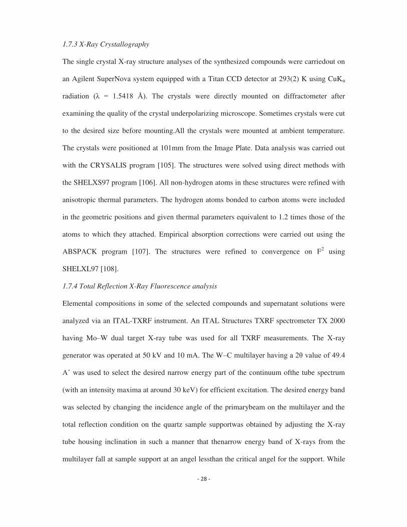
1.7.3 X-Ray Crystallography
The single crystal X-ray structure analyses of the synthesized compounds were carriedout on
an Agilent SuperNova system equipped with a Titan CCD detector at 293(2) K using CuK
were directly mounted on diffractometer after
examining the quality of the crystal underpolarizing microscope. Sometimes crystals were cut
to the desired size before mounting.All the crystals were mounted at ambient temperature.
The crystals were positioned at 101mm from the Image Plate. Data analysis was carried out
with the CRYSALIS program [105]. The structures were solved using direct methods with
the SHELXS97 program [106]. All non-hydrogen atoms in these structures were refined with
anisotropic thermal parameters. The hydrogen atoms bonded to carbon atoms were included
in the geometric positions and given thermal parameters equivalent to 1.2 times those of the
atoms to which they attached. Empirical absorption corrections were carried out using the
ABSPACK program [107]. The structures were refined to convergence on F2 using
SHELXL97 [108].
1.7.4 Total Reflection X-Ray Fluorescence analysis
Elemental compositions in some of the selected compounds and supernatant solutions were
analyzed via an ITAL-TXRF instrument. An ITAL Structures TXRF spectrometer TX 2000
having Mo W dual target X-ray tube was used for all TXRF measurements. The X-ray
generator was operated at 50 kV and 10 mA. The W
ired narrow energy part of the continuum ofthe tube spectrum
(with an intensity maxima at around 30 keV) for efficient excitation. The desired energy band
was selected by changing the incidence angle of the primarybeam on the multilayer and the
total reflection condition on the quartz sample supportwas obtained by adjusting the X-ray
tube housing inclination in such a manner that thenarrow energy band of X-rays from the
multilayer fall at sample support at an angel lessthan the critical angel for the support. While

doing this adjustment for TXRF conditions, the angle between multilayer and X-ray beam
incident on it was kept constant in theearlier optimized position. All the necessary multilayer
and tube inclination movements were done by motors controlled by TXRF32 program,
provided with the instrument. Thefulfilment of the TXRF condition was checked by
measuring the intensity of characteristic X-ray line of a suitable single element standard (e.g.
Ga) at differentinclination values of tube housing. The tube housing inclination giving
maximumintensity of the single element standard X-ray line indicates that the TXRF
conditions aresatisfied. This position is used for TXRF measurements. The X-rays were
detected andmeasured using a Si(Li) detector having a resolution of 139 eV (FWHM) at Mn
1.7.5 Electrospray ionization mass spectrometric analysis
ESI MS detection of positive ions was recorded in CH2Cl2/CH3OH/CH3CN using
aMicromass-Q-TOFMICRO instrument. The sample was introduced into the source witha
syringe pump. Nitrogen was employed as both the drying and spraying gas with a source
temperature of 900oC. The cone voltage was set to 30 V, the voltage applied on thecapillary
was 1162 kV and the sample solution flow rate was 5 mL min-1. Spectrum was recorded from
m/z of 100 1000.
1.8 Scope of the present work
Considering the above facts and the importance of the actinide complexes of nitrogen-oxygen
donor atoms based ligands in the field of nuclear fuel cycle, different series of ligands having
carbamoyl groups (piperidine carboxamide and teraalkyl urea based ligands), nitrogen,
carbamoyl groups (pyrazole carboxamide, picolinamide based ligands) and N-oxide,
carbamoyl groups (N-oxo picolinamide based ligands) have been synthesized and their
complex chemistry with uranyl and palldium(II) ions is explored. All the ligands and the
complexes have been characterized with the help of microanalytical, physico-chemical and

spectroscopic tools. The structures of some of the selected metal complexes have been
determined by X-ray diffraction method.
For clarity the present work is subdivided as follows:
a) Synthesis, structural and solvent extraction studies of piperidine urea and tetraalkyl urea
with uranyl(VI) ions
b) Synthesis, structural and coordination studies of N-oxo picolinamide complexes with
uranyl(VI) and lanthanide(III) ions
c) Synthesis, structural and theoretical studies of carbamoyl pyrazole compounds of the
uranyl(VI) and Pd(II) ions
d) Synthesis, structural and theoretical studies of picolinamide complexes with palladium(II)
and uranyl (VI) ions.
1.9 References
1. L.R. Morss, N.M. Edelstein, J. Fuger, J.J. Katz, The Chemistry of the Actinide and
Transactinide Elements, Springer , Netherlands (2006).
2. C.J. Burns, M.P. Neu, H. Boukhalfa, K.E. Gutowshi, N.J. Bridges, R.D. Rogers, J.A.
McCleverty, T.J. Meyer, G.F.R. Parkin, (Eds.), Comprehensive Coordination Chemistry
II , Elsevier, Oxford, 3 (2004) 189.
3. M. Ephritikhine, Dalton Trans. (2006) 2501.
4. S. Kant, Int. J. Nucl. Ener. Sci. Tech. 1 (2005) 204.
5. R.K. Sinha, A. Kakodkar, Nucl. Eng. Des. 236 (2006) 683-700.
6.
Shuh, K.N. Raymond, Chem. Commun. 47 (2011) 63
M.P. Sigalas, I.
7.
8. Thaddeus S. Franczyk, Kenneth R. Czerwinski, Kenneth N. Raymond, J. Am. Chem.
Soc. 114 (1992) 8138 8146.

9. Lu Zhou, Mike Bosscher, Changsheng Zhang, Salih Ozcubukcu, Liang Zhang, Wen
Zhang, Charles J. Li, Jianzhao Liu, Mark P. Jensen, Luhua Lai, Chuan He, Nat. Chem. 6
(2014) 236 241.
10. Aaron C. Sather, Orion B. Berryman, Jr. Julius Rebek, J. Am. Chem. Soc. 132 (2010)
13572 13574.
11. P.N. Pathak, D.R. Prabhu, R.B. Ruikar, V.K. Manchanda, Solvent Extr. Ion Exch. 20
12.
13. R. Ruhela, A.K. Singh, B.S. Tomar, R.C. Hubli, RSC Adv. 4 (2014) 24344-24350.
14. A. Dakshinamoorthy, P.S. Dhami, P.W. Naik, N.L. Dudwadkar, S.K. Munshi, P.K. Dey, V.
Venugopal, Desalination 232 (2008) 26-36.
15. G.H. Rizvi, J.N. Mathur, M.S. Murali, R.H. Iyer, Sep. Sci. Technol. 31 (1996) 1805-1816.
16. I. Gren -Schmitt, S.F. Wolf, in The
chemistry of the actinide and trans actinide elements, Vol. 1, 3rd ed. (Eds.: L. R. Morss, N.
M. Edelstein, J. Fuger, J. J. Katz), Springer, 2006.
17. L.L. Quill, Chem. Rev. 23 (1938) 87 155.
18. D.R. Lide, Ed., CRC Handbook of Chemistry and Physics: 86th Edition, Taylor & Francis,
86th edn., 2005.
19. Department of Health (2007), Understanding Radiation: Depleted Uranium, available at:
https://www.gov.uk/depleted-uranium-du-general-information-andtoxicology (Accessed:
April 2014).
20. T.J. Marks, Science 217 (1982) 989 997.
21. S.A. Cotton, Lanthanide and Actinide Chemistry, John Wiley & Sons, Ltd, Chichester,
First Edit., 2006.
22. N. Kaltsoyannis, P. Scott, The f elements, Oxford University Press, 1999.
23. M.R. MacDonald, M.E. Fieser, J.E. Bates, J.W. Ziller, F. Furche, W.J. Evans, J. Am.
Chem. Soc. 135 (2013) 13310.
24. I. Korobkov, S. Gambarotta, Trivalent Uranium: A Versatile Species for Molecular
Activation. In Prog. Inorg. Chem., John Wiley & Sons, Inc. (2005) 321.
25. M. Ephritikhine, Organometallics 32 (2013) 2464.
26. C.R. Graves, J.L. Kiplinger, Chem. Commun. (2009) 3831.
27. R.J. Baker, Chem. Eur. J. 18 (2012) 16258.
28. S. Fortier, G. Wu, T.W. Hayton, Inorg. Chem. 47 (2008) 4752.

29. L.A. Seaman, S. Fortier, G. Wu, T.W. Hayton, Inorg. Chem. 50 (2011) 636.
30. L.A. Seaman, G. Wu, N. Edelstein, W.W. Lukens, N. Magnani, T.W. Hayton, J. Am.
Chem. Soc. 134 (2012) 4931.
31. S. Fortier, J.R. Walensky, G. Wu, T.W. Hayton, J. Am. Chem. Soc. 133 (2011) 11732.
32. S. Fortier, T.W. Hayton, Coord. Chem. Rev. 254 (2010) 197.
33. E.A. Cuellar, T.J. Marks, Inorg. Chem. 20 (1981) 2129.
34. T.W. Hayton, Dalton Trans. 39 (2010) 1145.
35. Hurd W. Safford, A. Kuebel, J. Chem. Educ. 20 (1943) 88.
36. Department of Energy, Decommissioning of U.S. Uranium Production Facilities, February
1995.
37. J.H. Hill, J.W. Parks, "Uranium Enrichment in the United States," U.S. Energy Research
and Development Administration, CONF-750324-7, 5 Mar 75.
38. S. Nazare, J. Nucl. Mat. 124 (1984) 14.
39. J.C. Taylor, Coord. Chem. Rev. 20 (1976) 203.
40. A.F. Wells, Structural Inorganic Chemistry, Clarendon Press, Oxford (3rd edn, 1962).
41. Tetsuhiko Yoshimura, Chie Miyake, Shosuke Imoto, J. Nucl. Sci. Technol. 8 (1971) 498-
502.
42. Dipti Patel, Ashley J. Wooles, Emtithal Hashem, Harrison Omorodion, Robert J. Baker,
Stephen T. Liddle, New J. Chem. 39 (2015) 7559-7562.
43. A.S. Krass, Laser enrichement of uranium: The proliferation connection, Science 196 (1977)
721-731.
44. J. Boureston, C.D. Ferguson, Separation anxiety, Bull. Atomic Scientists 61 (2005) 14-18.
45. Trevor R. Griffiths, Vladimir A. Volkovich, J. Nucl. Mater. 274 (1999) 229-251.
46. Jean-Claude Berthet, Martine Nierlich, Michel Ephritikhine, Chem. Commun. (2004) 870 871.
47. S.A. Cotton, Lanthanides and Actinides, Macmillan, 1991.
48. Pierre Thuéry, Martine Nierlich, Bernardo Masci, Zouhair Asfari, Jacques Vicens, J.
Chem. Soc., Dalton Trans. (1999) 3151 3152.
49. Carol J. Burns, David L. Clark, Robert J. Donohoe, Paul B. Duval, Brian L. Scott, C.
Drew Tait, Inorg. Chem. 39 (2000) 5464-5468.
50. Y.S. Kim, ed., Uranium Mining Technology: Proceedings of a First Conference (U. of
Nevada, 1977).
51. Y. Kopylova, N. Guseva, A. Shestakova, A. Khvaschevskaya, K. Arakchaa, Earth
Environ. Sci. Trans. 27 (2015) 12034.
52. M.P. Elless, S.Y. Lee, Water Air Soil Pollut. 107 (1998) 147 162.
53. J.R. Lacher, J.D. Salzman, J.D. Park, Ind. Eng. Chem. 53 (1961) 282 284.
54. C.J. Hardy, D. Fairhurst, H.A.C. McKay, A.M. Willson, Trans. Faraday Soc. 60 (1964)
1626-1636.
55. E.V. Fedoseev, M.I. Aizenberg, A.V. Davydov, B.F. Myasoedov, J. Radioanal. Nucl.
Chem. 136 (1989) 395.
56. L.L. Shcherbakova, V.Yu. Mishin, E.M. Rubtsov, Soviet Radiokhim. 26 (1984) 698.
57. Y. Baskin, J.R. Ferraro, J. Inorg. Nucl. Chem. 30 (1968) 241.
58. Y. Baskin, N.S.K. Prasad, J. Inorg. Nucl. Chem. 25 (1963) 1011.
59. F.E. Beamish, W.A.E. Mcbryde, Anal. Chem. 25 (1953) 1613-1617.
60. F. Bonet, S. Grugeon, R. Herrera Urbina, K. Tekaia-Elhsissen, J.M. Tarascon, Solid State
Sci. 4 (2002) 665 670.
61. K.C. Taylor, Stud. Surf. Sci. Catal. 30 (1987) 97-116.
62. Christopher W. Corti, Platinum Met. Rev. 53 (2009) 198 202.
63. J.C. Wataha, C.T. Hanks, J. Oral Rehabil. 23 (1996) 309-320.
64. Morton Antler, Platinum Met. Rev. 26 (1982) 106-117.
65. Christopher R. King, Int. J. Radiat. Oncol. Biol. Phys. 47 (2000) 1165 1167.
66. N. Miksys, J.E. Cygler, J.M. Caudrelier, R.M. Thomson, Phys. Med. Biol. 61 (2016)
2705 2729.
67. C.J. Elsevier, Coord. Chem. Rev. 185/186 (1999) 809.
68. P.K. Byers, A.J. Canty, B.W. Skelton, A.H. White, J. Chem. Soc. Chem. Commun. (1986)
1722.
69. R. Uson, J. Fornies, R. Navarro, J. Organomet. Chem. 96 (1975) 307.
70. A.A. Timakov, V.N. Prusakov, Y.V. Drobyshevskii, Russ. J. Inorg. Chem. 27 (1982)
1704.
71. David E. Berry, Philippa Carrie, Kelli L. Fawkes, Bruce Rebner, Yao (Shirley) Xing, J.
Chem. Educ. 87 (2010) 533 534.
72. Barbara Milani, Anna Anzilutti, Lidia Vicentini, Andrea Sessanta o Santi, Ennio
Zangrando, Silvano Geremia, Giovanni Mestroni, Organometallics 16 (1997) 5064 5075.
73. Y. Pan, J.T. Mague, M.J. Fink, J. Am. Chem. Soc. 115 (1993) 3842.
74. R.U. Kirss, R. Eisenberg, Inorg. Chem. 28 (1989) 3372.
75. F.A. Cotton, G. Wilkinson, C.A. Murillo, M. Bochmann, Advanced Inorganic Chemistry;
6h Edition; John Wiley & Sons: New York, 1999.

76. S.A. Cotton, Ed.; Chemistry of Precious Metals; Springer, 1997.
77. E.G. Mednikov, L.F. Dahl, Phil. Trans. R. Soc. A 368 (2010) 1301 and references cited
therein.
78. K.R. Dixon, A. Dixon, Comprehensive Organometallics Chemistry II; Elsevier. 8 (1995)
200 and references cited therein.
79. (a) J.H. Duffus, Pure Appl. Chem. 74 (2002) 793. (b) S. Ahrland, J. Chatt, N.R. Davies,
Quart. Rev. 12 (1958) 265. (c) R.G. Pearson, J. Chem. Educ. 45 (1968) 581. (d) R.G.
Pearson, J. Chem. Educ. 45 (1968) 643.
80. F.A. Cotton, G. Wilkinson, P.L. Gaus, Basic Inorganic Chemistry; 3rd Edition, Wiley,
1994.
81. I. Nakanishi, K.Matsumoto, S. Ooi, Acta Crystallogr. Sect. C 47 (1991) 2073.
82. W.J. Louw, D.J.A. DeWaal, G.J. Kruger, J. Chem. Soc., Dalton Trans. (1976) 2365.
83. A.J. Blake, A.J. Holder, T.I. Hyde, M. Schröder, J. Chem. Soc., Chem. Commun. (1987)
987.
84. A.J. Blake, A.J. Holder, T.I. Hyde, Y.V. Roberts, A.J. Lavery, M. Schröder, J. Organomet.
Chem. 323 (1987) 261.
85. A.F. Wright, B.E.F. Fender, N. Bartlett, K. Leary, Inorg. Chem. 17 (1978) 748.
86. D.J. Gulliver, W. Levason, J. Chem. Soc., Dalton Trans. (1982) 1895.
87. L.R. Gray, D.J. Gulliver, W. Levason, M. Webster, J. Chem. Soc., Dalton Trans. (1983)
133.
88. A. Maleckis, M.S. Sanford, Organometallics 30 (2011) 6617.
89. S. Kannan, J.S. Gamare, K.V. Chetty, M.G.B. Drew, Polyhedron 26 (2007) 3810.
90. L. Nigond, C. Musikas, C.C. Cullerdier, Solv. Extr. Ion Exch. 12 (1994) 261.
91. M.S. Subramanian, J. Radioanal. Nucl. Chem. Lett. 187 (1994) 91.
92. S.S Biswas, P.S. Murty, A. Sethumadhavan, R. Kaimal, A.V. Sankaran, Anal. Lett. 24
(1991) 887.
93. J. Chen, J.C. Wang, Prog. Chem. 23 (2011) 1366.
94. M. Bosson, W. Levason, T. Patel, M.C. Popham, M. Webster, Polyhedron 20 (2001) 2055.
95. W. Levason, E.H. Newman, M. Webster, Polyhedron 19 (2000) 2697.
96. M.J. Hudson, F.W. Lewis, L.M. Harwood, Strategies and Tactics in Organic Synthesis 9
(2013) 177.
97. M.S. Nagar, P.B. Ruikar, J.N. Mathur, R.H. Iyer, Syn. React. Inorg. Met. 28 (1998) 621.

98. Y. Kulyako, D. Malikova, T. Trofimova, M. Chmutovaa, B. Myasoedova, J. Nucl. Sci.
Technol. 39 (2002) 302.
99. S. Kannan, K.V. Chetty, V. Venugopal, M.G.B. Drew, Dalton Trans. (2004) 3604.
100. Y. Sasakia, Y. Sugoa, S. Suzukia, S. Tachimoria, Solvent Extr. Ion Exch. 19 (2001) 91.
101. S. Kannan, M.A. Moody, C.L. Barnes, P.B. Duval, Inorg. Chem. 47 (2008) 4691.
102. J.C. Berthet, Y. Miquel, P.B. Iveson, M. Nierlich, P. Thuéry, C. Madic, M. Ephritikhine, J.
Chem. Soc., Dalton Trans. (2002) 3265.
103. P.J. Panak, A. Ge, Chem. Rev. 113 (2013) 1199.
104. A.I. Vogel, Textbook of Practical Organic Chemistry , 4th Ed., English Language Book
Society, Longman, London (1987).
105. CRYSALIS program, Oxford Diffraction Ltd., 2005.
106. G.M. Sheldrick, SHELXS, Program for Crystal Structure Solution, University of
Gottingen, Gottingen, Germany, 1997.
107. ABSPACK Program, Oxford Diffraction Ltd., 2005.
108. G.M. Sheldrick, SHELXL, Program for Crystal Structure Refinement, University of
Gottingen, Gottingen, Germany, 1997.


CHAPTER-2 2.1 Introduction
The coordination chemistry of actinides plays very important role for the development of new
methods concerning the processing of irradiated nuclear fuel and their storage [1 3]. The
selective separation of uranium-233 from the mixture of thorium and other fission products is
an important step [4] in the advanced heavy water reactor (AHWR) technology [5] of the
Indian atomic energy program, where upon the thorium oxide is used as a fuel material. In
recent years, incinerable organic amide based extractants have proved to be of particular
interest in the process chemistry of lanthanides and actinides from the nitric acid medium [6-
12]. It is reported that the N,N'-2-ethylhexyl iso-butyramide shows selective extraction
property for the uranium(VI) ion from a mixture containing thorium(IV) or plutonium(IV)
ion [13-15]. The solvent extraction method needs many steps such as: extraction, stripping,
precipitation and purification during the reprocessing of fuel materials. The neutral N-
cyclohexyl-2-pyrrolidone ligand shows selective precipitation of uranium(VI) ion (up to
68%) from the mixture containing various other ions [16] and with this ligand only two steps
are required namely precipitation and purification. It has also been reported recently that
isobutyramide based amide ligands show an excellent coordination property towards the
uranyl group and form air and moisture stable halide and thiolato complexes [17-18]. The
stability of these complexes was explained on the basis of very strong bonding between the
amide and uranyl group. The observed bond distances in these complexes are much shorter in
length compared to those of any of the uranyl-amide [19 21], uranyl-phosphine oxide [22-
26] and uranyl-pyrrolidone [27-28] complexes reported. Inspired by the results obtained, we
report herein the synthesis, characterization and the coordination chemistry of some urea
based ligands with uranyl nitrate, chloride, bromide and -diketonates) by
expecting even more strong bonding due to increased electron density on the O-atom of the
ligands by the mesomeric effect of two N-atoms placed both sides of C=O group.

2.2 Experimental 2.2.1 General Considerations
All uranyl -diketonates) were prepared according to the reported methods [29].
2.2.2 Synthesis of the ligands (Scheme 1)
2.2.2.1 Piperidine carboxamide ligands
2.2.2.1.1 Synthesis of C5H10NCON(CH3)2 (L1)
To a solution of N,N-dimethyl carbamoyl chloride (10.75 g, 0.1 mol) in (50 mL) benzene, a
solution of piperidine (8.52 g, 0.1 mol) and triethyl amine (12.1 g, 0.12 mol) in benzene (50
mL) was added slowly with stirring. The reaction mixture was refluxed for 2 h and treated
with 100 ml. of 5% HCl solution. The organic layer was separated, dried over anhydrous
sodium sulfate and filtered. The solution on evaporation yielded a colorless liquid. Yield:
62%. Anal. Calc. for C8H16N2O: C, 61.5; H, 10.25; N, 17.9. Found: C, 60.5; H, 10.1; N,
17.5%. 1H NMR (CDCl3, 25o -piperidyl CH2), 2.50 (s, 6H, N(CH3)2),
1.26 (br, 6H, piperidyl CH2). IR (cm-1
2.2.2.1.2 Synthesis of C5H10NCON(C2H5)2 (L2)
This was prepared similarly to L1 in 76% yield by taking N,N-diethyl carbamoyl chloride.
Anal. Calc. for C10H20N2O: C, 65.2; H, 10.9; N, 15.2. Found: C, 64.5; H, 10.3; N, 15.5%. 1H
NMR (CDCl3, 25 o -piperidyl CH2 and N(C2H5)2 CH2), 1.55 (br, 6H, 3,4,5-
piperidyl CH2), 1.09 (t, J = 7.0 Hz, 6H, N(C2H5)2 CH3). IR (cm-1 .
2.2.2.1.3 Synthesis of C5H10NCON(iC3H9)2 (L3)
This was prepared similarly to L1 in 94% yield by taking N,N-diisoproyl carbamoyl chloride.
Anal. Calc. for C12H24N2O: C, 67.9; H, 11.3; N, 13.2. Found: C, 66.6; H, 10.7; N, 13.0%. 1H
NMR (CDCl3, 25 o 48 (m, 2H, N(iPr2) CH), 2.91 (br, 4H, 2,6-piperidyl CH2), 1.44 (br,
6H, 3,4,5-piperidyl CH2), 1.15 (d, J = 6.6 Hz, 12H, N(iPr2) CH3). IR (cm-1
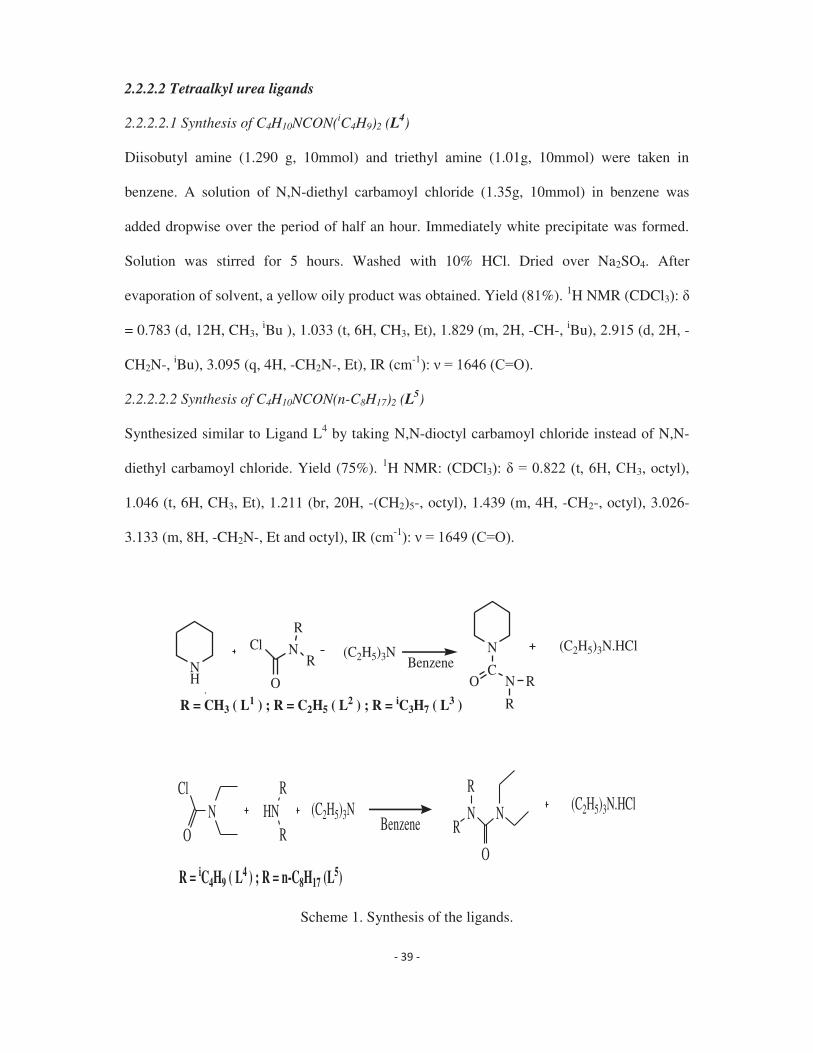
2.2.2.2 Tetraalkyl urea ligands
2.2.2.2.1 Synthesis of C4H10NCON(iC4H9)2 (L4)
Diisobutyl amine (1.290 g, 10mmol) and triethyl amine (1.01g, 10mmol) were taken in
benzene. A solution of N,N-diethyl carbamoyl chloride (1.35g, 10mmol) in benzene was
added dropwise over the period of half an hour. Immediately white precipitate was formed.
Solution was stirred for 5 hours. Washed with 10% HCl. Dried over Na2SO4. After
evaporation of solvent, a yellow oily product was obtained. Yield (81%). 1H NMR (CDCl3
= 0.783 (d, 12H, CH3, iBu ), 1.033 (t, 6H, CH3, Et), 1.829 (m, 2H, -CH-, iBu), 2.915 (d, 2H, -
CH2N-, iBu), 3.095 (q, 4H, -CH2N-, Et), IR (cm-1
2.2.2.2.2 Synthesis of C4H10NCON(n-C8H17)2 (L5)
Synthesized similar to Ligand L4 by taking N,N-dioctyl carbamoyl chloride instead of N,N-
diethyl carbamoyl chloride. Yield (75%). 1H NMR: (CDCl3 3, octyl),
1.046 (t, 6H, CH3, Et), 1.211 (br, 20H, -(CH2)5-, octyl), 1.439 (m, 4H, -CH2-, octyl), 3.026-
3.133 (m, 8H, -CH2N-, Et and octyl), IR (cm-1
(C2H5)3N (C2H5)3N.HClBenzene
R = CH3 ( L1 ) ; R = C2H5 ( L2 ) ; R = iC3H7 ( L3 )
NH
Cl N
O
R
RN
CNO R
R
NCl
OHN
R
RNN
O
R
R(C2H5)3N (C2H5)3N.HCl
Benzene
R = iC4H9 ( L4 ) ; R = n-C8H17 (L5)
Scheme 1. Synthesis of the ligands.

2L [UO2(NO3)2L2] (L = L1, L2, L3)
[UO2(DBM)2.2H2O] L [UO2(DBM)2.L] (L = L1, L2, L3)
(DBM = Dibenzoyl methanate)
[UO2(TTA)2.2H2O] L (L = L1, L2, L3)[UO2(TTA)2.L]
(TTA = Thenoyl trifluroacetonate)
[UO2X2, nH2O] 2L UO2X2L2(L = L1, L2, L3)
(X = Cl, Br)
[UO2(NO3)2.6H2O]
2L [UO2(NO3)2L2] (L = L4)
[UO2X2. nH2O] 2L UO2X2L2 (L = L4)
(X = Cl, Br)
[UO2(NO3)2. 6H2O]
Scheme 2. Synthesis of the uranyl compounds.
2.2.3 Synthesis of the compounds (Scheme 2)
2.2.3.1 Synthesis of [UO2(NO3)2{C5H10NCON(CH3)2}2] (1)
To a solution of N,N-dimethylpiperidine-1-carboxamide (200 mg, 1.29 mmol) in chloroform,
solid UO2(NO3)2.6H2O (300 mg, 0.59 mmol) was added with stirring. The solution was
allowed to stir until the entire uranyl nitrate dissolved to give a clear solution. This solution
was filtered and layered with isooctane. This solution on slow evaporation yielded a yellow
crystalline solid. This was filtered, washed with hexane and air dried. Yield: 375.5 mg (89%).
Elemental Analysis Calculated for [UO2(NO3)2{C5H10NCON(CH3)2}2], C16H32N6O10U (%):
Expected: C=27.2; H=4.5; N=11.9. Found: C= 26.7; H=4.2; N= 11.5. IR (Nujol, /cm-1):
1525 (C=O), 925 (U=Oassy). 1H NMR (CDCl3, 25 oC, 3.55 (br, 4H, 2,6-piperidyl
CH2), 3.14 (s, 6H, N(CH3)2), 1.63 (br, 6H, 3,4,5-piperidyl CH2).
2.2.3.2 Synthesis of [UO2(NO3)2{C5H10NCON(C2H5)2}2] (2)
Compound 2 was prepared in a similar way as compound 1 by taking N,N-diethylpiperidine-
1-carboxamide (240 mg, 1.3 mmol) and UO2(NO3)2.6H2O (300 mg, 0.59 mmol). Yield: 418.9
mg (92%). Elemental Analysis Calculated for [UO2(NO3)2{C5H10NCON(C2H5)2}2],
C20H40N6O10U (%): Expected: C=31.5; H=5.2; N=11.0. Found: C= 31.1; H=4.8; N= 10.7. IR

(Nujol, /cm-1): 1521 (C=O), 928 (U=Oassy). 1H NMR (CDCl3, 25 oC, 3.52 (br, 8H,
2,6-piperidyl CH2 and N(C2H5)2 CH2), 1.63 (br, 6H, 3,4,5-piperidyl CH2), 1.26 (s, 6H,
N(C2H5)2 CH3).
2.2.3.3 Synthesis of [UO2(NO3)2{C5H10NCON(iC3H7)2}2] (3)
Compound 3 was prepared in a similar way as compound 1 by taking N,N-
diisopropylpiperidine-1-carboxamide (270 mg, 1.27 mmol) and UO2(NO3)2.6H2O (300 mg,
0.59 mmol). Yield: 415.5 mg (85%). Elemental Analysis Calculated for
[UO2(NO3)2{C5H10NCON(iC3H7)2}2], C24H48N6O10U (%): Expected: C=35.2; H=5.9;
N=10.2. Found: C= 35.1; H=5.8; N= 10.1. IR (Nujol, /cm-1): 1522 (C=O), 932 (U=Oassy). 1H
NMR (CDCl3, 25 oC, 3.90 (br, 2H, N(iPr2) CH), 3.40 (br, 4H, 2,6-piperidyl CH2),
1.56 (br, 6H, 3,4,5-piperidyl CH2), 1.41 (d, J = 6.0 Hz, 12H, N(iPr2) CH3).
2.2.3.4 Synthesis of [UO2(C6H5COCHCOC6H5)2{C5H10NCON(CH3)2}] (4)
To a solution of N,N-dimethylpiperidine-1-carboxamide (80 mg, 0.51 mmol) in
dichloromethane, solid UO2(C6H5COCHCOC6H5)2.2H2O (350 mg, 0.47 mmol) was added
and the resulting mixture was refluxed for 2 h with constant stirring. The solution was filtered
and layered with dodecane. This on slow evaporation yielded an orange red crystalline solid
product. Yield: 373.4 mg (92%). Elemental Analysis Calculated for
[UO2(C6H5COCHCOC6H5)2{C5H10NCON(CH3)2}], C38H38N2O7U (%): Expected: C=52.3;
H=4.4; N=3.2. Found: C= 51.8; H=3.9; N= 3.0. IR (Nujol, /cm-1): 1591 (C=O, DBM), 1542
(C=O, Ligand), 907 (U=Oassy). 1H NMR (CDCl3, 25 oC,
8.0 (d, 4H, Ph, DBM), 7.58 (br, 8H, Ph, DBM), 7.24 (s, 2H, CH, DBM), 3.53 (br, 4H,
piperidyl CH2), 3.14 (br, 6H, N(CH3)2), 1.55 (br, 6H, piperidyl CH2).
2.2.3.5 Synthesis of [UO2(C6H5COCHCOC6H5)2{C5H10NCON(C2H5)2}] (5)
Compound 5 was prepared in a similar way as compound 4 by taking N,N-diethylpiperidine-
1-carboxamide (90 mg, 0.49 mmol) and UO2(C6H5COCHCOC6H5)2.2H2O (350 mg, 0.47

mmol). Yield: 393.8 mg (94%). Elemental Analysis Calculated for
[UO2(C6H5COCHCOC6H5)2{C5H10NCON(C2H5)2}], C40H42N2O7U (%): Expected: C=53.3;
H=4.7; N=3.1. Found: C= 52.8; H=4.5; N= 3.0. IR (Nujol, /cm-1): 1591 (C=O, DBM), 1542
(C=O, Ligand), 905 (U=Oassy). 1H NMR (CDCl3, 25 oC,
8.0 (d, 4H, DBM Ph), 7.58 (s, 8H, DBM Ph), 7.25 (s, 2H, DBM CH), 3.53 (br, 8H, piperidyl
CH2 and N(C2H5)2 CH2), 1.52 (s, 6H, piperidyl CH2), 1.23 (t, J = 6.6 Hz, 6H, N(C2H5)2 CH3).
2.2.3.6 Synthesis of [UO2(C6H5COCHCOC6H5)2{C5H10NCON(iC3H7)2}] (6)
Compound 6 was prepared in a similar way as compound 4 by taking N,N-
diisopropylpiperidine-1-carboxamide (105 mg, 0.5 mmol) and
UO2(C6H5COCHCOC6H5)2.2H2O (350 mg, 0.47 mmol). Yield: 384.4 mg (89%). Elemental
Analysis Calculated for [UO2(C6H5COCHCOC6H5)2{C5H10NCON(iC3H7)2}], C42H46N2O7U
(%): Expected: C=54.3; H=4.9; N=3.0. Found: C= 53.9; H=4.5; N= 2.8. IR (Nujol, /cm-1):
1592 (C=O, DBM), 1541 (C=O, Ligand), 901 (U=Oassy). 1H NMR (CDCl3, 25 oC,
8.61 (br, 4H, DBM Ph), 8.40 (br, 4H, DBM Ph), 8.0 (d, 4H, DBM Ph), 7.59 (s, 8H, DBM
Ph), 7.27 (s, 2H, DBM CH), 3.94 (s, 2H, N(iPr2) CH), 3.42 (s, 4H, piperidyl CH2), 1.49 (s,
18H, piperidyl CH2 and N(iPr2) CH3).
2.2.3.7 Synthesis of [UO2(C4H3SCOCHCOCF3)2{C5H10NCON(CH3)2}] (7)
To a dichloromethane solution of N,N-dimethylpiperidine-1-carboxamide (70 mg, 0.44
mmol), solid UO2(C4H3SCOCHCOCF3)2.2H2O (300 mg, 0.4 mmol) was added and refluxed
for 2 h with constant stirring. This solution was filtered and layered with isooctane. This
solution on slow evaporation yielded bright yellow colored needle shaped crystals. Yield:
327.2 mg (94%). Elemental Analysis Calculated for
[UO2(C4H3SCOCHCOCF3)2{C5H10NCON(CH3)2}], C24H24N2O7F6S2U (%): Expected:
C=33.2; H=2.8; N=3.2. Found: C= 32.8; H=2.7; N= 3.1. IR (Nujol, /cm-1): 1618 (C=O,
TTA), 1540 (C=O, Ligand), 919 (U=Oassy). 1H NMR (CDCl3, 25 oC,

thiophene CH), 7.83 (t, J = 6.6 Hz, 2H, thiophene CH), 7.28 (q, J = 4.5 Hz, 2H, thiophene
CH), 6.79 (s, 2H, TTA CH), 3.59 (br, 4H, piperidyl CH2), 3.22 (s, 6H, N(CH3)2), 1.63 (br,
6H, piperidyl CH2).
2.2.3.8 Synthesis of [UO2(C4H3SCOCHCOCF3)2{C5H10NCON(C2H5)2}] (8)
Compound 8 was prepared in a similar way as compound 7 by taking N,N-diethylpiperidine-
1-carboxamide (80 mg, 0.43 mmol) and UO2(C4H3SCOCHCOCF3)2.2H2O (300 mg, 0.4
mmol). Yield: 319.8 mg (89%). Elemental Analysis Calculated for
[UO2(C4H3SCOCHCOCF3)2{C5H10NCON(C2H5)2}], C26H28N2O7F6S2U (%): Expected:
C=34.8; H=3.1; N=3.1. Found: C= 34.6; H=2.9; N= 3.0. IR (Nujol, /cm-1): 1591 (C=O,
TTA), 1539 (C=O, Ligand), 922 (U=Oassy). 1H NMR (CDCl3, 25 oC,
thiophene CH), 7.84 (br, 2H, thiophene CH), 7.28 (d, J = 4.7 Hz, 2H, thiophene CH), 6.79 (s,
2H, TTA CH), 3.52 (br, 8H, piperidyl CH2 and N(C2H5)2 CH2), 1.61 (br, 6H, piperidyl CH2),
1.23 (br, 6H, N(C2H5)2 CH3).
2.2.3.9 Synthesis of [UO2(C4H3SCOCHCOCF3)2{C5H10NCON(iC3H7)2}] (9)
Compound 9 was prepared in a similar way as compound 7 by taking N,N-diisopropyl
piperidine-1-carboxamide (90mg, 0.42mmol) and UO2(C4H3SCOCHCOCF3)2.2H2O (300 mg,
0.4 mmol). Yield: 326.1 mg (88%). Elemental Analysis Calculated for
[UO2(C4H3SCOCHCOCF3)2{C5H10NCON(iC3H7)2}], C28H32N2O7F6S2U (%): Expected:
C=36.4; H=3.5; N=3.0. Found: C= 35.8; H=3.0; N= 3.0. IR (Nujol, /cm-1): 1618 (C=O,
TTA), 1540 (C=O, Ligand), 922 (U=Oassy). 1H NMR (CDCl3, 25 oC,
thiophene CH), 7.82 (d, 2H, thiophene CH), 7.29 (br, 2H, thiophene CH), 6.79 (s, 2H, TTA
CH), 3.91 (s, 2H, N(iPr2) CH), 3.46 (s, 4H, piperidyl CH2), 1.56 (s, 6H, piperidyl CH2), 1.46
(d, J = 6.6 Hz, 12H, N(iPr2) CH3).
2.2.3.10 Synthesis of [UO2Cl2{C5H10NCON(iC3H7)2}2] (10)
Solid UO3 (300 mg) was dissolved in 4 mL of concentrated HCl and the volume of the
solution was reduced completely to dryness on a hot plate. The orange colored solid thus
obtained was dissolved in 10 mL of methanol to give a clear solution. To this solution N,N-
diisopropylpiperidine-1-carboxamide was added and heated for 2 min. The volume of the
solution was reduced to 2mL, and 20 mL of diethyl ether was added into it with stirring. The
yellow product formed was filtered, washed with ether and dried in air. This product was
recrystallized from a chloroform/isooctane mixture to give a crystalline solid. Yield: 714.2
mg (89%). Elemental Analysis Calculated for [UO2Cl2{C5H10NCON(iC3H7)2}2],
C24H48N4O4Cl2U (%): Expected: C=37.6; H=6.3; N=7.3. Found: C= 37.2; H=6.0; N= 7.2. IR
(Nujol, /cm-1): 1524 (C=O), 924 (U=Oassy).
2.2.3.11 Synthesis of [UO2Br2{C5H10NCON(iC3H7)2}2] (11)
This was synthesized similar to 10, by taking concentrated HBr instead of HCl. Yield 85%.
Elemental Analysis Calculated for [UO2Br2{C5H10NCON(iC3H7)2}2], C24H48N4O4Br2U (%):
Expected: C=33.7; H=5.6; N=6.6. Found: C= 33.2; H=5.2; N= 6.2. IR (Nujol, /cm-1): 1525
(C=O), 928 (U=Oassy).
2.2.3.12 Synthesis of [UO2(NO3)2{C4H10NCON(iC4H9)2}2] (12)
UO2(NO3)2·6H2O (200 mg, 0.4 mmol) and ligand C4H10NCON(iC4H9)2}2 (182 mg, 0.8
mmol) were taken in dichloromethane (DCM) and stirred for 2 hours and all the uranyl
nitrate was dissolved. The solution was filtered and the filtrate is layered with isooctane to get
obtain yellow crystals of the compound. Yield: 331.9 mg (98%). Elemental Analysis
Calculated for [UO2(NO3)2{C4H10NCON(iC4H9)2}2], C26H56N6O10U (%): Expected: C=36.7;
H=6.6; N=9.9. Found: C= 36.2; H=6.9; N= 9.1. IR ( /cm-1): 1540 (C=O). 1H NMR (CDCl3,
25 oC, 0.879 (d, 12H, CH3, iBu), 1.252 (t, 6H, CH3, Et), 2.042 (m, 2H, CH , iBu),
3.272 (d, 2H, CH2N , iBu), 3.053 (m, br, 4H, CH2N , Et).
2.2.3.13 Synthesis of [UO2Cl2{C4H10NCON(iC4H9)2}2] (13)
Solid UO3 (114 mg, 0.4 mmol) was dissolved in 4 mL of concentrated HCl and the volume of
the solution was reduced completely to dryness on a hot plate to get orange powder of
UO2Cl2·XH2O. To a DCM solution of C4H10NCON(iC4H9)2}2 (182 mg, 0.8 mmol),
UO2Cl2·XH2O was added and stirred for 2 hours. The solution was filtered and the filtrate
was layered with isooctane to obtain yellow crystals of the compound. Yield: 311.3 mg
(98%). Elemental Analysis Calculated for [UO2Cl2{C4H10NCON(iC4H9)2}2],
C26H56N4O4Cl2U (%): Expected: C=39.1; H=7.0; N=7.0. Found: C= 38.6; H=7.9; N= 6.9. IR
( /cm-1): 1523 (C=O). 1H NMR: (CDCl3, 25 oC, 3, iBu), 1.290
(br, 6H, CH3, Et), 2.058 (m, 2H, CH , iBu), 3.429 (br, 2H, CH2N , iBu), 3.673 (br, 4H,
CH2N , Et).
2.2.3.14 Synthesis of [UO2Br2{C4H10NCON(iC4H9)2}2] (14)
This was synthesized similar to 13, by taking concentrated HBr instead of HCl. Yield 97%.
Elemental Analysis Calculated for [UO2Br2{C4H10NCON(iC4H9)2}2], C26H56N4O4Br2U (%):
Expected: C=35.2; H=6.3; N=6.3. Found: C= 34.7; H=6.8; N= 6.2. IR (Nujol, /cm-1): 1522
(C=O). 1H NMR (CDCl3, 25 oC, 3, iBu), 1.282 (t, 6H, CH3, Et),
2.057 (m, 2H, CH , iBu), 3.424 (d, 2H, CH2N , iBu), 3.676 (d, 4H, CH2N , Et).
2.2.4 Solvent extraction studies
Solvent extraction studies were performed by using a solution of N, N-diethyl-N ,N -dioctyl
urea in dodecane (0.2 M) with the required aqueous phase spiked with 233U or 239Pu tracers in
a thermostat water bath for 1 h at 25 ± 0.1 oC. Assay of organic and aqueous phases were
done in duplicate by alpha counting using a dioxane based liquid scintillator for 233U and
239Pu. The distribution ratio (D) is defined as the ratio of the concentration of uranium and
plutonium in the organic phase to that of the aqueous phase.

2.2.5 Separation studies
Stock solutions containing 5 g of [Th(NO3)4 . 6H2O], 200 mg of [La(NO3)3 .6H2O], 200 mg
of [Sm(NO3)3 .6H2O], 200 mg of [Eu(NO3)3 .6H2O] and 200 mg of [UO2(NO3)2 .6H2O ] were
prepared in 20 ml of 3 M HNO3. This solution was layered with 1 ml of N, N-diethyl-N ,N -
diisobutyl urea and allowed to stand for overnight. This process deposited yellow crystalline
solid material (Fig. 2.1), which was filtered, washed with water and then with hexane and
dried. The supernatants and the solutions of yellow solid in ethanol were used for the
elemental analysis using the total reflection X-ray fluorescence (TXRF) method.
Fig. 2.1 Stock solution of La, Sm, Eu, Th and U layered without (left) and with the ligand (right)

2.2.6 X-ray diffraction studies of compounds 3, 9, 10, 11, 12, 13 and 14
Selected crystallographic data for compounds 3, 9, 10, 11, 12, 13 and 14 are summarized in Table 2.1.
Table 2.1 Crystallographic data for compounds 3, 9, 10, 11, 12, 13 and 14
3 9 10 11 12 13 14 Empirical formula
C24H48N6O10U
C28H34N2O7S2F6U C24H48N4O4Cl2U C24H48N4O4Br2U C26H56N6O10U C26H56N4O4Cl2U C26H56N4O4Br2U
Formula weight 818.7 926.73 765.60 854.50 850.79 797.68 886.58 Crystal system Monoclinic Monoclinic Orthorhombic Orthorhombic Triclinic Monoclinic Monoclinic
Space group P 21/n P 21/n Pbca Pbca P P121/n1 P121/n1
a (Å) 8.9696(2) 16.0316(3) 15.2149(6) 15.2003(4) 9.4988(7) 9.1746(2) 8.9803(4)
b (Å) 18.2161(4) 9.03822(19) 12.2538(7) 12.3726(3) 10.8674(9) 14.7815(3) 13.5484(4)
c(Å) 10.2600(3) 23.4060(4) 16.9457(8) 16.9049(4) 18.8816(12) 13.3902(3) 15.0855(5)
o) ------ ------ ------ ------ 88.045(6) ------ ------
o) 102.403(3) 94.8813(18) ------ ------ 79.572(6) 98.738(2) 99.086(4)
o) ------ ------ ------ ------ 76.641(7) ------ ------
Volume (Å3) 1637.26 3379.16(12) 3159.4(3) 3179.24(13) 1865.0(2) 1794.84(7) 1812.40(11)
Z (formula unit) 2 4 4 4 2 2 2
Calculated density (g/cm3)
1.661 1.822 1.610 1.785 1.515 1.476 1.625

-1) 14.447 15.382 16.279 17.576 12.703 14.348 15.436
Reflections collected/unique
3088/2010 6283/4965 2957/1360 2981/1769 6875/3820 3347/2238 3428/2394
Data/restrains/ parameters
3088/36/202 6283/138/457 2957/0/165 2981/72/165 3820/108/401 2238/0/175 2394/0/175
Goodness of fit on F2
1.060 1.007 1.050 1.000 1.109 1.065 1.040
Final R1 indices
0.0425 0.0416 0.0798 0.0452 0.0785 0.0567 0.0487
wR2 indices (all data)
0.0688 0.0559 0.1308 0.0789 0.1825 0.1491 0.1185
2(Fo2)+(0.0649P)2] for 3 2(Fo
2)+(0.0643P)2 ] for 9 2(Fo2)+(0.1805P)2 + 0.8529P] for 10, w 2(Fo
2)+(0.0646P)2
+1.6221P] for 11 2(Fo2) + (0.0833P)2 + 1.1877P] for 12 2(Fo
2) + (0.0971P)2] for 13 2(Fo2) + (0.0809P)2] for 14,
where,P=(Fo2+2Fc
2)/3

2.2.7 Theoretical study
Experimentally derived crystal structures of UO2(NO3)2(L1)2 and Th(NO3)4(L
1)2 are further
subjected to gas phase optimizations with generalized-gradient approximated BP86 [30-31]
-TZVP basis set [32-33] and for C and H, def2-SV(P)
basis set [34] are used in conjugation with their corresponding auxiliary basis sets. Resolution of
Identity (RI) approximations are imposed to speed up the optimizations [35-39]. Single point
calculations on the optimized structures are carried out with meta-GGA M06-2X functional.
using TZVP basis set. We have benchmarked previously that M06-2X [40] is good for
predicting energetics for a series of actinyl complexes [41]. For both the actinides, def-TZVP
basis set is used to describe their valance orbitals while core orbitals (60 electrons) are modelled
with small-core effective core potential (SC-ECP). The effect of solvation media (water) was
incorporated during energy minimization using COSMO continuum salvation model [42] setting
dielectric constant values to 80. Further, analytical frequency calculations are performed to
verify the optimized structures (Table 2.2) as their potential minima and to derive the free energy
incorporated for 298.15 K. In addition, Mülliken population analysis (MPA) [43] (Table 2.3) is
also carried out to gauge on the charge distribution over the complexes. All the above
calculations are carried out using TURBOMOLE v 6.3.1 [44].
To decipher the nature of metal-ligand binding, energy decomposition analysis (EDA) [45-46]
(Table 2.4) is carried out at B3LYP/TZP level using pre-optimized structure as implemented in
ADF2013 programme package [47-48]. The total bonding energy bond) between the two
fragments is calculated using following equation:
bond pauli elast orb
pauli is destabilizing in nature and related to the exchange energy contributions. The
elast in co pauli gives the total steric

orb accounts for the orbital interactions between the fragments which
originates during relaxation and mixing of fragmented molecular orbitals. Scalar relativistic
corrections are incorporated into these calculations using Zeroth Order Regular Approximation
(ZORA) at integration level 4.5.
Finally, In order to scrutinize the percentage of metallic character and the atomic hybrid
decompositions of metal-carbonyl, we further performed natural localized molecular orbital
(NLMO) analysis (Table 2.5) on the pre-optimized structures at B3LYP/TZP level within the
framework of NBO 6.0 version [49] as implemented in ADF 2013 programme package. Natural
Population Analysis (Table 2.6) of the actinide-amide complexes was carried out to get an idea
about the involvement of valence orbitals in the bonding of the said complexes.
Separation factor (S.F) is calculated using the following equation,
S.F = ~exp (- U/Th/RT) for U/Th separation with L1 at 298.15K.
Table 2.2 Optimized structural parameters of actinide-amide complexes
U=O
(Å)
U/Th-OL1
(Å)
C=O
(Å)
(U/Th)-
O=CL1
U/Th-NO3
(Å)
O=U=O
UO2(NO3)2(L1)2 1.803,
1.804 (1.742, 1.746)
2.375, 2.396
(2.327, 2.350)
1.265, 1.268
(1.247, 1.282)
178.6, 153.2
(168.8, 153.8)
2.521-2.543
(2.532-2.545)
179.2 (179.6)
Th(NO3)4(L1)2 --- 2.379,
2.380 1.281, 1.281
174.4 2.519-2.551
---
Table 2.3 Net charge transfer from Mülliken Population Analysis of actinide-amide complexes.
Complex
UO2/Th Ligand Nitrate
[UO2(NO3)2.2L1] 0.397 +1.106 -1.503
[Th(NO3)4.2L1] 1.205 +0.517 -1.722

Table 2.4 Energy decomposition analysis of actinide-amide complexes at TZP/B3LYP/ZORA
level.
Complex Pauli repulsion
Epauli
Electrostatic interaction
Eelstata
Total steric interaction
Orbital interactions
Eorba
Total bonding energy
UO2(NO3)2(L1)2 115.07 -107.75
(61.40%)
7.33 -67.72
(38.60%)
-60.40
Th(NO3)4(L1)2 135.50 -130.14
(59.54%)
5.36 -88.47
(40.46%)
-83.11
aValues in the parenthesis represent the p Eelstat Eorb in total
Eelstat Eorb).
Table 2.5 Natural Localized Molecular Orbital (NLMO) analysis of actinide-amide complexes.a
Complex NLMO decomposition (%) Atomic hybrid decomposition (%)
NBOa M : O=CL1 M s/p/d/f
O=CL/L s/p/d
[UO2(NO3)2.2 L1]
BD U-O1(L1) 8.3:90.5 11.4/0/54.4/34.2 61/39/0
BD U-O2(L1) 8.4:90.6 11.3/0/55.3/33.3 60.9/39.1/0
LP O1 (L1) 2.6:92.2 0.3/0.9/29.3/69.6 0/100/0
LP O2 (L1) 3.0:91.8 0/0.6/29.7/69.7 0/100/0
[Th(NO3)4.2L1]
BD Th-O1(L1) 7.4:91.5 11.3/0/54.5/ 34.1 56.3/ 43.7/0
BD Th-O2(L1) 7.6:91.5 11.9/0.1/55.2/32.8 60.1/39.1/0
LP O1 (L1) 3.3:92.7 2.3/0.2/47.8/50.0 1/95.7/3.3
LP O2 (L1) 3.1:92.6 0.9/0.3/45.1/53.8 2/98.1/0.1
aBD and LP correspond to 2 center valence bond and 1 center lone pair, respectively.

Table 2.6 Natural Population Analysis of actinide-amide complexes.
M Natural Orbital populations
5f 6d 7s 7p
U(VI) 2.63 1.55 0.18 0.01
Th(IV) 0.86 1.12 0.18 0.01
2.3 Results and discussion
2.3.1 Complexation study of piperidine carboxamide ligands with the uranyl nitrate
The complexing ability of C5H10NCON(CH3)2, C5H10NCON(C2H5)2 and C5H10NCON(iC3H7)2
with uranyl nitrate was studied by using elemental analysis, IR and NMR spectroscopic
techniques. The reaction of [UO2(NO3)2.6H2O] with the piperidine carboxamide ligands yielded
the compounds 1 to 3 (Scheme 2). The C, H and N analysis revealed that the ratio of ligand to
uranyl nitrate is 2:1 in all these compounds.
N
CNO

Fig. 2.2 IR spectra of the free ligand L3 (above) and compound 3 (below)
The IR spectra of the compounds 1 3 show that the water molecules from the starting compound
[UO2(NO3)2 .6H2O] are completely replaced by the ligand and that the ligand is bonded through
the carbamoyl oxygen atom to the uranyl group (Fig. 2.2). The observed frequency differences
CO = 101-129 cm-1CO = CO (free ligand) - CO (coordinated) group are
consistent with the supposition that the carbamoyl group is bonded to the uranyl group directly
in the reported compounds. These differences are comparable in magnitude with those of the,
[UO2(NO3)2 (N-cyclohexyl,2-pyrrolidone)2] [27], [UO2(NO3)2 (1,3-dimethyl,2-imidazolidone)2]
[28], [UO2(NO3)2{iC3H7CON(iC4H9)2}2] [50] and [UO2(NO3)2(
iC3H7)2NCOCH2CON(iC3H7)2]
[51].
[UO2(NO3)2{C5H10NCON(iC3H7)2}2]
C5H10NCON(iC3H7)2 = NC
NO

[UO2(NO3)2{C5H10NCON(iC3H7)2}2]
C5H10NCON(iC3H7)2 = N
CO N
a
a
bb
cc
c
d
d
dd
Fig. 2.3 1H NMR spectrum of compound 3
Fig 2.3 showed the 1H NMR spectrum of complex 3. The multiplet at 3.90 ppm is attributed to
two CH protons (chemically equivalent due to C-N(diisopropyl) bond rotation at room
temperature) of the isopropyl groups in the ligand L3. These are the most deshielded protons
because the CH groups are attached with electronegetive N-atom. Next, the broad peak at 3.40
ppm is due to the four CH2 protons (chemically equivalent due to C-N(piperidine) bond rotation
at room temperature) of the piperidine ring at 2 and 6 position. This peak is broad rather than
well resolved multiplet because of quadrapolar effect of 14N-atom adjacent to the carbon atoms
at 2 and 6 position. The six CH2 protons of the piperidine ring at 3, 4 and 5 position resonate
together as a singlet at 1.56 ppm. At 1.41 ppm, the doublet is assigned to the twelve CH3 protons
of the isopropyl groups of the ligand. The methyl groups are adjacent to the CH groups of the
isopropyl groups, so the signal split into a doublet (n+1 rule, n=1). The spectrum also shows that
the protons of the piperidine carboxamide ligands are deshielded by ca. 0.5 0.8 ppm with
respect to the free ligand, indicating that the ligand is coordinated to the uranyl group in solution
also. It is apparent from the elemental analysis and IR spectral results that ligands are acting as
monodentate ligand in compounds 1 3 in the solid state to give similar structures to those
observed in the compounds of pyrrolidone [27,28] or other monodentate amides [19 21] with

the uranyl nitrate. Interestingly, all these complexes are air and moisture stable crystalline solids,
having good solubility in CHCl3 and CH2Cl2, and can be used as a starting compound for further
reactions. The structure of 3 has been determined by single crystal X-ray diffraction methods,
which confirms the IR and elemental analysis results.
2.3.2 Molecular structure of compound 3
Fig. 2.4 Molecular structure of compound 3
The molecular structure of the compound 3 is shown in Fig.2.4 and the selected interatomic
bond distances and angles for are given in Table 2.7. The structure contains a crystallographic
center of symmetry with the uranium atom surrounded by eight oxygen atoms in a hexagonal bi-
pyramidal geometry. Two uranyl oxygen atoms occupy the axial positions. There are two
bidentate nitrate ligands in the hexagonal equatorial plane together with two carbamoyl oxygen
atoms from the two monodentate piperidine carboxamide ligands.
This type of co-ordination is similar to that observed in the compounds of the monofunctional
ligands, phosphine oxide, phosphates and amides or pyrrolidones with uranyl nitrate such as:
[UO2(NO3)2(N-cyclohexylmethyl,2-pyrrolidone)2] [28], [UO2(NO3)2(1,3-dimethyl,2-
imidazolidone)2] [28], [UO2(NO3)2{iC3H7CON(iC4H9)2}2] [50], [UO2(NO3)(DMF)2] [19],
[UO2(NO3)2(tetrabutylglutaramide)2], [UO2(NO3)2(tetrabutylglutaramide)2] [21] and
[UO2(NO3)2(PhN(CH3)CO(CH3)NPh)2] [52]. The U O(amide) distance (2.378(6) Å) in 3 is
comparable in magnitude with those of earlier reported uranyl nitrate-amide compounds, such as

UO2(NO3)2(N-cyclohexylmethyl,2-pyrrolidone)2] (2.374(2)Å) [28], [UO2(NO3)2(1,3-dimethyl,2-
imidazolidone)2] (2.383(2)Å) [28], [UO2(NO3)2{iC3H7CON(iC4H9)2}2] (2.349(6)Å) [50],
[UO2(NO3)(DMF)2] (2.397(6) Å) [19], [UO2(NO3)2(tetrabutylglutaramide)2] (2.378(6) Å),
[UO2(NO3)2(dibutyldecanamide)2] (2.37(2) Å) [21] and [UO2(NO3)2 (PhN(CH3)CO(CH3)NPh)2]
(2.381(2) Å) [52]. The observed average U O(NO3) bond distance of 2.527(9) Å is normal. The
angles subtended at the metal atom show that the uranium atom has a slightly distorted
hexagonal bipyramidal geometry.
The shorter bond lengths observed in this compound can be explained on the basis of a strong pi
bonding interaction between the filled p orbitals of oxygen atom with those of the vacant d/f
orbitals of the uranyl group, which lead to the opening of the U O C bond angle to 149.3(6)o
(normally observed in number of metal amide compounds [53]) to 171.2(6)o. The observed
bond lengths and the U O C bond angles are compatible with the earlier reported correlation
diagram for the uranyl nitrate amide compounds [28]. In particular, the characteristic of shorter
bond distances and wider bond angles are reported for the iso-butyramide based ligands in the
compounds of [Th(NCS)4(iC3H7CON{iC3H7}2)3] [54], [UO2Cl2(
iC3H7CON{iC3H7}2)2],
[UO2Br2(iC3H7CON{secC4H9}2)2] [17] and [UO2(C6H3Cl2S)2 (
iC3H7CON{iC4H9}2)2] [18].
Table 2.7 Important bond distances (Å) and angles (o) for the compound 3 U1 O1 1.762(6) O1 U1 O2 87.4(2) U1 O2 2.378(6) O3 U1 O4 50.0(2) U1 O3 2.517(6) O2 U1 O4 65.2(2) U1 O4 2.538(6) U1 O2 C1 149.3(6) C1 O2 1.253(9)
2.3.3 Complexation study of piperidine carboxamide with the uranyl bis( -diketonates)
The reactions of L1 L3 with [UO2(OO)2.2H2O] (OO = C4H3SCOCHCOCF3 or
C6H5COCHCOC6H5) yielded the compounds 4 9 (Scheme 2). C, H and N analyses revealed that
-diketonate) is 1:1 in all the compounds. The IR spectra (Fig.
2.5) of 4 9 show that the water molecules from the starting compound [UO2(OO)2.2H2O] are

completely replaced by the ligand and furthermore the observed frequency difference for the
CO = 100 120 cm-1CO CO (free ligand) - CO (coordinated)) is consistent
with the supposition that the carbamoyl group is bonded to the uranyl group directly. This
difference is comparable in magnitude with those observed in
[UO2(DBM)2{iC3H7CON(iC3H7)2}2] [50], [UO2(NO3)2(N-cyclohexyl,2-pyrrolidone)2] [28] and
[UO2(NO3)2(1,3-dimethyl,2-imidazolidone)2] [28].
Fig. 2.5 IR spectra of L3 (above) and the compound 9 (below)
N
CNO
[UO2(C4H3SCOCHCOCF3)2{C5H10NCON(iC3H7)2}]
C5H10NCON(iC3H7)2 = NC
NO

[UO2(C4H3SCOCHCOCF3)2{C5H10NCON(iC3H7)2}]
C4H3SCOCHCOCF3 =S
CF3
O O
C5H10NCON(iC3H7)2 = N
CO N
a
b
cd
e
e
ff
ggg
h
h
h
h
Fig. 2.6 1HNMR spectrum of compound 9
Fig 2.6 showed the 1H NMR spectrum of complex 9. The triplet at 8.25 ppm, the doublet at 7.82
ppm and the broad peak at 7.29 ppm are attributed to the ring protons (two from two TTA
ligands) of the thiophene ring at 3, 5 and 4 position respectively. These protons are deshielded
because all are aromatic protons. At 6.79 ppm, the singlet is assigned to the CH protons (two
from two TTA ligands) adjacent to the keto/enol groups of the TTA ligand. Two CH protons
(chemically equivalent due to C-N(diisopropyl) bond rotation at room temperature) of the
isopropyl groups of the ligand L3 resonate as a broad peak at 3.91 ppm. This peak is broad rather
than well resolved multiplet because of quadrapolar effect of 14N-atom. Next, the broad peak at
3.46 ppm is due to the four ring protons (chemically equivalent due to C-N(piperidine) bond
rotation at room temperature) of the piperidine ring at 2 and 6 position. The six ring protons of
the piperidine ring at 3, 4 and 5 position together resonate as a singlet at 1.56 ppm. At 1.46 ppm,
the doublet is attributed to the twelve CH3 protons of the isopropyl groups of the ligand. The
methyl groups are adjacent to the CH groups of the isopropyl groups, so the signal split into a
doublet. The piperidine carboxamide protons are deshielded by ca. 0.5 ppm with respect to the
free ligand, indicating that the bonding between the ligand and uranyl group persists in solution.

The structure of 9 has been determined by the single crystal X-ray diffraction method and
confirms the spectral and analysis results.
2.3.4 Molecular structure of compound 9
Fig.2.7 The molecular structure of compound 9 (hydrogen atoms omitted for the clarity)
The structure of 9 is shown in Fig. 2.7 together with the numbering scheme, and selected bond
distances and angles are given in Table 2.8. The structure shows that the uranyl group is bonded
to two C4H3SCOCHCOCF3 groups and one piperidine carboxamide ligand to give a
coordination number of seven. The piperidine carboxamide ligand acts as a monodentate ligand
and is bonded through the carbamoyl oxygen to the uranyl group. Four oxygen from two
bidentate C4H3SCOCHCOCF3 groups and one oxygen from the piperidine carboxamide ligand
form the equatorial plane, which together with two oxygen atoms of the uranyl group form a
pentagonal bipyramidal geometry around the uranium(VI) ion. The thiophene ring is disordered
due to 180° rotation of this ring about the C16-C17 and C24-C25 bonds. A hybrid scattering
factor consisting of 50% sulphur and 50% carbon was used to model this disorder.

Table 2.8 Important bond distances (Å) and angles (o) for compound 9 U1 O1 1.754(5) O1 U1 O2 178.2(2) U1 O2 1.757(5) O6 U1 O7 70.06(17) U1 O3 2.375(5) O4 U1 O3 73.97(17) U1 O4 2.387(5) U1 O3 C1 155.6(5) U1 O5 2.393(5) O4 U1 O5 70.29(18) U1 O6 2.379(5) O5 U1 O6 71.18(18) U1 O7 2.383(5) O3 U1 O7 74.60(17) C1 O3 1.256(8)
Similar structures are also observed in the compounds of phosphine oxides, sulfoxides, ketones,
N- -diketoantes), viz., [UO2(DBM)2(OPPh3)] [55],
[UO2(DBM)2(C6H5CH2SOCH3)] [56], [UO2(TTA)2(DBA)] [57], [UO2(TTA)2(C5H5NO)] [58],
[UO2(TTA)2(camphor)] [59] and [UO2(DBM)2(iC3H7CON{iC3H7)2)] [50]. The observed U-O
amide bond distance (2.375(5) Å) is comparable in magnitude with that reported in
[UO2(DBM)2(iC3H7CON{iC3H7)2)] (2.379(5) Å) [50].
2.3.5 Complexation study of piperidine carboxamide ligands with the uranyl dihalides
The reaction of [UO2X2. nH2O] (X = Cl or Br) with the ligand L3 yielded compounds 10 and 11
(Scheme 2). However, similar reactions with L1 and L2 yielded hydroscopic products which
were therefore not characterized. C, H and N analyses of 10 and 11 revealed that the ratio of
ligand to uranyl halide is 2:1 in both compounds. The IR spectra (Fig. 2.8) of 10 and 11 show
that the water molecules from the starting compounds [UO2X2.nH2O] are replaced completely by
the ligand. The observed frequency difference for the carbamoyl group CO = 125 cm-1, where
CO CO (free ligand) - CO (coordinated)) shows that the carbamoyl group is bonded to the uranyl
group directly. This difference is comparable/greater in magnitude than those observed in
[UO2X2{iC3H7CON(iC4H9)2}2] (X = Cl or Br) [17], [UO2(NO3)2{
iC3H7CON(iC4H9)2}2] [50]
UO2(NO3)2(N-cyclohexyl,2-pyrrolidone)2] [28], [UO2(NO3)2(1,3-dimethyl,2-imidazolidone)2]
[28] and [UO2(NO3)2(iC3H7)2NCOCH2CON(iC3H7)2] [51].

Fig. 2.8 IR spectra of L3 (above) and the compound 10 (below)
The 1H NMR spectra of 10 and 11 are very similar to that of compound 3 (already discussed in
details earlier) since these complexes are derived from the same ligand (L3). The proton peaks of
the ligand in the spectrum of complexes 10 and 11 are deshielded with respect to the free ligand
indicating the bonding between the ligand and uranyl group persists in solution. It is apparent
from the IR and NMR spectra that the ligand bonds through the carbamoyl groups to the uranyl
N
CNO
C5H10NCON(iC3H7)2 = NC
NO
[UO2Cl2{C5H10NCON(iC3H7)2}2]

group. The structures of 10 and 11 have been determined by the single crystal X-ray diffraction
method, which confirms these spectral results.
2.3.6 Molecular structure of compounds 10 and 11
The structures of 10 and 11 are shown in Fig, 2.9 and 2.10, respectively, and selected bond
distances and angles are given in Table 2.9. The structures of both 10 and 11 show a
centrosymmetric uranium(VI) ion surrounded by four oxygen atoms and two halogen atoms in
an octahedral geometry. The two uranyl oxygen atoms occupy the axial positions. Two oxygen
atoms of the two piperidine carboxamide ligands together with two halogen atoms form the
equatorial square plane. This type of coordination is similar to that observed in the compounds
of uranyl chloro or bromo compounds such as [UO2X2{iC3H7CON(iC4H9)2}2] (X = Cl or Br)
[17] and [UO2Cl2(OPPh3)2] [60] with monodentate ligand. The average U-O(amide) distance of
2.322(10) Å in 10 and 2.317(5) Å in 11 are comparable in magnitude with those of earlier
reported uranyl halide-amide compounds [UO2X2{iC3H7CON(iC4H9)2}2] (2.3151(18) Å for X =
Cl and 2.281(4) Å for X = Br) [17]. The observed average U-Cl bond distance 2.683(5) Å in 10
and U-Br distance 2.8073(10) Å in 11 are normal [50, 60]. The angles subtended at the metal
atom show that the uranium atom has a slightly distorted octahedral geometry.
Fig.2.9 The molecular structure of compound 10
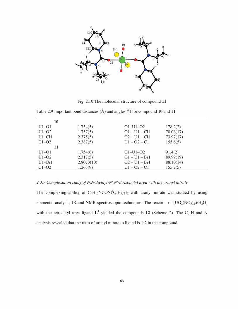
Fig. 2.10 The molecular structure of compound 11 Table 2.9 Important bond distances (Å) and angles (o) for compound 10 and 11
10 U1 O1 1.754(5) O1 U1 O2 178.2(2) U1 O2 1.757(5) O1 U1 Cl1 70.06(17) U1 Cl1 2.375(5) O2 U1 Cl1 73.97(17) C1 O2 2.387(5) U1 O2 C1 155.6(5)
11 U1 O1 1.754(6) O1 U1 O2 91.4(2) U1 O2 2.317(5) O1 U1 Br1 89.99(19) U1 Br1 2.8073(10) O2 U1 Br1 88.10(14) C1 O2 1.263(9) U1 O2 C1 155.2(5)
2.3.7 Complexation study of N,N-diethyl-N ,N -di-isobutyl urea with the uranyl nitrate
The complexing ability of C4H10NCON(iC4H9)2}2 with uranyl nitrate was studied by using
elemental analysis, IR and NMR spectroscopic techniques. The reaction of [UO2(NO3)2.6H2O]
with the tetraalkyl urea ligand L1 yielded the compounds 12 (Scheme 2). The C, H and N
analysis revealed that the ratio of uranyl nitrate to ligand is 1:2 in the compound.
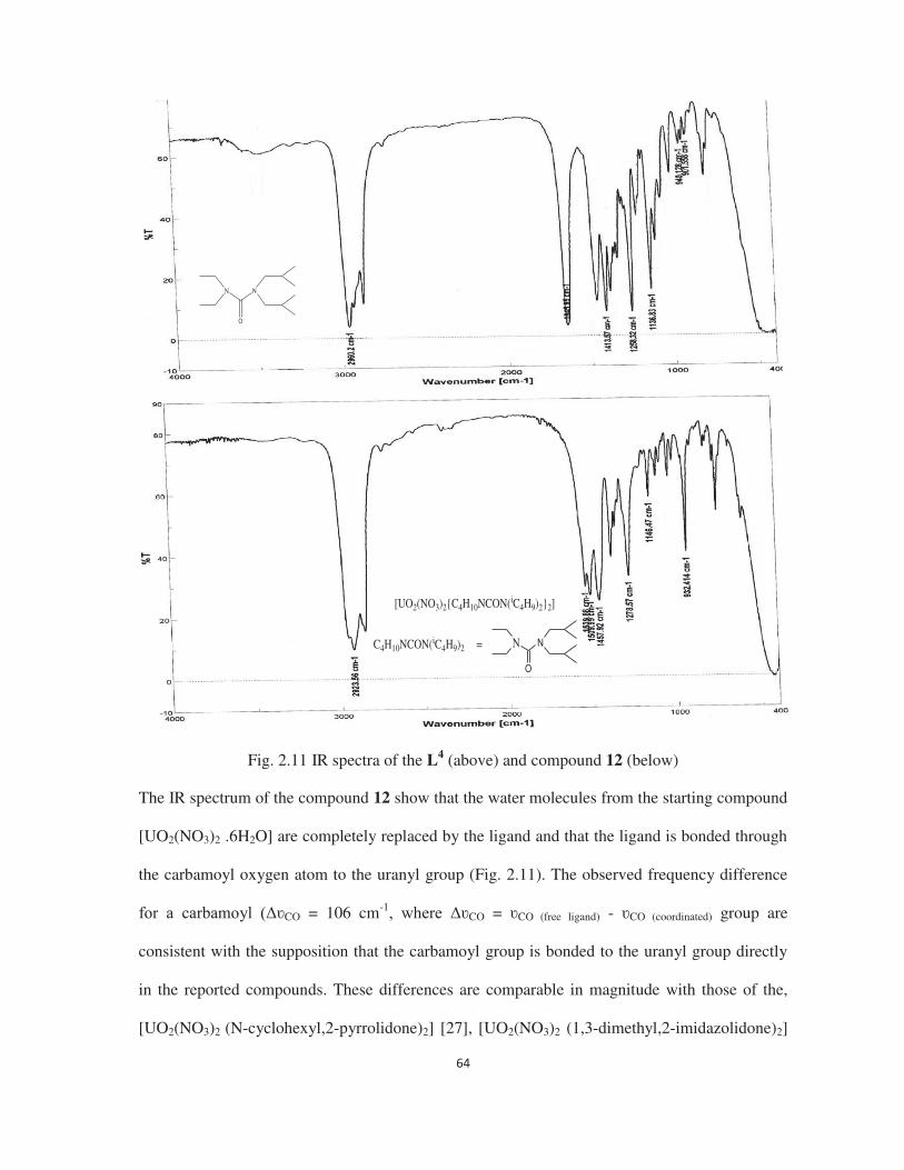
Fig. 2.11 IR spectra of the L4 (above) and compound 12 (below) The IR spectrum of the compound 12 show that the water molecules from the starting compound
[UO2(NO3)2 .6H2O] are completely replaced by the ligand and that the ligand is bonded through
the carbamoyl oxygen atom to the uranyl group (Fig. 2.11). The observed frequency difference
CO = 106 cm-1CO = CO (free ligand) - CO (coordinated) group are
consistent with the supposition that the carbamoyl group is bonded to the uranyl group directly
in the reported compounds. These differences are comparable in magnitude with those of the,
[UO2(NO3)2 (N-cyclohexyl,2-pyrrolidone)2] [27], [UO2(NO3)2 (1,3-dimethyl,2-imidazolidone)2]
N N
O
[UO2(NO3)2{C4H10NCON(iC4H9)2}2]
C4H10NCON(iC4H9)2 = N N
O

[28], [UO2(NO3)2{iC3H7CON(iC4H9)2}2] [50] and [UO2(NO3)2(
iC3H7)2NCOCH2CON(iC3H7)2]
[51].
[UO2(NO3)2{C4H10NCON(iC4H9)2}2]
C4H10NCON(iC4H9)2 = NC
N
Oa
a
b
b
c
c
d
d
e
e
ee
Fig. 2.12 1H NMR spectrum of compound 12 Fig 2.12 showed the 1H NMR spectrum of complex 12. The broad multiplet at 3.53 ppm is
assigned to four CH2 protons (chemically equivalent due to C-N(diisobutyl) bond rotation at
room temperature) of the isobutyl groups of the ligand L4. Next, four CH2 protons (chemically
equivalent due to C-N(diethyl) bond rotation at room temperature) of the ethyl groups resonate
as the broad multiplet at 3.27 ppm. These two peaks are broad because of quadrapolar effect of
14N-atom adjacent to the CH2 group protons. These are the deshielded protons in the spectrum
because they are attached with electronegative N-atom via one carbon atom. The multiplet at
2.04 ppm is attributed to two CH protons (chemically equivalent due to C-N(diisobutyl) bond
rotation at room temperature) of the isobutyl groups. Next, the six CH3 protons of the ethyl
groups of the ligand resonate as a triplet at 1.25 ppm. The signal is split into a triplet, because
the methyl group protons are coupled with two adjacent CH2 protons. At 0.88 ppm, the doublet
is due to the twelve CH3 protons of the isobutyl groups of the ligand. Here, the signal is split into
a doublet, because the methyl group protons are coupled with one adjacent CH protons. The
spectrum also shows that all the protons of the ligand are deshielded with respect to the free

ligand, indicating that the ligand is coordinated to the uranyl group in solution also. It is apparent
from the elemental analysis and IR spectral results that ligand is acting as monodentate ligand in
compounds 12 in the solid state to give similar structures to those observed in the compounds of
pyrrolidone [27,28] or other monodentate amides [19 21] with the uranyl nitrate. Interestingly,
the complex is air and moisture stable crystalline solids, having good solubility in CHCl3 and
CH2Cl2, and can be used as a starting compound for further reactions. The structure of 12 has
been determined by single crystal X-ray diffraction methods, which confirms the IR and
elemental analysis results.
2.3.8 Molecular structure of compound 12
The molecular structure of the compound 12 is shown in Fig. 2.13 and the selected interatomic
bond distances and angles for are given in Table 2.10. The structure contains a crystallographic
centre of symmetry with the uranium atom surrounded by eight oxygen atoms in a hexagonal bi-
pyramidal geometry. Two uranyl oxygen atoms occupy the axial positions. There are two
bidentate nitrate ligands in the hexagonal equatorial plane together with two carbamoyl oxygen
atoms from two monodentate tetraalkyl urea ligands. The U Oamide bond distances are 2.327(14)
Å (U1 O2) and 2.350(13) Å (U1 O7), which are comparable to the previously reported uranyl
nitrate complexes of the isobutyramide ligand [17].
Fig. 2.13 The molecular structure of compound 12

The observed U Oamide bond distances are shorter in length when compared to the corresponding
uranyl nitrate complexes with phosphine oxide ligands [UO2(NO3)2(Ph3PO)2] (2.327(14) and
2.350(13) Å vs. 2.359 (7) Å) [22]. The bond angles C1 O2 U1 and C14 O7 U1 are 168.8 (1)°
and 153.9 (1)°, respectively and further confirm the inverse relationship between the bond angles
(C O U) and bond distance (O U) in the amide uranyl system as described earlier [28].
However, such a short U Oamide bond distance and nearly linear bond angle 168.8(1)° is rare in
uranyl amide chemistry [17] and the presence of the short U ligand bond lengths and almost
linear bond angle indicates very strong bonding between the ligand and uranyl.
Table 2.10 Important bond distances (Å) and angles (o) for the compound 12 U1 O1 1.742(10) C1 O2 U1 168.8(14) U1 O2 2.350(13) C14 O7 U1 153.9(12) U1 O3 2.545(14) O1 U1 O6 179.6(5) U1 O4 2.532(12) O1 U1 O7 87.8(5) U1 O6 1.746 (9) O1 U1 O2 87.8(5) U1 O7 2.327(14) O6 U1 O7 92.4(5) U1 O8 2.534(14) O6 U1 O2 92.0(4) U1 O9 2.541(14)
2.3.9 Complexation study of N,N-diethyl-N ,N -di-isobutyl urea with the uranyl dihalides
The reaction of [UO2X2.nH2O] (X = Cl or Br) with the ligand L1 yielded compounds 13 and 14
(Scheme 2). Compounds 13 and 14 are soluble in most organic solvents and all are stable
towards moisture and air (in general the chloro and bromo compounds are moisture sensitive). C,
H and N analyses of 13 and 14 revealed that the ratio of ligand to uranyl halide is 2:1 in both
compounds. The IR spectra (Fig. 2.14) of 13 and 14 show that the water molecules from the
starting compounds [UO2X2.nH2O] are replaced completely by the ligand. The observed
frequency difference for the carbamoyl group CO = 123-124 cm-1CO CO (free ligand)
- CO (coordinated)) shows that the carbamoyl group is bonded to the uranyl group directly. This
difference is comparable/greater in magnitude than those observed in
[UO2X2{iC3H7CON(iC4H9)2}2] (X = Cl or Br) [17], [UO2(NO3)2{
iC3H7CON(iC4H9)2}2] [50]

UO2(NO3)2(N-cyclohexyl,2-pyrrolidone)2] [28], [UO2(NO3)2(1,3-dimethyl,2-imidazolidone)2]
[28] and [UO2(NO3)2(iC3H7)2NCOCH2CON(iC3H7)2] [51].
Fig. 2.14 IR spectra of L4 (above) and the compound 13 (below)
C4H10NCON(iC4H9)2 = N N
O
[UO2Cl2{C4H10NCON(iC4H9)2}2]
N N
O

[UO2Cl2{C4H10NCON(iC4H9)2}2]
C4H10NCON(iC4H9)2 = NC
N
Oa
a
b
b
c
c
d
d
e
e
ee
Fig. 2.15 1H NMR spectrum of compound 13
Fig 2.15 showed the 1H NMR spectrum of complex 13. The broad peak at 3.67 ppm is assigned
to four CH2 protons (chemically equivalent due to C-N(diisobutyl) bond rotation at room
temperature) of the isobutyl groups of the ligand L4. Next, at 3.43 ppm, the broad peak is due to
four CH2 protons (chemically equivalent due to C-N(diethyl) bond rotation at room temperature)
of the ethyl groups. These two peaks are broad because of quadrapolar effect of 14N-atom
adjacent to the CH2 group protons. These are the deshielded protons in the spectrum because
they are attached with electronegative N-atom via one carbon atom. The multiplet at 2.06 ppm is
attributed to two CH protons (chemically equivalent due to C-N(diisobutyl) bond rotation at
room temperature) of the isobutyl groups. The six CH3 protons of the ethyl groups of the ligand
resonate as a triplet at 1.29 ppm. The signal is split into a triplet, because the methyl group
protons are coupled with two adjacent CH2 protons. At 0.91 ppm, the doublet is due to the
twelve CH3 protons of two isobutyl groups of the ligand. The signal is split into a doublet,
because the methyl group protons are coupled with one adjacent CH protons. All the protons are
deshielded with respect to the free ligand indicating that the bonding between the ligand and
uranyl group persists in solution. It is apparent from the IR and NMR spectra that the ligand

bonds through the carbamoyl groups to the uranyl group. The structures of 13 and 14 have been
determined by the single crystal X-ray diffraction method, which confirms these spectral results.
2.3.10 Molecular structure of compounds 13 and 14
The structures of 13 and 14 are shown in Fig. 2.16 and 2.17, respectively, and selected bond
distances and angles are given in Table 2.11. Complex 13 and 14 [UO2X2·2L1 where X = Cl
and Br ] show a distorted octahedral geometry around the uranium centre. The two halide ions
and two ligands are trans to each other. The U Oamide bond distances are 2.264 (6) Å and 2.288
(6) Å in 13 and 14, respectively, which are very close to the reported uranyl chloride (2.315 (3)
Å) and uranyl bromide (2.281 (4) Å) isobutyramide complexes [17]. Again, these bond distances
are shorter when compared to the reported [UO2Cl2(Ph3PO)2] complexes [60(a)] (2.264 Å vs.
2.300 (8) Å) showing the strong bonding to uranyl compared to the phosphine oxide donor. The
C1 O2 U bond angles are 167.7 (6) and 167.3 (6) for complex 13 and 14, respectively, which
are in line with the previously reported isobutyramide complexes with uranyl halide (2.281
2.315(18) Å) [17]. From the abovementioned structural discussion it is evident that ligand L1
forms a stronger complex with uranyl dihalides when compared to that of phosphine oxide based
ligands, as well as having similar structural features as the isobutyramide ligands. Therefore, the
selectivity for uranyl has been explored as isobutyramide type ligands are known to be selective
for the uranyl over plutonium and thorium from nitric acid medium [13, 50].
Fig. 2.16 The molecular structure of compound 13

Fig. 2.17 The molecular structure of compound 14 Table 2.11 Important bond distances (Å) and angles (o) for compound 13 and 14
13 U1 O1 1.762(6) C1 O2 U 167.7(6) U1 O2 2.264(6) O1 U O2 90.9(3) U1 Cl 2.663(3) O1 U Cl 88.6(3)
14 U1 O1 1.763(6) C1 O2 U 167.3(6) U1 O2 2.288(6) O1 U O2 90.2(3) U1 Br 2.8118(12) O1 U Br 89.2(2)
2.3.11 Comparison of ligand (L1-L4) donor strength in the uranyl complexes by comparing the
uranyl stretching frequency in the vibrational spectroscopy
1) of the linear uranyl group (U=Oaxial) is infra-red
inactive but Raman active. On the other hand, the reverse response is true for the asymmetric
3) of the same. This is due to the mutual exclusion principle of any
molecule or group having centre of inversion akin to linear uranyl (O=U=O2+) group.
The central uranium atom in the uranyl complexes generally adopts six to eight coordination
number with two linear oxygen atoms at the axial positions and four to six donor atoms in the
equatorial positions. It is well proven that, the donor atoms at the equatorial positions form more
strong bond with the uranium atom, the bond strength of the equatorial bonds is increased which
ultimately weakens the axial bonds and decreases the axial bond strength and vice versa. Thus
the donor ability of the ligands can be compared indirectly by comparing either the symmetric

1) of the axial uranyl bond in the Raman spectroscopy or the asymmetric
3) of the same in the infra-red spectroscopy. Here, we have recorded the
infra-red spectra o -diketonate complexes with four newly
synthesized ligands (L1-L4) and systematically tabulated in table 2.12.
Table 2.12 Comparison of IR frequencies of uranyl complexes
Complex C=Ostr frequency of free ligands (cm-1)
C=Ostr frequency of the bonded ligands (cm-1)
CO
(cm-1) U=Oasy frequency of the axial bond (cm-1)
[UO2(NO3)2{C5H10NCON(CH3)2}2] (1)
1653 1525 128 925
[UO2(NO3)2{C5H10NCON(C2H5)2}2] (2)
1622 1521 101 928
[UO2(NO3)2{C5H10NCON(iC3H7)2}2
] (3) 1651 1522 129 931
[UO2(C6H5COCHCOC6H5)2{C5H10
NCON(CH3)2}] (4) 1653 1542 111 907
[UO2(C6H5COCHCOC6H5)2{C5H10
NCON(C2H5)2}] (5) 1622 1542 80 905
[UO2(C6H5COCHCOC6H5)2{C5H10
NCON(iC3H7)2}] (6) 1651 1541 110 901
[UO2(C4H3SCOCHCOCF3)2{C5H10
NCON(CH3)2}] (7) 1653 1540 113 919
[UO2(C4H3SCOCHCOCF3)2{C5H10
NCON(C2H5)2}] (8) 1622 1539 83 922
[UO2(C4H3SCOCHCOCF3)2{C5H10
NCON(iC3H7)2}] (9) 1651 1540 111 922
[UO2Cl2{C5H10NCON(iC3H7)2}2] (10)
1651 1524 127 924
[UO2Br2{C5H10NCON(iC3H7)2}2] (11)
1651 1525 126 928
[UO2(NO3)2{C4H10NCON(iC4H9)2}2
] (12) 1646 1540 106 932
[UO2Cl2{C4H10NCON(iC4H9)2}2] (13)
1646 1523 123 925
[UO2Br2{C4H10NCON(iC4H9)2}2] (14)
1646 1522 124 927
From the table it can be observed that, in the [UO2(NO3)2.6H2O],
[UO2(C6H5COCHCOC6H5)2.2H2O], [UO2(C4H3SCOCHCOCF3)2.2H2O] complexes of
piperidine carboxamide ligands, the order of U=Oasy frequency is (3) > (2) > (1), (4) > (5) > (6)
and (9) = (8) > (7) respectively, which signifies that donor strength order of the ligands is L1 >
L2 > L3 in these complexes. This can be explained on the basis of decreasing basicity of the
ligands with increasing bulkiness of the ligands since the electronic property is almost same for
all the ligands. Similarly, In the [UO2(NO3)2.6H2O] and uranyl dihalide complexes of tetraalkyl
urea complexes the order of U=Oasy frequency is (12) > (13) ~ (14), which signifies that donor
strength order of L4 ligand in these complexes is (13) ~ (14) > (12). This is as expected because
the ligands encounter lesser steric effect in the halide complexes (six coordinated) with respect
to the nitrate complexes (eight coordinated) when approaches toward the metal centre, hence the
basicity is decreased in the nitrate complexes with respect to the halide complexes where the
ligands get more chance to approach the metal centre closely.
2.3.12. Precipitation studies of U(VI) ions from the bulk of Th(IV) and Ln(III) ions
The EDXRF spectra of the initial solution, supernatant and precipitate are shown in Fig. 2.18.
From the EDXRF spectrum, it is clear that there is only thorium present in the supernatant
solution and no trace of uranium was detected. Similarly, the EDXRF spectrum of the precipitate
shows only the presence uranium and no thorium. This study clearly revealed that the ligand is
selective for uranium and precipitate uranium from the solution containing a large excess of
thorium. The observed UV-Vis spectra of the initial and supernatant (Fig. 2.19) solutions also
supported the abovementioned observations. This experiment was repeated with a nitric acid
solution containing a mixture of uranyl, thorium and lanthanide (La3+, Sm3+ and Eu3+) ions to
ascertain the selectivity of the ligand towards uranyl ions over lanthanide and thorium ions. This

process also selectively deposited uranyl ions from the solution containing a bulk of thorium and
lanthanide ions as confirmed by EDXRF analysis (Fig. 2.20).
Similarly, in the supernatant no traces of uranium are observed in the EDXRF spectra (Fig.
2.20(a)) and all the lanthanides are present in the supernatant as observed in the EDXRF spectra
(Fig. 2.20(b)). From the above mentioned precipitation studies, it is observed that the ligand L1
can be used to selectively precipitate uranium from a solution containing thorium and
lanthanides from a nitric acid medium.
Fig. 2.18 The EDXRF spectra of the precipitate, supernatant and initial solution.
Fig. 2.19 The UV-Vis spectra of the initial and supernatant solutions

Fig. 2.20 (a) EDXRF spectra of the initial and supernatant solutions containing a Rh source and (b) EDXRF spectra of the initial and supernatant solution containing a Ge secondary target. 2.3.13. Extraction studies of U(VI) and Pu(IV) with C4H10NCON(C8H17)2 from nitric acid
The extraction studies were carried out by using the ligand C4H10NCON(C8H17)2 in dodecane
with the U(VI) and Pu(IV) ions in tracer level (using the 233U and 239Pu) from nitric acid
medium to assess the feasibility of using this ligand for the extraction purpose. Distribution
ratios (D) for U(VI) and Pu(IV) as a function of nitric acid concentrations (Fig.2.21) shows
clearly that observed distribution values are very low for both the U(VI) and Pu(IV) under the
acid concentrations of 1 10 M. However, under any acid concentration, the D for Pu(IV) is
much smaller than those of U(VI). This observation is completely contradictory to the data
reported for any of the extractants (either mono-functional or bi- functional) with that of Pu(IV)
(DPu(IV) >> DU(VI) always) [6-9] and agrees well with the result reported for the isobutyramide
based extractant [13-15]. This shows clearly that the uranium can be selectively separated from
plutonium or vice versa.

Fig. 2.21 Variation of distribution ratio for U(VI) and Pu(IV) with HNO3
2.3.14 Theoretical studies
The molecular level insights on the origin of selectivity of ligand L1 obtained in experiments can
be understood through density functional theory (DFT) based calculations. The geometries
predicted using the BP86 functional of compound 1 are fairly comparable with that of the
experimental data (Fig. 2.22). The ligand L1 binds strongly to uranyl U OL1, which in turn
weakens the axial U=Oyl bond by a similar margin. The computed bond angle of one U O=CL1
is akin to the experimental value, whereas the second bond angle is overestimated by 10 degrees.
The U ONO3 bond lengths are very accurately predicted within 0.01 Å to the X-ray data.
Furthermore, the computed bond angle (O=U=O) is also predicted to be linear, which
corroborates well with our experimental data. Upon complexation, the computed C=O
vibrational frequency of the bound species is red-shifted by ~100 cm 1, which is in line with our
experimental IR shifts and thus consistent with the elongation of the C=O bond distance (by
~0.02 Å) upon metal binding. Similarly, the U=O stretching frequency is also red-shifted by 30
cm 1 with respect to the bare uranyl nitrate hexahydrate (the experimentally observed shift is 14
cm 1). However, for Th(IV) bound complexes we find this shift is somewhat larger (no complex
formation occur experimentally), which is reflected in the elongated C=O bond length (by 0.02
Å) when compared to the uranium species. To ascertain the thermodynamic favourability of
these metal complexation reactions, we have calculated the formation free energies for the two

actinide ions using the M06-2X functional and BP86 optimized structures (Table 2.13).
Furthermore, the computed separation factor of uranyl in the presence of Th(IV) with L1 is very
high (110.4), which explains the unavailability of uranium in the supernatant and the absence of
thorium precipitation in the observed complexation processes. The large negative entropy for
Th(IV) originates from the overcrowded coordination centre when compared to the uranium
centre. Energy decomposition analysis (EDA) was performed (TZP/B3LYP/ZORA) on the metal
bound species to segregate the contributions of the various repulsive-attractive energy
components from the total bonding energy. For both metal ions, favourable electrostatic
interactions (~60%) contributed more to the total bonding energy. However, orbital interactions,
originating from the orbital relaxation of fragmented orbitals from the excited to ground state
also seems to contribute an extent of ~40% with respect to the total attractive term. It can be
noted that for both actinides, the positive Pauli repulsion term cancels the attractive electrostatic
interaction resulting in a positive value for the total steric repulsion. The prominent electrostatic
interactions between L1 and the actinides arise from the net charge transfer from the ligand to the
metal (derived from Mülliken population analysis). When compared to Th bound complex, the
uranyl complex has higher ligand to uranyl charge transfer. Furthermore, the net charge on
nitrates is involved in neutralizing the tetra-positive thorium than uranium, which implies the
relatively stronger binding with nitrate for the former. A detailed molecular orbital analysis was
carried out through natural localized molecular orbital (NLMO) theory to understand the binding
behavior of L1 with the metal ions. We found that the lone pair of the carbonyl oxygen in the
ligand is strongly delocalized (~3%) in the U OL1 antibond wherein the f-orbital of U majorly
acts as the acceptor orbital (69%). However, for Th, both the d and f orbitals equally participate
as the acceptor orbital for the delocalization of the carbonyl lone pair electrons. The sigma bond
between U/Th and the carbonyl oxygen is formed by the involvement of ~8% uranium and
~92% carbonyl oxygen orbitals. In this bond, the metal orbitals have a contribution of roughly

11% s, 54% d and 34% f orbitals. In line with the NLMO results, the natural orbital population
shows lesser involvement of the 7s and 7p orbitals in ligand binding. In addition, between 6d
and 5f, greater participation of the 5f orbital is noticed for uranium, whereas the 6d orbital
contributes more for thorium.
Fig. 2.22 The optimized structures (bond length in Å) of the actinide amide complexes. The values in the brackets correspond to the X-ray structure data. Table 2.13 The computed formation free energies and separation factor (S.F) using the M06-2X
functional (kcal mol 1)
a S.F (UO2/Th) UO2 6.47 2.79 110.4 Th 3.68 a G = G(UO2) G(Th).
2.4 Conclusions
The piperidine based amide ligands form 2:1 complexes with uranyl chloride, bromide and
-diketonates). The uranyl chloro and bromo
compounds are air and moisture stable and soluble in common organic solvents. The structures
of the chloro and bromo complexes show that the uranium(VI) ion is surrounded by two
halogen, two uranyl and two amido oxygen atoms in an octahedral geometry. The structures of

-diketonates) complexes show hexagonal bipyramidal and
pentagonal bipyramidal geometries respectively around the uranium(VI) ion.
The selective recognition of one useful metal ion in the presence of other interfering ions is one
of the most important research areas in the field of nuclear waste management processes. In this
study, we have carried out combined experimental and electronic structure calculations to
understand the origin of selectivity of a structurally known but unexplored extractant, N,N-
diethyl-N ,N -diisobutyl urea. We have characterized the structure of the uranyl bound
complexes through X-ray crystallography. Furthermore, the extractant selectivity towards uranyl
ion in the presence of Th(IV) and other interfering lanthanide ions in a nitric acid medium was
explored using several spectroscopic techniques. Based on electronic structure calculations at the
DFT level of theory, we found that the preferential binding of uranyl ion to Th(IV) is modulated
by both steric and electronic factors. Thus, the combined experimental and theoretical studies are
extremely useful to understand the selectivity of heavy metal ions, which is relevant to the
backend of the nuclear fuel cycle.
2.5 References
1. L.R. Morss, N.M. Edelstein, J. Fuger, J.J. Katz, The Chemistry of the Actinide and
Transactinide Elements, Springer , Netherlands (2006).
2. C.J. Burns, M.P. Neu, H. Boukhalfa, K.E. Gutowshi, N.J. Bridges, R.D. Rogers, J.A.
McCleverty, T.J. Meyer, G.F.R. Parkin, (Eds.), Comprehensive Coordination Chemistry II ,
Elsevier, Oxford, 3 (2004) 189.
3. M. Ephritikhine, Dalton Trans. (2006) 2501.
4. S. Kant, Int. J. Nucl. Ener. Sci. Tech. 1 (2005) 204.
5. R. K. Sinha, A. Kakodkar, Nucl. Eng. Des. 236 (2006) 683.
6. C. Musikas, Inorg. Chim. Acta 140 (1987) 197.
7. C. Madic, M.J. Hudson, High Level Liquid Waste Partitioning by Means of Completely
Incinerable Extractants , European Commission, Nuclear Science and Technology,
EU18038EN, Luxembourg (1998).
8. L. Nigond, C. Musikas, C.C. Cullerdier, Solv. Extr. Ion Exch. 12 (1994) 261.

9. L. Nigond, N. Condamines, P.Y. Cordier, J. Livert, C. Madic, C.C. Cullerdier, C. Musikas,
Sep. Sci. Technol. 30 (1995) 2075.
10. Y. Sasaki, Y. Sugo, S. Suzuki, S. Tachimori, Solv. Extr. Ion Exch. 19 (2001) 91.
11. S. Tachimori, Y. Sasaki, S. Suzuki, Solv. Extr. Ion Exch. 20 (2002) 687.
12. Z. Zhu, Y. Sasaki, H. Suzuki, S. Suzuki, T. Kimura, Anal. Chim. Acta 527 (2004) 163.
13. P.N. Pathak, R. Veeraraghavan, D.R. Prabhu, G.R. Mahajan, V.K. Manchanda, Sep. Sci.
Technol. 34 (1999) 2601.
14. P.N. Pathak, L.B. Kumbhare, V.K. Manchanda, Solv. Extr. Ion Exch. 19 (2001) 105.
15. V.K. Manchanda, P.N. Pathak, Sep. Purif. Technol. 35 (2004) 85.
16. T.R. Varga, M. Sato, Zs. Fazekas, M. Harada, Y. Ikeda, H. Tomiyasu, Inorg. Chem.
Commun. 3 (2000) 637.
17. S. Kannan, C.L. Barnes, P.B. Duval, Chem. Commun. (2005) 5997.
18. S. Kannan, C.L. Barnes, P.B. Duval, Inorg. Chem. 44 (2005) 9137.
19. J.M. Gil, F.J.M. Gil, A. Perales, J. Fayos, M.M. Ripoll, Acta Crystallogr. Sect. C 39 (1983)
44.
20. P. Charpin, M. Lance, M. Nierlich, D. Vigner, Acta Crystallogr. Sect. C 43 (1987) 442.
21. P. Charpin, M. Lance, M. Nierlich, D. Vigner, Acta Crystallogr. Sect. C 42 (1986) 560.
22. N.W. Alcock, M.M. Roberts, D. Brown, J. Chem. Soc., Dalton Trans. (1982) 25.
23. B. Kanellakopulos, E. Dornberger, R. Maier, B. Nuber, H.G. Stammler, M.L. Ziegler, Z. An
org. Allg. Chem. 619 (1993) 593.
24. J.H. Burns, Inorg. Chem. 20 (1981) 3868.
25. A.R. de Aquino, G. Bombieri, P.C. Isolani, G. Vicentini, J.Z. Schpector, Inorg. Chim. Acta
306 (2000) 101.
26. G. Agostini, G. Giacometti, D.A. Clemente, M. Vicentini, Inorg. Chim. Acta 62 (1982) 237.
27. T.R. Varga, A.C. Benyei, Z. Fazekas, H. Tomiyasu, Y. Ikeda, Inorg. Chim. Acta 342 (2003)
291.
28. N. Koshino, M. Harada, M. Nogami, Y. Morita, T. Kikuchi, Y. Ikeda, Inorg. Chim. Acta 358
(2005) 1857.
29. K.M. Abubacker, N.S.K. Prasad, J. Inorg. Nucl. Chem. 16 (1961) 296.
30. A.D. Becke, Phys. Rev. A 38 (1988) 3098.
31. J.P. Perdew, Phys. Rev. B 33 (1986) 8822.
32. A. Schäfer, C. Huber, R. Ahlrichs, J. Chem. Phys. 100 (1994) 5829.
33. F. Weigend, R. Ahlrichs, Phys. Chem. Chem. Phys. 7 (2005) 3297.

34. A. Schäfer, H. Horn, R. Ahlrichs, J. Chem. Phys. 97 (1992) 2571.
35. J.L. Whitten, J. Chem. Phys. 58 (1973) 4496.
36. E.J. Baerends, D.E. Ellis, P. Ros, Chem. Phys. 2 (1973) 41.
37. B.I. Dunlap, J.W.D. Connolly, J.R. Sabin, J. Chem. Phys. 71 (1979) 3396.
38. C. Van Alsenoy, J. Comp. Chem. 9 (1988) 620.
39. K. Eichkorn, O. Treutler, H. Öhm, M. Häser, R. Ahlrichs, Chem. Phys. Letters. 40 (1995)
283.
40. Y. Zhao, D.G. Truhlar, Acc. Chem. Res. 41 (2008) 157.
41. J.P. Austin, N.A. Burton, I.H. Hillier, M. Sundararajan, M.A. Vincent, Phys. Chem. Chem.
Phys. 11 (2009) 1143.
42. A. Klamt, G. Schüürmann, J. Chem. Soc., Perkin Trans. 2 (1993) 799.
43. R. S. Mülliken, J. Chem. Phys. 23 (1955) 1833.
44. R. Ahlrichs, M. Bar, H. P. Baron, R. Bauernschmitt, S. Bocker, M. Ehrig, K. Eichkorn, S.
Elliot, F. Furche, F. Haase, M.Haser, H. Horn, C. Huber, U. Huniar, M. Kattannek, C.
Kolmel, M. Koolwitz, K. May, C. Ochsenfeld, H. Ohm, A. Schafer, U. Schneider, O.
Treutler, M. von Arnim, F. Weigend, P. Weis, H. Weiss, TURBOMOLE V6.3.1 2011, a
development of University of Karlsruhe and Forschungszentrum Karlsruhe GmbH, 1989-
2007, TURBOMOLE GmbH, since 2007; available from http://www.turbomole.com.
45. K. Morokuma, J. Chem. Phys. 55 (1971) 1236.
46. T. Ziegler, A. Rauk, Theor. Chim. Acta 46 (1977) 1.
47. G. te Velve, F.M. Bickelhaupt, E.J. Baerends, S.J.A. Gisbergen, van C.F. Guerra, J.G.
Snijders, T. Ziegler, J. Comput. Chem. 22 (2001) 931.
48. C. Fonseca Guerra, J.G. Snijders, G. teVelde, E. Baerends, J. Theor. Chem. Acc. 99 (1998)
391.
49. E.D. Glendening, C.R. Landis, F. Weinhold, J. comp.chem. 34 (2013) 1429.
50. S. Kannan, S.B. Deb, J.S. Gamare, M.G.B. Drew, Polyhedron 27 (2008) 2557.
51. G.J. Lumetta, B.K. McNamara, B.M. Rapko, R.L. Sell, R.D. Rogers, G. Broker, J.E.
Hutchison, Inorg. Chim. Acta 309 (2000) 103.
52. L.M. Zhu, B.L. Li, Z.B. Cao, Y. Zhang, Chin. J. Struct. Chem. 5 (2003) 521.
53. O. Clements, B.M. Rapko, B.P. Hay, Coord. Chem. Rev. 170 (1998) 203.
54. K.W. Bagnall, F. Benetollo, E. Forsellini, G. Bombieri, Polyhedron 11 (1992) 1765.
55. S. Alagar, K. Rajagopal, R.V. Krishnakumar, M. Subha Nandhini, S. Kannan, S. Natarajan,
Acta Crystallogr. E 59 (2003) m1-m3.

56. S. Kannan, M.R.A. Pillai, V. Venugopal, P.A. Droege, C.L. Barnes, Polyhedron 15 (1996)
97.
57. S. Kannan, M.R.A. Pillai, V. Venugopal, P.A. Droege, C.L. Barnes, E.O. chlemper,
Polyhedron 15 (1996) 465.
58. S. Kannan, A. Usman, H.K. Fun, Polyhedron 21 (2002) 2403.
59. S. Kannan, M.R.A. Pillai, V. Venugopal, P.A. Droege, C.L. Barnes, Inorg. Chim. Acta 254
(1997) 113.
60. (a) G. Bombieri, E. Forsellini, J.P. Day, W.I. Azeez, Dalton Trans. (1978) 677. (b) S.B.
Akona, J. Fawcett, J.H. Holloway, D.R. Russel, I. Leban, Acta Crystallogr. C 47 (1991) 45.


3.1 Introduction
Different groups around the world have developed many extractants which showed good
extraction properties for trivalent actinides. These processes are always associated with the co-
extraction of trivalent lanthanides present at large proportion in Purex raffinate [1-4]. This is
because of the chemical similarity of trivalent actinides with trivalent lanthanides. Several
bifunctional neutral extractants with various combinations of functional groups were synthesized
and tested for this purpose, sometimes known as Actinide Partitioning . The TRUEX process,
employed octyl(phenyl)-N,N-diisobutylcarbamoyl methyl phosphine oxide (CMPO) as
extractant (similarly carbamoyl methyl phosphonates (CMP)), in which C=O and P=O
functional groups are involved in bonding and it acts as a bidentate chelating ligand [5-6]. In
DIAMEX process, substituted malonamides such as, dimethyl-dibutyl-tetradecylmalonamide
(DMDBTDMA) and N,N'-dimethyl-N,N'-dioctylhexylethoxymalonamide (DMDOHEMA) were
used as extractants which contain two C=O functional groups in combination and these also act
as bidentate chelating ligands [7-9]. Apart from these, some acidic organophosphorus extractants
like di-isodecyl phosphoric acid (DIDPA) [10], Di-(2-ethylhexyl)phosphoric acid (DEHPA or
HDEHP) [11-12], di-nonyl phenyl phosphoric acid (DNPPA) [13-14], di-octyl phenyl
phosphoric acid (DOPPA) [15-16], were used to separate uranium or trivalent actinides from
lower acidic solution. All these extractants are composed of PO(OH) functional groups and
bonded with the metal ions through both P=O and P-O groups. Apart from these conventional
extractants, so many other bifunctional neutral extractants were synthesized and studied for the
coordination and extraction properties in the laboratory scale. For example, carbamoyl methyl
sulfoxide (CMSO) (combination of C=O and S=O functional groups) [17- -sulfoxo
-N-oxo phosphine oxide
(combination of P=O and N-O functional groups) [19], bis(diphenylphosphino)methane dioxide
(combination of two P=O functional groups) have also been studied previously [20]. Solid state

structural analyses of the coordination complexes of these ligands with actinides and lanthanides
reveal that, they can act either as bidentate chelating, bidentate bridging (in dimeric or polymeric
structure) or monodentate ligands. The mode of bonding for these ligands are controlled both by
steric as well as electronic properties. However, in this series, the study of the coordination and
extraction chemistry of bifunctional ligands containing the NO CO groups is very limited [21],
though the extraction and coordination chemistry of monofunctional N-oxide and amides are
well established [22-26]. 4-(5-nonyl) pyridine oxide and trioctylamine oxide were tested for the
extraction of uranium from different mineral acid solutions, which showed that the former acts
as a better extractant in dilute HNO3 and HCl media than that of TBP [22]. Structural studies on
the complexes of a mixed hydroxy N-oxo amide ligand, 2-hydroxy-2-(1-oxy-pyridin-2-yl)-N,N-
diphenylacetamide with La(NO3)3 showed that the ligand bonds through the hydroxyl and amide
O-atoms to metal centre and leaving the N-oxide group free (uncoordinated) [27]. In another
report, the N,N-di(pyridine N-oxide-2-yl)pyridine-2,6-dicarboxamide ligand forms 1:1 complex
with lanthanide nitrate and is bonded through both the N-oxide groups as bidentate chelating
mode, while both the amidic O atoms of the ligand are free [28]. These studies promoted us to
synthesize NO CO based ligands, N-oxo picolinamides and study their complex chemistry with
uranyl nitrate and lanthanide (La, Sm, Eu) nitrates to see the mode of bonding in solid state and
their potential for the separation of actinides.
3.2 Experimental 3.2.1 Synthesis of N-oxo pyridine 2-carboxamide ligands 3.2.1.1 Synthesis of C5H4NOCON(iC3H7)2 ( L
1)
A solution of 3-chloroperoxybenzoic acid (1.9 g, 70%, 11mmol) in dichloromethane (20 ml) was
added drop wise to a solution of N,N-diisopropyl 2-pyridine carboxamide (2.06 g, 10mmol) in
dichloromethane (15 ml) at 5 10 °C. After 16 h at room temperature, the reaction mixture was
cooled on an ice bath and sodium hydroxide (2 M solution in water, 14.5 ml, 29 mmol) was

added into it. After 15 min, the reaction mixture was diluted with chloroform (25 ml). The
organic layer was separated and washed with 2M NaOH solution, dried over sodium
carbonateand evaporated under reduced pressure. The residue was purified bysilica gel column
chromatography to give the final product (86% yield). Dichloromethane solution on slow
evaporation yielded colourless plate type crystals which was analysed by single crystal XRD
method. Anal.Calcd.for C12H18N2O2: C, 64.86; H, 8.11; N, 12.61. Found: C, 64.22; H, 8.42; N,
12.26. IR (ATR, /cm-1): 1628(s) (C=O), 1249(s) (N-O).1H NMR (25°C, 300 MHz, CDCl3) 8.25
(s, 1H), 7.36 (s, 3H), 3.56(t, J= 6.3 Hz, 2H), 1.55 (d, J= 6.6 Hz, 6H), 1.36 (s, 6H).
3.2.1.2 Synthesis of C5H4NOCON(iC4H9)2 (L2)
This was prepared similar to that of L1 by taking N,N-diisobutyl 2-pyridine carboxamide instead
of N,N-diisopropyl 2-pyridine carboxamide. Yield: 82%. Anal.Calcd.for C14H22N2O2: C, 67.20;
H, 8.80; N, 11.20. Found: C, 67.12; H, 8.98; N, 11.01. IR spectrum (ATR, /cm-1): 1639(s)
(C=O), 1207(s) (N-O).1H NMR (25°C,500 MHz, CDCl3) 8.06 (d, J= 3.0 Hz, 1H), 7.25 (t, J= 4.5
Hz, 1H), 7.21 (d, J= 3.0 Hz, 2H), 3.78 (dd, J= 13.25 Hz, J= 8.5 Hz, 1H), 3.07 (dd, J= 14.25 Hz,
J= 5.5 Hz, 1H), 2.77 (q, J= 5.5Hz, 1H), 2.68 (t, J= 10.0 Hz, 1H), 2.07 (t, J= 7.0 Hz, 1H), 1.76 (s,
1H), 0.92 (d, J= 6.5 Hz, 6H), 0.72 (d, J= 6.5 Hz, 3H), 0.62 (d, J= 6.0 Hz, 3H).
3.2.1.3 Synthesis of C5H4NOCONH(tC4H9) (L3)
This was prepared similar to that of L1 by taking N-tbutyl 2-pyridine carboxamide instead of
N,N-diisopropyl 2-pyridine carboxamide. Yield: 81%. Anal.Calcd.for C10H14N2O2: C, 61.86; H,
7.22; N, 14.43. Found: C, 61.67; H, 7.69; N, 14.31. IR (ATR, /cm-1): 1669(s) (C=O), 1227(s)
(N-O). 1H NMR (25°C,300 MHz, CDCl3) 11.2 (s, 1H),8.39 (d, J= 7.5 Hz, 1H), 8.22 (d, J= 6.0
Hz, 1H), 7.45 (t, J= 7.5 Hz, 1H), 7.37 (d, J= 6.0Hz, 1H), 1.45 (s, 9H).

3.2.2 Synthesis of uranyl complexes of N-oxo pyridine 2-carboxamide ligands 3.2.2.1 Synthesis of [UO2(NO3)2{C5H4NOCON(iC3H7)2}] (1)
To a solution of L1 (280 mg, 1.26mmol) in CHCl3 (20 mL), solid [UO2(NO3)2·6H2O] (318 mg,
0.63mmol) was added and stirred for one hour at room temperature and then at reflux condition
for 3-4 hours. This produces yellow precipitate at the bottom of the round bottom flask. It was
filtered, washed with CHCl3 and then dissolved in acetonitrile.The solution on slow evaporation
yielded yellow crystalline solid, which was filtered, washed with hexane, dried and analysed by
single crystal XRD method.Yield:91%. 1H NMR (25°C, 300 MHz,CD3COCD3
5.7 Hz, 1H], 8.06 [s, 1H], 7.97 [t, J= 6.3 Hz, 1H], 7.85 [t, J= 6.3 Hz, 1H], 3.93 [sep, J= 6.9 Hz,
1H], 1.70 [d, J= 6.6 Hz, 3H], 1.46 [d, J= 6.6 Hz, 3H], 1.39 [d, J= 6.6 Hz, 3H], 1.35 [d, J= 6.6 Hz,
3H]. IR (cm 1 =O), 1215(s) (N-O),924(s) (U=O). Analysis Calcd for
C12H18N4O10U: C, 23.38; H, 2.92; N, 9.09. Found: C, 23.52; H, 3.06; N, 9.26.
3.2.2.2 Synthesis of [UO2(NO3)2{C5H4NOCON(iC4H9)2}] (2)
This was prepared similarly to 1 by taking L2 (343 mg, 1.37 mmol) and [UO2(NO3)2·6H2O] (341
mg, 0.68mmol) in 87% yield. 1H NMR (25°C, 300 MHz,CD3COCD3
1H], 7.94 [s, 1H], 7.87 [d, J= 6.9 Hz, 1H], 7.81 [t, J= 6.6 Hz, 1H], 3.32 [br, 2H], 2.23[m, 2H],
0.99 [d, J= 6.6 Hz, 6H], 0.82 [d, J= 6.6 Hz, 6H]. IR (cm 1 =O), 1219(s) (N-
O),931(s) (U=O). Analysis Calcd for C14H22N4O10U: C, 26.09; H, 3.42; N, 8.70. Found: C,
26.21; H, 3.52; N, 8.47.
3.2.2.3 Synthesis of [UO2(NO3)2{C5H4NOCONH(tC4H9)}] (3)
This was prepared similarly to 1 by taking L3 (249 mg, 1.28 mmol) and [UO2(NO3)2·6H2O] (322
mg, 0.64mmol) in 86% yield. The acetonitrile solution on slow evaporation yielded yellow
crystalline solid, which was filtered, washed with hexane, dried and analysed by single crystal
XRD method. 1H NMR (25°C, 300 MHz,CD3COCD3

J= 4.8 Hz, 2H], 1.52 [s, 9H]. IR (cm 1 =O), 1205(s) (N-O), 930(s) (U=O).
Analysis Calcd for C10H14N4O10U: C, 20.41; H, 2.38; N, 9.52. Found: C, 20.21; H, 2.47; N, 9.46.
3.2.3 Synthesis of lanthanide complexes of N-oxo pyridine 2-carboxamide ligands
3.2.3.1 Lanthanum complexes
3.2.3.1.1 Synthesis of [La(NO3)3(H2O){C5H4NOCON(iC3H7)2}2] (4)
To a solution of L1 (411 mg, 1.85mmol) in CH2Cl2 (20 mL), solid [La(NO3)3·6H2O] (267 mg,
0.62mmol) was added and stirred at room temperature for one hour which produces white
precipitate at the bottom of the flask. It was filtered, washed with CHCl3dried. Yield: 89%. 1H
NMR (25°C, 300 MHz, CD3COCD3
3.43 [sep, J= 6.9 Hz, 1H], 1.47 [d, J= 6.6 Hz, 6H], 1.28 [d, J= 6.6 Hz, 6H]. IR (cm 1
1594(s) (C=O), 1220(s) (N-O). Analysis Calcd for C24H38N7O14La: C, 36.60; H, 4.83; N, 12.45.
Found: C, 36.49; H, 4.96; N, 12.56.
3.2.3.1.2 Synthesis of [La(NO3)3(H2O){C5H4NOCON(iC4H9)2}2] (5)
This was prepared by taking solid L2 (480 mg, 1.92mmol) and [La(NO3)3·6H2O] (277 mg,
0.64mmol) in CH2Cl2 and under reflux condition all solid [La(NO3)2·6H2O] slowly got
disappeared and white product was precipitated. Yield: 86%. IR (cm 1 =O),
1227(s) (N-O). Analysis Calcd for C28H46N7O14La: C, 39.86; H, 5.46; N, 11.63. Found: C,
39.67; H, 5.62; N, 11.79.
3.2.3.2 Samarium complexes
3.2.3.2.1 Synthesis of [Sm(NO3)2(H2O){C5H4NOCON(iC3H7)2}2] (6)
To a solution of L1 (400 mg, 1.80mmol) in CH2Cl2 (20 mL), solid [Sm(NO3)2·6H2O] (267 mg,
0.60mmol) was added and stirred at reflux condition which dissolves all solid [Sm(NO3)2·6H2O]
and clear solution was obtained. Then the solvent was evaporated, washed with CHCl3and
dried.Yield: 88%. 1H NMR (25°C, 300 MHz, CD3COCD3
[s, 1H], 1.45 [d, J= 4.8 Hz, 6H], 1.26 [s, 3H], 1.10 [d, J= 5.4 Hz, 3H]. IR (cm 1

(C=O), 1215(s) (N-O). Analysis Calcd for C24H38N7O14Sm: C, 36.07; H, 4.76; N, 12.28. Found:
C, 36.02; H, 4.85; N, 12.39.
3.2.3.2.2 Synthesis of [Sm(NO3)2(H2O){C5H4NOCON(iC4H9)2}2] (7)
This was prepared similarly to 6 by taking L2 (533 mg, 2.13mmol) and [Sm(NO3)2·6H2O] (316
mg, 0.71mmol) in 86% yield. IR (cm 1 =O), 1222(s) (N-O). Analysis Calcd for
C28H46N7O14Sm: C, 39.33; H, 5.38; N, 11.47. Found: C, 39.54; H, 5.42; N, 11.62.
3.2.3.3 Europium complexes
3.2.3.3.1 Synthesis of [Eu(NO3)3(H2O){C5H4NOCON(iC3H7)2}2] (8)
To a solution of L1 (433 mg, 1.95mmol) in CH2Cl2 (20 mL), solid [Eu(NO3)3·6H2O] (290 mg,
0.65mmol) was added and stirred at reflux condition for one hour which produces white
precipitate at the bottom of the flask. It was filtered, washed with CHCl3 and then dissolved in
dichloromethane. Dichloromethane solution on slow evaporation yielded white needle type
crystals which was analysed by single crystal XRD method.Yield: 91%. 1H NMR (25°C, 300
MHz,CD3COCD3
1H], 1.61 [d, J= 6.6 Hz, 3H], 1.53 [d, J= 6.6 Hz, 3H], 1.28 [d, J= 6.6 Hz, 3H], 1.09 [d, J= 6.9 Hz,
3H]. IR (cm 1 =O), 1219(s) (N-O). Analysis Calcd for C24H38N7O14Eu: C, 36.0;
H, 4.75; N, 12.25. Found: C, 36.08; H, 4.91; N, 12.38.
3.2.3.3.2 Synthesis of [Eu(NO3)3(H2O){C5H4NOCON(iC4H9)2}2] (9)
This was prepared by taking solid L2 (473 mg, 1.89mmol) and [Eu(NO3)3·6H2O] (281 mg,
0.63mmol) in CH2Cl2 and under reflux condition all solid [Eu(NO3)2·6H2O] slowly got
disappeared and white product was precipitated. Yield: 87%. IR (cm 1 =O),
1220(s) (N-O). AnalysisCalcd for C28H46N7O14Eu: C, 39.25; H, 5.37; N, 11.45. Found: C, 39.13;
H, 5.49; N, 11.61.

3.2.4 X-ray crystallography
Selected crystallographic data for the compounds L1, 3 and 8 are summarized in Table 3.1.
Table 3.1 Crystallographic data for compounds L1, 3 and 8
L1 3 8 Empirical formula C12H18N2O2 C10H14N4O10U C24H38N7O14Eu Formula weight 222.28 588.28 799.96 Crystal system Monoclinic Monoclinic triclinic Space group P 1 21/c 1 P 1 21/c 1 P -1 a (Å) 6.3595(4) 15.8049(4) 9.0106(5) b (Å) 7.4334(4) 10.4286(2) 12.8895(9) c(Å) 26.7575(14) 10.2359(3) 15.6708(7)
o) 90 90 107.570(5) o) 89.779(5) 98.514(2) 90.619(4) o) 90 90 90.415(5)
Volume (Å3) 1264.89(13) 1668.52(7) 1734.92(18) Z (formula unit) 4 4 2 Density (g/cm3) 1.167 2.342 1.532
-1) 0.647 27.962 13.562 Reflections collected/unique 2381/1639 3241/2152 6640/4510 Data/restrains/parameters 2381/0/149 3241/0/230 6640/0/424 Goodness of fit on F2 1.105 1.070 1.146 Final R1 0.0985 0.1105 0.1099 wR2 indices (all data) 0.1357 0.1329 0.1491
2(Fo
2) + (0.1132P)2 + 2.0722P] for L1 2(Fo2) + (0.2000P)2] for 3 and w =
2(Fo2)+(0.2000P)2] for 8, where P = (Fo
2 + 2Fc2)/3.
3.3 Results and discussion
3.3.1 Complexation study of N-oxo pyridine2-carboxamide ligands with uranyl nitrate
The complexing ability of C5H4NOCON(iC3H7)2, C5H4NOCON(iC4H9)2 and
C5H4NOCONH(tC4H9) with uranyl nitrate was studied by using elemental analysis, IR and NMR
spectroscopic techniques. The reaction of [UO2(NO3)2.6H2O] with the N-oxo pyridine 2-
carboxamide ligands yielded the compounds 1 to 3. The C, H and N analysis revealed that the
ratio of uranyl nitrate to ligand is 1:1 in all these compounds.
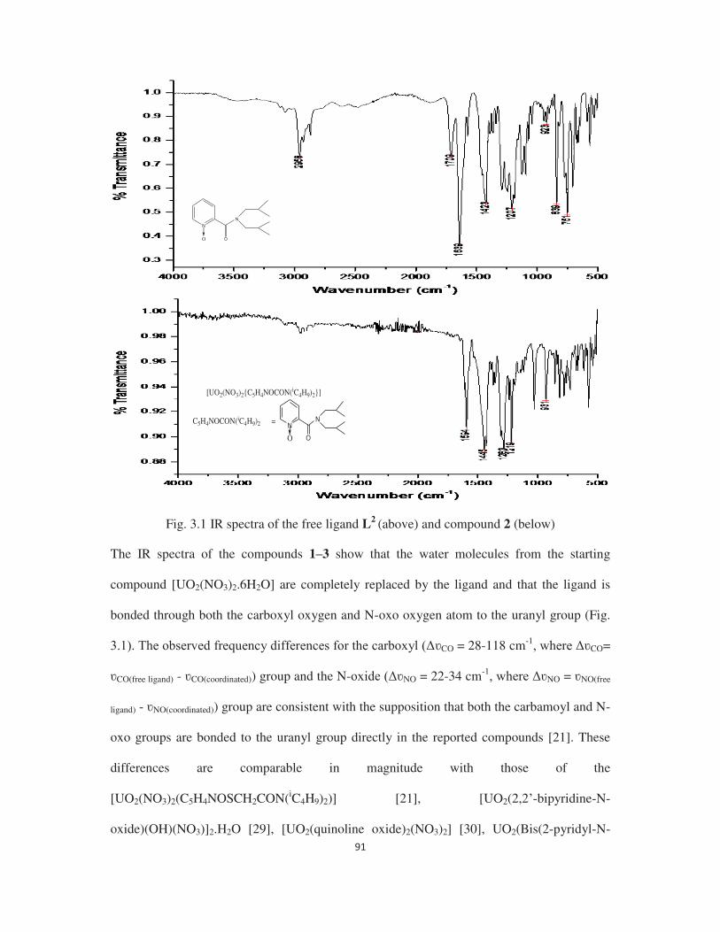
Fig. 3.1 IR spectra of the free ligand L2 (above) and compound 2 (below) The IR spectra of the compounds 1 3 show that the water molecules from the starting
compound [UO2(NO3)2.6H2O] are completely replaced by the ligand and that the ligand is
bonded through both the carboxyl oxygen and N-oxo oxygen atom to the uranyl group (Fig.
CO = 28-118 cm-1CO=
CO(free ligand) - CO(coordinated)) group and the N- NO = 22-34 cm-1NO = NO(free
ligand) - NO(coordinated)) group are consistent with the supposition that both the carbamoyl and N-
oxo groups are bonded to the uranyl group directly in the reported compounds [21]. These
differences are comparable in magnitude with those of the
[UO2(NO3)2(C5H4NOSCH2CON(iC4H9)2)] [21], [UO2(2,2 -bipyridine-N-
oxide)(OH)(NO3)]2.H2O [29], [UO2(quinoline oxide)2(NO3)2] [30], UO2(Bis(2-pyridyl-N-
NN
OO
NN
OO
[UO2(NO3)2{C5H4NOCON(iC4H9)2}]
C5H4NOCON(iC4H9)2 =
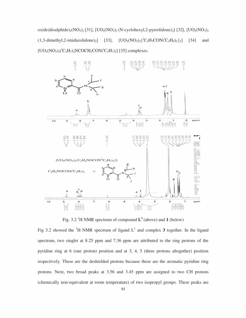
oxide)disulphide)2(NO3)2 [31], [UO2(NO3)2 (N-cyclohexyl,2-pyrrolidone)2] [32], [UO2(NO3)2
(1,3-dimethyl,2-imidazolidone)2] [33], [UO2(NO3)2{iC3H7CON(iC4H9)2}2] [34] and
[UO2(NO3)2(iC3H7)2NCOCH2CON(iC3H7)2] [35] complexes.
N
O O
N
C5H4NOCON(iC3H7)2 =N
O O
N
[UO2(NO3)2{C5H4NOCON(iC3H7)2}]
Fig. 3.2 1H NMR spectrum of compound L1 (above) and 1 (below)
Fig 3.2 showed the 1H NMR spectrum of ligand L1 and complex 3 together. In the ligand
spectrum, two singlet at 8.25 ppm and 7.36 ppm are attributed to the ring protons of the
pyridine ring at 6 (one proton) position and at 3, 4, 5 (three protons altogether) position
respectively. These are the deshielded protons because these are the aromatic pyridine ring
protons. Next, two broad peaks at 3.56 and 3.45 ppm are assigned to two CH protons
(chemically non-equivalent at room temperature) of two isopropyl groups. These peaks are

broad because of quadrapolar effect of 14N-atom adjacent to the CH groups of the isopropyl
groups. Six CH3 protons (chemically non-equivalent at room temperature) of one substituted
isopropyl groups resonate as doublets at 1.58 and 1.52 ppm. Both the signals are split into a
doublet, because the methyl group protons are coupled with one adjacent CH proton. Two
singlet peaks at 1.36 and 1.09 ppm are due to six CH3 protons (chemically non-equivalent at
room temperature) of the other substituted isopropyl groups. Spectrum of the complex 3 is
also similar to the ligand L1 except two additional peaks are appeared at 3.45 and 2.03 ppm
due to the residual water impurity and CH3COCH3 molecule in the solvent acetone-d6. All the
proton peaks of the ligand in the spectrum of complex 3 are deshielded by ca. 0.2-0.6 ppm
with respect to the free ligand, indicating that the ligand is coordinated to the uranyl group in
solution also. It is apparent from the IR spectral analysis that ligands are acting as bidentate
ligand and bonded through both carboxyl oxygen and N-oxo oxygen (both the C=Ostr and N-
Ostr frequencies are reduced in the compounds with respect to the free ligands) to the uranyl
group in compounds 1 3 in the solid state to give similar structures to those observed in the
compounds of (N,N-dialkylcarbamoyl methyl) (2-pyridyl-N-oxide) sulfide [21] with the
uranyl nitrate. The structure of 3 has been determined by single crystal X-ray diffraction
method, which confirms the IR and elemental analysis results.
3.3.2 Molecular structure of 3
Fig. 3.3 Molecular structure of [UO2(NO3)2{C5H4NOCONH(tC4H9)}]

The molecular structure of the compound 3 is shown in Fig. 3.3 and the selected interatomic
bond distances and angles for are given in Table 3.2. The uranium atom is surrounded by
eight oxygen atoms in a distorted hexagonal bi-pyramidal geometry. Two uranyl oxygen
atoms occupy the axial positions. The four oxygen atoms of the two nitrate groups and the
two oxygen atoms of the ligand forms the equatorial hexagonal plane. The ligand acts as a
bidentate chelating ligand and is bonded through both the N-oxo and amido oxygen atoms to
the uranyl group.
This type of co-ordination is similar to that observed in the complexes of the bifunctional
ligands with uranyl nitrate such as, carbamoyl methyl phosphonate complexes
[UO2(NO3)2{Ph(EtO)POCH2CONEt2}], [UO2(NO3)2(Ph2POCH2CONEt2)] [36], malonamide
complex [UO2(NO3)2(iC3H7)2NCOCH2CON(iC3H7)2 -sulfoxo phosphine oxide
complex [UO2(NO3)2{(iPrO)2POCH2SO(p-MeC6H4)}] [37] or bis(carbamoylmethyl) sulphide
complex UO2(NO3)2(iBu2NCOCH2SCH2CONiBu2)] [38] etc. The observed bond distance
for U O(amide) (2.430(9) Å) in 3 is close to the values observed in earlier reported uranyl
nitrate-amide complexes, such as [UO2(NO3)2(C5H4NOSCH2CON(iC4H9)2)] (2.393 (12)Å)
[21], UO2(NO3)2(N-cyclohexylmethyl,2-pyrrolidone)2] (2.374(2)Å) [32], [UO2(NO3)2(1,3-
dimethyl,2-imidazolidone)2] (2.383(2)Å) [33], [UO2(NO3)2(tetrabutylglutaramide)2]
(2.378(6) Å) [39], [UO2(NO3)2(dibutyldecanamide)2] (2.37(2) Å) [39] and
[UO2(NO3)2(PhN(CH3)CO(CH3)NPh)2] (2.381(2) Å) [40]. The U O(N-oxide) distance
(2.360(12) Å) in 3 is comparable in magnitude with those of earlier reported uranyl nitrate-N-
oxide complex, such as [UO2(NO3)2(C5H4NOSCH2CON(iC4H9)2)] (2.389 (12)Å) [21]. The
observed average U O(NO3) bond distance of 2.498(14) Å is normal. The angles subtended
at the metal atom show that the uranium atom has a slightly distorted hexagonal bipyramidal
geometry.

Table 3.2 Important bond distances (Å) and angles (o) for the compound 3 U1 O1 1.761(12) O1 U1 O2 178.1(6) U1 O2 1.762(11) O3 U1 O4 50.5(4) U1 O3 2.479(17) O6 U1 O7 50.8(4) U1 O9 2.360(12) O9 U1 O10 66.0(4) U1 O10 2.430(9) U1 O10 C1 131.4(9) C1 O10 1.23(2) U1 O9 N4 117.0(10)
The preliminary structural studies of compound 1 also confirms the chelating mode bonding
for the ligand with uranyl nitrate and confirms the connectivity between ligand and metal ion.
3.3.3 Comparison of ligand (L1-L3) donor strength in the uranyl complexes by comparing the
uranyl stretching frequency in the vibrational spectroscopy
Here, we have recorded the infra- -diketonate
complexes with three newly synthesized ligands (L1-L3) and systematically tabulated in table
3.3.
Table 3.3 Comparison of IR frequencies of uranyl complexes
Complex C=Ostr frequency of free ligands (cm-1)
C=Ostr
frequency of the bonded ligands (cm-1)
CO
(cm-1) NO
(cm-1) U=Oasy
frequency of the axial bond (cm-1)
[UO2(NO3)2{C5H4NOCON (iC3H7)2}] (1)
1628 1510 128 34 924
[UO2(NO3)2{C5H4NOCON (iC4H9)2}] (2)
1639 1594 101 12 931
[UO2(NO3)2{C5H4NOCONH (tC4H9)}] (3)
1669 1641 129 22 930
From the table it can be observed that, in the [UO2(NO3)2.6H2O] complexes of N-Oxo
pyridine 2-carboxamide ligands, the order of U=Oasy frequency is (2) > (3) > (1), which
signifies that donor strength order of the ligands is L1 > L3 > L2 in these complexes. The
increase in donor strength of ligand L1 with respect to the ligand L2 can be explained on the
basis of increase in steric bulkiness of the later with respect to the former since the electronic

property is almost same for both the ligands. The decrease in basicity of the amidic oxygen
atom in ligand L3 due to mono N-alkyl substituted nature may be the reason behind the less
donor strength of this ligand with respect to ligand L1, here ligand basicity is the dominating
factor. But the lesser donor capacity of the ligand L2 can be explained on the basis of more
steric bulkiness of the ligand L2 (N, N-disubstituition) with respect to the ligand L3 (N-mono
substituition), here steric factor is the dominating factor.
3.3.4 Complexation study of N-oxo pyridine2-carboxamide ligands with lanthanide nitrates
The reaction of ligands L1 L3 with lanthanide nitrate hexahydrate [Ln(NO3)3·6H2O] (where,
Ln = La, Sm, Eu) yielded compounds 4 9. C, H, and N analyses revealed that the ratio of
lanthanide nitrate to ligand is 1:2 in all compounds. The IR spectra of 4 9 (Fig. 3.4 for 4, 6
and 8) show that the water molecules from the starting compound [Ln(NO3)3·6H2O] are not
completely replaced by the ligand and that the ligand is bonded only through the N-oxo
oxygen atom to the lanthanide ions. The observed frequency differences for the N-oxide
NO = 29-34 cm-1NO = NO(free ligand) - NO(coordinated)) group are consistent with the
supposition that only the N-oxo group is bonded to the lanthanide ions directly in the reported
compounds.
(a)
NN
OO
[La(NO3)3(H2O){C5H4NOCON(iC3H7)2}2]
C5H4NOCON(iC3H7)2 =

(c)
Fig. 3.4 IR spectra of (a) compound 4, (b) compound 6, (c) compound 8
Fig 3.5 showed the 1H NMR spectrum of (a) complex 4, (b) complex 6 and (c) complex 8
together. These spectra are exactly similar to that of ligand L1 (already discussed in details
earlier) since these complexes are derived from the same ligand. The proton peaks of the
ligand in the spectrum of all complexes are deshielded by ca. 0.2-0.3 ppm with respect to the
free ligand, indicating that the ligand is coordinated to the lanthanide ions in solution also. It
is apparent from the IR spectral analysis that ligands are acting as monodentate ligand and
bonded only through N-oxo oxygen (only the N-Ostr frequencies are reduced in the
compounds with respect to the free ligands) to the lanthanide ions in compounds 4 9 in the
NN
OO
[Sm(NO3)3(H2O){C5H4NOCON(iC3H7)2}2]
C5H4NOCON(iC3H7)2 =
NN
OO
[Eu(NO3)3(H2O){C5H4NOCON(iC3H7)2}2]
C5H4NOCON(iC3H7)2 =

solid state. The structure of 8 has been determined by single crystal X-ray diffraction
methods, which confirms the IR and elemental analysis results.
C5H4NOCON(iC3H7)2 =N
O O
N
[La(NO3)3(H2O){C5H4NOCON(iC3H7)2}2]
(a)
C5H4NOCON(iC3H7)2 =N
O O
N
[Sm(NO3)3(H2O){C5H4NOCON(iC3H7)2}2]
(b)

C5H4NOCON(iC3H7)2 =N
O O
N
[Eu(NO3)3(H2O){C5H4NOCON(iC3H7)2}2]
(c)
Fig. 3.5 1H NMR spectrum of (a) compound 4, (b) compound 6, (c) compound 8
3.3.5 Molecular structure of compound 8
Fig. 3.6 Molecular structure of [Eu(NO3)3(H2O){C5H4NOCON(iC3H7)2}2]
The molecular structure of the compound 8 is shown in Fig. 3.6 and the selected interatomic
bond distances and angles for are given in Table 3.4. The europium atom is surrounded by
nine oxygen atoms in a distorted tricapped trigonal prismatic geometry. Six oxygen atoms
from three bidentate nitrate groups, two oxygen atoms from two N-oxo group of the ligands
and one oxygen atom from one water molecule satisfy the coordination number of nine. The
ligand acts as a monodentate ligand and is bonded only through the N-oxo oxygen atom to
the europium atom while amide group is free. Two ligands are placed mutually syn to each

other. The pyridyl rings of both the ligands are essentially non coplanar with the tortion angle
of 72.37o.
The observed bond distance for Eu O(N-oxide) (2.345(8) Å) in 8 is close to the values observed
in earlier reported lanthanide nitrate-N-oxide complexes, such as [Eu(nicotinic acid N-
oxide)3(H2O)2]n. 4n H2O (2.379 (5)Å) [41], [Eu(2,6-bis((1H-pyrazol-1-yl)methyl)pyridine 1-
oxide)2(NO3)3] (2.376(13) Å) [42], {[Eu(2,6-Bis(diphenyl-N,N-
diethylcarbamoylmethylphosphine oxide)-pyridine N-
Oxide)(NO3)3]·(Me2CO)0.75·(H2O)0.3}4 (2.467 (3)Å) [43], Eu(2,6-Bis[bis(2-
(trifluoromethyl)phenyl)phosphinoylmethyl]pyridine N-Oxide)(NO3)3 (2.424(2) Å) [44]. The
observed average Eu O(NO3) bond distance of 2.496(11) Å is normal.
Table 3.4 Important bond distances (Å) and angles (o) for the compound 8 Eu1-O11 2.335(8) O11-Eu1-O13 149.1(4) Eu1-O13 2.355(8) O6-Eu1-O5 50.3(3) Eu1-O8 2.507(10) O9-Eu1-O8 51.1(3) Eu1-O6 2.486(11) O3-Eu1-O2 51.0(5) Eu1-O2 2.496(11) N4-O11-Eu1 133.9(8) Eu1-O1 2.418(11) N6-O13-Eu1 133.7(8) N1-O3-Eu1 97.4(11)
3.4 Conclusions
N-oxo pyridine 2-carboxamide ligands showed coordination diversity towards uranyl nitrate
and lanthanide nitrates. The reaction between uranyl nitrate and N-oxo pyridine 2-
carboxamide yielded complexes with 1:1 stoichiometry. In these complexes, the ligands are
bonded with the uranyl group in a bidentate chelating fashion through both the amidic oxygen
and N-oxide oxygen atoms. The geometry around the uranium is distorted hexagonal
bipyramid. On the other hand, the lanthanide nitrates (Ln = La, Sm, Eu) form 1:2 complexes
with N-oxo pyridine 2-carboxamide ligands. The ligands act as monodetate ligands and bond
through the N-oxide oxygen atom with metal centre and leaving the amido oxygen atom free.
The geometry around the europium(III) ion is distorted tricapped trigonal prism.
3.5 References
1. J.N. Mathur, M.S.Murali, K.L. Nash, Solvent Extr. Ion Exch. 19 (2001) 357-390.
2. Seraj A. Ansari, Priyanath Pathak, Prasanta K. Mohapatra, Vijay K. Manchanda, Chem.
Rev. 112 (2012) 1751 1772.
3. G.R. Mahajan, D.R. Prabhu, V.K. Manchanda, L.P. Badheka, Waste Manage. 18 (1998)
125-133.
4. Henk H. Dam, David N. Reinhoudt, Willem Verboom, New J. Chem. 31 (2007) 1620-
1632.
5. E. Philip Horwitz , Dale C. Kalina , Herbert Diamond , George F. Vandegrift, Wallace W.
Schulz, Solvent Extr. Ion Exch. 3 (1985) 75-109.
6. W.W. Schulz, E.P. Horwitz, Sep. Sci. Technol. 23 (1988) 1191-1210.
7. A.B Patil, P. Pathak, V.S Shinde, S.V Godbole, P.K Mohapatra, Dalton Trans. 42 (2013)
1519-1529.
8. Eil-Hee Lee, Jae-Gwan Lim, Dong-Yong Chung, Jae-Hyung Yoo, Kwang-Wook Kim,
Nucl. Eng. Technol. 41 (2009) 319-326.
9. Benoît Gannaz, Mark R. Antonio, Renato Chiarizia, Clément Hill, Gérard Cote, Dalton
Trans. (2006) 4553-4562.
10. Sujoy Biswas, D.K. Singh, K.N. Hareendran, J.N. Sharma, S.B. Roy, J. Radioanal. Nucl.
Chem. 284 (2010) 201 205.
11. Taichi Sato, J. Inorg. Nucl. Chem. 24 (1962) 699-706.
12. F.E. Kosinski, H. Bostian, J. Inorg. Nucl. Chem. 31 (1969) 3623-3631.
13. Ahmed F. Abdel-Magied, Mostafa I. Amin, Int. J. Ind. Chem. 7 (2016) 21 28.
14. S. Biswas, P.N. Pathak, D.K. Singh, S.B. Roy, V.K. Manchanda, Int. J. Miner. Process.
104-105 (2012) 17 23.
15. F.J. Hurst, D.J. Crouse, Ind. Eng. Chem., Process Des. Develop. 13 (1974) 286-291.
16. P.D. Mithapara, V.K. Manchanda, P.R. Natarajan, J. Radioanal. Nucl. Chem. Art. 83/2
(1984) 301-307.
17. Shanmugaperumal Kannan, Kuna Venugopal Chetty, Venkatarama Venugopal, Michael
G.B. Drew, Dalton Trans. (2004) 3604 3610.
18. S.B. Deb, S. Kannan, M.G.B. Drew, J. Coord. Chem. 63 (2010) 3620-3626.
19. Sabrina Ouizem, Daniel Rosario-Amorin, Diane A. Dickie, Benjamin P. Hay, Robert T.
Paine, Polyhedron 101 (2015) 37 47.
20.

21. Bal Govind Vats, Jayashree S. Gamare, S. Kannan, I.C. Pius, D.M. Noronha, M. Kumar,
Inorg. Chim. Acta 467 (2017) 1 6.
22. M. Ejaz, Sep. Sci. 10 (1975) 425-446.
23. -1038.
24. Neelam Kumari, D.R. Prabhu, A.S. Kanekar, P.N. Pathak, Ind. Eng. Chem. Res. 51 (2012)
14535 14542.
25. Kevin McCann, Bruce J. Mincher, Nicholas C. Schmitt, Jenifer C. Braley, Ind. Eng.
26. Kevin McCann, Jessica A. Drader, Jenifer C. Braley, Sep. Purif. Rev.
doi.org/10.1080/15422119.2017.1321018.
27. Sylvie Pailloux, Iris Binyamin, Lorraine M. Deck, Benjamin P. Hay, Eileen N. Duesler,
Lev N. Zakharov, W. Scott Kassel, Arnold L. Rheingold, Robert T. Pain, Polyhedron 28
(2009) 3979 3984.
28. Hai-Jun Zhang, Ru-Hu Gou, Lan Yan, Ru-Dong Yang, Spectrochim. Acta, Part A 66
(2007) 289 294.
29. Zbigniew Hnatejko, Stefan Lis,
(2011) 880 885.
30. Zbigniew Hnatejko, J. Rare Earths 30 (2012) 552.
31. I.S. Ahuja , C.L. Yadava, Shailendra Tripathi, Synth. React. Inorg. Met.-Org. Chem. 18
(1988) 953-963.
32. T.R. Varga, A.C. Benyei, Z. Fazekas, H. Tomiyasu, Y. Ikeda, Inorg. Chim. Acta 342
(2003) 291.
33. N. Koshino, M. Harada, M. Nogami, Y. Morita, T. Kikuchi, Y. Ikeda, Inorg. Chim. Acta
358 (2005) 1857.
34. S. Kannan, S.B. Deb, J.S. Gamare, M.G.B. Drew, Polyhedron 27 (2008) 2557.
G.J. Lumetta, B.K. McNamara, B.M. Rapko, R.L. Sell, R.D. Rogers, G. Broker, J.E.
Hutchison, Inorg. Chim. Acta 309 (2000) 103.
36. L.J. Caudle, E.N. Duesler, R.T. Paine, Inorg. Chim. Acta 110 (1985) 91 100.
37. D.T. Cromer, R.R. Ryan, S. Karthikeyan, R.T. Paine, Inorg. Chim. Acta 172 (1990) 165
172.
38. S.B. Deb, J.S. Gamare, S. Kannan, M.G.B. Drew, Polyhedron 28 (2009) 2673 2678.
39. P. Charpin, M. Lance, M. Nierlich, D. Vigner, Acta Crystallogr. Sect. C 42 (1986) 560.
40. O. Clements, B.M. Rapko, B.P. Hay, Coord. Chem. Rev. 170 (1998) 203.
41. Jiang-Gao Mao, Hong-Jie Zhang, Jia-Zuan Ni, Shu-Bin Wang, Thomas C.W Mak,
Polyhedron 17 (1998) 3999 4009.
42. Sabrina Ouizema, Daniel Rosario-Amorina, Diane A. Dickiea, Benjamin P. Hay, Robert
T. Paine, Polyhedron 101 (2015) 37 47.
43. Daniel Rosario-Amorin, Sabrina Ouizem, Diane A. Dickie, Yufeng Wen, Robert T. Paine,
Jian Gao, John K. Grey, Ana de Bettencourt-Dias, Benjamin P. Hay, Lætitia H. Delmau,
Inorg. Chem. 52 (2013) 3063 3083.
44. Sylvie Pailloux, Cornel Edicome Shirima, Alisha D. Ray, Eileen N. Duesler, Robert T.
Paine, John R. Klaehn, Michael E. McIlwain, Benjamin P. Hay, Inorg. Chem. 48 (2009)
3104 3113.


4.1 Introduction
The speciality of actinide coordination chemistry is their high coordination number and their
structures depend on the steric effect provided by the bonded ligands like main group metal
atoms [1-3]. It also modulates the mode of bonding of the bonded ligands and stoichiometry
of the metal ligand complexes. Like other actinides, uranium also forms stable complexes
with ligands consist of hard donor atoms like O, N, etc, which often form five or six
membered cyclic chelates. It is evident from the previous studies that, five membered
chelating ligands are more sterically controlled than six or higher membered chelating ligands
[4]. In past, we had synthesized two types of carbamoyl methyl pyrazole based ligands (Fig.
1a, b) which form six membered chelating ring with uranyl nitrate irrespective to the steric
demand of the respective ligands and relative orientation of the donor atoms in the ground
state [5].
In continuation of our interest on the complex chemistry of uranyl and other metal ions with
newly synthesized ligands [6], we report herein the synthesis, characterization, and complex
chemistry of the analogous carbamoyl pyrazole based ligands (Fig. 1c, d) with uranyl as well
as palladium (II) ions. Since these ligands are having a soft pyrazolyl nitrogen atom apart
from hard amidic oxygen atom, they can form stable complexes with soft metal ion like
palladium. These ligands can act as monodentate or bidentate ligands while bonding with
palladium ions. Comparison of the complex chemistry between these two types (carbamoyl
methyl pyrazole and carbamoyl pyrazole) of ligands is also done to understand the effect of
ring size on complex formation. Density functional theory is done to validate and explain our
experimental observation.

N
N
N
O
R
R
N
N
N
O
R
R
CH3
H3C
N
N
O
N
R
RN
N
O
N
R
R
CH3
H3C
(a) (b) (c) (d)
Fig. 1 (a) Carbamoyl methyl pyrazole, (b) carbamoyl methyl (3,5-dimethyl pyrazole), (c)
carbamoyl pyrazole, (d) carbamoyl (3,5-dimethyl pyrazole)
4.2 Experimental
4.2.1 Synthesis of the carbamoyl pyrazole ligands
4.2.1.1 Synthesis of C3H3N2CON(CH3)2 (L1)
A solution of pyrazole (10 g, 0.14 mol) and triethyl amine (17.5 g, 0.17 mol) in benzene (50
mL) was added slowly to a solution of N,N-dimethyl carbamoyl chloride (15.8 g, 0.14 mol)
in benzene (50 mL) with stirring. The reaction mixture was refluxed for 6 h and treated with
100 mL of 5% HCl solution. The organic layer was separated, dried over anhydrous sodium
sulfate, and filtered. Removal of the solvent in vacuum yielded a colorless solution of L1 in
59% yield. 1H NMR (25 oC, CDCl3 3], 6.35 [q, 1H, pz], 7.64 [d, 1H,
pz], 8.12 [q, 1H, pz]. IR (cm 1 =O). Analysis Calcd for C6H9N3O: C, 51.8; H,
6.5; N, 30.2. Found: C, 51.1; H, 6.1; N, 29.7.
4.2.1.2 Synthesis of C3H3N2CON(C2H5)2 (L2)
This was prepared similarly as for L1 by taking N,N-diethyl carbamoyl chloride (20 g, 0.14
mol) and pyrazole (10 g, 0.14 mol) in 73% yield. 1H NMR (25 °C, CDCl3
CH3], 3.61 [br, 4H, NCH2], 6.34 [q, 1H, pz], 7.62 [d, 1H, pz], 8.13 [q, 1H, pz]. IR (cm 1

1687(C=O). Analysis Calcd for C8H13N3O: C, 57.5; H, 7.8; N, 25.1. Found: C, 56.9; H, 7.4;
N, 24.7.
4.2.1.3 Synthesis of C3H3N2CON(iC3H7)2 (L3)
This was prepared similarly to L1 by taking N,N-diisopropyl carbamoyl chloride (25 g, 0.15
mol) and pyrazole (10 g, 0.14 mol) in 90% yield. 1H NMR (25 °C, CDCl3
CH3], 4.09 [m, 2H, CH, iPr], 6.33 [q, 1H, pz], 7.60 [d, 1H, pz], 8.03 [q, 1H, pz]. IR (cm 1
= 1687(C=O). Analysis Calcd for C10H17N3O: C, 61.5; H, 8.7; N, 21.5. Found: C, 61.2; H,
8.4; N, 21.1.
4.2.1.4 Synthesis of C5H7N2CON(CH3)2 (L4)
This was prepared similarly to L1 by taking N,N-dimethyl carbamoyl chloride (12 g, 0.11
mol) and dimethyl pyrazole (10 g, 0.11 mol) in 80% yield. 1H NMR (25 °C, CDCl3
[s, 3H, CH3, dmpz], 2.39 [s, 3H, CH3, dmpz ], 3.09 [s, 6H, NCH3], 5.89 [s, 1H, dmpz]. IR
(cm 1 =O). Analysis Calcd for C8H13N3O: C, 57.5; H, 7.8; N, 25.1. Found: C,
57.1; H, 7.2; N, 24.8.
4.2.1.5 Synthesis of C5H7N2CON(C2H5)2 (L5)
This was prepared similarly to L1 by taking N,N-diethyl carbamoyl chloride (15 g, 0.11 mol)
and dimethyl pyrazole (10 g, 0.11 mol) in 84% yield. 1H NMR (25 °C, CDCl3
6H, CH3, C2H5], 2.21 [s, 3H, CH3, dmpz], 2.37 [s, 3H, CH3, dmpz], 3.45 [s, 4H, NCH2], 5.87
[s, 1H, dmpz]. IR (cm 1 =O). Analysis Calcd for C10H17N3O: C, 61.5; H, 8.7; N,
21.5. Found: C, 60.9; H, 8.5; N, 21.0.
4.2.1.6 Synthesis of C5H7N2CON(iC3H7)2 (L6)
This was prepared similarly to L1 by taking N,N-diisopropyl carbamoyl chloride (17 g, 0.11
mol) and dimethyl pyrazole (10 g, 0.11 mol) in 86% yield. 1H NMR (25 °C, CDCl3
[d, 12H, CH3, iPr], 2.16 [2, 3H, CH3, dmpz], 2.28 [s, 3H, CH3, dmpz], 3.66 [m, 2H, CH, iPr],

5.81 [s, 1H, dmpz]. IR (cm 1 =O). Analysis Calcd for C12H21N3O: C, 64.6; H,
9.4; N, 18.8. Found: C, 64.3; H, 9.1; N, 18.5.
4.2.2 Synthesis of uranyl complexes of carbamoyl pyrazole ligands
4.2.2.1 Synthesis of [UO2(NO3)2{C3H3N2CON(CH3)2}] (1)
To a solution of L1 (200 mg, 1.44 mmol) in CH2Cl2 (20 mL), solid [UO2(NO3)2·6H2O] (300
mg, 0.59 mmol) was added and stirred for few minutes until all [UO2(NO3)2·6H2O] dissolved
to give a clear solution. This solution was filtered and layered with iso-octane. The solution
on slow evaporation yielded yellow crystalline solid, which was filtered, washed with
hexane, and dried. Yield: 85%. 1H NMR (25°C, CD3COCD3 3], 3.98
[br, 3H, NCH3], 7.22 [br, 1H, pz], 9.26 [br, 2H, pz]. IR (cm 1 =O), 935(U=O).
Analysis Calcd for C6H9N5O9U: C, 13.5; H, 1.7; N, 13.5. Found: C, 13.4; H, 1.6; N, 13.2.
4.2.2.2 Synthesis of [UO2(NO3)2{C3H3N2CON(C2H5)2}] (2)
This was prepared similarly to 1 by taking L2 (220 mg, 1.37 mmol) and [UO2(NO3)2·6H2O]
(300 mg, 0.59 mmol) in 91% yield. 1H NMR (25°C, CD3COCD3 3],
4.15 [br, 4H, NCH2], 7.4 [s, 1H, pz], 8.98 [s, 1H, pz], 9.24 [s, 2H, pz]. 1H NMR (25°C,
CDCl3 3], 4.06 [br, 4H, NCH2], 7.12 [s, 1H, pz], 8.44 [s, 1H, pz], 9.4 [s,
2H, pz]. IR (cm 1 =O), 941 (U=O) 941. ES-MS (CH2Cl2): m/z = 666
[UO2(NO3)(L2)2]
+, 517 [UO2(NO3)(L2)H2O]+, 499 [UO2(NO3)(L
2)]+. Analysis Calcd for
C8H13N5O9U: C, 17.1; H, 2.3; N, 12.5. Found: C, 17.0; H, 2.3; N, 12.4.
4.2.2.3 Synthesis of [UO2(NO3)2{C3H3N2CON(iC3H7)2}] (3)
This was prepared similarly to 1 by taking L3 (250 mg, 1.28 mmol) and [UO2(NO3)2·6H2O]
(300 mg, 0.59 mmol) in 86% yield. 1H NMR (25°C, CD3COCD3 3], 1.80
[s, 3H, CH3], 4.42 [m, 2H, CH, iPr], 7.21 [s, 1H, pz], 8.95 [s, 1H, pz], 9.2 [s, 2H, pz]. 1H
NMR (25°C, CDCl3 3], 4.06 [br, 4H, NCH2], 7.12 [br 1H, pz], 8.44 [br,

1H, pz], 9.4 [br, 2H, pz]. IR (cm 1 1643(C=O), 939 (U=O). Analysis Calcd for
C10H17N5O9U: C, 20.4; H, 2.9; N, 11.9. Found: C, 20.2; H, 2.7; N, 11.8.
4.2.2.4 Synthesis of [UO2(NO3)2(H2O)2{C5H7N2CON(CH3)2}2] (4)
This was prepared similarly to 1 by taking L4 (220 mg, 1.37 mmol) and [UO2(NO3)2·6H2O]
(300 mg, 0.59 mmol) in acetone. The acetone solution on slow evaporation yielded yellow
crystalline solid, which was filtered, washed with ether, and dried. Yield: 96%. 1H NMR (25
°C, CD3COCD3 3, dmpz], 2.31 [s, 3H, CH3, dmpz], 3.05 [s, 6H, NCH3],
5.94 [s, 1H, dmpz]. IR(cm 1 3200(H2O), 1690 (C=O), 935 (U=O). Analysis Calcd
for C16H30N8O12U: C, 25.1; H, 3.9; N, 14.6. Found: C, 25.0; H, 3.7; N, 14.9.
4.2.2.5 Synthesis of [UO2(NO3)2(H2O)2{C5H7N2CON(C2H5)2}2] (5)
This was prepared similarly to 4 by taking L5 (250 mg, 1.28 mmol) and [UO2(NO3)2·6H2O]
(300 mg, 0.59 mmol) in 92% yield. 1H NMR (25°C, CD3COCD3 3,
C2H5], 2.13 [s, 3H, CH3, dmpz], 2.30 [s, 3H, CH3, dmpz], 3.41 [s, 4H, NCH2], 5.92 [s, 1H,
dmpz]. IR (cm 1 3200 (H2O), 1691 (C=O), 941 (U=O). Analysis Calcd for
C20H38N8O12U: C, 29.3; H, 4.6; N, 13.6. Found: C, 29.1; H, 4.5; N, 13.3.
4.2.2.6 Synthesis of [UO2(NO3)2(H2O)2{C5H7N2CON(iC3H7)2}2] (6)
This was prepared similarly to 4 by taking L6 (280 mg, 1.25 mmol) and [UO2(NO3)2·6H2O]
(300 mg, 0.59 mmol) in 93% yield. 1H NMR (25 °C, CD3COCD3 3,
iPr], 2.12 [2, 3H, CH3, dmpz], 2.27 [s, 3H, CH3, dmpz], 3.74 [m, 2H, CH, iPr], 5.91 [s, 1H,
dmpz]. IR (cm 1 3200 (H2O), 1687 (C=O), 941 (U=O). Analysis Calcd for
C24H46N8O12U: C, 32.9; H, 5.3; N, 12.8. Found: C, 32.7; H, 5.2; N, 12.7.
4.2.2.7 Synthesis of [UO2(C6H5COCHCOC6H5)2{C3H3N2CON(CH3)2}] (7)
To a hot chloroform (30 mL) solution of L1 (70 mg, 0.5 mmol), solid
[UO2(C6H5COCHCOC6H5)2·2H2O] (350 mg, 0.47 mmol) was added and refluxed for 2 h.
The clear solution was filtered and layered with iso-octane. The solution on slow evaporation

yielded an orange colored product in 86% yield. 1H NMR (25°C, CDCl3
NCH3], 6.91 [s, 1H, pz], 7.37 [s, 2H, DBM], 7.53 (t, 1H, pz), 7.61 [m, 12H, C6H5, DBM], 8.0
[d, 1H, pz], 8.46 [m, 8H, C6H5, DBM]. IR (cm 1 =O) (L1), 1591 (C=O) (DBM),
904 (U-O). Analysis Calcd for C36H31N3O7U: C, 50.5; H,3.6; N, 4.9. Found: C, 49.7; H, 3.4;
N, 4.7.
4.2.2.8 Synthesis of [UO2(C6H5COCHCOC6H5)2{C3H3N2CON(C2H5)2}] (8)
This was prepared similarly to 7 by taking L2 (80 mg, 0.48 mmol) and
[UO2(C6H5COCHCOC6H5).2H2O] (350 mg, 0.47 mmol) in 90% yield. 1H NMR (25°C,
CD3COCD3 3], 3.55 [m, 4H, NCH2], 6.37 [s, 1H, pz], 7.40 [s, 2H,
DBM], 7.65 [m, 13H, C6H5 + pz], 8.6 [m, 8H, C6H5]. IR (cm 1 =O) (L2), 1591
(C=O) (DBM), 902 (U=O). ES-MS (CH2Cl2): m/z = 678 [UO2(DBM)(L2)(H2O)]+, 660
[UO2(DBM)(L2)]+. Analysis Calcd for C38H35N3O7U: C, 51.6; H, 3.9; N, 4.8. Found: C, 51.0;
H, 3.7; N, 4.5.
4.2.3 Synthesis of palladium complexes of carbamoyl pyrazole ligands
4.2.3.1 Synthesis of [PdCl2{C3H3N2CON(CH3)2}2] (9)
To an acetonitrile solution of PdCl2 (200 mg, 1.1 mmol), C3H3N2CON{CH3}2 (314 mg, 2.25
mmol) was added and refluxed for 2 h. This solution was evaporated to dryness completely,
washed with hexane and dried. The resulting residue was extracted in CH2Cl2 and filtered to
remove insoluble materials. The solution was layered with 5 ml of iso-octane and allowed to
evaporate slowly. This process deposited brownish yellow colored product in 91% yield. 1H
NMR (25oC, CDCl3 3], 6.45 [q, 1H, pz], 7.63 [d, 1H, pz], 8.12 [q, 1H,
pz]. IR (cm-1): 2{C6H9N3O}2]: C, 31.6; H, 4.0; N, 18.4.
Found: C, 31.1; H, 3.8; N, 18.1%.

4.2.3.2 Synthesis of [PdCl2{C5H7N2CON(C2H5)2}2] (10)
This was prepared similarly to 1 by taking PdCl2 (200 mg, 1.1 mmol) and
C3H3N2CON{C2H5}2 (377 mg, 2.24 mmol) in 88% yield. 1H NMR (25oC, CDCl3
[br, 6H, CH3], 3.61 [q, 4H, NCH2], 6.42 [q, 1H, pz], 7.29(d, 1H, pz], 7.99 [q, 1H, pz]. IR (cm-
1 -MS (CH3CN): 475 (100%,) [PdL2Cl+]: Anal. Calc. for
[PdCl2{C8H13N3O}2]: C, 37.5; H, 5.1; N, 16.4. Found: C, 37.2; H, 4.7; N, 16.3%.
4.2.3.3 Synthesis of [PdCl2{C3H3N2CON(iC3H7)2}2] (11)
This was prepared similarly to 1 by taking PdCl2 (200 mg, 1.1 mmol) and
C3H3N2CON{iC3H7}2 (440 mg, 2.25 mmol) in 91% yield. 1H NMR (25oC, CDCl3):
[d, 12H, CH3], 3.77 [m, 2H, CH, iPr], 6.37 [q, 1H, pz], 7.60 (d, 1H, pz], 8.02 [q, 1H, pz]. IR
(cm-12{C10H17N3O}2]: C, 42.3; H, 6.0; N, 14.8.
Found: C, 41.9; H, 6.2; N, 14.4%.
4.2.3.4 Synthesis of [PdCl2{C5H7N2CON(CH3)2}2] (12)
This was prepared similarly to 1 by taking PdCl2 (200 mg, 1.1 mmol) and
C5H7N2CON{CH3}2 (377 mg, 2.25 mmol) in 90% yield. 1H NMR (25oC, CDCl3
3H, CH3, dmpz], 2.87 [d, 3H, CH3, dmpz], 2.98 [br, 3H, NCH3], 3.34 [br, 3H, NCH3], 5.97
[s, 1H, dmpz]. IR (cm-12{C8H13N3O}2]: C, 37.5; H,
5.1; N, 16.4. Found: C, 37.4; H, 4.9; N, 16.1%.
4.2.3.5 Synthesis of [PdCl2{C5H7N2CON(C2H5)2}2] (13)
This was prepared similarly to 1 by taking PdCl2 (200 mg, 1.1 mmol) and
C5H7N2CON{C2H5}2 (440 mg, 2.24 mmol) in 89% yield. 1H NMR (25oC, CDCl3
[br, 3H, CH3, C2H5], 1.47 [br, 3H, CH3, C2H5], 2.31 [s, 3H, CH3, dmpz], 2.86[s, 3H, CH3,
dmpz], 3.43 [br, 2H, NCH2 ], 3.73 [br, 2H, NCH2], 5.95 [s, 1H, dmpz]. IR (cm-1
(C=O). ESI-MS (CH3CN): 573 (100%,) [PdL2Cl+]: Anal. Calc. for [PdCl2{C10H17N3O}2]: C,
42.3; H, 6.0; N, 14.8. Found: C, 42.0; H, 6.1; N, 14.6%.

4.2.3.6 Synthesis of [PdCl2{C5H7N2CON(iC3H7)2}2] (14)
This was prepared similarly to 1 by taking PdCl2 (200 mg, 1.1 mmol) and
C5H7N2CON{iC3H7}2 (504 mg, 2.24 mmol) in 91% yield. 1H NMR (25oC, CDCl3
[d, 12H, CH3, iPr], 2.31 [2, 3H, CH3, dmpz], 2.93 [s, 3H, CH3, dmpz], 3.67 [m, 2H, CH, iPr],
5.93 [s, 1H, dmpz]. IR (cm-1 ). Anal. Calc. for [PdCl2{C12H21N3O}2]: C,
46.2; H, 6.8; N, 13.5. Found: C, 46.0; H, 6.6; N, 13.2%.

4.2.4 X-ray crystallography
Selected crystallographic data for the compounds 2, 5, 8, 10, and 13 are summarized in Tables 4.1.
2 5 8 10 13 Empirical formula C8H13N5O9U C20H38N8O12U C38H35N3O7U C16H26N6O2Cl2Pd C20H34N6O2Cl2Pd Formula weight 561.25 820.59 883.73 511.74 567.85 Crystal system Monoclinic Monoclinic Monoclinic Monoclinic Monoclinic Space group P21/c P21/n P21/n C2/c P21/c a (Å) 8.3229(6) 10.207(2) 10.2750(5) 22.3590(7) 7.1517(4) b (Å) 18.0208(11) 8.5355(17) 21.6295(10) 7.2273(2) 11.4886(5) c(Å) 10.6066(8) 18.095(4) 15.8792(7) 13.6515(4) 14.8111(6)
o) 100.902(8) 95.74(2) 100.913(4) 111.791(3) 92.600(4) Volume (Å3) 1562.12(19) 1568.6(6) 3465.2(3) 2048.37(10) 1215.67(9) Z (formula unit) 4 2 4 4 2 Density (g/cm3) 2.386 1.737 1.694 1.659 1.551
-1) 10.445 5.240 4.738 1.191 1.012 Reflections collected/unique 9807/4518 9787/4376 17433/9611 5722/2904 7036/3384 Data/restrains/parameters 4518/0/210 4376/31/202 9611/0/444 2904/0/126 3384/0/146 Goodness of fit on F2 1.038 1.010 1.073 1.041 1.087 Final R1 0.0755 0.0658 0.0639 0.0256 0.0243 wR2 indices (all data) 0.1855 0.1496 0.1133 0.0577 0.0578
2(Fo
2) + (0.1166P)2 + 0.000P] for 2 2(Fo2) + (0.0667P)2 + 0.000P] for 5 2(Fo
2) + (0.0378P)2 + 3.2432P] for 8, w = 2(Fo
2)+(0.0231P)2 + 0.7398P] for 10 2(Fo2)+(0.0251P)2 + 0.5630P] for 13, where P = (Fo
2 + 2Fc2)/3.

4.2.5 Theoretical calculations
For uranium complexes, full geometry optimization for the N,N-dimethyl analog of the
ligands a d and complexes (A D) has been carried out applying a popular nonlocal
correlated hybrid density functional, namely, B3LYP. Gaussian type atomic basis functions,
6-31+G(d), are adopted for H, C, N, and O atoms, and for U atom, a very recently suggested
basis set, SARC-ZORA [7a], is used for all the calculations. SARC-ZORA basis sets are
segmented all-electron scalar relativistic basis sets in which the coefficients of contracted
GTOs have been optimized for use with the ZORA scalar relativistic Hamiltonian. These
particular basis sets for U are obtained from Extensible Computational Chemistry
Environment Basis Set Database, Pacific Northwest National Laboratory [7b]. The quasi-
Newton Raphson based algorithm has been applied to carry out geometry optimization to
locate the minimum energy structure in each case. Hessian calculations have also been
carried out to check the nature of the equilibrium geometry. Macroscopic solvation effect of
solvent water has been incorporated in energy calculation through polarizable continuum
model (PCM). All these calculations have been carried out applying GAMESS suit of ab
initio program on a LINUX cluster platform [7c].
For palladium complexes, full geometry optimization of the ligands (L2 and L5) and
complexes (10 and 13) has been carried out applying a recently developed density functional
with
functions, 6-31+G(d) are adopted for H, C, N, O and Cl atoms and for Pd atom 3-21G basis
sets are used for all the calculations. Recently, it has been reported that for transition metal
dispersion corrections as well as other popular DFT functionals not including dispersion. The
functional adopted is observed to offer relatively small statistical errors when considering the
overall structure as well as selected distances [8]. It is worthwhile mentioning that basis sets

like LANL2DZ ECP and aug-cc-pVDZ-pp were also used for palladium, but such
calculations were unsuccessful due to SCF convergence failure or optimization to
unreasonable geometries.
4.3 Results and discussion
4.3.1. Synthesis of carbamoyl pyrazole and carbamoyl 3,5-dimethyl pyrazole ligands
Reaction of pyrazole and 3,5-dimethyl pyrazole with corresponding N,N-dialkyl carbamoyl
chloride in benzene in presence of triethyl amine as base yielded the corresponding ligands
(L1 to L6). The IR spectra of all ligands (Fig. 4.1) show the presence of the carbamoyl group
in the synthesized compounds.
N
N N
O

Fig. 4.1 IR spectra of ligand L2 (above) and L5 (below)
Fig 4.2 showed the 1H NMR spectrum of ligand L2. The three singlet peaks at 8.13, 7.62 and
6.34 ppm are due to each ring proton of the pyrazole ring at 5, 3 and 4 position respectively.
Next, at 3.61 ppm, the broad peak is assigned to four CH2 protons (chemically equivalent due
to C-N(diethyl) bond rotation at room temperature) of the ethyl groups. This peak is broad
because of quadrapolar effect of 14N-atom adjacent to the CH2 group protons. Six CH3
protons (chemically equivalent due to C-N(diethyl) bond rotation at room temperature) of
two ethyl groups of the ligand resonate as a triplet at 1.27 ppm. The signal is split into a
triplet, because the methyl group protons are coupled with two adjacent CH2 protons. The C,
H, N analysis supports the expected stoichiometry for the compounds.
N
N N
O

N
N
O
N
a
b
c d
d
e
e
Fig. 4.2 1H NMR spectrum of ligand L2
4.3.2 Synthesis and structural studies of uranyl complexes
4.3.2.1 Synthesis and complexation studies of carbamoyl pyrazole ligands with uranyl nitrate
The reaction of ligands L1 L3 with [UO2(NO3)2·6H2O] yielded compounds 1 3. C, H, and N
analyses revealed that the ratio of ligand to uranyl nitrate is 1:1 in all compounds. The IR
spectra of 1 3 (Fig. 4.3 for 2) show that the water molecules from the starting compound
[UO2(NO3)2·6H2O] are completely replaced by the ligand and that the ligand is bonded
through the carbamoyl oxygen atom to the uranyl group. The observed frequency difference
for a carbamoyl ( CO = 20 40 cm 1, where CO CO(free ligand) CO(coordinated)) group is
consistent with the supposition that the carbamoyl group is bonded to the uranyl group
directly. This difference is comparable in magnitude with those observed in [UO2(NO3)2(N-
cyclohexyl-2-pyrrolidone)2] [9a], [UO2(NO3)2(1,3-dimethyl-2-imidazolidone)2] [9b],
[UO2(NO3)2{iC3H7CON(iC4H9)2}2] [10], [UO2(NO3)2(
iC3H7)2NCOCH2CON(iC3H7)2] [11]
and [UO2(NO3)2(C15H27N3O)] [5].
Fig 4.4 showed the 1H NMR spectrum of complex 2. The spectrum is exactly similar to that
of ligand L2 (already discussed in details earlier) since these complexes are derived from the

same ligand. Some of the proton peaks of the ligand in the spectrum of the complex are
deshielded (maximum up to 1.3 ppm) with respect to the free ligand, indicating that the
bonding between pyrazolyl nitrogen and uranyl group persists in solution [5]. It is apparent
from IR and NMR spectral results that the ligand acts as a bidentate chelating ligand and
bonds through the carbamoyl and pyrazolyl nitrogen to uranyl group. The structure of 2 has
been determined by single crystal X-ray diffraction methods and confirms the spectral and
analysis results.
Fig. 4.3 IR spectrum of complex 2
NN N
O
[UO2(NO3)2{C3H3N2CON(C2H5)2}]
C3H3N2CON(C2H5)2 =

NN
O
N
a
b
c d
d
e
e
[UO2(NO3)2{C3H3N2CON(C2H5)2}]
C3H3N2CON(C2H5)2 =
Fig. 4.4 1H NMR spectrum of complex 2
Electrospray ionization mass spectrometric detection of positive ions for 2 (Fig. 4.5) in
CH2Cl2 shows three intense peaks at the m/z values of 499.1 (61%), 517.15 (63%), and
666.24 (100%). These peaks are assigned to the species [UO2(NO3)L]+ (69%),
[UO2(NO3)(H2O)L]+ (71%), and [UO2(NO3)L2]+ (100%) (where, L = C3H3N2CON(C2H5)2),
respectively. This study shows clearly that the ligand retains its bonding with the metal ion in
solution and also that the compound undergoes disproportionation to give a mixture of 1:1
and 1:2 complexes [6a].
Fig. 4.5 ESI-MS spectrum of complex 2

4.3.2.2 Molecular structure of compound 2
The structure of 2 is shown in Fig. 4.6, and selected bond distances and angles are given in
Table 4.2. The structure of 2 shows that the uranium atom is surrounded by one nitrogen and
seven oxygen atoms in a hexagonal bipyramidal geometry. Four oxygen atoms of the two
bidentate nitrate groups, together with one oxygen and one nitrogen atom of bidentate
carbamoyl pyrazole ligand, form the equatorial hexagonal plane. The UO5N atoms in the
equatorial plane show a root mean square (rms) deviation of 0.082 Å. The two uranyl oxygen
atoms occupy the axial positions.
Fig. 4.6 The molecular structure of compound 2
This type of coordination is similar to that observed in the compounds of the bifunctional
ligands, malonamide, carbamoyl methyl phosphonate, carbamoyl methyl phosphine oxide,
and carbamoyl methyl pyrazole with uranyl nitrate, such as [UO2(NO3)2({iC3H7)2NCO}2]
[11], [UO2(NO3)2(iC3H7O)2POCH2CONEt2] [12a], [UO2(NO3)2(C6H5)2POCH2CONEt2]
[12b], [UO2(NO3)2.C6H5SOCH2CONBu2] [6a] and [UO2(NO3)2(C15H27N3O)] [5]. The
U O(amide) distance (2.392(8) Å) in 2 is comparable in magnitude with those of earlier
reported uranyl nitrate-amide compounds, such as [UO2(NO3)2(N,N-dimethylformamide)2]
(2.397(6) Å) [13], [UO2(NO3)2(tetrabutylglutaramide)2] (2.378(6)Å [14],
[UO2(NO3)2(dibutyldecanamide)2] (2.37(2) Å) [15] and [UO2(NO3)2(C15H27N3O)] (2.364(7)
Å) [5]. The U N(pyrazole) distance (2.544(9) Å) is very close to the values observed in

[UO2(NO3)2(C15H27N3O)] (2.554(9)Å) [5], [UO2(NO3)2(phenanthroline)] (2.556(2)Å) [16]
and [UO2(terpyridine)](OTf)2 ((2.567(6), 2.592(6) Å) [17]. The observed average U O(NO3)
bond distance 2.485(9) Å is normal [6,9,11 15]. The angles subtended at the metal atom
show that the uranium atom has a slightly distorted hexagonal bipyramidal geometry.
Table 4.2 Important bond distances (Å) and angles (o) for the compound 2 U1 O1 1.775(9) U1 O51 2.467(9) U1 O2 1.778(9) U1 O53 2.494(9) U1 O11 2.392(8) U1 O43 2.513(9) U1 O41 2.466(8) U1 N17 2.544(9) O1 U1 O2 178.4(4) O51 U1 O53 50.7(3) O41 U1 O43 50.7(3) O11 U1 N17 61.7(3) O53 U1 N17 66.3(3) C12 O11 U1 127.4(7)
4.3.2.3 Synthesis and complexation studies of carbamoyl 3,5-dimethyl pyrazole ligands with
uranyl nitrate
The reaction of ligands L4 L6 with [UO2(NO3)2·6H2O] yielded the compounds 4 6. The C,
H, and N analysis revealed that the ratio of ligand to uranyl nitrate is 2:1 in all compounds.
The IR spectra of 4 6 (Fig. 4.7 for 5) show that the water molecules from the starting
compound [UO2(NO3)2·6H2O] have not been completely replaced by the ligand and that the
ligand is uncoordinated in the complex [18].
Fig. 4.7 IR spectrum of complex 5
NN N
O
C5H7N2CON(C2H5)2 =
[UO2(NO3)2(H2O)2{C5H7N2CON(C2H5)2}2]

Fig 4.8 showed the 1H NMR spectrum of complex 5. The singlet peak at 5.93 ppm is
attributed to the ring proton of the pyrazole ring at 4-position. Next, at 3.42 ppm, the doublet
is assigned to four CH2 protons (chemically equivalent due to C-N(diethyl) bond rotation at
room temperature) of two ethyl groups. Two singlet peaks at 2.30 and 2.14 ppm are due to six
CH3 protons attached with the pyrazole ring at 3 and 5 position. Finally, six CH3 protons
(chemically equivalent due to C-N(diethyl) bond rotation at room temperature) of two ethyl
groups of the ligand resonate as a singlet at 1.18 ppm. The spectrum also shows that the
peaks are broadened and their positions are very similar to those of free ligands. This
indicates clearly that there is no bonding between ligand and metal in solution. In order to
find out the nature of bonding between the carbamoyl 3,5-dimethyl pyrazole ligand and
uranyl nitrate in solid state, the structure of 5 has been determined by single crystal X-ray
diffraction methods.
NN
O
N
a b
b
e
e
C5H7N2CON(C2H5)2 =
[UO2(NO3)2(H2O)2{C5H7N2CON(C2H5)2}2]
c
d
Fig. 4.8 1H NMR spectrum of complex 5

4.3.2.4 Molecular structure of compound 5
The structure of 5 is shown in Fig. 4.9, and selected bond distances and angles are given in
Table 4.3. The structure consists of centrosymmetric [UO2(NO3)2·2H2O] groups, bridged by
carbamoyl, dimethyl pyrazole ligand via O H···O and O H···N hydrogen bonds. The
structure shows that there is no direct bonding between ligand and that the uranyl ion and the
uranium(VI) ion are surrounded by eight oxygen atoms to give hexagonal bipyramidal
geometry. The ligand forms a second sphere coordination [19] compound with
[UO2(NO3)2·2H2O]. Such types of second sphere compounds with uranyl nitrate are known
previously for the weak donor ligands, such as crown ethers [18] or alcohols [20] but not with
a strong donor ligand amide. However, some compounds of phosphine oxide with the
transition metal ions show second sphere coordination [20b,c]. The average distances for
U Ouranyl (1.711(7) Å) [6,9,11-17], U OH2O (2.434(7)Å) [18,20] and U ONO3 (2.488(7)Å)
[6,9,11 15] agree well with the values reported earlier. The hydrogen bonding
Owater· · ·Ocarbonyl and Owater· · ·N distances and angles are within the accepted values. Thus,
O(1) H(1)· · ·O(7) (x 1, y, z) has dimensions O·· ·O, 2.760(9) Å, O H·· ·O, 155°, and
H···O, 1.98 Å while O(1) H(2)·· ·N (2) (0.5 x, y + 0.5, 0.5 z) has dimensions O···N,
2.691(10) Å, O H··N, 152°, and H·· ·N, 1.92 Å.
Fig. 4.9 The molecular structure of compound 5

It is noteworthy that the analogous pyrazole ligands L1 L3 form inner sphere complexes with
uranyl nitrate and directly bond to the metal center in the bidentate fashion via the carbamoyl
oxygen and pyrazolyl nitrogen atoms. However, such type of bonding is not seen with the
ligands L4 L6. It is interesting to note further that the analogous carbamoyl methyl pyrazole
and carbamoyl methyl, 3,5-dimethyl pyrazole ligands form an inner sphere complex with the
uranyl nitrate [5] and also act as bidentate chelating ligands. These observations could be
explained purely on the basis of a steric effect due to the interaction between the methyl
groups at the 3 and 5 positions of the pyrazole group with the carbamoyl oxygen and
pyrazolyl nitrogen atoms of the ligands during metal ligand bond formation. However, the
analogous carbamoyl methyl pyrazole type of ligands form a six member metallocyclic ring
with the uranyl ion [6a] and here, the methyl groups at the 3 and 5 positions are positioned
well away from the donor groups. These differences are consistent with the earlier reports
that the steric effects play an important role during complex formation when ligands form a
five member metallocyclic ring [4].
Table 4.3 Important bond distances (Å) and angles (o) for the compound 5 U1 O100 1.711(7) U1 O1 2.434(7) U1 O11 2.484(7) U1 O13 2.492(6) O1 H1 0.838(10) O1 H2 0.841(10) O100 U1 O100 179.999(1) O1 U1 O1 180.0 O1 U1 O13 63.7(2) O11 U1 O13 128.9(2)
4.3.2.5 Synthesis and complexation studies of carbamoyl pyrazole ligands with uranyl
bis(dibenzoylmethanate)
The reaction of L1 L2 with [UO2(C6H5COCHCOC6H5)2·2H2O] yielded the compounds 7 and
8. C, H, and N analyses revealed that the ratio of ligand to uranyl bis(dibenzoylmethanate) is
1:1 in both compounds. The IR spectra of 7 and 8 show that the water molecules from the
starting compound [UO2(C6H5COCHCOC6H5)2·2H2O] are completely replaced by the ligand,

and furthermore, the observed frequency difference for a carbamoyl ( CO = 35 cm 1, where
CO CO(free ligand) CO(coordinated)) group is consistent with the supposition that the
carbamoyl group is bonded to the uranyl group directly. This difference is comparable in
magnitude with those observed in [UO2(DBM)2{iC3H7CON(iC3H7)2}2] (where DBM =
C6H5COCHCOC6H5) [10], [UO2(NO3)2(N-cyclohexyl-2-pyrrolidone)2] [9a], [UO2(NO3)2(1,
3-dimethyl-2-imidazolidone)2] [9b], [UO2(NO3)2(iC3H7)2NCOCH2CON (iC3H7)2] [11] and
[UO2(NO3)2(C15H27N3O)] [13].
The 1H NMR spectra of 7 8 show the expected peaks and integrations. The pyrazolyl protons
are deshielded by ca. 0.5 ppm with respect to the free ligand indicating that the bonding
between ligand and uranyl group persists in solution [6a]. The structure of 8 has been
determined by single crystal X-ray diffraction methods and confirms the spectral and analysis
results. The ES-MS spectrum of 8 (Fig. 4.10) in CH2Cl2 shows peaks at the m/z values of
660.2 (100%), 678.2 (24%), and 849(18%). The peaks at 660.2 and 678.2 could be assigned
to the species [UO2(DBM)L]+ and [UO2(DBM)L(H2O)]+, respectively, thus showing clearly
that the ligand retains bonding with the metal ion in solution.
Fig. 4.10 ESI-MS spectrum of complex 8

4.3.2.6 Molecular structure of compound 8
The structure of 8 is shown in Fig. 4.11 together with the numbering scheme, and selected
bond distances and angles are given in Table 4.4. The structure shows that the uranyl group is
bonded to two C6H5COCHCOC6H5 groups and a carbamoyl pyrazole ligand to give a
coordination number of seven. The pyrazole ligand acts as a monodentate ligand and is
bonded through the carbamoyl oxygen atom to the uranyl group. Four oxygens from two
bidentate C6H5COCHCOC6H5 groups and one oxygen from the carbamoyl pyrazole ligand
form the equatorial plane, and together with two oxygen atoms of the uranyl group form a
pentagonal bipyramidal geometry around the uranium(VI) ion. The five oxygen atoms in the
equatorial plane show an rms deviation of 0.062 Å. Similar structures are also observed in the
compounds of phosphine oxides, sulfoxides, ketones, N-oxides, and amides with the uranyl
-diketoantes), viz, [UO2(DBM)2(OPPh3)] [21a], [UO2(DBM)2(C6H5SOCH3)] [21b],
[UO2(TTA)2(C5H5NO)] [21c], [UO2(DBM)2(camphor] [21d],
[UO2(DBM)2(iC3H7CON{iC3H7}2)] [6], and [UO2(DBM)2(C4H9CON{C4H9}2)] [21e]. The
observed U O amide bond distance (2.437(5)Å) is much longer in length compared to any of
the amide uranyl bond distances reported, and the U O C angle is very close to linear
(177.8(5)°). These dimensions are unusual in metal amide chemistry [22] with U O(amide)
distance being longer and the U O C angle being larger than usual. The inverse relation
between the M O bond distance and M O C bond angle is reported in the literature [9b] and
could be explained on the
to the vacant f/d orbitals of metal center thereby shortening of M O bond distance with the
opening in M O C bond angle [10, 23a,b]. However, the observed dimensions in 8 could be
explained purely on the basis of steric effects caused by the interaction between the pyrazole
group and the bulky phenyl groups of the diketonate units, which may lead to the formation
of a longer M O bond with larger M O C bond angle.

Fig. 4.11 The molecular structure of compound 8
Table 4.4 Important bond distances (Å) and angles (o) for the compound 8 U1 O1 1.772(4) U1 O2 1.774(4) U1 O11 2.350(5) U1 O15 2.318(5) U1 O41 2.326(5) U1 O45 2.358(5) U1 O71 2.437(5) C72 O71 1.221(8) O1 U1 O2 179.6(2) O15 U1 O41 77.07(16) O15 U1 O11 69.88(16) O41 U1 O45 70.72(16) O45 U1 O71 71.93(16) C72 O71 U1 177.8(5)
4.3.2.7 Comparison of ligand (L1-L6) donor strength in the uranyl complexes by comparing
the uranyl stretching frequency in the vibrational spectroscopy
Here, we have recorded the infra- -diketonate complexes
with six newly synthesized ligands (L1-L6) and systematically tabulated in table 4.5.
Table 4.5 Comparison of IR frequencies of uranyl complexes
Complex C=Ostr frequency of free ligands (cm-1)
C=Ostr frequency of the bonded ligands (cm-1)
CO (cm-1)
U=Oasy frequency of the axial bond (cm-1)
[UO2(NO3)2{C3H3N2CON (CH3)2}] (1)
1693 1653 40 935
[UO2(NO3)2{C3H3N2CON (C2H5)2}] (2)
1687 1654 33 941
[UO2(NO3)2{C3H3N2CON(iC3
H7)2}] (3) 1687 1643 44 939

[UO2(NO3)2(H2O)2{C5H7N2CON(CH3)2}2] (4)
1697 1690 7 935
[UO2(NO3)2(H2O)2{C5H7N2CON(C2H5)2}2] (5)
1691 1691 0 941
[UO2(NO3)2(H2O)2{C5H7N2CON(iC3H7)2}2] (6)
1693 1687 6 941
[UO2(C6H5COCHCOC6H5)2
{C3H3N2CON(CH3)2}] (7) 1693 1656 37 904
[UO2(C6H5COCHCOC6H5)2
{C3H3N2CON(C2H5)2}] (8) 1687 1650 37 902
From the table it can be observed that, in the [UO2(NO3)2.6H2O] complexes of carbamoyl
pyrazole ligands (L1-L3), the order of U=Oasy frequency is (2) > (3) > (1), which signifies that
donor strength order of the ligands is L1 > L3 > L2 in these complexes. The increase of donor
strength of ligand L1 with respect to the ligand L2 and L3 can be explained on the basis of
increase in steric bulkiness (more alkyl group) of L2 and L3 with respect to L1, since the
electronic property is almost same for all the ligands. Between L2 and L3, the ethyl group is a
poor donor as compare to isopropyl group, which leads to a slight difference in the frequency.
4.3.2.8 Theoretical study
Full geometry optimization of carbamoyl pyrazole and carbamoyl methyl pyrazole ligands
has been carried out applying a correlated nonlocal hybrid density functional, namely,
B3LYP. SARC-ZORA basis sets for U and Gaussian type atomic basis functions 6-31+G(d)
for H, C, N, and O atoms are applied for all the calculations. Table 4.6 displays the calculated
atomic charge density over N1 and O1 atoms of the corresponding ligands and O atoms of
H2O molecule. It is clearly observed that charge density increases over N1 and O1 atoms
(Fig. 4.12) on putting CH3 groups as expected. This indicates the formation of coordinated
(Fig. 4.12) has been optimized, adopting the same level of theory, and selected geometrical
parameters are listed in the same Table 4.6. It is observed that, in case of complex B, the

distance between U and N1 is longer than that in other cases by ~0.15 Å. The calculated
distance suggests that due to steric factor N1 atom fails to approach U ion close in
comparison to other complexes making the binding of this ligand weak to make the complex.
This is reflected in the calculated binding energy (BE) of the complexes as displayed in Table
4.6. Binding energy has been calculated from the theoretical BE curve as shown in Fig. 4.13.
This curve has been generated by calculating the total energy of the complex, keeping the
ligand at different distances from the central ion of the complex until it goes to dissociation
limit. The calculated binding energy has been refined, considering PCM macroscopic solvent
model, and the results do show a similar trend as reflected in the table. To form a stable
complex, a suitable geometry and proper orbital orientation of the central ion and chelating
sites of the ligand is essential. In the present case, the chelating atoms of the ligand and the
central ion should be in a plane to have a good orbital overlap to result in a stable complex.
Calculated angle between N1 O1 U and O1 U O(NO2) planes are also listed in Table 4.6.
It suggests that, in case of complex B, chelating site of the ligand N1 is out of plane from the
remaining atoms by ~14°. This configuration may not allow good overlap between N1 and U
ion orbitals. As a result, this complex is expected to be weaker compared to the other
complexes. In fact, X-ray crystallography data suggests that two H2O molecules stay in the
first solvation layer and this ligand stays in the second solvation layer. This should make the
complex more stable due to additional four H-bonding between four H atoms of two H2O
molecules and the ligands as shown in Fig. 4.9. Binding energy curve of the complex E with
two H2O molecules in place of the ligand (complex B) is depicted in Fig. 4.14, which
suggests complex E to be more stable than complex B by 1.9 kcal/mol.

Table 4.6 Selected Geometrical and Other Parameters of the Complex Calculated Applying
B3LYP/6-31+G(d) Level of Theorya
Mulliken atomic charge density in the corresponding ligands (au)
selected bond distances (Å) in the complex
complex N1 O1 U N1 U N2 U O1 O1 U O(NO2)) (degree)
binding energy
(kcal/mol)b A 0.13 0.24 2.91 3.75 2.77 1.7 27.8 (17.0)
B 0.20 0.28 3.06 3.80 2.75 14.1 24.7 (14.6)
C 0.19 0.30 2.92 3.84 2.69 7.6 28.8 (18.0)
D 0.20 0.29 2.93 3.81 2.73 9.4 28.5 (17.6)
E 2.75 0.6 27.6 (16.9)
aFor U atom, SARC-ZORA basis function has been considered.
bBinding energy has been calculated in gas phase as well as in water medium applying a macroscopic solvent model, namely, PCM. Binding energy has been calculated by moving away the ligand from U ion to a distance of 2.5 times of the equilibrium distance of U N2 as shown in Figure 4.12 or U O1 (in case of complex E). The values in parentheses show the calculated binding energy including solvent effect (water).
Fig. 4.12 Fully optimized minimum energy structure for the [UO2(NO3)2] complex with the N,N-dimethyl analog of the ligands (A D).

Fig. 4.13 Binding energy curve for the complex B
Fig. 4.14 Minimum energy structure and binding energy curve for [UO2(NO3)2·2H2O]
4.3.3 Synthesis and structural studies of palladium complexes
4.3.3.1 Synthesis and complexation studies of carbamoyl pyrazole and carbamoyl 3,5-
dimethyl pyrazole ligands with palladium chloride
The reaction of ligands C3H3N2CON{CH3}2 (L1), C3H3N2CON{C2H5}2 (L2),
C3H3N2CON{iC3H7}2 (L3), C5H7N2CON{CH3}2 (L4), C5H7N2CON{C2H5}2 (L5), and
C5H7N2CON{iC3H7} (L6) with PdCl2(CH3CN)2 yielded the compounds [PdCl2L2] (where L =
L1, 9; L2, 10; L3, 11; L4, 12; L5, 13; L6, 14). The C, H and N analyses revealed that the ratio
of ligand to metal ratio is 2:1 in all these compounds 9-14. The IR spectra of 9-14 (Fig. 4.15
for 10) show that the absorption frequency for the carbamoyl groups in all compounds is ca.

3 27 cm-1 more than that of the corresponding free ligands. It clearly shows that the
carbamoyl group is un-coordinated in all the compounds and that the ligand bonds through
CO
frequency difference (between free and coordinated) is more for the pyrazole based ligands
(22 27 cm-1) than the dimethyl pyrazole based ligands (3 14 cm-1), which indicates that the
pyrazole ligands are more strongly bonded to the metal centre than the dimethyl pyrazole
ligands.
Fig. 4.15 IR spectrum of complex 10
NN
O
N
a b
b
e
e
C5H7N2CON(C2H5)2 =
c
d
[PdCl2{C5H7N2CON(C2H5)2}2]
Fig. 4.16 1H NMR spectrum of complex 13
NN N
O
C5H7N2CON(C2H5)2 =
[PdCl2{C5H7N2CON(C2H5)2}2]

Fig 4.16 showed the 1H NMR spectrum of complex 13. The spectrum is exactly similar to
that of complex 5 (already discussed in details earlier) since both these complexes are derived
from the same ligand L5. The spectrum shows that the pyrazole protons are deshielded with
respect to the free ligands proving further evidence for the coordination of the ligand to the
metal ion. The CH2 and CH3 groups attached to the nitrogen atom show a broad line
indicating that the complexes exhibit a syn-anti type isomerization in solution. This type of
isomerisation was also observed in previously reported palladium(II) chloride pyrazole
compounds such as [trans-PdCl2(C5H7N2CH2OCH2CH2OCH3)2] [24], [trans-
PdCl2(C3H3NCH2CON{iC4H9}2)2] [6b], [trans-PdCl2(C5H7N2CH2OH)2] [25], [trans-
PdCl2(C3H3N2CH2CH2OH)2] [26] and [trans-PdCl2(C10H9N2CH2OH)2] [27]. The structures
for compounds 10 and 13 have been determined by single crystal X-ray diffraction methods
and confirm our interpretation of the spectral data.
4.3.3.2 Molecular structure of compound 10
The molecular structure of 10 which contains a crystallographic centre of symmetry is shown
in Fig. 4.17, together with the atom numbering scheme. The important bond lengths and
angles are given in Table 4.6. The structure shows that the palladium(II) ion is bonded to two
trans-C3H3N2CON{C2H5}2 and two trans-chloride ligands to give a square planar
coordination geometry. This agrees well with the earlier reported palladium(II) chloride
pyrazole compounds. Both the ligands act as monodentate ligands and are bonded through the
pyrazole nitrogen atom to the palladium(II) chloride. The observed Pd Cl (2.2905(4) Å) and
Pd N (2.0114(5) Å) distances agree well with previously reported values [28,24,6b,25 27].
The position of the NCONC2H5 groups show that the molecule exists in the anti-
conformation, similar to those reported for [trans-PdCl2(C5H7N2CH2OCH2CH2OCH3)2] [24],
[trans-PdCl2(C5H7N2CH2OH)2] [25], [trans-PdCl2(C3H3N2CH2CH2OH)2] [26] [trans-
PdCl2(C10H9N2CH2OH)2] [27] and [trans-PdCl2(C3H3NCH2CON{iC4H9}2)2] [6b]. The
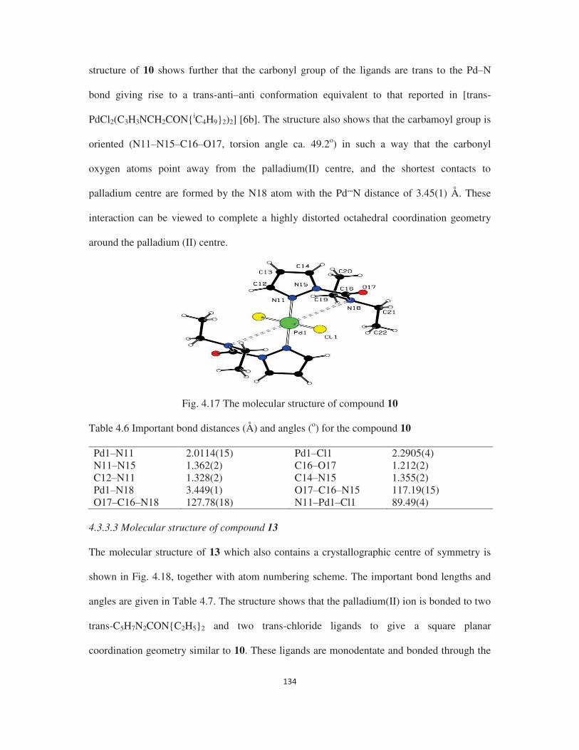
structure of 10 shows further that the carbonyl group of the ligands are trans to the Pd N
bond giving rise to a trans-anti anti conformation equivalent to that reported in [trans-
PdCl2(C3H3NCH2CON{iC4H9}2)2] [6b]. The structure also shows that the carbamoyl group is
oriented (N11 N15 C16 O17, torsion angle ca. 49.2o) in such a way that the carbonyl
oxygen atoms point away from the palladium(II) centre, and the shortest contacts to
palladium centre are formed by the N18 atom with the Pd...N distance of 3.45(1) Å. These
interaction can be viewed to complete a highly distorted octahedral coordination geometry
around the palladium (II) centre.
Fig. 4.17 The molecular structure of compound 10 Table 4.6 Important bond distances (Å) and angles (o) for the compound 10 Pd1 N11 2.0114(15) Pd1 Cl1 2.2905(4) N11 N15 1.362(2) C16 O17 1.212(2) C12 N11 1.328(2) C14 N15 1.355(2) Pd1 N18 3.449(1) O17 C16 N15 117.19(15) O17 C16 N18 127.78(18) N11 Pd1 Cl1 89.49(4)
4.3.3.3 Molecular structure of compound 13
The molecular structure of 13 which also contains a crystallographic centre of symmetry is
shown in Fig. 4.18, together with atom numbering scheme. The important bond lengths and
angles are given in Table 4.7. The structure shows that the palladium(II) ion is bonded to two
trans-C5H7N2CON{C2H5}2 and two trans-chloride ligands to give a square planar
coordination geometry similar to 10. These ligands are monodentate and bonded through the

pyrazole nitrogen atom to the palladium(II) chloride. The observed Pd Cl (2.3115(4) Å) and
Pd N (2.0217(13) Å) distances agree well with those of previously reported values [6b, 24,
25-27, 28]. The observed Pd N distance (2.0217(13) Å) in 13 is slightly longer that in 10
(2.0114(5) Å), and supports our conclusion from the IR spectra that the pyrazole ligands form
stronger bonds with palladium(II) than dimethyl pyrazole ligands. The position of the
NCONC2H5 groups show that the molecule exists in the anti-conformation, similar to that
reported for [trans-PdCl2(C5H7N2CH2OCH2CH2OCH3)2] [24], [trans-
PdCl2(C3H3NCH2CON{iC4H9}2)2] [6b], [trans-PdCl2(C5H7N2CH2OH)2] [25], [trans-
PdCl2(C3H3N2CH2CH2OH)2] [26], and [trans-PdCl2(C10H9N2CH2OH)2] [27]. This structure
also shows that the carbonyl group of the ligand is cis to the Pd N bond, thus giving rise to a
trans-anti syn conformation. The structure also shows that the carbamoyl group is oriented
(N11 N15 C16 O16, torsion angle ca. 132.8o) in such a way that the carbonyl oxygen atoms
point towards the palladium(II) centre, with a Pd...O distance of 3.226(1) Å. These interaction
can be viewed to complete a highly distorted octahedral coordination geometry around the
palladium(II) centre. This type of weak Pd...O interaction with distorted octahedral geometry
around palladium(II) ion is by no means unknown and has been reported previously in
compounds in which the bond lengths are as follows: 2.651 Å in [{(2,4,6-
trimethoxyphenyl)3P}2Pd(BF4)2] [29], 3.09 Å in [trans-PdCl2(C5H7N2CH2OCH2CH2OCH3)2]
[24], 2.95 Å in [trans-PdCl2(Me2NNC10H14O)2] [30], 2.92 3.03 Å in [trans-
PdBr2(Me2NNC10H14O)2] [31], 3.476 Å in [trans-PdCl25-C5H5}- 5-
C5H4}CHN(C6H4OH))2] [32] and 3.232 3.548 Å in [trans-PdCl2(Me-AcGIcN)2] (where Me-
AcGIcN = methyl, 3,4,6-tri O-acetyl, 2-amino, 2- -D glucopyranoside) [33], 2.708 Å
in [PdCl2(8-(2-pyridyl methyl) quinoline] [34] and 2.630 2.673 Å in [PdCl2 hexakis(8-
quinolyoxymethyl)benzene] [35]. The structural studies clearly reveal that the methyl group

at position 13 of the pyrazole group plays an important role in determining the conformation
around the palladium(II) chloride.
Fig. 4.18 The molecular structure of compound 13
Table 4.7 Important bond distances (Å) and angles (o) for the compound 13 Pd1 N11 2.0217(13) Pd1 Cl1 2.3115(4) N11 N15 1.3747(16) C16 O17 1.2182(19) Pd1 O16 3.226(1) O16 C16 N15 118.88(14) O16 C16 N17 125.65(15) N11 Pd1 Cl1 90.15(4)
4.3.3.4 Theoretical studies
Geometry of the two ligands (L2 and L5) and the corresponding Pd(II) chloride complexes
(10 and 13) has been fully optimized to obtain the most stable structures applying the
includes an empirical dispersion correction. Geometry
optimization has been carried out with different possible conformations as the initial structure
for the ligands. Both ligands have an anti conformation with respect to carbonyl O and ring N
(N11) in their most stable structure under isolated condition as shown in Fig. 4.19. For the
two complexes, X-ray structures have been used as the input structures for optimization.
Geometrical parameters of both the complexes in the most stable structures have been
observed to be very close (bond length ±0.1 Å and bond angle ±1o) to the X-ray
crystallography data. Calculated Mulliken atomic charges for both the ligands are listed in
Table 4.8. Based on these values, ligand L5 is expected to be a better donor than ligand L2 as
the negative charge over the N11 atom is predicted to be greater in L5 (-0.28 a.u.) than that in

L2 (0.13 a.u.). The most stable structures of the Pd(II) chloride complexes with these two
ligands are also displayed in Fig. 4.19. Complex 10 does show the ligand L2 to retain its
conformation. However in complex 13, the two ligands L5 undergo rotation along the N15
C16 bond and as a result carbamoyl O and ring N (N11) makes a syn conformation in the
most stable structure. This produces a distorted octahedral Pd(II) chloride complex with this
simple ligand, L5. Due to the presence of CH3 groups in ligand L5, two ligands may feel
steric hindrance to approach Pd(II)Cl2 moiety in its original anti conformation. To reduce
steric repulsion, the ligands undergo rotation along N15 C16 bond to form the complex in
Pd1 N11
N15) which is 73.0o in complex 13 compared to 50.2o in complex 10. Note that the respective
dihedral angles in the X-ray crystal structures are 60.8o and 45.2o. It can be concluded that the
larger dihedral angle in 13 reduces the steric repulsion between the CH3 group of one ligand
and the C2H5 group of the other. Calculated Mulliken atomic charges over selected atoms of
these complexes are supplied in Table 4.8. In the case of 10, the central Pd atom is highly
negative (-1.61 a.u.) in contrast to complex 13 where the central Pd atom is hardly negative (-
0.10 a.u.). This suggests that the pyrazole based ligand L2 is the better donor and thus forms a
stronger Pd N bond than the dimethyl pyrazole based ligand L5. This prediction is supported
by IR results where the shift in carbonyl frequency is larger for the pyrazol based ligand than
for the dimethyl pyrazole based ligands as discussed in the above section. The binding energy
of these ligands with Pd(II)Cl2 to form respective complexes has been calculated via
dissociation energy curves as shown in Fig. 4.20a. Potential energy curves have been
calculated by keeping the two ligands symmetrically away from the central Pd atom at a fixed
distance till the potential energy curve reaches the dissociation limit. From these curves, the
binding energy (BE) per ligand of 13 is predicted to be larger by 5.5 kcal/mol than that of 10.
It will be noted that these BE values apparently contradict our previous conclusion that the

pyrazole based ligand L2 forms a stronger Pd N bond than the dimethyl pyrazole based
ligand L5. However, in case of 13 the stability due interaction between carbamoyl O with Pd
is very small as they are far off (3.226 Å). In fact, visualization of specific frontier molecular
orbital (FMO) in 13 suggests an interaction between the pz orbital of O and the dxz orbital of
Pd atom as depicted in Fig. 4.20b, particularly as the spatial orientation of the two orbitals
which have the same symmetry (shown by color) is favorable for the interaction. This
additional interaction in complex 5 may be the reason for the extra stability of complex 5
over 2. The geometry of the complex formed between the Pd(II)Cl2 unit and ligand L2
keeping the N11 and O atoms in the syn conformation by rotating along the N15 C16 bond
as shown in Fig. 4.20b, has also been optimized at the same level of theory. The complex
thus formed has been observed to be less stable than 10 by 6.0 kcal/mol. The calculated
Pd N1 N2) is larger by 1.2o than that in complex 10. To examine the
other structural possibility for 13, geometry of the complex formed between Pd(II)Cl2 unit
and ligand L5 keeping N11 and O atoms the in anti conformation has also been optimized.
This complex has been predicted to be less stable than complex 13 by 2.2 kcal/mol. The
Pd N1 N2) in the new complex is smaller by 11.3o than that
in complex 13.
Fig. 4.19 Minimum energy structures of two ligands (L2 and L5) and their complexes (10 and 13) optimized with DFT.

Fig. 4.20 (a) Dissociation energy curves of Pd(II)Cl2 complexes 10 and 13 calculated onal. (b) A frontier molecular orbital (FMO) of complex 13
showing the interaction between of O and Pd atoms. Table 4.8 Calculated Mulliken atomic charges (in a.u.) for selected atoms in L2, L5, 10 and
13 (Fig. 4.19 for atom numbering).
Atom L2 L5 10 13 Pd -1.61 -0.10 Cl -0.23 -0.32 N11 -0.13 -0.28 +0.33 +0.30 N15 -0.10 -0.07 -0.04 +0.02 N18/N17 -0.24 -0.23 +0.01 +0.15 O -0.51 -0.48 -0.36 -0.28 4.4 Conclusions
The coordination chemistry of the bifunctional ligand, carbamoyl pyrazole with uranyl
nitrate, shows that it acts as a bidentate chelating ligand bonding through both the carbamoyl
oxygen and pyrazolyl nitrogen atoms. However, the analogous carbamoyl 3,5-dimethyl
pyrazole ligand forms a second sphere complex with the uranyl nitrate. The complex
chemistry of carbamoyl pyrazole ligands could be explained on the basis of steric effect, and
this effect controls the complexation reaction.
Whereas both the carbamoyl pyrazole and carbamoyl 3,5-dimethyl pyrazole ligands act as
monodentate ligands and bonded through the N atom with the palladium (II) chloride. The
complex chemistry of substituted pyrazole with palladium(II) chloride shows that geometry
around the palladium(II) chloride is sterically controlled to give a highly distorted octahedral

geometry around palladium(II) ion. Theoretical studies revealed that the orientation of the
carbonyl group attached to the nitrogen atom is also sterically controlled by the methyl group
at position 5 of the pyrazolyl ring during complexation to give different conformations.
4.5 References
1. Anne E.V. Gorden, Michael A. DeVore, Branson A. Maynard, Inorg. Chem. 52 (2013)
2. Gabriella Bombieri, Inorg. Chim. Acta 139 (1987) 21-32.
3. Simon Cotton; Lanthanide and Actinide Chemistry, John Wiley & Sons Ltd, 2006.
4. (a) R.D. Hancock, A.E. Martell, Chem. Rev. 89 (1989) 1875 1914. (b) A.E. Martell, R.D.
Hancock, Metal complexes in aqueous solution; Plenum Press: New York, 1996.
5. S. Kannan, J.S. Gamare, K.V. Chetty, M.G.B. Drew, Polyhedron 26 (2007) 3810 3816.
6. (a) S. Kannan, K.V. Chetty, V. Venugopal, M.G.B. Drew, Dalton Trans. (2004)
3604 3610. (b) S. Kannan, M.G.B. Drew, Inorg. Chim. Acta (2007) 3647 3649. (c) S.B.
Deb, J.S. Gamare, S. Kannan, M.G.B. Drew, Polyhedron 28 (2009) 2673 2678.
7. (a) A.P. Dimitrios, N. Frank, J. Chem. Theory Comput. 7 (2011) 677 684. (b) K.L.
Schuchardt, B.T. Didier, T. Elsethagen, L. Sun, V. Gurumoorthi, J. Chase, J. Li, T.L.
Windus, J. Chem. Inf. Model. 47 (2007) 1045 1052. (c) W. Schmidt, K.K. Baldridge, J.A.
Boatz, S.T. Elbert, M.S. Gordon, J.H. Jensen, S. Koseki, N. Matsunaga, K.A. Nguyen, S.J.
Su, T.L. Windus, M. Dupuis, J.A. Montgomery, J. Comput. Chem. 14 (1993) 1347 1363.
8. Y. Minenkov, A. Singstad, G. Occhipinti, V.R. Jensen, Dalton Trans. 41 (2012) 5526.
9. (a) T.R. Varga, A.C. Benyei, Z. Fazekas, H. Tomiyasu, Y. Ikeda, Inorg. Chim. Acta 342
(2003) 291 294. (b) N. Koshino, M. Harada, M. Nogami, Y. Morita, T. Kikuchi, Y. Ikeda,
Inorg. Chim. Acta 358 (2005) 1857 1864.
10. S. Kannan, S.B. Deb, J.S. Gamare, M.G.B. Drew, Polyhedron 27 (2008) 2557 2562.
11. A.R. de Aquino, G. Bombieri, P.C. Isolani, G. Vicentini, Zukerman Schpector, J. Inorg.
Chim. Acta 306 (2000) 101 106.
12. (a) S.M. Bowen, E.N. Duesler, R.T. Paine, Inorg. Chem. 21 (1982) 261 265. (b) J.
Caudle, E.N. Duesler, R.T. Paine, Inorg. Chim. Acta 110 (1985) 91 100.
13. J.M. Gil, F.J.M. Gil, A. Perales, J. Fayos, M.M. Ripoll, Acta Crystallogr. C 39 (1983)
44 45.
14. P. Charpin, M. Lance, M. Nierlich, D. Vigner, Acta Crystallogr. C 43 (1987) 442 445.
15. P. Charpin, M. Lance, M. Nierlich, D. Vigner, Acta Crystallogr. C 42 (1986) 560 563.

16. (a) J.C. Berthet, M. Nierlich, M. Ephritikhine, Chem. Commun. (2003) 1660 1661. (b)
J.C. Berthet, M. Nierlich, M. Ephritikhine, Dalton Trans. (2004) 2814 2821.
17. (a) N.W. Alcock, D.J. Flanders, D. Brown, Dalton Trans. (1985) 1001 1007. (b) N.W.
Alcock, D.J. Flanders, M. Pennington, D. Brown, Acta Crystallogr. C 44 (1988) 247 250.
18. (a) R.D. Rogers, A.H. Bond, W.G. Hipple, A.N. Rollins, R.F. Henry, Inorg. Chem. 30
(1991) 2671 2679. (b) G. Bombieri, G. De Paoli, A. Cassol, A. Immirzi, Inorg. Chim.
Acta 18 (1976) L23 L24. (c) P.G. Eller, R.A. Penneman, Inorg. Chem. 15 (1976)
2439 2442. (d) S. Kannan, S.S. Raj, H.K. Fun, Polyhedron 20 (2001) 2145 2150.
19. H.M. Colquhoun, J.F. Stoddart, D.J. Williams, Angew. Chem., Int. Ed. Engl. 25 (1986)
487 507.
20. (a) C. Villiers, P. Thuery, M. Ephritikhine, Polyhedron 23 (2004) 1613 1618. (b) A.M.J.
Lees, A.W.G. Platt, Acta Crystallogr. C 65 (2009) m10 m13. (c) M. Pourayoubi, J.A.
Golen, M.R. Chaijan, V. Divjakovic, M. Negari, A.L. Rheingold, Acta Crystallogr. C 67
(2011) m160 m164.
21. (a) S. Alagar, M. Subha Nandhini, R.V. Krishnakumar, S. Kannan, S. Natarajan, Acta
Crystallogr. E 59 (2003) m524 m526. (b) S. Kannan, V. Venugopal, M.R.A. Pillai, P.A.
Droege, C.L. Barnes, Polyhedron 15 (1996) 97 101. (c) S. Kannan, A. Usman, H.K. Fun,
Polyhedron 21 (2002) 2403 2407. (d) S. Kannan, M.R.A. Pillai, V. Venugopal, P.A.
Droege, C.L.Barnes, Inorg. Chim. Acta 254 (1997) 113 117. (e) S. Rajagopal, S. Kannan,
V. Venugopal, R.V. Krishnakumar, M. Subha Nandhini, S.J. Natarajan, Chem. Res.
(2003) 468 469.
22. O. Clement, B.M. Rapko, B.P. Hay, Coord. Chem. Rev. 170 (1998) 203 243.
23. (a) S. Kannan, C.L. Banes, P.B. Duval, Chem. Commun. (2005) 5997 5998. (b) S.
Kannan, C.L. Barnes, P.B. Duval, Inorg. Chem. 44 (2005) 9137 9139.
24. A. Boixassa, J. Pons, X. Solans, M.F. Bardia, J. Ros, Inorg. Chim. Acta 346 (2003) 151.
25. A. Boixassa, J. Pons, A. Virgili, X. Solans, M.F. Bardia, J. Ros, Inorg. Chim. Acta 340
(2002) 49.
26. A. Boixassa, J. Pons, X. Solans, M.F. Bardia, J. Ros, Inorg. Chim. Acta 357 (2004) 733.
27. J.A. Perez, V. Montoya, J.A. Ayllon, M.F. Bardia, T. Calvet, J. Pons, Inorg. Chim. Acta
394 (2013) 21.
28. K. Li, J. Darkwa, I.A. Guzei, S.F. Mapolie, J. Organomet. Chem. 660 (2000) 108.
29. K.R. Dunbar, J.S. Sun, Chem. Commun. (1994) 2387.

30. M.F.N.N. Carvalho, M.T. Duarte, R. Herrmann, Collect. Czech. Chem. Commun. 71
(2003) 302.
31. M.F.N.N. Carvalho, A.M. Galvao, A.S.D. Ferreira, J. Organomet. Chem. 694 (2009) 2061.
32. S. Perez, C. Lopez, A. Caubet, X. Solans, M.F. Bardia, M. Gich, E. Molins, J. Organomet.
Chem. 692 (2007) 2402.
33. S.A. Johannesen, B.O. Petersen, J.O. Duus, T. Skrydstrup, Inorg. Chem. 46 (2007) 4326.
34. M.R.A. Al-Mandhary, P.J. Steel, Inorg. Chim. Acta 351 (2003) 7.
35. M.R.A. Al-Mandhary, C.M. Fitchett, P.J. Steel, Aust. J. Chem. 59 (2006) 307.

5.1 Introduction
The separation of the metal ions from high level liquid waste after spent fuel reprocessing is
very challenging [1-2] and an important step in the back end of fuel cycle in nuclear industry
to reduce the long term radiotoxicity of the waste and further utilization of the precious
elements. N-substituted amides are proposed to be very promising extractants for the solvent
extraction removal of actinides from nitric acid medium [3-5]. In this respect, N-heterocyclic
amide ligands are found to be a potential candidate to form stable complex with actinides and
show structural variety in the solid state [6]. This type of ligands has an additional advantage
compared to the simple N-substituted amide ligands. These ligands contain both hard oxygen
and soft nitrogen donors in combination, which can help to extract both hard actinides as well
as soft fission product elements during solvent extraction from the high level liquid waste
solution [7].
N-heterocyclic amides such as picolinamide or pyrazole 2-carboxamide show a great
tenability in bonding while forming complexes with different type of metal ions. In few
cases, it acts as a chelating bidentate ligand with either pyridine-N and amido-O or pyridine-
N and amido-N as donors to the metal center [8-12], while in other cases, it acts as a
monodentate ligand with either pyridine N or amido O atom as donor centers [13-14]. The
type of metal ions and electronic as well as steric factor of the ligands are the reason behind
the bonding diversity in the coordination complexes. These findings clearly indicate that by
tuning the alkyl group of the amides, it is possible to extract/separate metal ions of interest
from any mixture of metal ions which is directly relevant to nuclear-waste management
processes.
Although many structures of picolinic acid with metal complexes including palladium are
known [15-19], the corresponding picolinamide complexes with palladium are reported here
for the first time. In fact, to the best of our knowledge, only one X-ray structure of Pd(II) with

picolinamide is reported to date [20], where the coordination is with methyl palladium and
not simple Pd(II) ion. Since this ligands have hard donor group (carbamoyl) in addition to
soft nitrogen group, it is expected to form complexes with actinides/lanthanides during
separation process. So, the understanding of complexes chemistry of this ligands with
actinides is also important.
We report the synthesis, characterization and structural studies of Pd(II) and uranyl (VI)
complexes derived from picolinamide based ligands. The Pd/ U complexes synthesized and
characterized herein are aimed to find the bonding mode for these ligands with
palladium(II)/uranyl(VI) ions. This study will be useful for designing new ligands for the
separation of palladium from high level liquid waste in solution. Particularly, we are very
much interested in understanding the crucial role played by increasing the bulkiness of the
ligand upon complexation with Pd(II) ion.
5.2 Experimental 5.2.1 Synthesis of pyridine 2-carboxamide ligands 5.2.1.1 Synthesis of C5H4NCON(iC3H7)2 ( L
1)
A solution of 2-Pyridine carbonyl chloride (2.12 g, 15 mmol) in CH2Cl2 (30 mL) was slowly
added to a solution of di-isopropyl amine (1.52 g, 15 mmol) and triethyl amine (4.2 mL, 30
mmol) in CH2Cl2 (15 mL) at 0°C. After the addition, the mixture was slowly brought to room
temperature and stirred for about 24 h. The solution was treated with 50 ml of 10% HCl and
the organic layer was separated. The separated organic solution was dried over Na2SO4,
filtered and the solvent was evaporated in vacuo to give the final product (69% yield). Anal.
Calcd. for C12H18N2O: C, 69.90; H, 8.73; N, 13.59. Found: C, 69.22; H, 8.99; N, 13.26. IR
(ATR, /cm-1): 1624(s) (C=O). 1H NMR (300 MHz, CDCl3) 8.62 (s, 1H), 7.88 (s, 1H), 7.48
(d, J= 9.3Hz, 1H), 7.30 (d, J= 5.1Hz, 1H), 3.78 (q, J= 6.3Hz, 1H), 3.56 (d, J= 6.3Hz, 1H),
1.55 (t, J= 6.3Hz, 6H), 1.19 (s, 6H).

5.2.1.2 Synthesis of C5H4NCON(iC4H9)2 (L2)
This was prepared similar to that of L1 by taking di-isobutyl amine instead of di-isopropyl
amine. Yield: 72%. Anal. Calcd. for C14H22N2O: C, 71.79; H, 9.40; N, 11.97. Found: C,
71.42; H, 9.68; N, 12.11. IR spectrum (ATR, /cm-1): 1628(s) (C=O). 1H NMR (300 MHz,
CDCl3) 8.60 (s, 1H), 7.82 (t, J= 7.2Hz, 1H), 7.57 (d, J= 7.5Hz, 1H), 7.36 (d, J= 4.5Hz, 1H),
3.37 (d, J= 7.5Hz, 2H), 3.28 (d, J= 7.5Hz, 2H), 2.15 (p, J= 6.6Hz, 1H), 1.77 (p, J= 6.3Hz,
1H), 1.00 (d, J= 6.6Hz, 6H), 0.73 (s, 6H).
5.2.1.3 Synthesis of C5H4NCONH(tC4H9) (L3)
This was prepared similar to that of L1 by taking tert-butylamine. Yield: 65%. Anal. Calcd.
for C10H14N2O: C, 67.42; H, 7.87; N, 15.73. Found: C, 67.07; H, 7.99; N, 15.41. IR (ATR,
/cm-1): 1675(s) (C=O), 3373(m) (-NH). 1H NMR (300 MHz, CDCl3) 8.51 (d, J= 3.9Hz, 1H),
8.17 (d, J= 7.8Hz, 1H), 8.04 (s, 1H, NH), 7.84 (t, J= 7.5Hz, 1H), 7.40 (t, J= 6.0Hz, 1H), 1.48
(s, 9H).
5.2.2 Synthesis of uranyl complexes of pyridine 2-carboxamide ligands
5.2.2.1 Synthesis of [UO2(NO3)2{C5H4NCON(iC3H7)2}] (1)
To a solution of N,N-diisopropyl pyridine-2-carboxamide (300 mg, 1.46 mmol) (L1) in
chloroform, solid UO2(NO3)2.6H2O (365 mg, 0.71 mmol) was added with stirring. The
solution was allowed to stir until all uranyl nitrate dissolved to give a clear solution. This
solution was filtered and layered with isooctane. This solution on slow evaporation yielded a
yellow crystalline solid. This was filtered, washed with hexane and air dried. Yield: 89%.
Anal. Calc. for C12H18N4O9U: C, 24.0; H, 3.0; N, 9.33. Found: C, 24.21; H, 3.38; N, 9.52. IR
-1) : 1590 (C=O), 935 (U=Oassy). 1HNMR (300 MHz, CD3COCD3) 8.56 (d, J= 4.8Hz,
1H), 7.95 (t, J= 6.9Hz, 1H), 7.53 (t, J= 8.4Hz, 2H), 1.54 (t, J= 8.1Hz, 6H), 1.22 (s, 6H).

5.2.2.2 Synthesis of [UO2(NO3)2{C5H4NCON(iC4H9)2}] (2)
This was prepared similarly to 1 by taking UO2(NO3)2.6H2O (300 mg, 0.60 mmol) and L2
(293 mg, 1.25 mmol). Yield 87%. Anal. Calc. For C14H22N4O9U: C, 26.75; H, 3.50; N, 8.92.
-1) 1590 (C=O), 934 (U=Oassy). : 1H NMR (300
MHz, CD3COCD3) 8.59 (s, 1H), 7.91 (s, 1H), 7.57 (s, 1H), 7.45 (s, 1H), 3.38 (d, J= 6.9Hz,
2H), 3.31 (d, J= 6.3Hz, 2H), 1.79 (d, J= 5.1Hz, 1H), 1.26 (s, 1H), 0.95 (d, J= 5.7Hz, 6H),
0.69 (d, J= 5.7Hz, 6H).
5.2.2.3 Synthesis of [UO2(NO3)2{C5H4NCONH(tC4H9)}] (3)
This was prepared similarly to 1 by taking UO2(NO3)2.6H2O (300 mg, 0.60 mmol) and L3
(240 mg, 1.3 mmol). Yield 86%. Anal. Calc. For C10H14N4O9U: C, 20.98; H, 2.45; N, 9.79.
-1) : 1620 (C=O), 899 (U=Oassy). 1H NMR
(300 MHz, CD3COCD3) 8.67 (d, J= 18.3Hz, 1H), 8.13 (s, 2H), 7.72 (s, 1H), 1.56 (s, 9H).
5.2.3 Synthesis of palladium complexes of pyridine 2-carboxamide ligands
5.2.3.1 Synthesis of [PdCl2{C5H4NCON(iC3H7)2 }2] (4)
To an acetonitrile solution of PdCl2 (200 mg, 1.13 mmol), L1 (466 mg, 2.26 mmol) was
added and refluxed for 2 h. This solution was evaporated to dryness completely, washed with
hexane and dried. The resulting residue was extracted in CH2Cl2 and filtered to remove
insoluble materials. The solution was layered with 5 ml of iso-octane and allowed to
evaporate slowly. This process deposited brownish yellow colored product (yield. 91%).
Anal. Calc. For C24H36N4O2Cl2Pd: C, 48.86; H, 6.11; N, 9.50. Found: C, 48.71; H, 6.48; N,
-1) 1621 (C=O). 1H NMR (300 MHz, DMSO-d6): 8.54 (s, 1H), 7.88 (t, J=
8.1Hz, 1H), 7.61 (q, J= 6.9Hz, 2H), 3.60 (d, J= 6.0Hz, 2H), 1.43 (d, J= 3.3Hz, 6H), 1.11 (s,
6H).
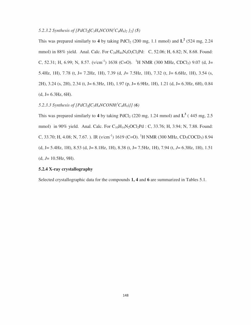
5.2.3.2 Synthesis of [PdCl2{C5H4NCON(iC4H9)2 }2] (5)
This was prepared similarly to 4 by taking PdCl2 (200 mg, 1.1 mmol) and L2 (524 mg, 2.24
mmol) in 88% yield. Anal. Calc. For C28H44N4O2Cl2Pd: C, 52.06; H, 6.82; N, 8.68. Found:
C, 52.31 -1) 1638 (C=O). 1H NMR (300 MHz, CDCl3) 9.07 (d, J=
5.4Hz, 1H), 7.78 (t, J= 7.2Hz, 1H), 7.39 (d, J= 7.5Hz, 1H), 7.32 (t, J= 6.6Hz, 1H), 3.54 (s,
2H), 3.24 (s, 2H), 2.34 (t, J= 6.3Hz, 1H), 1.97 (p, J= 6.9Hz, 1H), 1.21 (d, J= 6.3Hz, 6H), 0.84
(d, J= 6.3Hz, 6H).
5.2.3.3 Synthesis of [PdCl2{C5H4NCONH(tC4H9)}] (6)
This was prepared similarly to 4 by taking PdCl2 (220 mg, 1.24 mmol) and L3 ( 445 mg, 2.5
mmol) in 90% yield. Anal. Calc. For C10H14N2OCl2Pd : C, 33.76; H, 3.94; N, 7.88. Found:
C, 33.70; H, 4.08; N, 7.67. ). -1) 1619 (C=O). 1H NMR (300 MHz, CD3COCD3) 8.94
(d, J= 5.4Hz, 1H), 8.53 (d, J= 8.1Hz, 1H), 8.38 (t, J= 7.5Hz, 1H), 7.94 (t, J= 6.3Hz, 1H), 1.51
(d, J= 10.5Hz, 9H).
5.2.4 X-ray crystallography
Selected crystallographic data for the compounds 1, 4 and 6 are summarized in Tables 5.1.

Table 5.1 Crystallographic data for compounds 1, 4 and 6
1 4 6 Empirical formula C12H18N4O9U C28H42N5O2Cl2Pd C12H17N3OCl2Pd Formula weight 600.32 657.99 396.61 Crystal system monoclinic triclinic monoclinic Space group P 1 21/n 1 P -1 P 1 21/a 1 a (Å) 10.9779(9) 7.5131(10) 19.631(5) b (Å) 14.6010(4) 9.7076(12) 10.5763(8) c(Å) 12.4580(9) 12.2336(17) 12.855(3)
o) 111.894(8) 76.728(12) 141.81(5) Volume (Å3) 1852.9(2) 836.2(2) 1650.3(13) Z (formula unit) 4 1 4 Calculated density (g/cm3)
2.152 1.334 1.596
-1) 25.154 6.203 12.021 Reflections collected/unique
3442/2304 3003/2803 3075/2127
Data/restrains/ parameters
3442/0/240 3003/48/184 3075/0/176
Goodness of fit on F2 0.983 1.128 1.096 Final R1 indices
0.0824 0.0790 0.1021
wR2 indices (all data) 0.1141 0.0878 0.1239
2(Fo
2) + (0.1516P)2] for 1 2(Fo2) + (0.1690P)2 + 1.3617P] for 4 2(Fo
2) + (0.2000P)2] for 6, where P = (Fo
2 + 2Fc2)/3.
5.3 Results and discussion
5.3.1. Synthesis and characterization of N-substituted pyridine 2-carboxamide ligands
Reaction of 2-pyridine carbonyl chloride with corresponding N-substituted amines in presence
of triethylamine as base in benzene yielded the corresponding ligands (L1 to L3). The IR
spectra of all ligands (Fig. 5.1) show the presence of the carbamoyl group in the synthesized
compounds.

Fig. 5.1 IR spectra of ligand L1 (above) and L3 (below)
Fig 5.2 showed the 1H NMR spectrum of ligand L2. The singlet at 8.60 ppm, the triplet at
7.82 ppm, the doublet at 7.57 ppm and the doublet at 7.36 ppm are attributed to the ring
protons of the pyridine ring at 6, 5, 3 and 4-position respectively. At 3.37 and 3.28 ppm, the
doublets are assigned to four CH2 protons (chemically non-equivalent at room temperature)
of two isobutyl groups. Both the signals are split into a doublet, because the methylene group
protons are coupled with one adjacent CH proton. Similarly, two multiplets at 2.15 and 1.77
NN
O
N
HN
O

ppm are attributed to two CH protons (chemically non-equivalent at room temperature) of
two isobutyl groups. Finally, twelve CH3 protons (chemically non-equivalent at room
temperature) of two substituted isobutyl groups resonate as two doublets at 1.00 and 0.71
ppm. Both the signals are split into a doublet, because the methyl group protons are coupled
with one adjacent CH proton. The C, H, N analysis supports the expected stoichiometry for
the compounds.
N
O
Na
b c
d
e
f
g
h
i
ij
j
Fig. 5.2 1H NMR spectrum of ligand L2
5.3.2 Synthesis and structural studies of uranyl complexes
5.3.2.1 Synthesis and complexation studies of pyridine 2-carboxamide ligands with uranyl
nitrate
The reaction of ligands L1 L3 with [UO2(NO3)2·6H2O] yielded compounds 1 3. C, H, and N
analyses revealed that the ratio of ligand to uranyl nitrate is 1:1 in all compounds. The IR
spectra of 1 3 (Fig. 5.3 for 2) show that the water molecules from the starting compound
[UO2(NO3)2·6H2O] are completely replaced by the ligand and that the ligand is bonded
through the amide oxygen atom to the uranyl group. The observed frequency difference for a

carbamoyl ( CO = 34 55 cm 1, where CO CO(free ligand) CO(coordinated)) group is
consistent with the supposition that the carbamoyl group is bonded to the uranyl group
directly. This difference is comparable in magnitude with those observed in [UO2(NO3)2(N-
cyclohexyl-2-pyrrolidone)2] [21], [UO2(NO3)2(1,3-dimethyl-2-imidazolidone)2] [22]
[UO2(NO3)2{iC3H7CON(iC4H9)2}2] [23], [UO2(NO3)2(
iC3H7)2NCOCH2CON(iC3H7)2] [24],
and [UO2(NO3)2(C15H27N3O)] [25].
4000 3500 3000 2500 2000 1500 1000 500
0.92
0.94
0.96
0.98
1.00
1.02
3582
2955 23
49 2190
2040
1962
1590
1523
1434 1367
1273
1094
1023
934
807
743 671
627
573
535.2
9217
% Tr
ansm
ittanc
e
Wavenumber (Cm-1)
Fig. 5.3 IR spectrum of complex 2
Fig 5.4 showed the 1H NMR spectrum of complex 1. The singlet at 8.65 ppm and the triplet
at 7.95 ppm are assigned to the ring protons of the pyridine ring at 6 and 5 position
respectively. The multiplet at 7.53 ppm is due to two ring protons of the pyridine ring at 3
and 4 position. Next, two doublets at 1.54 and 1.22 ppm are attributed to twelve CH3 protons
(chemically non-equivalent at room temperature) of two substituted isopropyl groups. Two
additional peaks are appeared at 3.56 and 2.09 ppm due to the residual water impurity and
CH3COCH3 molecule in the solvent acetone-d6. The pyridyl protons are deshielded by ca.
0.2-0.3 ppm with respect to the free ligand indicating that the bonding between pyrazolyl
nitrogen and uranyl group persists in solution. It is apparent from IR and NMR spectral
NN
O
[UO2(NO3)2{C5H4NCON(iC4H9)2}]
C5H4NCON(iC4H9)2 =

results that the ligand acts as a bidentate chelating ligand and bonds through the carbamoyl
and pyrazolyl nitrogen to uranyl group. The structure of 1 has been determined by single
crystal X-ray diffraction methods and confirms the spectral and analysis results.
C5H4NCON(iC3H7)2
N
O
Na
b cd
[UO2(NO3)2{C5H4NCON(iC3H7)2}]
e
f
g
g
hh
=
Fig. 5.4 1H NMR spectrum of complex 1
5.3.2.2 Molecular structure of compound 1
The structure of 1 is shown in Fig. 5.5, and selected bond distances and angles are given in
Table 5.2. The structure of 1 shows that the uranium atom is surrounded by one nitrogen and
seven oxygen atoms in a slightly distorted hexagonal bipyramidal geometry. Four oxygen
atoms of the two bidentate nitrate groups, together with one oxygen and one nitrogen atom of
bidentate carboxamide ligand, form the equatorial hexagonal plane. The UO5N atoms in the
equatorial plane show a root mean square (rms) deviation of 0.082 Å. The two uranyl oxygen
atoms occupy the axial positions. This type of coordination is similar to that observed in the
compounds of the bifunctional ligands, malonamide, carbamoyl methyl phosphonate,
carbamoyl methyl phosphine oxide, and carbamoyl methyl pyrazole with uranyl nitrate, such
as [UO2(NO3)2{(iC3H7)2NCO}2] [26], [UO2(NO3)2(iC3H7O)2POCH2CONEt2] [27a],
UO2(NO3)2(C6H5)2POCH2CONEt2] [27b] and UO2(NO3)2(C15H27N3O)] [25]. The U O(amide)

distance (2.377(11) Å) in 1 is comparable in magnitude with those of earlier reported uranyl
nitrate-amide compounds, such as [UO2(NO3)2(N,N-dimethylformamide)2] (2.397(6) Å) [28],
[UO2(NO3)2(tetrabutylglutaramide)2] (2.378(6) Å) [29], [UO2(NO3)2(dibutyldecanamide)2]
(2.37(2) Å) [30], and [UO2(NO3)2(C15H27N3O)] (2.364(7) Å) [25]. The U N(pyridyl) distance
(2.625(14) Å) is very close to the values observed in [UO2(NO3)2(C15H27N3O)] (2.554(9)Å)
[25], [UO2(NO3)2(phenanthroline)] (2.556(2)Å) [31], and [UO2(terpyridine)](OTf)2
((2.567(6), 2.592(6) Å) [32]. The observed average U O(NO3) bond distance 2.493(14) Å is
normal [21,22,25-30]. The angles subtended at the metal atom show that the uranium atom
has slightly distorted hexagonal bipyramidal geometry.
Fig. 5.5 The molecular structure of compound 1
Table 5.2 Important bond distances (Å) and angles (o) for the compound 1
U1-O1 1.727(17) O3-U1-N1 62.6(5) U1-O2 1.738(16) O1-U1-O2 179.4(8) U1-N1 2.615(15) O8-U1-O7 50.8(5) U1-O3 2.364(12) O5-U1-O4 50.5(4) U1-O5 2.499(15) N1-U1-N3 154.7(5) U1-O8 2.459(15) N1-U1-N2 92.8(5) U1-O4 2.501(14) O5-U1-N1 176.5(5) U1-O7 2.507(16) O5-U1-N3 24.7(5)
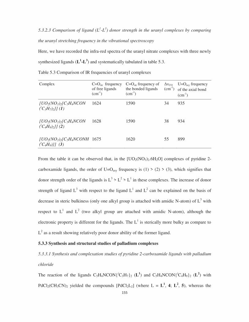
5.3.2.3 Comparison of ligand (L1-L3) donor strength in the uranyl complexes by comparing
the uranyl stretching frequency in the vibrational spectroscopy
Here, we have recorded the infra-red spectra of the uranyl nitrate complexes with three newly
synthesized ligands (L1-L3) and systematically tabulated in table 5.3.
Table 5.3 Comparison of IR frequencies of uranyl complexes
Complex C=Ostr frequency of free ligands (cm-1)
C=Ostr frequency of the bonded ligands (cm-1)
CO
(cm-1) U=Oasy frequency of the axial bond (cm-1)
[UO2(NO3)2{C5H4NCON (iC3H7)2}] (1)
1624 1590 34 935
[UO2(NO3)2{C5H4NCON (iC4H9)2}] (2)
1628 1590 38 934
[UO2(NO3)2{C5H4NCONH(tC4H9)}] (3)
1675 1620 55 899
From the table it can be observed that, in the [UO2(NO3)2.6H2O] complexes of pyridine 2-
carboxamide ligands, the order of U=Oasy frequency is (1) > (2) > (3), which signifies that
donor strength order of the ligands is L3 > L2 > L1 in these complexes. The increase of donor
strength of ligand L3 with respect to the ligand L1 and L2 can be explained on the basis of
decrease in steric bulkiness (only one alkyl group is attached with amidic N-atom) of L3 with
respect to L1 and L2 (two alkyl group are attached with amidic N-atom), although the
electronic property is different for the ligands. The L1 is sterically more bulky as compare to
L2 as a result showing relatively poor donor ability of the former ligand.
5.3.3 Synthesis and structural studies of palladium complexes
5.3.3.1 Synthesis and complexation studies of pyridine 2-carboxamide ligands with palladium
chloride
The reaction of the ligands C5H4NCON{iC3H7}2 (L1) and C5H4NCON{iC4H9}2 (L2) with
PdCl2(CH3CN)2 yielded the compounds [PdCl2L2] (where L = L1, 4; L2, 5), whereas the

similar reaction with C5H4NCONH{tC4H9} (L1) yielded the compound [PdCl2L] (where L =
L3, 6). The IR spectra (Fig. 5.6 for 5) of 4 5 show that the absorption frequencies of the C=O
group in these compounds are almost unshifted with respect to the corresponding free
ligands. It clearly shows that the C=O group is un-coordinated in these compounds and that
the ligands are bonded through the pyridyl nitrogen atom to the metal centre. But the IR
spectrum of compound 6 shows that the absorption frequency for the C=O groups in this
compound is 56 cm-1 red shifted with respect to the corresponding free ligands, which clearly
reveals that the amide group is directly coordinated to the Pd(II) centre in this compound. The
CO frequency difference (between free and coordinated) is more
for the pyrazole based ligands (22 27 cm-1) than the dimethyl pyrazole based ligands (3 14
cm-1), which indicates that the pyrazole ligands are more strongly bonded to the metal centre
than the dimethyl pyrazole ligands. Fig 5.7 showed the 1H NMR spectrum of complex 5. The
spectrum is exactly similar to that of ligand L2 (already discussed in details earlier) since the
complex is derived from the ligand L2. However some of the proton peaks of the ligand in the
spectrum of the complex are deshielded up to 0.5 ppm with respect to the free ligand proving
further evidence for the coordination of the ligand to the metal ion. The structures for
compounds 4 and 6 have been determined by single crystal X-ray diffraction methods and
confirm our interpretation of the spectral data.

4000 3500 3000 2500 2000 1500 1000 500
0.92
0.93
0.94
0.95
0.96
0.97
0.98
0.99
1.00
2952
2008
1638
1423
1287
1096
944
761
675
%Tran
smitta
nce
Wavenumber(cm-1)
Fig. 5.6 IR spectrum of complex 5
C5H4NCON(iC4H9)2
[PdCl2{C5H4NCON(iC4H9)2}2]
=N
O
Na
b cd
e
f
g
h
i
ijj
Fig. 5.7 1H NMR spectrum of complex 5
5.3.3.2 Molecular Structure of compound 4
The molecular structure of 4 which contains a crystallographic centre of symmetry is shown
in Fig. 5.8, together with the atom numbering scheme. The important bond lengths and angles
are given in Table 5.4. The structure shows that the palladium(II) ion is bonded to two trans-
C5H4NCON{iC3H7}2 ligands and two trans-chloride ligands to give a square planar
NN
O
C5H4NCON(iC4H9)2 =
[PdCl2{C5H4NCON(iC4H9)2}2]

coordination. The oxygen atom of the carbamoyl group shows week interaction (3.068 Å)
with palladium centre, which can be viewed to complete a highly distorted octahedral
coordination geometry around the palladium (II) centre. Similar type of coordination is
observed in palladium(II) chloride compounds of carbamoyl methyl pyrazole [14] and
carbamoyl pyrazole ligands [13]. Both the ligands act as monodentate ligands and are bonded
through the pyridyl nitrogen atom to the palladium(II) chloride. The observed Pd Cl
(2.3043(19) Å) and Pd N (2.005(6)Å) distances agree well with previously reported values
[33-34].
Fig. 5.8 The molecular structure of compound 4
Table 5.4 Important bond distances (Å) and angles (o) for the compound 4
Pd1-Cl 2.3043(19) Cl-Pd1-Cl 180.0 Pd1-N1 2.005(6) N1-Pd1-Cl 89.75(17) N1-C1 1.327(11) N1-Pd1-N1 180.0 N1-C5 1.356(10) C1-N1-Pd1 118.8(6) O1-C6 1.240(10) C5-N1-Pd1 123.8(5)
5.3.3.3 Molecular Structure of compound 6
The molecular structure of 6 is shown in Fig. 5.9, together with the atom numbering scheme.
The important bond lengths and angles are given in Table 5.5. The structure shows that the
palladium(II) ion is bonded to one pyridine carboxamide C5H4NCONH{tC4H9} and two
chloride ligands in cis fashion to give a square planar coordination geometry. The

C5H4NCONH{tC4H9} ligand acts as a bidentate chelating ligand and are bonds through both
the pyridyl nitrogen and carbamoyl oxygen to the palladium(II) chloride. The observed Pd Cl
(2.279 (3) Å), Pd N (2.015(9 Å) [35-36] and Pd-O (2.019(9 Å) [20] distances are normal
agree well with previously reported values.
Fig. 5.9 The molecular structure of compound 6
Table 5.5 Important bond distances (Å) and angles (o) for the compound 6
Pd1-Cl1 2.264(4) Cl1-Pd1-Cl2 91.01(13) Pd1-Cl2 2.279(3) O1-Pd1-Cl2 91.1(2) Pd1-O1 2.019(9) O1-Pd1-Cl1 177.7(2) Pd1-N1 2.015(9) N1-Pd1-Cl2 172.1(3) O1-C6 1.294(13) N1-Pd1-Cl1 96.9(3) N1-Pd1-O1 81.0(3)
5.3.3.4 Computational studies
To gain valuable insights on the structure and coordination binding motifs of the
picolinamide ligand to Pd, we have carried out electronic structure calculations at the density
functional theory level. Our optimized structural parameters of species 4 and 6 are in good
agreement with the experimental data (Fig. 5.10). Careful analysis reveals a large deviation in
the structures between 4 and 6 depends on the binding modes of the ligand (i.e. monodentate
vs chelation). In complex 4, there are two L1 (trans to each other) each coordinating with Pd
primarily through the nitrogens (Pd-N bonds are 2.04 Å) and the carbamoyl oxygens are
weakly interacting with Pd (with a bond distance of 3.15 Å). Further, the computed Pd-N

bond lengths in both 4 and 6 are within 0.04Å, which is in good agreement with the
experimental estimates. The Pd-Cl bond lengths are also well reproduced within 0.05Å for
both complexes against the experimental data. Further, in 4, the C=O of the ligand is very
weakly interacting with Pd (3.15Å) as compared to species 6, whereas a direct strong
coordination (Pd-O = 2.11Å) is noted. Although the Pd-O distance seems large to be
considered as an interacting distance, a careful inspection of the optimized structure reveals
that the oxygens are not rotated away and still facing the Pd centre, thus some weak
electrostatic interaction is noted.
Fig. 5.10 Optimized structures (Å) of 4. The complexation energy for 4 (1:2) and 6 (1:1) are -63.4 kJ mol-1 and -77.3 kJ mol-1
respectively (Table 5.6) again suggesting both are facile processes with a preference for the
L3 forming 1:1 complex over the L1 as 1:2. Further, we have also optimized structure of 1:1
complex with L3. We find that this species is less favourable (by -59.8 kJ mol-1) as compared
to 6. Due to the steric hindrance of the ligand L1, a chelate binding motif is less preferable,
thus this species is not experimentally observed. The C=O stretching frequency for 4 and 6
are 1643 cm-1 and 1594 cm-1 respectively which follows the experimental trends. It is very
evident that there is a small blue shift in 4 and a red shift in 6 suggesting, in complex 4 the
C=O oxygen is not bound to Pd and forms a distorted octahedral complex where as in 6 it is
directly coordinated to Pd as a bidentate ligand forming a square planar complex.
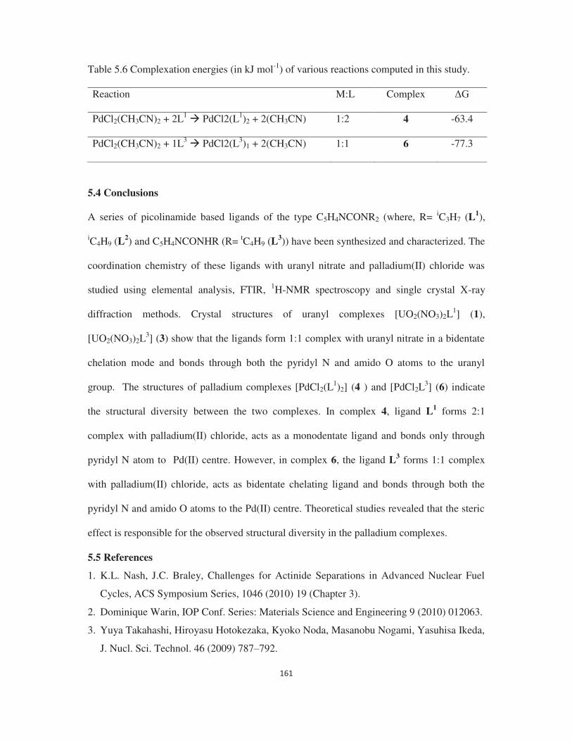
Table 5.6 Complexation energies (in kJ mol-1) of various reactions computed in this study.
Reaction M:L Complex
PdCl2(CH3CN)2 + 2L1 PdCl2(L1)2 + 2(CH3CN) 1:2 4 -63.4
PdCl2(CH3CN)2 + 1L3 PdCl2(L3)1 + 2(CH3CN) 1:1 6 -77.3
5.4 Conclusions
A series of picolinamide based ligands of the type C5H4NCONR2 (where, R= iC3H7 (L1),
iC4H9 (L2) and C5H4NCONHR (R= tC4H9 (L
3)) have been synthesized and characterized. The
coordination chemistry of these ligands with uranyl nitrate and palladium(II) chloride was
studied using elemental analysis, FTIR, 1H-NMR spectroscopy and single crystal X-ray
diffraction methods. Crystal structures of uranyl complexes [UO2(NO3)2L1] (1),
[UO2(NO3)2L3] (3) show that the ligands form 1:1 complex with uranyl nitrate in a bidentate
chelation mode and bonds through both the pyridyl N and amido O atoms to the uranyl
group. The structures of palladium complexes [PdCl2(L1)2] (4 ) and [PdCl2L
3] (6) indicate
the structural diversity between the two complexes. In complex 4, ligand L1 forms 2:1
complex with palladium(II) chloride, acts as a monodentate ligand and bonds only through
pyridyl N atom to Pd(II) centre. However, in complex 6, the ligand L3 forms 1:1 complex
with palladium(II) chloride, acts as bidentate chelating ligand and bonds through both the
pyridyl N and amido O atoms to the Pd(II) centre. Theoretical studies revealed that the steric
effect is responsible for the observed structural diversity in the palladium complexes.
5.5 References
1. K.L. Nash, J.C. Braley, Challenges for Actinide Separations in Advanced Nuclear Fuel
Cycles, ACS Symposium Series, 1046 (2010) 19 (Chapter 3).
2. Dominique Warin, IOP Conf. Series: Materials Science and Engineering 9 (2010) 012063.
3. Yuya Takahashi, Hiroyasu Hotokezaka, Kyoko Noda, Masanobu Nogami, Yasuhisa Ikeda,
J. Nucl. Sci. Technol. 46 (2009) 787 792.

4. Fa-tang Li, Biao Wu, Rui-hong Liu, Xiao-jing Wang, Lan-ju Chen, Di-shun Zhao, Chem.
Eng. J. 274 (2015) 192 199.
5. P.N. Pathak, R. Veeraraghavan, D.R. Prabhu, G.R. Mahajan, V.K. Manchanda, Sep. Sci.
Technol. 34 (1999) 2601.
6. Yingjie Zhang, Mohan Bhadbhade, Jiabin Gao, Inna Karatchevtseva, Jason R. Price,
Gregory R. Lumpkin, Inorg. Chem. Commun. 37 (2013) 219 221.
7. Debasish Das, Bal Govind Vats, Shanmugaperumal Kannan, Dilip K. Maity, Michael G.B.
Drew, Polyhedron 54 (2013) 104 109.
8. J. Qi, H. Ma, X. Li, Z. Zhou, M.C.K. Choi, A.S.C. Chan, Q. Yang, Chem. Commun.
(2003) 1294.
9. H.R. Khavasi, A. Ghanbarpour, A.A. Tehrani, RSC Adv. 6 (2016) 2422.
10. L. Huang, B. Gu, W. Su, P. Yin, H. Li, RSC Adv. 5 (2015) 76296.
11. L. Kirsten, V.D. Schwade, L. Selter, A. Hagenbach, P.C. Piquini, E.S. Lang, U. Abram,
Eur. J. Inorg. Chem. 21 (2015) 3748.
12. I. Lumb, M.S. Hundal, M. Corbella, V. Gómez, G. Hundal, Eur. J. Inorg. Chem. (2013)
4799.
13. D. Das, B.G. Vats, S. Kannan, D.K. Maity, M.G.B. Drew, Polyhedron 54 (2013) 104.
14. S. Kannan, M.G.B. Drew, Inorg. Chim. Acta 360 (2007) 3647.
15. M.K. Biswas, S.C. Patra, A.N. Maity, S. Ke, T. Weyhermuller, P. Ghosh, Chem.
Commun. 49 (2013) 4522.
16. M. Roger, L.M.P. Lima, M. Frindel, C. Platas-Iglesias, J.F. Gestin, R. Delgado, V.
Patinec, R. Tripier, Inorg. Chem. 52 (2013) 5246.
17. S. Yun, H. Seo, M. Song, S. Jin, S.K. Kang, Y. Kim, J. Organomet. Chem. 724 (2013)
244.
18. A.R. Rodríguez, Z. Garda, E. Ruscsák, D.E. Gómez, A. de Blas, T. Rodríguez-Blas,
L.M.P. Lima, M. Beyler, R. Tripier, G. Tircsó, C. Platas-Iglesias, Dalton Trans. 44 (2015)
5017.
19. D.C. Zhao, Y.Y. Hu, H. Ding, H.Y. Guo, X.B. Cui, X. Zhang, Q.S. Huo, J.Q. Xu, Dalton
Trans. 44 (2015) 8971.
20. M.J. Green, G.J.P. Britovsek, K.J. Cavell, F. Gerhards, B.F. Yates, K. Frankcombe, B.W.
Skelton, A.H. White, J. Chem. Soc., Dalton Trans. (1998) 1137.
21. T.R. Varga, A.C. Benyei, Z. Fazekas, H. Tomiyasu, Y. Ikeda, Inorg. Chim. Acta 342

22. N. Koshino, M. Harada, M. Nogami, Y. Morita, T. Kikuchi, Y. Ikeda, Inorg. Chim. Acta
358 (2005) 1857 1864.
23. S. Kannan, S.B. Deb, J.S. Gamare, M.G.B. Drew, Polyhedron 27 (2008) 2557.
24. G.J. Lumetta, B.K. McNamara, B.M. Rapko, R.L. Sell, R.D. Rogers, G. Broker, J.E.
Hutchison, Inorg. Chim. Acta 309 (2000) 103.
25.
26. A.R. de Aquino, G. Bombieri, P.C. Isolani, G. Vicentini, Zukerman Schpector, J. Inorg.
27. J.
Caudle, E.N. Duesler, R.T. Paine, Inorg. Chim. Acta 110 (1985) 91 100.
28. P. Charpin, M. Lance, M. Nierlich, D. Vigner, Acta Crystallogr. C 43 (1987) 442 445.
29. J. M. Gil, F.J.M. Gil, A. Perales, J. Fayos, M.M. Ripoll, Acta Crystallogr. C 39 (1983)
44 45.
30. P. Charpin, M. Lance, M. Nierlich, D. Vigner, Acta Crystallogr. C 42 (1986) 560 563.
31. (a) J.C. Berthet, M. Nierlich, M. Ephritikhine, Chem. Commu
32.
Alcock, D.J. Flanders, M. Pennington, D. Brown, Acta Crystallogr. C 44 (
33. Maximilian N. Kopylovich, Jamal Lasri, M. Fatima C. Guedes da Silva´, Armando J. L.
Pombeiro, Dalton Trans. (2009) 3074 3084.
34. Poulami Pattanayak, Jahar Lal Pratihar, Debprasad Patra, Paula Brandão, Vitor Felix,
Surajit Chattopadhyay, Polyhedron 79 (2014) 43 51.
35. A.R. Balavardhana Rao, Samudranil Pal, J. Organomet. Chem. 731 (2013) 67-72.
36. Srinivasa Budagumpi, Yinshan Liu, Hongsuk Suh, Il Kim, J. Organomet. Chem. 696
(2011) 1887-1894.