structural and functional analysis of wnt receptors …
TRANSCRIPT

From the Department of Physiology and Pharmacology
Karolinska Institutet, Stockholm, Sweden
STRUCTURAL AND FUNCTIONAL ANALYSIS OF WNT RECEPTORS - WITH
EMPHASIS ON FZD6
Julian Petersen
Stockholm 2016

All previously published papers were reproduced with permission from the publisher.
Cover has been designed with: http://www.tagxedo.com
Published by Karolinska Institutet.
Printed by E-Print AB 2016
© Julian Petersen, 2016
ISBN 978-91-7676-138-0

i
Stockholm 2016
STRUCTURAL AND FUNCTIONAL ANALYSIS OF WNT RECEPTORS - WITH EMPHASIS ON FZD6
THESIS FOR DOCTORAL DEGREE (Ph.D.) Public defense occurs Friday 29 th of January 2016 at 9.00 am
Karolinska Institutet, Nanna Svartz väg 2, 17177 Stockholm
by
Julian Petersen
Principal Supervisor: Associate Prof. Gunnar Schulte Karolinska Institutet Department of Physiology and Pharmacology Co-supervisor: Prof. Roger Sunahara University of California, San Diego Department of Pharmacology
Opponent: Prof. Nevin Alan Lambert Augusta University Department of Pharmacology & Toxicology Examination Board: Associate Prof. Jonas Fuxe Karolinska Institutet Department of Medical Biochemistry & Biophysics Prof. Thomas Sakmar Karolinska Institutet Department of Neurobiology, Care Sciences and Society Prof. Tommy Andersson Lund University Department of Translational Medicine


i
ἓν οἶδα ὅτι οὐδὲν οἶδα The more I learn, the more I learn how little I know. -- Socrates

ii

iii
ABSTRACT
Many cellular processes are dependent on the activation of G protein-coupled
receptors (GPCRs). The development of drugs that can specifically target GPCRs
and their corresponding signalling cascades is of high interest. Today, almost
1000 different GPCRs are known and even though about 40 % of all prescribed
pharmaceuticals on the market target GPCRs either directly or indirectly, only a
small fraction of receptors are druggable.
One specific Class of GPCRs are Frizzled (FZD), which belong to the Class F of
GPCRs. Nineteen mammalian WNTs can bind to 10 FZDs in various
combinations resulting in the activation of different downstream pathways
such as WNT/β-catenin, WNT/planar cell polarity and WNT/Ca2+. These
signaling pathways are highly involved in cell polarity, embryonic development,
formation of neural synapses, cell proliferation, differentiation and many other
processes in developing and adult organisms. Mutations and/or misregulation
within the pathways are linked to many diseases ranging from cancer,
inflammatory diseases to metabolic and neurological disorders. Therefore,
targeting WNT receptors pharmacologically would be advantageous. Currently
very little is known in regard to structure and precise function of Class Frizzled
receptors. Particular the link between FZDs and heterotrimeric G proteins is a
matter of discussion in the field. Furthermore, the lack of high throughput
screening assays hampers the development of small compounds targeting FZDs.
This thesis provides advanced insight into structural and functional aspects of
WNT receptors with the focus on FZD6, from which not only future research but
also drug discovery could benefit from. In addition, this thesis intends to
provide novel tools to study this unconventinal class of receptors, which will
allow extending the knowledge to the entire Class F and possible to other
GPCRs.

iv
LIST OF SCIENTIFIC PAPERS
I. Disheveled regulates precoupling of heterotrimeric G proteins to
Frizzled 6.
Kilander MB*, Petersen J*, Andressen KW, Ganji RS, Levy FO,
Schuster J, Dahl N, Bryja V, Schulte G FASEB J. 2014 May;28(5):2293-
305
II. Agonist-induced dimer dissociation as a macromolecular step in G protein-coupled receptor signaling
Julian Petersen*, Shane Wright*, David Rodríguez, Noa Lahav, Aviv
Vromen, Johan Strömqvist, Stefan Wennmalm, Assaf Friedler, Jens
Carlsson and Gunnar Schulte manuscript
III. Identification of a molecular switch in TM6/7 in Frizzled 6 regulating
receptor activation
Julian Petersen*, Belma Hot*, Kateřina Straková, Jana Valnohová,
Evgeny Ivashkin, Maria Consuelo Alonso Canizal, Carsten Hoffmann,
Igor Adameyko, Vita Bryja, David Rodríguez, Jens Carlsson and
Gunnar Schulte manuscript
IV. Asymmetry of VANGL2 in migrating lymphocytes as a tool to monitor
activity of the mammalian WNT/planar cell polarity pathway
Markéta Kaucká*, Julian Petersen*, Pavlína Janovská, Tomasz Radaszkiewicz, Lucie Smyčková, Avais M Daulat, Jean-Paul Borg, Gunnar Schulte, Vitezslav Bryja Cell Communication and Signaling (2015) 13:2
* These authors contributed equally.

v
LIST OF SCIENTIFIC PAPERS NOT INCLUDED IN THE THESIS
Related published papers and database entry involving the author, which are
not included in the thesis:
I. WNT stimulation dissociates a Frizzled 4 inactive state complex with
G12/13 Elisa Arthofer, Julian Petersen, Belma Hot, Kateřina Straková, J.
Silvio Gutkind, Gunnar Schulte manuscript
II. The Concise Guide to PHARMACOLOGY 2013/14: overview CGTP
Collaborator
British Journal of Pharmacology (2013) 170(8):1449-58. III. WNT/Frizzled signaling: receptor-ligand selectivity with focus on FZD-G
protein signaling and its physiological relevance: IUPHAR Review 3
Dijksterhuis JP*, Petersen J*, Schulte G
British Journal of Pharmacology (2014) 171(5):1195-209.
IV. Frizzled Class GPCRs entry on the IUPHAR database
Contributor
http://www.guidetopharmacology.org/GRAC/FamilyIntroductionFo
rward?familyId=25
* These authors contributed equally.

vi

vii
CONTENTS
1 Introduction .................................................................................................. 1
1.1 The world of G protein-coupled receptors ........................................ 3
1.1.1 Discovery of the Frizzled Class of receptors .......................... 4
1.1.2 The WNT family ...................................................................... 6
1.1.3 WNT/FZD signaling ................................................................. 8
1.1.4 Frizzled 6 ................................................................................. 9
1.1.5 Other WNT receptors ........................................................... 10
1.1.6 FZD binding partners ............................................................ 13
1.1.7 Structure, function and transport ....................................... 15
1.1.8 Oligomerization .................................................................... 18
1.1.9 Mechanisms of G protein activation ................................... 20
1.1.10 Ligands and G proteins ........................................................ 22
2 Specific Aims ............................................................................................... 25
3 Materials and Methods .............................................................................. 26
3.1 Fluorescent recovery after photobleaching .................................... 26
3.2 Fluorescent cross correlation spectroscopy .................................... 27
3.3 Circular dichroism (CD) ..................................................................... 29
3.4 The isothermal titration calorimetry ............................................... 30
3.5 Cellular thermal shift assay .............................................................. 31
4 Results and Discussion ............................................................................... 32
4.1 FZD6-G protein interaction: collision coupling vs inactive state
assembly and G protein selectivity ............................................................ 32

viii
4.2 Bifunctional role of Dishevelled ....................................................... 33
4.3 Dynamic dimerization of FZD6 ......................................................... 36
4.4 The missing element in WNT/FZD signaling .................................... 39
4.5 Structural analysis and discovery of a molecular switch ................ 40
4.6 Monitoring activity of the WNT/planar cell polarity pathway ....... 42
5 General discussion and future perspectives ............................................. 45
6 Conclusion .................................................................................................. 50
7 Acknowledgements .................................................................................... 51
8 References .................................................................................................. 54

ix

x
LIST OF ABBREVIATIONS
7TMD seven transmembrane domain
aa amino acid
Arg arginine
B2AR beta-2 adrenergic receptor
CD circular dichroism
CETSA cellular thermal shift assay
CK1 casein Kinase 1
CLL chronic lymphocytic leukemia
CRD cysteine-rich domain
Cys cysteine
dcFRAP dual-color fluorescence recovery after photobleaching
DEP dishevelled, eg-10 and pleckstrin
DIX dishevelled and axin
DVL dishevelled
ER endoplasmic reticulum
ERK1/2 extracellular signal-regulated kinases
FCCS fluorescence cross-correlation spectroscopy
FCS fluorescence correlation spectroscopy
FRAP fluorescence recovery after photobleaching
FRET förster resonance energy transfer
FZD frizzled
GDP guanosine diphosphate
GEF guanine nucleotide Exchange Factor
Glu glutamate
GPCR G protein–coupled receptor
GRK G protein-coupled receptor kinase
GTP guanosine triphosphate
HTS high throughput screening
IC intracellular

xi
ITC Isothermal titration calorimetry
IUPHAR the international union of basic and clinical pharmacology
LRP5/6 low-density lipoprotein-related receptors 5 and 6
Lys lysine
MAPK mitogen-activated protein kinase
PCP planar cell polarity
PDZ Psd-95/Disc large/ZO-1 homologous
P-ERK1/2 phosphorylated ERK1/2
PM plasma membrane
RGS regulators of G protein signaling
ROR1/2 receptor tyrosine kinase-like orphan receptor 1 and 2
RYK receptor-like tyrosine kinase
SFRP secreted frizzled-related protein
SMO smoothened
TCF T-cell factor
Thr threonine
TM transmembane
TSH receptor thyrotropin receptor
Tyr tyrosine
UV-Vis ultraviolet-visible spectrophotometry
Vangle1/2 van gogh-like protein 1 and 2
WNT wingless/int-1
wt wild type

xii

1
1 INTRODUCTION
Cells are spatially defined by a plasma membrane that serves as a border
between the cell interior and the surrounding environment. Exterior signals are
detected by receptors embedded in the cell membrane and the encoding
information is transferred into the cell interior. Such signal transduction can be
accomplished in various ways. The majority of receptors for hormones and
neurotransmitters mediate the signal to effector proteins located inside of the
cell via the activation of heterotrimeric GTP-binding proteins (Gilman 1987,
Dijksterhuis, Petersen et al. 2014). The superfamily of G protein-coupled
receptors (GPCR) belongs to the largest protein-coding gene family in
humans (McPherson, Marra et al. 2001). Almost 1000 various GPCRs have been
identified to date. Their ligands range from biogenic amines, peptides,
glycoproteins, lipids, nucleotides, to ions and proteases. GPCRs thus participate
in innumerable physiological functions and even sensing exogenous stimuli such
as taste, smell and light perception are mediated by this receptor family. GPCRs
are divided into different classes based on their sequence homology and
functional similarity. The first classification scheme that has been proposed for
GPCRs divided them, on the basic of sequence homology, into six classes:
Class A (rhodopsin-like), Class B (secretin receptor family), Class C
(metabotropic glutamate), Class D (fungal mating pheromone receptors), Class
E (cyclic AMP receptors) and Class F (frizzled/smoothened). Out of these, only
classes D and E are not found in vertebrates (Kolakowski 1994).
An alternative classification scheme called "GRAFS" divides vertebrate GPCRs
into five classes (Glutamate, Rhodopsin, Adhesion, Frizzled/Taste2, Secretin)
(Schioth and Fredriksson 2005, Bjarnadottir, Gloriam et al. 2006).

2
Furthermore, the International Union of Basic and Clinical Pharmacology
(IUPHAR) published a concise guide for the nomenclature of GPCRs which I will
use in this thesis (Alexander, Benson et al. 2013).
The largest class by far is the rhodopsin-like receptor (Class A), which accounts
for nearly 85 % of the GPCR genes. Frizzleds (FZDs) form a significantly smaller
group, consisting of 10 FZDs and Smoothened (SMO) that are known to be
seven-pass membrane proteins with receptor function. Despite the small size of
this group, the complexity is demonstrated by the fact, that 10 FZDs can be
activated by various ligands from the wingless/int (WNT) protein family
consisting of 19 different members. Any combination between WNTs and FZDs,
and thus their activation, is theoretically possible.
However, very little is known about the specificity and selectivity between
different WNTs and FZDs (Schulte 2010).
WNT signaling pathways have been extensively studied and they are known to
be crucial for cell-to-cell communication, differentiation, and morphogenesis
during embryonic development and later in adulthood. Not surprisingly, the
deregulation of WNT signaling is manifested in a high number of developmental
defects, inherited diseases, and many types of cancer (Clevers 2006).
Even though FZDs consist of seven trans-membrane domains and according to
the International Union of Basic and Clinical Pharmacology are classified as a
group of GPCRs, it is still a matter of debate if FZDs indeed signal through
heterotrimeric G proteins in a physiologically relevant manner.
This thesis aims to give a more detailed understanding of WNT receptors with a
strong emphasis on FZD6 by investigating the receptor within the cell
membrane itself and combine these findings with the mechanism, which
underlie binding of WNTs to FZDs and subsequent signal initiation.

3
1.1 THE WORLD OF G PROTEIN-COUPLED RECEPTORS
The very first explicit postulation for the existence of membrane-embedded
receptors goes back to the British pharmacologist J. N. Langley. He wrote in
1905:
”I conclude then that in all cells two constituents at least must be
distinguished, 1) substance concerned with carrying out the chief functions of
the cells, such as contraction, secretion, the formation of special metabolic
products, and 2) receptive substances especially liable to change and capable
of setting the chief substance in action.” (Langley 1905)
Langley postulated two very important functions of these hypothetical
receptor structures: 1) They react on chemical substances and stimuli
probably due to specific binding and 2) they operate on effectors within the
cell and can alter their function.
Since then time has passed and many researchers have questioned Langley.
However, the work on GPCRs became so important and valuable that two
Nobel Prizes were awarded so far in this research area. Alfred G. Gilman and
Martin Rodbell were given in 1994 the Nobel Prize in medicine "for their
discovery of G proteins and the role of these proteins in signal transduction in
cells". Eighteen years later, in 2012, Robert Lefkowitz and Brian K. Kobilka were
awarded the Nobel Prize in Chemistry "for their studies of G protein–coupled
receptors".
But what is so special about these GPCRs?
GPCRs are composed of seven transmembrane helices. The binding sites for
small and medium ligands are located in the membrane interior and for larger
ligands rather in the extracellular regions of the receptor.

4
The N-terminus is extracellular and the C-terminal end is
localized inside the cell. A disulfide bridge, which covalently connects the first
and second intracellular loop is conserved in many GPCRs. Due to
sequence homologies within the seven trans-membrane-domains (7TMD),
GPCRs can be divided into different classes as mentioned previously
(Kolakowski 1994, Schioth and Fredriksson 2005, Bjarnadottir, Gloriam et al.
2006, Alexander, Benson et al. 2013).
When a ligand binds to its receptor, the binding site to G proteins is exposed by
a conformational transition. Once the association of an active ligand-receptor
complex is achieved, it subsequently leads to a ligand-receptor-G protein
complex and the initiation of a signal cascade (Kobilka 2007).
While the function of classical GPCRs is already fairly well understood, FZDs are
one of the less studied family of GPCRs (Schulte 2010).
1.1.1 Discovery of the Frizzled Class of receptors
The so-called frizzled gene was first identified in Drosophila melanogaster in a
screen for mutations that disrupt the polarity of epidermal cells in the adult fly
(Wang, Macke et al. 1996). The discovered and responsible pathway – known as
Planar Cell Polarity (PCP) - is key for the proper development of tissue polarity
and has been identified in the 1980s (Vinson, Conover et al. 1989, Slusarski,
Corces et al. 1997, Strutt, Weber et al. 1997, Gubb, Green et al. 1999). Key
members of the PCP pathway are FZDs (Vinson, Conover et al. 1989),
Dishevelled (Slusarski, Corces et al. 1997), Van Gogh (Vang) (Taylor, Abramova
et al. 1998) and starry night (also known as Flamingo) (Chae, Kim et al. 1999).

5
FZDs can be found in a vast variety of organisms ranging from humans over
Drosophila melanogaster, Caenorhabditis elegans to sponge Suberites
domuncula (Adell, Nefkens et al. 2003) and Hydra vulgaris (Minobe, Fei et al.
2000).
Ten isoforms of FZDs can be found in vertebrates, four in Drosophila
melanogaster and three in Caenorhabditis elegans (Huang and Klein 2004).
All vertebrate isoforms can be divided into four main clusters based on their
homology (Fredriksson, Lagerstrom et al. 2003). FZD1, FZD2 and FZD7 form the
first cluster sharing approximately 75 % identity whereas the second group
formed by FZD5 and FZD8 share 70 %. The third cluster comprised by FZD4, FZD9
and FZD10 share 65 % and the last cluster (FZD3 and FZD6) share 50 % amino acid
identity. All FZD genes from different clusters share between 20 % and 40 %
sequence similarity.
FZDs are bound and activated by the lipoglycoproteins of the WNT family
(Willert, Brown et al. 2003; Schulte, 2010). However, expect of WNTs other
molecules have been described to bind and activate FZDs. For example, Norrin
– a protein that in humans is encoded by the NDP gene – was shown to bind
and activate FZD4 to subsequently induce -catenin stabilization (Niehrs 2004,
Xu, Wang et al. 2004). Dickkopf (DKK), Wise/Sclerostin and WNT inhibitory
factor (WIFs) have been shown to be upregulated, when WNT signaling is
activated, therefore antagonizing it (Niehrs 2006, MacDonald, Tamai et al.
2009, Cruciat and Niehrs 2013). Soluble Frizzled-related proteins (SFRPs)
contain a FZD-like cysteine rich domain, which can bind and sequester WNTs
(Hsieh, Rattner et al. 1999, Jones and Jomary 2002). However, SFRPs under
certain circumstances, also bind and activate Frizzled receptors (Bovolenta,
Esteve et al. 2008).

6
Furthermore, R-spondin is known to act through leucine-rich repeat-containing
G protein-coupled receptors (LGRs) thereby sensitizing cells to WNT responses
(Carmon, Gong et al. 2011).
Every receptor in the FZD family has a signal sequence in the very end of the
extracellular N-terminal that is required for adequate insertion of the protein
into the membrane. This signaling sequence is followed by a cysteine-rich
domain (CRD), which is the orthosteric ligand binding site that binds WNTs
(Dann, Hsieh et al. 2001, Janda, Waghray et al. 2012)
1.1.2 The WNT family
The first member of the WNT ligand family was int1, which was discovered in
mouse, where it was activated as a proto-oncogene by the integration of the
mouse mammary tumor virus (MMTV), leading to the development of breast
cancer (Nusse and Varmus 1982). With the identification of Wingless (wg), the
WNT1 ortholog in Drosophila melanogaster (Cabrera, Martinez-Arias et al.
1987, Rijsewijk, Schuermann et al. 1987), it became clear that WNT genes are
also significantly involved in embryonic development (Nusse and Varmus 1992).
Their mutations were shown to contribute to disturbances in the wings
architecture as well as in the segment polarity (Sharma, Sharma et al. 1973,
Nusslein-Volhard and Wieschaus 1980). The term "WNT" originated from the
fusion of "Wingless" and "Integration 1" (Nusse and Varmus 1992).
WNT proteins not only play a key role in the regulation of various stages of
embryonic development, such as the creation of patterning, but they are also
involved in controlling the differentiation, proliferation, invasion, polarity and
apoptosis of cells (Miller 2002). In adulthood, disturbances of the WNT signaling
pathways can cause various diseases such as many types of cancer (Polakis
2000), osteoporosis (Patel and Karsenty 2002) and many others.

7
The human WNT family comprises of 19 different members that appear highly
conserved in a variety of organisms.
WNTs are secreted as lipid modified glycoproteins, which have a length of 350
to 400 amino acids (aa). Other characteristic features of WNTs include a signal
sequence for their secretion, a few highly charged residues and a large number
of N-linked glycosylation sites (Willert, Brown et al. 2003). In addition, WNT
proteins are characterized by a highly conserved distribution of 23 cysteines.
In addition to the classification of WNTs due to their amino acid sequence
homology (Miller 2002), a further classification of the WNTs has been
established considering the ability of individual WNTs to induce the
transformation of murine mammary gland cells (C57MG) and thus cause breast
cancer (Wong, Gavin et al. 1994, Polakis 2000). Members, which cause a strong
transformation, were WNT-1, WNT-3, WNT-3A and WNT-7A. Little or no
transformative causing WNTs were WNT-2, WNT-4, WNT-5A, WNT-5B, WNT-6,
WNT-7B and WNT-11 (Kikuchi and Yamamoto 2007, Kikuchi, Yamamoto et al.
2007).
Historically, WNT-induced signaling has been classified into “canonical WNT
signaling” and "non-canonical WNT signaling" based on the involvement
ofcatenin. However, this classification is no longer retained since it was
shown that canonical WNT-3A is also able to induce β-catenin independent
signaling through the activation of Rho and Rho kinases (Kuhl, Sheldahl et al.
2000) or G proteins (Halleskog and Schulte 2013). Moreover, under certain
conditions, WNT-5A (previously classified as “non-canonical WNT”) can induce
both β-catenin dependent as well as β-catenin-independent pathways (Mikels
and Nusse 2006).

8
1.1.3 WNT/FZD signaling
In general, there are at least three different branches of WNT signaling
pathways described in the literature. The rather well-studied -catenin-
dependent WNT/β-catenin signaling pathway, as well as -catenin-independent
WNT/PCP (planar cell polarity) - and WNT/Ca2+ signaling pathway, which have
been extensively studied in Drosophila and Xenopus. However, little is known
about their biological function in mammals.
The WNT/PCP pathway is known to control cell polarity and cell movements
during gastrulation (Heisenberg, Tada et al. 2000) and especially FZD3 and FZD6
seem to have a major role in neural tube closure and in the planar polarity of
inner-ear sensory hair cells (Wang, Guo et al. 2006).
WNT/PCP-signaling leads to intracellular polarization of proteins such as
Dishevelled (DVL), Prickle and Vangl, resulting in activation of small GTPases
such as Rho and Rac, and consequently in oriented alignment of the
cytoskeleton filaments.
The WNT/Ca2+ signaling pathway has been linked to an increase in intracellular
Ca2+ concentration through heterotrimeric G protein activated phospholipase C
and phosphodiesterase (Slusarski, Corces et al. 1997, Ahumada, Slusarski et al.
2002). The intracellular Ca2+ increase leads to a further activation of
calcium/calmodulin-dependent protein kinase II (CaMKII), and the protein
kinase C (Kuhl, Sheldahl et al. 2000). Although their importance is not fully
understood yet, a function for cell proliferation and migration is suspected
(Kuhl, Sheldahl et al. 2000).
Further function of the WNT/Ca2+ signaling pathway appears to be the
inhibition of the WNT/β-catenin signaling pathway.

9
Here, the CamKII- and TAK1-mediated activation of mitogen-activated protein
kinase NLK (NLK-MAPK), phosphorylates the T-cell factor (TCF), and thereby
inhibits the binding of the β-catenin/TCF complex to the DNA and thus prevents
the transcription (Ishitani, Kishida et al. 2003).
In the context of heterotrimeric G proteins, it was reported that WNT-5A
induces G protein-dependent signaling to ERK1/2 and WNT-5A was linked to
the regulation of proinflammatory responses in mouse primary microglia cells
(Halleskog, Dijksterhuis et al. 2012).
1.1.4 Frizzled 6
Historically, FZD6 has been linked to the PCP pathway, where it is important for
the proper orientation of hair follicles (Wang, Badea et al. 2006), auditory
sensory cells and for neural tube closure (Wang, Guo et al. 2006). Furthermore,
it has been shown that FZD6 negatively regulates the -catenin dependent
pathway (Golan, Yaniv et al. 2004). Despite a positive correlation between FZD6
expression and WNT-3A induced β-catenin activation in human mesenchymal
stem cells was shown, the knockdown of FZD6 in these cells had no influence on
this activation (Kolben, Perobner et al. 2012).
Lately it has been shown that mutation of FZD6 can result in defects in nail and
claw formation. The mutated form of FZD6 shows abolished membranous levels
and nonfunctional WNT/FZD signaling in mammals (Frojmark, Schuster et al.
2011). In this context it is not surprising that FZD6 deficiency disrupts the
differentiation process of nail development (Cui, Klar et al. 2013). Furthermore,
FZD6 is a critical component linked to the development of B cell lymphocytic
leukemia (Wu, Zierold et al. 2009).

10
For the purpose of this thesis it is important to mention that FZD6 is highly
expressed in the lung epithelium and leads to cancer once miss-regulated
occurs (van Helden, Godschalk et al. 2010, Piga, van Dartel et al. 2014) 1.
Using a mono color FRAP approach, our group has previously analyzed the
WNT-isoform-specific abilities to promote changes in FZD6 molecular mobility
and grouped them accordingly. Among the WNTs tested, fast and transient
mobility shifters were: WNT-2B and WNT-7A. Intermediate and transient
shifters were: WNT-1, WNT-5A, WNT-9B and WNT-10B and slow and persistent
shifters were WNT-3A and WNT-4. WNT-5B and WNT-11 did not show any
affect in the molecular mobility upon stimulation (Kilander, Dahlstrom et al.
2014).
Most interestingly, with the use of pertussis toxin and pertussis toxin-resistant
mutants of Gi, the study linked the change in mobility shift to the dissociation
of the heterotrimeric G protein (in case of WNT-5A) (Kilander, Dahlstrom et al.
2014).
1.1.5 Other WNT receptors: LRP5/6, ROR1/2, Vangl1/2 and RYK
In addition to FZDs, other membrane receptors have been associated with WNT
signaling, which I will describe only briefly:
1.1.5.1 LRP5/6:
The low-density lipoprotein-related proteins 5 and 6 (LRP5 and LRP6) belong to
the type I transmembrane proteins with a single membrane spanning domain
and play an essential role in the signal transduction of WNT/-catenin signaling
(Pinson, Brennan et al. 2000).
1 http://www.proteinatlas.org/ENSG00000164930-FZD6/cancer/tissue/lung+cancer

11
The extracellular N-terminus of LRP5/6 represents the main part of the protein
(He, Semenov et al. 2004), and an interaction of this domain with various WNT
proteins was experimentally be detected. WNT-1 and WNT-4 were shown to
bind LRP5 (Mao, Wang et al. 2001, Kato, Patel et al. 2002), whereas WNT-1,
WNT-3A, WNT-9B and XWNT-8 show preferential binding to LRP6 (Itasaki, Jones
et al. 2003, Liu, Bafico et al. 2003, Tamai, Zeng et al. 2004, Bourhis, Tam et al.
2010). However, the WNT/LRP5/6 interaction seems to be significantly weaker
compared to WNT/FZD binding (He, Semenov et al. 2004). Recent evidence also
indicates that selective recruitment of LRP5/6 and receptor tyrosine kinases
such as ROR1/2 and RYK, is important for WNT signaling outcome (Hendrickx
and Leyns 2008, van Amerongen, Mikels et al. 2008).
However, it appears that LRP5/6 cannot initiate WNT/-catenin signaling
without the presence of FZDs (Dijksterhuis, Baljinnyam et al. 2015) and DVL is
required for the phosphorylation of LRP6 (Bilic, Huang et al. 2007)
1.1.5.2 ROR1/2:
Receptor tyrosine kinase-like orphan receptor 1 and 2 (ROR 1 and ROR2) play
an important role in neuronal development, bone formation, and in the
development of organs such as the lungs and brain (Minami, Oishi et al. 2010).
ROR1/2 have a similar CRD domain compared to FZD (Xu and Nusse 1998) and
an intracellular kinase domain, which appears to be dispensable for the transfer
to intracellular signaling cascades (Hikasa, Shibata et al. 2002).
In case of ROR1, it is thought that WNT-5A binds to the CRD of ROR1 and then
interacts with FZD and initiates the -catenin-independent signaling pathway.
“Canonical” WNTs, on the other hand, were reported to antagonize ROR1
signaling (Grumolato, Liu et al. 2010).

12
ROR1 seems to have a high potential as a diagnostic and possibly as prognostic
marker, since it is uniformly expressed in CLL patients when compared to
healthy individuals (Baskar, Kwong et al. 2008, Uhrmacher, Schmidt et al. 2011)
Activation of the non-canonical JNK signaling pathway via ROR2 has been
shown to be WNT-5A-dependent (Unterseher, Hefele et al. 2004). Further,
overexpression experiments with ROR2 revealed an inhibitory function of this
receptor on the -catenin dependent WNT pathway (Mikels and Nusse 2006).
1.1.5.3 RYK:
The receptor-like tyrosine kinase (RYK) has an extracellular N-terminal domain,
known to bind WNT proteins (Patthy 2000), and an intracellular PDZ domain. In
experiments with human cells, interaction with WNT-1 and WNT-3A was shown
to be followed by the activation of the -catenin dependent WNT pathway.
Furthermore, it has been suggested that RYK thereby forms together with FZD
and WNT a trimeric complex that activates signal transduction via DVL (Lu,
Yamamoto et al. 2004).
1.1.5.4 Vangl1/2:
Van Gogh-like protein 1 and 2 (Vangl1/2) are four-pass transmembrane
proteins that are known to be components of the PCP pathway (Wu and
Mlodzik 2009). Vangl2 and ROR2 are known to act together to transduce the
signal after the binding of WNT-5A. ROR2 forms a receptor complex with
Vangl2 and phosphorylates Vangl2 in a WNT-dependent manner, which is
subsequently essential for establishing the planar cell polarity via Rho and
JNK (Gao, Song et al. 2011).

13
1.1.6 FZD binding partners
FZD receptors are known to interact directly or indirectly with many proteins
involved in WNT signaling such as LRP5/6, DVL1/2/3 and various G proteins.
So far, it is not well described how exactly DVL interacts with FZD. Is full
activation of the receptor leading to the binding of DVL to the receptor or does
DVL already exist in a pre-associated state? In other words, it is still unknown
whether the interaction between DVL and FZDs is static or dynamic and how
agonist treatment is altering this interaction.
This is rather difficult to analyze and study since available DVL antibodies are
poor for detection of endogenous DVL and co-expression with FZDs recruits
DVL to the membrane (Cong, Schweizer et al. 2004).
The DVL protein consists of three main domains: the N terminal Dishevelled and
Axin (DIX) domain, the Dishevelled, Eg-10 and Pleckstrin (DEP) domain and the
Psd-95/Disc large/ZO-1 homologous (PDZ) domain. The DIX domain is the main
domain involved in polymerization of DVL and the proteins typical punctate
appearance when overexpressed in vitro (Schwarz-Romond, Merrifield et al.
2005). The PDZ domain enables DVL to interact with other proteins including
Class Frizzled receptors whereas the DEP domain is functionally involved in the
regulation of small GTPases. DVL has been shown to be involved in the -
catenin-dependent as well as the –independent pathways such as PCP,
WNT/RAC, WNT/RHO and possibly also the WNT/Ca2+ pathway (Gao and Chen
2010).
Activation of the WNT/PCP signaling pathway requires the translocation of DVL
to the plasma membrane pointing at the importance of the cellular localization
of DVL (Park, Gray et al. 2005).

14
FZDs are - despite all debates - thought to interact with heterotrimeric G
proteins. In silico analysis proposed interaction of FZDs with G proteins
specifically of the Gαi/o, Gαq and Gαs groups (Moller, Vilo et al. 2001). In vivo
studies showed pertussis toxin sensitive inhibition of the FZD1 mediated -
catenin-dependent WNT signaling pathway through Gαo. Additionally, WNT/β-
catenin signaling could effectively be blocked by the knockdown of Gαq proteins
(Liu, DeCostanzo et al. 2001). Experiments using loss-of-function mutations in
DVL, overexpression of GSK3β and the use of mutant Gαo proteins in Drosophila
further supported the function of FZDs as guanine nucleotide exchange factors
(GEF) and localized G protein activity upstream of DVL (Katanaev, Ponzielli et al.
2005).
Inhibition of the FZD2-mediated -catenin-independent signaling pathway was
also achieved by pertussis toxin treatment and knockdown experiments of Gαo
and Gαt2 (Liu, Liu et al. 1999, Ahumada, Slusarski et al. 2002), which supports
the possibility of different G proteins being recruited to the same FZD
depending on the activating signal.
Loss-of-function approaches indicated a functional role of heterotrimeric G
proteins in WNT/FZD signaling (Slusarski, Corces et al. 1997, Liu, Liu et al. 1999,
Liu, Liu et al. 1999, Sheldahl, Park et al. 1999, Katanaev, Ponzielli et al. 2005,
Koval, Purvanov et al. 2011). More recently, experiments in mammalian cells
and tissue preparations confirmed that WNTs can evoke GDP/GTP exchange at
heterotrimeric G proteins at physiological stoichiometry of the involved
signaling components (Katanaev and Buestorf 2009, Kilander, Dijksterhuis et al.
2011, Kilander, Halleskog et al. 2011, Koval, Purvanov et al. 2011, Halleskog and
Schulte 2013).

15
Thus, even though WNT-FZD-G protein signaling is relevant in physiological
processes, underlying details of FZD-G protein communication and its
involvement WNT signaling remain unclear.
With regard to the FZD co-receptor LRP6 or the similar LRP5, it is generally
believed that binding of WNT to FZDs leads to the formation of a WNT-FZD-LRP
complex (van Amerongen, Mikels et al. 2008). This complex formation and the
subsequent recruitment of the scaffolding protein DVL lead to the activation of
LRP6/5 by CK1-mediated phosphorylation. Subsequently, the activated co-
receptor causes a redistribution of Axin to the cell membrane and leaves a
dysfunctional destruction complex in the cytosol (Stamos and Weis 2013).
However, the underlying stoichiometry remains unclear (Schulte 2015). This
causes β-catenin to accumulate in the cell and to enter the nucleus, where it
binds to the transcription factors TCF/LEF and regulates the expression of a
variety of proteins, including cyclin D1, c-myc, COX-2 and iNOS (Ramsay,
Ciznadija et al. 2003, Barker 2008, Du, Zhang et al. 2009, MacDonald, Tamai et
al. 2009). It has been thus proposed that WNTs induce co-clustering of FZDs and
DVL in LRP6-signalosomes, which in turn triggers the phosphorylation of LRP6
and promote Axin recruitment and -catenin stabilization (Bilic, Huang et al.
2007)
1.1.7 Structure, function and transport
Upon activation by extracellular agonists, GPCRs transmit signals to the cell
interior. Ligands can bind GPCRs either on the extracellular N-terminus and
part of the extracellular loops (for example glutamate receptors) (Niswender
and Conn 2010) or to a well-defined binding pocket within the
transmembrane helical bundle (Rhodopsin) (Landau, Pebay-Peyroula et al.
2003).

16
Figure 1: Superimposing the inactive conformational structures of the SMO receptor (magenta) and class A GPCRs (light green) from the side (Wang, Wu et al. 2013)
All GPCRs are activated
either by agonists, or
through a spontaneous
auto-activation of
receptors, referred to as
constitutive activity
(Trzaskowski, Latek et al.
2012). Full activation of
GPCRs is acquired through
a conformational change “switching” the receptor from its inactive to its
active state. This type of “switching” has been previously described for
classical GPCRs. The amino acids which form the so called D(E)RY motif in
most of the Class A GPCRs are conserved Glu, Arg and Tyr on the cytoplasmic
end of transmembrane domain 3 (TM3) (Fahmy and Sakmar 1993).
The Arginine is, with 96 % homology, the most conserved amino acid in Class
A GPCRs (Mirzadegan, Benko et al. 2003). In the inactive conformation of the
receptor, a number of hydrogen bonds are formed with the neighboring
amino acids Glu, Glu and Thr (Ramon, Cordomi et al. 2007, Cordomi, Ramon et
al. 2008), through which TM3 connects with TM6 and the charge of the Arg
within the D(E)RY motif is shielded/neutralized. This network surrounded by
the arginine is referred to as “ionic lock” (Vogel, Mahalingam et al. 2008).
Upon stabilization of the active conformation, a proton is added to this
network (Arnis, Fahmy et al. 1994, Knierim, Hofmann et al. 2007), de-
stabilizing the charge between the helices whereupon the side chain of the
arginine is used as docking site for the G protein.

17
Opening of the lock is accompanied by large conformational changes of TM6
in the agonist-G protein-bound β2 adrenergic receptor complex (Rasmussen,
DeVree et al. 2011). However, even though the D(E)RY motif exists in almost
all class GPCRs (Rosenbaum, Rasmussen et al. 2009), an “ionic lock” has not
been identified in all GPCRs. The “ionic lock” in the β1-adrenergic receptor, for
example, does not seem to have an essential role for the maintenance of the
inactive state (Warne, Serrano-Vega et al. 2008).
With regard to Class Frizzled receptors, which do not contain a D(E)RY motif, a
mechanism corresponding to the “ionic lock” has so far not been identified. In
addition, there is no crystal structure for any FZD reported so far, which makes
it difficult to pinpoint certain amino acids that could be relevant for FZD
activation. However, the high resolution crystal structure of the closely-related
smoothened (SMO) receptor has previously been published (Wang, Wu et al.
2013).
Interestingly, when superimposing the structures of SMO and class A GPCRs, all
receptors - despite their homology and functional differences – appear to be
structurally quite similar (Figure 1). However, most of the conserved motifs for
class A GPCRs are lacking and the structure reveals an unusually complex
arrangement of long extracellular loops (Wang, Wu et al. 2013).
With respect to FZDs, the G protein–receptor interface within the third
intracellular loop (i3) and the C-terminus (Wess 1998) overlaps substantially
with regions important for FZD–DVL interaction (Wong, Bourdelas et al. 2003,
Tauriello, Jordens et al. 2012). This overlap raises the question whether steric
hindrance or close collaboration play a role for FZD–DVL and FZD–G protein
interaction, signal integration and signal specification.

18
1.1.8 Oligomerization
For a long time, it was believed that GPCRs exist exclusively as monomers.
However, in the late 90s, first oligomerization on the vasopressin V2 receptor
(V2R) (Schoneberg, Yun et al. 1996), the gonadotropin-releasing hormone
receptor (GnRHR) (Grosse, Schoneberg et al. 1997) and the δ- opioid receptor
(Cvejic and Devi 1997) was shown. Since then, a vast number of GPCRs were
shown to form oligomeric complexes. Even in tissue, where GPCRs are
expressed endogenously, oligomerization of receptors has been detected
(Albizu, Cottet et al. 2010). Nevertheless, how GPCR oligomerization takes place
at the molecular level and what is the role of the oligomers in receptor function
and pharmacology has only been resolved for a few GPCRs. For the prototype
GPCRs, rhodopsin, β2-adrenergic- and the μ-opioid receptor, it was shown that
a monomer is sufficient for the binding of a G protein (Ernst, Gramse et al.
2007, Whorton, Bokoch et al. 2007, Whorton, Jastrzebska et al. 2008, Kuszak,
Pitchiaya et al. 2009, Rasmussen, DeVree et al. 2011). In the case of rhodopsin,
a monomer was detected as the smallest functional unit sufficient for the
binding of -arrestin and for the phosphorylation through GRK1 (Scheerer, Park
et al. 2008, Tsukamoto, Sinha et al. 2010, Bayburt, Vishnivetskiy et al. 2011).
Nevertheless, it is widely assumed that the reason for an interaction between
GPCRs is not always the activation of signal transduction itself. Rather
cooperative and allosteric effects have been described (Nature Reviews Drug
Discovery 2004).
From a pharmacological point of view it is of particular interest to study the
possibility of different signaling pathway activation via the formation of GPCR
oligomers.

19
As an example, reduction of Gi-coupling by co-expression of δ- and μ-opioid
receptors (George, Fan et al. 2000, Charles, Mostovskaya et al. 2003) or CCR5
and CCR2 chemokine receptors (Mellado, Rodriguez-Frade et al. 2001) has been
reported. With respect to homo-oligomers of the TSH-Receptor, however, both
Gs and Gq are activated: The TSH hormone binds first to the N-terminal high-
affinity binding site on the receptor followed by Gs C-terminal coupling. When
the low-affinity site on the N-terminus is occupied by TSH, an additional Gq
coupling to the oligomer occurs (Allen, Neumann et al. 2011). Another
mechanism lies in the initiation of signal transduction via transactivation within
a GPCR dimer. Here, the ligand binds initially at the N-terminus of one receptor.
This ligand then activates the G-protein coupling to the neighboring receptor.
This type of activation has been shown by signal deficient mutants of the
luteinizing hormone receptor that are only in combination capable of initiating
a signaling cascade (Rivero-Muller, Chou et al. 2010).
There are also GPCR oligomers described, which play an important role in their
transport to the plasma membrane (PM) or in their internalization.
The best known example here is the GABAB receptor that can be transported
from the endoplasmic reticulum (ER) to the plasma membrane only as an
oligomer. The subunit GABAB1 cannot access the plasma membrane due to a
coat protein-complex I (COPI) binding site on the C-terminus. COPI is a protein
complex that forms the shell of retrograde transport vesicles in the secretory
pathway. Only after interaction with GABAB2 this binding site is hidden. While
GABAB2 is also transported without the presence of GABAB1 to plasma
membrane, the converse is not possible. However, both subunits of the GABA
receptor are important for signaling: GABAB1 for ligand binding and GABAB2 for
the G protein coupling (Pin, Comps-Agrar et al. 2009).

20
In the mentioned phenomena it is, in most cases, unclear whether the
interacting GPCR exist only as dimers or even higher oligomers and whether
these di- or oligomers exist in a fixed, possibly functional, stoichiometry of
monomers and dimers (or oligomers).
Ligand-induced rearrangement, but not dissociation, of a dimeric metabotropic
glutamate receptor (mGluR) was linked to receptor activation (Xue, Rovira et al.
2015), whereas for the M1 muscarinic, β1-adrenergic and GABAB receptor,
dimer formation and dissociation of a dimer could be observed by single
molecule tracking (Hern, Baig et al. 2010, Calebiro, Rieken et al. 2013). The
dynamics of oligomers, however, is overall poorly studied.
In the case of receptor tyrosine kinases an influence of the monomer-dimer
relationship (M/D) is known for its activity (Maruyama 2014). However, such
monomer-dimer relation in regard to GPCRs regulation has hardly been taken
into consideration.
1.1.9 Mechanisms of G protein activation
The activation of G proteins requires an activated GPCR. In order to clarify the
interplay between G protein and GPCR or in other words - how G proteins
reach an activated receptor - I would like to introduce two different models
which are being discussed in the field (Hein and Bunemann 2009). Receptor and
G protein can interact by catalytic collision interaction (collision coupling) (Hein,
Frank et al. 2005, Digby, Lober et al. 2006), which means that a single receptor
can possibly activate multiple G proteins in succession. A collision coupling
mechanism for the 2-adrenergic receptor (B2AR) has been demonstrated
(Hein, Frank et al. 2005) and it has been estimated that the -adrenergic
receptor is able to activate about 100 Gs molecules in native membranes
(Ransnas and Insel 1988).

21
Furthermore, the 2-adrenergic receptor for example interacts with Gi via a
collision mechanism (Hein, Frank et al. 2005).
This mechanism explains in a more plausible way the observed signal gain on
G protein level, where a receptor can activate more G proteins at the same
time. The accumulation of G proteins to particular cell compartments or
specializations of the plasma membrane furthermore favors their selective
interactions with the receptor (Huang, Hepler et al. 1997).
As an alternative model, varying degree of pre-coupling, pre-assembly or
inactive-state preassembly between receptor and G protein are discussed
(Bruheim, Krobert et al. 2003, Qin, Dong et al. 2011, Drastichova and Novotny
2012). The pre-coupling, pre-assembly or inactive-state preassembly models
can in part explain the specificity of a GPCR-G protein complex and the rapid
intracellular signal response. However, the effect on signal amplification has its
limitations when considering these types of models. Here, the so called collision
coupling model that ensures rapid exchange of G proteins, seems to be
necessary to provide the enormous amplification reported for example for
rhodopsin (Oldham and Hamm 2008).
Nevertheless, one cannot rule out the possibility that a receptor changes from
one model to the other and therefore ensures both: rapid signaling and signal
amplification. Signal amplification can also occur downstream of G protein
activation. Localization of proteins in membrane signaling microdomains, such
as caveolae and lipid rafts, can further promote one model over the other.
In the absence of a ligand, the G alpha subunit is GDP bound and in complex
with Gβγ. The activation of a receptor results in a high affinity binding of the
G alpha subunit to the receptor.

22
The receptor then acts as guanine nucleotide exchange factor (GEF), thereby
supporting the release of GDP. This is followed by the binding of GTP, which is
present in significantly higher concentration than GDP in cells. The guanine
nucleotide exchange results in a conformational change of the G alpha subunit,
which either leads to the dissociation of G alpha and the Gβγ subunits or causes
a rearrangement of these subunits (Hepler and Gilman 1992, Bunemann, Frank
et al. 2003). Thereupon, G and also the Gβγ subunits activate downstream
events (Ford, Skiba et al. 1998). The G subunit contains an intrinsic GTPase
activity that hydrolyzes GTP to GDP and thus leads to the re-association of the
G subunit with the Gβγ complex, which terminates the cycle. However,
experiments with purified G proteins suggested that the GTPase activity is
biochemically considered too slow to terminate physiological processes (in the
range of 10-20 seconds for most G proteins 50 seconds for Gq and up to
7 min for Gz (Gilman 1987, Berstein, Blank et al. 1992). This contradiction led
to the postulation of regulatory proteins (regulators of G protein signaling
[RGS]) accelerating the GTPase activity, which could be isolated and their
GTPase-activating function has been demonstrated (Ross and Wilkie 2000).
1.1.10 Ligands and G proteins
Specific binding of a ligand to its receptor is a prerequisite for most of the drug
effects. GPCRs can trigger a number of different signaling cascades within the
cell after the specific binding of a particular ligand. According to their effects on
signal transduction, ligands may be present as 1) full agonists 2) partial
agonists 3) neutral antagonists 4) inverse agonists (Rosenbaum, Rasmussen et
al. 2009). In addition, in recent years, biased agonism or ligand bias for GPCRs,
has been introduced.

23
It describes the functional selectivity of a ligand (having many different
efficacies), activating upon binding to the receptor one or several possible
signal pathways, also referred to as "pluridimensional efficacy" (Violin and
Lefkowitz 2007, Kenakin 2011, Whalen, Rajagopal et al. 2011, Reiter, Ahn et al.
2012). More recent studies show that GPCRs can bypass G proteins and initiate
G protein-independent signaling through -arrestin. Thus, they are able to act
as agonists or inverse agonists at the same receptor and activate different
signaling pathways (Galandrin, Oligny-Longpre et al. 2007). In the same year
Violin and Lefkowitz confirmed that "biased-ligands" can selectively activate G
proteins or -arrestin (Violin and Lefkowitz 2007). For the chemical compound
JNJ7777120, for example, it was shown that it was a "biased ligand" for the
Histamine H4 receptor that selectively activates -arrestin, independent of the
G Protein (Rosethorne and Charlton 2011).
In the literature, many terms for biased agonism have been used: "agonist-
directed trafficking of receptor stimulus", "Agonist trafficking of receptor
signals" (Kenakin 1995, Berg, Maayani et al. 1998), "functional selectivity",
"biased agonism" (Kenakin 2007), "ligand biased efficacy", "collateral efficacy",
"pluridimensional efficacy" (Galandrin and Bouvier 2006).
Such "biased signaling" offers many new opportunities to target and modulate
specific signaling pathways and thus intervene with pathophysiological
processes (Violin and Lefkowitz 2007, Kenakin 2011).
Biased signaling in terms of WNT/FZD signaling has been proposed in a
systematic mapping study, where distinct WNT-FZD interactions where related
to individual signaling (Kilander, Halleskog et al. 2011, Dijksterhuis, Baljinnyam
et al. 2015). Furthermore, there is evidence that a single FZD can activate
multiple signaling pathways.

24
FZD3, FZD4 and FZD5 for example can act through possibly both, the -catenin
dependent and independent WNT signaling pathway (Burns, Zhang et al. 2008).
This is probably depending on the interaction of FZDs with downstream
proteins or on the combination of FZDs binding to different WNTs (Mikels and
Nusse 2006, van Amerongen, Mikels et al. 2008).
However, the underlying mechanisms that determine the outcome of WNT-
induced FZD activation are still poorly understood. First of all, the degree of
WNT-FZD binding specificity is largely unknown and the question if certain
WNT-FZD combinations could selectively activate certain signaling routes over
others has not been systematically addressed. Secondly, coupling selectivity of
activated FZDs to downstream signaling pathways remains obscure.
It has been shown that receptors can couple to more than one G protein
isoform (Kenakin 1995).
This is exemplified by the cannabinoid CB1 receptor: The ligand
“desacetyllevonantradol” acts with respect to Gi1 and Gi2 as a positive
agonist, however, in regard to Gi3 as an inverse agonist. “Methanandamide” is
an inverse agonist for Gi1 and Gi2, and a positive agonist for Gi3
(Mukhopadhyay and Howlett 2005). Corresponding observations could be
justified by different degrees of ligand selective conformations at the receptor.
This hypothesis, however, would be incompatible with the classic concept of
the "two state model".
25
2 SPECIFIC AIMS
This thesis aims to uncover new structural and functional aspects of WNT
receptors with a strong emphasis on FZD6 and to provide a new perspective on
molecular features of FZD in regard to the future use for drug development.
The specific aims were in particular:
- Solving the longstanding question of FZD-G protein coupling.
- Obtain a more detailed and structural-function understanding of Class
Frizzled receptors.
- Assess FZD6 dimerization and the functional relevance of agonist
induced dynamics
- Role of DVL for FZD6-G protein coupling
- Provide new tools to study FZDs for future research on Class Frizzled
receptors
- Develop a mammalian readout assay for the analysis of PCP protein
activity and their asymmetric localization

26
3 MATERIALS AND METHODS
3.1 FLUORESCENT RECOVERY AFTER PHOTOBLEACHING
Fluorescent recovery after photobleaching (FRAP) is a very powerful tool to
investigate the dynamics of proteins within living cells. The method describes
the measurement of the recovery of
fluorescence in a defined area in a
microscopic image after previous
bleaching of the fluorescence in this
particular area (Figure 2). With this
method the mobility and diffusion rate
of the analyzed proteins can be
determined and analyzed. For this
purpose, a fluorescent area is selected,
the fluorescence intensity measured
and then bleached by a short but strong
laser pulse only applied at this location.
Photobleaching is irreversibly
destructing the fluorescent molecules,
which is causing it to become non-
fluorescent. Subsequently, the
intensity in the selected area is being
followed for a certain period of time,
usually seconds to minutes, thereby
detecting the time it takes for fluorescent proteins to migrate into this area.
Figure 2 Fluorescence intensities of a Bleached-ROI in a time series, the bleached area is marked red. Illustrated are different types of possible kinetics depended on the properties of the proteins. For all examples a region of interest (ROI) within a cell is bleached (red box). Non bleached molecules are moving into the ROI. The speed of redistribution can be determined (Lippincott-Schwartz, Altan-Bonnet et al. 2003)

27
The gained intensity of the recovery curve is then needed to calculate the
diffusion coefficient, from which the percentage of the mobile proteins can be
determined (Figure 2).
The modified dual color FRAP (dcFRAP) method is using two fluorescently
labeled proteins allowing to study their interaction dynamics. Therefore cell
surface or specific protein mediated cross-linking is needed (Dorsch, Klotz et al.
2009, Lambert 2009).
This method has been one of the main breakthroughs in Paper I, where we
measured G protein binding to a FZD6 receptor and monitored its dissociation
upon agonist stimulation. In addition, with the modified dcFRAP method we
were able to look for receptor-receptor interaction and its dynamics in Paper II.
3.2 FLUORESCENT CROSS CORRELATION SPECTROSCOPY
To overcome the issue of overexpressing certain proteins and artificially cross
linking the cellular
membrane of living cells
another method has been
used. Fluorescent cross-
correlation spectroscopy
(FCCS) is a very sensitive
method with which near
physiological relevant levels
of receptors and proteins can
be studied in living cells.
Figure 3 Principle of fluorescence correlation spectroscopy: the fluorescence intensity originates from a well-defined volume and are recorded and correlated to yield estimates for the average number of molecules observed and the characteristic fluctuation time (Wenger and Rigneault 2010)

28
Fluorescence correlation spectroscopy (FCS) was developed in the early 70s by
Elson and Webb to measure the transport properties and concentrations of
fluorophores in solution (Bopp and Magdeburg 1972, Webb 1976) . In FCS, a
focused laser beam illuminates a voxel in a size of a femtoliter or smaller.
The fluorescent particles diffuse through the focus due to Brownian motion.
The light, which is emitted by the fluorophores while they passively diffuse
through the voxel is collected and detected using a confocal microscope.
Since the measurements take place in a very small observation volume, FCS is
also considered as a single-molecule technology. When a volume of one
femtoliter is observed with a 1 nM solution less than one particle (0.6) is
observed within the focus. An important extension of FCS is FCCS, also known
as two-color FCS (Schwille, Meyer-Almes et al. 1997). The principle based on
FCCS is the simultaneous excitation and detection of two spectrally separated
fluorophores. In addition to auto-correlation for each channel, the intensity
fluctuations of both channels can be cross-correlated. Particles, that give a
signal simultaneously in both channels, meaning both colors simultaneously
diffuse through the volume when they interact with each other, contribute to
the cross-correlation (Figure 3).
The cross-correlation amplitude provides information about the fraction
containing two colors diffusing at the same time through the focus. A series of
such in vitro binding studies have been conducted using the FCCS (Tewes, Tsikas
et al. 1998) 2.
2 Weidemann, T., Wachsmuth, M., Tewes, M., Rippe, K. and Langowski, J. (2002), Analysis of
Ligand Binding by Two-Colour Fluorescence Cross-Correlation Spectroscopy. Single Mol., 3: 49–61. doi: 10.1002/1438-5171(200204)3:1<49::AID-SIMO49>3.0.CO;2-T

29
The FCCS is an elegant, biophysical method that slowly finds its application also
in biological and pharmacological questions. Schmidt et al. showed with the
help of FCCS that the oligomerization of the amyloid precursor protein (APP) is
inhibited in the presence of the transmembrane Sortilin-related receptor
(SORLA) (Schmidt, Baum et al. 2012).
Due to its single molecule sensitivity, this method allowed us in paper II to
further dissect the dynamics in FZD6 dimerization without the use of artificial
cross linking. Together with dcFRAP, FCCS has been a very powerful tool in our
experimental setup to link and analyze receptor-receptor and receptor-G
protein dynamics.
3.3 CIRCULAR DICHROISM (CD)
The CD spectroscopy is an excellent method to gain helpful indications for
secondary structure of a protein or peptide. The term circular dichroism (CD)
refers to the different absorption of right and left circularly polarized light
through a sample in the Ultraviolet-Visible Spectrophotometry (UV-Vis).
In general, protein CD spectra can be divided into two characteristic spectral
regions: i) absorbing in a range from 160 to 230 nm, the so called peptide
region, and ii) a range of 230 to 300 nm, in which mostly aromatic acid are
detected and absorbed. The CD spectra of proteins in the peptide range below
250 nm are sensitive for the detection of secondary structures. This applies in
particular for the characterization of α-helical units (Sreerama and Woody
2004).
The UV-Vis CD spectrum of -helix is characterized by a positive band at 192 nm
and by two negative minima at 208 and 222 nm. Expectedly, coiled-coils show
in CD experiments also the typical signals for -helices.

30
While the absorption bands at 222 nm gives indications on the -helical
content, the 208 nm absorption band is an indication that the helix exists as
single or is present in multiform, as in coiled-coils (Lau, Taneja et al. 1984).
In contrast to the CD spectrum of a helical structures antiparallel -sheets are
indicated by a positive absorption band at 195 nm and a negative signal at 218
nm (Figure 4). Typical CD spectra are detected in a range of 260-178 nm within
30 to 60 minutes.
Figure 4 Characteristic UV-Vis circular dichroism (CD) spectra of -Helix, a -sheet and a disordered random coil structure
3.
Circular dichroism in combination with isothermal titration calorimetry (see
below) was used in paper II to validate TM minigenes as tools to interfere with
receptor dynamics.
3.4 THE ISOTHERMAL TITRATION CALORIMETRY
Isothermal titration calorimetry (ITC) is used to detect interactions between
substances of equal or different molecule classes.
3 http://www.fbs.leeds.ac.uk/facilities/cd/images/1.png

31
These range from interactions between two peptides, to hormone-receptor
binding, up to interactions between antibodies and their cell surface proteins.
ITC measurements are performed by titrations. If two binding partners interact,
heat is produced or released from the measuring system. This depends on
whether it is an endothermic or exothermic reaction, respectively. This heat
change is measured and used to calculate the change in enthalpy (∆H). This
value describes the change in the energy state of a system. In addition, by using
titration, the calorimeter can be used to determine the affinity, or the Kd-value
of the interacting partners (Freyer and Lewis 2008).
3.5 CELLULAR THERMAL SHIFT ASSAY
The folding of a protein is often stabilized by binding of other molecules. The
stabilization is manifested among others by increased thermal stability of the
protein in which the biological activity of the protein is retained even at
relatively high temperatures and denaturion starts only at higher temperatures
(Koshland 1958). The novel cellular thermal shift assay (CETSA) is very simple
and requires no labeling with dyes compared to the previous known thermal
shift assay (Niesen, Berglund et al. 2007). The CETSA method is most commonly
used to measure the thermal stability for a given protein at different
temperatures, with and without the presence of an active ingredient. The
heating in this case takes just about a minute. When an active ingredient binds,
the thermal stability of the protein is changing. This is referred to the melting
curve (Martinez Molina, Jafari et al. 2013, Jafari, Almqvist et al. 2014). We have
used this method in Paper III to compare the thermal shift of FZD6-wt receptors
compared to the mutant form thought to be influencing the conformation and
therefore the thermal stability of the protein.

32
4 RESULTS AND DISCUSSION
4.1 FZD6-G PROTEIN INTERACTION: COLLISION COUPLING VS INACTIVE
STATE ASSEMBLY AND G PROTEIN SELECTIVITY
Functional interaction of ligand, GPCR and the heterotrimeric G proteins as
transducers was summarized in the so called ternary complex model, which
describes the receptor-dependent allosteric interaction of ligand and G proteins
(De Lean, Stadel et al. 1980). However, as mentioned in the introduction
controversy still exists concerning whether GPCRs are pre-assembled/pre-
coupled with heterotrimeric G proteins or if the GPCR–G protein interaction is
based on random collision in the membrane (Neubig 1994, Oldham and Hamm
2008). Experimental evidence for all scenarios have been obtained (Hein, Frank
et al. 2005, Nobles, Benians et al. 2005, Gales, Van Durm et al. 2006, Qin, Dong
et al. 2011). Collision coupling postulates receptor–G proteins interaction only
upon receptor activation. Pre-assembly of inactive GPCRs with G proteins,
however, more readily explains receptor–G protein selectivity as well as the
rapid responses observed in cells (Oldham and Hamm 2008).
Along this line, I would like to make a clear difference between the term pre-
coupling and pre-assembly. Unfortunately, these semantic distinctions are not
universally applied or understood. Many scientists would not make a
distinction between these modes of interaction. Nevertheless, all terms
describe, to my understanding, the direct interaction of a G protein with its
receptor. Just the degree of interaction might be distinguishable by using the
different terms. Pre-coupling in this regard refers to an interaction of the G
protein with an activated receptor (agonist activated or constitutive active), in
which the G protein is about to become activated.

33
Pre-assembly or inactive-state-assembly refer to the interaction of an inactive
receptor with an inactive G protein. Upon activation of the receptor, one
might refer again to a pre-coupling. In general, a full activation of a receptor
might look like this:
Inactive-state-assembly/Pre-assembly -> pre-coupling -> coupling -> signaling
-> dissociation -> Inactive-state-assemble/pre-assemble
If, how and to what extent DVL plays a role in the context of FZD-G protein
interaction has neither been addressed nor even touched.
4.2 BIFUNCTIONAL ROLE OF DISHEVELLED
Even though FZDs consist of seven trans-membrane domains and according to
the International Union of Basic and Clinical Pharmacology belong to the group
of GPCRs, it is still a matter of debate if FZDs indeed signal through G proteins
and if their capability of G protein coupling has any physiological relevance.
The first work (paper I) in this thesis solved the longstanding problem of FZD-G
protein coupling. A suitable approach to investigate the pre-assembly or
inactive-state-assemble of G proteins and GPCRs is the FRAP method (see
materials and methods). FRAP permits the determination of whether GPCR and
G protein are separated in the membrane or already paired in the inactive
state. This method has already been used to investigate the interaction of β-
adrenergic receptors (Dorsch, Klotz et al. 2009), and recently the association of
inactive M3 acetylcholine receptors and Gq proteins were detected (Qin, Dong
et al. 2011).
By performing dcFRAP experiments, we showed that FZD6 is in an inactive-
state-assembly with Gi1 and Gq proteins.

34
This interaction could be disturbed by ADP-ribosylation of adenylate cyclase by
pertussis toxin (in the case
of Gi1) and WNT-5A
stimulation according to
the ternary complex
model. In addition, a
pathogenic C terminal
FZD6 Arg511Cys mutation
known to cause nail
dysplasia in human
(Frojmark, Schuster et al. 2011) did not show any interaction with Gi1 or Gq.
In regard to the scaffold protein DVL, both receptors (wt and Arg511Cys) were
interacting with DVL when overexpressed assessed by dcFRAP.
To further dissect this newly discovered FZD-G protein interaction, we used
cellular imaging in combination with FRAP and Förster resonance energy
transfer (FRET) and identified DVL as a regulator of G protein signaling.
Modulating DVL levels by either overexpression or siRNA resulted in the loss of
Gi1 and Gq inactive-state-assembly to FZD6 (Figure 5). This phenomenon has
been further explored in cells expressing endogenous levels of FZDs and the
same modulation of DVL levels, functionally impairing WNT-induced and G
protein-dependent signaling through P-ERK1/2.
By using different DVL3 deletion constructs we further defined specific domains
in this newly discovered dual role of DVL as a master regulator of FZD6-G
protein interaction (Figure 6).
Figure 5 Gαi1 and Gαq protein coupling efficiency from dcFRAP experiments (left axis) was correlated with DVL3 expression levels (right axis) in panDVL siRNA-treated HEK293 cells, normal HEK293 cells and in cells transfected with DVL3-FLAG (individual dcFRAP data for FZD6-Gαi1 and -Gαq coupling with overexpression of DVL1, 2, 3 or panDVL siRNA treatment are presented in Paper 2.

35
Figure 6 Domain mapping using deletion mutants of FLAG-tagged DVL3 identifies the DIX domain to be important for the negative regulation of FZD-G protein inactive-state-assemble upon overexpression.
Based on our data and what is known from classical GPCRs, we proposed in this
study a higher order complex model integrating the function of WNT/FZD/DVL
and heterotrimeric G proteins. This complex model was further analyzed in
paper II. Overall, the results in paper I shed light on underlying mechanisms of
WNT/FZD signaling specification and add new concepts on the role of
heterotrimeric G proteins and the scaffold protein DVL for WNT/FZD signaling.
We provided novel insight into previously unappreciated mechanisms with
potential impact on the field of biology of WNT-based cellular communication,
WNT/FZD pharmacology and WNT/FZD signal transduction. These data could
have further importance for the understanding of FZD transduction in
embryonic development, cancer and for the future development of FZD-
targeting drugs.
We used the same technique to address inactive-state-assembly of FZD4 and
FZD10 and G proteins.
In the case of FZD4, G12 and G13 turned out to be the G proteins able to
bind to the receptor whereas FZD10 seemed to be exclusively interacting in an
inactive-state-assembly fashion with G13 (unpublished data). This also
supports the selectivity and sensitivity of the method.

36
4.3 DYNAMIC DIMERIZATION OF FZD6
Following up on the results from Paper I, we were interested in the
stoichiometry underlying the balance between G protein and DVL coupling to
FZD6. In theory, a receptor dimer
would be necessary to
accommodate the binding proteins
(DVL and G protein proposed in
paper I) due to space limitations.
With the help of computational
protein modeling using the closely
related Smoothened (SMO)
structure (Wang, Wu et al. 2013),
we were able to model a
homodimer of FZD6, which pointed
to a TM4/TM5 dimer interface. The
presented dimer model was
complemented by dcFRAP and FCCS
experiments in combination with site directed mutagenesis to identify a TM4/5
interface homo-dimer of FZD6.
Interestingly, according to a recent publication, this interface is also the one
that most likely accomplishes G protein activation compared to dimers with a
TM1/TM1, TM5/6 and TM4/4 interface (Cordomi, Navarro et al. 2015).
According to the Cordomi et al. model, the open conformation of the -helical
domain of the G protein, observed in the crystal structure of the β2AR-Gs
complex, would sterically clash with the second protomer of a TM4/5 dimer
(Figure 7).
Figure 7 Hypothetical models of a TM4/5 GPCR dimer complex incorporating a G protein (Cordomi, Navarro et al. 2015).

37
One possible explanation for this
would be that the conformation of
the -helical domain observed in
the β2-adrenergic receptors -Gs
crystal might be under
physiological conditions smaller in
size. Though, in this study we
provided another possible
mechanism, which has been
previously been unrecognized:
dynamic, agonist-induced
dissociation of FZD6 dimer.
In the case of FZD6, agonist (WNT-
5A) stimulation led to the receptor
dissociation of the dimer and G
protein-dependent signaling
through P-ERK1/2 was induced
(Figure 8). This novel concept in
GPCR receptor activation characterized by the agonist-induced, transient
dissociation and re-association of a GPCR dimer, has not been shown previously
(Figure 9), provides however, another mode of activation and the possibility to
incorporate two FZD6, a G protein and DVL without the sterically clash as
mentioned above.
The bifunctional role of DVL as described in Paper I appears to have no
influence on the formation of a FZD6 homo dimer assessed by modulating DVL
levels by either overexpression or siRNA.
Figure 8 The dcFRAP assay in HEK293 cell expressing V5-FZD6-Cherry and FZD6-GFP allowed kinetic monitoring of receptor-receptor interactions upon WNT-5A addition (300 ng/ml). Mobile fractions were measured in the absence of agonist (0 min) and after 5, 10, 15, 20 min of WNT-5A stimulation. The transient increase of the FZD6-GFP mobile fraction at 5-10 min indicates decreased dimer formation. Error bars give s.e.m.; each bar represents 30-40 ROIs of 8-10 different experiments. (b) Dynamic FCCS experiments were performed to confirm the dynamic and agonist-dependent nature of the FZD6 dimer. (c) Based on the data we suggest a kinetic model of FZD6 de- and re-dimerization upon WNT-5A stimulation.

38
This suggests that FZD6 dimerization is rather receptor intrinsic and does not
require further binding partners. Next, we related the kinetics of FZD6
dissociation/re-association with agonist-induced signaling to ERK1/2.
Figure 9: GPCR receptor activation characterized by the agonist-induced, transient dissociation and re-
association of a GPCR dimer.
Genome editing of FZD6 expression level and modification of the proposed
dimer using expression of FZD6 derived TM5 minigenes led to a decrease of
basal P-ERK1/2 in mouse lung epithelial (MLE) cells, endogenously expressing
FZD6. However, with respect to the aformentioned minigenes, it is important to
mention that our experimental data provide evidence for an inactive state-
dimer, which is disrupted by the TM5 minigenes and kept inactive through
possibly negative allosteric binding to TM5 peptide.
Furthermore, WNT-5A stimulation of MLE cells led to a time-dependent
induction of P-ERK1/2 matching the dynamics in dimerization we obtained with
dcFRAP and FCCS supporting the idea of receptor dissociation playing a role in
signal initiation. Overall, in this study we proposed a novel mechanism for GPCR
activation induced by WNT-5A stimulation of FZD6.

39
Our findings shed light on the molecular mechanisms underlying GPCR/FZD
complex composition, stoichiometry and dynamics and offer insight into the
kinetics of WNT/FZD signal initiation. We further provide evidence for the
physiological relevance of FZD6 dimerization in lung epithelial cells by employing
dimer-interfering minigenes in cells that endogenously express FZD6 (paper II).
Therefore, targeting the dimer interface to impede with receptor activation in
vivo, might offer novel therapeutic possibilities. This study, in combination with
paper III, provides in addition evidence that the monomeric FZD6 is inducing
signaling and that the interaction of TM5 with monomeric wt FZD6 maintains an
inactive state possibly through action as a negative allosteric modulator (NAM).
Therfore, depending on the application, maintaining the dimer will promote a
functional inactive receptor complex and interfering with the dimer interface
can – depending on the mode of action - promote monomeric (active) FZD6 or
monomeric (inactive) FZD6 as shown for the TM5 minigenes.
4.4 THE MISSING ELEMENT IN WNT/FZD SIGNALING
Overall, the identification of the bifunctional role of DVL as crucial and
negatively regulating factor for FZD-G protein inactive-state-assembly (paper I)
together with the underlying dynamics in FZD6 activation (paper II) might
explain (i) previous difficulties to mechanistically identify FZD-G protein
coupling of FZDs and (ii) the unclear data on the involvement of G proteins and
DVL as well as their relative localization to each other in several cellular systems
(Liu, Rubin et al. 2005). Nevertheless, claiming that DVL is the missing link
between G protein coupling and FZDs would be an oversimplification. It is
important to mention that researchers have struggled to characterize FZDs to
the same degree as other GPCRs for which structures and robust signaling
readouts are available.

40
One of the more recent publications claims that the DVL-associating protein
Daple (homolog of GIV/Girdin) is a guanine nucleotide exchange factor (GEF),
which was suggested to serve as the missing factor in FZD signaling through G
proteins (Aznar, Midde et al. 2015). While I find the idea of a non-receptor GEF
appealing, the study was unable to provide sufficient evidence proving that
Daple is as effective in activating heterotrimeric G protein as GPCRs are.
However, according to the study Daple is “trapping” DVL to the cytoplasm and
competes for FZD binding at the plasma membrane. In regard to the
bifunctional role of DVL one might speculate that a delicate balance of DVL,
Daple and G protein might be required for full activation of Class Frizzled
receptors. Nonetheless, taken the bifunctional role of DVL into account, it
seems oversimplifed to generalize this to the entire FZD receptor family, since
FZD4 and FZD10 for example, did not show an altered G protein coupling when
DVL was knocked down or overexpressed (unpublished data).
4.5 STRUCTURAL ANALYSIS AND DISCOVERY OF A MOLECULAR SWITCH
Molecular modelling of FZD6, using the recently published high resolution
Smoothened structure as a template, identified a potentially interesting residue
that could have implications for receptor activation. This residue FZD6-R416 is
located in TM6 and likely forms hydrogen bonds with W493 and W495 located
in TM7.
Site directed mutagenesis of this particular residue resulted in a receptor with
high internalization dynamics and intrinsic constitutive activity with regard to
the positive regulation of G protein-dependent activation of mitogen-activated
protein kinases (MAPK) and the inhibition of -catenin-dependent WNT
signaling. Most interestingly, this residue – or an alternative basic residue such
as Lys in case of FZD4 and FZD9 – is conserved among almost all Class Frizzled

41
receptors and mining of available mutational data of different patient-derived
tumors revealed mutations in that residue in various FZDs with high frequency
in many forms of cancer (Figure 10). Detailed analysis of the internalization of
FZD6-R416A argues that the internalized receptor localizes to RAB5-positive
endosomes and lysotracker-positive lysosomes. In depth analysis of the
constitutive activity of the receptor suggests a central role of heterotrimeric G
proteins in signaling to the MAPK pathway and in the inhibition of the WNT/-
catenin pathway. Furthermore, FZD6-R416A leads to a reduction in basal LRP6
phosphorylation and DVL2 shift as shown by western blotting.
Overall one might speculate that the identified basic residue R presents a
general molecular switch among Class Frizzled receptors.
In order to obtain a proof-of-principle and the general importance of the R416A
constitutive activity, we extrapolated our findings to FZD10 also generating a
constitutively active variant of this receptor isoform, negatively regulating -
catenin activation and reducing the DVL shift. The findings in this study provide
unique insight into the WNT-induced molecular activation mechanisms of FZDs.
Figure 10 a) homology model of FZD6 based on the high resolution crystallographic structure of SMO. R416
6.33 (IC tip of TM6) is conserved in all human FZD’s except FZD4 & FZD9 (Lys) b) R416 in FZD6 is
mutated frequently in bladder and uterine cancers.

42
Further, we uncover central and well conserved mechanisms - similar to what is
known as the "ionic lock" in Class A GPCRs - that could provide molecular
mechanisms for the development of many different forms of cancer. Especially
in regard to the frequency R416 is mutated in cancer (Figure 10) and the strong
internalization phenotype, it could provide a suitable platform for high
throughput screening.
So far, we have not succeeded in identifying the factor involved in the
observed negative regulatory effect of FZD6 on -catenin (paper III). Though
purely speculative, together with the accumulating evidence that the Hippo
pathway might be regulating the WNT/FZD pathway through G12/13 and Gq
(Hergovich and Hemmings 2010, Varelas, Miller et al. 2010, Imajo, Miyatake et
al. 2012, Kim and Jho 2014, Wang, Ye et al. 2014, Park, Kim et al. 2015), which
in turn is regulating DVL, might give a plausible explanation in regard to the
negative regulation of -catening through FZD6.
4.6 MONITORING ACTIVITY OF THE WNT/PLANAR CELL POLARITY
PATHWAY
So far this thesis has focused mainly on the G protein signaling aspect of
WNT/FZD signaling. However, as mentioned earlier, the WNT/FZD pathway is
divided into several different signaling branches. Cell polarization is a
prerequisite for the control of cell shape, directional migration, asymmetric cell
division and function of cells within complex tissues.
One of the key pathways, which control cell polarity, is the WNT/PCP pathway.
WNT/PCP controls migration and cell polarity during development of all
multicellular organisms from cnidarians to human. The function of the
mammalian WNT/PCP machinery is still poorly understood and progress is

43
hampered mostly by experimental barriers and limitations of non-mammalian
experimental models.
A recent report showed that the WNT/PCP pathway is driving the pathogenesis
of chronic lymphocytic leukemia (CLL). It was shown that PCP pathway key
components such as Vangl2, Celsr1, Prickle1, FZD3, FZD7, DVL2, DVL3, and
casein kinase 1 (CK1)-ε are
upregulated in B
lymphocytes of patients
with CLL (Kaucka, Plevova
et al. 2013). Most
interestingly, in this study,
it was shown that patient-
derived B-cells, expressing
to a higher extend FZD3,
FZD7 and Prickle1, had a
less favorable prognosis
(Kaucka, Plevova et al.
2013). In the last paper of
this thesis (paper IV), we
used the well characterized
CLL cell line MEC1 to develop a useful and powerful model for studying PCP and
CLL behavior in vitro, which can easily be manipulated. It is for example, ideal
for high throughput screening of chemical compounds targeting mammalian
PCP signaling. We demonstrate that several PCP proteins are asymmetrically
localized in migrating MEC1 cells (Figure 11).
Figure 11 Subcellular localization of fluorescently-tagged PCP proteins in polarized MEC1 cells. (A) Photomicrograph of migrating polarized MEC1 cell with precisely defined leading and trailing edge. Vangl2-EGFP is significantly enriched in the trailing edge of the cell. To visualize the cytoskeleton, actin-RFP was co-transfected with the Vangl2 construct. Arrow indicates the direction of migration. Size bar = 10 μm. (B, C, D) Polarized structure of migrating MEC1 cells expressing ROR2-mCherry, DVL3-EYFP and β-arrestin 2. Arrows indicate the direction of cell migration. Size bars = 10 μm.

44
This is similar to the localization of PCP protein observed in Drosophila wing or
eye where polarized localization of PCP proteins serves as a useful readout of
PCP pathway activity (McNeill 2010).
By correlating asymmetric localization of fluorescently-tagged PCP proteins
such as Vangl2 with MEC1 cell migration and chemotaxis, we showed that
casein kinase 1 maintains the polarized localization of Vangl2 in the trailing
edge and at the same time is required for directed cell migration (paper IV).
Despite this, the highlight of this story is that for the first time clear polarization
of PCP proteins could be observed in vitro in cells that are not embedded in
tissue, and lack permanent cell-cell contact.
This work defines a long–awaited mammalian cell-based readout for analysis of
activity and asymmetric localization of PCP proteins, which was so far limited to
the powerful, but evolutionary distant, Drosophila system. The novel assay
developed in this study opens for the first time possibilities to study
mammalian PCP signaling in a cell culture assay. Moreover, the assay can be
easily automated, which allows the design of screens aimed at identification of
novel PCP pathway components and PCP pathway-targeting drugs.

45
5 GENERAL DISCUSSION AND FUTURE PERSPECTIVES
Class Frizzled and Class A receptors share many common aspects and at the
same time show substantial diversity. They share very similar structural
features (Figure 1) and all couple to heterotrimeric G proteins (Dijksterhuis,
Petersen et al. 2014, Kilander, Petersen et al. 2014). Nevertherless, it is still a
matter of debate if Class Frizzled receptors are indeed GPCRs and if a
physiological relevance lies behind the observed coupling to heterotrimeric G
proteins. However, this thesis provides not only possible new, previously
unappreciated mechanisms through which FZDs signal but also novel
explanations why it is so diffiult to study this unconventional type of GPCR.
Despite the novel structural and functional aspects of WNT
receptors provided in this thesis, the view on FZDs is diversely discussed in the
field and multiple pathways and numerous factors supporting or disrupting
signaling have been proposed (see introduction). Here, we propose a higher
order complex consisting of two FZD6 incorporating one DVL and a
heterotrimeric G protein. This however, needs to be elucidated in more detail
to reveal the exact stoichiometry between receptor, DVL and G proteins and
the underlying ligand-induced dynamics. Furthermore, we concluded that in the
case of FZD6 a dimeric receptor is required to form a functional inactive
receptor complex and, once activated, the monomeric FZD6 induces signaling.
In this context, numerous studies have shown that activation of GPCRs and G
proteins does not necessarily require the binding of a ligand to the receptor
(Robinson, Cohen et al. 1992, Porter, Hwa et al. 1996, Kenakin 2001, Seifert and
Wenzel-Seifert 2002). This is referred to as constitutive activity, where a
receptor activates G proteins in the absence of agonists.

46
Also mutations in the receptor (Alewijnse, Timmerman et al. 2000, Clark, Dever
et al. 2004) and truncation of the third intracellular loop & the c-terminus can
cause constitutive activity (Boone, Davis et al. 1993, Lefkowitz, Cotecchia et al.
1993, Paavola, Stephenson et al. 2011). Constitutive activity has not been
addressed for FZDs yet. In this thesis, we provide the first evidence that FZD6-wt
has intrinsic constitutive activity when overexpressed. A dynamic equilibrium
between inactive dimers and potentially active monomers is present (paper II)
and that mutations in TM6 increase ERK1/2 signaling through FZD6 (paper III).
Though highly speculative, FZD6-R416A could be a biased (constitutive active)
mutant. Since we propose a FZD6-wt dimer consisting of one DVL and one
G protein, one might speculate that FZD6 can activate either DVL or G protein
related signaling depending on the activation of the receptor. In this regard, the
FZD6-R416A would be “biased” towards G protein signaling since it has no effect
on DVL and induces agonist-independent ERK1/2 signaling. This theory is
supported by the fact, that the FZD6-R416A mutant neither co-localizes with
DVL2-GFP nor induces DVL2 shift in HEK293 cells. Furthermore, the negative
effect that was observed on LRP6 phosphorylation could be explained by the
lack of DVL interaction with FZD6 which might have important implications for
signaling. DVL was shown to be required for LRP6 phosphorylation and
aggregation (Bilic, Huang et al. 2007).
In addition, it would be interesting to investigate the mechanisms through
which DVL affects stoichiometry of FZD complexes. In other words, why is DVL
required for the interaction between FZD6 and G-proteins, but not a necessary
component for FZD6 dimer formation? Does DVL induce a particular
conformational change to achieve G protein binding?

47
Or is DVL promoting the recruitment of, for example, Daple to act as a GEF?
Furthermore, it is unclear if DVL is required for the receptor dynamics observed
in paper II.
Future studies will offer new insight into Class Frizzled receptors
and provide researchers with pharmacological tools to target them. To gain an
even deeper and more detailed understanding of FZDs, it would be of great
importance to define the smallest functional unit in regard to FZD activation
as it was shown for rhodopsin (Tsukamoto, Sinha et al. 2010, Bayburt,
Vishnivetskiy et al. 2011). This however, requires purification and reconstitution
of FZDs into so called HDL particles. The potential of such particles would be
immense. The aforementioned stoichometry can not only be better defined
but also the role of DVL, Daple, LRP5/6 and other proteins can be elucidated
using GTPS binding studies. Furthermore, WNT selectivity can be addressed
in a new and more physiological fashion, which is so far restricted to CRD
binding studies.
Using dcFRAP or the mentioned HDL-particles in combination with FZDs
lacking the WNT-binding domain, could also further answer the question if
receptor dimerization is a prerequisite for ligand binding and if one WNT is
sufficient to activate one or possible both receptors and if this is dependent on
different WNT/FZD combination?
From the drug developmental point of view and the connection
between impaired WNT signaling and various diseases, FZDs are a very
attractive target for drug therapy. There are however, several complicating
factors for drug development, most notably the lack of assays to monitor WNT-
FZD interaction or direct FZD activation.

48
The establishment of an HTS assay for FZDs has also been hampered because
coupling of a detectable tag to WNTs affects its biological activity, which is
important for the binding and activation to FZDs. Another complicating factor is
the lack of small molecule agonists or antagonists for FZDs. Development of
agonist and antagonist as potential treatment for diseases targeting conditions
with downregulated versus overexpressed WNT-signaling is of great
significance (Schulte 2010).
The development of selective drugs targeting GPCRs is challenging due to
several reasons i) there is a high degree of homology among many closely
related receptor subtypes that can regulate diverse physiologic function ii) a
single receptor can couple to different G proteins and initiate even G protein-
independent pathways iii) a receptor can allosterically be influenced by
receptor homo-dimers or hetero-dimers and iv) signaling behavior of GPCRs
might differ dependent on different cells or organs (Trzaskowski, Latek et al.
2012).
The improved understanding of signal transduction initiated by FZDs provided
in this thesis could reveal new mechanisms suitable as targets for new drugs.
Furthermore, the techniques employed in papers I, II and IV could open up new
avenues for high throughput screening assays for the development of small
molecules targeting WNT receptors.
In summary, the functional studies described in this thesis are
mostly incompatible with the classic concept of the two-state equilibrium
model of GPCR activation.

49
A multistate model would be more appropriate, where ligands can stabilize
specific conformations or subsets of activities (Deupi and Kobilka 2010) making
the complexity of GPCR signaling similar to the microprocessor work4.
4 Kenakin T. In: GPCR Molecular Pharmacology and Drug Targeting. Shifting Paradigms and New
Directions. Gilchrist A, editor. John Wiley & Sons: John Wiley Sons. Hoboken; 2010. pp. 1–26.
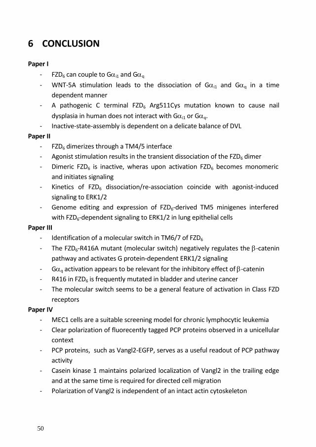
50
6 CONCLUSION Paper I
- FZD6 can couple to Gi1 and Gq
- WNT-5A stimulation leads to the dissociation of Gi1 and Gq in a time
dependent manner
- A pathogenic C terminal FZD6 Arg511Cys mutation known to cause nail
dysplasia in human does not interact with Gi1 or Gq.
- Inactive-state-assembly is dependent on a delicate balance of DVL
Paper II
- FZD6 dimerizes through a TM4/5 interface
- Agonist stimulation results in the transient dissociation of the FZD6 dimer
- Dimeric FZD6 is inactive, wheras upon activation FZD6 becomes monomeric
and initiates signaling
- Kinetics of FZD6 dissociation/re-association coincide with agonist-induced
signaling to ERK1/2
- Genome editing and expression of FZD6-derived TM5 minigenes interfered
with FZD6-dependent signaling to ERK1/2 in lung epithelial cells
Paper III
- Identification of a molecular switch in TM6/7 of FZD6
- The FZD6-R416A mutant (molecular switch) negatively regulates the -catenin
pathway and activates G protein-dependent ERK1/2 signaling
- Gq activation appears to be relevant for the inhibitory effect of -catenin
- R416 in FZD6 is frequently mutated in bladder and uterine cancer
- The molecular switch seems to be a general feature of activation in Class FZD
receptors
Paper IV
- MEC1 cells are a suitable screening model for chronic lymphocytic leukemia
- Clear polarization of fluorecently tagged PCP proteins observed in a unicellular
context
- PCP proteins, such as Vangl2-EGFP, serves as a useful readout of PCP pathway
activity
- Casein kinase 1 maintains polarized localization of Vangl2 in the trailing edge
and at the same time is required for directed cell migration
- Polarization of Vangl2 is independent of an intact actin cytoskeleton

51
7 ACKNOWLEDGEMENTS
Firstly, I would like to express my sincere gratitude to my advisor Associate
Professor Gunnar Schulte for the continuous support during my PhD studies,
for his patience, motivation, friendship and immense knowledge. Your guidance
helped me in all the time of research and writing of this thesis. Honestly, I could
not have imagined having a better advisor and mentor for my PhD study.
Besides my advisor, I would like to thank my co-supervisor Prof.
Roger Sunahara for his insightful comments and encouragement, but also for
the inspiring question, which incentivized me to widen my research from
various perspectives during my stay in his laboratory. Further, I would like to
thank the wolf pack for providing me with an excellent work environment.
My sincere thanks also go to my mentor Prof. Eddie Weizberg
and Dr. Peter Konrad, for their precious initial support back when I was
looking for a master thesis.
The entire Department of Pharmacology and Physiology and
especially the head Prof. Stefan Eriksson including the administration unit,
obtain my thanks for providing an excellent work environment and always
showing interest in the progression of my studies.
An enormous thank you also goes to my fellow lab-mates for the
stimulating discussions, for the fun we had at work and on numerous trips
outside the lab. Belma Hot aka Belissima Belma, Carina Halleskog, Elisa
Arthofer, Jacomijn Dijksterhuis, Jana Valnohova aka Jana Banana, Javier
Ortega, Katherina Strakova aka Coconut Katjo, Michaela Kilander, Shane C
Wright aka Shany Poo and Stefan Jäger.
Also I would like to cordially thank Bertil Fredholm for his
particularly constructive criticism of my work and stimulating ideas.

52
For the strenuous efforts, the professional introduction to the lab and the
unbelievable kindness I would like to especially thank Eva Lindgren.
Furthermore, the best corridor in the entire Karolinska Institutet
deserves special thanks for providing me with a stimulating research
environment and fun memories. Alice Costantini, Andrei Chagin, Annika
Olsson, Åsa Nordling, Anthi Faka, Bara Szarowska, Carina Nihlen, Catherine
Bell, Christa Zollbrecht, Evgeny Ivashkin aka Giant Jenia, Hannah Aucott,
Kazunori Sunadome, Igor Adameyko aka my future boss, Igor Cervenka,
Joanna Pascual Villani, Karuna Vuppalapati, Lei Li, Maria Peleli, Marin Jukic,
Maryam Khatibi Shahidi, Mattias Carlström, Meng Xie, Michael Hezel,
Michaela Sundqvist, Micke Elm, Natalia Akkuratova, Nina Kaukua, Phil
Newton, Sabine Vorrink, Simon Suter, Tatiana Chontorotzea, Thibault
Bouderlique aka ThiBoob, Tianle Goa, Tomas Schiffer and Vyacheslav
Dyachuk.
In addition I would like to express my gratitude to people
outside our department who made my life here in Sweden very pleasant and
we had a great time together. André Costa, Claudio Bertini, Gustavo De Abreu
Vieira, Jan Krivanek, Joel Bertlin, Martin Härring, Michael Hagemann-Jensen,
Moritz Lübke, Ollie Vieira Diksterhuis, Rebecca Liv, Rene Rizzo, Richard and
Johanna von Oldershausen, Simona Hanke, Stephanie Kilander, the
Jägergatan family especially, Aga Sowinska, Frank Bakker, Hoyin Lam, Jolinde
Kettelarij, Matheus Dyczynski, Shahul Hameed, Szabi Elias, Thibaud Richard
and Triin Pärn, to my friends from the most beautiful city in the world
(Hamburg for those who don’t know ;))

53
For providing me with high resolution pictures and the
permission to reuse figures and articles I would like to thank all journals and
especially Nasir Khan and Gemma Brugal for an example par excellence that
research is about sharing and changing ideas.
Special thanks to my family. Words cannot express how grateful
I am to my parents for all of the support that I have received from them.
Finally, and most importantly, I would like to express my love
and appreciation to the love of my life Marketa Kaucka Petersen, for her
support, encouragement and never-ending patience during my PhD.

54
8 REFERENCES
Adell, T., I. Nefkens and W. E. Muller (2003). "Polarity factor 'Frizzled' in the demosponge Suberites domuncula: identification, expression and localization of the receptor in the epithelium/pinacoderm(1)." FEBS Lett 554(3): 363-368.
Ahumada, A., D. C. Slusarski, X. Liu, R. T. Moon, C. C. Malbon and H. Y. Wang (2002). "Signaling of rat Frizzled-2 through phosphodiesterase and cyclic GMP." Science 298(5600): 2006-2010.
Albizu, L., M. Cottet, M. Kralikova, S. Stoev, R. Seyer, I. Brabet, T. Roux, H. Bazin, E. Bourrier, L. Lamarque, C. Breton, M. L. Rives, A. Newman, J. Javitch, E. Trinquet, M. Manning, J. P. Pin, B. Mouillac and T. Durroux (2010). "Time-resolved FRET between GPCR ligands reveals oligomers in native tissues." Nat Chem Biol 6(8): 587-594.
Alewijnse, A. E., H. Timmerman, E. H. Jacobs, M. J. Smit, E. Roovers, S. Cotecchia and R. Leurs (2000). "The effect of mutations in the DRY motif on the constitutive activity and structural instability of the histamine H(2) receptor." Mol Pharmacol 57(5): 890-898.
Alexander, S. P., H. E. Benson, E. Faccenda, A. J. Pawson, J. L. Sharman, M. Spedding, J. A. Peters, A. J. Harmar and C. Collaborators (2013). "The Concise Guide to PHARMACOLOGY 2013/14: G protein-coupled receptors." Br J Pharmacol 170(8): 1459-1581.
Allen, M. D., S. Neumann and M. C. Gershengorn (2011). "Occupancy of both sites on the thyrotropin (TSH) receptor dimer is necessary for phosphoinositide signaling." FASEB J 25(10): 3687-3694.
Arnis, S., K. Fahmy, K. P. Hofmann and T. P. Sakmar (1994). "A conserved carboxylic acid group mediates light-dependent proton uptake and signaling by rhodopsin." J Biol Chem 269(39): 23879-23881.
Aznar, N., K. K. Midde, Y. Dunkel, I. Lopez-Sanchez, Y. Pavlova, A. Marivin, J. Barbazan, F. Murray, U. Nitsche, K. P. Janssen, K. Willert, A. Goel, M. Abal, M. Garcia-Marcos and P. Ghosh (2015). "Daple is a novel non-receptor GEF required for trimeric G protein activation in Wnt signaling." Elife 4: e07091.
Barker, N. (2008). "The canonical Wnt/beta-catenin signalling pathway." Methods Mol Biol 468: 5-15.
Baskar, S., K. Y. Kwong, T. Hofer, J. M. Levy, M. G. Kennedy, E. Lee, L. M. Staudt, W. H. Wilson, A. Wiestner and C. Rader (2008). "Unique cell surface expression of receptor tyrosine kinase ROR1 in human B-cell chronic lymphocytic leukemia." Clin Cancer Res 14(2): 396-404.
Bayburt, T. H., S. A. Vishnivetskiy, M. A. McLean, T. Morizumi, C. C. Huang, J. J. Tesmer, O. P. Ernst, S. G. Sligar and V. V. Gurevich (2011). "Monomeric rhodopsin is sufficient for normal rhodopsin kinase (GRK1) phosphorylation and arrestin-1 binding." J Biol Chem 286(2): 1420-1428.
Berg, K. A., S. Maayani, J. Goldfarb, C. Scaramellini, P. Leff and W. P. Clarke (1998). "Effector pathway-dependent relative efficacy at serotonin type 2A and 2C receptors: evidence for agonist-directed trafficking of receptor stimulus." Mol Pharmacol 54(1): 94-104.
Berstein, G., J. L. Blank, D. Y. Jhon, J. H. Exton, S. G. Rhee and E. M. Ross (1992). "Phospholipase C-beta 1 is a GTPase-activating protein for Gq/11, its physiologic regulator." Cell 70(3): 411-418.
Bilic, J., Y. L. Huang, G. Davidson, T. Zimmermann, C. M. Cruciat, M. Bienz and C. Niehrs (2007). "Wnt induces LRP6 signalosomes and promotes dishevelled-dependent LRP6 phosphorylation." Science 316(5831): 1619-1622.
Bjarnadottir, T. K., D. E. Gloriam, S. H. Hellstrand, H. Kristiansson, R. Fredriksson and H. B. Schioth (2006). "Comprehensive repertoire and phylogenetic analysis of the G protein-coupled receptors in human and mouse." Genomics 88(3): 263-273.

55
Boone, C., N. G. Davis and G. F. Sprague, Jr. (1993). "Mutations that alter the third cytoplasmic loop of the a-factor receptor lead to a constitutive and hypersensitive phenotype." Proc Natl Acad Sci U S A 90(21): 9921-9925.
Bopp, K. P. and W. Magdeburg (1972). "[Possibilities and limitations of cortisone therapy in internal diseases]." Z Allgemeinmed 48(2): 43-53.
Bourhis, E., C. Tam, Y. Franke, J. F. Bazan, J. Ernst, J. Hwang, M. Costa, A. G. Cochran and R. N. Hannoush (2010). "Reconstitution of a frizzled8.Wnt3a.LRP6 signaling complex reveals multiple Wnt and Dkk1 binding sites on LRP6." J Biol Chem 285(12): 9172-9179.
Bovolenta, P., P. Esteve, J. M. Ruiz, E. Cisneros and J. Lopez-Rios (2008). "Beyond Wnt inhibition: new functions of secreted Frizzled-related proteins in development and disease." J Cell Sci 121(Pt 6): 737-746.
Bruheim, S., K. A. Krobert, K. W. Andressen and F. O. Levy (2003). "Unaltered agonist potency upon inducible 5-HT7(a) but not 5-HT4(b) receptor expression indicates agonist-independent association of 5-HT7(a) receptor and Gs." Receptors Channels 9(2): 107-116.
Bunemann, M., M. Frank and M. J. Lohse (2003). "Gi protein activation in intact cells involves subunit rearrangement rather than dissociation." Proc Natl Acad Sci U S A 100(26): 16077-16082.
Burns, C. J., J. Zhang, E. C. Brown, A. M. Van Bibber, J. Van Es, H. Clevers, T. O. Ishikawa, M. M. Taketo, M. L. Vetter and S. Fuhrmann (2008). "Investigation of Frizzled-5 during embryonic neural development in mouse." Dev Dyn 237(6): 1614-1626.
Cabrera, C. V., A. Martinez-Arias and M. Bate (1987). "The expression of three members of the achaete-scute gene complex correlates with neuroblast segregation in Drosophila." Cell 50(3): 425-433.
Calebiro, D., F. Rieken, J. Wagner, T. Sungkaworn, U. Zabel, A. Borzi, E. Cocucci, A. Zurn and M. J. Lohse (2013). "Single-molecule analysis of fluorescently labeled G-protein-coupled receptors reveals complexes with distinct dynamics and organization." Proc Natl Acad Sci U S A 110(2): 743-748.
Carmon, K. S., X. Gong, Q. Lin, A. Thomas and Q. Liu (2011). "R-spondins function as ligands of the orphan receptors LGR4 and LGR5 to regulate Wnt/beta-catenin signaling." Proc Natl Acad Sci U S A 108(28): 11452-11457.
Chae, J., M. J. Kim, J. H. Goo, S. Collier, D. Gubb, J. Charlton, P. N. Adler and W. J. Park (1999). "The Drosophila tissue polarity gene starry night encodes a member of the protocadherin family." Development 126(23): 5421-5429.
Charles, A. C., N. Mostovskaya, K. Asas, C. J. Evans, M. L. Dankovich and T. G. Hales (2003). "Coexpression of delta-opioid receptors with micro receptors in GH3 cells changes the functional response to micro agonists from inhibitory to excitatory." Mol Pharmacol 63(1): 89-95.
Clark, M. C., T. E. Dever, J. J. Dever, P. Xu, V. Rehder, M. A. Sosa and D. J. Baro (2004). "Arthropod 5-HT2 receptors: a neurohormonal receptor in decapod crustaceans that displays agonist independent activity resulting from an evolutionary alteration to the DRY motif." J Neurosci 24(13): 3421-3435.
Clevers, H. (2006). "Wnt/beta-catenin signaling in development and disease." Cell 127(3): 469-480.
Cong, F., L. Schweizer and H. Varmus (2004). "Wnt signals across the plasma membrane to activate the beta-catenin pathway by forming oligomers containing its receptors, Frizzled and LRP." Development 131(20): 5103-5115.
Cordomi, A., G. Navarro, M. S. Aymerich and R. Franco (2015). "Structures for G-Protein-Coupled Receptor Tetramers in Complex with G Proteins." Trends Biochem Sci 40(10): 548-551.

56
Cordomi, A., E. Ramon, P. Garriga and J. J. Perez (2008). "Molecular dynamics simulations of rhodopsin point mutants at the cytoplasmic side of helices 3 and 6." J Biomol Struct Dyn 25(6): 573-587.
Cruciat, C. M. and C. Niehrs (2013). "Secreted and transmembrane wnt inhibitors and activators." Cold Spring Harb Perspect Biol 5(3): a015081.
Cui, C. Y., J. Klar, P. Georgii-Heming, A. S. Frojmark, S. M. Baig, D. Schlessinger and N. Dahl (2013). "Frizzled6 deficiency disrupts the differentiation process of nail development." J Invest Dermatol 133(8): 1990-1997.
Cvejic, S. and L. A. Devi (1997). "Dimerization of the delta opioid receptor: implication for a role in receptor internalization." J Biol Chem 272(43): 26959-26964.
Dann, C. E., J. C. Hsieh, A. Rattner, D. Sharma, J. Nathans and D. J. Leahy (2001). "Insights into Wnt binding and signalling from the structures of two Frizzled cysteine-rich domains." Nature 412(6842): 86-90.
De Lean, A., J. M. Stadel and R. J. Lefkowitz (1980). "A ternary complex model explains the agonist-specific binding properties of the adenylate cyclase-coupled beta-adrenergic receptor." J Biol Chem 255(15): 7108-7117.
Deupi, X. and B. K. Kobilka (2010). "Energy landscapes as a tool to integrate GPCR structure, dynamics, and function." Physiology (Bethesda) 25(5): 293-303.
Digby, G. J., R. M. Lober, P. R. Sethi and N. A. Lambert (2006). "Some G protein heterotrimers physically dissociate in living cells." Proc Natl Acad Sci U S A 103(47): 17789-17794.
Dijksterhuis, J. P., B. Baljinnyam, K. Stanger, H. O. Sercan, Y. Ji, O. Andres, J. S. Rubin, R. N. Hannoush and G. Schulte (2015). "Systematic mapping of WNT-FZD protein interactions reveals functional selectivity by distinct WNT-FZD pairs." J Biol Chem 290(11): 6789-6798.
Dijksterhuis, J. P., J. Petersen and G. Schulte (2014). "WNT/Frizzled signalling: receptor-ligand selectivity with focus on FZD-G protein signalling and its physiological relevance: IUPHAR Review 3." Br J Pharmacol 171(5): 1195-1209.
Dorsch, S., K. N. Klotz, S. Engelhardt, M. J. Lohse and M. Bunemann (2009). "Analysis of receptor oligomerization by FRAP microscopy." Nat Methods 6(3): 225-230.
Drastichova, Z. and J. Novotny (2012). "Identification of a preassembled TRH receptor-G(q/11) protein complex in HEK293 cells." Cell Struct Funct 37(1): 1-12.
Du, Q., X. Zhang, J. Cardinal, Z. Cao, Z. Guo, L. Shao and D. A. Geller (2009). "Wnt/beta-catenin signaling regulates cytokine-induced human inducible nitric oxide synthase expression by inhibiting nuclear factor-kappaB activation in cancer cells." Cancer Res 69(9): 3764-3771.
Ernst, O. P., V. Gramse, M. Kolbe, K. P. Hofmann and M. Heck (2007). "Monomeric G protein-coupled receptor rhodopsin in solution activates its G protein transducin at the diffusion limit." Proc Natl Acad Sci U S A 104(26): 10859-10864.
Fahmy, K. and T. P. Sakmar (1993). "Regulation of the rhodopsin-transducin interaction by a highly conserved carboxylic acid group." Biochemistry 32(28): 7229-7236.
Ford, C. E., N. P. Skiba, H. Bae, Y. Daaka, E. Reuveny, L. R. Shekter, R. Rosal, G. Weng, C. S. Yang, R. Iyengar, R. J. Miller, L. Y. Jan, R. J. Lefkowitz and H. E. Hamm (1998). "Molecular basis for interactions of G protein betagamma subunits with effectors." Science 280(5367): 1271-1274.
Fredriksson, R., M. C. Lagerstrom, L. G. Lundin and H. B. Schioth (2003). "The G-protein-coupled receptors in the human genome form five main families. Phylogenetic analysis, paralogon groups, and fingerprints." Mol Pharmacol 63(6): 1256-1272.

57
Freyer, M. W. and E. A. Lewis (2008). "Isothermal titration calorimetry: experimental design, data analysis, and probing macromolecule/ligand binding and kinetic interactions." Methods Cell Biol 84: 79-113.
Frojmark, A. S., J. Schuster, M. Sobol, M. Entesarian, M. B. Kilander, D. Gabrikova, S. Nawaz, S. M. Baig, G. Schulte, J. Klar and N. Dahl (2011). "Mutations in Frizzled 6 cause isolated autosomal-recessive nail dysplasia." Am J Hum Genet 88(6): 852-860.
Galandrin, S. and M. Bouvier (2006). "Distinct signaling profiles of beta1 and beta2 adrenergic receptor ligands toward adenylyl cyclase and mitogen-activated protein kinase reveals the pluridimensionality of efficacy." Mol Pharmacol 70(5): 1575-1584.
Galandrin, S., G. Oligny-Longpre and M. Bouvier (2007). "The evasive nature of drug efficacy: implications for drug discovery." Trends Pharmacol Sci 28(8): 423-430.
Gales, C., J. J. Van Durm, S. Schaak, S. Pontier, Y. Percherancier, M. Audet, H. Paris and M. Bouvier (2006). "Probing the activation-promoted structural rearrangements in preassembled receptor-G protein complexes." Nat Struct Mol Biol 13(9): 778-786.
Gao, B., H. Song, K. Bishop, G. Elliot, L. Garrett, M. A. English, P. Andre, J. Robinson, R. Sood, Y. Minami, A. N. Economides and Y. Yang (2011). "Wnt signaling gradients establish planar cell polarity by inducing Vangl2 phosphorylation through Ror2." Dev Cell 20(2): 163-176.
Gao, C. and Y. G. Chen (2010). "Dishevelled: The hub of Wnt signaling." Cell Signal 22(5): 717-727.
George, S. R., T. Fan, Z. Xie, R. Tse, V. Tam, G. Varghese and B. F. O'Dowd (2000). "Oligomerization of mu- and delta-opioid receptors. Generation of novel functional properties." J Biol Chem 275(34): 26128-26135.
Gilman, A. G. (1987). "G proteins: transducers of receptor-generated signals." Annu Rev Biochem 56: 615-649.
Golan, T., A. Yaniv, A. Bafico, G. Liu and A. Gazit (2004). "The human Frizzled 6 (HFz6) acts as a negative regulator of the canonical Wnt. beta-catenin signaling cascade." J Biol Chem 279(15): 14879-14888.
Grosse, R., T. Schoneberg, G. Schultz and T. Gudermann (1997). "Inhibition of gonadotropin-releasing hormone receptor signaling by expression of a splice variant of the human receptor." Mol Endocrinol 11(9): 1305-1318.
Grumolato, L., G. Liu, P. Mong, R. Mudbhary, R. Biswas, R. Arroyave, S. Vijayakumar, A. N. Economides and S. A. Aaronson (2010). "Canonical and noncanonical Wnts use a common mechanism to activate completely unrelated coreceptors." Genes Dev 24(22): 2517-2530.
Gubb, D., C. Green, D. Huen, D. Coulson, G. Johnson, D. Tree, S. Collier and J. Roote (1999). "The balance between isoforms of the prickle LIM domain protein is critical for planar polarity in Drosophila imaginal discs." Genes Dev 13(17): 2315-2327.
Halleskog, C., J. P. Dijksterhuis, M. B. Kilander, J. Becerril-Ortega, J. C. Villaescusa, E. Lindgren, E. Arenas and G. Schulte (2012). "Heterotrimeric G protein-dependent WNT-5A signaling to ERK1/2 mediates distinct aspects of microglia proinflammatory transformation." J Neuroinflammation 9: 111.
Halleskog, C. and G. Schulte (2013). "Pertussis toxin-sensitive heterotrimeric G(alphai/o) proteins mediate WNT/beta-catenin and WNT/ERK1/2 signaling in mouse primary microglia stimulated with purified WNT-3A." Cell Signal 25(4): 822-828.
He, X., M. Semenov, K. Tamai and X. Zeng (2004). "LDL receptor-related proteins 5 and 6 in Wnt/beta-catenin signaling: arrows point the way." Development 131(8): 1663-1677.

58
Hein, P. and M. Bunemann (2009). "Coupling mode of receptors and G proteins." Naunyn Schmiedebergs Arch Pharmacol 379(5): 435-443.
Hein, P., M. Frank, C. Hoffmann, M. J. Lohse and M. Bunemann (2005). "Dynamics of receptor/G protein coupling in living cells." EMBO J 24(23): 4106-4114.
Heisenberg, C. P., M. Tada, G. J. Rauch, L. Saude, M. L. Concha, R. Geisler, D. L. Stemple, J. C. Smith and S. W. Wilson (2000). "Silberblick/Wnt11 mediates convergent extension movements during zebrafish gastrulation." Nature 405(6782): 76-81.
Hendrickx, M. and L. Leyns (2008). "Non-conventional Frizzled ligands and Wnt receptors." Dev Growth Differ 50(4): 229-243.
Hepler, J. R. and A. G. Gilman (1992). "G proteins." Trends Biochem Sci 17(10): 383-387.
Hergovich, A. and B. A. Hemmings (2010). "TAZ-mediated crosstalk between Wnt and Hippo signaling." Dev Cell 18(4): 508-509.
Hern, J. A., A. H. Baig, G. I. Mashanov, B. Birdsall, J. E. Corrie, S. Lazareno, J. E. Molloy and N. J. Birdsall (2010). "Formation and dissociation of M1 muscarinic receptor dimers seen by total internal reflection fluorescence imaging of single molecules." Proc Natl Acad Sci U S A 107(6): 2693-2698.
Hikasa, H., M. Shibata, I. Hiratani and M. Taira (2002). "The Xenopus receptor tyrosine kinase Xror2 modulates morphogenetic movements of the axial mesoderm and neuroectoderm via Wnt signaling." Development 129(22): 5227-5239.
Hsieh, J. C., A. Rattner, P. M. Smallwood and J. Nathans (1999). "Biochemical characterization of Wnt-frizzled interactions using a soluble, biologically active vertebrate Wnt protein." Proc Natl Acad Sci U S A 96(7): 3546-3551.
Huang, C., J. R. Hepler, L. T. Chen, A. G. Gilman, R. G. Anderson and S. M. Mumby (1997). "Organization of G proteins and adenylyl cyclase at the plasma membrane." Mol Biol Cell 8(12): 2365-2378.
Huang, H. C. and P. S. Klein (2004). "The Frizzled family: receptors for multiple signal transduction pathways." Genome Biol 5(7): 234.
Imajo, M., K. Miyatake, A. Iimura, A. Miyamoto and E. Nishida (2012). "A molecular mechanism that links Hippo signalling to the inhibition of Wnt/beta-catenin signalling." EMBO J 31(5): 1109-1122.
Ishitani, T., S. Kishida, J. Hyodo-Miura, N. Ueno, J. Yasuda, M. Waterman, H. Shibuya, R. T. Moon, J. Ninomiya-Tsuji and K. Matsumoto (2003). "The TAK1-NLK mitogen-activated protein kinase cascade functions in the Wnt-5a/Ca(2+) pathway to antagonize Wnt/beta-catenin signaling." Mol Cell Biol 23(1): 131-139.
Itasaki, N., C. M. Jones, S. Mercurio, A. Rowe, P. M. Domingos, J. C. Smith and R. Krumlauf (2003). "Wise, a context-dependent activator and inhibitor of Wnt signalling." Development 130(18): 4295-4305.
Jafari, R., H. Almqvist, H. Axelsson, M. Ignatushchenko, T. Lundback, P. Nordlund and D. Martinez Molina (2014). "The cellular thermal shift assay for evaluating drug target interactions in cells." Nat Protoc 9(9): 2100-2122.
Janda, C. Y., D. Waghray, A. M. Levin, C. Thomas and K. C. Garcia (2012). "Structural basis of Wnt recognition by Frizzled." Science 337(6090): 59-64.
Jones, S. E. and C. Jomary (2002). "Secreted Frizzled-related proteins: searching for relationships and patterns." Bioessays 24(9): 811-820.

59
Katanaev, V. L. and S. Buestorf (2009). "Frizzled Proteins are bona fide G Protein-Coupled Receptors." Nature Precedings hdl:10101/npre.2009.2765.1.
Katanaev, V. L., R. Ponzielli, M. Semeriva and A. Tomlinson (2005). "Trimeric G protein-dependent frizzled signaling in Drosophila." Cell 120(1): 111-122.
Kato, M., M. S. Patel, R. Levasseur, I. Lobov, B. H. Chang, D. A. Glass, 2nd, C. Hartmann, L. Li, T. H. Hwang, C. F. Brayton, R. A. Lang, G. Karsenty and L. Chan (2002). "Cbfa1-independent decrease in osteoblast proliferation, osteopenia, and persistent embryonic eye vascularization in mice deficient in Lrp5, a Wnt coreceptor." J Cell Biol 157(2): 303-314.
Kaucka, M., K. Plevova, S. Pavlova, P. Janovska, A. Mishra, J. Verner, J. Prochazkova, P. Krejci, J. Kotaskova, P. Ovesna, B. Tichy, Y. Brychtova, M. Doubek, A. Kozubik, J. Mayer, S. Pospisilova and V. Bryja (2013). "The planar cell polarity pathway drives pathogenesis of chronic lymphocytic leukemia by the regulation of B-lymphocyte migration." Cancer Res 73(5): 1491-1501.
Kenakin, T. (1995). "Agonist-receptor efficacy. II. Agonist trafficking of receptor signals." Trends Pharmacol Sci 16(7): 232-238.
Kenakin, T. (2001). "Inverse, protean, and ligand-selective agonism: matters of receptor conformation." FASEB J 15(3): 598-611.
Kenakin, T. (2007). "Functional selectivity through protean and biased agonism: who steers the ship?" Mol Pharmacol 72(6): 1393-1401.
Kenakin, T. (2011). "Functional selectivity and biased receptor signaling." J Pharmacol Exp Ther 336(2): 296-302.
Kikuchi, A. and H. Yamamoto (2007). "Regulation of Wnt signalling by receptor-mediated endocytosis." J Biochem 141(4): 443-451.
Kikuchi, A., H. Yamamoto and S. Kishida (2007). "Multiplicity of the interactions of Wnt proteins and their receptors." Cell Signal 19(4): 659-671.
Kilander, M. B., J. Dahlstrom and G. Schulte (2014). "Assessment of Frizzled 6 membrane mobility by FRAP supports G protein coupling and reveals WNT-Frizzled selectivity." Cell Signal 26(9): 1943-1949.
Kilander, M. B., J. P. Dijksterhuis, R. S. Ganji, V. Bryja and G. Schulte (2011). "WNT-5A stimulates the GDP/GTP exchange at pertussis toxin-sensitive heterotrimeric G proteins." Cell Signal 23(3): 550-554.
Kilander, M. B., C. Halleskog and G. Schulte (2011). "Recombinant WNTs differentially activate beta-catenin-dependent and -independent signalling in mouse microglia-like cells." Acta Physiol (Oxf) 203(3): 363-372.
Kilander, M. B., J. Petersen, K. W. Andressen, R. S. Ganji, F. O. Levy, J. Schuster, N. Dahl, V. Bryja and G. Schulte (2014). "Disheveled regulates precoupling of heterotrimeric G proteins to Frizzled 6." FASEB J 28(5): 2293-2305.
Kim, M. and E. H. Jho (2014). "Cross-talk between Wnt/beta-catenin and Hippo signaling pathways: a brief review." BMB Rep 47(10): 540-545.
Knierim, B., K. P. Hofmann, O. P. Ernst and W. L. Hubbell (2007). "Sequence of late molecular events in the activation of rhodopsin." Proc Natl Acad Sci U S A 104(51): 20290-20295.
Kobilka, B. K. (2007). "G protein coupled receptor structure and activation." Biochim Biophys Acta 1768(4): 794-807.

60
Kolakowski, L. F., Jr. (1994). "GCRDb: a G-protein-coupled receptor database." Receptors Channels 2(1): 1-7.
Kolben, T., I. Perobner, K. Fernsebner, F. Lechner, C. Geissler, L. Ruiz-Heinrich, S. Capovilla, M. Jochum and P. Neth (2012). "Dissecting the impact of Frizzled receptors in Wnt/beta-catenin signaling of human mesenchymal stem cells." Biol Chem 393(12): 1433-1447.
Koshland, D. E. (1958). "Application of a Theory of Enzyme Specificity to Protein Synthesis." Proc Natl Acad Sci U S A 44(2): 98-104.
Koval, A., V. Purvanov, D. Egger-Adam and V. L. Katanaev (2011). "Yellow submarine of the Wnt/Frizzled signaling: submerging from the G protein harbor to the targets." Biochem Pharmacol 82(10): 1311-1319.
Kuhl, M., L. C. Sheldahl, C. C. Malbon and R. T. Moon (2000). "Ca(2+)/calmodulin-dependent protein kinase II is stimulated by Wnt and Frizzled homologs and promotes ventral cell fates in Xenopus." J Biol Chem 275(17): 12701-12711.
Kuhl, M., L. C. Sheldahl, M. Park, J. R. Miller and R. T. Moon (2000). "The Wnt/Ca2+ pathway: a new vertebrate Wnt signaling pathway takes shape." Trends Genet 16(7): 279-283.
Kuszak, A. J., S. Pitchiaya, J. P. Anand, H. I. Mosberg, N. G. Walter and R. K. Sunahara (2009). "Purification and functional reconstitution of monomeric mu-opioid receptors: allosteric modulation of agonist binding by Gi2." J Biol Chem 284(39): 26732-26741.
Lambert, N. A. (2009). "Uncoupling diffusion and binding in FRAP experiments." Nat Methods 6(3): 183; author reply 183-184.
Landau, E. M., E. Pebay-Peyroula and R. Neutze (2003). "Structural and mechanistic insight from high resolution structures of archaeal rhodopsins." FEBS Lett 555(1): 51-56.
Langley, J. N. (1905). "On the reaction of cells and of nerve-endings to certain poisons, chiefly as regards the reaction of striated muscle to nicotine and to curari." J Physiol 33(4-5): 374-413.
Lau, S. Y., A. K. Taneja and R. S. Hodges (1984). "Synthesis of a model protein of defined secondary and quaternary structure. Effect of chain length on the stabilization and formation of two-stranded alpha-helical coiled-coils." J Biol Chem 259(21): 13253-13261.
Lefkowitz, R. J., S. Cotecchia, P. Samama and T. Costa (1993). "Constitutive activity of receptors coupled to guanine nucleotide regulatory proteins." Trends Pharmacol Sci 14(8): 303-307.
Lippincott-Schwartz, J., N. Altan-Bonnet and G. H. Patterson (2003). "Photobleaching and photoactivation: following protein dynamics in living cells." Nat Cell Biol Suppl: S7-14.
Liu, G., A. Bafico, V. K. Harris and S. A. Aaronson (2003). "A novel mechanism for Wnt activation of canonical signaling through the LRP6 receptor." Mol Cell Biol 23(16): 5825-5835.
Liu, T., A. J. DeCostanzo, X. Liu, H. Wang, S. Hallagan, R. T. Moon and C. C. Malbon (2001). "G protein signaling from activated rat frizzled-1 to the beta-catenin-Lef-Tcf pathway." Science 292(5522): 1718-1722.
Liu, T., X. Liu, H. Wang, R. T. Moon and C. C. Malbon (1999). "Activation of rat frizzled-1 promotes Wnt signaling and differentiation of mouse F9 teratocarcinoma cells via pathways that require Galpha(q) and Galpha(o) function." J Biol Chem 274(47): 33539-33544.
Liu, X., T. Liu, D. C. Slusarski, J. Yang-Snyder, C. C. Malbon, R. T. Moon and H. Wang (1999). "Activation of a frizzled-2/beta-adrenergic receptor chimera promotes Wnt signaling and differentiation of mouse F9 teratocarcinoma cells via Galphao and Galphat." Proc Natl Acad Sci U S A 96(25): 14383-14388.

61
Liu, X., J. S. Rubin and A. R. Kimmel (2005). "Rapid, Wnt-induced changes in GSK3beta associations that regulate beta-catenin stabilization are mediated by Galpha proteins." Curr Biol 15(22): 1989-1997.
Lu, W., V. Yamamoto, B. Ortega and D. Baltimore (2004). "Mammalian Ryk is a Wnt coreceptor required for stimulation of neurite outgrowth." Cell 119(1): 97-108.
MacDonald, B. T., K. Tamai and X. He (2009). "Wnt/beta-catenin signaling: components, mechanisms, and diseases." Dev Cell 17(1): 9-26.
Mao, J., J. Wang, B. Liu, W. Pan, G. H. Farr, 3rd, C. Flynn, H. Yuan, S. Takada, D. Kimelman, L. Li and D. Wu (2001). "Low-density lipoprotein receptor-related protein-5 binds to Axin and regulates the canonical Wnt signaling pathway." Mol Cell 7(4): 801-809.
Martinez Molina, D., R. Jafari, M. Ignatushchenko, T. Seki, E. A. Larsson, C. Dan, L. Sreekumar, Y. Cao and P. Nordlund (2013). "Monitoring drug target engagement in cells and tissues using the cellular thermal shift assay." Science 341(6141): 84-87.
Maruyama, I. N. (2014). "Mechanisms of activation of receptor tyrosine kinases: monomers or dimers." Cells 3(2): 304-330.
McNeill, H. (2010). "Planar cell polarity: keeping hairs straight is not so simple." Cold Spring Harb Perspect Biol 2(2): a003376.
McPherson, J. D., M. Marra, L. Hillier, R. H. Waterston, A. Chinwalla, J. Wallis, M. Sekhon, K. Wylie, E. R. Mardis, R. K. Wilson, R. Fulton, T. A. Kucaba, C. Wagner-McPherson, W. B. Barbazuk, S. G. Gregory, S. J. Humphray, L. French, R. S. Evans, G. Bethel, A. Whittaker, J. L. Holden, O. T. McCann, A. Dunham, C. Soderlund, C. E. Scott, D. R. Bentley, G. Schuler, H. C. Chen, W. Jang, E. D. Green, J. R. Idol, V. V. Maduro, K. T. Montgomery, E. Lee, A. Miller, S. Emerling, Kucherlapati, R. Gibbs, S. Scherer, J. H. Gorrell, E. Sodergren, K. Clerc-Blankenburg, P. Tabor, S. Naylor, D. Garcia, P. J. de Jong, J. J. Catanese, N. Nowak, K. Osoegawa, S. Qin, L. Rowen, A. Madan, M. Dors, L. Hood, B. Trask, C. Friedman, H. Massa, V. G. Cheung, I. R. Kirsch, T. Reid, R. Yonescu, J. Weissenbach, T. Bruls, R. Heilig, E. Branscomb, A. Olsen, N. Doggett, J. F. Cheng, T. Hawkins, R. M. Myers, J. Shang, L. Ramirez, J. Schmutz, O. Velasquez, K. Dixon, N. E. Stone, D. R. Cox, D. Haussler, W. J. Kent, T. Furey, S. Rogic, S. Kennedy, S. Jones, A. Rosenthal, G. Wen, M. Schilhabel, G. Gloeckner, G. Nyakatura, R. Siebert, B. Schlegelberger, J. Korenberg, X. N. Chen, A. Fujiyama, M. Hattori, A. Toyoda, T. Yada, H. S. Park, Y. Sakaki, N. Shimizu, S. Asakawa, K. Kawasaki, T. Sasaki, A. Shintani, A. Shimizu, K. Shibuya, J. Kudoh, S. Minoshima, J. Ramser, P. Seranski, C. Hoff, A. Poustka, R. Reinhardt, H. Lehrach and C. International Human Genome Mapping (2001). "A physical map of the human genome." Nature 409(6822): 934-941.
Mellado, M., J. M. Rodriguez-Frade, A. J. Vila-Coro, S. Fernandez, A. Martin de Ana, D. R. Jones, J. L. Toran and A. C. Martinez (2001). "Chemokine receptor homo- or heterodimerization activates distinct signaling pathways." EMBO J 20(10): 2497-2507.
Mikels, A. J. and R. Nusse (2006). "Purified Wnt5a protein activates or inhibits beta-catenin-TCF signaling depending on receptor context." PLoS Biol 4(4): e115.
Miller, J. R. (2002). "The Wnts." Genome Biol 3(1): REVIEWS3001.
Minami, Y., I. Oishi, M. Endo and M. Nishita (2010). "Ror-family receptor tyrosine kinases in noncanonical Wnt signaling: their implications in developmental morphogenesis and human diseases." Dev Dyn 239(1): 1-15.
Minobe, S., K. Fei, L. Yan, M. Sarras Jr and M. Werle (2000). "Identification and characterization of the epithelial polarity receptor "Frizzled" in Hydra vulgaris." Dev Genes Evol 210(5): 258-262.

62
Mirzadegan, T., G. Benko, S. Filipek and K. Palczewski (2003). "Sequence analyses of G-protein-coupled receptors: similarities to rhodopsin." Biochemistry 42(10): 2759-2767.
Moller, S., J. Vilo and M. D. Croning (2001). "Prediction of the coupling specificity of G protein coupled receptors to their G proteins." Bioinformatics 17 Suppl 1: S174-181.
Mukhopadhyay, S. and A. C. Howlett (2005). "Chemically distinct ligands promote differential CB1 cannabinoid receptor-Gi protein interactions." Mol Pharmacol 67(6): 2016-2024.
Nature Reviews Drug Discovery, G. Q. P. (2004). "The state of GPCR research in 2004." Nat Rev Drug Discov 3(7): 575, 577-626.
Neubig, R. R. (1994). "Membrane organization in G-protein mechanisms." FASEB J 8(12): 939-946.
Niehrs, C. (2004). "Norrin and frizzled; a new vein for the eye." Dev Cell 6(4): 453-454.
Niehrs, C. (2006). "Function and biological roles of the Dickkopf family of Wnt modulators." Oncogene 25(57): 7469-7481.
Niesen, F. H., H. Berglund and M. Vedadi (2007). "The use of differential scanning fluorimetry to detect ligand interactions that promote protein stability." Nat Protoc 2(9): 2212-2221.
Niswender, C. M. and P. J. Conn (2010). "Metabotropic glutamate receptors: physiology, pharmacology, and disease." Annu Rev Pharmacol Toxicol 50: 295-322.
Nobles, M., A. Benians and A. Tinker (2005). "Heterotrimeric G proteins precouple with G protein-coupled receptors in living cells." Proc Natl Acad Sci U S A 102(51): 18706-18711.
Nusse, R. and H. E. Varmus (1982). "Many tumors induced by the mouse mammary tumor virus contain a provirus integrated in the same region of the host genome." Cell 31(1): 99-109.
Nusse, R. and H. E. Varmus (1992). "Wnt genes." Cell 69(7): 1073-1087.
Nusslein-Volhard, C. and E. Wieschaus (1980). "Mutations affecting segment number and polarity in Drosophila." Nature 287(5785): 795-801.
Oldham, W. M. and H. E. Hamm (2008). "Heterotrimeric G protein activation by G-protein-coupled receptors." Nat Rev Mol Cell Biol 9(1): 60-71.
Paavola, K. J., J. R. Stephenson, S. L. Ritter, S. P. Alter and R. A. Hall (2011). "The N terminus of the adhesion G protein-coupled receptor GPR56 controls receptor signaling activity." J Biol Chem 286(33): 28914-28921.
Park, H. W., Y. C. Kim, B. Yu, T. Moroishi, J. S. Mo, S. W. Plouffe, Z. Meng, K. C. Lin, F. X. Yu, C. M. Alexander, C. Y. Wang and K. L. Guan (2015). "Alternative Wnt Signaling Activates YAP/TAZ." Cell 162(4): 780-794.
Park, T. J., R. S. Gray, A. Sato, R. Habas and J. B. Wallingford (2005). "Subcellular localization and signaling properties of dishevelled in developing vertebrate embryos." Curr Biol 15(11): 1039-1044.
Patel, M. S. and G. Karsenty (2002). "Regulation of bone formation and vision by LRP5." N Engl J Med 346(20): 1572-1574.
Patthy, L. (2000). "The WIF module." Trends Biochem Sci 25(1): 12-13.
Piga, R., D. van Dartel, A. Bunschoten, I. van der Stelt and J. Keijer (2014). "Role of Frizzled6 in the molecular mechanism of beta-carotene action in the lung." Toxicology 320: 67-73.

63
Pin, J. P., L. Comps-Agrar, D. Maurel, C. Monnier, M. L. Rives, E. Trinquet, J. Kniazeff, P. Rondard and L. Prezeau (2009). "G-protein-coupled receptor oligomers: two or more for what? Lessons from mGlu and GABAB receptors." J Physiol 587(Pt 22): 5337-5344.
Pinson, K. I., J. Brennan, S. Monkley, B. J. Avery and W. C. Skarnes (2000). "An LDL-receptor-related protein mediates Wnt signalling in mice." Nature 407(6803): 535-538.
Polakis, P. (2000). "Wnt signaling and cancer." Genes Dev 14(15): 1837-1851.
Porter, J. E., J. Hwa and D. M. Perez (1996). "Activation of the alpha1b-adrenergic receptor is initiated by disruption of an interhelical salt bridge constraint." J Biol Chem 271(45): 28318-28323.
Qin, K., C. Dong, G. Wu and N. A. Lambert (2011). "Inactive-state preassembly of G(q)-coupled receptors and G(q) heterotrimers." Nat Chem Biol 7(10): 740-747.
Ramon, E., A. Cordomi, L. Bosch, E. Y. Zernii, Senin, II, J. Manyosa, P. P. Philippov, J. J. Perez and P. Garriga (2007). "Critical role of electrostatic interactions of amino acids at the cytoplasmic region of helices 3 and 6 in rhodopsin conformational properties and activation." J Biol Chem 282(19): 14272-14282.
Ramsay, R. G., D. Ciznadija, M. Vanevski and T. Mantamadiotis (2003). "Transcriptional regulation of cyclo-oxygenase expression: three pillars of control." Int J Immunopathol Pharmacol 16(2 Suppl): 59-67.
Ransnas, L. A. and P. A. Insel (1988). "Subunit dissociation is the mechanism for hormonal activation of the Gs protein in native membranes." J Biol Chem 263(33): 17239-17242.
Rasmussen, S. G., B. T. DeVree, Y. Zou, A. C. Kruse, K. Y. Chung, T. S. Kobilka, F. S. Thian, P. S. Chae, E. Pardon, D. Calinski, J. M. Mathiesen, S. T. Shah, J. A. Lyons, M. Caffrey, S. H. Gellman, J. Steyaert, G. Skiniotis, W. I. Weis, R. K. Sunahara and B. K. Kobilka (2011). "Crystal structure of the beta2 adrenergic receptor-Gs protein complex." Nature 477(7366): 549-555.
Reiter, E., S. Ahn, A. K. Shukla and R. J. Lefkowitz (2012). "Molecular mechanism of beta-arrestin-biased agonism at seven-transmembrane receptors." Annu Rev Pharmacol Toxicol 52: 179-197.
Rijsewijk, F., M. Schuermann, E. Wagenaar, P. Parren, D. Weigel and R. Nusse (1987). "The Drosophila homolog of the mouse mammary oncogene int-1 is identical to the segment polarity gene wingless." Cell 50(4): 649-657.
Rivero-Muller, A., Y. Y. Chou, I. Ji, S. Lajic, A. C. Hanyaloglu, K. Jonas, N. Rahman, T. H. Ji and I. Huhtaniemi (2010). "Rescue of defective G protein-coupled receptor function in vivo by intermolecular cooperation." Proc Natl Acad Sci U S A 107(5): 2319-2324.
Robinson, P. R., G. B. Cohen, E. A. Zhukovsky and D. D. Oprian (1992). "Constitutively active mutants of rhodopsin." Neuron 9(4): 719-725.
Rosenbaum, D. M., S. G. Rasmussen and B. K. Kobilka (2009). "The structure and function of G-protein-coupled receptors." Nature 459(7245): 356-363.
Rosethorne, E. M. and S. J. Charlton (2011). "Agonist-biased signaling at the histamine H4 receptor: JNJ7777120 recruits beta-arrestin without activating G proteins." Mol Pharmacol 79(4): 749-757.
Ross, E. M. and T. M. Wilkie (2000). "GTPase-activating proteins for heterotrimeric G proteins: regulators of G protein signaling (RGS) and RGS-like proteins." Annu Rev Biochem 69: 795-827.
Scheerer, P., J. H. Park, P. W. Hildebrand, Y. J. Kim, N. Krauss, H. W. Choe, K. P. Hofmann and O. P. Ernst (2008). "Crystal structure of opsin in its G-protein-interacting conformation." Nature 455(7212): 497-502.

64
Schioth, H. B. and R. Fredriksson (2005). "The GRAFS classification system of G-protein coupled receptors in comparative perspective." Gen Comp Endocrinol 142(1-2): 94-101.
Schmidt, V., K. Baum, A. Lao, K. Rateitschak, Y. Schmitz, A. Teichmann, B. Wiesner, C. M. Petersen, A. Nykjaer, J. Wolf, O. Wolkenhauer and T. E. Willnow (2012). "Quantitative modelling of amyloidogenic processing and its influence by SORLA in Alzheimer's disease." EMBO J 31(1): 187-200.
Schoneberg, T., J. Yun, D. Wenkert and J. Wess (1996). "Functional rescue of mutant V2 vasopressin receptors causing nephrogenic diabetes insipidus by a co-expressed receptor polypeptide." EMBO J 15(6): 1283-1291.
Schulte, G. (2010). "International Union of Basic and Clinical Pharmacology. LXXX. The class Frizzled receptors." Pharmacol Rev 62(4): 632-667.
Schulte, G. (2015). "Frizzleds and WNT/beta-catenin signaling - The black box of ligand-receptor selectivity, complex stoichiometry and activation kinetics." Eur J Pharmacol 763(Pt B): 191-195.
Schwarz-Romond, T., C. Merrifield, B. J. Nichols and M. Bienz (2005). "The Wnt signalling effector Dishevelled forms dynamic protein assemblies rather than stable associations with cytoplasmic vesicles." J Cell Sci 118(Pt 22): 5269-5277.
Schwille, P., F. J. Meyer-Almes and R. Rigler (1997). "Dual-color fluorescence cross-correlation spectroscopy for multicomponent diffusional analysis in solution." Biophys J 72(4): 1878-1886.
Seifert, R. and K. Wenzel-Seifert (2002). "Constitutive activity of G-protein-coupled receptors: cause of disease and common property of wild-type receptors." Naunyn Schmiedebergs Arch Pharmacol 366(5): 381-416.
Sharma, B., R. P. Sharma and R. Prakash (1973). "Enhancement of ethylemethane sulfunate-induced frequency in Drosophila by dimethyl sulphoxide." Experientia 29(8): 1029-1030.
Sheldahl, L. C., M. Park, C. C. Malbon and R. T. Moon (1999). "Protein kinase C is differentially stimulated by Wnt and Frizzled homologs in a G-protein-dependent manner." Current Biology 9(13): 695-698.
Slusarski, D. C., V. G. Corces and R. T. Moon (1997). "Interaction of Wnt and a Frizzled homologue triggers G-protein-linked phosphatidylinositol signalling." Nature 390(6658): 410-413.
Sreerama, N. and R. W. Woody (2004). "Computation and analysis of protein circular dichroism spectra." Methods Enzymol 383: 318-351.
Stamos, J. L. and W. I. Weis (2013). "The beta-catenin destruction complex." Cold Spring Harb Perspect Biol 5(1): a007898.
Strutt, D. I., U. Weber and M. Mlodzik (1997). "The role of RhoA in tissue polarity and Frizzled signalling." Nature 387(6630): 292-295.
Tamai, K., X. Zeng, C. Liu, X. Zhang, Y. Harada, Z. Chang and X. He (2004). "A mechanism for Wnt coreceptor activation." Mol Cell 13(1): 149-156.
Tauriello, D. V., I. Jordens, K. Kirchner, J. W. Slootstra, T. Kruitwagen, B. A. Bouwman, M. Noutsou, S. G. Rudiger, K. Schwamborn, A. Schambony and M. M. Maurice (2012). "Wnt/beta-catenin signaling requires interaction of the Dishevelled DEP domain and C terminus with a discontinuous motif in Frizzled." Proc Natl Acad Sci U S A 109(14): E812-820.
Taylor, J., N. Abramova, J. Charlton and P. N. Adler (1998). "Van Gogh: a new Drosophila tissue polarity gene." Genetics 150(1): 199-210.

65
Tewes, K. S., D. Tsikas, F. M. Gutzki and J. C. Frolich (1998). "Measurement of thromboxane B2 in platelet-rich human plasma by gas chromatography-mass spectrometry and gas chromatography-tandem mass spectrometry following extractive pentafluorobenzyl esterification." Anal Biochem 261(1): 121-124.
Trzaskowski, B., D. Latek, S. Yuan, U. Ghoshdastider, A. Debinski and S. Filipek (2012). "Action of molecular switches in GPCRs--theoretical and experimental studies." Curr Med Chem 19(8): 1090-1109.
Tsukamoto, H., A. Sinha, M. DeWitt and D. L. Farrens (2010). "Monomeric rhodopsin is the minimal functional unit required for arrestin binding." J Mol Biol 399(3): 501-511.
Uhrmacher, S., C. Schmidt, F. Erdfelder, S. J. Poll-Wolbeck, I. Gehrke, M. Hallek and K. A. Kreuzer (2011). "Use of the receptor tyrosine kinase-like orphan receptor 1 (ROR1) as a diagnostic tool in chronic lymphocytic leukemia (CLL)." Leuk Res 35(10): 1360-1366.
Unterseher, F., J. A. Hefele, K. Giehl, E. M. De Robertis, D. Wedlich and A. Schambony (2004). "Paraxial protocadherin coordinates cell polarity during convergent extension via Rho A and JNK." EMBO J 23(16): 3259-3269.
van Amerongen, R., A. Mikels and R. Nusse (2008). "Alternative wnt signaling is initiated by distinct receptors." Sci Signal 1(35): re9.
van Helden, Y. G., R. W. Godschalk, S. G. Heil, A. Bunschoten, S. Hessel, J. Amengual, M. L. Bonet, J. von Lintig, F. J. van Schooten and J. Keijer (2010). "Downregulation of Fzd6 and Cthrc1 and upregulation of olfactory receptors and protocadherins by dietary beta-carotene in lungs of Bcmo1-/- mice." Carcinogenesis 31(8): 1329-1337.
Wang, C., H. Wu, V. Katritch, G. W. Han, X. P. Huang, W. Liu, F. Y. Siu, B. L. Roth, V. Cherezov and R. C. Stevens (2013). "Structure of the human smoothened receptor bound to an antitumour agent." Nature 497(7449): 338-343.
Wang, Y., T. Badea and J. Nathans (2006). "Order from disorder: Self-organization in mammalian hair patterning." Proc Natl Acad Sci U S A 103(52): 19800-19805.
Wang, Y., N. Guo and J. Nathans (2006). "The role of Frizzled3 and Frizzled6 in neural tube closure and in the planar polarity of inner-ear sensory hair cells." J Neurosci 26(8): 2147-2156.
Wang, Y., J. P. Macke, B. S. Abella, K. Andreasson, P. Worley, D. J. Gilbert, N. G. Copeland, N. A. Jenkins and J. Nathans (1996). "A large family of putative transmembrane receptors homologous to the product of the Drosophila tissue polarity gene frizzled." J Biol Chem 271(8): 4468-4476.
Wang, Z., J. Ye, Y. Deng, Z. Yan, S. Denduluri and T. C. He (2014). "Wnt Hippo: A balanced act or dynamic duo?" Genes Dis 1(2): 127-128.
Varelas, X., B. W. Miller, R. Sopko, S. Song, A. Gregorieff, F. A. Fellouse, R. Sakuma, T. Pawson, W. Hunziker, H. McNeill, J. L. Wrana and L. Attisano (2010). "The Hippo pathway regulates Wnt/beta-catenin signaling." Dev Cell 18(4): 579-591.
Warne, T., M. J. Serrano-Vega, J. G. Baker, R. Moukhametzianov, P. C. Edwards, R. Henderson, A. G. Leslie, C. G. Tate and G. F. Schertler (2008). "Structure of a beta1-adrenergic G-protein-coupled receptor." Nature 454(7203): 486-491.
Webb, W. W. (1976). "Applications of fluorescence correlation spectroscopy." Q Rev Biophys 9(1): 49-68.
Wenger, J. and H. Rigneault (2010). "Photonic methods to enhance fluorescence correlation spectroscopy and single molecule fluorescence detection." Int J Mol Sci 11(1): 206-221.

66
Wess, J. (1998). "Molecular basis of receptor/G-protein-coupling selectivity." Pharmacol Ther 80(3): 231-264.
Whalen, E. J., S. Rajagopal and R. J. Lefkowitz (2011). "Therapeutic potential of beta-arrestin- and G protein-biased agonists." Trends Mol Med 17(3): 126-139.
Whorton, M. R., M. P. Bokoch, S. G. Rasmussen, B. Huang, R. N. Zare, B. Kobilka and R. K. Sunahara (2007). "A monomeric G protein-coupled receptor isolated in a high-density lipoprotein particle efficiently activates its G protein." Proc Natl Acad Sci U S A 104(18): 7682-7687.
Whorton, M. R., B. Jastrzebska, P. S. Park, D. Fotiadis, A. Engel, K. Palczewski and R. K. Sunahara (2008). "Efficient coupling of transducin to monomeric rhodopsin in a phospholipid bilayer." J Biol Chem 283(7): 4387-4394.
Willert, K., J. D. Brown, E. Danenberg, A. W. Duncan, I. L. Weissman, T. Reya, J. R. Yates, 3rd and R. Nusse (2003). "Wnt proteins are lipid-modified and can act as stem cell growth factors." Nature 423(6938): 448-452.
Vinson, C. R., S. Conover and P. N. Adler (1989). "A Drosophila tissue polarity locus encodes a protein containing seven potential transmembrane domains." Nature 338(6212): 263-264.
Violin, J. D. and R. J. Lefkowitz (2007). "Beta-arrestin-biased ligands at seven-transmembrane receptors." Trends Pharmacol Sci 28(8): 416-422.
Vogel, R., M. Mahalingam, S. Ludeke, T. Huber, F. Siebert and T. P. Sakmar (2008). "Functional role of the "ionic lock"--an interhelical hydrogen-bond network in family A heptahelical receptors." J Mol Biol 380(4): 648-655.
Wong, G. T., B. J. Gavin and A. P. McMahon (1994). "Differential transformation of mammary epithelial cells by Wnt genes." Mol Cell Biol 14(9): 6278-6286.
Wong, H. C., A. Bourdelas, A. Krauss, H. J. Lee, Y. Shao, D. Wu, M. Mlodzik, D. L. Shi and J. Zheng (2003). "Direct binding of the PDZ domain of Dishevelled to a conserved internal sequence in the C-terminal region of Frizzled." Mol Cell 12(5): 1251-1260.
Wu, J. and M. Mlodzik (2009). "A quest for the mechanism regulating global planar cell polarity of tissues." Trends Cell Biol 19(7): 295-305.
Wu, Q. L., C. Zierold and E. A. Ranheim (2009). "Dysregulation of Frizzled 6 is a critical component of B-cell leukemogenesis in a mouse model of chronic lymphocytic leukemia." Blood 113(13): 3031-3039.
Xu, Q., Y. Wang, A. Dabdoub, P. M. Smallwood, J. Williams, C. Woods, M. W. Kelley, L. Jiang, W. Tasman, K. Zhang and J. Nathans (2004). "Vascular development in the retina and inner ear: control by Norrin and Frizzled-4, a high-affinity ligand-receptor pair." Cell 116(6): 883-895.
Xu, Y. K. and R. Nusse (1998). "The Frizzled CRD domain is conserved in diverse proteins including several receptor tyrosine kinases." Curr Biol 8(12): R405-406.
Xue, L., X. Rovira, P. Scholler, H. Zhao, J. Liu, J. P. Pin and P. Rondard (2015). "Major ligand-induced rearrangement of the heptahelical domain interface in a GPCR dimer." Nat Chem Biol 11(2): 134-140.