stroke neuroimaging – some basics · stroke neuroimaging – some basics ... •to understand the...
TRANSCRIPT

Stroke neuroimaging –some basics
Dr Hedley EMSLEY PhD MRCP(UK)Consultant Neurologist &
Honorary Lecturer
Learning objectives
• To revise basic neuro‐anatomy
• To compare normal and abnormal scans with particular reference to stroke pathology
• To be able to relate the imaging report to the scan
• To understand the medical interpretation of scans and the implications for management
• To develop a level of knowledge to be able to understand current research in the area of language processing

Stroke – why do a brain scan?
• Stroke aetiology, natural history, investigation and management all differ by pathological type– 80% ischaemic– 15% intracerebral haemorrhage– 5% subarachnoid haemorrhage
• Is there haemorrhage?• Are there early signs of ischaemia?• Is there evidence of a stroke mimic (eg tumour)?• Is there salvageable tissue? • Is there an identifiable stenosis or occlusion?

Which type of scan?
• Computed axial tomography (CAT or CT)– Plain (unenhanced)– Contrast enhanced– CT angiography– CT perfusion
• Magnetic resonance imaging (MRI)– T1, T2, FLAIR– Diffusion weighted imaging (DWI)– Gradient echo– MR angiography (time of flight, contrast)

CT
• Cross sectional image formed from large series of X‐ray images taken around a single axis of rotation
• X‐rays blocked (or attenuated) to varying degree by different tissues
• Rapid and suitable for sick patient (eg acute stroke patient)
• Readily available 24 hours a day
• Gold standard for visualisation of haemorrhage
• Main concern is radiation exposure

iwt

iwt

iwt
iwt

iwt
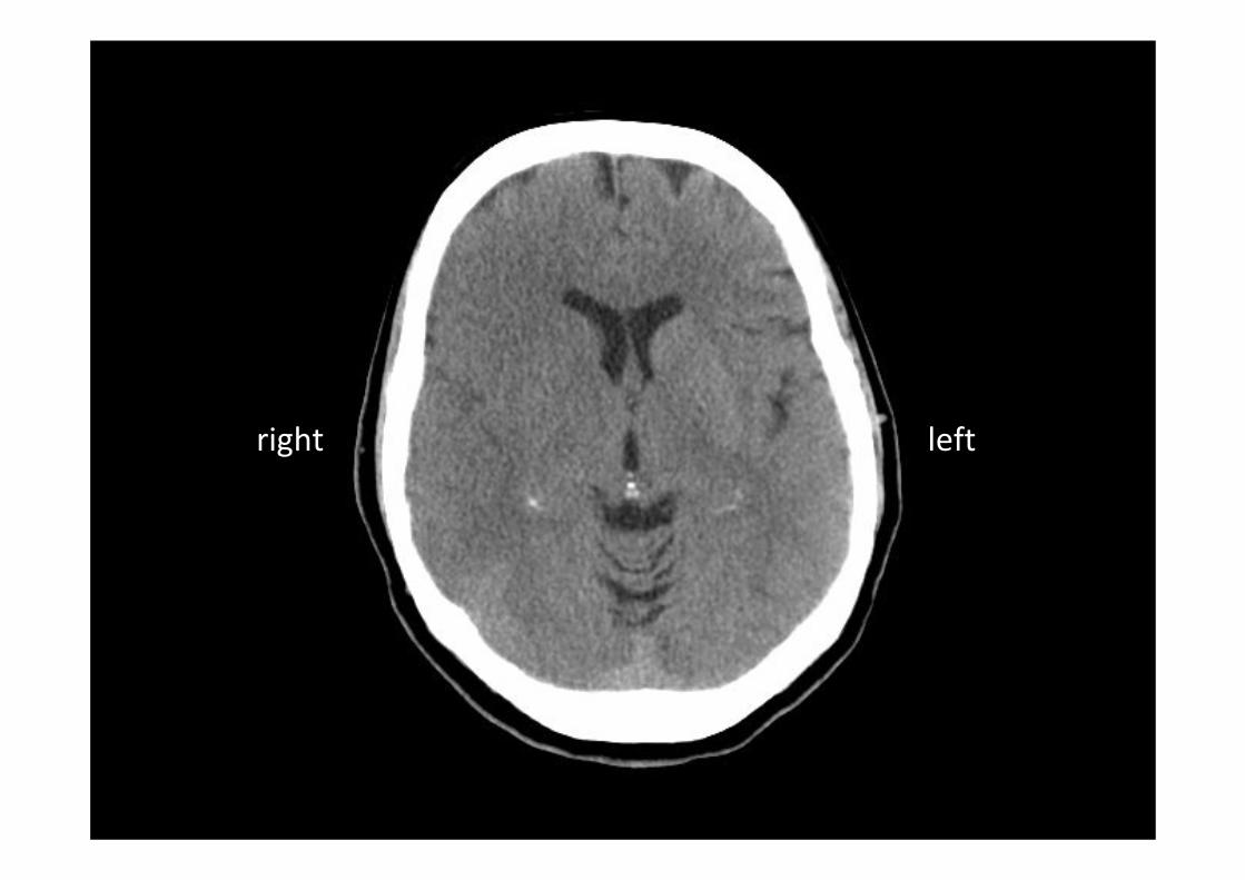
right left

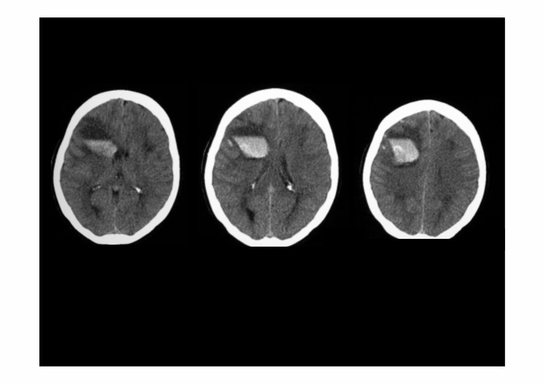
Acute CT. Early right MCAterritory ischaemia
CT perfusion. Reduced cerebral blood flow.
CT perfusion. Reduced cerebral blood volume.

MRI
• Excellent contrast between different soft tissues (including brain)
• No ionising radiation• Powerful magnetic field aligns nuclear magnetisation of hydrogen atoms within body water (including brain)
• Radio frequency fields then used to alter the alignment of the magnetisation
• Cross sectional images then generated from resultant electromagnetic signals
• Contrast agents (eg gadolinium) enhance appearance of blood vessels, tumours, inflammation


iwt

Vascular territories
• Concept of anterior and posterior (vertebrobasilar) circulation
• Circle of Willis
• Anterior, middle and posterior cerebral artery territories
• Oxfordshire Community Stroke Project (OCSP) classification

• Total anterior circulation syndrome (TACS)– hemiplegia and homonymous hemianopiacontralateral to the lesion, and
– either aphasia or visuospatialdisturbance
– with or without sensory deficit contralateral to the lesion
• Partial anterior circulation syndrome (PACS)– one or more of unilateral motor or sensory deficit, aphasia or visuospatialdysfunction (combined or not with homonymous hemianopia)
– motor or sensory deficit may be less extensive than in lacunarsyndromes (eg hand alone)
OCSP classification

OCSP classification
• Lacunar syndrome (LACS)– no visual field defect– no new disturbance of higher cortical function
– pure motor hemiparesis
– pure sensory deficit– sensorimotor stroke– ataxic hemiparesis– At least 2 of 3 contiguous areas (face, arm, leg), limb in its entirety
• Posterior circulation syndrome (POCS)– any one of:– cranial nerve impairment
– unilateral or bilateral motor or sensory deficit
– disorder of conjugate eye movement
– cerebellar dysfunction– homonymous hemianopia
– cortical blindness

iwt
• Low density
• Wedge shaped
• Grey & white matter
• Within known arterial vascular territory
• Proportionally little mass effect
Recognising an infarct

iwt

iwt

iwt

iwt

iwt
Early signs of ischaemia
• Hyperdense artery signs eg MCA
• Loss of insular ribbon
• Loss of grey/white matter differentiation in relation to caudate, lentiform nucleus, internal capsule
• Hypodense caudate or lentiform nuclei

Acute CT. Early right MCAterritory ischaemia
Repeat CT 1 day later. Malignant MCA infarction led to hemicraniectomy.
DW MRI at 5 days. Restricted diffusion pattern right hemisphere.

iwt

iwt
Day 1

iwt
Day 2

iwt
Day 4

Summary
• Role of imaging
• Types of imaging
• Vascular territories
• OCSP classification
• Recognition of infarct / early ischaemia
• Example scans