stroke and vte : a deadly combination amjad almahameed, md, mph, facp division of cardiology beth...
TRANSCRIPT
Stroke and VTE:A Deadly Combination
Amjad AlMahameed, MD, MPH, FACPDivision of Cardiology
Beth Israel Deaconess Medical CenterBoston, MA

Historical and Projected Stroke Deaths
Historical Data
Projected Data
Projected values are the product of future age-race-sex–specific mortality rates and US Census Bureau projections
Stroke 2003

50% of early post stroke
deaths have PE on autopsy studies
VTE in stroke patients has worse prognosis than nonstroke pts:
50% present as sudden deathMost patients do not have clinical evidence of DVT
VTE and Death in Stroke patients
up to 25% of early post
stroke deaths are secondary
to PE
Stroke 2000
Did u know?
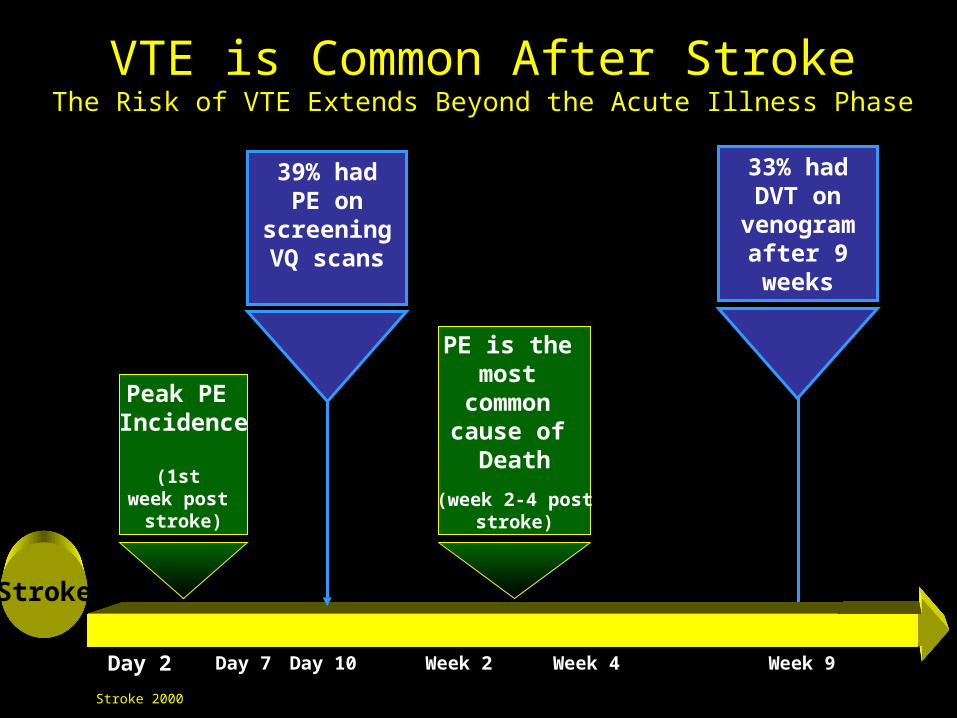
Peak PE Incidence
(1st week post
stroke)
PE is the most
common cause of
Death
(week 2-4 poststroke)
33% had DVT on
venogram after 9 weeks
Stroke
Day 2 Day 7 Day 10 Week 2 Week 4 Week 9
39% had PE on
screening VQ scans
VTE is Common After StrokeThe Risk of VTE Extends Beyond the Acute Illness Phase
Stroke 2000
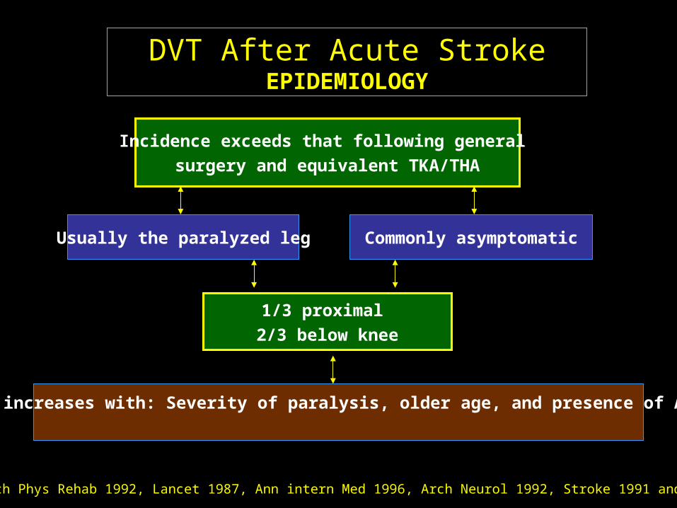
DVT After Acute Stroke EPIDEMIOLOGY
Arch Phys Rehab 1992, Lancet 1987, Ann intern Med 1996, Arch Neurol 1992, Stroke 1991 and 2001
Usually the paralyzed leg Commonly asymptomatic
1/3 proximal
2/3 below knee
Risk increases with: Severity of paralysis, older age, and presence of A fib
Incidence exceeds that following general
surgery and equivalent TKA/THA
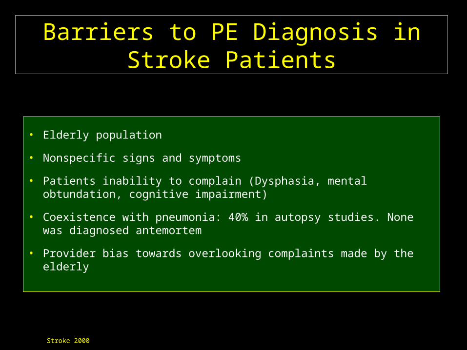
Barriers to PE Diagnosis in Stroke Patients
• Elderly population
• Nonspecific signs and symptoms
• Patients inability to complain (Dysphasia, mental obtundation, cognitive impairment)
• Coexistence with pneumonia: 40% in autopsy studies. None was diagnosed antemortem
• Provider bias towards overlooking complaints made by the elderly
Stroke 2000

Consequences of ALL Proximal DVT After Stroke
All PE (silent & clinical)
50%
Clinical PE37%
Large PE10%
Fatal PE 10-15%
Stroke 2000

Consequences of Asymptomatic Below-Knee (BK) DVT After Stroke
ProximalProximalExtensionExtension
20%20%
Silent PE 33%Usually small sizePE, not massive
Asymptomatic below the knee DVT is not as benign as once thought!!

Clinically Apparent ButUntreated DVT
Post ThromboticSyndrome 30-90%
Fatal PE37%
Stroke 2000
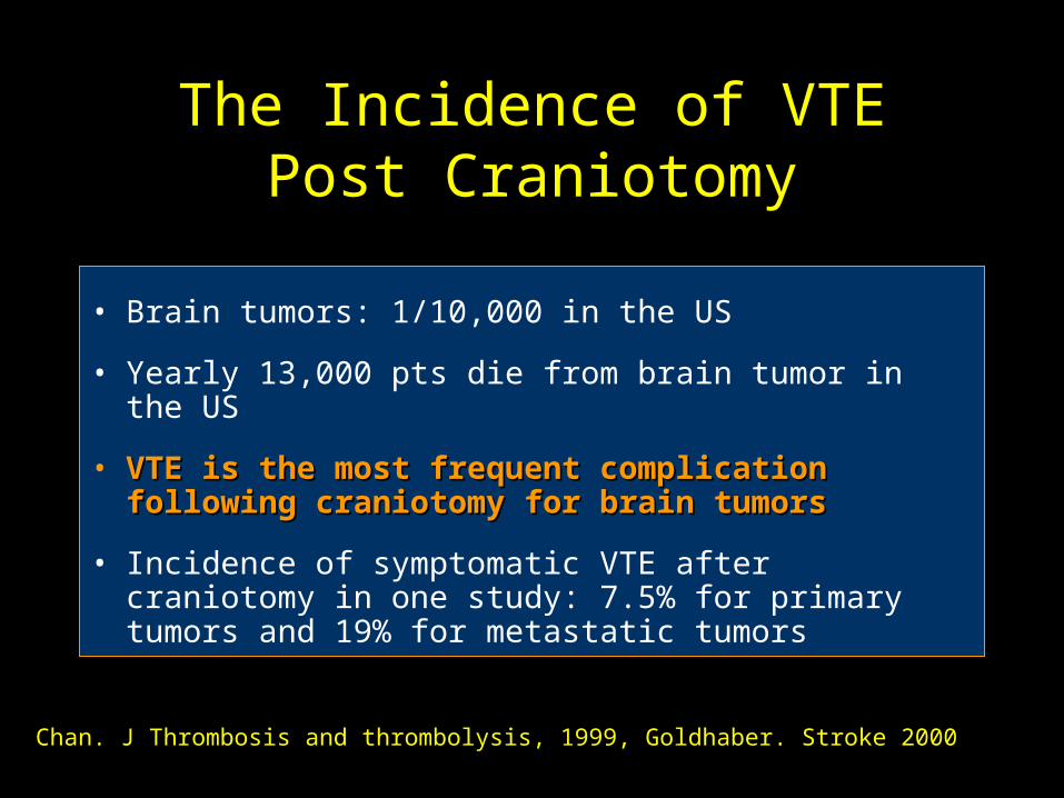
The Incidence of VTE Post Craniotomy
• Brain tumors: 1/10,000 in the US
• Yearly 13,000 pts die from brain tumor in the US
• VTE is the most frequent complication following VTE is the most frequent complication following craniotomy for brain tumorscraniotomy for brain tumors
• Incidence of symptomatic VTE after craniotomy in one study: 7.5% for primary tumors and 19% for metastatic tumors
Chan. J Thrombosis and thrombolysis, 1999, Goldhaber. Stroke 2000

VTE in the Acute Treatment Phase After Spinal Cord Injury
PE is the third most common cause of
death in this population (2)
The risk of fatal PE after SCI has NOT decreased over the past 25 years (3)
(1) Brach 1977, Rossi 1980, Myllynen 1985, Petaja 1989, Geerts 1994(2) Waring 1991, De Vivo 1999(3) De Vivo 1999
In the absence of thromboprophylaxis, objective evidence of
DVT 67-100% (1)

The risk of VTE post neurological events (stroke, spinal cord injury/surgery, and
craniotomy) is even higher in the presence of other risk factors for VTE
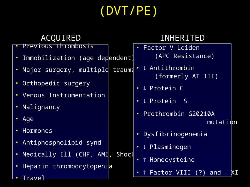
Risk Factors for VTE (DVT/PE)
ACQUIREDACQUIRED• Previous thrombosis
• Immobilization (age dependent)
• Major surgery, multiple trauma
• Orthopedic surgery
• Venous Instrumentation
• Malignancy
• Age
• Hormones
• Antiphospholipid synd
• Medically Ill (CHF, AMI, Shock)
• Heparin thrombocytopenia
• Travel
• Factor V Leiden (APC Resistance)
• Antithrombin (formerly AT III)
• Protein C
• Protein S
• Prothrombin G20210A mutation
• Dysfibrinogenemia
• Plasminogen
• Homocysteine
• Factor VIII (?) and XI
INHERITEDINHERITED
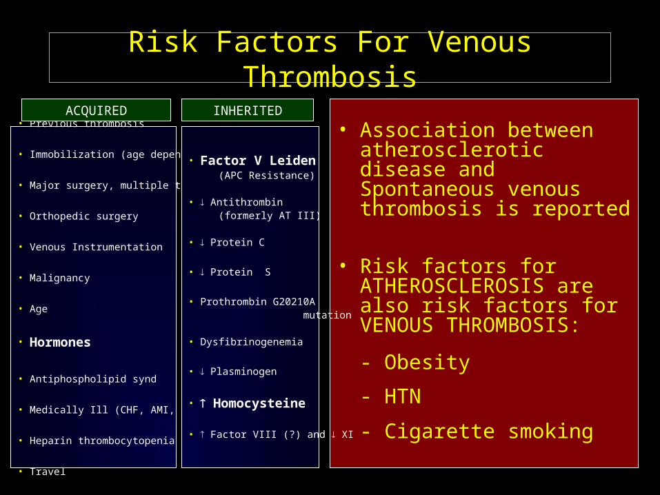
Risk Factors For Venous Thrombosis
• Association between atherosclerotic disease and Spontaneous venous thrombosis is reported
• Risk factors for ATHEROSCLEROSIS are also risk factors for VENOUS THROMBOSIS:
- Obesity
- HTN
- Cigarette smoking
• Previous thrombosis
• Immobilization (age dependent)
• Major surgery, multiple trauma
• Orthopedic surgery
• Venous Instrumentation
• Malignancy
• Age
• Hormones
• Antiphospholipid synd
• Medically Ill (CHF, AMI, Shock)
• Heparin thrombocytopenia
• Travel
• Factor V Leiden (APC Resistance)
• Antithrombin (formerly AT III)
• Protein C
• Protein S
• Prothrombin G20210A mutation
• Dysfibrinogenemia
• Plasminogen
• Homocysteine
• Factor VIII (?) and XI
ACQUIRED INHERITED

Primary Prevention Strategies

Prevention of Venous Thromboembolism
• Low dose heparin• Adjusted dose heparin• LMWH• Warfarin• Danaparoid• Refludan• Dextran, Aspirin• Fondaparinux• Ximelagatran?
• Elastic stockings• IPC• Early ambulation• IVC filter• External pneumatic plantar
compression
PharmacologicPharmacologic NonpharmacologicNonpharmacologic

Current Strategies:Mechanical Prophylaxis
• Intermittent pneumatic compression (IPC)
• Pneumatic plantar compression (foot pump)

Clot Formation
In real life: poor compliance

Contraindications for Use of PCD
Severe PAD (ABI < 0.4)
Diabetic neuropathy with sensory loss
Dermatological diseases
Presence of acute DVT at time of application
Kay 1986, Merret 1993, CLOTS 2001
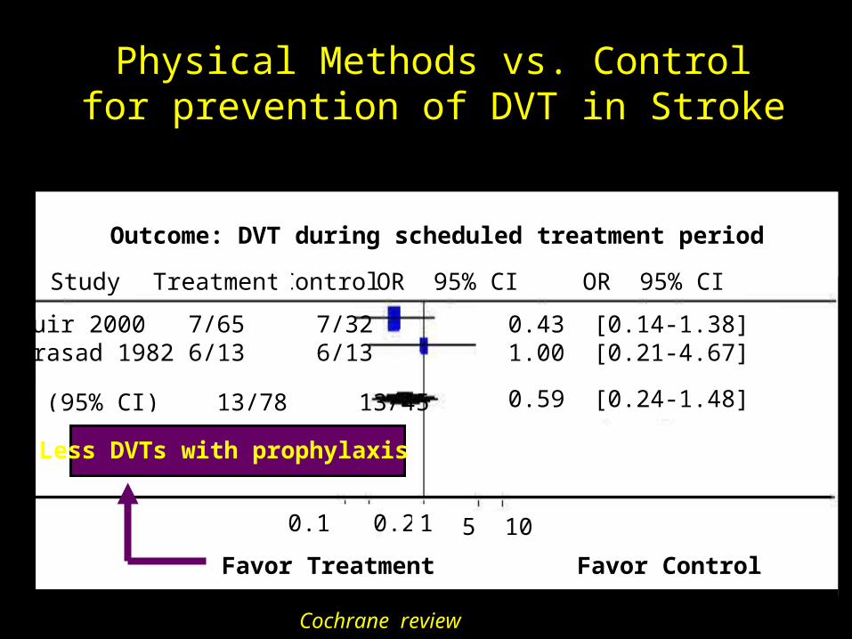
Outcome: DVT during scheduled treatment period
0.43 [0.14-1.38]1.00 [0.21-4.67]
OR 95% CITreatment Control OR 95% CIStudy
Muir 2000 7/65 7/32Prasad 1982 6/13 6/13
Total (95% CI) 13/78 13/45
0.1 0.2 1 5 10
Favor Treatment Favor Control
Physical Methods vs. Control for prevention of DVT in Stroke
0.59 [0.24-1.48]
Less DVTs with prophylaxis
Cochrane review

Physical Methods vs. Control for prevention of DVT in Stroke
Favor Treatment Favor Control
5.06 [0.96-26.78]5.06 [0.96-26.78]
Outcome: death from any cause during scheduled treatment period
0.1 0.2 1 5 10
Muir 2000 9/13 4/13
Total (95% CI) 9/13 4/13
OR 95% CITreatment Control OR 95% CIStudy
Less deaths with prophylaxis
Cochrane review

Systematic Review of Trials Comparing LMWH w UFH in Acute Ischemic Stroke: Effects on DVT
Results expressed as Peto odds ratio (OR) with a fixed-effects model. OR <1 suggests LMWH superior to UFH.
Enoxaparin vs. standard UFH
Hilbom 1998 14/106 24/106
Subtotal 14/106 24/106
Total (95% CI) 55/414 65/291
0.53 (0.26-1.06)0.53 (0.26-1.06)
0.52 (0.35-0.79)
0.1 0.2 6 10
Counsell C, Sandercock P (Cochrane Review)
Less DVT with LMWH vs. UFH

VTE Prophylaxis Post Trauma, SCIs and Craniotomy

Leg DVT in Trauma Patients
Fisher, J Ortho Trauma 1995, Kaufman, Angiology 1983, Geerts, NEJM 1996, KUDSK, Am J Surgery 1989 Geerts Injury 1996
RRR for LMWH vs. UFH
All DVT: 30% reduction (p=0.01)
Proximal DVT: 58% reduction (p=0.01)
Incidence w/o prophylaxis Incidence w prophylaxis
US studiesUp to 30%
Venography28-63%
Enoxaparin31% (40/129)
UFH44% (60/136)

UFH 5000 q 8 hrs and IPC vs. Lovenox 30 mg q 12 hrs in Acute SCI
• 1995-1998, 27 acute SCI centers in USA and Canada
• Patients > 15 years
• Traumatic SCI (C2-T12)within 72 hours
• American Spinal Injury Association (ASIA) impairment classification A, B, and C
• Bleeding (SC, ICH, other site), Coagulopathy, GI bleed within 2 weeks
• Pregnancy
• Inability to use IPC, perform US or venography, administer Heparin or contrast agents
• Uncontrolled HTN, S Creat. > 2.0, requirement for AC, Spinal surgery planned in 2 weeks, use of ASA, NSAIDS, or Ketorolac
Exclusion CriteriaInclusion Criteria
Spinal Cord Injury Thromboprophylaxis Investigators, J Trauma 2003;54:1116-1126

2 Weeks Acute Treatment Phase
VTE: termination from the study
2 weeks(bilateral venography + DUS)
6 Weeks Rehabilitation Phase
No VTE
No VTEEnoxaparin 40 mg q.d.
UFH 5000 U q 8h + ICP
8 weeks(DUS)
STUDY DESIGN
Randomization
< 72 hours After injury
UFH 5000 U q 8h + ICP
Enoxaparin 30 mg q 12 h
246 pts
230 pts
Spinal Cord Injury Thromboprophylaxis Investigators, J Trauma 2003;54:1116-1126

Outcome UFH + IPC Enoxaparin P Value
(n = 49) (%) (n = 58) (%) All VTE 31 (63.3) 38 (65.5) 0.81
DVT 22 (44.9) 35 (60.3) 0.11
PE 9 (18.4) 3 (5.2) 0.03
Symptomatic DVT 1 (2.0) 1 (1.7) -
Fatal PE 0 (0.0) 0 (0.0) -
Efficacy Outcomes in Assessable Patients with Adequate Proximal and Distal Diagnostic Imaging or Evidence of PE (N = 107)
Spinal Cord Injury Thromboprophylaxis Investigators, J Trauma 2003;54:1116-1126
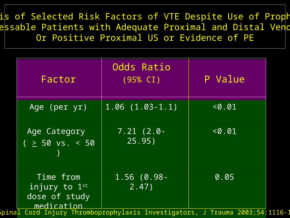
FactorOdds Ratio
(95% CI) P Value
Age (per yr) 1.06 (1.03-1.1) <0.01
Age Category
( > 50 vs. < 50 )
7.21 (2.0-25.95) <0.01
Time from injury to 1st dose of study
medication
1.56 (0.98-2.47) 0.05
Days to venography 1.26 (0.97-1.63) 0.05
Analysis of Selected Risk Factors of VTE Despite Use of ProphylaxisIn Assessable Patients with Adequate Proximal and Distal Venography
Or Positive Proximal US or Evidence of PE
Spinal Cord Injury Thromboprophylaxis Investigators, J Trauma 2003;54:1116-1126

Outcome UFH + IPC Enoxaparin P Value
(n = 249) (%) (n = 230) (%) Major Bleeding 13 (5.3) 6 (2.6) 0.14
Minor Bleeding 44 (17.9) 34 (14.8) 0.43
Discontinuation
b/c of bleeding
9 (3.7) 6 (2.6) 0.51
Deaths 2 (0.8) 2 (0.9) 0.95
Safety Events during Acute Treatment in theRandomized Population (N = 476)
Spinal Cord Injury Thromboprophylaxis Investigators, J Trauma 2003;54:1116-1126

LMWH for VTE prophylaxis after Craniotomy and Spinal Surgery
307 patients post elective craniotomy or spinal surgery97% had brain tumors
All received graduated compression stockings
Enoxaparin 40 mg/d
RANDOMIZED
Placebo
DVT: 17% DVT: 32%42% RRR
NEJM 1998;339:80-85
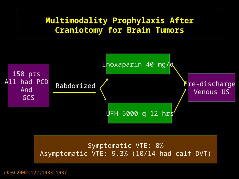
Multimodality Prophylaxis After Craniotomy for Brain Tumors
150 pts All had PCD
And GCS
Rabdomized
Enoxaparin 40 mg/d
UFH 5000 q 12 hrs
Pre-discharge Venous US
Symptomatic VTE: 0%Asymptomatic VTE: 9.3% (10/14 had calf DVT)
Chest 2002;122;1933-1937

Strategies for decreasing the Incidence of VTE after Acute Ischemic Stroke, SCI,
and Post craniotomy
• Effective and optimal in-patient prophylaxis
• ? Extended out of hospital prophylaxis
• ? Early diagnosis of subclinical VTE (screening serial US, D-Dimer, MRV)
• ? Multi-modality approach
• Prompt diagnosis and treatment of clinically apparent VTE (saves life and prevents recurrence)