striatal volumes in pediatric bipolar patients with and without comorbid adhd
TRANSCRIPT

Psychiatry Research: Neuroimaging 194 (2011) 14–20
Contents lists available at ScienceDirect
Psychiatry Research: Neuroimaging
j ourna l homepage: www.e lsev ie r.com/ locate /psychresns
Striatal volumes in pediatric bipolar patients with and without comorbid ADHD
Isabelle Yisha Liu a, Meghan Howe b, Amy Garrett b,c, Asya Karchemskiy b,c, Ryan Kelley b,c, Dylan Alegria b,c,Allan Reiss b,c, Kiki Chang b,⁎a Washington University School of Medicine, Saint Louis, MO, United Statesb Pediatric Bipolar Disorders Program, Stanford University School of Medicine, Stanford, CA, United Statesc Center for Interdisciplinary Brain Sciences Research, Stanford University, Stanford, CA, United States
⁎ Corresponding author at: 401 Quarry Rd, MC 5719650 723 5511; fax: +1 650 723 5531.
E-mail address: [email protected] (K. Chang).
0925-4927/$ – see front matter © 2011 Elsevier Irelanddoi:10.1016/j.pscychresns.2011.06.008
a b s t r a c t
a r t i c l e i n f oArticle history:Received 18 August 2010Received in revised form 13 June 2011Accepted 14 June 2011
Keywords:Magnetic resonance imagingCaudate nucleusBasal gangliaStriatum
The most prevalent comorbid disorder in pediatric bipolar disorder (BD) is attention-deficit/hyperactivitydisorder (ADHD). As caudate volume abnormalities have been demonstrated in both BD and ADHD, this studysought to determine whether these findings could be attributed to separable effects from either diagnosis.High resolution anatomical magnetic resonance (MRI) images were obtained from youth in 4 groups: BD withcomorbid ADHD (n=17), BD without comorbid ADHD (n=12), youth with ADHD alone (n=11), andhealthy control subjects (n=24). Caudate, putamen, and globus pallidus volumes were manually traced foreach subject using BrainImageJava software by a reliable rater blinded to diagnosis. There was a significanteffect of diagnosis on striatal volumes, with ADHD associated with decreased caudate and putamen volumes,and BD associated with increased caudate, putamen, and globus pallidus volumes. Thus, the presence orabsence of comorbid ADHD in patients with BD was associated with distinct alterations in caudate volumes,suggesting that these groups have different, but related, mechanisms of neuropathology.
, Stanford, CA 94305. Tel.: +1
Ltd. All rights reserved.
© 2011 Elsevier Ireland Ltd. All rights reserved.
1. Introduction
There has been a vast increase in our research knowledge ofpediatric-onset bipolar disorder (BD) over the past decade. Neverthe-less, many questions regarding the underlying pathophysiology of thedisease remain unanswered. Finding a common biological marker of BDis complicated by the heterogeneity of the disorder, particularly as itpresents frequently with comorbid disorders. The most prevalentcomorbid disorder with pediatric BD is attention deficit/hyperactivitydisorder (ADHD)(DelBello et al., 2004;Adler et al., 2005). Thesepatientsshow impairments in attention, impulse control, and executive functionin addition to the mood dysregulation that affects all patients with BD.Studying BD patient populations based on the presence or absence ofADHDmay create greater homogeneity within patient groups. Thismayaid in distinguishing neural abnormalities unique to each disorder.
Several studies have used structural magnetic resonance imaging(sMRI) to examine regional brain volumes in both BD and ADHD.While anumber of studies have demonstrated reduced caudate nucleus volumesin patients with ADHD as compared with healthy controls (Krain andCastellanos, 2006a; Schneider et al., 2006), such studies on patients withBD have been few and inconsistent. Wilke and colleagues found overallvolume increases in the basal ganglia of adolescentswith BD (Wilke et al.,
2004), while DelBello reported increased putamen volumes compared tohealthy controls (DelBello et al., 2004). In contrast, other studies inpediatric BD samples found no abnormalities in putamen (Sanches et al.,2005) or caudate volumes (Chang et al., 2005; Sanches et al., 2005).Furthermore, in the most relevant recent study that examined striatalvolumes among youths with BD and/or ADHD, caudate and putamenvolumes were decreased in subjects with ADHD alone, but no differencesin striatal volumes were found between the subjects with BD alone orthose with BD+ADHD as compared with controls (Lopez-Larson et al.,2009). Additionally, two studies of first degree relatives or offspring ofbipolar parents did not show significant changes in striatal volumes ascompared with controls (Singh et al., 2008; Hajek et al., 2009).
In this study, we compared the volumes of the caudate andputamen in pediatric patients with BD+ADHD, BD alone, ADHD alone,and healthy controls. We hypothesized that subjects with ADHDwould have decreased striatal volumes compared to healthy controls,while subjects with BD alone would have increased striatal volumes.Thus, when both disorders co-occur (ADHD+BD), we hypothesizedthat individuals would present with intermediate striatal volumesthat do not significantly differ from healthy controls.
2. Methods
2.1. Subjects
This study was approved by the Stanford University IRB, and allsubjects gave oral and written informed consent or assent before

Fig. 1. Coronal view of all regions of interest.
15I.Y. Liu et al. / Psychiatry Research: Neuroimaging 194 (2011) 14–20
participation. Fortypatientswere recruited fromthe StanfordUniversitychild and adolescent psychiatry clinic. Participants included 17 with BDand ADHD (BD+ADHD group), 12 with BD only (BD−ADHD group),and 11with ADHDonly (ADHD group). Twenty-four healthy volunteerswere recruited from the community (Healthy Control group). Partici-pants were included if they were 9–18 years old, had IQN70, had nocontraindications to undergoing an MRI scan, and did not have anymajor neurological disorders, medical conditions, or developmentaldisorders (such as a pervasive developmental disorder) which mayaffect brain functioning. Diagnoses were made by a trained clinicianusing the Washington University in St. Louis Kiddie Schedule forAffective Disorders and Schizophrenia (WASH-U-KSADS) (Geller et al.,1996) and the Schedule for Affective Disorders and Schizophrenia forSchool-age Children, Present and Lifetime (Kaufman et al., 1997). Alldiagnoses were confirmed by a board-certified child psychiatrist (KC).Subjects with BD met DSM-IV criteria for bipolar I disorder. ADHDparticipants met DSM-IV criteria for attention-deficit hyperactivitydisorder (inattentive, hyperactive, or combined) with no additionaldiagnoses of any other psychiatric disorders. Healthy volunteers did nothave any current or lifetimeDSM-IV diagnoses. Parental diagnoseswerealso obtained using the Structured Clinical Interview for DSM-IV Axis IDisorders (First et al., 1995).
2.2. Image acquisition
Subjects discontinued use of psychostimulants for 24 h prior tomagnetic resonance imaging (MRI) due to a concurrent functional MRIstudy. Participants were allowed to continue taking other medicationssuch as mood stabilizers and antidepressants to prevent mooddestabilization.
All imaging procedures were conducted at the Lucas Center forMagnetic Resonance Spectroscopy and Imaging at Stanford Universi-ty, Palo Alto, CA. Magnetic resonance images of each subject's brainwere acquired with a Signa 3.0-T scanner (GE Medical Systems,Milwaukee,WI). Images were acquired in the coronal plane, using a 3-D volumetric radiofrequency spoiled gradient echo pulse sequencewith the following scan parameters: TR=35 ms, TE=6ms, flipangle=45°, number of excitations=1, image matrix=256×192field of view=24 cm, slice thickness=1.5 mm, 124 slices, acquiredresolution=1.5×0.9×1.2 mm3. The images were reconstructed as a124×256×256 matrix with a 1.5×0.9×0.9 mm3 spatial resolution.
Seven of the BD−ADHD subjects were scanned with the samepulse sequence parameters, but using a different headcoil due to theirconcurrent participation in another study. Before combining thesedata, we systematically investigated the effects of each headcoil typeon measures of caudate volume. Four subjects (2 male, 2 female) withno family history of psychiatric illness were scanned using bothheadcoils and using the same pulse sequences and volumetricmeasures as used in this study. Reliability analysis performed by asingle measure intraclass correlation calculated in SPSS 17.0 foundexcellent intraclass correlations (ICC) for all measures: Volume of graymatter ICC=0.99, white matter ICC=0.99, and cerebrospinal fluidICC=0.98. Measures of total caudate volume were also highlycomparable between headcoils. Right caudate total volume had anICC of 0.99, and left caudate total volume had an ICC of 0.99. Thus,headcoil type did not contribute significant variance to themeasure ofstriatal volumes for this study, and we therefore included these sevensubjects in our analyses.
2.3. Volumetric analysis
Images were imported to the program BrainImageJava, version5.3.7 (BIJ; Center for Interdisciplinary Brain Sciences Research; http://spnl.stanford.edu). Non-brain tissue was removed using a semi-automated process, and the images were corrected for field biasartifact before importing into BIJ. Images were positionally normal-
ized (rigid body transformation) based on positions of the anteriorand posterior commissures (Talairach and Tournoux, 1988). Totalbrain volume (TBV) was calculated as the sum of the total tissue andtotal CSF of the cerebrum, cerebellum, ventricles, and brainstem.
All regions of interest (ROIs) were outlined by a single trainedrater blinded to diagnosis and to the subjects' identity (IL). Highintraclass reliability (ICCN0.9) was first established with a set of goldstandard ROIs traced on a separate set of control images. A briefdescription of each ROI follows, and an example of a coronal slice withoutlined ROIs is seen in Fig. 1. These tracings are in accordance withpreviously published descriptions (Murphy et al., 1992; Chang et al.,2005).
2.3.1. CaudateTracing the caudate began at the most anterior slice where the
gray matter of the head of the caudate was visible and proceededposteriorly until gray matter of the caudate tail was no longer visible.Themedial border was the lateral ventricle, and the lateral border wasthe internal capsule. When a small gray matter connection was seenbetween the putamen and the caudate, the inferior border of thecaudatewasmoved to a straight line between this graymatter and themost inferior point of the lateral ventricle. This served to exclude thenucleus accumbens from the ROI.
2.3.2. Lenticular nucleusThe lenticular nucleus includes the putamen and the globus pallidus.
Tracing began at the most anterior slice where the putamen becamevisible lateral to the caudate. The lateral borderwas the external capsuleand themedial border was the internal capsule. The inferior borderwasthe medial border until the anterior commissure became visible, atwhich point it became the anterior commissure. The superior borderwas the corona radiata and the internal capsule.
2.3.3. Globus pallidus and putamenFrom the lenticular nucleus ROI, the globus pallidus ROIwas isolated
by tracing between the globus pallidus and the putamen and deletingthe putamen from the ROI. The globus palliduswas not originally part ofthe hypothesis, but due to this method of isolating the putamen ROI, wedid exploratory measurements of GP volumes. Putamenmeasurementswere taken indirectly by subtracting the globus pallidus volumes fromthe lenticular nucleus volumes.

16 I.Y. Liu et al. / Psychiatry Research: Neuroimaging 194 (2011) 14–20
2.4. Statistical analysis
One-way analyses of variance (ANOVAs) were performed tocompare the four subject groups on demographic and clinical variables.All volumetric analyses of individual structures used linear regressionmodels in SPSS software. Binary variables were centered as+0.5/−0.5.These includedbipolardiagnosis (yes or no) andADHDdiagnosis (yesorno). The linear variable used as a covariate, total cerebral tissue, wascentered by subtracting the global mean value from each value. Acorrected threshold of p=0.05/3 models=0.0167 was used todetermine significance. In order to use these parametric statistics, thedatawerefirst examined for normality. Thedatawere also examined foroutliers, andone outlier datapointwas found andverified tobe accurate.Follow-up hemispheric analyses were conducted as exploratory andthus no correction formultiple comparisonswasperformed.Medicationexposure was included as a factor to investigate whether effects ofdiagnosis were due to medications.
3. Results
3.1. Demographic variables and total brain volume
Neither age (F=1.59, d.f.=3, p=0.20), total brain volume (TBV)(F=1.17, d.f.=3, p=0.33), nor IQ (F=1.76, d.f.=3, p=0.17)exhibited significant differences between groups (Tables 1 and 2).However, we covaried for TBV in our analyses of striatal volumes aswe found high correlations between TBV and striatal volumes. TheBD+ADHD and BD−ADHD groups did not differ in scores on theYoung Mania Rating Scale (F=0.06, d.f.=1, p=0.80), Children'sDepression Index (F=0.14, d.f.=1, p=0.71), age of BD onset(F=0.07, d.f.=1, p=0.79), or ratings on the Children's GlobalAssessment Scale (F=1.01, d.f.=1, p=0.33) (Table 1). The groupsdiffered in current medication usage (see Table 1). All subjects withADHD (with or without BD) had combined type. Twenty-five of the 29subjects (86%)with BD had at least one parentwith BD. ADHD subjectshad parents with no DSM-IV diagnoses other than ADHD, and healthycontrols had parents with no DSM-IV diagnoses. ADHD participantsand healthy controls also had no first degree relatives with BD. Five ofthe 12 subjects (42%) with BD only had a parent with ADHD, while 14of the 17 subjects with BD+ADHD (82%) had a parent with ADHD.Information on whether bipolar subjects were manic, depressed, or
Table 1Descriptive and clinical measures by groupa.
Bipolar+ADHD Bipolar–
N 17 12Age mean (SD) 14.4 (2.6) 15.8 (2.Gender (%male) 76% 42%IQc 109 (11.7) 109 (9.0Age of BD onset (S.D.) 12.6 (2.4) 12.9 (3.YMRS mean (SD)d 14.1 (8.4) 15.1 (10C-GAS mean (S.D.)e 54.6 (8.2) 50.8 (8.CDI mean (S.D.)f 14.3 (8.2) 16.3 (9.% exposed to any psychotropic 94% 25%% exposed to antidepressants 94% 17%% exposed to mood stabilizersg 71% 25%% exposed to antipsychotics h 29% 17%% exposed to stimulants 73% 17%
a Attention-deficit/hyperactivity disorder only (ADHD), bipolar disorder with comorbid Acontrols (Control).
b p values obtained from 4-group analyses of variance: main effect of groupc As measured by Wechsler Abbreviated Scale of Intelligenced Young Mania Rating Scalee Children's Global Assessment Scalef Children's Depression Inventoryg Mood stabilizers include lithium, valproate, lamotrigine, and carbamazepine.h Includes both typical and atypical antipsychotics.
euthymic at the time of the scan was not collected outside of theYMRS and CDI scales given. No subjects had symptoms of psychosis.
3.2. ROI volumetric regression analyses
The brain region volumetric measurements in cm3 are given inTable 2 and Fig. 2. The results of the three regressionmodels are given inTable 3. For themodel of total caudate volume, themain effect of bipolardiagnosis and the main effect of ADHD diagnosis were both significant(see Table 3), and in opposite directions (e.g. bipolar diagnosis wasassociated with a larger caudate, while ADHD diagnosis was associatedwith a smaller caudate). The interaction was not significant.
A similar pattern was seen for total putamen volume. The maineffect of bipolar diagnosis, the main effect of ADHD diagnosis, and theinteraction of bipolar×ADHD were significant. Again, those with abipolar diagnosis had a larger putamen, while those with an ADHDdiagnosis had a smaller putamen volume.
The main effect of bipolar diagnosis was significant for total globuspallidus volume, such that bipolar diagnosis was associated with alarger volume. No other factors were significant for globus pallidusvolume.
After the initial three models were examined and found to besignificant, we performed exploratory analyses on left and righthemispheric volumes for the different structures. For left caudate, themain effect of bipolar was significant (p=0.0001, positive effect), themain effect of ADHDwas significant (p=0.0001, negative effect), whilethe interactionwasnot significant (p=0.045). For the right caudate, themain effect of bipolar was significant (p=0.0001, positive effect),the main effect of ADHD was significant (p=0.0001, negative effect),while the interaction was not significant (p=0.155). For the leftputamen, the effects were as follows: bipolar: p=0.0001 positive;ADHD: p=0.0001 negative; interaction p=0.002. For the rightputamen, the effects were as follows: bipolar: p=0.0001 positive;ADHD: p=0.0001 negative: interaction p=0.057. For the left GP, theeffects were as follows: bipolar p=0.0001 positive; ADHD; p=0.274,interaction p=0.046. For the right GP, the effects were as follows:bipolar p=0.138; ADHD p=0.253; interaction p=0.760. We alsoperformed exploratory analyses investigating whether the caudatehead or body/tail were differently affected. Both the head and the body/tail showed similar effects as the total caudate (caudate head: maineffect of bipolar diagnosis: p=0.0001, main effect of ADHD diagnosis:p=0.0001, the interaction of bipolar×ADHD was not significant NS;
ADHD ADHD Controls p b
11 245) 13.4 (3.3) 14.2 (2.7) 0.20
82% 71% 0.14) 114 (14.2) 116 (8.3) 0.170) — — 0.79.5) — — 0.805) — — 0.335) — — 0.71
73% 0%18% 0%0% 0%9% 0%73% 0%
DHD (BD+ADHD), bipolar disorder without comorbid ADHD (BD−ADHD), and healthy
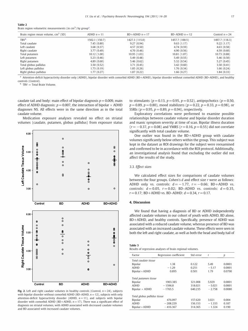
Table 2Brain region volumetric measurements (in cm3) by groupa.
Brain region mean volume, cm3 (SD) ADHD n=11 BD+ADHD n=17 BD–ADHD n=12 Control n=24
TBVb 1502.1 (150.7) 1427.3 (115.0) 1457.7 (100.5) 1497.7 (118.3)Total caudate 7.45 (0.80) 9.27 (0.94) 9.63 (1.17) 9.22 (1.17)Left caudate 3.68 (0.37) 4.57 (0.50) 4.74 (0.59) 4.63 (0.58)Right caudate 3.77 (0.49) 4.70 (0.46) 4.90 (0.56) 4.59 (0.60)Total putamen 10.12 (1.00) 10.95 (1.03) 10.81 (1.07) 10.73 (0.88)Left putamen 5.23 (0.46) 5.49 (0.48) 5.49 (0.55) 5.46 (0.50)Right putamen 4.89 (0.60) 5.46 (0.62) 5.32 (0.54) 5.27 (0.45)Total globus pallidus 3.50 (0.52) 3.71 (0.45) 3.42 (0.60) 3.50 (0.41)Left globus pallidus 1.73 (0.19) 1.85 (0.26) 1.76 (0.34) 1.66 (0.24)Right globus pallidus 1.77 (0.27) 1.87 (0.22) 1.66 (0.27) 1.84 (0.33)
a Attention-deficit hyperactivity disorder only (ADHD), bipolar disorder with comorbid ADHD (BD+ADHD), bipolar disorder without comorbid ADHD (BD–ADHD), and healthycontrols (Control).
b TBV = Total Brain Volume.
17I.Y. Liu et al. / Psychiatry Research: Neuroimaging 194 (2011) 14–20
caudate tail and body: main effect of bipolar diagnosis p=0.009, maineffect of ADHD diagnosis: p=0.007, the interaction of bipolar × ADHDdiagnoses NS. All effects were in the same direction as in the totalcaudate volume.
Medication exposure analyses revealed no effect on striatalvolumes (caudate, putamen, globus pallidus) from exposure status
Fig. 2. Left and right caudate volumes in healthy controls (Control, n=24), subjectswith bipolar disorder without comorbid ADHD (BD–ADHD, n=12), subjects with onlyattention-deficit hyperactivity disorder (ADHD, n=11), and subjects with bipolardisorder with comorbid ADHD (BD+ADHD, n=17). There was a significant effect ofdiagnosis on striatal volumes, with ADHD associated with decreased caudate volumesand BD associated with increased caudate volumes.
to stimulants (p=0.13, p=0.95, p=0.52), antipsychotics (p=0.50,p=0.89, p=0.80), mood stabilizers (p=0.22, p=0.33, p=0.98), orSSRIs (p=0.95, p=0.89, p=0.94), respectively.
Exploratory correlations were performed to examine possiblerelationships between caudate volume and bipolar disorder durationand manic symptom severity at time of scan. Bipolar illness duration(r=−0.37, p=0.08) and YMRS (r=0.16, p=0.55) did not correlatesignificantly with total caudate volume.
One outlier was found in the BD+ADHD group with caudatevolumes significantly below others within the group. This subject waskept in the dataset as ROI drawings for the subject were reexaminedand confirmed to be in accordancewith the ROI protocol. Additionally,an investigational analysis found that excluding the outlier did notaffect the results of the study.
3.3. Effect sizes
We calculated effect sizes for comparisons of caudate volumesbetween the four groups. Cohen's d and effect size r were as follows:ADHD only vs. controls: d=−1.77, r=−0.66; BD+ADHD vs.controls: d=0.05, r=0.02; BD–ADHD vs. controls: d=0.35,r=0.17; BD+ADHD vs. BD–ADHD: d=0.34, r=0.17.
4. Discussion
We found that having a diagnosis of BD or ADHD independentlyaffected caudate volumes in our cohort of youth with ADHD, BD alone,BD+ADHD, and healthy controls. Specifically, presence of ADHD wasassociated with a reduced caudate volume, whereas presence of BDwasassociatedwith an increased caudate volume. These effects were seen inboth the left and right caudate, as well as both the head and body/tail of
Table 3Results of regression analyses of brain regional volumes.
Factor Regression coefficient Std error t p
Total caudate tissueBipolar 1.38 0.122 5.49 0.0001ADHD −1.29 0.251 −5.17 0.0001Bipolar×ADHD 0.895 0.501 1.79 0.0790
Total putamen tissueBipolar 1903.385 321.008 5.929 0.0001ADHD −1596.8 318.021 −5.021 0.0001Bipolar × ADHD −1765.5 640.235 −2.758 0.0080
Total globus pallidus tissueBipolar 476.097 157.620 3.021 0.004ADHD −208.229 156.153 −1.333 0.187Bipolar×ADHD −416.367 314.365 −1.324 0.190

18 I.Y. Liu et al. / Psychiatry Research: Neuroimaging 194 (2011) 14–20
the caudate, and psychotropic medication exposure did not affect thesefindings. Effect size calculations revealed a large effect size for thedifference in caudate volume between ADHD subjects and healthycontrols, andmediumeffect sizes for BD−ADHDsubjects comparedwithcontrols, and BD only subjects compared with BD+ADHD subjects.
We could grossly restate these findings by stating that patients withADHD have decreased caudate volumes, patients with BD haveincreased caudate volumes, and those with both ADHD and BD havenormal caudate volumes. However, it would likely be an oversimplifi-cation to conclude that those with both disorders have an intermediatevolume because the effects on the caudate of the disorders “canceled”each other. Previous studies regarding the role of the caudate in bothexecutive functioning and mood regulation suggest more informedinterpretations.
A number of animal studies of lesions of the caudate nucleus havebeen performed inmany species, and a comprehensive reviewof thesedata concluded that while early studies on motor function suggestedthat the caudate serves to inhibit behaviors initiated in the cortex, itsfunction is actually much more complex and nuanced (White, 2009).Based on studies in monkeys, cats, and rats, the authors conclude thatcaudate lesions affect memory function, and specifically reinforcedstimulus-response learning. Observational studies of basal ganglialesions in humans indicate that the caudate is involved in cognitivefunctions, including planning and sequencing, memory, and sustainedattention (Benke et al., 2003; Grahn et al., 2008; White, 2009).Functional MRI studies in healthy control populations have foundcaudate activation during executive function tasks, particularlyresponse inhibition (Aron et al., 2003; Luna and Sweeney, 2004; Aliet al., 2010), but also goal achievement (Tricomi and Fiez, 2008),learning (Nomura and Reber, 2008), reward prediction (Haruno andKawato, 2006), reward-related learning (Haruno et al., 2004), andplanning and set-shifting (Monchi et al., 2006).
With these functions, it is not surprising that the caudate has beenconsistently associated with ADHD, which is characterized byinattention and/or impulsivity/hyperactivity (Sonuga-Barke, 2005).Reductions in caudate volume have been widely reported inparticipants with ADHD (Krain and Castellanos, 2006; Schneider etal., 2006). However, Castellanos et al. have suggested that caudatevolumes in ADHD may “normalize” over the trajectory of develop-ment from childhood to adulthood (Castellanos et al., 2002). A recentDTI study also suggests normalization of the caudate during lateadolescence (Silk et al., 2009). This is consistent with findings ofincreased caudate volume in older adolescents (ages 15 to 19) withADHD (Mataro et al., 1997; Garrett et al., 2008). Thus, our findings ofsmaller caudate volumes in children with ADHD are consistent withprevious reports in early/mid adolescence, as our subjects had a meanage of 13.4±3 years.
The finding of increased caudate volumes in subjects with BDsuggests that abnormalities in the caudate are also associated with BDseparately, which may have several explanations. First, regardingbrain anatomy, bigger is not always better (Foster et al., 1999; Roth etal., 2010), so increased caudate volume may indicate an abnormality,albeit with a different etiology. This different abnormality may resultin a different set of impairments than those seen in ADHD, as cognitiveimpairments have indeed been reported in BD. A longitudinal studyover 30 months found that adults with BD had cognitive deficitsduring all mood states, including depressive, manic, and euthymic(Malhi et al., 2007). However, while not all patients with BD havebeen found to have these cognitive deficits (Jamrozinski, 2010), mostof these studies did not separate out the effects of comorbid ADHD.The only study that examined the effects of comorbid ADHD onneuropsychological functioning in pediatric BD found significantcognitive impairment in the ADHD group and the BD+ADHD group,but not in the BD only group (Rucklidge, 2006).
While increased caudate volume may not mediate cognitive impair-ment in BD, it may be directly related tomood regulation difficulties. The
caudate is a component in limbic circuits that also include the amygdalaandprefrontal cortex (Ring andSerra-Mestres, 2002).Mooddysfunctionoften develops in neurodegenerative disorders that affect the caudate,including Parkinson's (Gotham et al., 1986) and Huntington's Disease(Peyser and Folstein, 1990). Additionally, recent studies have implicatedthe caudate in other mood disorders such as major depressive disorder(Lee et al., 2008), and subjects with major depression have beenreported to show decreased activation in the caudate in response torewards (Pizzagalli et al., 2009). In healthy control subjects, a recentstudy reported robust caudate activation to negative but not positiveemotion-related picture content, and suggested that the caudate plays arole in emotional withdrawal (Carretie et al., 2009). Thus, abnormalcontrol of emotion-related processes could be related to abnormalcaudate volume in BD subjects. Still, it is unclear why caudate volumewould be increased in the context of mood regulation, but decreased inthe context of behavioral response inhibition.
Lopez-Larson and colleagues performed a similar analysis across thesame four groups in youth with BD only, ADHD only, and both BD andADHD and healthy controls (Lopez-Larson et al., 2009). Consistent withour results, they found decreased caudate volumes in subjects withADHD only as compared to each of the other three groups. However,unlike our study, they did not find volumetric differences in striatalstructures in subjects with BD alone as compared to either healthycontrols or those with BD+ADHD. This difference in findings may bedue to a difference in our study populations. The subjects in their studywere significantly younger (meanageof 10.8 vs. 14.4 years in our study)with ahigher proportion of prepubertal patients. It is very possible thesestructural differences do not appear until later in development, whendisease duration, medication exposure, and development all play a rolein affecting brain structures. Our study also included a predominance ofBD subjects who had a family history of the disorder, which may be amore genetically and neurobiologically homogeneous group than thegroup in the Lopez-Larson study. Finally, our statistical approachdiffered from that of Lopez-Larson et al.
Wealso found thatADHDandBDdiagnoseshad significant effects onputamen and globus pallidus volumes. Putamen volumes followed thesame patterns as for caudate, whereas GP volume was affected only byBD diagnosis (increased). Putamen volume has previously been foundincreased in some (DelBello et al., 2004) but not all studies of youthwithBD (Ahn et al., 2007) or in offspring of bipolar patients (Hajek et al.,2009). However, putamen activation has been found to be elevated infMRI studies of bipolar youth (Blumberg et al., 2003; Chang et al., 2004;Rich et al., 2006). Globus pallidus volume has not been reported to beabnormal in youth with BD (DelBello et al., 2004; Ahn et al., 2007). Inyouth with ADHD, both putamen and globus pallidus volumes havebeen reported to be decreased compared with controls (Ellison-Wrightet al., 2008; Qiu et al., 2009). Thus, our findings in general are in accorddirectionally with these previous findings: increased striatal volume inBD, decreased striatal volume in ADHD.
It should be noted that psychotropic medications have beenassociated with changes in regional cortical volumes, includingstriatum. For example, two studies reported increases in basal ganglialvolumes with chronic exposure to typical antipsychotics (Corson et al.,1999; Lang et al., 2001), while one reported decreased basal gangliavolumeswith chronic exposure to atypical antipsychotics (Corson et al.,1999). A recent systematic review of 33 articles found that typicalantipsychotic exposure, even for a short duration (b12 weeks), wasassociated with increased basal ganglial volumes (Navari and Dazzan,2009). Exposure to atypicals did not have a consistent association withbasal ganglial volumes, with one study finding increased volumes(Massana et al., 2005),while several other studies finding unchanged ordecreased volumes (Heitmiller et al., 2004; Navari and Dazzan, 2009).
Our groups differed significantly by medication exposure. Unfor-tunately, we could not compare medication-exposed to medication-naive subjects within each group statistically, as the groups wereunbalanced in this regard, and the number of subjects would have

19I.Y. Liu et al. / Psychiatry Research: Neuroimaging 194 (2011) 14–20
been too small in each group to perform valid statistical comparisons.Specifically, the ratios of medication-exposed to medication-naivesubjects for each group were as follows: BD−ADHD: 3/9, ADHD 8/3,and BD+ADHD 16/1. We attempted to address this question using aregression analysis that showed exposure to each type of medicationdid not have a significant effect on striatal volumes. However, thispost-hoc medication analysis was underpowered and thus we cannotconclude that medication had no effect. Additionally, as discussedabove, psychotropic medications have been shown to affect brainvolumes, so our findings should be interpreted in this context andtaken as preliminary.
It should be noted that only 25% (3/12) of the BD−ADHD group hadbeen exposed to psychotropic medications. It is possible that this groupis slightly healthier than the other groups in the study; however, clinicalcharacteristics, including similar duration of illness and CGAS scores,suggest that this group did not differ significantly from the other groupsin severity of illness.
There were several limitations to our study, including the relativelysmall sample sizes, particularly within the BD−ADHD group. While thestudy was adequately powered to find significant differences betweengroups, other less robust differences may have beenmissed. Effect sizesfor comparisons between the four groups on caudate volumes wereprovided for future hypothesis testing. Also, while we addressed theheadcoil difference in the methods section, this difference could haveconfounded our analysis in unknownways. Lastly, as is the case with allregional structural studies of the brain, a structural difference does notnecessarily correspond to a functional difference. Recent fMRI studieshave suggested functional abnormalities in the caudate nuclei of bipolarpatients (Wessa et al., 2007). However, more studies are needed toelucidate the function of the caudate in BD with and without comorbidADHD.
Nonetheless, our findings suggest that different caudate abnormal-ities may be associated with ADHD and BD. Future studies should takethis difference into account, particularly when studying youth with BDand comorbid ADHD, as differential functional deficits may lead torelatively normal volumetric findings.
Acknowledgments
This work was supported in part by NIMH grant MH64460, a grantfrom the Klingenstein Third Generation Foundation, the Hahn family,and a grant from the Stanford University Undergraduate ResearchPrograms, administered by the Office of the Vice Provost for Under-graduate Education.Wealso thank theHowardHughesMedical Institutefor support of our research.
References
Adler, C.M., Delbello, M.P., Mills, N.P., Schmithorst, V., Holland, S., Strakowski, S.M.,2005. Comorbid ADHD is associated with altered patterns of neuronal activation inadolescents with bipolar disorder performing a simple attention task. BipolarDisorders 7, 577–588.
Ahn, M.S., Breeze, J.L., Makris, N., Kennedy, D.N., Hodge, S.M., Herbert, M.R., Seidman, L.J., Biederman, J., Caviness, V.S., Frazier, J.A., 2007. Anatomic brain magneticresonance imaging of the basal ganglia in pediatric bipolar disorder. Journal ofAffective Disorders 104, 147–154.
Ali, N., Green, D.W., Kherif, F., Devlin, J.T., Price, C.J., 2010. The role of the left head ofcaudate in suppressing irrelevant words. Journal of Cognitive Neuroscience 22 (10),2369–2386.
Aron, A.R., Schlaghecken, F., Fletcher, P.C., Bullmore, E.T., Eimer, M., Barker, R., Sahakian,B.J., Robbins, T.W., 2003. Inhibition of subliminally primed responses is mediatedby the caudate and thalamus: evidence from functional MRI and Huntington'sdisease. Brain 126, 713–723.
Benke, T., Delazer, M., Bartha, L., Auer, A., 2003. Basal ganglia lesions and the theory offronto-subcortical loops: neuropsychological findings in two patients with leftcaudate lesions. Neurocase 9, 70–85.
Blumberg, H.P., Martin, A., Kaufman, J., Leung, H.C., Skudlarski, P., Lacadie, C., Fulbright,R.K., Gore, J.C., Charney, D.S., Krystal, J.H., Peterson, B.S., 2003. Frontostriatalabnormalities in adolescents with bipolar disorder: preliminary observations fromfunctional MRI. The American Journal of Psychiatry 160, 1345–1347.
Carretie, L., Rios,M., de la Gandara, B.S., Tapia,M., Albert, J., Lopez-Martin, S., Alvarez-Linera,J., 2009. The striatum beyond reward: caudate responds intensely to unpleasantpictures. Neuroscience 164, 1615–1622.
Castellanos, F.X., Lee, P.P., Sharp, W., Jeffries, N.O., Greenstein, D.K., Clasen, L.S.,Blumenthal, J.D., James, R.S., Ebens, C.L., Walter, J.M., Zijdenbos, A., Evans, A.C.,Giedd, J.N., Rapoport, J.L., 2002. Developmental trajectories of brain volumeabnormalities in children and adolescents with attention-deficit/hyperactivitydisorder. JAMA 288, 1740–1748.
Chang, K., Adleman, N.E., Dienes, K., Simeonova, D.I., Menon, V., Reiss, A., 2004.Anomalous prefrontal-subcortical activation in familial pediatric bipolar disorder: afunctional magnetic resonance imaging investigation. Archives of GeneralPsychiatry 61, 781–792.
Chang, K., Karchemskiy, A., Barnea-Goraly, N., Garrett, A., Simeonova, D.I., Reiss, A.,2005. Reduced amygdalar graymatter volume in familial pediatric bipolar disorder.Journal of the American Academy of Child and Adolescent Psychiatry 44, 565–573.
Corson, P.W., Nopoulos, P., Miller, D.D., Arndt, S., Andreasen, N.C., 1999. Change in basalganglia volume over 2 years in patients with schizophrenia: typical versus atypicalneuroleptics. The American Journal of Psychiatry 156, 1200–1204.
DelBello, M.P., Zimmerman, M.E., Mills, N.P., Getz, G.E., Strakowski, S.M., 2004. Magneticresonance imaging analysis of amygdala and other subcortical brain regions inadolescents with bipolar disorder. Bipolar Disorders 6, 43–52.
Ellison-Wright, I., Ellison-Wright, Z., Bullmore, E., 2008. Structural brain change inAttention Deficit Hyperactivity Disorder identified by meta-analysis. BMCPsychiatry 8, 51.
First, M.B., Spitzer, R.L., Gibbon, M., Williams, J.B.W., 1995. Structured Clinical Interviewfor DSM-IV Axis I Disorders — Patient Edition (SCID-I/P, version 2.0). New YorkState Psychiatric Institute, New York.
Foster, J.K., Meikle, A., Goodson, G., Mayes, A.R., Howard, M., Sunram, S.I., Cezayirli, E.,Roberts, N., 1999. The hippocampus and delayed recall: bigger is not necessarilybetter? Memory 7, 715–732.
Garrett, A., Penniman, L., Epstein, J.N., Casey, B.J., Hinshaw, S.P., Glover, G., Tonev, S.,Vitolo, A., Davidson, M., Spicer, J., Greenhill, L.L., Reiss, A.L., 2008. Neuroanatomicalabnormalities in adolescents with attention-deficit/hyperactivity disorder. Journalof the American Academy of Child and Adolescent Psychiatry 47, 1321–1328.
Geller, B., Zimerman, B., Williams, M., 1996. WASH-U-KSADS (Washington Universityin St. Louis Kiddie Schedule for Affective Disorders and Schizophrenia). Washing-ton University, St. Louis.
Gotham, A.M., Brown, R.G., Marsden, C.D., 1986. Depression in Parkinson's disease: aquantitative and qualitative analysis. Journal of Neurology, Neurosurgery, andPsychiatry 49, 381–389.
Grahn, J.A., Parkinson, J.A., Owen, A.M., 2008. The cognitive functions of the caudatenucleus. Progress in Neurobiology 86, 141–155.
Hajek, T., Gunde, E., Slaney, C., Propper, L., MacQueen, G., Duffy, A., Alda, M., 2009.Striatal volumes in affected and unaffected relatives of bipolar patients—high-riskstudy. Journal of Psychiatric Research 43, 724–729.
Haruno,M.,Kawato,M., 2006.Different neural correlatesof reward expectationandrewardexpectation error in theputamenandcaudate nucleus during stimulus–action–rewardassociation learning. Journal of Neurophysiology 95, 948–959.
Haruno, M., Kuroda, T., Doya, K., Toyama, K., Kimura, M., Samejima, K., Imamizu, H.,Kawato, M., 2004. A neural correlate of reward-based behavioral learning incaudate nucleus: a functional magnetic resonance imaging study of a stochasticdecision task. The Journal of Neuroscience 24, 1660–1665.
Heitmiller, D.R., Nopoulos, P.C., Andreasen, N.C., 2004. Changes in caudate volume afterexposure to atypical neuroleptics in patients with schizophrenia may be sex-dependent. Schizophrenia Research 66, 137–142.
Jamrozinski, K., 2010. Do euthymic bipolar patients have normal cognitive functioning?Current Opinion in Psychiatry 23 (3), 255–260.
Kaufman, J., Birmaher, B., Brent, D., Rao, U., Flynn, C., Moreci, P., Williamson, D., Ryan,N., 1997. Schedule for Affective Disorders and Schizophrenia for School-AgeChildren-Present and Lifetime Version (K-SADS-PL): initial reliability and validitydata. Journal of the American Academy of Child and Adolescent Psychiatry 36,980–988.
Krain, A.L., Castellanos, F.X., 2006. Brain development and ADHD. Clinical PsychologyReview 26, 433–444.
Lang, D.J., Kopala, L.C., Vandorpe, R.A., Rui, Q., Smith, G.N., Goghari, V.M., Honer, W.G.,2001. AnMRI study of basal ganglia volumes in first-episode schizophrenia patientstreated with risperidone. The American Journal of Psychiatry 158, 625–631.
Lee, B.T., Seok, J.H., Lee, B.C., Cho, S.W., Yoon, B.J., Lee, K.U., Chae, J.H., Choi, I.G., Ham, B.J.,2008. Neural correlates of affective processing in response to sad and angry facialstimuli in patients with major depressive disorder. Progress in Neuro-Psychophar-macology & Biological Psychiatry 32, 778–785.
Lopez-Larson, M., Michael, E.S., Terry, J.E., Breeze, J.L., Hodge, S.M., Tang, L., Kennedy,D.N., Moore, C.M., Makris, N., Caviness, V.S., Frazier, J.A., 2009. Subcorticaldifferences among youths with attention-deficit/hyperactivity disorder comparedto those with bipolar disorder with and without attention-deficit/hyperactivitydisorder. Journal of Child and Adolescent Psychopharmacology 19, 31–39.
Luna, B., Sweeney, J.A., 2004. The emergence of collaborative brain function: FMRIstudies of the development of response inhibition. Annals of the New YorkAcademy of Sciences 1021, 296–309.
Malhi, G.S., Ivanovski, B., Hadzi-Pavlovic, D., Mitchell, P.B., Vieta, E., Sachdev, P., 2007.Neuropsychological deficits and functional impairment in bipolar depression,hypomania and euthymia. Bipolar Disorders 9, 114–125.
Massana, G., Salgado-Pineda, P., Junque, C., Perez, M., Baeza, I., Pons, A., Massana, J.,Navarro, V., Blanch, J., Morer, A., Mercader, J.M., Bernardo, M., 2005. Volumechanges in gray matter in first-episode neuroleptic-naive schizophrenic patientstreated with risperidone. Journal of Clinical Psychopharmacology 25, 111–117.

20 I.Y. Liu et al. / Psychiatry Research: Neuroimaging 194 (2011) 14–20
Mataro, M., Garcia-Sanchez, C., Junque, C., Estevez-Gonzalez, A., Pujol, J., 1997. Magneticresonance imaging measurement of the caudate nucleus in adolescents withattention-deficit hyperactivity disorder and its relationship with neuropsycholog-ical and behavioral measures. Archives of Neurology 54, 963–968.
Monchi, O., Petrides, M., Strafella, A.P., Worsley, K.J., Doyon, J., 2006. Functional role ofthe basal ganglia in the planning and execution of actions. Annals of Neurology 59,257–264.
Murphy, D.G., DeCarli, C., Schapiro, M.B., Rapoport, S.I., Horwitz, B., 1992. Age-relateddifferences in volumes of subcortical nuclei, brain matter, and cerebrospinal fluid inhealthymen asmeasured withmagnetic resonance imaging. Archives of Neurology49, 839–845.
Navari, S., Dazzan, P., 2009. Do antipsychotic drugs affect brain structure? A systematicand critical review of MRI findings. Psychological Medicine 39, 1763–1777.
Nomura, E.M., Reber, P.J., 2008. A review of medial temporal lobe and caudatecontributions to visual category learning. Neuroscience and Biobehavioral Reviews32, 279–291.
Peyser, C.E., Folstein, S.E., 1990. Huntington's disease as amodel formood disorders. Cluesfrom neuropathology and neurochemistry. Molecular and Chemical Neuropathology12, 99–119.
Pizzagalli, D.A., Holmes, A.J., Dillon, D.G., Goetz, E.L., Birk, J.L., Bogdan, R., Dougherty,D.D., Iosifescu, D.V., Rauch, S.L., Fava, M., 2009. Reduced caudate and nucleusaccumbens response to rewards in unmedicated individuals with majordepressive disorder. The American Journal of Psychiatry 166, 702–710.
Qiu, A., Crocetti, D., Adler, M., Mahone, E.M., Denckla, M.B., Miller, M.I., Mostofsky, S.H.,2009. Basal ganglia volume and shape in children with attention deficithyperactivity disorder. The American Journal of Psychiatry 166, 74–82.
Rich, B.A., Vinton, D.T., Roberson-Nay, R., Hommer, R.E., Berghorst, L.H., McClure, E.B.,Fromm, S.J., Pine, D.S., Leibenluft, E., 2006. Limbic hyperactivationduringprocessing ofneutral facial expressions in children with bipolar disorder. Proceedings of theNational Academy of Sciences of the United States of America 103, 8900–8905.
Ring, H.A., Serra-Mestres, J., 2002. Neuropsychiatry of the basal ganglia. Journal ofNeurology, Neurosurgery, and Psychiatry 72, 12–21.
Roth 2nd, T.C., Brodin, A., Smulders, T.V., LaDage, L.D., Pravosudov, V.V., 2010. Is biggeralways better? A critical appraisal of the use of volumetric analysis in the study of
the hippocampus. Philosophical Transactions of the Royal Society of London. SeriesB, Biological Sciences 365, 915–931.
Rucklidge, J.J., 2006. Impact of ADHD on the neurocognitive functioning of adolescentswith bipolar disorder. Biological Psychiatry 60, 921–928.
Sanches, M., Roberts, R.L., Sassi, R.B., Axelson, D., Nicoletti, M., Brambilla, P., Hatch, J.P.,Keshavan, M.S., Ryan, N.D., Birmaher, B., Soares, J.C., 2005. Developmentalabnormalities in striatum in young bipolar patients: a preliminary study. BipolarDisorders 7, 153–158.
Schneider, M., Retz, W., Coogan, A., Thome, J., Rosler, M., 2006. Anatomical and functionalbrain imaging in adult attention-deficit/hyperactivity disorder (ADHD)—a neurologicalview. EuropeanArchives of Psychiatry andClinicalNeuroscience 256 (Suppl 1), i32–i41.
Silk, T.J., Vance, A., Rinehart, N., Bradshaw, J.L., Cunnington, R., 2009. Structuraldevelopment of the basal ganglia in attention deficit hyperactivity disorder: adiffusion tensor imaging study. Psychiatry Research: Neuroimaging 172, 220–225.
Singh, M.K., Delbello, M.P., Adler, C.M., Stanford, K.E., Strakowski, S.M., 2008.Neuroanatomical characterization of child offspring of bipolar parents. Journal ofthe American Academy of Child and Adolescent Psychiatry 47, 526–531.
Sonuga-Barke, E.J., 2005. Causal models of attention-deficit/hyperactivity disorder:from common simple deficits to multiple developmental pathways. BiologicalPsychiatry 57, 1231–1238.
Talairach, J., Tournoux, P., 1988. Co-Planar Stereotaxis Atlas of the Human Brain:3-Dimensional Proportional System: An Approach to Cerebral Imaging. Thieme,New York.
Tricomi, E., Fiez, J.A., 2008. Feedback signals in the caudate reflect goal achievement ona declarative memory task. NeuroImage 41, 1154–1167.
Wessa, M., Houenou, J., Paillere-Martinot, M.L., Berthoz, S., Artiges, E., Leboyer, M.,Martinot, J.L., 2007. Fronto-striatal overactivation in euthymic bipolar patientsduring an emotional go/nogo task. The American Journal of Psychiatry 164,638–646.
White, N.M., 2009. Some highlights of research on the effects of caudate nucleus lesionsover the past 200 years. Behavioural Brain Research 199, 3–23.
Wilke, M., Kowatch, R.A., DelBello, M.P., Mills, N.P., Holland, S.K., 2004. Voxel-basedmorphometry in adolescents with bipolar disorder: first results. PsychiatryResearch: Neuroimaging 131, 57–69.