streptokinase: cloning, escherichia coli streptokinase solution, using the casein/plasminogen plate...
TRANSCRIPT
Proc. Nati. Acad. Sci. USAVol. 81, pp. 3557-3561, June 1984Microbiology
Streptokinase: Cloning, expression, and excretion byEscherichia coli
(recombinant DNA/streptococci/plasminogen activator/periplasmic transport)
HORST MALKE* AND JOSEPH J. FERRETTIt*Academy of Sciences of the German Democratic Republic, Central Institute of Microbiology and Experimental Therapy, DDR-69 Jena, German DemocraticRepublic; and tDepartment of Microbiology and Immunology, University of Oklahoma Health Sciences Center, Oklahoma City, OK 73190
Communicated by Maclyn McCarty, February 16, 1984
ABSTRACT Genomic DNA from Streptococcus equisimilisstrain H46A was cloned in Escherichia coli by using the bacte-riophage X replacement vector L47 and an in vitro packagingsystem. A casein/plasminogen overlay technique was used toscreen the phage bank for recombinants carrying the strepto-kinase gene (skc). The gene was present with a frequency of 1in 836 recombinants, and 10 independent clones containing skcwere isolated and physically characterized. One recombinantclone was used to subclone skc in E. coli plasmid vectors. Plas-mid pMF2 [10.4 kilobases (kb)] consisting of pACYC184 witha 6.4-kb H46A DNA fragment in the EcoRI site and pMF5 (6.9kb) carrying a 2.5-kb fragment in the Pst I site of pBR322 wereamong the recombinant plasmids determining streptokinaseproduction in three different E. coli host strains. Expression ofskc was independent of its orientation in either vector, indicat-ing that its own promoter was present and functional in E.coli. However, expression in pBR322 was more efficient in oneorientation than in the other, suggesting that one or both of thebla gene promoters contributed to skc expression. Several linesof evidence, including proof obtained by the immunodiffusiontechnique, established the identity of E. coli streptokinase.Testing cell-free culture supernatant fluids, osmotic shock flu-ids, and sonicates of osmotically shocked cells for streptoki-nase activity revealed the substance to be present in all threeprincipal locations, indicating that E. coli cells were capable ofreleasing substantial amounts of streptokinase into the culturemedium.
Streptokinases are a well-defined group of proteins exportedby many strains of hemolytic streptococci to the growth me-dium. They interact stoichiometrically with the enzymatical-ly inert plasma plasminogen to yield the active enzyme plas-min. The plasmin so formed then degrades, by limited prote-olysis, the fibrin network to form soluble products (1, 2).Although, unlike other plasminogen activators, streptoki-nases are not proteases, the recently determined amino acidsequence of one streptokinase species revealed homology tothe sequences of bovine trypsin and Streptomyces griseusproteases, suggesting that it evolved from a seine protease(3).The role of streptokinases in the pathogenicity of strepto-
cocci is unclear. Potentially, these substances may be deter-minants of virulence that contribute to the invasiveness ofthe organisms by preventing the formation of fibrin barriersaround infectious lesions. Physical and immunological dif-ferences, paralleled by differences in substrate specificity,testify to the molecular heterogeneity of streptokinases fromdifferent sources (4, 5). Although these proteins are closelyrelated in function, the genetic basis of their heterogeneity isunknown. To achieve a better understanding of the geneticaspects of this important streptococcal product, we have un-
dertaken to clone a streptokinase gene from a group C Strep-tococcus and report here its expression in Escherichia coli.Besides providing approaches to studying the molecular andepidemiological relationships between streptokinases, thecloning of this gene should provide alternative organisms forcommercial streptokinase production. As a drug, streptoki-nase has a place in thrombolytic therapy (6).
MATERIALS AND METHODSMicroorganisms and Culture Conditions. The bacterial
strains, phages, and plasmids used are listed in Table 1. S.equisimilis H46A cells were grown in brain heart infusionbroth (Difco) as standing cultures at 370C. E. coli strainswere grown in LB medium (16) with added selective agents[ampicillin (Ap), 50-100 gg/ml; tetracycline (Tc), 12.5pg/ml; chloramphenicol (Cm, 30 tkg/ml] if required. r, Re-sistant; s, sensitive. Solid and liquid media for the prepara-tion of X phage lysates and for titering phage were preparedas described by Davis et al. (17).DNA Preparation. Strain H46A cells were lysed with mu-
tanolysin (Dainippon Pharmaceutical, Osaka, Japan), andchromosomal DNA was treated with phenol and further pu-rified by CsCl density gradient centrifugation (18). The aver-age size of this DNA was 50 kilobases (kb). Plasmid DNAwas isolated from cleared E. coli lysates obtained by treat-ment with lysozyme and sodium dodecyl sulfate (19). Twosuccessive centrifugations to equilibrium in CsCl/ethidiumbromide gradients were used to purify this DNA. Plasmidscreening in E. coli was done according to the "mini pr p"method of Ish-Horowicz and Burke (20). BacteriophageDNA was prepared from high-titer lysates [about 1010plaque-forming units (pfu)/ml] after potassium acetate/so-dium dodecyl sulfate precipitation of denatured proteins, asdescribed by Cameron et al. (21).
Cloning Procedures. Twenty replicate 10-pg amounts ofH46A DNA were partially digested with Sau3A, pooled, ex-tracted with phenol, and fractionated by centrifugationthrough a 10-40% linear sucrose gradient (19). One micro-gram of DNA fragments in the 4- to 15-kb range was ligatedwith 1 jig of XL47 DNA completely digested with BamHI.We used 0.8 pug of this ligated DNA to package it into Xphage heads under conditions specified by the supplier(Amersham) of the A DNA in vitro packaging kit. Recombi-nant phage carrying the streptokinase gene were used for theextraction ofDNA, which was subcloned as explained in Re-sults. Transformation of the E. coli strains was as describedby Dagert and Ehrlich (22).
Detection and Assay of Streptokinase Activity. Phageplaques or bacterial colonies producing streptokinase wereroutinely detected by taking advantage of the caseinolyticactivity of activated plasminogen. Plates with developed
Abbreviations: Ap, ampicillin; Tc, tetracycline; Cm, chlorampheni-col; r, resistant; s, sensitive; kb, kilobases; pfu, plaque-formingunits.
3557
The publication costs of this article were defrayed in part by page chargepayment. This article must therefore be hereby marked "advertisement"in accordance with 18 U.S.C. §1734 solely to indicate this fact.
3558 Microbiology: Malke and Ferretti
Table 1. Bacterial strains, phages, and plasmids used
Strain Properties Ref. or source
Streptococcus equisimilisH46A Serological group C strain producing streptokinase 1
Escherichia coliHB101 F- hsdS20 recAl3 ara-14 leuB6 proA2 lacYl galK2 rpsL20 xyl-5 mtl-I thi-J supE44 X-; 7
cloning host294 Endonuclease I-negative, endonuclease R-negative, thiamin-requiring; cloning host 8CP78 F-, requiring threonine, leucine, histidine, arginine, and thiamin; cloning host 9WL87 recBC; host for propagating recombinant XL47 phage AmershamWL95 metB supE supF hsdR tonA trpR P2k; host for selecting recombinant XL47 Amersham
PhagesXcI857 Bacteriophage X with thermolabile cI gene product 10XL47 X replacement vector Ref. 11, Amersham
PlasmidspBR322 E. coli plasmid vector; ampicillin resistant, tetracycline resistant 12, 13pACYC184 E. coli plasmid vector; chloramphenicol resistant, tetracycline resistant 14
Symbols and genetic markers are used in accordance with Bachmann (15).
plaques or colonies were overlayed with 9 ml of 50 mMTris HCI, pH 8.1/150 mM NaCl containing 90 mg of agar oragarose, 100 ,ug of human plasminogen, and 1 ml of skimmilk. After incubation for a minimum of 2 hr at 37°C, clearzones around plaques or colonies indicated phage or bacteri-al clones carrying the streptokinase gene. Streptokinase ac-tivity of culture supernatant fluids or cellular fractions wasestimated by comparison with dilutions of a standard puri-fied streptokinase solution, using the casein/plasminogenplate technique. The area of the lysis zone surrounding thewells cut into the agarose medium and filled with the sam-ples can be correlated to the amount of streptokinase.Recovery of Extracellular, Periplasmic, and Cytoplasmic
Streptokinase. Extracellular streptokinase was assayed inchloroform-treated culture supernatant fluids obtained bycentrifugation of overnight liquid cultures. Periplasmic pro-tein was prepared by the minishock procedure as describedby Hazelbauer and Harayama (23). The cytoplasmic proteinfraction was obtained by subjecting the osmotically shockedcells to sonication in 0.5 mM MgCl2 at 0WC, using four inter-mittent 20-sec pulses at maximal output.Immunodiffusion. Two-dimensional immunodiffusion was
performed on 1% agarose plates containing barbital buffer.Enzymes and Reagents. Restriction enzymes and T4 DNA
ligase were purchased from New England Biolabs. Humanplasminogen came from Sigma or was a gift of Dieter Ger-lach. The latter also prepared standard streptokinase (55,000units/mg of protein) from H46A and generously provided uswith monospecific streptokinase antibody purified by affini-ty chromatography.
RESULTSConstruction of a Genomic Library of H46A in XL47. S.
equisimilis strain H46A total cell DNA partially digestedwith Sau3A and XL47 DNA completely cut with BamHIwere mixed and ligated. An aliquot of phage resulting fromthe in vitro packaging was plated on strain WL87 to give thetotal number of pfu and on strain WL95(P2) to determine theproportion of recombinant phage resistant to phage P2-medi-ated interference (11). The packaging efficiency was 105 to-tal pfu/0.4 ,g of vector DNA. Of the total phage, a propor-tion of 20% were particles carrying recombinant genomes.Assuming that the average insert size was about 10 kb (seebelow) and the size of the Streptococcus genome is 2000 kb(24), the number of recombinant phage particles required tohave any target DNA sequence represented at a probabilityof 0.99 is approximately 900 (25). The yield of recombinantphages resulting from ligation and in vitro packaging exceed-ed this figure by a factor of about 20.
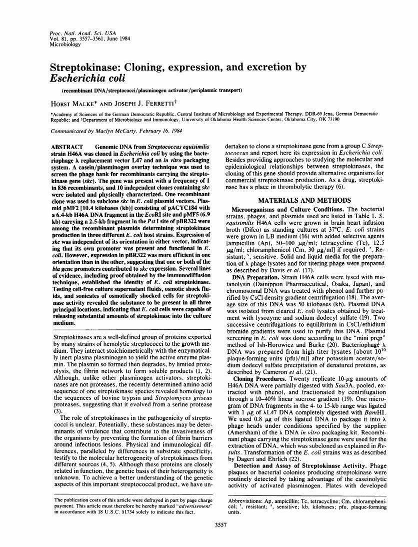
Detection of Streptokinase-Producing Plaques. The plas-minogen-casein overlay technique was used to detect strep-tokinase-producing plaques. Of 8360 recombinant phagesplated, 10 yielded plaques showing distinct zones of casein-olysis (Fig. 1). There was no background in the assay, indi-cating that neither E. coli nor phage X produced detectableamounts of proteases that cleave casein under these condi-tions. The frequency of the streptokinase gene, henceforthreferred to as skc, in the phage bank agreed well with thetheoretical expectations based on the fraction of the H46Agenome in a single recombinant phage (0.5%).
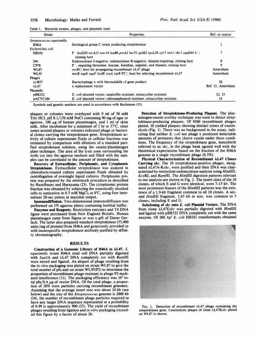
Physical Characterization of Recombinant XL47 ClonesCarrying skc. The 10 streptokinase-positive phages, desig-nated XL47A-Kskc, were purified and their DNA was char-acterized by restriction endonuclease analysis using HindIll,EcoRI, and BamHI. The HindIII digestion patterns relevantto our analysis are shown in Fig. 2. The insert sizes of the 10clones, of which E and G were identical, were 7-15 kb. Themost prominent feature of the HindIII patterns was the exis-tence of a 1.9-kb fragment common to all 10 clones. A sec-ond HindIII fragment, 2.65 kb in size, was common to 5clones, including E and G.
Subcloning of skc onto E. coli Plasmid Vectors. The DNAfrom clone XL47Eskc was partially digested with HindIIIand ligated with pBR322 DNA completely cut with the sameenzyme. Of 260 Apr E. coli HB101 transformants obtained
FIG. 1. Detection of recombinant XL47 phage containing thestreptokinase gene. Caseinolytic plaque of clone XL47Kskc platedon WL87 is shown.
Proc. NatL Acad Sci. USA 81 (1984)
Proc. NatL. Acad Sci. USA 81 (1984) 3559
FIG. 2. Analysis of the DNA of phage clones XL47A-Kskc byHindlI digestion. Lanes: 1, XL47; 2, XL47Askc; 3, XL47Bskc; 4,XL47Cskc; 5, XL47Dskc; 6, XL47Eskc; 7, XL47Fskc; 8, XL47Gskc;9, XL47Hskc; 10, XL47Iskc; 11, XL47Kskc; 12, X; 13, pBR322 di-gested with Ava II.
with the ligation mixture, 37 were TcW. One of these gave apositive streptokinase reaction. pMF1 (11.8 kb), the plasmidcarried by the streptokinase-positive HB101 clone, consistedof pBR322 carrying a 7.4-kb insert in its HindIII site. A frag-ment of the same size was identifiable in partial HindIII di-gests of XL47Eskc DNA, indicating that the pBR322 insertdid not represent scrambled sequences. The insert consisted
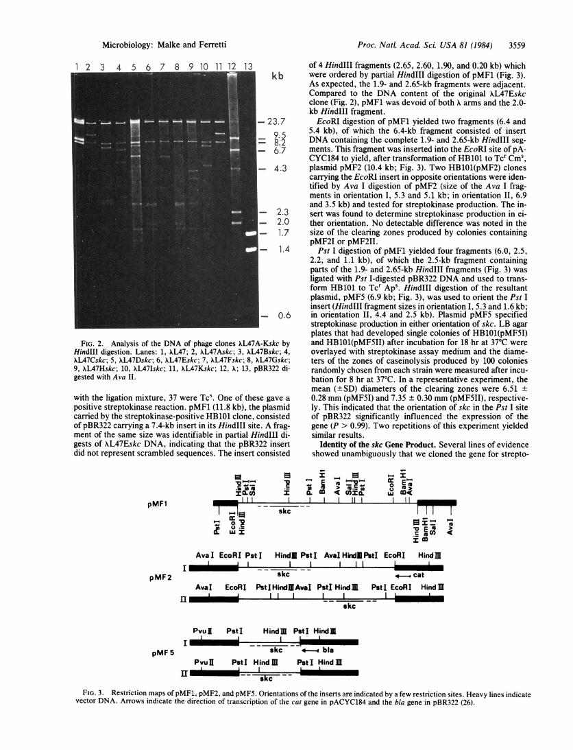
pMFl
pMF2
"Oa
_ l II Iskc
IAvaI EcoRI PstIII I
of 4 HindIII fragments (2.65, 2.60, 1.90, and 0.20 kb) whichwere ordered by partial HindIII digestion of pMF1 (Fig. 3).As expected, the 1.9- and 2.65-kb fragments were adjacent.Compared to the DNA content of the original XL47Eskcclone (Fig. 2), pMF1 was devoid of both X arms and the 2.0-kb HindIII fragment.EcoRI digestion of pMF1 yielded two fragments (6.4 and
5.4 kb), of which the 6.4-kb fragment consisted of insertDNA containing the complete 1.9- and 2.65-kb HindIII seg-ments. This fragment was inserted into the EcoRI site of pA-CYC184 to yield, after transformation of HB101 to Tcr Cms,plasmid pMF2 (10.4 kb; Fig. 3). Two HB101(pMF2) clonescarrying the EcoRI insert in opposite orientations were iden-tified by Ava I digestion of pMF2 (size of the Ava I frag-ments in orientation I, 5.3 and 5.1 kb; in orientation II, 6.9and 3.5 kb) and tested for streptokinase production. The in-sert was found to determine streptokinase production in ei-ther orientation. No detectable difference was noted in thesize of the clearing zones produced by colonies containingpMF2I or pMF2II.Pst I digestion of pMF1 yielded four fragments (6.0, 2.5,
2.2, and 1.1 kb), of which the 2.5-kb fragment containingparts of the 1.9- and 2.65-kb HindIII fragments (Fig. 3) wasligated with Pst I-digested pBR322 DNA and used to trans-form HB101 to Tcr Aps. HindIII digestion of the resultantplasmid, pMFS (6.9 kb; Fig. 3), was used to orient the Pst Iinsert (HindIll fragment sizes in orientation I, 5.3 and 1.6 kb;in orientation II, 4.4 and 2.5 kb). Plasmid pMFS specifiedstreptokinase production in either orientation of skc. LB agarplates that had developed single colonies of HB101(pMFSI)and HB101(pMFSII) after incubation for 18 hr at 37°C wereoverlayed with streptokinase assay medium and the diame-ters of the zones of caseinolysis produced by 100 coloniesrandomly chosen from each strain were measured after incu-bation for 8 hr at 37°C. In a representative experiment, themean (±SD) diameters of the clearing zones were 6.51 ±0.28 mm (pMF5I) and 7.35 ± 0.30 mm (pMFSII), respective-ly. This indicated that the orientation of skc in the Pst I siteof pBR322 significantly influenced the expression of thegene (P > 0.99). Two repetitions of this experiment yieldedsimilar results.
Identity of the skc Gene Product. Several lines of evidenceshowed unambiguously that we cloned the gene for strepto-
_-_
o. m cnX. , m11 r n
11 10 _X
co
Hindl PstI AvalHindiPstI EcoRII I I
skc 4-
Hindlg
-_ catAval EcoRI PstIHindlAvaI PstI HindJ
II I II I.
I I I-
skc
PstI EcoRI Hind JRI L
pMF 5
Pvu 1 Pst I Hindm PstI HindmI I
skc *- blaPvuU PstI Hind m PstI Hind ml
_IIir --- - is
FIG. 3. Restriction maps of pMF1, pMF2, and pMF5. Orientations of the inserts are indicated by a few restriction sites. Heavy lines indicatevector DNA. Arrows indicate the direction of transcription of the cat gene in pACYC184 and the bla gene in pBR322 (26).
.. .
Microbiology: Malke and Ferretti
.,JIM,la.;: o c& u
LU Y
3560 Microbiology: Malke and Ferretti
S._ .
B 0
0
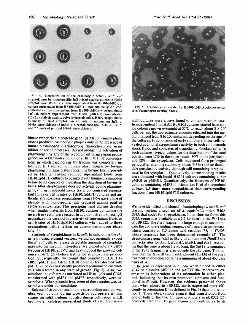
j SFIG. 4. Neutralization of the caseinolytic activity of E. coli
streptokinase by monospecific IgG raised against authentic H46Astreptokinase. Wells: a, culture supernatant from HB101(pMF1); b,culture supernatant from HB101(pMF1) + nonimmune IgG; c, con-centrated culture supernatant from HB101(pMF1) + streptokinaseIgG; d, culture supernatant from HB101(pBR322) concentrated(10x) by dialysis against polyethylene glycol; e, H46A streptokinase(5 units); f, H46A streptokinase -(5 units) + nonimmune IgG; g,H46A streptokinase (5 units) + streptokinase IgG; h-k, 20, 10, 5,and 2.5 units of purified H46A streptokinase.
kinase rather than a protease gene. (i) All 10 primary phageclones produced caseinolytic plaques only in the presence ofhuman plasminogen; (ii) diisopropyl fluorophosphate, an in-hibitor of serine proteases, did not abolish the activation ofplasminogen by any of the recombinant phages upon propa-gation on WL87 under conditions (10 mM final concentra-tion) in which caseinolysis by trypsin was completely in-hibited; (iii) replacing human plasminogen by bovineplasminogen in agar plates containing bovine fibrin (provid-ed by Fletcher Taylor) required supernatant fluids fromHB1O1(pMF1) cultures to be mixed with human plasminogenbefore being capable of mediating the digestion of bovine fi-brin (H46A streptokinase does not activate bovine plasmino-gen); (iv) in immunodiffusion tests, concentrated superna-tant fluids or cell lysates of HB1O1(pMF1) cultures and au-thentic streptokinase preparations from H46A gave a line ofidentity with monospecific IgG prepared against purifiedH46A streptokinase. This precipitin band did not developwhen similar materials from HB101 cultures containing theinsert-free vector were tested. In addition, streptokinase IgGneutralized the caseinolytic activity of supernatant fluids orcell lysates of HB1O1(pMF1) cultures when added to thesepreparations before testing on casein/plasminogen plates(Fig. 4).
Synthesis of Streptokinase in E. coli. In subcloning the skcgene by using plasmid vectors, we did not originally expectthe E. coli cells to release detectable amounts of streptoki-nase into the medium. Therefore, we cloned into a K c1857lysogen of HB101 at 29°C and heat-induced the growing col-onies at 42°C (27) before testing for streptokinase produc-tion. Subsequently, we found that uninduced HB101 (Xc1857, pMF1) and A-free HB101 colonies transformed withpMF1, -2, or -5 consistently produced pronounced caseinol-ysis when tested in any state of growth (Fig. 5). Also, twoadditional E. coli strains unrelated to HB101 (294 and CP78)transformed with pMF1 and pMF5, respectively, were ca-seinolytic. When plasmid-free, none of these strains was ca-seinolytic under our conditions.Release of streptokinase into the surrounding medium was
observed not only during growth of appropriate E. colistrains on solid medium but also during cultivation in LBbroth-i.e., cell-free supernatant fluids of saturated over-
FIG. 5. Caseinolysis mediated by HB101(pMF1) colonies on ca-sein/plasminogen overlay plates.
night cultures were always found to contain streptokinase.In independent 5-ml HB101(pMF1) cultures started from sin-gle colonies grown overnight at 37°C to reach about 3 x 10⁹cells per ml, the approximate amounts released into the me-dium ranged from 8 to 100 units/ml, depending on the age ofthe cultures. Fractionation of early stationary phase cells re-vealed additional streptokinase activity in both cold osmoticshock fluids and sonicates of osmotically shocked cells. Insuch cultures, typical values for the distribution of the totalactivity were 17% in the supernatant, 30% in the periplasm,and 52% in the cytoplasm. Cells incubated for a prolongedperiod after attaining stationary phase (_8 hr) had no detect-able periplasmic activity although still containing streptoki-nase in the cytoplasm. Qualitatively, corresponding resultswere obtained with liquid HB101 cultures containing eitherpMF5I or pMF5II. Quantitatively, the fractions of HB101cultures containing pMF5 in orientation II of skc containedat least 1.5 times more streptokinase than correspondingfractions from HB1O1(pMF5I) cultures.
DISCUSSIONWe have identified and cloned in bacteriophage K and E. coliplasmid vectors a segment of S. equisimilis strain H46ADNA that codes for streptokinase. In its shortest form, thisDNA segment is available as a 2.5-kb insert in the Pst I siteof pBR322. The Pst I fragment is large enough to accommo-date the complete coding sequence of mature streptokinase,which consists of 415 amino acid residues (Mr = 47,408)whose sequence has been determined recently (3). Thestreptokinase gene (skc) is likely to contain one HindIII sitebut lacks sites for Ava I, BamHI, EcoRI, and Pst I. Assum-ing that the gene is about 1.3 kb long, the Sal I site containedin the Pst I fragment is also outside the skc gene. This im-plies that the HindIII/Sal I subfragment (1.5 kb) of the Pst Ifragment in question contains a minimum of about 400 basepairs of skc.The gene is expressed in E. coli when cloned in either
KL47 or plasmids pBR322 and pACYC184. Moreover, ex-pression is independent of its orientation in either plas-mid, indicating that its own promoter is present and func-tional in E. coli. However, the evidence presented showsthat, when cloned in pBR322, skc is expressed more effi-ciently in orientation II (as defined in Fig. 3) than in orienta-tion I. These observations suggest that transcription fromone or both of the two bla gene promoters in pBR322 (26)proceeds into the skc gene region and contributes to its
Proc. Natl. Acad Sci. USA 81 (1984)
Proc. NatL. Acad Sci. USA 81 (1984) 3561
expression. If this idea is true, note that it defines the orien-tation of skc in the Pst I fragment (Fig. 3). The finding thatthe level of skc expression in pMF2 is orientation indepen-dent may mean that either the cat gene promoter of pA-CYC184 is weaker than the skc promoter or there exists atranscription termination signal(s) in H46A DNA sequencesupstream from the skc promoter.
Streptokinase produced by E. coli has the same substratespecificity as that of the streptococcal donor strain. Theidentity of the E. coli product has also been demonstrated bythe immunodiffusion technique. In Streptococcus, streptoki-nase is a secretory protein, suggesting that its immature formis synthesized with a signal sequence at its amino terminus(28). The existence of a signal sequence has also been sug-gested by Jackson and Tang (3) on the basis of their hypothe-sis that the streptokinase gene has evolved from a serine pro-tease gene by duplication and fusion, with one copy of theputative signal sequence being conserved in the central partof the processed protein. Clearly, nucleotide sequencing ofskc is necessary to test the validity of this attractive hypothe-sis.
In E. coli cultures, we find streptokinase activity in allthree principal locations. Our data suggest that the extracel-lular activity is not attributable to leakage of the protein outof dead cells, but we do not know whether or not the export-ed activity results from correct processing and active secre-tion. In any event, the phenomenon is conspicuous andquantitatively significant. From a pragmatic point of view, itallows reliable detection of the skc gene product in E. coliclones without the requirement of fractionating or lysing thecells. The main barrier for the release of the E. coli streptoki-nase seems to be the inner membrane; once it is passed, theperiplasmic protein is released upon prolonged incubation.The presence of extracellular and periplasmic streptokinasein cultures of strains carrying skc in two different plasmids ineither orientation indicates that excretion is not entirely dueto signal sequences provided by the attached proximal se-quences of the cat or bla genes of the vectors. Rather, theputative streptokinase-specific signal sequence can be ex-pected to play a crucial role in excretion of the substance bythe heterologous host. Assuming that authentic and E. colistreptokinase have the same specific activity, the amounts ofextracellular streptokinase activity (8-100 units/ml, depend-ing on the age of saturated cultures containing 3 x 10⁹ viablecells per ml) correspond to 0.15-1.82 mg per liter of cultureor about 600-7000 streptokinase molecules released per cell.Of the three drugs available for thrombolytic therapy, the
cDNA sequences of urokinase and tissue-type plasminogenactivator have been cloned before (29, 30). Another plasmin-ogen activator substance produced by Staphylococcus aure-us, staphylokinase, has also been cloned and is expressed inE. coli (31). The cloning of the streptokinase gene adds to thearsenal of sequences relevant to problems of plasminogenactivation and opens up approaches for studying unresolvedquestions of theoretical and practical significance in theblood clotting field. For the genetics of streptococci, wherethe study of virulence genes by the recombinant DNA ap-proach is still in its initial stages (26, 32), our work has shownthe feasibility of cloning and expressing genes in phage Xvectors. The procedure could be particularly useful for clon-ing determinants whose products are detrimental to the nor-mal physiology of the cloning host and readily detectable bysimple plate assays.
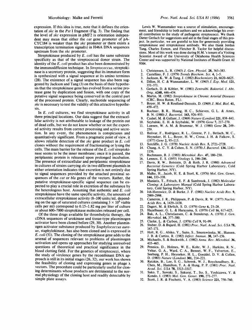
Lewis W. Wannamaker was a source of stimulation, encourage-ment, and friendship to both authors and we acknowledge his over-all contribution to the study of pathogenic streptococci. We thankDieter Gerlach for suggestions and aid in the final stages of this pro-ject. In particular, we are grateful to him for generously providingstreptokinase and streptokinase antibody. We also thank JordanTang, Charles Esmon, and Fletcher B. Taylor for helpful discus-sions. Most of this work was done during H.M.'s tenure of a VisitingScientist Award of the University of Oklahoma Health SciencesCenter and was supported by National Institutes of Health Grant Al9304.
1. Christensen, L. R. (1945) J. Gen. Physiol. 28, 363-383.2. Castellino, F. J. (1979) Trends Biochem. Sci. 4, 1-5.3. Jackson, K. W. & Tang, J. (1982) Biochemistry 21, 6620-6625.4. Dillon, H. C. & Wannamaker, L. W. (1965) J. Exp. Med. 121,
351-371.5. Gerlach, D. & Kohler, W. (1981) Zentralbl. Bakteriol. I. Abt.
Orig. A248, 446-454.6. Martin, M. (1982) Streptokinase in Chronic Arterial Diseases
(CRC, Boca Raton, FL).7. Boyer, H. W. & Roulland-Dussoix, D. (1969) J. Mol. Biol. 41,
459-472.8. Bochner, B. R., Huang, H. C., Schieven, G. L. & Ames,
B. N. (1980) J. Bacteriol. 143, 926-933.9. Cashel, M. & Gallant, J. (1969) Nature (London) 221, 838-841.
10. Szybalski, E. H. & Szybalski, W. (1979) Gene 7, 217-270.11. Loenen, W. A. M. & Brammar, W. J. (1980) Gene 20, 249-
259.12. Bolivar, F., Rodriguez, R. L., Greene, P. J., Betlach, M. C.,
Heyneker, H. L., Boyer, H. W., Crosa, J. H. & Falkow, S.(1977) Gene 2, 95-113.
13. Sutcliffe, J. G. (1978) Nucleic Acids Res. 5, 2721-2728.14. Chang, A. C. Y. & Cohen, S. N. (1978) J. Bacterol. 134, 1141-
1156.15. Bachmann, B. J. (1983) Microbiol. Rev. 47, 180-230.16. Lennox, E. S. (1955) Virology 1, 190-206.17. Davis, R. W., Botstein, D. & Roth, J. R. (1980) Advanced
Bacterial Genetics (Cold Spring Harbor Laboratory, ColdSpring Harbor, NY).
18. Malke, H., Jacob, H. E. & Storl, K. (1976) Mol. Gen. Genet.144, 333-338.
19. Maniatis, T., Fritsch, E. F. & Sambrook, J. (1982) MolecularCloning: A Laboratory Manual (Cold Spring Harbor Labora-tory, Cold Spring Harbor, NY).
20. Ish-Horowicz, D. & Burke, J. F. (1981) Nucleic Acids Res. 9,2989-2998.
21. Cameron, J. R., Philippsen, P. & Davis, R. W. (1977) NucleicAcids Res. 4, 1429-1438.
22. Dagert, M. & Ehrlich, S. D. (1979) Gene 6, 23-28.23. Hazelbauer, G. L. & Harayama, S. (1979) Cell 16, 617-625.24. Bak, A. L., Christiansen, C. & Stenderup, A. (1970) J. Gen.
Microbiol. 64, 377-380.25. Clarke, L. & Carbon, J. (1976) Cell 9, 91-99.26. Stuber, D. & Bujard, H. (1981) Proc. Natl. Acad. Sci. USA 78,
167-171.27. Holt, R. G., Abiko, Y., Saito, S., Smorawinska, M., Hansen,
J. B. & Curtiss, R. (1982) Infect. Immun. 38, 147-156.28. Michaelis, S. & Beckwith, J. (1982) Annu. Rev. Microbiol. 36,
435-465.29. Pennica, D., Holmes, W. E., Kohr, W. J., Harkins, R. N.,
Vehar, G. A., Ward, C. A., Bennet, W. F., Yelverton, E.,Seeburg, P. H., Heyneker, H. L., Goeddel, D. V. & Collen,D. (1983) Nature (London) 301, 214-221.
30. Ratzkin, B., Lee, S. G., Schrenk, W. J., Roychoudhury, R.,Chen, M., Hamilton, T. A. & Hung, P. P. (1981) Proc. Natl.Acad. Sci. USA 78, 3313-3317.
31. Sako, T., Sawaki, S., Sakurai, T., Ito, S., Yoshizawa, Y. &Kondo, I. (1983) Mol. Gen. Genet. 190, 271-277.
32. Scott, J. R. & Fischetti, V. A. (1983) Science 221, 758-760.
Microbiology: Malke and Ferretti