strategies for increasing pancreatic tumor...
TRANSCRIPT
Strategies for Increasing Pancreatic TumorImmunogenicityBurles A. Johnson III1, Mark Yarchoan1, Valerie Lee1, Daniel A. Laheru1, andElizabeth M. Jaffee1,2
Abstract
Immunotherapy has changed the standard of care for multipledeadly cancers, including lung, head and neck, gastric, and somecolorectal cancers. However, single-agent immunotherapy hashad little effect in pancreatic ductal adenocarcinoma (PDAC).Increasing evidence suggests that the PDACmicroenvironment iscomprised of an intricate network of signals between immunecells, PDAC cells, and stroma, resulting in an immunosuppressiveenvironment resistant to single-agent immunotherapies. In thisreview, we discuss differences between immunotherapy-sensitive
cancers and PDAC, the complex interactions between PDACstroma and suppressive tumor-infiltrating cells that facilitatePDAC development and progression, the immunologic targetswithin these complex networks that are druggable, and datasupporting combination drug approaches thatmodulatemultiplePDAC signals, which should lead to improved clinical outcomes.Clin Cancer Res; 23(7); 1656–69. �2017 AACR.
See all articles in this CCR Focus section, "Pancreatic Cancer:Challenge and Inspiration."
IntroductionCurrent estimates predict pancreatic ductal adenocarcinoma
(PDAC) to overtake breast cancer and become the third mostcommon cause of cancer-related death in the United States (1, 2).Only 20% to 30% of patients with PDAC have resectable diseaseat diagnosis, and the majority of patients who undergo surgicalresection subsequently relapse (3–7). Most patients present withmetastatic disease at diagnosis and have only a 2% five-yearsurvival (2). To date, the rate of successful clinical trials inpancreatic cancer remains low (8). Of the many therapies inves-tigated in large clinical trials over the past two decades, only twosystemic therapies have demonstrated a statistically significantand clinically meaningful improvement in overall survival (OS)as compared with gemcitabine alone (9, 10). As a result, the five-year survival rate for PDAC has improved only marginally sincethe 1970s, from3% to 7% (2). This highlights the continued needfor new and effective therapies in PDAC.
Immune checkpoint immunotherapies have produced unprec-edented clinical benefits in a variety of different cancers, includinglung cancer, which was previously thought to be nonimmuneresponsive (11). However, clinical trials using single-agent check-point immunotherapy in PDAC have been unsuccessful thus far.
This may be explained by increasing evidence that suggests thatPDAC creates a potently immunosuppressive microenvironmentvia activation ofmultiple regulatorymechanisms (12, 13), where-by interactions between the tumor, stroma, and immune cells inthe pancreatic tumor microenvironment (TME) result in cancerprogression (Fig. 1). In this review, we discuss potentialapproaches to increasing immunogenicity, or immune respon-siveness, to PDAC. Specifically, we (i) examine the challenges indeveloping successful immunotherapies for PDAC; (ii) describethe complex immune components of the TME and discuss howthe immune system, pancreatic tumor cells, microbiome, andstromal signals suppress immune-mediated attack; and (iii) dis-cuss novel multiagent therapeutic strategies to target signalswithin this integrated immunosuppressive network that are underdevelopment in clinical trials. Current standard-of-care therapyand clinical trials in progress are also reviewed by Manji andcolleagues in this CCR Focus (14).
Clinical Challenges in DevelopingImmunotherapies for PDAC
There is mounting evidence that immune-mediated inflamma-tion is an integral component of the environment that supportsPDAC development and progression (15). Genomic analysesshow that PDAC frequently upregulates multiple pathwaysinvolved in acquired immune suppression, and upregulation ofthese pathways is associated with poor survival (16). This mayexplain why early human clinical studies involving immunother-apy monotherapy in PDAC have been discouraging. Althoughtreatment with single-agent immune checkpoint inhibitors target-ing cytotoxic T-lymphocyte–associated protein 4 (CTLA-4) andprogrammed cell death protein 1 (PD-1) causes meaningfulobjective responses in many tumor types (11, 17–20), only 1 of27 patients with PDAC responded to the CTLA-4 inhibitor ipili-mumab (21), and 0 of 14 patients with PDAC had an objectiveresponse to anti–programmed death-ligand 1 (PD-L1) therapy(22). Recently completed and planned immunotherapy clinical
1Department of Oncology, Sidney Kimmel Comprehensive Cancer Center,Bloomberg-Kimmel Institute for Cancer Immunotherapy, Johns Hopkins Uni-versity, Baltimore, Maryland. 2Department of Pathology, Sidney Kimmel Com-prehensive Cancer Center, Bloomberg-Kimmel Institute for Cancer Immuno-therapy, Johns Hopkins University, Baltimore, Maryland.
Note: B.A. Johnson III and M. Yarchoan contributed equally to this article.
Corresponding Author: Elizabeth M. Jaffee, Sidney Kimmel Cancer Center atJohns Hopkins University School of Medicine, 1650 Orleans Street, CRB1 4M06,Baltimore, MD 21287. Phone: 410-955-2957; Fax: 410-614-8216; E-mail:[email protected]
doi: 10.1158/1078-0432.CCR-16-2318
�2017 American Association for Cancer Research.
CCRFOCUS
Clin Cancer Res; 23(7) April 1, 20171656
on July 6, 2018. © 2017 American Association for Cancer Research. clincancerres.aacrjournals.org Downloaded from

© 2017 American Association for Cancer Research
CCL2CCL2
CCL2
CD40 agonist +gem
CSF1Rblockade
B cell
Breg
IL35
Tregs
Tregs
Stroma-associatedfibroblasts
Pancreatictumor cells
Fibrosis
Stromal hyaluronicacid
Stroma
FAK1
Fusobacterium
MDSC B cells
CXCL13
Treg ActivatedTreg
GemMHC
B7
TCR
PD-1
PD-L
1
CTLA-4
IDO-expressing DC
Macrophage
CD4+ T cells
CD8+ T cells
CD8+ T cellGM-CSF
CD4+ T cell
B
C
A
G
A
E D
D
B
F
PDAC microenvironment
H
γδ T cells
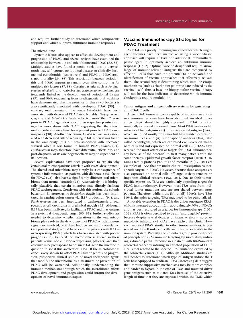
Figure 1.
Mechanisms within the PDAC TME drive resistance to therapies. PDAC comprises complex interactions between T cells, B cells, antigen-presenting cells (APC),pancreatic tumor cells, and stromal elements. These interactions result in a profoundly immunosuppressive TME, and, consequently, single-agentimmunotherapy has been largely ineffective. However, emerging preclinical data have suggested that combination therapy may dramatically affect OS.Current trial design is being driven largely by these data. This figure summarizes major pathways in PDAC tumorigenesis that are being manipulatedin clinical trials for patients with metastatic PDAC. Except for G, which represents in part indoleamine 2,3 dioxygenase (IDO)-activated regulatory T cells (Treg)in tumor-draining lymph nodes (TDLN) from a melanoma model (40), this figure represents data known exclusively from PDAC models. A, Tregs and gd T cellsblock effector T cell (Teff) division and drive PDAC growth, whereas gd T cells block T-cell infiltration (47). B, Myeloid-derived suppressor cells (MDSC) andmacrophages are mobilized into the TME by PDAC-derived granulocyte macrophage-colony stimulating factor (GM-CSF) and chemokine (C-C motif) ligand 2(CCL2), respectively (145, 182, 183). C, Macrophages block CD4þ T cell entry into the PDAC microenvironment. CD40 is expressed on these CD4þ T cells, andactivation of the CD40 pathway concurrentlywith gemcitabine can drive T-cell infiltration (140).D, Stromal-associated fibroblasts produce chemokine (C-X-Cmotif)ligand 13 (CXCL13), which recruits regulatory B cells (Breg) into the TME. These Bregs produce IL35, which drives PDAC progression (136, 184). These Bregs may beinhibited by Bruton tyrosine kinase (BTK) inhibitors, such as ibrutinib (137). E, Tumor-infiltrating macrophages stimulate PDAC progression. Blockade of the colony-stimulating factor-1 receptor (CSF1R) expressed by macrophages can lead to macrophage depletion, cytotoxic T-lymphocyte–associated protein 4 (CTLA-4)upregulation on CD8þ T cells, and programmed death-ligand 1 (PD-L1) upregulation on pancreatic tumor cells (146, 147). F, Stromal elements create a physicalbarrier to immune infiltration and therapeutic agents. Stromal fibroblasts block Treg accumulation and PDAC progression (62), but targeting other stromalelements has achieved encouraging results. Stromal hyaluronic acid deposition results in decreased vascular patency (72, 73), and focal adhesion kinase-1 (FAK1)drives stromal fibrosis (68). Inhibition of either target has led to decreased PDAC progression when combined with chemotherapy in preclinical models. G, IDOinduction in dendritic cells (DC) by tumors activates Tregs via major histocompatibility complex (MHC) and CTLA-4 pathways (40, 131). In phase II studies,gemcitabine-based therapy synergizes with IDO inhibition (via indoximod) to improve response rates in PDAC (133), possibly via transient depletion of Tregs (39).This provides an immune system reset, allowing for chemotherapy-mediated elimination of previously activated Tregs, followed by indoximod-mediated inhibitionof subsequent Treg activation. H, Recent evidence suggests the Fusobacterium found within the PDAC microenvironment drives PDAC progression, but themechanism of this is unknown (91). Gem, gemcitabine; PD-1, programmed cell death protein 1; PD-L1, programmed death-ligand 1; TCR, T-cell receptor.
Increasing Pancreatic Tumor Immunity
www.aacrjournals.org Clin Cancer Res; 23(7) April 1, 2017 1657
on July 6, 2018. © 2017 American Association for Cancer Research. clincancerres.aacrjournals.org Downloaded from

trials for patients with PDAC have been reviewed in detail else-where (23–27). Although single-agent immunotherapies havefailed to showbenefit in PDAC, increasing data support the testingof combinatorial approaches that target multiple suppressivemechanisms. In addition to examining geneticmutations inPDACtumor samples, which is reviewed byDreyer and colleagues in thisCCR Focus (28), performing RNA sequencing to determine whichimmune escape mechanisms are upregulated [e.g., PD-1 andindoleamine 2,3 dioxygenase (IDO)] may allow us to furtherpersonalize therapy for patients by combining immunotherapyagents with chemotherapy to reset the immune system (29). Thismay be critical specifically in patients with PDAC as the failure ofsingle-agent checkpoint therapy indicates that the PDAC TME ismore complicated and suppressive than in other more immuno-genic cancers. This would also have the advantage of being able todetermine a tumor's immunogenicity upfront, before initiatingtreatment. As we better understand the role of the multipleimmunologic contributors to PDAC growth, it should be possibleto design multiagent immunotherapies that target multiple path-ways, leading to increased antitumor immunity.
The multiple immunosuppressive components of the PDACTME collectively suppress effector T cells (cells that recognize and
kill tumor cells), preventing immune-mediated destruction(Fig. 1). Accumulation of effector CD4þ and CD8þ T cells inhuman PDAC are associated with improved OS (30–32). Aspancreatic lesions progress, tumor-infiltrating CD8þ effector Tcells decrease while suppressive regulatory T cells (Treg) comprisea higher percentage of theCD4þ T-cell compartment (33), leadingto a low number of tumor-infiltrating lymphocytes (TIL) and ahigh number of immunosuppressive cells (13). Thus, PDAC isconsidered to be a poorly immune responsive cancer. By contrast,highly immune responsive solid tumors are characterized by ahigh number of TILs at baseline and a high response rate toimmune checkpoint inhibitors (34). Although PDAC is poorlyimmunogenic, that is likely due to having a more complex andsuppressive TME, not because the immune system does notrecognize the tumor. Discovery of the complex immune pathwaysinvolved in PDAC progression and immune escape (summarizedin Fig. 1) has led to additional novel PDAC immunotherapytargets (Table 1). Increasing data suggest that poorly immune-responsive cancers like PDAC require multiagent therapy to elicitan immune response. One multipronged approach involves vac-cines, which stimulate accumulation of lymphoid aggregates inPDAC (ref. 35; Fig. 2). One likely reason why vaccines have not
Table 1. A list of notable immunotherapies in clinical development for PDAC
Therapeutic target and agentsunder investigation for PDAC Preclinical rationale Clinical evidence and ongoing trials
PD-1/PD-L1 PD-1/PD-L1 inhibition has activity in a wide number oftumors. PD-L1 expression is upregulated in a subsetof PDAC and is associated with shortened survival(43, 161).
Responses were observed in a subset of patients with MMR-deficient pancreatic cancer (56), and additional trials in MMR-deficient disease are ongoing (NCT01876511 andNCT02465060). None of 14 pancreatic patients responded in astudy of single-agent nivolumab (22). Multiple combinationimmunotherapy trials are ongoing (NCT02558894,NCT02268825, NCT02472977, NCT02243371, andNCT02777710).
Nivolumab
Pembrolizumab
Durvalumab
CTLA-4 Anti–CTLA-4 therapy may reduce intratumoral Tregsand shift the threshold needed for T-cell activation.A trial of ipilimumab failed to show convincingclinical activity, but a possible delayed responsewasobserved in one patient (21).
Multiple combination trials are ongoing, including combinationswith PD-1 inhibition and/or therapeutic vaccines (NCT02558894and NCT01896869).
IpilimumabTremelimumab
IDO1 IDO1 mediates tumor immunosuppression inpreclinical models (non-PDAC), and PDACfrequently overexpresses IDO as a mechanism ofimmune escape (132, 162, 163).
Evidence of clinical activity was observed in combination withchemotherapy (133). A clinical trial is ongoing in combinationwith gemcitabine-based chemotherapy (NCT02077881).
Indoximod
BTK BTK is involved with B-cell receptor signaling and isalso expressed by macrophages. In preclinicalmodels, ibrutinib synergizes with gemcitabine toincrease antitumor immunity (137).
Clinical trials are ongoing in combination with gemcitabine-basedchemotherapy in PDAC (NCT02562898 and NCT02436668).Ibrutinib
CD40 CD40 is expressed on B cells, DCs, and other cell types.CD40 agonists inhibit PDAC stroma, increase CCL2levels and interferon gamma (IFN-g) in the TME, andsynergize with chemotherapy (145, 164).
Evidence of clinical activity was observed in an early-stage clinicaltrial in PDAC (141). Additional trials of monotherapy orcombination with gemcitabine-based chemotherapy areongoing (NCT02588443 and NCT02829099).
RO7009789 (CP-870,893)JNJ-64457107
CCR2 CCR2 recruits suppressive macrophages to theimmunosuppressive TME in PDAC, and CCR2inhibition depletes tumor-infiltrating macrophagesand improves survival in a preclinical model (145).
CCR2 inhibition has shown safety and possible evidence of clinicalactivity in combination with chemotherapy. Clinical trials incombination with chemotherapy in PDAC are ongoing(NCT02345408 and NCT02732938).
CCX872
PF04136309
CSF1R CSF1R inhibition reprograms tumor-associatedmacrophages andupregulates immune checkpoints.Synergistic activity has been observed with immunecheckpoint inhibitors in preclinical models of PDAC(146, 147).
Multiple agents are in clinical trials in metastatic PDAC incombination with PD-1 inhibitors (NCT02526017, NCT02777710,NCT02829723, and NCT02713529).
Cabiralizumab (FPA008)
Pexidartinib (PLX3397)BLZ945AMG 820
CXCR4 CXCR4 blockade abrogated metastasis in preclinicalmodels (151) and synergized with PD-L1 therapy toincrease antitumor immunity (158).
CXCR4 inhibitor is in clinical trial in combination with PD-L1blockade to treat advanced solid tumors, including PDAC(NCT27037072).
LY2510924
Abbreviations: BTK, Bruton tyrosine kinase; CCL2, chemokine (C-Cmotif) ligand2; CCR2, C-C chemokine receptor type 2; CSF1R, colony-stimulating factor-1 receptor;CXCR4, C-X-C chemokine receptor type 4; DC, dendritic cell; MMR, mismatch repair.
CCRFOCUS
Clin Cancer Res; 23(7) April 1, 2017 Clinical Cancer Research1658
on July 6, 2018. © 2017 American Association for Cancer Research. clincancerres.aacrjournals.org Downloaded from

stimulated effective antitumor responses, despite inducing lym-phoid infiltration, is that vaccines also upregulate T-cell–inhibi-tory pathways such as the PD-1/PD-L1 pathway (36). Althoughvaccine therapy has thus far been unsuccessful, we believe thatthese lymphoid infiltrates represent increased immunogenicity,and, speculatively, that patients with vaccine-induced infiltrationof lymphoid aggregates may benefit from a combinationapproach involving vaccine plus costimulatory blockade. Also,upregulation of immune checkpoint pathways after vaccine ther-apy may be a biomarker of increased immunogenicity and sug-gests that these patientsmay also respond to checkpoint blockade.It is also possible that vaccines upregulate multiple immuneescape mechanisms, and elucidation of these would be necessaryto ensure vaccine efficacy. As chemotherapy transiently depletessuppressive Tregs in PDAC patients (37–39), chemotherapyshould be considered in addition to administration of an immu-nomodulatory agent to attempt to overcome the potent immu-nosuppressive TME.
The TME's Role in PDAC Development andProgressionImmune checkpoints and immune checkpoint inhibitors
There are many immune signaling pathways that regulateantitumor immunity, which involve costimulatory and inhibitory
receptors (immune checkpoints) on T cells. Most studies ofimmunomodulatory agents in PDAC have examined the role ofthe inhibitory costimulatory receptors CTLA-4 and PD-1. Bothreceptors are critical in activation and suppressive activity of Tregs(40) and exist primarily to prevent autoimmunity and excessiveimmune responses to infection (41). However, tumors alsoinduce Treg activation and suppression via these pathways, lead-ing to dampened antitumor immune responses (40). This conceptis critical, because it suggests that the immune system is notignorant of PDAC; rather, the immune system detects PDAC butis instructed by the tumor not to attack it (42). Thus, inhibition ofimmunosuppression, rather than immune activation alone, iscritical to achieve durable clinical responses. Consistent withthis, CTLA-4 and PD-pathway expression are upregulated inPDAC (43–45), and both are associated with worse survival(44, 46). Furthermore, PD-1 is expressed on multiple PDAC-infiltrating T-cell subsets, including Tregs and CD4þ andCD8þ effector T cells (37). Additionally, PDAC-infiltrating gdT cells were recently identified, which represent a subset ofsuppressive T cells that express PD-L1 and suppress effectorT-cell activation (47). Collectively, these studies indicate thatimmune checkpoint inhibition may be a target for PDAC-related immunotherapy.
Anumber of principles have emerged that characterize immunecheckpoint pathways. First, these pathways develop in response to
© 2017 American Association for Cancer Research
Tumor TumorTumor
Tregs
MDSCs
B cell
PD-1/PD-L1
Interferon-γ
Macrophages Macrophages Macrophages
Interferon-γ
Intratumoral lymphoidaggregate
Intratumoral lymphoidaggregate
Checkpoint inhibitorCD8+ T cellCD4+ T cell
Pre-vaccine Post-vaccine
Vaccine +cyclophosphamide
Post-vaccine +checkpoint inhibitor
Figure 2.
Therapeutic vaccine immunotherapy for PDAC requires multiple steps to overcome immunosuppression. PDAC and other poorly immune-responsive cancersare characterized by low numbers of TILs, low levels of PD-L1 expression, and high numbers of immunosuppressive cells, such as Tregs and myeloid-derivedsuppressor cells (MDSC), at baseline (left; ref. 13). Using a vaccine approach will require at least two immunotherapeutics to achieve an immune response.In step 1 (center), a therapeutic vaccine is used to induce accumulation of lymphoid aggregates (35). These lymphocytes secrete interferon-g and other solublefactors that induce high levels of PD-L1/PD-1 expression on epithelial tumor cells and on immune cells (185). Vaccines can also be combined with other therapies,such as cyclophosphamide, to deplete immunosuppressive cells in the TME (29). In step 2 (right), the addition of a PD-pathway inhibitor to a vaccine-primedtumor inhibits PD-L1/PD-1 signaling to increase lymphocyte proliferation and activation and promote tumor eradication (36). The hypothesis that vaccinetherapy can synergize with immune checkpoint inhibition is currently under clinical investigation in multiple trials in PDAC.
Increasing Pancreatic Tumor Immunity
www.aacrjournals.org Clin Cancer Res; 23(7) April 1, 2017 1659
on July 6, 2018. © 2017 American Association for Cancer Research. clincancerres.aacrjournals.org Downloaded from

the genetic changes that occur within developing tumors and areshaped by the evolving inflammatory response to these geneticchanges. Second, there are many inhibitory and activating sig-naling pathways (48, 49), butmuch still needs tobe learned abouttheir role in different cancer types. Although melanoma, lungcarcinoma, and renal cell carcinoma respond to blockade ofone checkpoint pathway (i.e., PD-1/PD-L1 or CTLA-4; refs. 11,17–20), most cancers will likely require combination therapy tofully activate T-cell responses. Figure 1 depicts a nonexhaustivedescription of the broad range of suppressive mechanisms inPDAC, which account for single-agent immunotherapy havinglimited clinical activity. Increasing preclinical evidence (seebelow) suggests that combining checkpoint inhibition with othertargeted therapy may improve clinical efficacy. Third, additionalstudies are needed to understand primary (patients who donot respond) and secondary (patients who initially respondbut then recur) resistance to these agents.
Although immune checkpoint inhibitors have thus farfailed as single agents to demonstrate convincing clinicalactivity in PDAC, there may be subgroups of PDAC that aremore likely to respond to these agents as monotherapy.Predictive biomarkers have now been used in multiple cancertypes to identify patients who may be more likely to respondto immune checkpoint inhibitors. For example, expression ofPD-L1 is used to identify patients who should receive front-line PD-1 inhibitor immunotherapy instead of chemotherapyin non–small cell lung cancer (NSCLC; ref. 50). In gastroin-testinal malignancies, including PDAC, one emerging bio-marker of response to immune checkpoint inhibitors is mis-match repair deficiency (MMR-d), which results in a failure torepair errors in base pair mismatches in tumor DNA (e.g., C-Tinstead of C-G), leading to microsatellite instability (MSI;ref. 51). In unselected populations of colorectal cancer, littleto no clinical activity was reported in the initial clinical trialsof immune checkpoint inhibitors. However, the PD-1 inhib-itor pembrolizumab demonstrated significant clinical activityin the small subset of colorectal cancers (�5% of advanceddisease; ref. 52) with MMR-d (53). This activity is likely due tothe high baseline immunogenicity of the MMR-d cancersubtype, as evidenced by the increased lymphoid infiltrationin MMR-d colorectal carcinomas at baseline, as well as thehigh expression of multiple immune checkpoints, includingPD-L1 (54, 55).
Mismatch repair status is not routinely checked in PDAC, andwe are aware of only four reported cases of MMR-d pancreaticcancer treated with a PD-1 inhibitor. Of these four cases, onepatient had a partial response to pembrolizumab, and the otherthree achieved stable disease (56). Additional basket trials ofsingle-agent PD-1 inhibition inMMR-d cancers (includingPDAC)are ongoing. AlthoughMMR-dPDAC is a small subset of all PDAC(13%–17.4% in prior studies; refs. 57–59), these preliminary datasuggest that single-agent immune checkpoint inhibitorsmay havemeaningful clinical activity in such cases. These studies alsosuggest that it is important to perform genetic sequencing studieson all patient tumors to better define each cancer's biology and toidentify potential therapeutic options that may otherwise bemissed.
StromaThe dense stroma surrounding pancreatic cancers creates a
hypovascular environment that can block the penetration of
chemotherapeutics and facilitate immune escape. T cells werefirst demonstrated in the late 1990s to form aggregates inthe fibrotic tissue of pancreatic cancer samples (60), leading tothe current hypothesis that interactions between stroma, lym-phocytes, and antigen-presenting cells (APC) create a complexTME that makes overcoming immunosuppression difficult.Initial studies demonstrated that tumor incidence and metas-tasis increased when an increased proportion of pancreaticstellate cells were coinjected with PDAC cells, identifyingthe stroma as a potential target for therapeutic intervention(61). However, in preclinical models of PDAC, simple deple-tion of fibroblasts led to increased Treg accumulation anddecreased survival, suggesting that the relationship betweenPDAC and stroma may be more complex than previouslyappreciated (62). This may explain why depletion of fibro-blasts via inhibition of Hedgehog signaling, although leadingto disease stabilization in some preclinical studies, ultimatelyfailed in other preclinical models and clinical trials (63–65).These conflicting data are described in more detail elsewhere(66) and may reflect heterogeneity between fibroblasts (67) ordifferent systems used.
However, targeting other factors that drive stromalfibrosis haveelicited encouraging preclinical data in PDAC that may overcomethe limitations of targeting fibroblasts alone and also facilitateeffector T-cell access and TME activation. As one example, inhi-bition of focal adhesion kinase-1 (FAK1), a tyrosine kinaseexpressed on PDAC cells and stroma that drives stromal fibrosis,with the selective inhibitor VS-4718 improves responses to che-motherapy and immunotherapy in a preclinical model of PDAC(68). Unfortunately, three previous clinical trials studying single-agent FAK inhibition in patients with solid tumors, includingPDAC, demonstrated no objective responses (69–71). However,several trials of combination FAK inhibition with gemcitabineand/or PD-1 blockade are now ongoing (NCT02758587,NCT02651727, and NCT02546531). Additionally, targeting hya-luronic acid (HA) restored vascular patency in apreclinicalmodel,and improved OS in patients with high HA content (72–74).Ongoing trials are examiningHAdepletionwithpegylated recom-binant human hyaluronidase (PEGPH20) plus standard-of-carechemotherapy for PDAC (NCT02715804, NCT02487277, andNCT01959139).
The stroma also produces factors, such as the proinflamma-tory cytokine IL6, which are associated with poorer survivalwhen expressed in peripheral blood of patients with PDAC(46, 75). Unfortunately, no objective responses were noted inany patients who received single-agent IL6 blockade in a phase Itrial of patients with solid tumors, including nine patients withPDAC (76). More recently, Lesinski and colleagues demonstrat-ed that blockade of IL6, which upregulates PD-L1 in viralmodels (77), synergized with PD-L1 inhibition to increaselymphocyte infiltration and improve CD8þ T-cell–dependentantitumor immunity (78). This suggests that in addition tothe stroma functioning as a physical barrier for immune infil-tration, the stroma actively suppresses T-cell infiltration viaproduction of soluble factors, and blocking IL6 may increasePDAC immunogenicity via upregulation of PD-L1. Speculative-ly, instead of complete stromal depletion, targeting thesoluble factors produced may lead to improved outcomes. AsIL6 is known to promote chronic inflammation (79), targetingother mechanisms driving chronic inflammation, such as IL17,may also be relevant (80, 81). Overall, the stroma is complex
CCRFOCUS
Clin Cancer Res; 23(7) April 1, 2017 Clinical Cancer Research1660
on July 6, 2018. © 2017 American Association for Cancer Research. clincancerres.aacrjournals.org Downloaded from
and requires further study to determine which componentssupport and which suppress antitumor immune responses.
The microbiomeSystemic factors also appear to affect the development and
progression of PDAC, and several reviews have examined therelationship between the oral microbiome and PDAC (82, 83).Multiple studies have found a possible relationship betweentooth loss, self-reported periodontal disease, or clinically docu-mented periodontitis (respectively) and PDAC or PDAC-asso-ciated mortality (84–86). This association between periodon-titis and PDAC appears to remain even after controlling formultiple risk factors (87, 88). Certain bacteria, such as Porphyr-omonas gingivalis and Actinobacillus actinomycetemcomitans, arefrequently linked to the development of periodontal disease(89), and RNA sequencing from prediagnostic oral washingshave demonstrated that the presence of these two bacteria isalso significantly associated with developing PDAC (90). Incontrast, oral bacteria of the genus Leptotrichia have beenassociated with decreased PDAC risk. Notably, Porphyromonasgingivalis and Leptotrichia levels collected more than 2 yearsprior to PDAC diagnosis retained their respective positive andnegative associations with PDAC, suggesting that the alteredoral microbiome may have been present prior to PDAC carci-nogenesis (90). Another bacterium, Fusobacterium, was associ-ated with decreased risk of acquiring PDAC when it was foundin the oral cavity (90) but was associated with decreasedsurvival when it was found in human PDAC tissues (91).Fusobacterium may, therefore, have differential effects pre- andpost-diagnosis, or its carcinogenic effects may be dependent onits location.
Several explanations have been proposed to explain whycertain oral microorganisms correlate with PDAC development.The altered oral microbiota may simply be a consequence ofsystemic inflammation, as patients with diabetes, a risk factorfor PDAC (92), also have a significantly different oral micro-biome than normal controls (93). Alternatively, it is biologi-cally plausible that certain microbes may directly facilitatePDAC carcinogenesis. Consistent with this notion, the colonicbacterium Enterotoxigenic Bacteroides fragilis has been impli-cated in causing colon cancer via IL17 production (94), andPorphyromonas has been implicated in carcinogenesis of oralsquamous cell carcinoma in preclinical models (95). AlthoughIL17 has been implicated in facilitating PDAC and may emergeas a potential therapeutic target (80, 81), further studies areneeded to determine whether alterations in the oral micro-biome play a role in the development of PDAC, which immunesignals are involved, or if these findings are simply correlative.One potential study would be to examine patients with IL17R-overexpressing PDAC, which has been associated with poorerprognosis (80), to see if the microbiome is altered in thesepatients versus non–IL17R-overexpressing patients, and thencolonize mice predisposed to obtain PDAC with the microbe inquestion to see if this accelerates PDAC. If the microbiome isconclusively shown to affect PDAC tumorigenesis or progres-sion, prospective clinical studies of novel therapeutic agentsthat modify the microbiome as a treatment or prevention ofPDAC will be warranted. Additionally, understanding theimmune mechanisms through which the microbiome affectsPDAC development and progression could inform the devel-opment of novel immunotherapies.
Vaccine Immunotherapy Strategies forPDAC Treatment
As PDAC is a poorly immunogenic cancer for which single-agent vaccines have been ineffective, using a vaccine-basedapproach will require at least one additional immunothera-peutic agent to optimally achieve an antitumor immuneresponse (Fig. 2). Optimal vaccine design will require know-ledge of immune-relevant antigens that are recognized byeffector T cells that have the potential to be activated andidentification of vaccine approaches that effectively activatethem. The second step is determining which immune escapemechanisms (such as checkpoint pathways) are induced by thevaccine itself. Thus, a baseline biopsy before vaccine therapywill not be the best indicator to determine which immunecheckpoints require modulation.
Tumor antigens and antigen delivery systems for generatinganti-PDAC T cells
A few PDAC tumor antigens capable of inducing an antitu-mor immune response have been identified. An ideal tumorantigen target should be highly expressed in PDAC cells andminimally expressed in normal tissue. Most PDAC antigens fallinto one of two categories: (i) tumor-associated antigens (TAA),which are found mostly on tumor but have limited expressionon normal cells, and (ii) tumor-specific antigens (TSA), alsocalled neoantigens, which are expressed exclusively on malig-nant cells and not expressed on normal cells (96). TAAs havereceived the most attention as targets for PDAC immunother-apy because of the potential to treat many patients with thesame therapy. Epidermal growth factor receptor (HER/EGFR/ERBB) family proteins (97, 98) and mesothelin (99–101) areexamples of TAAs that are under clinical investigation as ther-apeutic targets in PDAC. However, because these antigens arealso expressed on normal cells, off-target toxicity remains animportant clinical concern (102, 103). Due to their tumor-specific expression, TSAs are particularly appealing targets forPDAC immunotherapy. However, most TSAs arise from indi-vidual tumor mutations and are not shared between mostpatients. Therefore, while most (if not all) PDACs have TSAs(104), therapies targeting TSAs may need to be personalized.
A notable exception in PDAC is the driver oncogene KRAS,which is mutated at codon 12 in approximately 90% of PDACsand has been explored as a target for immunotherapy (105–108). KRAS is often described to be an "undruggable" protein,because despite several decades of intensive efforts, no phar-macologic inhibitors of KRAS have reached the clinic. How-ever, mutated KRAS, similar to other tumor antigens, is pre-sented on the cell surface of cells and, thus, is accessible to theimmune system. Recently, the Rosenberg group provided proofof principle for KRAS immune targeting by successfully induc-ing a durable partial response in a patient with KRAS-mutantcolorectal cancer by infusing an enriched population of CD8þ
T cells that reacted to the specific KRAS mutation expressed bythe colorectal cancer (109). Although additional studies arestill needed to determine which type of antigen induce the Tcells best equipped to eradicate PDAC, increasing data suggestthat immune-suppressive mechanisms may be more complexand harder to bypass in the case of TAAs and mutated drivergene antigens such as mutated Kras because of the extensivelength of time that they are expressed within the TME, which
Increasing Pancreatic Tumor Immunity
www.aacrjournals.org Clin Cancer Res; 23(7) April 1, 2017 1661
on July 6, 2018. © 2017 American Association for Cancer Research. clincancerres.aacrjournals.org Downloaded from

suggest these antigens have undergone immunoediting andsubsequent immune escape (96).
Many different platforms are available for inducing TAA- andTSA-specific T cells, including various vaccine and adoptive T-cellstrategies. Notable antigen targets in PDAC and the therapiestargeting these antigens are reviewed in Table 2. A number ofvaccine delivery systems under development include plasmidDNA, polypeptide, and modified viral and bacterial approaches.In addition, new adjuvants under clinical development activatespecific innate immune responses, via Toll-like receptors andSTING pathways (110, 111). Chimeric antigen receptor (CAR)T cells, which are genetically engineered to express an antigenreceptor specific for amalignancy-related target, are a platform fortargeted immunotherapy that has shown promise in treatinghematologic malignancies (112–114) and is now under clinicalinvestigation in PDAC. Recently, CAR T cells have been developedthat target MUC1, a cell membrane protein that is overexpressedin PDAC and other cancers (115, 116). In preclinical studies,miceharboring pancreatic cancer xenografts had increased OS whenthey received MUC1-specific CAR T-cell therapy (116). CAR Tcells targeting MUC1 are currently in clinical trials for solidtumors, including metastatic PDAC (NCT02587689). CAR Tcells targeting mesothelin, a glycoprotein overexpressed in PDAC(100), are also being explored in human clinical trials for PDAC(NCT01583686; ref. 117). However, no objective radiographicresponses were reported in the initial PDAC clinical trial resultsfor this agent (118). Although additional single-agent studies ofthese novel targeted immunotherapies are ongoing, it is likely
that these targeted approaches will need to be combined withother therapies to overcome the immunosuppressive signalswithin the TME.
Although most therapeutic cancer vaccines are categorized bytheir antigen target, whole-cell vaccines deliver many tumorantigens without the need for specific knowledge of the relevanttarget. Autologous vaccines use the patient's own tumor as anantigen source, whereas allogeneic vaccines are derived fromanother patient's tumor. Allogeneic vaccines are more conve-nient and pragmatic, because a single vaccine can be used totreat many patients by presenting many relevant PDAC TAAs(119, 120), whereas autologous vaccines must be personalizedfrom each patient's individual tumor. It is usually not feasibleto utilize autologous tumor cells due to the lack of adequatetumor specimen.
The most studied whole-cell vaccine platform in humanPDAC trials is composed of two allogeneic granulocyte mac-rophage-colony stimulating factor (GM-CSF) secretingpancreatic tumor cell lines (GVAX). The PDAC GVAX has beencombined with CRS-207, an attenuated Listeria monocytogenes–based vaccine targeting mesothelin. Although the combinationof GVAX plus CRS-207 showed encouraging results in earlyphase II studies (121), unfortunately an interim analysis ofphase IIb data failed to demonstrate improved OS comparedwith chemotherapy alone. A different whole-cell vaccine, algen-pantucel-L, also recently failed to produce a clinical benefit in arecent phase III study, despite promising phase II data (122).These mixed clinical results suggest that although whole-cell
Table 2. A nonexhaustive list of antigen targets for pancreatic cancer immunotherapies and notable therapies targeting these antigens
Target Expression Notable immunotherapies against antigen target
Mesothelin Highly overexpressed in virtually all pancreatic cancersand also expressed at lower levels in pleura,peritoneum, and pericardium (100).
CRS-207 (Aduro Biotech); live-attenuated Listeriamonocytogenes engineered to secrete mesothelin (99, 121)
Amatuximab (MORAb-009, Morphotek); monoclonal antibody(165)
DMOT4039A (Genentech); antibody–drug conjugate (166)Anetumab ravtansine (BAY 94-9343, Bayer); antibody–drugconjugate (167)
Anti-mesothelin CART cells (University of Pennsylvania, NationalCancer Institute; ref. 118)
CEA Glycosylated homotypic/heterotypic cell-surfaceintracellular adhesion molecule, overexpressed in56%–98% of pancreatic cancers and also expressedon oncofetal tissues (168).
CEA peptide vaccine (CAP1-6D) emulsified in montanide andGM-CSF (169)
TRICOM-CEA(6D); poxvirus-based vaccine expressingcostimulatory molecules and CEA (170, 171)
AVX701 (AlphaVax); poxvirus-based vaccine expressingcostimulatory molecules and CEA (170, 172)
GI-6207 (GlobeImmune/Celgene); recombinant yeast-CEAvaccine (173)
MUC1 Transmembrane glycoprotein, overexpressed in�90% of pancreatic tumors. Also low levels ofexpression on ductal and glandular epithelial cells.However, cancer-associated MUC1 is structurallydifferent from normal MUC1 (hypoglycosylated) andmay function as a TSA (174).
MUC1-peptide–pulsed dendritic cells (175)Autologous dendritic cell vaccine (176)
MUC1-peptide vaccine with SB-AS2 adjuvant (177)Adoptive transferwithMUC1 peptide–pulsedDCs and activated Tlymphocytes (178)
HER/EGFR/ERBB family proteins(e.g., HER1, HER2, HER3)
Cell-surface receptors implicated in tumor growth.HER2/neu is overexpressed in approximately 50%and EGFR in approximately 70% of pancreaticcancers, and expression correlates with poorsurvival (97, 98). These proteins are also expressedat lower levels in normal tissues.
Cetuximab (Erbitux; Lilly); EGFR antibody previously failed inclinical trials alone or in combination with cytotoxic agents butmay induce innate and adaptive immune responses that couldsynergize with novel immunotherapies (179, 180)
MM-141 (Merrimack Pharmaceuticals); bispecific antibodytargeting IGF-1R and ErbB3 (HER3; ref. 181)
Mutated KRAS Intracellular GTPase important for cell growth andsurvival, mutated in up to 90% of pancreatic cancers(16, 106). Mutated KRAS is a TSA.
GI-4000 (GlobeImmune); attenuated yeast-expressing mutatedRAS proteins (107)
TG01 (Targovax); mutated RAS peptide vaccine coadministeredwith GM-CSF as an adjuvant (108)
CCRFOCUS
Clin Cancer Res; 23(7) April 1, 2017 Clinical Cancer Research1662
on July 6, 2018. © 2017 American Association for Cancer Research. clincancerres.aacrjournals.org Downloaded from

vaccination monotherapy induces TAA specific T cells, it islikely not enough to overcome the potently immunosuppres-sive TME of PDAC (35, 123–125).
Despite these recent clinical setbacks, whole-cell vaccines maybe an important component of combination strategies for PDACimmunotherapy. We and others have shown that GVAX andother vaccines may prime the TME for treatment with animmune checkpoint inhibitor by inducing high levels of PD-L1expression on epithelial tumor cells and intratumoral lymphoidaggregates (35). The upregulation of immunosuppressive regu-latory mechanisms by PDAC suggests that whole-cell vaccinesshould be combined with other immune therapies to maximizeantitumor efficacy. Combination therapy with GVAX and PD-1blockade improves survival in tumor-bearing mice (36). Thishypothesis that whole-cell vaccine therapy can convert an immu-nosuppressive tumor into a tumor responsive to immunecheckpoint blockade is currently being tested with combinationPD-1 inhibitor and GVAX in patients with surgically resectableand borderline resectable PDAC (NCT02451982 andNCT02648282). Additionally, GVAX and CRS-207 are now inclinical development in combination with the PD-1 inhibitornivolumab in a phase II trial (STELLAR, NCT02243371).
Treating PDAC via combination therapyOther combination approaches are actively being tested in
patients with PDAC. These approaches include combiningimmunomodulatory agents with one another or with chemo-therapy (Table 1). Gemcitabine-based chemotherapy is oftenused as the chemotherapy backbone in these combinationimmunotherapy trials, because it has been shown to increasetumor antigen availability, and transiently deplete immuno-suppressive Tregs and myeloid-derived suppressor cells(MDSC) in the PDAC TME (37–39, 126). Lower numbers ofintratumoral Tregs are associated with increased disease-freesurvival after pancreatectomy (30), suggesting that Treg accu-mulation is an important determinant of survival in patientswith PDAC. We and others have demonstrated that low-dosecyclophosphamide can also deplete Tregs, modulate the TME,and maximize clinical responses to immunotherapy (123, 127,128). Another approach is combination therapy with epige-netic modulators, as epigenetic therapy appears to be immu-nomodulatory (129), and epigenetic therapy in PDAC isreviewed by Evan and colleagues in this CCR Focus (130).Immunotherapies in clinical development for PDAC in com-bination with standard chemotherapy include the IDO inhib-itor indoximod, the Bruton tyrosine kinase (BTK) inhibitoribrutinib, CD40 agonists, and C-C chemokine receptor type 2(CCR2) inhibitors (Table 1). IDO is a tryptophan-catabolizingenzyme that, when activated via tumors or another inflamma-tory stimulus, activates suppressive activity in dendritic cells(DC) and leads to Treg activation (40, 131, 132). In a phase IIstudy of untreated metastatic PDAC, the combination ofindoximod plus gemcitabine/nab-paclitaxel demonstrated aresponse rate of 45% (133). This appears favorable comparedwith the 23% historical response rate of patients treated withgemcitabine/nab-paclitaxel alone in phase III studies (10) butmust be tempered with phase II data demonstrating a 48%overall response rate with this chemotherapy combination(134). Another suppressive cell involved in Treg generationis the regulatory B cell (Breg), which has been implicated inconverting resting CD4þ T cells to Tregs in a breast cancer
model (135), and promotes tumorigenesis in PDAC (136).Although identifying a specific Breg inhibitor is an area ofactive study, targeting BTK, which is expressed by tumor-infiltrating B cells and myeloid cells, with ibrutinib synergizeswith gemcitabine to inhibit murine PDAC growth (137).Ibrutinib is currently in clinical trials in combination withgemcitabine and nab-paclitaxel in the first-line setting formetastatic PDAC (NCT02562898 and NCT02436668).
CD40 is a TNF receptor superfamily member that isexpressed by many cells, including B cells, DCs, monocytes,endothelial cells, and fibroblasts (138). CD40 agonists havebeen shown to activate APCs and promote tumor regression(139), and synergize with gemcitabine in mice to increaseintratumoral effector T-cell infiltration and induce T-cell–dependent PDAC tumor regression (140). CD40 agonists(NCT02588443 and NCT02829099) and CCR2 blockade(NCT02732938) are currently being tested in clinical trialsin multiple settings (141, 142).
The presence of tumor-infiltrating macrophages (TIM) isassociated with poorer outcomes in patients with resectedPDAC (143, 144). CCR2 is a chemokine receptor involved inthe recruitment of immunosuppressive macrophages; CCR2inhibition depletes CCR2-expressing TIMs and improves sur-vival in mouse models (145). CCR2 blockade (NCT02732938)is currently being tested in combination with gemcitabine/nab-paclitaxel in a phase Ib/II study (142).
Another receptor whose inhibition facilitates depletion ofTIMs in preclinical models is the colony-stimulating factor-1receptor (CSF1R), which synergized with gemcitabine toincrease effector T-cell infiltration and slow pancreatic tumorgrowth (146). CSF1R inhibition also increased expression ofcheckpoint molecules on PDAC tumor cells and T cells, andwhen combined with checkpoint blockade and gemcitabine,further slowed murine PDAC growth (147). Multiple humantrials are examining whether targeting CSF1R synergizeswith PD-pathway blockade in solid tumors, including PDAC(NCT02526017 and NCT02777710).
The C–X–C chemokine receptor type 4 (CXCR4) is a che-mokine receptor whose expression in human pancreatic tis-sues is associated with a poorer prognosis (148–150). CXCR4blockade abrogated invasion and metastasis (151–153), andtransfection of CXCR4 into pancreatic tumor cells increasedtheir metastatic potential (154). Gemcitabine upregulatesCXCR4 expression in human pancreatic cancer cells (155),which may be a mechanism of acquired resistance to gemci-tabine (155–157). Inhibiting CXCR4 synergized with anti–PD-L1 blockade to decrease tumor size in a mouse PDACmodel (158). Based on this encouraging preclinical data,clinical trials are examining the combination of CXCR4inhibition with PD-pathway blockade in advanced solidtumors, including PDAC (NCT02737072, NCT02472977, andNCT02826486).
Due to the suppressive nature of the PDAC TME, it is likelythat multiple suppressive cell types will need to be targeted inorder to improve clinical outcomes. The Treg, the APC, and,speculatively, the Breg are the three cell subtypes that appearto most potently suppress immune responses in PDAC. Che-motherapy should be the backbone of most trials in metastaticPDAC due to its immunomodulatory effect and already prov-en (although modest) survival benefit. Targeting Treg suppres-sion via the PD pathway is reasonable if done with
Increasing Pancreatic Tumor Immunity
www.aacrjournals.org Clin Cancer Res; 23(7) April 1, 2017 1663
on July 6, 2018. © 2017 American Association for Cancer Research. clincancerres.aacrjournals.org Downloaded from

chemotherapy (to transiently eliminate already establishedTregs to "reset" the immune system) and in combination withat least one other immunomodulatory agent that affectsanother immune cell type, preferably either suppressive APCsor Bregs. Targeting the IDO pathway is attractive due to itsinduction of tolerogenic DCs and Treg activation, and theencouraging phase II results in PDAC. Synergy with IDOinhibition and PD-pathway inhibitors or chemotherapy inearly studies with other tumor types suggests that combinationtherapy with an IDO inhibitor, PD pathway, and chemother-apy may be efficacious if not overly toxic (159, 160). Similarly,promising data in early studies with combination macrophagetargeting (via CCR2 inhibition) and FOLFIRINOX in patientswith borderline resectable or locally advanced PDAC makeCCR2 an appealing target (142).
Future DirectionsThe failure of single-agent immunotherapy in PDAC (21, 22)
at first glance suggests that immunotherapy may not have a rolein future management of PDAC. However, the documentedinvolvement of an integrated suppressive network of immunecells and stroma in PDAC development and progression sug-gests that a combination approach involving chemotherapy,immunotherapy, targeted therapy against stromal elements,and other modalities will be necessary in order to improvesurvival. Combination therapy, including strategies to boostadaptive immunity, break systemic tolerance, and increase
tumor immunogenicity, has the potential to revolutionizePDAC treatment. Increasing our understanding of the PDACTME, and how therapies affect the suppressive milieu, will helpidentify the best potential targets for therapeutic developmentand testing in clinical trials.
Disclosure of Potential Conflicts of InterestE. M. Jaffee reports receiving commercial research grants from Aduro
Biotech and Bristol-Myers Squibb, is a consultant/advisory boardmember for Adaptive Biotech, and has the potential to receive royaltiesfrom GVAX as a result of a licensing agreement with Aduro Biotech andJohns Hopkins University. No potential conflicts of interest were disclosedby the other authors.
Grant SupportThis work was supported in part by the Viragh Foundation and the
Skip Viragh Pancreatic Cancer Center at Johns Hopkins (D.A. Laheru andE.M. Jaffee), the Bloomberg-Kimmel Institute for Cancer Immunotherapy(all authors), an NCI SPORE in Gastrointestinal Cancers P50 CA062924(E.M. Jaffee), NIH R01 CA184926 (E.M. Jaffee), and NIH T32 CA009071(B.A. Johnson and M. Yarchoan). Research is also supported by a StandUp To Cancer – Lustgarten Foundation Pancreatic Cancer ConvergenceDream Team Translational Research Grant (E.M. Jaffee; grant numberSU2C-AACR-DT14-14). Stand Up To Cancer is a program of the Enter-tainment Industry Foundation administered by the American Associationfor Cancer Research.
Received December 8, 2016; revised January 23, 2017; accepted January 27,2017; published online April 3, 2017.
References1. Miller KD, Siegel RL, Lin CC, Mariotto AB, Kramer JL, Rowland JH, et al.
Cancer treatment and survivorship statistics, 2016. CA Cancer J Clin2016;66:271–89.
2. Siegel RL, Miller KD, Jemal A. Cancer statistics, 2016. CA Cancer J Clin2016;66:7–30.
3. Li D, Xie K, Wolff R, Abbruzzese JL. Pancreatic cancer. Lancet 2004;363:1049–57.
4. Mahipal A, Frakes J, Hoffe S, Kim R. Management of borderlineresectable pancreatic cancer. World J Gastrointest Oncol 2015;7:241–9.
5. Regine WF, Winter KA, Abrams RA, Safran H, Hoffman JP, Konski A,et al. Fluorouracil vs gemcitabine chemotherapy before and afterfluorouracil-based chemoradiation following resection of pancreaticadenocarcinoma: a randomized controlled trial. JAMA 2008;299:1019–26.
6. Oettle H, Post S, Neuhaus P, Gellert K, Langrehr J, Ridwelski K, et al.Adjuvant chemotherapy with gemcitabine vs observation in patientsundergoing curative-intent resection of pancreatic cancer: a randomizedcontrolled trial. JAMA 2007;297:267–77.
7. Neoptolemos JP, Stocken DD, Bassi C, Ghaneh P, Cunningham D,Goldstein D, et al. Adjuvant chemotherapy with fluorouracil plus folinicacid vs gemcitabine following pancreatic cancer resection: a randomizedcontrolled trial. JAMA 2010;304:1073–81.
8. Kundranda M, Kachaamy T. Promising new therapies in advanced pan-creatic adenocarcinomas. Future Oncol 2014;10:2629–41.
9. Conroy T, Desseigne F, Ychou M, Bouch�e O, Guimbaud R, B�ecouarn Y,et al. FOLFIRINOX versus gemcitabine for metastatic pancreatic cancer. NEngl J Med 2011;364:1817–25.
10. Von Hoff DD, Ervin T, Arena FP, Chiorean EG, Infante J, Moore M, et al.Increased survival in pancreatic cancer with nab-paclitaxel plus gemcita-bine. N Engl J Med 2013;369:1691–703.
11. Brahmer J, Reckamp KL, Baas P, Crin�o L, Eberhardt WEE, Pod-dubskaya E, et al. Nivolumab versus docetaxel in advancedsquamous-cell non-small-cell lung cancer. N Engl J Med 2015;373:123–35.
12. Laheru D, Jaffee EM. Immunotherapy for pancreatic cancer - sciencedriving clinical progress. Nat Rev Cancer 2005;5:459–67.
13. Zheng L, Xue J, Jaffee EM, Habtezion A. Role of immune cells andimmune-based therapies in pancreatitis and pancreatic ductal adenocar-cinoma. Gastroenterology 2013;144:1230–40.
14. Manji GA, Olive KP, Saenger YM, Oberstein P. Current and emergingtherapies in metastatic pancreatic cancer. Clin Cancer Res 2017;23:1670–8.
15. Steele CW, Jamieson NB, Evans TRJ, McKay CJ, Sansom OJ, Morton JP,et al. Exploiting inflammation for therapeutic gain in pancreatic cancer. BrJ Cancer 2013;108:997–1003.
16. Bailey P, Chang DK, Nones K, Johns AL, Patch A-M, Gingras M-C, et al.Genomic analyses identify molecular subtypes of pancreatic cancer.Nature 2016;531:47–52.
17. Weber JS, D'Angelo SP, Minor D, Hodi FS, Gutzmer R, Neyns B, et al.Nivolumab versus chemotherapy in patients with advanced melanomawho progressed after anti-CTLA-4 treatment (CheckMate 037): a rando-mised, controlled, open-label, phase 3 trial. Lancet Oncol 2015;16:375–84.
18. Borghaei H, Paz-Ares L, Horn L, Spigel DR, Steins M, Ready NE, et al.Nivolumab versus docetaxel in advanced nonsquamous non–small-celllung cancer. N Engl J Med 2015;373:1627–39.
19. Motzer RJ, Escudier B, McDermott DF, George S, Hammers HJ, Srinivas S,et al. Nivolumab versus everolimus in advanced renal-cell carcinoma. NEngl J Med 2015;373:1803–13.
20. Hodi FS, O'Day SJ, McDermott DF, Weber RW, Sosman JA, Haanen JB,et al. Improved survival with ipilimumab in patients with metastaticmelanoma. N Engl J Med 2010;363:711–23.
21. Royal RE, Levy C, Turner K, Mathur A, Hughes M, Kammula US, et al.Phase 2 trial of single agent Ipilimumab (anti-CTLA-4) for locallyadvanced or metastatic pancreatic adenocarcinoma. J Immunother 2010;33:828–33.
22. Brahmer JR, Tykodi SS, Chow LQM, Hwu W-J, Topalian SL, Hwu P, et al.Safety and activity of anti-PD-L1 antibody in patients with advancedcancer. N Engl J Med 2012;366:2455–65.
CCRFOCUS
Clin Cancer Res; 23(7) April 1, 2017 Clinical Cancer Research1664
on July 6, 2018. © 2017 American Association for Cancer Research. clincancerres.aacrjournals.org Downloaded from

23. Kunk PR, Bauer TW, Slingluff CL, Rahma OE. From bench to bedside acomprehensive review of pancreatic cancer immunotherapy. J Immun-other Cancer 2016;4:14.
24. Kotteas E, Saif MW, Syrigos K. Immunotherapy for pancreatic cancer.J Cancer Res Clin Oncol 2016;142:1795–805.
25. Foley K, Kim V, Jaffee E, Zheng L. Current progress in immunotherapy forpancreatic cancer. Cancer Lett 2016;381:244–51.
26. Ko AH.Progress in the treatment of metastatic pancreatic cancer and thesearch for next opportunities. J Clin Oncol 2015;33:1779–86.
27. Varghese AM, LoweryMA, Yu KH,O'Reilly EM. Current management andfuture directions in metastatic pancreatic adenocarcinoma. Cancer2016;122:3765–75.
28. Dreyer SB, Chang DK, Bailey P, Biankin AV. Pancreatic cancer genomes:implications for clinical management and therapeutic development. ClinCancer Res 2017;23:1638–46.
29. Ercolini AM, Ladle BH, Manning EA, Pfannenstiel LW, Armstrong TDMa-chiels J-PH, et al. Recruitment of latent pools of high-avidity CD8(þ) Tcells to the antitumor immune response. J Exp Med 2005;201:1591–602.
30. Liu L, Zhao G, Wu W, Rong Y, Jin D, Wang D, et al. Low intratumoralregulatory T cells and high peritumoral CD8(þ) T cells relate tolong-term survival in patients with pancreatic ductal adenocarcino-ma after pancreatectomy. Cancer Immunol Immunother 2016;65:73–82.
31. Fukunaga A, Miyamoto M, Cho Y, Murakami S, Kawarada Y, OshikiriT, et al. CD8þ tumor-infiltrating lymphocytes together with CD4þ
tumor-infiltrating lymphocytes and dendritic cells improve the prog-nosis of patients with pancreatic adenocarcinoma. Pancreas 2004;28:e26–31.
32. Ino Y, Yamazaki-Itoh R, Shimada K, Iwasaki M, Kosuge T, Kanai Y, et al.Immune cell infiltration as an indicator of the immune microenviron-ment of pancreatic cancer. Br J Cancer 2013;108:914–23.
33. Hiraoka N, Onozato K, Kosuge T, Hirohashi S. Prevalence of FOXP3þ
regulatory T cells increases during the progression of pancreatic ductaladenocarcinoma and its premalignant lesions. Clin Cancer Res 2006;12:5423–34.
34. Taube JM, Klein A, Brahmer JR, Xu H, Pan X, Kim JH, et al. Association ofPD-1, PD-1 ligands, and other features of the tumor immune microen-vironment with response to anti-PD-1 therapy. Clin Cancer Res 2014;20:5064–74.
35. Lutz ER, Wu AA, Bigelow E, Sharma R, Mo G, Soares K, et al. Immu-notherapy converts nonimmunogenic pancreatic tumors into immu-nogenic foci of immune regulation. Cancer Immunol Res 2014;2:616–31.
36. Soares KC, Rucki AA, Wu AA, Olino K, Xiao Q, Chai Y, et al. PD-1/PD-L1 blockade together with vaccine therapy facilitates effectorT-cell infiltration into pancreatic tumors. J Immunother 2015;38:1–11.
37. Shibuya KC, Goel VK, Xiong W, Sham JG, Pollack SM, Leahy AM, et al.Pancreatic ductal adenocarcinoma contains an effector and regulatoryimmune cell infiltrate that is altered by multimodal neoadjuvant treat-ment. PLoS One 2014;9:e96565.
38. Homma Y, Taniguchi K, Nakazawa M, Matsuyama R, Mori R, Takeda K,et al. Changes in the immune cell population and cell proliferation inperipheral blood after gemcitabine-based chemotherapy for pancreaticcancer. Clin Transl Oncol 2014;16:330–5.
39. Eriksson E, Wenthe J, Irenaeus S, Loskog A, Ullenhag G. Gemcitabinereduces MDSCs, tregs and TGFb-1 while restoring the teff/treg ratio inpatients with pancreatic cancer. J Transl Med 2016;14:282.
40. Sharma MD, Baban B, Chandler P, Hou D-Y, Singh N, Yagita H, et al.Plasmacytoid dendritic cells from mouse tumor-draining lymph nodesdirectly activate mature Tregs via indoleamine 2,3-dioxygenase. J ClinInvest 2007;117:2570–82.
41. Fife BT, Bluestone JA. Control of peripheral T-cell tolerance andautoimmunity via the CTLA-4 and PD-1 pathways. Immunol Rev2008;224:166–82.
42. Munn DH, Mellor AL. Indoleamine 2,3-dioxygenase and tumor-inducedtolerance. J Clin Invest 2007;117:1147–54.
43. LoosM, Giese NA, Kleeff J, Giese T, GaidaMM, Bergmann F, et al. Clinicalsignificance and regulation of the costimulatory molecule B7-H1 inpancreatic cancer. Cancer Lett 2008;268:98–109.
44. Nomi T, ShoM, Akahori T, Hamada K, Kubo A, Kanehiro H, et al. Clinicalsignificance and therapeutic potential of the programmed death-1 ligand/programmed death-1 pathway in human pancreatic cancer. Clin CancerRes 2007;13:2151–7.
45. Zhang Y, Velez-Delgado A, Mathew E, Li D, Mendez FM, Flannagan K,et al.Myeloid cells are required for PD-1/PD-L1 checkpoint activation andthe establishment of an immunosuppressive environment in pancreaticcancer. Gut 2016;66:124–36.
46. Farren MR, Mace TA, Geyer S, Mikhail S, Wu C, Ciombor K, et al.Systemic Immune Activity Predicts Overall Survival in Treatment-Na€�ve Patients with Metastatic Pancreatic Cancer. Clin Cancer Res2016;22:2565–74.
47. Daley D, Zambirinis CP, Seifert L, AkkadN,MohanN,WerbaG, et al. gd Tcells support pancreatic oncogenesis by restraining ab t cell activation.Cell 2016;166:1485–1499.e15.
48. Sharma P, Allison JP. The future of immune checkpoint therapy. Science2015;348:56–61.
49. S´ledzin´ska A, Menger L, Bergerhoff K, Peggs KS, Quezada SA. Negativeimmune checkpoints on T lymphocytes and their relevance to cancerimmunotherapy. Mol Oncol 2015;9:1936–65.
50. ReckM, Rodríguez-AbreuD, RobinsonAG,Hui R, Cso��szi T, F€ul€op A, et al.Pembrolizumab versus chemotherapy for PD-L1–positive non–small-celllung cancer. N Engl J Med 2016;375:1823–33.
51. Poulogiannis G, Frayling IM, ArendsMJ. DNAmismatch repair deficiencyin sporadic colorectal cancer and Lynch syndrome. Histopathology2010;56:167–79.
52. Koopman M, Kortman GAM, Mekenkamp L, Ligtenberg MJL, Hooger-brugge N, Antonini NF, et al. Deficient mismatch repair system inpatients with sporadic advanced colorectal cancer. Br J Cancer 2009;100:266–73.
53. Le DT, Uram JN,WangH, Bartlett BR, KemberlingH, Eyring AD, et al. PD-1 blockade in tumors with mismatch-repair deficiency. N Engl J Med2015;372:2509–20.
54. Kim H, Jen J, Vogelstein B, Hamilton SR. Clinical and pathologicalcharacteristics of sporadic colorectal carcinomas with DNA repli-cation errors in microsatellite sequences. Am J Pathol 1994;145:148–56.
55. LlosaNJ, CruiseM, TamA,Wicks EC,Hechenbleikner EM, Taube JM, et al.The vigorous immunemicroenvironment ofmicrosatellite instable coloncancer is balanced by multiple counter-inhibitory checkpoints. CancerDiscov 2015;5:43–51.
56. Le D, Uram J, Wang H, Kemberling H, Eyring A, Bartlett B, et al. PD-1blockade in mismatch repair deficient non-colorectal gastrointestinalcancers. J Clin Oncol 2016;34:195.
57. Yamamoto H, Itoh F, Nakamura H, Fukushima H, Sasaki S, Perucho M,et al. Genetic and clinical features of human pancreatic ductal adeno-carcinomas with widespread microsatellite instability. Cancer Res 2001;61:3139–44.
58. Nakata B, Wang YQ, Yashiro M, Nishioka N, Tanaka H, Ohira M, et al.Prognostic value of microsatellite instability in resectable pancreaticcancer. Clin Cancer Res 2002;8:2536–40.
59. OttenhofNA,Morsink FHM, TenKate F, vanNoordenCJF,OfferhausGJA.Multivariate analysis of immunohistochemical evaluation of proteinexpression in pancreatic ductal adenocarcinoma reveals prognostic sig-nificance for persistent Smad4 expression only. Cell Oncol 2012;35:119–26.
60. Ademmer K, Ebert M, M€uller-Ostermeyer F, Friess H, B€uchler MW,Schubert W, et al. Effector T lymphocyte subsets in human pancreaticcancer: detection of CD8þCD18þ cells andCD8þCD103þ cells bymulti-epitope imaging. Clin Exp Immunol 1998;112:21–6.
61. Hwang RF,Moore T, Arumugam T, Ramachandran V, Amos KD, Rivera A,et al. Cancer-associated stromal fibroblasts promote pancreatic tumorprogression. Cancer Res 2008;68:918–26.
62. €Ozdemir BC, Pentcheva-Hoang T, Carstens JL, Zheng X,WuC-C, SimpsonTR, et al. Depletion of carcinoma-associated fibroblasts and fibrosisinduces immunosuppression and accelerates pancreas cancer withreduced survival. Cancer Cell 2014;25:719–34.
63. Olive KP, Jacobetz MA, Davidson CJ, Gopinathan A, McIntyre D, HonessD, et al. Inhibition of Hedgehog signaling enhances delivery of chemo-therapy in a mouse model of pancreatic cancer. Science 2009;324:1457–61.
Increasing Pancreatic Tumor Immunity
www.aacrjournals.org Clin Cancer Res; 23(7) April 1, 2017 1665
on July 6, 2018. © 2017 American Association for Cancer Research. clincancerres.aacrjournals.org Downloaded from
64. Rhim AD, Oberstein PE, Thomas DH, Mirek ET, Palermo CF,Sastra SA, et al. Stromal elements act to restrain, rather thansupport, pancreatic ductal adenocarcinoma. Cancer Cell 2014;25:735–47.
65. CatenacciDVT, JunttilaMR,Karrison T, BaharyN,HoribaMN,NattamSR,et al. Randomized phase Ib/II study of gemcitabine plus placebo orvismodegib, a Hedgehog pathway inhibitor, in patients with metastaticpancreatic cancer. J Clin Oncol 2015;33:4284–92.
66. Mei L, DuW,MaWW. Targeting stromalmicroenvironment in pancreaticductal adenocarcinoma: controversies and promises. J GastrointestOncol2016;7:487–94.
67. Ikenaga N, Ohuchida K, Mizumoto K, Cui L, Kayashima T, Morimatsu K,et al. CD10þpancreatic stellate cells enhance the progression of pancreaticcancer. Gastroenterology 2010;139:1041–1051.e8.
68. Jiang H, Hegde S, Knolhoff BL, Zhu Y, Herndon JM, Meyer MA, et al.Targeting focal adhesion kinase renders pancreatic cancers responsive tocheckpoint immunotherapy. Nat Med 2016;22:851–60.
69. Infante JR, CamidgeDR,Mileshkin LR, Chen EX,Hicks RJ, RischinD, et al.Safety, pharmacokinetic, and pharmacodynamic phase I dose-escalationtrial of PF-00562271, an inhibitor of focal adhesion kinase, in advancedsolid tumors. J Clin Oncol 2012;30:1527–33.
70. Jones SF, Siu LL, Bendell JC, Cleary JM, Razak ARA, Infante JR, et al. Aphase I study of VS-6063, a second-generation focal adhesion kinaseinhibitor, inpatientswith advanced solid tumors. InvestNewDrugs 2015;33:1100–7.
71. Soria JC, GanHK, Blagden SP, Plummer R, ArkenauHT, RansonM, et al. Aphase I, pharmacokinetic andpharmacodynamic studyofGSK2256098, afocal adhesion kinase inhibitor, in patients with advanced solid tumors.Ann Oncol 2016;27:2268–2274.
72. Provenzano PP, Cuevas C, Chang AE, Goel VK, VonHoff DD,HingoraniSR. Enzymatic targeting of the stroma ablates physical barriers totreatment of pancreatic ductal adenocarcinoma. Cancer Cell 2012;21:418–29.
73. Jacobetz MA, Chan DS, Neesse A, Bapiro TE, Cook N, Frese KK, et al.Hyaluronan impairs vascular function and drug delivery in a mousemodel of pancreatic cancer. Gut 2013;62:112–20.
74. Hingorani SR,HarrisWP, Beck JT, Berdov BA,Wagner SA, Pshevlotsky EM,et al. Phase Ib study of PEGylated recombinant humanhyaluronidase andgemcitabine in patients with advanced pancreatic cancer. Clin Cancer Res2016;22:2848–54.
75. Mace TA, Ameen Z, Collins A, Wojcik S, Mair M, Young GS, et al.Pancreatic cancer-associated stellate cells promote differentiation ofmye-loid-derived suppressor cells in a STAT3-dependent manner. Cancer Res2013;73:3007–18.
76. Angevin E, Tabernero J, Elez E, Cohen SJ, Bahleda Rvan Laethem J-L, et al.A phase I/II, multiple-dose, dose-escalation study of siltuximab, an anti-interleukin-6 monoclonal antibody, in patients with advanced solidtumors. Clin Cancer Res 2014;20:2192–204.
77. Jin Y-H, Hou W, Kang HS, Koh C-S, Kim BS. The Role of Interleukin-6 inthe expression of PD-1 and PDL-1 on central nervous system cellsfollowing infection with theiler's murine encephalomyelitis virus. J Virol2013;87:11538–51.
78. Mace TA, Shakya R, Pitarresi JR, Swanson B, McQuinn CW, Loftus S, et al.IL-6 and PD-L1 antibody blockade combination therapy reduces tumourprogression in murine models of pancreatic cancer. Gut 2016 Oct 21.pii: gutjnl-2016-311585. [Epub ahead of print].
79. Scheller J, Garbers C, Rose-John S. Interleukin-6: From basic biology toselective blockade of pro-inflammatory activities. Semin Immunol 2014;26:2–12.
80. Wu H-H, Hwang-Verslues WW, Lee W-H, Huang C-K, Wei P-CChen C-L,et al. Targeting IL-17B–IL-17RB signaling with an anti–IL-17RB antibodyblocks pancreatic cancer metastasis by silencing multiple chemokines. JExp Med 2015;212:333–49.
81. McAllister F, Bailey JM, Alsina J, Nirschl CJ, Sharma R, Fan H, et al.Oncogenic Kras activates a hematopoietic-to-epithelial IL-17 signal-ing axis in preinvasive pancreatic neoplasia. Cancer Cell 2014;25:621–37.
82. MichaudDS, Izard J.Microbiota, oralmicrobiome, and pancreatic cancer.Cancer J 2014;20:203–6.
83. Zambirinis CP, Pushalkar S, Saxena D, Miller G. Pancreatic cancer,inflammation, and microbiome. Cancer J 2014;20:195–202.
84. Stolzenberg-Solomon RZ, Dodd KW, Blaser MJ, Virtamo J, Taylor PR,Albanes D. Tooth loss, pancreatic cancer, and Helicobacter pylori. Am JClin Nutr 2003;78:176–81.
85. Hujoel PP, Drangsholt M, Spiekerman C,Weiss NS. An exploration of theperiodontitis-cancer association. Ann Epidemiol 2003;13:312–6.
86. Ahn J, Segers S, Hayes RB. Periodontal disease, Porphyromonas gingivalisserum antibody levels and orodigestive cancer mortality. Carcinogenesis2012;33:1055–8.
87. MichaudDS, JoshipuraK,Giovannucci E, FuchsCS.Aprospective studyofperiodontal disease and pancreatic cancer in US male health profes-sionals. J Natl Cancer Inst 2007;99:171–5.
88. Michaud DS, Liu Y, Meyer M, Giovannucci E, Joshipura K. Periodontaldisease, tooth loss, and cancer risk in male health professionals: aprospective cohort study. Lancet Oncol 2008;9:550–8.
89. PihlstromBL,Michalowicz BS, JohnsonNW. Periodontal diseases. Lancet2005;366:1809–20.
90. Fan X, Alekseyenko A V, Wu J, Peters BA, Jacobs EJ, Gapstur SM, et al.Human oral microbiome and prospective risk for pancreatic cancer:a population-based nested case–control study. Gut 2016 Oct 14. pii:gutjnl-2016-312580. [Epub ahead of print].
91. Mitsuhashi K, Nosho K, Sukawa Y, Matsunaga Y, Ito M, Kurihara H, et al.Association of fusobacterium species in pancreatic cancer tissues withmolecular features and prognosis. Oncotarget 2015;6:7209–20.
92. Maisonneuve P, Lowenfels AB. Risk factors for pancreatic cancer: asummary review of meta-analytical studies. Int J Epidemiol 2015;44:186–98.
93. Wang C, Li J. Pathogenic microorganisms and pancreatic cancer. Gastro-intest Tumors 2015;2:41–7.
94. Wu S, Rhee K-J, Albesiano E, Rabizadeh S, Wu XYen H-R, et al. A humancolonic commensal promotes colon tumorigenesis via activation ofT helper type 17 T cell responses. Nat Med 2009;15:1016–22.
95. Perera M, Al-Hebshi NN, Speicher DJ, Perera I, Johnson NW. Emergingrole of bacteria in oral carcinogenesis: a review with special reference toperio-pathogenic bacteria. J Oral Microbiol 2016;8:32762.
96. Ward JP, Gubin MM, Schreiber RD. The role of neoantigens in naturallyoccurring and therapeutically induced immune responses to cancer. AdvImmunol 2016;130:25–74.
97. Yamanaka Y, Friess H, Kobrin MS, B€uchler M, Kunz J, Beger HG, et al.Overexpression of HER2/neu oncogene in human pancreatic carcinoma.Hum Pathol 1993;24:1127–34.
98. Lei S, Appert HE, Nakata B, Domenico DR, Kim K, Howard JM. Over-expression of HER2/neu oncogene in pancreatic cancer correlates withshortened survival. Int J Pancreatol 1995;17:15–21.
99. LeDT, Brockstedt DG,Nir-Paz R, Hampl J,Mathur S, Nemunaitis J, et al. Alive-attenuated Listeria vaccine (ANZ-100) and a live-attenuated Listeriavaccine expressing mesothelin (CRS-207) for advanced cancers: phase Istudies of safety and immune induction. Clin Cancer Res 2012;18:858–68.
100. Argani P, Iacobuzio-Donahue C, Ryu B, Rosty C, Goggins M, Wilentz RE,et al. Mesothelin is overexpressed in the vast majority of ductal adeno-carcinomas of the pancreas: identification of a new pancreatic cancermarker by serial analysis of gene expression (SAGE). Clin Cancer Res2001;7:3862–8.
101. Thomas AM, Santarsiero LM, Lutz ER, Armstrong TD, Chen Y-C, HuangL-Q, et al. Mesothelin-specific CD8(þ) T cell responses provide evi-dence of in vivo cross-priming by antigen-presenting cells in vacci-nated pancreatic cancer patients. J Exp Med 2004;200:297–306.
102. Lamers CH, Sleijfer S, van Steenbergen S, van Elzakker P, van Krimpen B,Groot C, et al. Treatment of metastatic renal cell carcinoma with CAIXCAR-engineered T cells: clinical evaluation andmanagement of on-targettoxicity. Mol Ther 2013;21:904–12.
103. Morgan RA, Yang JC, KitanoM,DudleyME, Laurencot CM, Rosenberg SA.Case report of a serious adverse event following the administration ofT cells transduced with a chimeric antigen receptor recognizing ERBB2.Mol Ther 2010;18:843–51.
104. Tran E, Ahmadzadeh M, Lu Y-C, Gros A, Turcotte S, Robbins PF, et al.Immunogenicity of somaticmutations inhuman gastrointestinal cancers.Science 2015;350:1387–90.
105. Smit VT, Boot AJ, Smits AM, FleurenGJ, Cornelisse CJ, Bos JL. KRAS codon12 mutations occur very frequently in pancreatic adenocarcinomas.Nucleic Acids Res 1988;16:7773–82.
CCRFOCUS
Clin Cancer Res; 23(7) April 1, 2017 Clinical Cancer Research1666
on July 6, 2018. © 2017 American Association for Cancer Research. clincancerres.aacrjournals.org Downloaded from
106. Wed�en S, Klemp M, Gladhaug IP, Møller M, Eriksen JA, Gaudernack G,et al. Long-term follow-up of patients with resected pancreatic cancerfollowing vaccination against mutant K-ras. Int J Cancer 2011;128:1120–8.
107. Muscarella P, Wilfong L, Ross S, Richards D, Raynov J, Fisher W, et al. Arandomized, placebo-controlled, double blind, multicenter phase IIadjuvant trial of the efficacy, immunogenicity, and safety of GI-4000plus gem versus gem alone in patients with resected pancreas cancer withactivating RAS mutations/survival and immun. J Clin Oncol 30, 2012(suppl; abstr e14501).
108. Palmer D, Dueland S, Valle J, Otterhaug T, Eriksen J, Muller H, et al. Aprospective, single-arm, phase I/II trial of RAS peptide vaccine TG01/GM-CSF and gemcitabine as adjuvant therapy for patients withresected pancreatic adenocarcinoma. J Clin Oncol 33, 2015 (suppl;abstr 4121).
109. Tran E, Robbins PF, Lu Y-C, Prickett TD, Gartner JJ, Jia L, et al. T-celltransfer therapy targeting mutant KRAS in cancer. N Engl J Med2016;375:2255–62.
110. Baird JR, Friedman D, Cottam B, Dubensky TW, Kanne DB, Bambina S,et al. Radiotherapy combined with novel STING-targeting oligonu-cleotides results in regression of established tumors. Cancer Res2016;76:50–61.
111. Vaz J, Andersson R. Intervention on toll-like receptors in pancreaticcancer. World J Gastroenterol 2014;20:5808–17.
112. Brentjens RJ, Rivi�ere I, Park JH,DavilaML,Wang X, Stefanski J, et al. Safetyand persistence of adoptively transferred autologous CD19-targeted Tcells in patients with relapsed or chemotherapy refractory B-cell leuke-mias. Blood 2011;118:4817–28.
113. Porter DL, Levine BL, Kalos M, Bagg A, June CH. Chimeric antigenreceptor-modified T cells in chronic lymphoid leukemia. N Engl J Med2011;365:725–33.
114. Kochenderfer JN, Wilson WH, Janik JE, Dudley ME, Stetler-Steven-son M, Feldman SA, et al. Eradication of B-lineage cells andregression of lymphoma in a patient treated with autologous Tcells genetically engineered to recognize CD19. Blood 2010;116:4099–102.
115. Vlad AM, Kettel JC, Alajez NM, Carlos CA, Finn OJ. MUC1 immunobiol-ogy: from discovery to clinical applications. Adv Immunol 2004;82:249–93.
116. Posey AD, Schwab RD, Boesteanu AC, Steentoft C, Mandel U, Engels B,et al. Engineered CAR T cells targeting the cancer-associated Tn-glycoformof the membrane mucin MUC1 control adenocarcinoma. Immunity2016;44:1444–54.
117. MorelloA, SadelainM,Adusumilli PS.Mesothelin-targetedCARs:DrivingT cells to solid tumors. Cancer Discov 2016;6:133–46.
118. Beatty GL, O'Hara MH, Nelson AM, McGarvey M, Torigian DA, Lacey SF,et al. Safety and antitumor activity of chimeric antigen receptormodifiedTcells in patients with chemotherapy refractory metastatic pancreaticcancer. J Clin Oncol 33, 2015 (suppl; abstr 3007).
119. Kawakami Y, Eliyahu S, Delgado CH, Robbins PF, Rivoltini L, TopalianSL, et al. Cloning of the gene coding for a shared human melanomaantigen recognized by autologous T cells infiltrating into tumor. ProcNatlAcad Sci U S A 1994;91:3515–9.
120. Huang AY, Golumbek P, Ahmadzadeh M, Jaffee E, Pardoll D, Levitsky H.Role of bone marrow-derived cells in presenting MHC class I-restrictedtumor antigens. Science 1994;264:961–5.
121. LeDT,Wang-GillamA, Picozzi V, Greten TF, Crocenzi T, Springett G, et al.Safety and survival with GVAX pancreas prime and Listeria Monocyto-genes-expressing mesothelin (CRS-207) boost vaccines for metastaticpancreatic cancer. J Clin Oncol 2015;33:1325–33.
122. Hardacre JM,MulcahyM, SmallW, TalamontiM,Obel J, Krishnamurthi S,et al. Addition of algenpantucel-L immunotherapy to standard adjuvanttherapy for pancreatic cancer: a phase 2 study. J Gastrointest Surg 2013;17:94–100.
123. Laheru D, Lutz E, Burke J, Biedrzycki B, Solt S, Onners B, et al. Allogeneicgranulocyte macrophage colony-stimulating factor-secreting tumorimmunotherapy alone or in sequence with cyclophosphamide for met-astatic pancreatic cancer: a pilot study of safety, feasibility, and immuneactivation. Clin Cancer Res 2008;14:1455–63.
124. Murata S, Ladle BH, Kim PS, Lutz ER, Wolpoe ME, Ivie SE, et al. OX40costimulation synergizes with GM-CSF whole-cell vaccination to over-
come establishedCD8þ T cell tolerance to an endogenous tumor antigen.J Immunol 2006;176:974–83.
125. Keenan BP, Saenger Y, Kafrouni MI, Leubner A, Lauer P, Maitra A, et al. AListeria vaccine and depletion of T-regulatory cells activate immunityagainst early stage pancreatic intraepithelial neoplasms and prolongsurvival of mice. Gastroenterology 2014;146:1784–94.e6.
126. Shevchenko I, Karakhanova S, Soltek S, Link J, Bayry J,Werner J, et al. Low-dose gemcitabine depletes regulatory T cells and improves survival in theorthotopic Panc02 model of pancreatic cancer. Int J Cancer 2013;133:98–107.
127. Leao IC, Ganesan P, Armstrong TD, Jaffee EM. Effective depletionof regulatory T cells allows the recruitment of mesothelin-specificCD8 T cells to the antitumor immune response against a mesothelin-expressing mouse pancreatic adenocarcinoma. Clin Transl Sci 2008;1:228–39.
128. Hermans IF, Chong TW, Palmowski MJ, Harris AL, Cerundolo V. Syner-gistic effect of metronomic dosing of cyclophosphamide combined withspecific antitumor immunotherapy in a murine melanoma model. Can-cer Res 2003;63:8408–13.
129. Chiappinelli KB, Zahnow CA, Ahuja N, Baylin SB. Combining epigeneticand immunotherapy to combat cancer. Cancer Res 2016;76:1683–9.
130. Evan GI, Hah N, Littlewood TD, Sodir NM, Campos T, Downes M, et al.Re-engineering the pancreas tumor microenvironment: a "regenerativeprogram" hacked. Clin Cancer Res 2017;23:1647–55.
131. Munn DH, Sharma MD, Hou D, Baban B, Lee JR, Antonia SJ, et al.Expression of indoleamine 2,3-dioxygenase by plasmacytoid dendriticcells in tumor-draining lymph nodes. J Clin Invest 2004;114:280–90.
132. Johnson BA, Baban B, Mellor AL. Targeting the immunoregulatory indo-leamine 2,3 dioxygenase pathway in immunotherapy. Immunotherapy2009;1:645–61.
133. Bahary N, Garrido-Laguna I, Cinar P, O'Rourke MA, Somer BG, Nayak-Kapoor A, et al. Phase 2 trial of the indoleamine 2,3-dioxygenase pathway(IDO) inhibitor indoximod plus gemcitabine/nab-paclitaxel for thetreatment of metastatic pancreas cancer: interim analysis. J Clin Oncol34, 2016 (suppl; abstr 3020).
134. Von Hoff DD, Ramanathan RK, Borad MJ, Laheru DA, Smith LS, WoodTE, et al. Gemcitabine plus nab-paclitaxel is an active regimen inpatients with advanced pancreatic cancer: a phase I/II trial. J ClinOncol 2011;29:4548–54.
135. Olkhanud PB, Damdinsuren B, Bodogai M, Gress RE, Sen R, Wejksza K,et al. Tumor-evoked regulatory b cells promote breast cancermetastasis byconverting resting CD4þ T cells to T-regulatory cells. Cancer Res2011;71:3505–15.
136. Pylayeva-Gupta Y, Das S, Handler JS, Hajdu CH, Coffre M, Koralov SB,et al. IL35-producing b cells promote the development of pancreaticneoplasia. Cancer Discov 2016;6:247–55.
137. Gunderson AJ, KanedaMM, Tsujikawa T,Nguyen AV, Affara NI, Ruffell B,et al. Bruton tyrosine kinase-dependent immune cell cross-talk drivespancreas cancer. Cancer Discov 2016;6:270–85.
138. Vonderheide RH.Prospect of targeting the CD40 pathway for cancertherapy. Clin Cancer Res 2007;13:1083–8.
139. Beatty GL, Chiorean EG, FishmanMP, Saboury B, TeitelbaumUR, SunW,et al. CD40 agonists alter tumor stroma and show efficacy againstpancreatic carcinoma in mice and humans. Science 2011;331:1612–6.
140. Beatty GL, Winograd R, Evans RA, Long KB, Luque SL, Lee JW, et al.Exclusion of T cells from pancreatic carcinomas in mice is regulated byLy6C(low) F4/80(þ) extratumoral macrophages. Gastroenterology2015;149:201–10.
141. BeattyGL, TorigianDA, Chiorean EG, Saboury B, Brothers A, Alavi A, et al.A phase I study of an agonist CD40 monoclonal antibody (CP-870,893)in combination with gemcitabine in patients with advanced pancreaticductal adenocarcinoma. Clin Cancer Res 2013;19:6286–95.
142. Nywening TM,Wang-Gillam A, Sanford DE, Belt BA, Panni RZ, CusworthBM, et al. Targeting tumour-associated macrophages with CCR2 inhibi-tion in combination with FOLFIRINOX in patients with borderlineresectable and locally advanced pancreatic cancer: a single-centre,open-label, dose-finding, non-randomised, phase 1b trial. Lancet Oncol2016;17:651–62.
143. Kurahara H, Shinchi H, Mataki Y, Maemura K, Noma H, Kubo F, et al.Significance ofM2-polarized tumor-associatedmacrophage in pancreaticcancer. J Surg Res 2011;167:e211–9.
Increasing Pancreatic Tumor Immunity
www.aacrjournals.org Clin Cancer Res; 23(7) April 1, 2017 1667
on July 6, 2018. © 2017 American Association for Cancer Research. clincancerres.aacrjournals.org Downloaded from

144. Kurahara H, Takao S, Maemura K, Mataki Y, Kuwahata T, Maeda K, et al.M2-polarized tumor-associated macrophage infiltration of regionallymph nodes is associated with nodal lymphangiogenesis and occultnodal involvement in pN0 pancreatic cancer. Pancreas 2013;42:155–9.
145. Sanford DE, Belt BA, Panni RZ, Mayer A, Deshpande AD, Carpenter D,et al. Inflammatory monocyte mobilization decreases patient survival inpancreatic cancer: a role for targeting the CCL2/CCR2 axis. Clin CancerRes 2013;19:3404–15.
146. Mitchem JB, Brennan DJ, Knolhoff BL, Belt BA, Zhu Y, Sanford DE, et al.Targeting tumor-infiltrating macrophages decreases tumor-initiatingcells, relieves immunosuppression, and improves chemotherapeuticresponses. Cancer Res 2013;73:1128–41.
147. Zhu Y, Knolhoff BL,MeyerMA, Nywening TM,West BL, Luo J, et al. CSF1/CSF1R blockade reprograms tumor-infiltrating macrophages andimproves response to T-cell checkpoint immunotherapy in pancreaticcancer models. Cancer Res 2014;74:5057–69.
148. Heinrich EL, Lee W, Lu J, Lowy AM, Kim J. Chemokine CXCL12 activatesdual CXCR4 and CXCR7-mediated signaling pathways in pancreaticcancer cells. J Transl Med 2012;10:68.
149. Wang Z, Ma Q, Liu Q, Yu H, Zhao L, Shen S, et al. Blockade of SDF-1/CXCR4 signalling inhibits pancreatic cancer progression in vitrovia inactivation of canonical Wnt pathway. Br J Cancer 2008;99:1695–703.
150. Krieg A, Riemer JC, Telan LA, Gabbert HE, Knoefel WT. CXCR4–APrognostic and Clinicopathological Biomarker for Pancreatic DuctalAdenocarcinoma: A Meta-Analysis. Batra SK, editor. PLoS One 2015;10:e0130192.
151. Hermann PC, Huber SL, Herrler T, Aicher A, Ellwart JW, Guba M, et al.Distinct populations of cancer stem cells determine tumor growth andmetastatic activity in human pancreatic cancer. Cell Stem Cell 2007;1:313–23.
152. XuQ,WangZ,ChenX,DuanW, Lei J, ZongL, et al. Stromal-derived factor-1a/CXCL12-CXCR4 chemotactic pathway promotes perineural invasionin pancreatic cancer. Oncotarget 2015;6:4717–32.
153. Mori T, Doi R, Koizumi M, Toyoda E, Ito D, Kami K, et al. CXCR4antagonist inhibits stromal cell-derived factor 1-induced migrationand invasion of human pancreatic cancer. Mol Cancer Ther 2004;3:29–37.
154. Saur D, Seidler B, Schneider G, Alg€ul H, Beck R, Senekowitsch-SchmidtkeR, et al. CXCR4 expression increases liver and lung metastasis in a mousemodel of pancreatic cancer. Gastroenterology 2005;129:1237–50.
155. Arora S, Bhardwaj A, Singh S, Srivastava SK, McClellan S, Nirodi CS, et al.An undesired effect of chemotherapy: gemcitabine promotes pancreaticcancer cell invasiveness through reactive oxygen species-dependent,nuclear factor kB- and hypoxia-inducible factor 1a-mediated up-regula-tion of CXCR4. J Biol Chem 2013;288:21197–207.
156. Balic A, Sørensen MD, Trabulo SM, Sainz B, Cioffi M, Vieira CR, et al.Chloroquine targets pancreatic cancer stem cells via inhibition of CXCR4and Hedgehog signaling. Mol Cancer Ther 2014;13:1758–71.
157. Singh S, Srivastava SK, Bhardwaj A, Owen LB, Singh AP. CXCL12-CXCR4signalling axis confers gemcitabine resistance to pancreatic cancer cells: anovel target for therapy. Br J Cancer 2010;103:1671–9.
158. Feig C, Jones JO, Kraman M, Wells RJB, Deonarine A, Chan DS, et al.Targeting CXCL12 from FAP-expressing carcinoma-associated fibroblastssynergizes with anti-PD-L1 immunotherapy in pancreatic cancer. ProcNatl Acad Sci U S A 2013;110:20212–7.
159. Zakharia Y, Drabick JJ, Khleif S, McWilliams RR, Munn D, Link CJ, et al.Updates on phase 1b/2 trial of the indoleamine 2,3-dioxygenase pathway(IDO) inhibitor indoximodplus checkpoint inhibitors for the treatmentofunresectable stage 3 or 4 melanoma. J Clin Oncol 34, 2016 (suppl; abstr3075).
160. ColmanH,Mott F, Spira AI, Johnson TS, Zakharia Y, Vahanian NN, et al. Aphase Ib/2 study of the combination of the IDO pathway inhibitor indox-imod and temozolomide for adult patients with temozolomide-refractoryprimarymalignant brain tumors: Safety analysis and preliminary efficacy ofthe phase Ib component. J Clin Oncol 33, 2015 (suppl; abstr 2070).
161. Birnbaum DJ, Finetti P, Lopresti A, Gilabert M, Poizat F, Turrini O, et al.Prognostic value of PDL1 expression in pancreatic cancer. Oncotarget2016;7:71198–71210.
162. MunnDH,MellorAL. IDO in the tumormicroenvironment: inflammation,counter-regulation, and tolerance. Trends Immunol 2016;37:193–207.
163. Witkiewicz A, Williams TK, Cozzitorto J, Durkan B, Showalter SL, Yeo CJ,et al. Expression of Indoleamine 2,3-Dioxygenase inmetastatic pancreaticductal adenocarcinoma recruits regulatory T cells to avoid immunedetection. J Am Coll Surg 2008;206:849–54.
164. Long KB,GladneyWL, TookerGM,GrahamK, Fraietta JA, Beatty GL. IFNg
and ccl2 cooperate to redirect tumor-infiltrating monocytes to degradefibrosis and enhance chemotherapy efficacy in pancreatic carcinoma.Cancer Discov 2016;6:400–13.
165. Fujisaka Y, Kurata T, Tanaka K, Kudo T, Okamoto K, Tsurutani J, et al.Phase I study of amatuximab, a novel monoclonal antibody to mesothe-lin, in Japanese patients with advanced solid tumors. Invest New Drugs2015;33:380–8.
166. Weekes CD, Lamberts LE, Borad MJ, Voortman J, McWilliams RR, Dia-mond JR, et al. Phase I study of DMOT4039A, an antibody–drug con-jugate targeting mesothelin, in patients with unresectable pancreatic orplatinum-resistant ovarian cancer. Mol Cancer Ther 2016;15:439–47.
167. Golfier S, Kopitz C, Kahnert A, Heisler I, Schatz CA, Stelte-Ludwig B, et al.Anetumab ravtansine: a novel mesothelin-targeting antibody-drugconjugate cures tumors with heterogeneous target expression favored bybystander effect. Mol Cancer Ther 2014;13:1537–48.
168. Yamaguchi K, Enjoji M, Tsuneyoshi M. Pancreatoduodenal carcinoma: aclinicopathologic study of 304 patients and immunohistochemicalobservation for CEA and CA19–9. J Surg Oncol 1991;47:148–54.
169. Geynisman DM, Zha Y, Kunnavakkam R, Aklilu M, Catenacci DV, PoliteBN, et al. A randomized pilot phase I study ofmodified carcinoembryonicantigen (CEA) peptide (CAP1-6D)/montanide/GM-CSF-vaccine inpatientswithpancreatic adenocarcinoma. J Immunother cancer 2013;1:8.
170. Huang EH, Kaufman HL. CEA-based vaccines. Expert Rev Vaccines2002;1:49–63.
171. Marshall JL, Gulley JL, Arlen PM, Beetham PK, Tsang K-Y, Slack R, et al.Phase I study of sequential vaccinationswith fowlpox-CEA(6D)-TRICOMalone and sequentially with vaccinia-CEA(6D)-TRICOM, with and with-out granulocyte-macrophage colony-stimulating factor, in patients withcarcinoembryonic antigen-expressing carcinomas. J Clin Oncol 2005;23:720–31.
172. Morse M, Hobeika A, Gwin W, Osada T, Gelles J, Rushing C, et al. Phase Istudy of alphaviral vector (AVX701) in colorectal cancer patients: com-parison of immune responses in stage III and stage IV patients. J Immun-other Cancer 2015;3:444.
173. Bilusic M, Heery CR, Arlen PM, Rauckhorst M, Apelian D, Tsang KY, et al.Phase I trial of a recombinant yeast-CEA vaccine (GI-6207) in adults withmetastatic CEA-expressing carcinoma. Cancer Immunol Immunother2014;63:225–34.
174. Qu CF, Li Y, Song YJ, Rizvi SMA, Raja C, Zhang D, et al. MUC1 expressionin primary andmetastatic pancreatic cancer cells for in vitro treatment by(213)Bi-C595 radioimmunoconjugate. Br J Cancer 2004;91:2086–93.
175. Rong Y, Qin X, Jin D, Lou W, Wu L, Wang D, et al. A phase I pilot trial ofMUC1-peptide-pulsed dendritic cells in the treatment of advanced pan-creatic cancer. Clin Exp Med 2012;12:173–80.
176. Kimura Y, Tsukada J, Tomoda T, Takahashi H, Imai K, Shimamura K,et al. Clinical and immunologic evaluation of dendritic cell-basedimmunotherapy in combination with gemcitabine and/or S-1 inpatients with advanced pancreatic carcinoma. Pancreas 2012;41:195–205.
177. Ramanathan RK, Lee KM, McKolanis J, Hitbold E, Schraut W, Moser AJ,et al. Phase I study of a MUC1 vaccine composed of different doses ofMUC1 peptide with SB-AS2 adjuvant in resected and locally advancedpancreatic cancer. Cancer Immunol Immunother 2005;54:254–64.
178. Kondo H, Hazama S, Kawaoka T, Yoshino S, Yoshida S, Tokuno K, et al.Adoptive immunotherapy for pancreatic cancer using MUC1 peptide-pulsed dendritic cells and activated T lymphocytes. Anticancer Res2008;28:379–87.
179. Bauman JE, Grandis JR. Targeting secondary immune responses to cetux-imab: CD137 and the outside story. J Clin Invest 2014;124:2371–5.
180. Yang X, Zhang X, Mortenson ED, Radkevich-Brown O, Wang Y, Fu Y-X.Cetuximab-mediated tumor regression depends on innate and adaptiveimmune responses. Mol Ther 2013;21:91–100.
181. Fitzgerald JB, Johnson BW, Baum J, Adams S, Iadevaia S, Tang J, et al.MM-141, an IGF-IR- and ErbB3-directed bispecific antibody, overcomes net-work adaptations that limit activity of IGF-IR inhibitors. Mol Cancer Ther2014;13:410–25.
Clin Cancer Res; 23(7) April 1, 2017 Clinical Cancer Research1668
CCRFOCUS
on July 6, 2018. © 2017 American Association for Cancer Research. clincancerres.aacrjournals.org Downloaded from
182. Bayne LJ, BeattyGL, Jhala N, Clark CE, RhimAD, Stanger BZ, et al. Tumor-derived granulocyte-macrophage colony-stimulating factor regulatesmyeloid inflammation and T cell immunity in pancreatic cancer. CancerCell 2012;21:822–35.
183. Pylayeva-Gupta Y, Lee KE, Hajdu CH, Miller G, Bar-Sagi D. OncogenicKras-induced GM-CSF production promotes the development of pancre-atic neoplasia. Cancer Cell 2012;21:836–47.
184. Nicholl MB, Ledgewood CL, Chen X, Bai Q, Qin C, Cook KM, et al. IL-35promotes pancreas cancer growth through enhancement of proliferationand inhibition of apoptosis: evidence for a role as an autocrine growthfactor. Cytokine 2014;70:126–33.
185. Topalian SL, Drake CG, Pardoll DM. Targeting the PD-1/B7-H1(PD-L1)pathway to activate anti-tumor immunity. Curr Opin Immunol 2012;24:207–12.
www.aacrjournals.org Clin Cancer Res; 23(7) April 1, 2017 1669
Increasing Pancreatic Tumor Immunity
on July 6, 2018. © 2017 American Association for Cancer Research. clincancerres.aacrjournals.org Downloaded from

2017;23:1656-1669. Clin Cancer Res Burles A. Johnson III, Mark Yarchoan, Valerie Lee, et al. Strategies for Increasing Pancreatic Tumor Immunogenicity
Updated version
http://clincancerres.aacrjournals.org/content/23/7/1656
Access the most recent version of this article at:
Cited articles
http://clincancerres.aacrjournals.org/content/23/7/1656.full#ref-list-1
This article cites 177 articles, 60 of which you can access for free at:
Citing articles
http://clincancerres.aacrjournals.org/content/23/7/1656.full#related-urls
This article has been cited by 4 HighWire-hosted articles. Access the articles at:
E-mail alerts related to this article or journal.Sign up to receive free email-alerts
Subscriptions
Reprints and
To order reprints of this article or to subscribe to the journal, contact the AACR Publications Department at
Permissions
Rightslink site. Click on "Request Permissions" which will take you to the Copyright Clearance Center's (CCC)
.http://clincancerres.aacrjournals.org/content/23/7/1656To request permission to re-use all or part of this article, use this link
on July 6, 2018. © 2017 American Association for Cancer Research. clincancerres.aacrjournals.org Downloaded from