strange relationship between cancer and hemophilia...
TRANSCRIPT
Archives of the Balkan Medical UnionCopyright © 2017 Balkan Medical Union
vol. 52, no. 2, pp. 201-205June 2017
RÉSUMÉ
Relation étrange entre cancer et hémophilie: rapport de cas
La relation entre le cancer et l’hémostase, même an-cienne, n’est pas assez étudiée. Les métastases intesti-nales dues au cancer du poumon sont rares; elles sont diagnostiquées principalement dans les stades avancés de la maladie. Nous rapportons le cas d’un homme de 38 ans, non fumeur, atteint d’hémophilie A, qui s’est présenté pour toux et hémoptysie depuis un mois. Au cours des 3 derniers jours, le patient avait également des douleurs dans l’hypogastre. Le scanner a diagnos-tiqué une tumeur endo-bronchique gauche. L’examen histopathologique a établi le diagnostic de carcinome pulmonaire à cellules squameuses G3. Deux mois plus tard, il présentait encore une péritonite aiguë avec obstruction intestinale. L’intervention chirurgicale d’urgence a montré une sténose tumorale de l’iléon distal. Le diagnostic final était: un adénocarcinome bronchopulmonaire gauche peu différencié, aux
ABSTRACT
The relationship between cancer and hemostasis, even old, is not sufficiently studied. Intestinal metastases from a lung cancer are rare, mostly found in the ad-vanced stages of the disease. We report the case of a 38 yo man, nonsmoker, with hemophilia A, who presented for cough and hemoptysis since one month. During the last 3 days, the patient had also pain in the hypogas-trium. The CT scan diagnosed a left endobronchial tu-mor. The histopathological exam established the diag-nosis of squamous cell lung carcinoma G3. Two months later, he presented again with acute peritonitis and in-testinal obstruction. The emergency surgical interven-tion showed a distal ileum tumoral stenosis. The final diagnosis was: a poorly differentiated left bronchopul-monary adenocarcinoma with peritoneal and terminal ileum metastases, complicated with bowel obstruction and acute peritonitis, in a patient with hemophilia A.
Key words: endobronchial tumor, intestinal metasta-ses, peritonitis.
CASE REPORT
STRANGE RELATIONSHIP BETWEEN CANCER AND HEMOPHILIA
Camelia Diaconu1,2, Mihaela Adela Iancu1, Giorgiana Dediu3, Alice Balaceanu1,3, Bianca Paraschiv3, Ovidiu Gabriel Bratu1,4, Valentin Enache2
1 University of Medicine and Pharmacy „Carol Davila“2 Clinical Emergency Hospital of Bucharest, Romania3 Clinical Emergency Hospital „Sf. Ioan“, Bucharest, Romania4 University Emergency Central Military Hospital „Dr. Carol Davila“, Bucharest, Romania
Corresponding author: Dr. Camelia Diaconu, FACC, FESC, FACP, FEFIM
University of Medicine „Carol Davila“, Internal Medicine Clinic, Clinical Emergency
Hospital of Bucharest
Romania, 8 Calea Floreasca, 1st district, Bucharest, 014461
Phone: 0040 726 377 300
E-mail: [email protected]

Strange relationship between cancer and hemophilia – DIACONU et al
202 / vol. 52, no. 2
INTRODUCTION
Due to improvements in the quality of treat-ment, the life span of patients with hemophilia has increased. With ageing, patients with hemophilia may develop diseases not previously seen in these patients, such as malignant disease, that represent a new chal-lenge for physicians.
CASE PRESENTATION
A 38 yo men, nonsmoker, with hemophilia A, presented to the Emergency Department (ED) for cough and hemoptysis since one month. During the last 3 days the patient had also abdominal pain in the hypogastrium. The physical exam revealed a pale patient, without fever, with symmetrical chest move-ments and vesicular breath sounds, without crackles, SatO
2 94% while breathing ambient air, rhythmic
heart sounds, blood pressure 100/60 mm Hg, ab-dominal pain in the hypogastrium, without signs of peritoneal irritation, normal liver and spleen, normal intestinal transit. Laboratory tests showed a mild normocytic normochromic anemia and infla-mmatory syndrome. Chest X-Ray performed in the ED revealed an opacity in the left lung, parahilar,
and right lung hyperinflation (Figure 1). Abdominal ultrasonography showed free intraperitoneal f luid, 25 mm, in the lower abdomen. The patient was admitted to the internal medicine clinic and a thoraco-abdominal CT scan was performed, that revealed a left endobronchial tumor, enlarged re-troperitoneal and mesenteric lymph nodes (Figures 2, 3). Bronchial endoscopy with lavage showed sub-total obstruction of the left primitive bronchus, with congestive and infiltrative appearance. The histopathological exam established the diagnosis of squamous cell lung carcinoma G3. Upper and lower digestive endoscopy were performed, with normal results. The patient was referred to PET-CT scan, from occipital level up to the proximal 1/3 of the thighs, that showed a T
4N
3M
x left pulmonary tumor
and colonic tumor T2-T
3N
2bM
1 (Figures 4,5). The
patient was sent for oncological evaluation. Two months later, he presented again to our ED with acute peritonitis and intestinal obstruction (Figure 6) and he was admitted to the surgery clinic. The emergency surgical intervention showed a distal ileum tumoral stenosis. At this moment, we have considered the differential diagnosis between two synchronous tumors in a hemophilic patient and a primary lung tumor with intestinal metastases.
métastases de l›iléon péritonéal et terminal, compliqué d’ une obstruction intestinale et une péritonite aiguë chez un patient atteint d’hémophilie A.
Mots-clés: tumeur endo-bronchique, métastases in-testinales, péritonite.
Figure 1. Thoracic X-Ray: opacity in the left lung, parahi-lar, right lung hyperinflation.
Figure 2. Thoracic CT scan. Left endobronchial tumor and enlarged mediastinal lymph nodes.

Archives of the Balkan Medical Union
June 2017 / 203
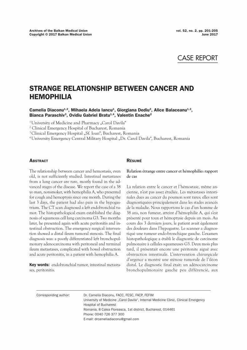
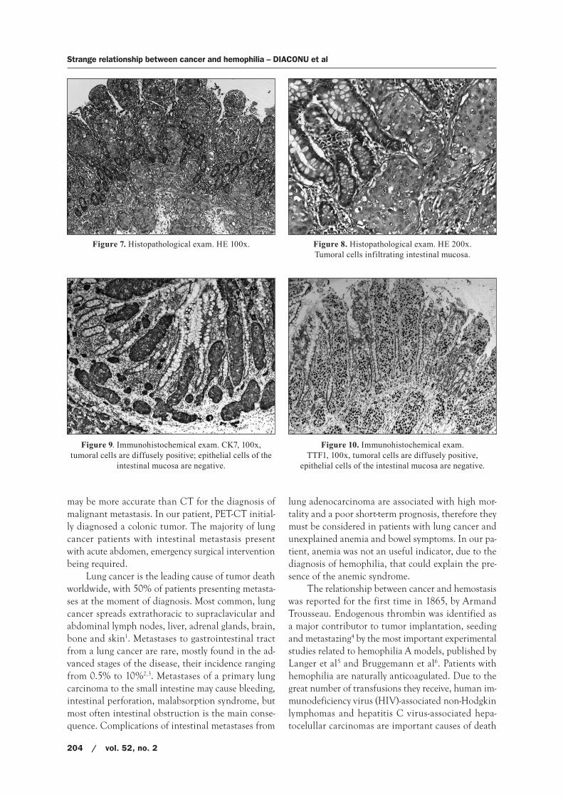
The histopathological exam of the small bowel frag-ment diagnosed a poorly differentiated carcinoma. Immunohistochemical exam demonstrated CK7 and TTF1 diffusely positive in tumor cells, CEA focally positive in tumor cells, Ck20 and CD X2 negative in tumor cells, positive internal control, P63 nega-tive. Conclusion: the histopathological exam corre-lated with immunohistochemical tests sustain the diagnosis of an intestinal metastasis from a poorly differentiated adenocarcinoma with bronchopulmo-nary origin (Figures 7-10).
The final diagnosis was: a poorly differentiated left bronchopulmonary adenocarcinoma with perito-neal and terminal ileum metastases, complicated with bowel obstruction and acute peritonitis, in a patient with hemophilia A. The evolution was unfavorable, the patient died 6 weeks later.
DISCUSSION
The case that we presented is rare and has two major particularities: the first one is the diagnosis of bronchopulmonary
adenocarcinoma in a relatively young hemophilic man, nonsmoker, without exposure to any other risk factors, who presented for cough and hemop-tysis;
the second one is the unusual presence of ileal me-tastasis from bronchopulmonary adenocarcinoma since the onset of respiratory symptoms. Initially, we took into consideration the diagnosis of two synchronous cancers.
The early diagnosis of ileal metastasis can be very difficult, as in the case presented, due to the unclear clinical symptoms. The initial abdominal CT scan was not able to identify the ileal tumor. PET-CT
Figure 3. Thoracic CT scan. Left endobronchial tumor.
Figure 5. PET-CT. Colonic tumor (probably multicentric) with enlarged lymph nodes and peritoneal metastasis,
metabolically active. Stage T3N2BM1.
Figure 4. PET-CT. Left lung tumor and metabolically active metastasis. Stage T4N3Mx.
Figure 6. Abdominal X-Ray showing intestinal obstruction (air levels).
Strange relationship between cancer and hemophilia – DIACONU et al
204 / vol. 52, no. 2
may be more accurate than CT for the diagnosis of malignant metastasis. In our patient, PET-CT initial-ly diagnosed a colonic tumor. The majority of lung cancer patients with intestinal metastasis present with acute abdomen, emergency surgical intervention being required.
Lung cancer is the leading cause of tumor death worldwide, with 50% of patients presenting metasta-ses at the moment of diagnosis. Most common, lung cancer spreads extrathoracic to supraclavicular and abdominal lymph nodes, liver, adrenal glands, brain, bone and skin1. Metastases to gastrointestinal tract from a lung cancer are rare, mostly found in the ad-vanced stages of the disease, their incidence ranging from 0.5% to 10%2,3. Metastases of a primary lung carcinoma to the small intestine may cause bleeding, intestinal perforation, malabsorption syndrome, but most often intestinal obstruction is the main conse-quence. Complications of intestinal metastases from
lung adenocarcinoma are associated with high mor-tality and a poor short-term prognosis, therefore they must be considered in patients with lung cancer and unexplained anemia and bowel symptoms. In our pa-tient, anemia was not an useful indicator, due to the diagnosis of hemophilia, that could explain the pre-sence of the anemic syndrome.
The relationship between cancer and hemostasis was reported for the first time in 1865, by Armand Trousseau. Endogenous thrombin was identified as a major contributor to tumor implantation, seeding and metastazing4 by the most important experimental studies related to hemophilia A models, published by Langer et al5 and Bruggemann et al6. Patients with hemophilia are naturally anticoagulated. Due to the great number of transfusions they receive, human im-munodeficiency virus (HIV)-associated non-Hodgkin lymphomas and hepatitis C virus-associated hepa-tocelullar carcinomas are important causes of death
Figure 7. Histopathological exam. HE 100x.
Figure 9. Immunohistochemical exam. CK7, 100x, tumoral cells are diffusely positive; epithelial cells of the
intestinal mucosa are negative.
Figure 8. Histopathological exam. HE 200x. Tumoral cells infiltrating intestinal mucosa.
Figure 10. Immunohistochemical exam. TTF1, 100x, tumoral cells are diffusely positive,
epithelial cells of the intestinal mucosa are negative.

Archives of the Balkan Medical Union
June 2017 / 205
among the virus-infected ageing hemophiliacs7,8. A study conducted in Netherlands between 1992-2001 found that the death rate due to neoplasms was 1.5 times higher in hemophiliacs than in the general population9. An UK study published in 2007, on behalf of the United Kingdom Hemophilia Centre Doctors Organization, analyzed mortality rates and causes of death in 6018 hemophilic patients unin-fected with HIV and concluded that while mortality from liver cancer and Hodgkin disease was increased as compared to general population, there was no evidence of increased mortality for other cancers10. Very interestingly, the authors reported a reduction of cancer-related mortality with increasing severity of hemophilia10. Moreover, in a review on epidemiologi-cal data related to cancer and hemophilia, non-HIV/HCV-related cancers represented the cause of death in 8-16% of the patients with hemophilia, lower than the mortality rate in the matched general male popu-lation11. There is an attractive hypothesis in the litera-ture, that hemophilia could confer a kind of protec-tion against cancer, that is only a speculation for the moment. This hypothesis needs further investigation in clinical trials with large populations, followed for many years.
One important problem that physicians face when dealing with hemophilic patients is related to the treatment. Diagnostic and therapeutic procedures in hemophiliacs might be complicated by adverse hemorrhagic events. This concern is the reason why in clinical practice hemophiliacs receive suboptimal anticancer treatment. The literature is scarce in infor-mation about cancer treatment in hemophiliacs. The largest study on this subject was published in 2012, on 122 patients monitored in 21 Italian hemophilia centers12 . In accordance with the previous findings of UK researchers10, they found that non-virus re lated cancers were less frequent in patients with severe forms of hemophilia as compared to patients with milder forms of the disease. A very interesting find-ing was that hemorrhagic events were more frequent in patients receiving chemotherapy or radiotherapy than in those surgically treated, hemophilia experts recommending replacement therapy not only at the time of invasive procedures but also during chemo-therapy or radiotherapy13.
In conclusion, the presence of hemorrhagic events in hemophilic patients should be careful-ly investigated, especially if they associate other
symptoms, in order to not miss a malignant disease. Although rare, intestinal metastasis may appear in the evolution of lung cancer and should be taken into consideration in patients with abdominal symptoms.
No acknowledgements
REFERENCES
1. Navani N, Spiro SG. Symptoms and signs of lung can-cer. In: Spiro SG, Huber RM, Janes SM. (eds). Thoracic Malignancies. Eur Respir Mon 2009;44:71-87.
2. De Palma GD, Masone S, Rega M, et al. Metastatic tumors to the stomach: clinical and endoscopic features. World J Gastroenterol 2006;12:7326-28.
3. Antler AS, Ough Y, Pitchumoni CS, et al. Gastrointestinal metastases from malignant tumors of the lung. Cancer 1982;49:170-2.
4. Trousseau A. Phlegmasia alba dolens. In: Trousseau A, ed. Clinique medicinale de l’Hotel-Dieu de Paris. Paris, France: JB Bailliere et fils; 1865. p. 645-712.
5. Langer F, Amirkhosravi A, Ingersoll SB, et al. Experimental metastasis and primary tumor growth in mice with hemo-philia A. J Thromb Haemost 2006; 4: 1056-62.
6. Bruggemann LW, Versteeg HH, Niers TM, et al. Experimental melanoma metastasis in lungs of mice with congenital coagulation disorders. J Cell Biol Med 2008; 12: 2622-7.
7. Darby SC, Ewart DW, Giangrande PL, et al. Mortality from liver cancer and liver disease in haemophilic men and boys in UK given blood products contaminated with hepatitis C. UK Haemophilia Centre Directors’ Organisation. Lancet 1997; 350: 1425-31.
8. Ragni MV, Belle SH, Bass D, et al. Clinical characteristics and blood product usage in AIDS-associated lymphoma in haemophiliacs: a case-control study. Haemophilia 1998; 4: 826-35.
9. Plug I, van der Bom JG, Peters M, et al. Mortality and causes of death in patients with hemophilia, 1992-2001: a prospec-tive cohort study. J Thromb Haemost 2006; 4: 510-6.
10. Darby SC, Kan SW, Spooner RJ, et al. Mortality rates, life expectancy, and causes of death in people with hemophilia A or B in the United Kingdom who were not infected with HIV. Blood 2007; 110: 815-25.
11. Miesbach W, Seifried E. Does hemophilia influence can-cer-related mortality in HIV-negative patients? Haemophilia 2011; 17: 55-60.
12. Tagliaferri A, Di Perna C, Santoro C, et al; on behalf the Italian Association of Hemophilia Centers. Cancers in patients with hemophilia: a retrospective study from the Italian Association of Hemophilia Centers. J Thromb Haemost 2012; 10: 90-5.
13. Mannucci PM, Schutgens RE, Santagostino E, Mauser- Bunschoten EP. How I treat age-related morbidities in el-derly persons with hemophilia. Blood 2009; 114: 5256-63.