steroid conversions with the cyp106a subfamily from ... · moral support throughout my work,...
TRANSCRIPT

Steroid conversions with the CYP106A subfamily
from Bacillus megaterium
Kumulative Dissertation
zur Erlangung des Grades Doktor der
Naturwissenschaften (Dr. rer. nat.)
der Naturwissenschaftlich-Technischen Fakultät III
Chemie, Pharmazie, Bio- und Werkstoffwissenschaften
der Universität des Saarlandes
von
Dipl.-Ing. Flora Marta Kiss
Saarbrücken
2015

Tag des Kolloquiums: 23. 10. 2015
Dekan: Prof. Dr. Dirk Bähre
Berichterstatter: Prof. Dr. Rita Bernhardt
Prof. Dr. Gert-Wieland Kohring
Vorsitz: Prof. Dr. Karin Römisch
Akad. Mitarbeiter: Dr. Yutaka Suzuki

“Courage doesn’t always roar.
Sometimes courage is the quiet voice at the end of the day saying,
‘I will try again tomorrow’.”
Mary Anne Radmacher

Acknowledgements
I would like to thank Prof. Dr. Rita Bernhardt for her guidance during the past three years and
providing me with the opportunity to take part in the Marie Curie ITN program. I’m grateful for such a
great fellowship that allowed me to meet the crème de la crème of the P450 community, to make
professional connections as well as good friends, and to travel around the world. Thanks to Dr. Frank
Hannemann for the discussions, constructive suggestions and reassuring words especially at the
beginning of my studies in the most critical times. Many thanks to Dr. Josef Zapp for the NMR
measurement and data evaluation of the extensive amount of samples that I produced during these three
years. His hard work allowed me to complete the three publications on which this thesis is based. I would
like to express my special gratitude to Prof. Dr. John Woodley, from the Technical University of
Denmark, who made my two-month exchange possible. I thank him for the stimulating discussions, his
keen interest in the progress of my work, valuable comments and encouragement. I would also like to
thank my Marie Curie fellow and friend Marie Lundemo, with who I worked side-by-side during my stay
in Denmark. I’m grateful for her enthusiasm, hard work and sacrificed long working days while she was
organizing her wedding. My sincere thanks go to our technicians Wolfgang Reinle and Birgit Heider-
Lips for the excellent protein purifications, and their help in all technical matters. Many thanks to Adrian
Gerber for letting me follow him everywhere in the lab at the very beginning of my work, when I had
honestly no clue what I was supposed to be doing, for his help in HPLC matters and for his assistance in
the administrative work, together with Jens Neunzig. Special thanks to Daniela Schmitz, to who I am
particularly grateful for introducing me to cell culture handling, for the helpful discussions, advice and
moral support throughout my work, especially during my first publication. I am also grateful to my
Ausländer-office mates, Azzam Mosa and Yogan Khatri. Azzam was always willing to help, whether it
was sharing experiences, cookies or tea, and was always very supportive from my first day in the lab.
Yogan was an enormous help in the last year of my PhD work: thanks to him I was able to publish my
third paper and learn new techniques in the lab. I would like to thank my friend, the chemist and hip-hop
enthusiast Alexander “Sascha” Schifrin, who did not only provide me with a place to stay upon arrival in
Saarbrücken, he introduced me to his friends, and made me feel welcome, which I really needed, living
alone for the first time in a foreign country. I will always be grateful for the numerous favors he was
willing to do, his guidance in the lab and around the university. Thanks to him, I had the chance to meet
and become friends with Irina, Oriana, Mael and María, who were a tremendous help in starting to live a
happy life in Saarbrücken. This includes Tanja Sagadin, my fellow vegetarian, special thanks for always
being there when I needed help in the lab, teaching me the basics of protein purification and also for the
great times in and outside of the lab. Thanks to Lina, Benni and Simone for the cheerful laughs coming
to my office, which always made me smile and for the initiative and organization of extracurricular
activities. Special acknowledgements go to all my colleagues from the Institute of Biochemistry for their
scientific support, the great moments, the “company outflights” and the good atmosphere during the last

years. I would like to thank both my families (all the Kisses and the Souzas) and friends at home and
around the world, who always believed in me more than I believed in myself. Despite being far away, we
found our ways to keep in touch. I would like to thank my father, who taught me that being excellent in
only one thing is not a must, nor the key to success and my mother, who encouraged me to take this
fellowship and believes without a doubt that I will change the world. Thanks to my sister and my brother,
who never stopped annoying me about being a nerd and an eternal student. Without them, I probably
would have ended up pursuing my other dream as a dancer .
I want to thank above all my husband, best friend and life-coach Nico, for his love, motivation,
endless faith in me, his help and effort correcting my papers/presentations, proofreading the present work
and keeping my head above water. Te lo dedico a ti, mi vida.
Thanks to the People Programme (Marie Curie Actions) of the European Union's 7th Framework
Programme (FP7/2007-2013), P4FIFTY - FP7 PEOPLE ITN 2011-289217 for providing the financial
support to complete this work.

Scientific contributions
This work is based on three original research papers reproduced in Chapter 2. The original manuscripts
are printed with the kind permissions from Springer Science and Business Media, Applied Microbiology
and Biotechnology (2.1 Kiss et al. (2015)), Elsevier, FEBS letters (2.2 Kiss et al. (2015)) and BioMed
Central, Microbial Cell Factories (2.3 Kiss et al. (2015)).
2.1 Kiss et al. (2015)
The author performed all the laboratory work and data analysis involved in this study (in vitro and in vivo
biotransformations, spectroscopic studies, HPLC analyses, product purification and data evaluation) and
drafted the manuscript. The NMR measurements and structure determination was performed by Dr. Josef
Zapp (Institute of Pharmaceutical Biology, Saarland University), the mass-determination of the
compounds by HRMS was completed by Tobias K. F. Dier and Prof. Dr. Dietrich A. Volmer (Institute
of Bioanalytical Chemistry, Saarland University). Dr. Daniela Schmitz and Prof Dr. Rita Bernhardt both
participated in the design of the project and contributed to the manuscript drafting.
2.2 Kiss et al. (2015)
The author performed all the laboratory experiments, analyzed and interpreted the data and drafted the
manuscript. Dr. Josef Zapp (Institute of Pharmaceutical Biology, Saarland University) performed the
NMR measurements and the structure determination of the converted steroids. Dr. Yogan Khatri and
Prof. Dr. Rita Bernhardt designed the project and assisted in interpreting the results and drafting the
manuscript.
2.3 Kiss et al. (2015)
The author and Marie Therese Lundemo (CAPEC-PROCESS, Department of Chemical and Biochemical
Engineering, Technical University of Denmark) contributed equally to the biochemical and
biotechnological experiments, the data analysis and the drafting of the manuscript. The NMR
measurement and the structure determination of the produced hydroxysteroid was performed by Dr. Josef
Zapp (Institute of Pharmaceutical Biology, Saarland University). Prof. Dr. John M. Woodley (CAPEC-
PROCESS, Department of Chemical and Biochemical Engineering, Technical University of Denmark)
and Prof. Dr. Rita Bernhardt took part in the design of the project, the interpretation of the data and
contributed to the manuscript drafting.

Table of contents
Abstract ................................................................................................................................................... 1
Zusammenfassung ................................................................................................................................ 2
1. Introduction .................................................................................................................................... 3
1.1. Cytochromes P450 ................................................................................................................. 3
1.1.1. General aspects ...................................................................................................................... 3
1.1.2. Structural features .................................................................................................................. 5
1.1.3. Catalytic cycle of cytochromes P450 ..................................................................................... 6
1.1.4. Biotechnological application of cytochromes P450 .............................................................. 7
1.2. Cytochromes P450 from Bacillus megaterium ...................................................................... 9
1.2.1. CYP106A1 from Bacillus megaterium DSM 319 ............................................................... 10
1.2.2. CYP106A2 from Bacillus megaterium ATCC 13368 ......................................................... 10
1.3. Steroid hormones and steroidal drugs .................................................................................. 12
1.4. Aim and outline of the work ................................................................................................ 13
2. Scientific articles.......................................................................................................................... 15
2.1. Kiss et al. (2015) .................................................................................................................. 15
Comparison of CYP106A1 and CYP106A2 from Bacillus megaterium - identification of a novel 11-oxidase activity
2.2. Kiss et al. (2015) .................................................................................................................. 44
Identification of new substrates for the CYP106A1-mediated 11-oxidation and investigation of the reaction mechanism
2.3. Kiss et al. (2015) .................................................................................................................. 58
Process development for the production of 15β-hydroxycyproterone acetate using Bacillus megaterium expressing
CYP106A2 as whole-cell biocatalyst
3. Discussion and outlook .............................................................................................................. 75
4. List of abbreviations ................................................................................................................... 83
5. Appendix ....................................................................................................................................... 84
5.1. Investigating the applicability of a fluorescence assay for the analysis of cortisol-cortisone
turnover by CYP106A1 ....................................................................................................................... 84
5.2. XTT-based cell proliferation assay using the LNCaP cell line, cyproterone acetate and its
15β-hydroxy product ........................................................................................................................... 85
6. References ..................................................................................................................................... 86

1
Abstract
Regio- and stereoselective hydroxylation represents a major challenge for synthetic chemistry.
The cytochrome P450 subfamily CYP106A efficiently catalyzes such reactions in steroids, di-, and
triterpenes. The well-studied CYP106A2, from B. megaterium ATCC 13368, is a promising candidate
for the pharmaceutical industry. It shares 63% amino acid sequence identity with CYP106A1 from
B. megaterium DSM 319 which was recently identified. The global objective of the work was the in depth
characterization of the CYP106A subfamily concerning the bioconversion of steroids, to explore their
potential application as industrial biocatalysts.
A focused steroid library was screened with the CYP106A subfamily. Binding studies, in vitro
and in vivo reactions allowed the comparison of enzyme activity, product pattern and product structures.
13 new substrates were identified for CYP106A1 and 7 for CYP106A2. The hydroxylase activity was
confirmed at positons 6β, 7β, 9α and 15β, in addition to an unprecedented 11-oxidase activity.
The 11-oxidase activity of CYP106A1 was further studied, identifying 3 11β-hydroxysteroids as
novel substrates for 11-oxidation. The reaction mechanism was also investigated, resulting in a large
inverse kinetic isotope effect (~0.44) suggesting the ferric peroxoanion as the reactive intermediate.
The CYP106A2 based B. megaterium whole-cell system has shown effective
15β-hydroxycyproterone acetate production. The conversion was scaled up from shake flask to bench-
top bioreactor, the reaction-bottlenecks were identified and addressed, demonstrating a successful process
development with a product formation of 0.43 g/L, approaching industrial process requirements and a
future large-scale application.

2
Zusammenfassung
Regio- und stereoselektive Hydroxylierung stellt für die synthetische Chemie eine ernste
Herausforderung dar. Die CYP106A-Subfamilie der Cytochrome P450 katalysiert diesen Reaktionstyp
auf effiziente Weise für Steroide, Di- und Triterpene. Das intensiv untersuchte CYP106A2 aus
B. megaterium ATCC 13368 teilt 63% Sequenzidentität mit CYP106A1 aus B. megaterium DSM 319,
das erst kürzlich identifiziert wurde. Das Ziel der Arbeit war, beide Enzyme in Bezug auf den Umsatz
von Steroiden zu charakterisieren und ihre potenzielle Anwendung als industrielle Biokatalysatoren zu
erkunden.
Ein Steroidbibliothek-Screening wurde mit den CYP106A-Enzymen vorgenommen.
Bindungsstudien, in vitro- und in vivo-Reaktionen ermöglichen den Vergleich der Enzymaktivität, der
Produktmuster und der Produktstrukturen. 13 neue Substrate wurden für CYP106A1 und 7 für
CYP106A2 identifiziert. Die Hydroxylaseaktivität wurde für die Positonen 6β, 7β, 9α und 15β, zusätzlich
zu einer 11-Oxidaseaktivität bestätigt.
Die 11-Oxidaseaktivität von CYP106A1 wurde untersucht und 3 11β-Hydroxysteroide als neue
Substrate für die 11-Oxidation identifiziert. Der in mechanistischen Studien große inverse kinetische
Isotopeneffekt (~ 0,44) deutet auf ein Eisen(III)-peroxoanion als reaktives Zwischenprodukt hin.
Das CYP106A2-B. megaterium-Ganzzellsystem hat sich als effizienter Katalysator für die
Herstellung von 15β-Hydroxycyproteronacetat erwiesen. Die Reaktion wurde von Schüttelkolben auf
einen Tischbioreaktor hochskaliert, die Hauptengpässe der Reaktion beseitigt und somit ein Prozess
erfolgreich etabliert. Die Produktausbeute von 0.43 g/L, bietet eine Basis für eine zukünftige Anwendung
im industriellen Maßstab.

3
1. Introduction
1.1. Cytochromes P450
1.1.1. General aspects
Cytochrome P450 enzymes (P450s) are heme-containing monooxygenases, constituting one of
the largest and oldest enzyme families, found in almost every life form, from prokaryotes to eukaryotes
[1]. They are involved in various biological processes such as carbon-source degradation, secondary
metabolite formation in prokaryotes, lower eukaryotes and plants, as well as in steroid hormone
biosynthesis and xenobiotic metabolism in humans [2-4]. Their first experimental evidence dates to 1955,
when the oxidation of xenobiotic compounds was detected in rabbit liver microsomes [5, 6]. This was
followed by the independent discovery of a carbon monoxide binding pigment in rat and pig livers by
Klingenberg and Garfinkel in 1958, respectively, showing an absorption maximum at 450 nm [7, 8]. In
1962, this microsomal carbon monoxide binding pigment was named cytochrome P450 by Omura and
Sato [9], who demonstrated the presence of iron-bound protoporphyrin IX in the enzyme. The
cytochrome denotes the hemoprotein nature, while P stands for “pigment” and 450 refers to the
characteristic absorption maximum observed at 450 nm during complex formation with CO (Soret peak).
This characteristic absorption is induced by the thiolate group of a conserved cysteine, forming the fifth
ligand of the heme iron [10], thus assigning these enzymes as heme thiolate proteins [11]. Their unusual
spectral property is still in use for the quantitative determination of the P450 content of a protein probe
[4].
Today the P450 database (http://drnelson.uthsc.edu/CytochromeP450.html) lists more than
21000 representatives. The amount of P450 genes varies enormously in different species. For instance,
7446 P450 genes are found in plants, 6313 in animals, 1254 in bacteria, 27 in archaea and only 2 are
discovered in viruses. They are classified according to the guidelines of a systematic nomenclature,
introduced by Nebert et al. in 1989 [12]. The enzymes are divided into families based on their amino acid
sequence identity (> 40 %) and are given the CYP designation for Cytochrome P450 followed by a
number (Figure 1). The letter behind the family number represents the subfamily, which requires a
similarity of more than 55 % in their amino acid sequences. The last number is unique for each P450,
indicating the individual isoenzyme.
Figure 1 - Nomenclature of P450s exemplified by CYP106A1
Categorized as external monooxygenases, P450s are able to activate molecular oxygen with the
support of one or more redox-partners, which transfer electrons from the cofactor, usually nicotinamide
4
adenine dinucleotide phosphate (NAD(P)H), to the P450 heme [4]. The general P450-catalyzed reaction
is performed by inserting a single oxygen atom, delivered from molecular oxygen, into the aliphatic or
aromatic substrate, while the remaining oxygen atom is reduced to water (Figure 2):
Figure 2 - General reaction mechanism catalyzed by P450s
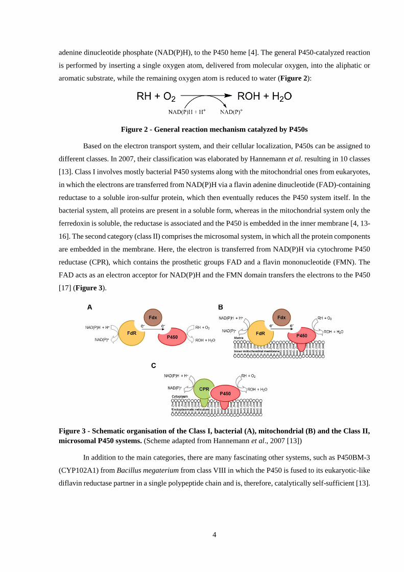
Based on the electron transport system, and their cellular localization, P450s can be assigned to
different classes. In 2007, their classification was elaborated by Hannemann et al. resulting in 10 classes
[13]. Class I involves mostly bacterial P450 systems along with the mitochondrial ones from eukaryotes,
in which the electrons are transferred from NAD(P)H via a flavin adenine dinucleotide (FAD)-containing
reductase to a soluble iron-sulfur protein, which then eventually reduces the P450 system itself. In the
bacterial system, all proteins are present in a soluble form, whereas in the mitochondrial system only the
ferredoxin is soluble, the reductase is associated and the P450 is embedded in the inner membrane [4, 13-
16]. The second category (class II) comprises the microsomal system, in which all the protein components
are embedded in the membrane. Here, the electron is transferred from NAD(P)H via cytochrome P450
reductase (CPR), which contains the prosthetic groups FAD and a flavin mononucleotide (FMN). The
FAD acts as an electron acceptor for NAD(P)H and the FMN domain transfers the electrons to the P450
[17] (Figure 3).
Figure 3 - Schematic organisation of the Class I, bacterial (A), mitochondrial (B) and the Class II,
microsomal P450 systems. (Scheme adapted from Hannemann et al., 2007 [13])
In addition to the main categories, there are many fascinating other systems, such as P450BM-3
(CYP102A1) from Bacillus megaterium from class VIII in which the P450 is fused to its eukaryotic-like
diflavin reductase partner in a single polypeptide chain and is, therefore, catalytically self-sufficient [13].

5
1.1.2. Structural features
Although P450s from different gene families often demonstrate less than 20 % sequence identity,
their structural organization shows similar folding and conserved topography (Figure 4), indicating a
common mechanism of oxygen activation [18]. They consist of two domains, the first one being the α
domain, rich in helices, including the highly conserved core formed by a four-helix bundle (D, L and I
and the antiparallel E-helix) besides two other helices (J and K) [19]. The second one is the β domain
with two beta-sheets (β1 and β2) in addition to another highly preserved structural motif, the meander
loop at the C-terminal end of the K-helix, stabilized by the conserved sequence ExxR [20]. The heme is
located between the distal I-helix and the proximal L-helix bound via a cysteine thiolate, including the
signature heme-binding motive FxxGx(H/R)xCxG. The long I-helix forms a wall for the substrate-
binding pocket and contains the conserved consensus sequence (A/G)GxS(E/D)T. The well preserved
threonine in the active center is believed to be involved in the activation of molecular oxygen throughout
the catalytic cycle [21].
Figure 4 - Topographic map showing the secondary structural elements of cytochromes P450,
exemplified by P450 BM3. The blue cylinders represent α-helices, the orange arrows the β-strands. The
random coil structures are shown as black lines, linking the individual secondary structure elements. The
elements are grouped in two domains, as first described by Poulos et al., 1987 [22] (Figure modified after
Peterson and Graham, 1998 [23]).
There are six regions identified by Gotoh named substrate recognition sites (SRS) having
particular importance in the recognition and binding of substrates: the B'-helix region (SRS1), parts of
helices F and G (SRS2 and SRS3), a portion of the I-helix (SRS4), the β4 hairpin structure (SRS5) and
the area between K-helix and β2 (SRS6) [24]. The SRSs are considered to be the most variable regions,

6
showing structural rearrangement of the protein upon substrate binding in favor of the catalytic reaction,
according to the proposed model of induced fit by Koshland [21, 25, 26].
1.1.3. Catalytic cycle of cytochromes P450
P450s were shown to catalyze a vast number of reactions with abundant substrate diversity. The
ability to perform these different reactions using the same catalytic cycle lies in the changing oxidation
states of the iron atom. Seven intermediates including three oxidation states form the complex catalytic
reaction mechanism of P450s, identified for CYP101 [21]. An overview of the reaction cycle is shown
in Figure 5.
Figure 5 - Catalytic cycle of cytochrome P450 monooxygenases. The numbers (1-7) represent the
actual state of the enzyme: 1 – Low-spin substrate-free state. 2 – High-spin enzyme-substrate complex.
3 - High-spin ferrous state. 4 – Oxy-ferrous state. 5a - Ferric peroxo intermediate. 5b – Ferric
hydroperoxo intermediate/compound 0. 6 – High-valent iron-oxo state/compound I. 7 - Product oxidation
and release. RH and ROH illustrate the substrate and the product, respectively. The three unproductive
shunt-pathways are marked with dashed grey arrows and the reduced oxygen products are shown as
outlets. (Figure adapted from Makris et al., 2002 [27] and Denisov et al., 2005 [21])
The cycle starts with the substrate-free, inactive ferric state (Fe (III)) of the heme iron, where the
sixth coordination center is occupied by a water molecule (1). Upon substrate binding, the water at the
distal site of the heme is partially or completely displaced, resulting in an increase of the redox potential
(by 100-130 mV) and a transition from the low spin to the five-times coordinated high spin Fe (III)
complex (2). This triggers the first electron transfer from NAD(P)H to the heme iron, producing a high
spin ferrous iron (Fe II) (3). The subsequent binding of molecular oxygen, generates the oxy-ferrous
intermediate, one of the key intermediates of the cycle (4). The following step is a second electron
transfer, yielding the highly reactive peroxoanion state (5a), which is then protonated at the distal oxygen

7
atom to form the hydroperoxo intermediate, Compound 0 (Cpd 0) (5b). The next protonation step of the
distal oxygen atom and the subsequent cleavage of the O-O bond leads to the release of a water molecule
and the formation of the so-called Compound I (Cpd I) (6), a high-valent iron-oxo complex. This Fe(IV)O
porphyrin cation radical initiates the oxygen atom transfer to the substrate (7) and the initial state is
restored.
However, the depicted cycle is not always complete, there are three “uncoupling” pathways
which can abort the catalytic cycle, being unproductive regarding substrate oxidation, yet still consuming
NAD(P)H. The first one is the auto-oxidation of the oxy-ferrous complex (4) while forming the
superoxide radical. The second one is known as the peroxide shunt, in which hydrogen peroxide is
released from the hydroperoxo intermediate (5b) resulting in the substrate-bound high spin complex (2).
Some P450s utilize the peroxide shunt in reverse, as a short cut for their reactions, introducing the oxygen
to the substrate directly from the hydrogen peroxide [28]. The third possibility is the so-called oxidase
shunt, where the activated oxygen atom of Cpd I (6) is protonated and reduced to form water rather than
being inserted into the substrate.
1.1.4. Biotechnological application of cytochromes P450
Throughout their long evolution, P450s became versatile biocatalysts, performing a large variety
of reactions, such as: hydroxylations, dehydrogenations, epoxidations, sulfoxidations, deaminations, N-,
O-, S-dealkylations and many more [4, 29] (Figure 6). In addition to their diverse reaction spectrum they
convert a broad range of substrates, from fatty acids, terpenes and steroids to drugs, organic solvents and
even toxins. Nevertheless, what makes these enzymes particularly interesting for industrial biocatalysis
is the ability to incorporate molecular oxygen into non-activated carbons in a regio- and/or stereoselective
fashion at neutral pH and moderate temperatures [30-33]. Such selective hydroxylation of aromatic or
aliphatic C-H bonds is hard to achieve by classical chemical synthesis, which often requires functional
group protection, suffers from side reactions and high temperature operation [34-36].
Figure 6 - Examples of the diverse oxidation reactions catalyzed by P450s.
X: -C, -N, -S. (Figure adapted from Bornschauer and Buchholz, 2005 [37])
In the last few years, significant progress has been made to reduce limitations and expand the
application of P450s for synthetic processes [29, 38]. By means of protein engineering, such as site-
directed mutagenesis or by directed (molecular) evolution techniques, the enzyme activity, stability,

8
substrate specificity and solvent tolerance has been successfully improved [39]. Yet, despite their
impressive synthetic potential, it was also recognized that isolated P450s are challenging to implement
in industry. Their constant need for the expensive cofactor supply and for the corresponding redox
partners along with their instability and low activity under process conditions all raise limitations for such
an approach [31, 40]. To circumvent this, the focus was shifted to the whole-cell system application of
P450s. The application of a microbial host has proven to be a promising strategy by providing endogenous
cofactor regeneration, potential co-expression of homologous or heterologous redox partners and
protected environment for the biocatalyst [41]. In addition, the P450 stability was shown to be enhanced
in the cellular milieu, since the H2O2 formed during uncoupling reactions is rendered harmless by the
cellular catalase [42]. In spite of its many advantages, whole-cell catalysis also faces some important
limitations, such as the transcellular transport limitation, low solubility/inhibition/toxicity of both
substrate and product, inadequate NAD(P)H supply and unwanted side-reactions [40, 43].
Since cytochrome P450 substrates are mostly hydrophobic organic compounds, their transport in
proximity of the whole-cell catalyst, present in the aqueous phase, as well as through the cell membrane,
is a major challenge. To enable the substrate uptake, different membrane permeabilization methods
(physical, chemical, and molecular engineering approaches) can be applied, depending on the
microorganism. These methods may include the disintegration of the membrane by ultrasonication,
freeze-thawing, using surface active substances such as Triton X, Tween, saponins or chelating agents
such as ethylenediaminetetraacetic acid (EDTA) and even the co-expression of membrane transport
proteins [44-48]. The faster transport of the substrate to the catalyst can be facilitated by using immiscible
or miscible organic solvents. Two-phase systems with immiscible solvents are of particular interest, in
which the organic phase serves as a substrate reservoir and facilitates the product removal, while the
aqueous phase protects the biocatalyst from degradation [49, 50]. Another option is to apply an organic
co-solvent such as ethanol, methanol or dimethyl sulfoxide (DMSO) in higher concentrations to increase
substrate solubility. However, in these cases the solvent tolerance of the biocatalyst has to be considered.
A further possibility, which circumvents the use of damaging organic solvents, yet still increases
both membrane permeability and substrate solubility, is the use of cyclodextrins (CD). CDs are
oligosaccharides, consisting of cyclically arranged (α-1,4)-linked α-D-glucopyranose units, forming a
lipophilic central cavity and a hydrophilic outer surface. Depending on the number of units they are called
α-, β- and γ-CDs containing six, seven or eight glucopyranose units, respectively. These basic CDs can
be further improved by chemical modifications resulting in derivatives with improved solubility and
stability such as 2-hydroxypropyl-β-cyclodextrin (2-HP-β-CD) [51, 52]. Due to their hydrophobic cone-
like cavity, CDs are able to form inclusion complexes with a variety of organic molecules. Based on this
outstanding property, they have been widely used in medicine and healthcare for improved solubility,
chemical stability and bioavailability of drugs [53]. Since CDs are inert to microorganisms and were
shown to be beneficial to respiratory-chain activity [54] their use in microbial transformations of
hydrophobic substrates has also been exemplified [55-57]. Besides improving substrate solubility, they

9
have also been used to avoid substrate/product toxicity and/or inhibition during the biocatalytic process
by complexation of the corresponding molecule in the hydrophobic central cavity, thus removing it from
the aqueous environment present in the cell [58].
At present, the practical application of P450s is limited mostly to the production of fine chemicals
such as pharmaceuticals, flavors, fragrances and (human) drug metabolites [42]. Among the remarkable
examples of biotechnological P450 applications is the artificial multi-enzyme cascade for the production
of the anti-malarial drug artemisinic acid in Saccharomyces cerevisiae (S. cerevisiae) [59], the synthesis
of a taxol precursor in Escherichia coli (E. coli) [60] and the production of pregnenolone, progesterone
and finally hydrocortisone using S. cerevisiae [61, 62]. Several further developments have also
contributed to raise attention to the commercial potential of P450s, such as the 11β-hydroxlation of
cortexolone to hydrocortisone by Curvularia sp. [43, 63], the 6β-hydroxylation of compactin to
pravastatin [64, 65], the biotransformation of Vitamin D3 to 1α,25-dihydroxyvitamin D3 [66, 67], or their
role in anthocyanin biosynthesis in blue or violet flowers [68, 69]. The production of human drug
metabolites is also a significant aspect of P450 applications due to their importance in diagnostics and
drug development, for the assessment of drug-induced side effects, toxicity and possible drug-drug
interactions. Their production by classical chemistry or using liver homogenates faces limitations, yet,
the recombinant expression of human drug-metabolizing P450s turned out to be a promising alternative.
This was successfully demonstrated by Novartis Pharma AG (Basel, Switzerland), who established an
E. coli host co-expressing human P450s with human CPR [70]. Nonetheless, these mammalian systems
might be limited due to the insufficient expression of the membrane-associated enzymes, hence bacterial,
soluble P450s could be considered as suitable alternatives.
1.2. Cytochromes P450 from Bacillus megaterium
Bacillus megaterium (B. megaterium) is a gram-positive, rod shaped soil-bacterium which has
been proven to be an ideal expression host for industrial biotechnology, mainly applied to protein
production [71, 72]. It is used as a production strain for industrial enzymes such as, penicillin G acylase
or different amylases, polyhydroxybutirate and vitamin B12 [73]. Beyond these applications, in the past
decade B. megaterium has gained interest as host for novel recombinant enzymes and therapeutic proteins
due to its many advantageous properties: it has a stable plasmid replication system, is not a pathogen,
lacks alkaline proteases, does not form endotoxins, has a high protein secretion capacity directly into the
medium and it is able to grow on a variety of carbon sources, which allows for low cost cultivation [74-
77].
Amongst the most interesting proteins present in B. megaterium are P450s [78] such as:
CYP102A1 (P450 BM3), a self-sufficient and so far one of the most thoroughly studied bacterial P450
[79]; CYP106A1 (P450 BM1) [80], recently functionally characterized by Brill et al. (2014) [81] and
Lee et al. (2014) [82]; and finally CYP106A2 from B. megaterium ATCC 13368 [83, 84], which was

10
extensively investigated in the past four decades and its biotechnological relevance has been
demonstrated in the conversion of steroids, di- and triterpenes [48, 85-89].
1.2.1. CYP106A1 from Bacillus megaterium DSM 319
CYP106A1 from B. megaterium ATCC 14581 is a soluble ~47.7 kDa protein consisting of 410
amino acids. It was first identified by He et al. in 1989, and has been investigated at the level of gene
regulation and gene expression in the following years [90, 91]. In 2011, the genome of B. megaterium
DSM 319 was sequenced [92], which led to the identification of several P450 genes, including the one
encoding CYP106A1. The enzyme was purified and characterized by Brill et al., who demonstrated its
successful application in a whole-cell system for the hydroxylation of the triterpene, 11-keto-β-boswellic
acid, at 7β and 15α positions [81]. Recently, CYP106A1 was also isolated from the B. megaterium strain
ATCC 14581 by Lee et al., showing successful in vitro steroid transformations, however, the product
structures were left unidentified [82]. Its closest homologue is the well-studied CYP106A2 from
B. megaterium ATCC 13368, with whom it shares 63 % amino acid sequence identity and 76 % similarity.
Based on the high sequence identity between the two subfamily members, CYP106A1 was proposed to
be an equally or even more promising catalyst in terms of steroidal drug or drug metabolite production,
leading to a possible transition of the pharmaceutical industry towards greener processes.
1.2.2. CYP106A2 from Bacillus megaterium ATCC 13368
CYP106A2 from B. megaterium ATCC 13368 is a soluble ~47 kDa protein consisting of 410
amino acids. It is one of the few cloned bacterial steroid hydroxylases, also known as 15β-hydroxylase,
which originates from its favored hydroxylation position on 3-oxo-Δ4 steroids. The discovery of
B. megaterium ATCC 13368-mediated steroid oxidation goes back to 1958, when the 15β-hydroxylation
of progesterone was described by McAleer et al. (1958) [93]. Only later, in 1975 was the role of the
cytochrome P450 as steroid hydroxylase recognized by Berg et al., who also identified the potential
components of the steroid hydroxylase system [83] followed by the isolation, purification and functional
characterization of the enzyme [84, 94, 95]. Finally in 1993, the cloning, sequencing and heterologous
expression of CYP106A2 took place in E.coli and B. subtilis [96]. Although its natural electron transfer
partners are not known, its activity was successfully reconstituted using bovine adrenal redox partners,
as well as by putidaredoxin and putidaredoxin reductase [48, 97-99].
In the past two decades, CYP106A2 was profoundly investigated as a potential industrial
biocatalyst, applying the enzyme in whole-cell systems using both E. coli [48, 86, 98] and B. megaterium
as expression hosts [87-89, 100]. Using E.coli, the transport of hydrophobic substrates across the outer
membrane was found to be limited [101], thus further approaches were focused on using the gram-
positive B. megaterium as host. The substrate spectrum of CYP106A2 was originally thought to be
limited to 3-oxo-Δ4-steroids [83], yet recent studies have shown that the hydroxylation of
3-hydroxy-Δ5-steroids, di- and triterpenes is also feasible [87-89, 100]. The native substrate of
CYP106A2 and its biological function are still unidentified. However, as a consequence of on-going

11
natural substrate library screenings, the list of its substrates is steadily growing. An overview of the so
far identified CYP106A2 substrates is shown in Table 1.
Table 1 - Overview of the formerly identified substrates of CYP106A2
(Table adapted from Janocha, 2013 [102])
CYP106A2 substrates References
Testosterone Berg et al. 1976, Berg et al. 1979a
Progesterone Berg et al. 1976, Berg et al. 1979a
17α-Hydroxyprogesteron Berg et al. 1976, Berg et al. 1979a
20α-Dihydroprogesterone Berg et al. 1976, Berg et al. 1979a
Deoxycorticosterone (DOC) Berg et al. 1976, Berg et al. 1979a
Corticosterone Berg et al. 1976, Berg et al. 1979a
Androstendione Berg et al. 1976, Berg et al. 1979a
Anilin Berg und Rafter 1981
6-Fluor-16-Methyl-DOC Rauschenbach et al. 1993
Betulinic acid Chatterjee et al. 2000
6β-Hydroxyprogesterone Lisurek 2004
15β-Hydroxyprogesterone Lisurek 2004
Cholestenone Lisurek 2004
Spironolactone Lisurek 2004
11-Deoxycortisol Virus 2006
4-Pregnen-20β-ol-3-one Bleif 2007
11-Keto-β-boswellic acid Bleif 2007
Dihydrochinopimaric acid Bleif 2007
Ethisterone Bleif 2007
17α-Methyltestosterone Bleif 2007
4-Pregnen-17α,20α,21-triol-3-one Bleif 2007
Abietic acid Bleif 2012
Oleanolic acid Bleif 2012
Ursolic acid Bleif 2012
Glycyrrhetinic acid Bleif 2012
Digitoxigenin Schmitz 2013
Prednisone Schmitz 2013
Dexamethasone Schmitz 2013
Dehydroepiandrosterone Schmitz 2013
Pregnenolone Schmitz 2013
Dipterocarpol Schmitz 2013
Betulin Schmitz 2013
Though its original name, 15β-hydroxylase, suggests hydroxylation at the 15β position, the long-
thought strict regioselectivity was contradicted by the identification of 6β, 7α/β, 9α, 11α and 15α-hydroxy
derivatives [85, 103, 104]. This by-product formation has been extensively studied for progesterone,
where the 6β, 9α and 11α derivatives are of pharmaceutical interest (contraceptives, hormone replacement
therapy) [103]. To enhance the previously observed C11-hydroxylation, a combinatory strategy was
applied, joining directed evolution and rational protein design to change the selectivity of the enzyme

12
from 15β to 11α in the hydroxylation of progesterone [85]. An impressive increase in regioselectivity
towards C11 was achieved forming a particularly interesting reaction product due to its inhibitory effect
on 11β-hydroxysteroid dehydrogenase [105].
1.3. Steroid hormones and steroidal drugs
Steroids are terpenoid lipids with a characteristic gonane skeleton comprising seventeen carbon
atoms in a four-ring structure, forming three cyclohexane rings (rings A, B, and C) and one cyclopentane
ring (ring D) (Figure 7). Various derivatives exist, which principally differ in the oxidation state of the
rings and in the functional groups attached to the four-ring system.
Figure 7 - Sterane, the parent ring structure of steroid compounds.
The four-ring structure (A-D) and the numbering of the carbon atoms (1-17) are shown.
Steroids play an important role as biological and chemical compounds in many living systems
and have been identified in animals (e.g.: cholesterol, corticosteroids, sex hormones, bile acids, vitamin
D, neurosteroids), plants (e.g.: phytosterols, diosgenin) and lower eukaryotes such as yeast and fungi
(e.g.: ergosterol, ergosteroids) [106, 107]. In humans, steroidal hormones are synthesized in steroidogenic
tissues such as gonads and adrenal glands from cholesterol. Their natural roles include the regulation of
electrolyte and water homeostasis, cholesterol levels, control of sexual differentiation and reproduction,
as well as cardiovascular and neuroprotective functions [108-111]. Steroid hormones are also known to
control cell proliferation and tissue differentiation, to regulate signal transduction pathways and cell-to-
cell communication processes [112-114].
Drugs based on the steroid structure are of great pharmaceutical importance being widely used
in almost all fields of healthcare, from anti-microbial, anti-viral, anti-fungal agents to the treatment of
hormone-dependent cancer forms, autoimmune and allergic disorders [106]. To date, steroids represent
one of the largest sectors in the pharmaceutical industry with more than 300 approved drugs known [115,
116]. Steroid analogues with altered functional groups are favored over their natural counterparts, due to
their improved therapeutic features such as, reduced side effects, higher potency and better
pharmacokinetics [117]. Moreover, steroid hydroxylation is considered to be one of the most important
reactions in steroid functionalization, since the derivatives can have enhanced biological activity and/or
be further modified in drug development [118-120]. The synthesis of these functionalized derivatives is
a real challenge for synthetic steroid chemistry, suffering from low predictability and specificity, yet, the
combination of classical chemistry and biocatalysis was found to be successful to produce these steroids

13
for the pharmaceutical industry [107]. Microbial steroid hydroxylation using heterologously expressed
P450s is considered to be a promising tool to produce such hydroxysteroids. Unfortunately, the
biotechnological application of eukaryotic P450s is restricted due to their membrane-associated assembly
leading to limitations in their expression and catalytic activities [13, 29]. In contrast, bacterial P450s are
soluble proteins, which can be overexpressed to very high amounts allowing higher productivities in
biotransformation and representing a promising alternative for industrial applications [48, 99].
1.4. Aim and outline of the work
The global objective of this work was the in-depth characterization of the bacterial CYP106A
subfamily in the bioconversion of steroid compounds to explore and expand their potential application as
biocatalysts in industrial-scale steroid hydroxylations (Figure 8). CYP106A2 from B. megaterium ATCC
13368 was extensively studied since the late 1970s and shown to hydroxylate steroids, di- and triterpenes
in a regio- and stereoselective manner. Its lesser known counterpart, CYP106A1 from B. megaterium
DSM 319, sharing high sequence identity, was also recognized as a terpene- and steroid-hydroxylase,
however, its potential as a biocatalyst was not fully investigated.
Figure 8 - Summary of the motivation, previous work and contents of this thesis. The orange box
represents the global aim of the work, the yellow contains the state of the art. The blue and the grey
rectangles symbolize the published and unpublished results, respectively, with the corresponding chapter
number.
For a detailed analysis of the CYP106A subfamily members and a comparison of their steroid
hydroxylating capacity, a focused steroid library was screened. The initial substrate-screening was based
on their high-spin shift induction via difference spectroscopy, also allowing the determination of
dissociation constants (KD). Since type I shift-induction is a good indication but not a prerequisite for

14
successful conversion, all steroids were subjected to further in vitro assays and subsequent analysis of
product formation by high performance liquid chromatography (HPLC). Out of the 19 converted steroids,
6 were chosen for further investigation. Since functionalized steroids could be of pharmaceutical
importance as drugs or drug metabolites, the emphasis was placed on the analysis of enzyme activity and
product identification for the evaluation of potential biotechnological applications. Substrate conversions
were performed using formerly established B. megaterium MS941 whole-cell systems, overexpressing
each CYP106 enzyme. The conversion products were isolated by preparative HPLC and their structural
determination was achieved by NMR spectroscopy. The obtained results are summarized in Chapter 2.1
of this thesis.
In Chapter 2.2, the newly identified CYP106A1 11-oxidase activity, obtained as a result in
Chapter 2.1, was further investigated. Three 11β-hydroxysteroid-analogues (prednisolone,
dexamethasone and 11β-hydroxyandrostenedione) were screened with CYP106A1. The 11-oxidation of
the substrates was confirmed by NMR in all cases, however, single 11-keto product formation was only
obtained for 11β-hydroxyandrostenedione. The latter reaction was chosen to investigate the mechanism
of the reaction using kinetic solvent isotope effect.
The final section (Chapter 2.3) describes the results obtained in a collaborative project with the
Technical University of Denmark, where the bioconversion of the pharmaceutically relevant
antiandrogen, cyproterone acetate, was investigated using a CYP106A2-based B. megaterium whole-cell
system. Since the product, 15β-hydroxycyproterone acetate, is of pharmaceutical interest, the aim was to
scale-up the reaction from shake flasks to bioreactors to model an efficient, yet greener and cost-effective
production. To improve the yield and product titers for a future large-scale application, the main
bottlenecks of the reaction were identified and addressed.

15
2. Scientific articles
The obtained results presented in this work are published in the articles listed below:
2.1. Kiss et al. (2015)
Comparison of CYP106A1 and CYP106A2 from Bacillus megaterium - identification of a novel
11-oxidase activity
Flora Marta Kiss, Daniela Schmitz, Josef Zapp, Tobias K. F. Dier, Dietrich A. Volmer, Rita
Bernhardt
Applied Microbiology and Biotechnology 2015 Apr 24 doi: 10.1007/s00253-015-6563-8
Reprinted with the permission of Springer Science and Business Media.

16
Comparison of CYP106A1 and CYP106A2 from Bacillus
megaterium - identification of a novel 11-oxidase activity
Flora Marta Kiss1, Daniela Schmitz1, Josef Zapp2, Tobias K. F. Dier3, Dietrich A. Volmer3,
Rita Bernhardt1*
Abstract
The CYP106A subfamily hydroxylates steroids, di- and triterpenes in a regio- and stereoselective manner,
which is a challenging task for synthetic chemistry. The well-studied CYP106A2 enzyme, from the
Bacillus megaterium strain ATCC 13368, is a highly promising candidate for the pharmaceutical
industry. It shares 63 % amino acid sequence identity with CYP106A1 from B. megaterium DSM319,
which was recently characterized. A focused steroid library was screened with both, CYP106A1 and
CYP106A2. Nineteen out of the 23 tested steroids were successfully converted by both enzymes during
in vitro and in vivo reactions. Thirteen new substrates were identified for CYP106A1, while the substrate
spectrum of CYP106A2 was extended by 7 new members. Finally, 6 chosen steroids were further studied
on a preparative scale employing a recombinant B. megaterium MS941 whole-cell system, yielding
sufficient amounts of product for structure characterization by nuclear magnetic resonance. The
hydroxylase activity was confirmed at positons 6β, 7β, 9α and 15β. In addition, the CYP106A subfamily
showed unprecedented 11-oxidase activity, converting 11β-hydroxysteroids to their 11-keto derivatives.
This novel reaction and the diverse hydroxylation positions on pharmaceutically relevant compounds
underline the role of the CYP106A subfamily in drug development and production.
Keywords: CYP106A1, CYP106A2, cortisol, cortisone, 11β-hydroxysteroid dehydrogenation
Introduction
For the past decade cytochrome P450 enzymes
(P450s) have been studied as candidates for
pharmaceutical drug and drug precursor
development. This large superfamily of
heme cofactor-containing monooxygenases
(http://drnelson.uthsc.edu/CytochromeP450.html)
is involved in the catalysis of a wide range of
reactions from hydroxylations, epoxidations and
deaminations to dealkylations and C-C bond
cleavage. Not only do they perform versatile
reactions, they also convert a broad range of
substrates. P450s participate in the biosynthesis of
1Institute of Biochemistry, Saarland University, D-66123
Saarbruecken, Germany
2Institute of Pharmaceutical Biology, Saarland University,
D-66123 Saarbruecken, Germany
3Institute of Bioanalytical Chemistry, Saarland University,
D-66123 Saarbruecken, German
*Address correspondence to: Professor Dr. Rita Bernhardt
Email: [email protected]
endogenous steroids, fatty acids and vitamins as
well as in the elimination of exogenous
compounds, xenobiotics and drugs (Bernhardt,
2006; Urlacher et al., 2004). They are thus suitable
for the production of pharmaceutical compounds
and provide a more economically and ecologically
sustainable alternative to chemical synthesis, even
more so, where the latter has failed to produce
sufficient amounts of product (Bernhardt and
Urlacher, 2014; Urlacher and Girhard, 2012). The
chemical synthesis of steroids is very complex and
costly, particularly when performing
regio- and stereo-selective hydroxylations.
Microbial/enzymatic transformations are,
therefore, favored by the industry as a straight-
forward, greener and more cost-effective solution
(Donova and Egorova, 2012; Tong and Dong,
2009).
The heterologous expression of bacterial P450
enzymes was shown to be successful in several
host organisms, making the Class I bacterial P450
systems preferable over their membrane-bound
mammalian counterparts, due to their solubility

17
and easy handling (Bernhardt, 2006; Hannemann
et al., 2007; Venkataraman et al., 2014). Although
their expression and purification is possible at an
analytical scale, the use of isolated P450s in
industrial applications is not feasible, mainly due
to their instability under process conditions and
the need for a constant supply of the rather
expensive cofactor NADP(H) (Bernhardt and
Urlacher, 2014; O'Reilly et al., 2011; Urlacher and
Eiben, 2006). The latter has been addressed by
both enzymatic and chemical methods (Chefson
and Auclair, 2006; Hollmann et al., 2006), along
with the progress towards improved
thermostability (Li et al., 2007) and solvent
tolerance (Seng Wong et al., 2004). A different
possible workaround is the construction of whole-
cell systems, where the P450 is expressed by a
microbial host organism. These constructs have
the advantage of stabilizing the P450 integrity and
electron transfer and provide cofactor regeneration
by the cellular metabolism. Numerous organisms
have been described to realize the heterologous
expression of P450s, including the most widely
used, Escherichia coli (Agematu et al., 2006;
Bracco et al., 2013; Makino et al., 2014).
Nevertheless, there are cases when using E. coli
can lead to limitations, such as the restricted
transport of large hydrophobic molecules across
the cell membrane (Janocha and Bernhardt, 2013;
Zehentgruber et al., 2010). Another candidate,
Bacillus megaterium, has recently aroused greater
interest as a genetically modified host organism
for biotechnological/industrial approaches,
especially for the expression of heterologous
proteins. On account of having its own P450
expression system, it does not require the addition
of the heme-precursor (δ-aminolevulinic acid) in
contrast to E.coli which lacks natural P450 genes
and heme proteins. Its attractive characteristics
also include: growth on various carbon sources,
high plasmid stability and no alkaline protease or
endotoxin production (Bleif et al., 2012; Korneli
et al., 2013; Vary et al., 2007).
From a biotechnological standpoint,
CYP106A2 from the B. megaterium strain ATCC
13368 (Berg et al., 1976; Berg et al., 1979) and its
unique hydroxylating properties raised interest in
the past years (Hannemann et al., 2006; Virus and
Bernhardt, 2008). CYP106A2 being extensively
studied since the late 1970s, is a highly active
steroid hydroxylase, catalyzing mainly the
15β-hydroxylation of 3-oxo-Δ4-steroids (Berg et
al., 1976; Berg et al., 1979). Furthermore, the
enzyme was shown to hydroxylate di- and
triterpenes (Bleif et al., 2011; Bleif et al., 2012;
Schmitz et al., 2012) and recently was also
described as a regioselective hydroxylase for the
3-hydroxy-Δ5 steroid dehydroepiandrosterone
(Schmitz et al., 2014). Although its original name,
15β-hydroxylase, suggests mainly hydroxylation
at the 15β position, the long-thought strict
regioselectivity was contradicted by the
identification of 6β, 7α/β, 9α, 11α and 15α-
hydroxy products (Lisurek et al., 2004; Lisurek et
al., 2008; Nguyen et al., 2012; Virus et al., 2006).
Even though, these hydroxylations occur in lower
proportions, the resulting products are of industrial
interest. This means a unique potential for
industrial applications, since there is a wide
variety of compounds that could serve as
substrates for this enzyme. Its natural substrate is
yet to be identified and its substrate range is
continuously broadening as a result of library
screening (Schmitz et al., 2012; Schmitz et al.,
2014).
The lesser known CYP106A1 shares 63 %
amino acid sequence identity and 76 % similarity
with its well-studied sibling, CYP106A2 (He et
al., 1995; He et al., 1989). CYP106A1 from
B. megaterium DSM319 was recently purified and
characterized by our group (Brill et al., 2014) and
successfully applied in a whole-cell system for the
hydroxylation of a triterpene, 11-keto-β-boswellic
acid (KBA), at 7β and 15α positions. The
CYP106A1-dependent conversion showed a
different product pattern compared with
CYP106A2 (Bleif et al., 2012), resulting in two
additional products, identified as 7β-hydroxy-
KBA and 7β,15α-dihydroxy KBA. Furthermore,
CYP106A1 was also characterized in vitro from
another B. megaterium strain, ATCC 14581 by
Lee et al., who recognized some steroids as
convertible substrates, but leaving product
structures unidentified (Lee et al., 2014). Based on
the high sequence identity between the two
enzymes, CYP106A1 was proposed to be an

18
equally promising candidate in a possible
transition of the pharmaceutical industry towards
greener processes. In order to exploit the potential
of CYP106A1 and to reveal the anticipated
differences in activity and selectivity between the
two subfamily members, we decided to focus on
steroid conversions. Steroids are pharmaceuticals
of great importance. Drugs based on the steroid
structure are widely used in almost all fields of
healthcare, from anti-microbial and anti-viral
agents to the treatment of hormone-dependent
cancer forms (Donova and Egorova, 2012).
Moreover, steroid hydroxylation is considered to
be one of the most important reactions in steroid
functionalization, since the derivatives have
enhanced biological activity or can be further
modified in drug development (Choudhary et al.,
2011; Choudhary et al., 2005; Janeczko et al.,
2009).
Here we tested a focused substrate library with
both CYP106A subfamily members and extended
their substrate ranges with respect to different
steroid molecules. Nineteen of the 23 screened
steroids were successfully converted by both
enzymes during in vitro and in vivo reactions.
Using recombinant B. megaterium MS941 whole-
cell systems, overexpressing each enzyme,
sufficient amounts of product were obtained for
structure characterization by nuclear magnetic
resonance (NMR). Besides confirming the
production of mono- and dihydroxylated products
at 6β, 7β, 9α and 15β positions, we were able to
demonstrate a previously unknown reaction
among these P450s, an 11β-hydroxysteroid
dehydrogenase activity. Our recent findings
underline the role of the CYP106A subfamily in
the field of drug development and production.
Materials and methods
Reagents and chemicals
All steroid substrates were purchased from Sigma-
Aldrich (Sigma Aldrich Biochemie Gmbh,
Germany), all other chemicals were obtained from
standard sources and of highest grade available.
Expression and purification of the enzymes
The expression and purification of the CYP106A1
and CYP106A2 proteins was performed as
described previously (Brill et al., 2014; Lisurek et
al., 2004; Simgen et al., 2000). For the
reconstituted in vitro system, a truncated form of
bovine adrenodoxin (Adx4-108) was used in
combination with bovine adrenodoxin reductase
(AdR). Their expression and purification was
completed as described elsewhere (Sagara et al.,
1993; Uhlmann et al., 1992).
UV-visible absorbance spectroscopy
To analyze the characteristics of the purified
CYP106A family members, the UV-visible
absorbance spectrum of the proteins was recorded
in a range of 200-700 nm with a double beam
spectrophotometer (UV-2101 PC, Shimadzu,
Japan). The spectra were constantly analyzed
during the purification process, approaching a
Q value (A417/A280) higher then 1.5, suggesting a
high amount of well-folded, heme-containing and
active P450s. The concentration of the proteins for
further experiments was determined by CO
difference spectroscopy according to the method
of Omura and Sato (Omura and Sato, 1964), using
an extinction coefficient for the CO-bound P450
of 91 mM-1 cm-1.
Substrate binding assay
The substrate binding difference spectra were
investigated using a double-beam
spectrophotometer (UV-2101PC, Shimadzu,
Japan) and tandem quartz cuvettes. The reaction
was carried out in 800 µl total volume. One
chamber of each cuvette contained 10 µM solution
of the enzymes in 50 mM potassium phosphate
buffer pH 7.4, while the other chamber was filled
with the buffer only. The substrate was dissolved
in DMSO at a stock concentration of 20 mM. The
enzyme solution was then titrated with increasing
concentrations of the substrate. In each step the
same amount of substrate was also added to the
buffer-containing chamber of the reference
cuvette. After each titration step, the spectrum was
recorded between 350 nm and 500 nm. The Kd
value was calculated after the titration of the
substrate until saturation. The data was analyzed
by plotting the peak-to-through differences against
the concentrations of the substrate. The data was
fitted in Origin (OriginLab Corporations,

19
Massachusetts, USA) using hyperbolic regression.
The Kd values are averaged from three
independent measurements.
In vitro conversions
The in vitro conversion of the substrates was
carried out with a reconstituted system in a final
volume of 250 µL at 30 °C for 60 min in 50 mM
potassium phosphate buffer (pH 7.4), containing
20 % (v/v) glycerol. The reconstituted system
contained AdR (1 µM), the truncated form of
bovine Adx4-108 (10 µM), CYP106A1 and
CYP106A2 (1 µM), an NADPH-regenerating
system [MgCl2 (1 mM), glucose-6-phosphate
(5 mM), glucose-6-phosphate dehydrogenase
(1 U), and NADPH (0.1 µM)] and the substrate
(200 µM). The reaction was started by adding
NADPH (100 µM) and stopped by the addition of
250 μl of ethyl acetate, mixed vigorously, and
extracted twice. After evaporating the combined
organic phases to dryness, the residues were
dissolved in the high-performance liquid
chromatography (HPLC) mobile phase and
subjected to HPLC analysis.
In vivo conversions
The in vivo steroid conversions were performed
using the recombinant B. megaterium MS941
strain, derived from the strain DSM319 (Wittchen
and Meinhardt, 1995), lacking the major
extracellular protease gene nprM. The
B. megaterium MS941 strain was transformed
with the corresponding plasmid pSMF2.1B
(containing CYP106A1 cloned into SpeI/MluI
sites (Brill et al., 2014)) or pSMF2.1C (containing
CYP106A2 cloned into SpeI/MluI sites (Bleif et
al., 2012)) applying a polyethylene glycol-
mediated technique using protoplasts (Barg et al.,
2005). To make sure that the conversion was
catalyzed by the anticipated enzyme, the
conversions were compared to transformations
with the wild type strain MS941 (lacking the
pSMF2.1 plasmid, but naturally containing
cytochrome P450 genes) as a control. According
to these results, the wild type strain did not show
any conversion.
Precultures were inoculated from -80 °C
glycerol stock, using 25 ml complex TB medium
(24 g/l yeast extract, 12 g/l soytone, 2.31 g/l
KH2PO4 and 12.5 g/l K2HPO4) supplemented with
10 µg/ml tetracycline and incubated overnight, at
150 rpm, 30 °C. The main culture (supplemented
with the corresponding amount of tetracycline)
was inoculated with 1% of the culture volume
from the overnight culture. Following the
inoculation of the main culture, it was further
incubated for 2-3 hours, until it reached an
OD578 = 0.4 when 0.5 g/l xylose solution was
added to induce the expression. Cells were
cultivated for 24 h under the same conditions prior
to the addition of the substrate. For analytical
purposes, the experiments were performed in 2 ml
reaction tubes with a 500 µl volume of the freshly
aliquoted main culture. The transformations
required the use of an Eppendorf thermomixer
(Eppendorf, Hamburg, Germany) for the
continuous mixing with 1000 rpm, keeping the
temperature constant at 30°C, for 1-4 h. For the
preparative scale conversions, 50 ml culture
volume was used in 300 ml baffled shake flasks,
inoculated with 500 µl of the precultures, induced
and cultivated for 24 h at 30°C, before the addition
of the substrates. Conversions at a larger scale
were performed with resting cells, either adding
the substrate directly 24 hours after the expression
or after harvesting the cells and suspending them
in 100 mM potassium phosphate buffer (pH 7.4).
The steroids were added as ethanolic solution to
the culture medium (the use of ethanol did not
exceed 5% of the culture volume). Following the
corresponding conversion time, the reaction was
stopped and the steroids were extracted twice by
the addition of 50 ml ethyl acetate. The organic
phase was dried over anhydrous MgSO4 and
concentrated to dryness in a rotatory evaporator
(Büchi R-114). The yellowish residue was
dissolved in the mobile phase of the HPLC and
filtered through a sterile syringe filter (Rotilabo
syringe filter, 0.22 μm, Carl Roth GmbH,
Karlsruhe, Germany). The products were
separated by preparative HPLC, according to its
retention time. The collected fractions were
evaporated to dryness and analyzed by NMR
spectroscopy and high-resolution mass
spectrometry (HRMS).

20
HPLC analysis
The HPLC analysis was performed on a Jasco
system consisting of a Pu-980 HPLC pump, an
AS-950 sampler, a UV-975 UV/Vis detector, and
an LG-980–02 gradient unit (Jasco, Gross-
Umstadt, Germany). A reversed-phase ec MN
Nucleodor C18 (3µM, 4.0x125mm) column
(Macherey-Nagel, Betlehem, PA, USA) was used
to carry out the experiments, kept at an oven
temperature of 40 °C. The steroids were eluted
from the column using a gradient method, starting
with a mobile phase ratio of 1:9 of ACN:H2O and
increasing it to 1:1. The used flow rate was
1 ml/min and the UV detection of the substrate and
product was accomplished at 240 or 254 nm. In the
case of the isolation of conversion products the
conditions of the preparative reversed-phase
HPLC (ec MN Nucleodur C18 VP (5µM,
8x250mm), Macherey-Nagel, Betlehem, PA,
USA) varied according to the size of the column,
consequently the maximum injectable amount of
sample could reach 1 ml and the flow rate
2.5 ml/min.
The conversion and product distribution were
calculated from the relative peak area (area %) of
the HPLC chromatograms. Following the
conversion, all peak areas were summed up and
the respective product peak area was divided by
the total area of all peaks. The results are presented
as conversion % and product formation %.
HRMS analysis
Analyses were performed using a solariX 7 Tesla
FTICR mass spectrometer (Bruker Daltonik,
Bremen, Germany). All samples were ionized by
atmospheric pressure chemical ionization (APCI)
in negative ionization mode, using the following
parameters: dry temperature 350 °C, vaporizing
temperature 350 °C, corona needle 40000 nA,
capillary voltage 2000 V, end plate offset -500 V,
estimated R. P. (400 m/z) 70000. The calculated
exact and measured accurate masses are presented
next to each identified compound name in the
NMR section.
NMR characterization of the metabolites
The NMR spectra were recorded in CDCl3 or
CD3OD with a Bruker DRX 500 or a Bruker
Avance 500 NMR spectrometer at 300 K. The
chemical shifts were relative to CHCl3 at δ 7.26 or
CH3OD at δ 3.30 (1H NMR) and CDCl3 at δ 77.00
or CD3OD at δ 49.00 (13C NMR) respectively
using the standard δ notation in parts per million.
The 1D NMR (1H and 13C NMR, DEPT135) and
the 2D NMR spectra (gs-HH-COSY, gs-NOESY,
gs-HSQCED, and gs-HMBC) were recorded using
the BRUKER pulse program library. All
assignments were based on extensive NMR
spectral evidence. (For detailed substrate and
product structures, see Table 4.)
6-hydroxyandrost-4-ene-3,17-dione
Product A4 (2.6 mg) in the conversion of
androstenedione with CYP106A1 (HRMS (APCI)
calculated exact mass [Da] C19H26O3 [M+TFA-H]-
415.1732; measured accurate mass [Da] 415,1733,
error [ppm] -0.44). Its 1H NMR data matched
those in literature (Kirk et al., 1990). 1H NMR
(CDCl3, 500 MHz): δ 0.95 s (3xH-18), 0.98 ddd
(12.3, 10.8 and 4.2 Hz, H-9), 1.29 m (H-12a), 1.31
m (H-14), 1.33 m (H-7a), 1.41 s (3xH-19), 1.53 m
(H-11a), 1.63 m (H-15a), 1.66 m (6-OH), 1.70 m
(H-11b), 1.73 m (H-1a), 1.89 ddd (13.3, 4.2 and
3.0 Hz, H-12b), 1.99 ddd (13.2, 4.0 and 3.0 Hz, H-
15b), 2.06 ddd (13.3, 5.0 and 3.0 Hz, H-1b), 2.12
m (H-16a), 2.14 m (H-7b), 2.18 m (H-8), 2.41 m
(H-2a), 2.49 m (H-16b), 2.53 m (H-2b), 4.41 ddd
(3x2.5 Hz, H-6), 5.84 brs (H-4). 13C NMR (CDCl3,
125 MHz): δ 13.79 (CH3, C-18), 19.59 (CH3, C-
19), 20.29 (CH2, C-11), 21.72 (CH2, C-15), 29.43
(CH, C-8), 31.30 (CH2, C-12), 34.20 (CH2, C-2),
35.76 (CH2, C-16), 37.13 (CH2, C-1), 37.22 (CH2,
C-7), 38.05 (C, C-10), 47.63 (C, C-13), 50.93 (CH,
C-14), 53.68 (CH, C-9), 72.94 (CH, C-6), 126.64
(CH, C-4), 167.43 (C, C-5), 199.97 (C, C-3),
220.33 (C, C-17).
7-hydroxyandrost-4-ene-3,17-dione
Product A2 (3 mg) in the conversion of
androstenedione with CYP106A1 (HRMS (APCI)
calculated exact mass [Da] C19H26O3 [M+TFA-H]-
415.1732; measured accurate mass [Da] 415,1731,
error [ppm] 0.06). Its 1H NMR data matched those
in literature (Kirk et al., 1990): 1H NMR (CDCl3,
500 MHz): δ 0.95 s (3xH-18), 1.01 ddd (12.4, 10.5
and 4.0 Hz, H-9), 1.24 s (3xH-19), 1.26 ddd

21
(2x13.3 and 4.2 Hz, H-12a), 1.47 m (H-14), 1.50
m (H-11a), 1.57 m (7-OH), 1.67 ddd (13.5, 13.5
and 5.0 Hz, H-1a), 1.75 m (H-11b), 1.76 ddd
(3x10.5 Hz, H-8), 1.88 ddd (13.3, 5.0 and 3.3 Hz,
H-12b), 1.94 m (H-15a), 2.06 ddd (13.5, 5.0 and
3.3 Hz, H-1b), 2.13 ddd (19.5 and 2x9.5 Hz, H-
16a), 2.32 m (H-15b), 2.39 m (H-2a), 2.45 m (H-
2b), 2.48 m (H-16b), 2.49 m (H-6a), 2.58 dd (14.0
and 5.2 Hz, H-6b), 3.59 m (H-7), 5.78 d (2.0 Hz,
H-4). 13C NMR (CDCl3, 125 MHz): δ 13.96 (CH3,
C-18), 17.38 (CH3, C-19), 20.37 (CH2, C-11),
24.98 (CH2, C-15), 31.22 (CH2, C-12), 33.89
(CH2, C-2), 35.65 (CH2, C-1), 35.97 (CH2, C-16),
38.04 (C, C-10), 42.59 (CH, C-8), 42.69 (CH2, C-
6), 48.01 (C, C-13), 50.52 (CH, C-14), 50.78 (CH,
C-9), 74.37 (CH, C-7), 125.09 (CH, C-4), 166.35
(C, C-5), 198.95 (C, C-3), 220.39 (C, C-17).
15-hydroxyandrost-4-ene-3,17-dione
Product B6 (3.7 mg) in the conversion of
androstenedione with CYP106A2 (HRMS (APCI)
calculated exact mass [Da] C19H26O3 [M+TFA-H]-
415.1732; measured accurate mass [Da] 415,1752,
error [ppm] 4.78). Its 13C NMR data matched those
in literature (Mineki et al., 1995) 1H NMR (CDCl3,
500 MHz): δ 1.04 ddd (14.8, 10.7 and 4.2 Hz, H-
9), 1.22 m (H-7a), 1.23 s (3xH-18), 1.26 s (3xH-
19), 1.29 m (H-12a), 1.30 m (H-14), 1.52 m (H-
11a), 1.74 m (H-1a), 1.75 m (H-11b), 1.85 ddd
(13.2, 4.8 and 3.2 Hz, H-12b), 2.06 m (H-1b), 2.11
m (H-8), 2.15 m (H-7b), 2.36 m (H-2a), 2.37 m
(H-6a), 2.43 m (H-2b), 2.50 m (H-6b), 2.52 dd
(19.6 and 6.0 Hz, H-16a), 2.59 dd (19.6 and 1.3
Hz, H-16b), 4.59 ddd (6.0, 4.5 and 1.3 Hz, H-15),
5.77 brs (H-4). 13C NMR (CDCl3, 125 MHz): δ
17.26 (CH3, C-19), 17.59 (CH3, C-18), 20.26
(CH2, C-11), 30.47 (CH2, C-7), 31.44 (CH, C-8),
32.46 (CH2, C-6), 32.61 (CH2, C-12), 33.91 (CH2,
C-2), 35.73 (CH2, C-1), 38.80 (C, C-10), 46.70 (C,
C-13), 47.06 (CH2, C-16), 54.24 (CH, C-9), 54.96
(CH, C-14), 67.16 (CH, C-15), 124.23 (CH, C-4),
170.12 (C, C-5), 199.30 (C, C-3), 219.05 (C, C-
17).
7-dihydroxyandrost-4-ene-3,17-dione
Product B3 (2 mg) in the conversion of
androstenedione with CYP106A2 (HRMS (APCI)
calculated exact mass [Da] C19H26O4 [M+TFA-H]-
431.1682; measured accurate mass [Da] 431,1685,
error [ppm] 0.81. The 1H and 13C NMR spectra
showed signals for two secondary hydroxyl
groups with similar chemical shifts to those of the
monohydroxylated androstenedione derivatives
A2 and B6. The positions of the hydroxyl groups
at C-7 and C-15 were supported by the results of
2D NMR experiments (HHCOSY, HSQC and
HMBC). For example, H-7 (δH 3.74 ddd) showed
correlations to the H-6a and H-6b (2.58 m, 2H)
and H-8 (2.12 ddd) in the HHCOSY, H-15 could
be assigned by its vicinal couplings to H-14, H-
16a and H-16b in the HHCOSY and its 3JCH
correlation with carbonyl C-17 in the HMBC. The
-orientation for both hydroxyls could be
concluded by results of the NOESY spectrum. H-
7 and H-15 showed an effect to each other and
both to the -orientated H-14; H-7 showed an
additional effect to the -orientated H-9.
Therefore, both H-7 and H-15 were in -position
and as a consequence their corresponding
hydroxyls -orientated. To our knowledge the
structure of this dihydroxylated androstenedione
has not been reported so far. 1H NMR (CDCl3, 500
MHz): δ 1.06 ddd (12.5, 10.7 and 4.2 Hz, H-9),
1.26 s (3xH-18), 1.27 m (H-12a), 1.28 s (3xH-19),
1.40 dd (10.7 and 4.5 Hz, H-14), 1.58 m (H-11a),
1.68 ddd (2x13.5 and 5.0 Hz, H-1a), 1.76 m (H-
11b), 1.83 ddd (12.7, 4.0 and 2.5 Hz, H-12b), ),
2.08 ddd (13.5, 5.0 and 3.3 Hz, H-1b), 2.12 ddd
(3x 10.7 Hz, H-8) 2.38 m (H-2a), 2.44 m (H-2b),
2.53 dd (20.0 and 8.0 Hz, H-16a), 2.58 m (2H, H-
6a and H-6b), 2.67 dd (20.0 and 1.2 Hz, H-16b),
3.74 ddd (9.5 and 2x8.0 Hz, H-7), 4.66 ddd (7.0,
4.5 and 1.2 Hz, H-15), 5.79 brs (H-4). 13C NMR
(CDCl3, 125 MHz): δ 17.34 (CH3, C-19), 17.65
(CH3, C-18), 20.38 (CH2, C-11), 32.16 (CH2, C-
12), 33.86 (CH2, C-2), 35.58 (CH2, C-1), 38.01 (C,
C-10), 38.56 (CH, C-8), 42.46 (CH2, C-6), 44.53
(CH2, C-16), 47.06 (C, C-13), 51.01 (CH, C-9),
55.07 (CH, C-14), 68.52 (CH, C-15), 73.74 (CH,
C-7), 125.37 (CH, C-4), 165.67 (C, C-5), 198.90
(C, C-3), 219.50 (C, C-17).
15-hydroxycorticosterone
Product D1 (1.2 mg) in the conversion of
corticosterone with CYP106A2 (HRMS (APCI)

22
calculated exact mass [Da] C21H30O5 [M-H]-
361.2015; measured accurate mass [Da] 361.2020,
error [ppm] 1.27). Its NMR data showed
resonances for an additional hydroxyl group (δC
70.24; δH 4.41 m), which could be located at
position 15 by means of 2D NMR. 1H NMR
(CDCl3, 500 MHz): δ 1.02 m (H-14), 1.06 m (H-
9), 1.15 m (H-7a), 1.19 s (3xH-18), 1.48 s (3xH-
19), 1.62 dd (13.8 and 3.3 Hz, H-12a), 1.86 ddd
(2x13.5 and 4.5 Hz, H-1a), 2.08 dd (13.8 and 2.8
Hz, H-12b), 2.21 ddd (13.5, 5.0 and 4.0 Hz, H-1b),
2.28 m (2H, H-6a and H-7b), 2.33 m (2H, H-16a
and b), 2.34 m (H-17), 2.37 m (H-2a), 2.38 m (H-
8), 2.49 m (H-2b), 2.55 m (H-6b), 3.23 dd (2x 4.5
Hz, 21-OH), 4.19 dd (18.5 and 4.5 Hz, H-21a),
4.24 dd (18.5 and 4.5 Hz, H-21b), 4.41 m (H-15),
5.70 d (2.0 Hz, H-4). 13C NMR (CDCl3, 125
MHz): δ 18.65 (CH3, C-18), 20.80 (CH3, C-19),
27.78 (CH, C-8), 31.70 (CH2, C-7), 31.82 (CH2,
C-6), 33.79 (CH2, C-2), 34.78 (C, C-10), 35.04
(CH2, C-1), 35.53 (CH2, C-16), 43.46 (C, C-13),
49.13 (CH2, C-12), 56.64 (CH, C-9), 59.80 (CH,
C-17), 61.74 (CH, C-14), 67.80 (CH, C-11), 69.22
(CH2, C-21), 70.24 (CH, C-15), 122.55 (CH, C-4),
171.36 (C, C-5), 199.32 (C, C-3), 208.77 (C, C-
20).
11-dehydrocorticosterone
Product C5 (2.5 mg) and D4 (1 mg) in the
conversion of corticosterone with CYP106A1 and
CYP106A2 respectively (HRMS (APCI)
calculated exact mass [Da] C21H28O4 [M-H]-
343.1909; measured accurate mass [Da] 343.1921,
error [ppm] 3.34). The NMR spectra of C5 lacked
of resonances for the 11-hydroxy group but
revealed an additional carbonyl (δC 207.49) for C-
11. 1H NMR (CDCl3, 500 MHz): δ 0.67 s (3xH-
18), 1.27 m (H-7a), 1.41 s (3xH-19), 1.45 m (H-
15a), 1.63 ddd (2x14.5 and 4.5 Hz, H-1a), 1.80 m
(H-14), 1.92 m (2H, H-9 and H-16a), 1.93 m (H-
8), 1.95 m (H-15b), 1.98 m (H-7b), 2.29 m (H-
16b), 2.30 m (2H, H-2a and H-6a), 2.44 m (H-6b),
2.47 d (12.0 Hz, H-12a), 2.48 ddd (17.0, 14.5 and
5.0 Hz, H-2b), 2.56 d (12.0 Hz, H-12b), 2.68 dd
(2x9.5 Hz, H-17), 2.77 ddd (14.5, 5.0 and 3.3 Hz,
H-1b), 3.16 dd (2x 3.5 Hz, 21-OH), 4.14 dd (19.5
and 3.5 Hz, H-21a), 4.19 dd (19.5 and 3.5 Hz, H-
21b), 5.73 brs (H-4). 13C NMR (CDCl3, 125
MHz): δ 14.35 (CH3, C-18), 17.15 (CH3, C-19),
23.34 (CH2, C-16), 24.00 (CH2, C-15), 32.13
(CH2, C-7), 32.15 (CH2, C-6), 33.69 (CH2, C-2),
34.74 (CH2, C-1), 36.74 (CH, C-8), 38.16 (C, C-
10), 47.50 (C, C-13), 54.93 (CH, C-14), 56.20
(CH2, C-12), 57.56 (CH, C-17), 62.67 (CH, C-9),
69.24 (CH2, C-21), 124.70 (CH, C-4), 168.03 (C,
C-5), 199.53 (C, C-3), 207.49 (C, C-11), 208.98
(C, C-20).
9-hydroxy-11-dehydrocorticosterone
Product C2 (1 mg) in the conversion of
corticosterone with CYP106A1 (HRMS (APCI)
calculated exact mass [Da] C21H28O5 [M-H]-
359.1859; measured accurate mass [Da] 343.1867,
error [ppm] 2.45). C2 was the only product in our
conversion study that formed a tertiary hydroxyl
group with the P450 enzymes. Its resonance line
at δC 79.31 showed 3JCH couplings to H-1a, H-12a
and methyl H-19 in the HMBC spectrum
indicating that C-9 bared the additional hydroxyl
group. As H-9 in the substrate 9-OH of the
conversion product C2 was -oriented. This was
obvious by the cross peaks found in NOESY
spectrum. Key correlations were found between
H-8 and the methyls H-18 and H-19, which
therefore must be positioned on the same side of
the steroid skeleton. Only two citations from the
fifties could be found for the structure of 9-
hydroxy-11-dehydrocorticosterone but both
without NMR data. 1H NMR (CDCl3, 500 MHz):
δ 0.66 s (3xH-18), 1.39 m (H-15a), 1.49 s (3xH-
19), 1.66 m (H-7a), 1.71 m (H-7b), 1.82 m (H-
15b), 1.94 m (H-16a), 2.10 m (H-1a), 2.17 m (H-
8), 2.22 m (H-14), 2.26 m (H-16b), 2.30 ddd (15.0,
4.7 and 2.0 Hz, H-6a), 2.37 m (H-2a), 2.38 d (12.0
Hz, H-12a), 2.47 m (H-2b), 2.48 m (H-6b), 2.50 m
(H-1b), 2.73 dd (2x9.5 Hz, H-17), 3.05 dd (12.0
and 1.0 Hz, H-12b), 3.16 dd (2x 5.0 Hz, 21-OH),
4.13 dd (19.2 and 4.7 Hz, H-21a), 4.19 dd (19.2
and 4.7 Hz, H-21b), 5.86 d (2.0 Hz, H-4). 13C
NMR (CDCl3, 125 MHz): δ 13.91 (CH3, C-18),
18.89 (CH3, C-19), 23.32 (CH2, C-16), 23.72
(CH2, C-15), 24.52 (CH2, C-7), 28.15 (CH2, C-1),
31.29 (CH2, C-6), 33.74 (CH2, C-2), 38.65 (CH,
C-8), 43.66 (C, C-10), 47.11 (C, C-13), 48.73 (CH,
C-14), 51.98 (CH2, C-12), 57.41 (CH, C-17),
69.28 (CH2, C-21), 79.31 (C, C-9), 127.68 (CH, C-

23
4), 165.89 (C, C-5), 198.83 (C, C-3), 206.83 (C,
C-11), 209.09 (C, C-20).
6β-hydroxy-11-deoxycorticosterone
(6β-hydroxy-DOC)
Product E5 (1.1 mg) in the conversion of 11-
deoxycorticosterone with CYP106A1 (HRMS
(APCI) calculated exact mass [Da] C21H30O4 [M-
H]- 345.2066; measured accurate mass [Da]
345.2076, error [ppm] 2.82). Selected 1H NMR
data were published by Matsuzaki (Matsuzaki et
al., 1995). Our 1H NMR of E4 matched these data. 1H NMR (CDCl3, 500 MHz): δ 0.73 s (3xH-18),
0.96 ddd (12.5, 11.0 and 4.5 Hz, H-9), 1.17 m (H-
14), 1.28 m (H-7a), 1.38 m (H-12a), 1.39 s (3xH-
19), 1.40 m (H-15a) 1.49 m (H-11a), 1.63 m (H-
11b), 1.73 m (H-1a), 1.77 m (H-16a), 1.79 m (H-
15b), 1.97 ddd (12.0, 3.5 and 2.5 Hz, H-12b), 2.01
m (H-8), 2.03 m (H-7b), 2.04 ddd (10.5, 5.0 and
2.8 Hz, H-1b), 2.23 m (H-16b), 2.40 dddd (17.3,
4.5, 3.0 and 1.0 Hz, H-2a), 2.47 brd (9.0 Hz, H-
17), 2.49 m (H-16b), 2.53 ddd (17.3, 15.0 and 5.0
Hz, H-2b), 3.25 dd (2x4.5 Hz,far 19-OH), 4.17 dd
(19.0 and 4.5 Hz, H-21a), 4.22 dd (19.0 and 4.5
Hz, H-21b), 4.37 dd (2x2.5 Hz, H-6), 5.83 brs (H-
4). 13C NMR (CDCl3, 125 MHz): δ 13.50 (CH3, C-
18), 19.53 (CH3, C-19), 20.87 (CH2, C-11), 22.91
(CH2, C-16), 24.43 (CH2, C-15), 29.74 (CH, C-8),
34.20 (CH2, C-2), 37.12 (CH2, C-1), 37.94 (C, C-
10), 38.35 (2xCH2, C-7 and C-12), 44.75 (C, C-
13), 53.38 (CH, C-9), 56.06 (CH, C-14), 59.09
(CH, C-17), 69.69 (CH2, C-21), 73.03 (CH, C-6),
126.51 (CH, C-4), 167.60 (C, C-5), 200.08 (C, C-
3), 210.06 (C, C-20).
15-hydroxy-11-deoxycorticosterone
(15β-hydroxy-DOC)
Product E4 (3.5 mg) and F5 (4 mg) in the
conversion of 11-deoxycorticosterone with
CYP106A1 and CYP106A2 respectively (HRMS
(APCI) calculated exact mass [Da] C21H30O4 [M-
H]- 345.2066; measured accurate mass [Da]
345.2055, error [ppm] -3.06). In comparison to 11-
deoxycorticosterone the NMR data of E4 and F5
showed signals for an additional secondary
hydroxyl group (δH 4.38 ddd, δC 70.34). Its
position at C-15 could be deduced by vicinal
couplings of the methin proton with H-14 (δH
1.04) and H-16a (δH 2.32) in the HHCOSY and
with C-13 (δC 44.29) and C-17 (δC 59.28) in
HMBC. The -orientation of the hydroxyl was
obvious by the NOESY effects of H-15 with H-14
and H-16a, both in -position. In addition to these
results the chemical shifts of the epimeric 15-
hydroxy-11-deoxycorticosterone (Faramarzi et
al., 2003) differed significantly from our values. 1H NMR (CDCl3, 500 MHz): δ 0.96 s (3xH-18),
1.04 m (H-9), 1.06 m (H-14), 1.16 m (H-7a), 1.22
s (3xH-19), 1.38 m (H-12a), 1.43 dddd (3x 13.5
and 3.5 Hz, H-11a), 1.66 dddd (13.5 and 3x 4.0
Hz, H-11b), 1.73 ddd (2x13.5 and 5.0 Hz, H-1a),
1.95 m (H-12b), 1.97 m (H-8), 2.05 ddd (13.5, 5.0
and 3.0 Hz, H-1b), 2.14 m (H-7b), 2.32 m (H-16a),
2.33 m (2H, H-6a and H-6b), 2.38 m (H-2a), 2.43
m (2H, H-2b and H-17), 2.49 m (H-16b), 4.18 d
(19.0 Hz, H-21a), 4.24 d (19.0 Hz, H-21b), 4.38
ddd (3x5.5 Hz, H-15), 5.75 brs (H-4). 13C NMR
(CDCl3, 125 MHz): δ 15.98 (CH3, C-18), 17.29
(CH3, C-19), 20.84 (CH2, C-11), 31.11 (CH2, C-
7), 31.74 (CH, C-8), 32.61 (CH2, C-16), 33.92
(CH2, C-2), 35.78 (CH2, C-1), 36.03 (CH2, C-6),
38.69 (C, C-10), 39.87 (CH2, C-12), 44.29 (C, C-
13), 53.94 (CH, C-9), 59.28 (CH, C-17), 60.37
(CH, C-14), 69.39 (CH2, C-21), 70.34 (CH, C-15),
124.09 (CH, C-4), 170.46 (C, C-5), 199.35 (C, C-
3), 209.01 (C, C-20).
7, 15-dihydroxy-11-deoxycorticosterone
(7β, 15β-dihydroxy-DOC)
Product E2 (1.8 mg) and F4 (3 mg) in the
conversion of 11-deoxycorticosterone with
CYP106A1 and CYP106A2 respectively (HRMS
(APCI) calculated exact mass [Da] C21H30O5 [M-
H]- 361.2015; measured accurate mass [Da]
361.2019, error [ppm] 1.16). The NMR spectra of
E2 and F4 were similar to those of E4, especially
for the resonances of C-15 (δC 71.55) and H-15 (δH
4.50 ddd, 3x5.5 Hz) as well as the coupling pattern
of H-15. But E2 beared an additional secondary
hydroxyl group with resonances at δH 3.62 (ddd,
9.5 and 2x8.0 Hz) and δC 74.36. These resonances
and the coupling pattern were close to those of the
7-hydroxy group found for B3. Therefore, the
structure of E2 was elucidated as 7, 15-
dihydroxy-11-deoxycorticosterone, a structure
which has not been published so far. 2D NMR

24
measurements supported the structure and led to
the full assignments. 1H NMR (CDCl3, 500 MHz):
δ 0.99 s (3xH-18), 1.03 m (H-9), 1.12 dd (10.8 and
5.0 Hz, H-14), 1.25 s (3xH-19), 1.33 ddd (2x13.0
and 4.0 Hz, H-12a), 1.50 dddd (3x 13.5 and 4.0
Hz, H-11a), 1.67 m (H-1a), 1.68 m (H-11b), 1.95
ddd (13.0, 4.0 and 3.0 Hz, H-12b), 2.05 m (H-8),
2.07 m (H-1b), 2.33 m (2H, H-16a and H-16b),
2.35 m (H-2a), 2.36 m (H-17), 2.41 m (H-2b), 2.55
m (2H, H-6a and H-6b), 3.62 ddd (9.5 and 2x8.0
Hz, H-7), 4.22 s (2H, H-21a and H-21b), 4.50 ddd
(3x5.5 Hz, H-15), 5.77 brs (H-4). 13C NMR
(CDCl3, 125 MHz): δ 15.63 (CH3, C-18), 17.25
(CH3, C-19), 20.74 (CH2, C-11), 33.54 (CH2, C-
16), 33.89 (CH2, C-2), 35.66 (CH2, C-1), 37.95 (C,
C-10), 38.52 (CH, C-8), 39.36 (CH2, C-12), 42.60
(CH2, C-6), 44.47 (C, C-13), 50.74 (CH, C-9),
59.01 (CH, C-17), 60.11 (CH, C-14), 69.35 (CH2,
C-21), 71.55 (CH, C-15), 74.36 (CH, C-7), 125.10
(CH, C-4), 166.10 (C, C-5), 198.99 (C, C-3),
209.24 (C, C-20).
15-hydroxy-11-deoxycortisol
(15-hydroxy-RSS)
Product G4 (5 mg) and H3 (5 mg) in the
conversion of RSS with CYP106A1 and
CYP106A2 respectively (HRMS (APCI)
calculated exact mass [Da] C21H30O5 [M-H]-
361.2015; measured accurate mass [Da] 361.2013,
error [ppm] 0.72). Its NMR data showed
resonances for an additional hydroxyl group (δC
70.29; δH 4.31 ddd), which could be located at
position 15 by means of 2D NMR. Although
15-hydroxy-11-deoxycortisol is a known
structure NMR data were not available in
literature. It should be mentioned that the coupling
constants found for H-15 (7.8, 6.0 and 2.3 Hz)
differed completely from those found for it in 15-
hydroxy-11-deoxycorticosterone (3x5.5 Hz) and
in 15-hydroxyandrost-4-ene-3,17-dione (6.0, 4.5
and 1.3 Hz). This is caused by the high flexibility
of the trans-annulated cyclopentane ring and by
the strong influence of the C-17 substitution
pattern of its preferred configuration. 1H NMR
(CD3OD, 500 MHz): δ 0.89 s (3xH-18), 1.04 ddd
(12.3, 10.8 and 4.0 Hz, H-9), 1.13 dddd (13.8,
12.5, 11.5 and 4.0 Hz, H-7a), 1.25 s (3xH-19), 1.41
ddd (12.5, 4.0 and 2.8 Hz, H-12a), 1.48 ddd (2x
13.5 and 4.0 Hz, H-11a), 1.67 m (H-11b), 1.71 m
(H-1a), 1.72 dd (11.5 and 6.0 Hz, H-14), 1.86 ddd
(2x12.5 and 4.0 Hz, H-12b), 2.03 ddd (2x11.2 and
3.2 Hz, H-8), 2.06 dd (15.5 and 7.5 Hz, H-16a),
2.10 ddd (13.5, 5.2 and 3.0 Hz, H-1b), 2.20 dddd
(12.5, 5.7 and 2x 2.8 Hz, H-7b), 2.30 m (H-2a),
2.32 m (H-6a), 2.48 ddd (17.0, 15.0 and 5.3 Hz, H-
2b), 2.53 dddd (2x15, 5.5 and 1.7 Hz, H-6b), 2.73
dd (15.5 and 2.3 Hz, H-16b), 4.27 d (19.2 Hz, H-
21a), 4.31 ddd (7.8, 6.0 and 2.3 Hz, H-15), 4.62 d
(19.2 Hz, H-21b), 5.72 brs (H-4). 13C NMR
(CD3OD, 125 MHz): δ 17.63 (CH3, C-19), 18.04
(CH3, C-18), 21.68 (CH2, C-11), 32.66 (CH2, C-
7), 32.72 (CH2, C-12), 32.94 (CH, C-8), 33.90
(CH2, C-6), 34.73 (CH2, C-2), 36.81 (CH2, C-1),
40.16 (C, C-10), 47.54 (CH2, C-16), 48.40 (C, C-
13), 55.32 (CH, C-9), 55.73 (CH, C-14), 67.70
(CH2, C-21), 70.29 (CH, C-15), 90.27 (C, C-17),
124.17 (CH, C-4), 175.17 (C, C-5), 202.35 (C, C-
3), 212.64 (C, C-20).
7, 15-dihydroxy-11-deoxycortisol
(7, 15-dihydroxy-RSS)
Product G1 (3.5 mg) in the conversion of RSS with
CYP106A1 (HRMS (APCI) calculated exact mass
[Da] C21H30O6 [M-H]- 377.1964; measured
accurate mass [Da] 377.1976, error [ppm] 3.01).
Its 1H and 13C NMR spectra revealed two
additional secondary hydroxyl groups. One of
them (δC 71.26; δH 4.56 ddd , 8.0, 5.6 and 2.6 Hz)
gave similar values as found for the 15-hydroxy
group in H3. The other one (δC 74.67; δH 3.68 ddd,
2x10.0 and 6.5 Hz) showed vicinal couplings to
the isochronic protons of H-6 (δH 2.56 m, 2H) and
to H-8 (δH 2.06 m) in the HHCOSY. NOESY cross
peaks between H-7 and the -oriented H-9, H-14
and H-15 revealed the stereochemistry at C-7 with
the hydroxy group in -orientation. The structure
of 7, 15-dihydroxy-11-deoxycortisol has not
beeen published so far. 1H NMR (CDCl3, 500
MHz): δ 1.03 s (3xH-18), 1.06 ddd (2x12.0 and 3.7
Hz, H-9), 1.26 s (3xH-19), 1.39 m (H-12a), 1.48
m (H-11a), 1.67 m (H-1a), 1.70 m (H-12b), 1.72
m (H-11b), 1.77 dd (11.0 and 5.5 Hz, H-14), 2.06
m (H-8), 2.07 m (H-1b), 2.22 dd (15.5 and 8.0 Hz,
H-16a), 2.40 m (H-2a), 2.45 m (H-2b), 2.56 m
(2H, H-6a and H-6b), 2.63 dd (15.5 and 2.5 Hz, H-
16b), 3.68 ddd (2x10.0 and 6.5 Hz, H-7), 4.36 d

25
(19.8 Hz, H-21a), 4.56 ddd (8.0, 5.6 and 2.6 Hz,
H-15), 4.70 d (19.8 Hz, H-21b), 5.77 brs (H-4). 13C
NMR (CDCl3, 125 MHz): δ 17.28 (CH3, C-19),
17.62 (CH3, C-18), 20.19 (CH2, C-11), 30.22
(CH2, C-12), 33.90 (CH2, C-2), 35.65 (CH2, C-1),
37.95 (C, C-10), 38.25 (CH, C-8), 42.65 (CH2, C-
6), 43.25 (CH2, C-16), 48.70 (C, C-13), 50.33
(CH, C-9), 53.96 (CH, C-14), 67.71 (CH2, C-21),
71.26 (CH, C-15), 74.67 (CH, C-7), 87.65 (C, C-
17), 125.19 (CH, C-4), 165.99 (C, C-5), 198.98 (C,
C-3), 212.29 (C, C-20)
Cortisone
Product I6 (1 mg) in the conversion of cortisol
with CYP106A1 (HRMS (APCI) calculated exact
mass [Da] C21H28O5 [M-H]- 359.1858; measured
accurate mass [Da] 359.1842, error [ppm] -4.62).
Its 1H NMR data matched those in literature (Kirk
et al., 1990). 1H NMR (CD3OD, 500 MHz): δ 0.60
s (3xH-18), 1.33 m (H-7a), 1.42 s (3xH-19), 1.46
m (H-15a), 1.70 m (H-16a), 1.72 m (H-1a), 1.92 m
(H-15b), 2.01 m (H-8), 2.02 m (H-7b), 2.04 d (12.5
Hz, H-12a), 2.11 d (10.5 Hz, H-9), 2.23 ddd (17.0
and 2x 3.8 Hz, H-2a), 2.33 ddd (15.0, 4.0 and 2.5
Hz, H-6a), 2.45 m (H-14), 2.50 m (H-2b), 2.52 m
(H-6b), 2.70 m (H-1b), 2.72 m (H-16b), 2.95 d
(12.5 Hz, H-12b), 4.22 d (19.5 Hz, H-21a), 4.60 d
(19.5 Hz, H-21b), 5.71 brs (H-4). 13C NMR
(CD3OD, 125 MHz): δ 16.15 (CH3, C-18), 17.58
(CH3, C-19), 24.14 (CH2, C-15), 33.43 (CH2, C-
6), 33.58 (CH2, C-7), 34.44 (CH2, C-2), 35.23
(CH2, C-16), 35.62 (CH2, C-1), 37.88 (CH, C-8),
39.60 (C, C-10), 50.98 (CH, C-14), 51.45 (CH2, C-
12), 52.24 (C, C-13), 63.47 (CH, C-9), 67.88
(CH2, C-21), 89.35 (C, C-17), 124.76 (CH, C-4),
172.87 (C, C-5), 202.45 (C, C-3), 212.01 (C, C-
11), 212.78 (C, C-20).
6-hydroxycortisone
The only under in vivo conditions observed
product (2 mg) downstream K1 in the conversion
of cortisone with CYP106A1 (HRMS (APCI)
calculated exact mass [Da] C21H28O6 [M-H]-
375.1808; measured accurate mass [Da] 375.1792,
error [ppm] -4.22). Comparison of its 1H and 13C
NMR data with those of the 6-hydroxy
derivatives, A4 and E5 showed striking
similarities especially for the hydroxyl moiety.
The results of 2D NMR analysis supported the
structure. 1H NMR (CDCl3, 500 MHz): δ 0.69 s
(3xH-18), 1.26 m (H-16a), 1.53 m (H-7a), 1.57 m
(H-15a), 1.60 s (3xH-19), 1.64 m (H-1a), 1.88 m
(H-9), 2.03 m (H-15b), 2.14 m (H-12a), 2.15 m
(H-7b), 2.32 m (H-16b), 2.36 m (H-2a), 2.38 m
(H-14), 2.42 m (H-8), 2.57 ddd (17.5, 15.0 and 5.0
Hz, H-2b), 2.81 m (H-12b), 2.82 m (H-1b), 4.29 d
(19.8 Hz, H-21a), 4.38 dd (2x3.0 Hz, H-6), 4.65 d
(19.8 Hz, H-21b), 5.83 brs (H-4). 13C NMR
(CDCl3, 125 MHz): δ 16.00 (CH3, C-18), 19.15
(CH3, C-19), 23.42 (CH2, C-15), 30.62 (CH, C-8),
32.06 (CH2, C-16), 34.04 (CH2, C-2), 35.98 (CH2,
C-1), 37.81 (C, C-10), 38.91 (CH2, C-7), 49.37
(CH, C-14), 49.76 (CH2, C-12), 51.68 (C, C-13),
62.17 (CH, C-9), 67.54 (CH2, C-21), 72.41 (CH,
C-6), 88.06 (C, C-17), 127.20 (CH, C-4), 166.06
(C, C-5), 199.94 (C, C-3), 208.47 (C, C-11),
211.53 (C, C-20).
15-hydroxycortisone
Product K3 and L3 (1 mg) in the conversion of
cortisone with CYP106A1 and with CYP106A2
respectively (HRMS (APCI) calculated exact
mass [Da] C21H28O6 [M-H]- 375.1808; measured
accurate mass [Da] 375.1822, error [ppm] 3.87).
Due to the low amount only a 1H NMR and a
HHCOSY could be recorded for the samples,
indicating an additional secondary hydroxyl group
for the conversion product. Comparison of the
chemical shift and the coupling pattern of the
CHOH resonance (4.62 dd 7.5, 5.5 and 2.0 Hz)
with those of all the other conversion products led
to the structure of 15-hydroxycortisone. 1H NMR
(CDCl3, 500 MHz): δ 0.67 s (3xH-18) 1.47 s (3xH-
19), 4.33 d (19.8 Hz, H-21a), 4.62 ddd (7.5, 5.5
and 2.0 Hz, H-15), 4.77 d (19.8 Hz, H-21b), 5.77
brs (H-4).
Results
Expression, purification and spectral
characterization of the proteins
The proteins were expressed and purified (to
homogeneity) using a recombinant E. coli C43
(DE3) strain. The UV-Vis spectra of both proteins
showed characteristic absorbance peaks at 356,
417, 534, 567 nm in the oxidized form. In the
carbon monoxide-bound reduced form, a peak at

26
450 nm was clearly observed without a visible
peak at 420 nm suggesting no inactive P450 form
(Schenkman and Jansson, 1998) (Fig. S1).
Substrate binding
To compare the steroid hydroxylating capacity of
the CYP106A subfamily members, a focused
library of 23 steroids was screened for high-spin
shift induction using difference spectroscopy
(Table 1). The study was performed using
difference spectroscopy due to its higher
sensitivity, since the extent of the obtained shifts
was very small, in all cases less than 10 %. The
library included 14 compounds formerly tested
with CYP106A2 and 9 additional 3-oxo-steroids,
derivatives of already known substrates of
CYP106A2. P450 substrates usually induce a shift
of the heme iron to the high-spin state, resulting in
a so-called type I shift spectrum (Schenkman et al.,
1981). It is known that type I shift-inducing
compounds are converted more often than those
which do not induce a high spin shift. However, it
has been shown previously that even compounds
which do not induce a clear type I shift can be
converted by P450s (Ferrero et al., 2012; Girhard
et al., 2010; Schmitz et al., 2014; Simgen et al.,
2000), yet the induction of a type I shift does not
necessarily lead to product formation (Khatri et
al., 2013; Schmitz et al., 2012). In our case, there
are two examples listed in Table 1, where type I
shift-inducing compounds were not converted
(adrenosterone, 19-hydroxyandrostenedione),
while successful conversion was observed with
pregnenolone and 17-hydroxypregnenolone
inducing no shift. The type I binding assay allows
us a straight forward comparison of the binding
behavior of CYP106A1 and CYP106A2. Only
four steroids, β-estradiol, estrone, pregnenolone
and 17β-hydroxypregnenolone, did not induce any
spin-shift alteration in CYP106A1 and
CYP106A2. There was only one steroid,
19-nortestosterone, which despite being converted
by both enzymes, did not induce any spectral shift
with CYP106A2, but induced a type I shift with
CYP106A1. Out of 23 steroids tested, 18 showed
a clear type I shift induction by both enzymes and
product formation was observed with all but four
steroids (Table 1). As expected, the two CYP106A
family members show similar steroid binding
behavior according to spin-state alterations,
caused by the displacement of the water molecule
as sixth heme iron ligand.
Catalytic activity and dissociation constants
The catalytic activity of the CYP106A subfamily
was compared with respect to all 23 steroids
(Table 1). Most of the steroids contained the
3-oxo-Δ4 moiety, others possessed 3-oxo-Δ2,4 or
3-hydroxy-Δ5 structures. Following the HPLC
analysis, 19 out of 23 steroids, except
19-hydroxyandrostenedione, adrenosterone,
β-estradiol and estrone, were observed to be
converted by both subfamily members, with
detectable conversion products. Though all 19
steroids were converted both by CYP106A1 and
CYP106A2, differences in the product pattern
and/or activity were observed when studying
3-oxo-Δ4-steroids (Table 2). There was no clear
correlation found between the steroid structures,
the differences in conversion velocity or side
product formation.
However, testing C18-, C19-, C20- or C21-
steroids, CYP106A2 seemed in all cases to be
more selective than CYP106A1, showing one or
two main products. In most cases CYP106A1
produced the same main product, but a higher
number of side-products. CYP106A2 also showed
slightly higher conversion velocity in all
transformations except for 11β-hydroxysteroids.
To get deeper insight into the structural basis
for the activity and selectivity of steroid
hydroxylation, the substrates were narrowed down
to six 3-oxo-Δ4 steroids (Table 4) whose
conversion showed significant differences
between CYP106A1 and CYP106A2, either in
product pattern or enzyme activity. Since the main
product of CYP106A2 was already identified for
some of these substrates (Table 1), the recognized
differences in the product pattern produced by
CYP106A1 suggested hidden potentials in terms
of novel products. The dissociation constant of
these steroids and their in vitro and in vivo
conversion with the resulting product(s) was
therefore further investigated.
The binding of the six substrates induced a type
I shift with both CYP106A enzymes (Table 1).

27
Table 1 Overview of the 23 tested steroids concerning their binding and conversion with CYP106A1 and CYP106A2
Steroid In vitro conversion Known product formation by
CYP106A2
Induction of high spin shift
CYP106A1 CYP106A2 CYP106A1 CYP106A2
Testosterone + + 15βa + +
17α-methyltestosterone + + n.d. + +
19-nortestosterone + + n.d. + -
Ethisterone + + n.d. + +
Progesterone + + 15βa (11α, 6β, 9α) + +
17α-hydroxyrogesterone + + 15βa + +
Androstenedione + + 15βa + +
11-deoxycorticosterone (DOC) + + 15βa + +
11-deoxycortisol (RSS) + + n.d + +
Corticosterone + + 15βa + +
Cortisol + + n.d. + +
Cortisone + + n.d. + +
Prednisolone + + n.d. + +
Prednisone + + n.d. + +
Dexamethasone + + n.d. + +
11β-hydroxyandrostenedione + + n.d. + +
19-hydroxyandrostenedione - - - + +
Adrenosterone - - - + +
β-estradiol - - - - -
Estrone - - - - -
Pregnenolone + + 7βb - -
17α-hydroxypregnenolone + + 7β (7α)b - -
Dehydroepiandrosterone + + 7β (7α)b + +
+ / - = positive and negative outcomes of what is stated in the column headers, respectively, n.d hydroxylation position not determined a Berg et al. (1976) b Schmitz et al. (2014)
The equilibrium dissociation constants were
determined to investigate the binding affinities
towards both enzymes (Table 3), each of which
was titrated with increasing concentrations of the
corresponding substrates. Saturation of binding
was observed in all cases and the binding constants
were estimated by hyperbolic regression. In Fig.
S2, the binding curves of three substrates tested in
this study (corticosterone, cortisol and cortisone to
CYP106A1 and CYP106A2) are shown as an
example. Interestingly, both enzymes bind the six
steroids with similar affinities, with dissociation
constants ranging from 50 µM for
11-deoxycorticosterone (DOC) to 543 µM for
cortisol (CYP106A2) and from 68 µM for DOC to
464 µM for cortisone (CYP106A1). The binding
affinities of corticosterone, cortisone and cortisol
showed the highest differences, being 2.5 fold and
1.7 fold higher for corticosterone and cortisone,
respectively, with CYP106A1 compared with
CYP106A2, whereas for cortisol the Kd value was
0.6 fold lower with CYP106A1 than with
CYP106A2. The binding affinities are in the
higher micromolar range with both enzymes,
except for androstenedione and DOC.
Table 2 Summary of the in vitro substrate conversion of steroid compounds with the CYP106A subfamily members
Steroids Conversion with CYP106A1 and CYP106A2
3-oxo-Δ4 steroids Conversion, similar or same product pattern and activity with exceptions
(generally more side-product formation for CYP106A1)
3-oxo-Δ2,4 steroids Conversion, same product pattern
3-hydroxy-Δ5 steroids Conversion, same product pattern
Other steroids No conversion (19-hydroxyandrostenedione, adrenosterone, β-estradiol, estrone)

28
In vitro conversions
To shed more light on the differences in the
hydroxylation pattern of both CYP106A
subfamily members, the in vitro conversion of the
six chosen steroids was further investigated and
the main and some minor conversion products
were identified. In order to compare both enzymes
with one another and with previously published
results, the redox system from bovine adrenals,
known to be an efficient electron supplier for
bacterial P450s, especially for CYP106A2 (Ewen
et al., 2012; Virus et al., 2006), was used in this
work as described elsewhere (Bleif et al., 2012;
Schmitz et al., 2012).
Androstenedione was successfully converted
by both enzymes, but with different velocities.
Using equal enzyme concentrations, after 30
minutes CYP106A2 converted nearly 100 % of the
substrate, producing two main products, B3 and
B6, as well as several side products (Fig. 1b). In
contrast, CYP106A1 could reach a maximal 84 %
total conversion after 60 minutes (Fig. 1a).
Consequently, the activity of CYP106A1 was
considered to be lower towards androstenedione,
though it also formed two main products, A2 and
A4, besides a few minor metabolites. A2, A4 and
A6 showed very similar retention times as the
CYP106A2’s products, B3, B6 and B7
respectively. The minor products A2 and B3 had
shorter retention times, thus higher
hydrophobicity.
Within 30 minutes, CYP106A1 converted
41 % of corticosterone into one main product (C5)
with low amounts of minor product formation
(C1-C4). No significant increase in conversion
was observed with longer incubation times
(Fig. 1c). The conversion with CYP106A2
resulted in a rather different product pattern. After
30 minutes, CYP106A2 converted 47 % of the
substrate mainly to D1 and D2, whose peaks
increased over time, resulting in 57 % conversion
within 60 minutes (Fig. 1d). The CYP106A1 main
and minor products (except for C1) showed
matching retention times with the CYP106A2
metabolites.
The activity of the CYP106A subfamily
members was considered to be similar towards
DOC, since they both converted 100 % of the
substrate within 60 minutes to one main (E4, F5)
and several minor products (Fig. 1e, f). The main
product (E4, F5) of both enzymes, as well as the
minor products (E2, F4), had identical retention
times. In addition, the minor product ratios
increased from 30 to 60 minutes of conversion,
along with the decrease of the main product peak,
hence these products were thought to be further
oxidized derivatives of the main product.
CYP106A2 showed complete conversion of
11-deoxycortisol (RSS) into one main (H3) and
four minor products (H1-H5), already within 30
minutes (Fig. 1h). CYP106A1 showed similar
activity towards the substrate with lower
conversion rate and produced a main product (G4)
with the same retention time as the CYP106A2
main product (H3) (Fig. 1g). Both enzymes
showed side product formation of which G1 and
H2 also had identical retention times.
After 4 hours CYP106A1 transformed 77 % of
cortisol, while CYP106A2 converted only 55 %.
Therefore, the activity of CYP106A2 was
considered to be lower towards cortisol.
CYP106A1 produced two main products, I5
downstream and I6 upstream the substrate peak
and four side products (I1-I4) (Fig. 1i).
Interestingly, using CYP106A1 only the ratio of
I3, I4 and I5 increased, while the amounts of the
substrate and I6 simultaneously decreased over
time. According to the used reversed-phase HPLC
system, I6 was believed to be more hydrophobic
than the substrate (Fig. 1i). Cortisol conversion
with CYP106A2 produced somewhat different
results. After 1 hour, cortisol was converted into
Table 3 Overview of the substrate binding to CYP106A1
and CYP106A2 showing the respective Kd values (R2 ≥ 0.98 in all cases)
Steroid Kd value [µM]
CYP106A1 CYP106A2
Androstenedione 77 ± 2a 81 ± 10b
Corticosterone 471 ± 14c 185 ± 8c
11-deoxycorticosterone (DOC) 68 ± 2a 50 ± 1c
11-deoxycortisol (RSS) 161 ± 5a 102 ± 4c
Cortisol 307 ± 19c 543 ± 31c
Cortisone 464 ± 8c 260 ± 7c
a Brill (2013) b Schmitz et al. (2014) cTested in this work

29
J1 and J3 with low amounts of J2 and J4. The
amount of J1 and J3 increased throughout the
substrate conversion, showing again more
hydrophobic side-product formation (Fig. 1j). The
CYP106A1 products I2, I5 and I6 showed exactly
the same retention times as J1, J3 and J4 from
CYP106A2, suggesting identical structures.
When studying the conversion of cortisone,
93% of the substrate was converted by CYP106A2
within 1 hour, while CYP106A1 showed
significantly lower activity with 58% conversion
and reaching only 76% during the 4 hour reaction
time (Fig. 1k, l). Besides the difference in their
activities, both subfamily members produced one
main product (K3 and L3) with the same retention
time and two-three minor side products.
Interestingly, the main products, K3 and L3 had
matching retention times not only with each other,
but also with I5 and J3 from the cortisol
conversion (Fig. 1i, j).
In vivo whole-cell conversions and product
identification
Following the comparison of the in vitro activities
of both enzymes, which revealed some differences
in the product pattern, we aimed to identify the
main products via NMR spectroscopy. For this,
200 µM final concentration of the substrate was
added to B. megaterium resting cells
overexpressing the corresponding P450, 24 hours
after induction. Samples were taken at 1 h, 2 h,
4 h, 8 h and 24 h to follow the main and side
product formations over time for the final isolation
of the main products. Compared with the in vitro
results, the product pattern slightly differed using
the whole-cell catalyst (Fig. S3). However, the
main products remained present in all cases. The
novel peaks observed in the in vivo system are
most likely further metabolized intermediate
products by enzymes naturally present in the strain
or remaining traces of the cultivation medium. For
this reason, we decided to purify the
corresponding main products and some of the
minor products already identified during the in
vitro experiments. To avoid confusion, the
products seen in vitro (Fig. 1) were labelled the
same fashion as on the in vivo HPLC
chromatograms (Fig. S3). The structures of all six
substrates and their identified conversion products
are summarized in Table 4.
In vivo, CYP106A1 showed a lower
conversion rate with androstenedione compared
with CYP106A2, consistent with the in vitro
observations. However, both enzymes reached
100 % substrate consumption after 24 hours (data
not shown). CYP106A1 showed the same product
distribution after 4 hours (Fig. S3a) as in vitro
(Fig. 1a), while CYP106A2 showed a slightly
altered one (Fig. S3b). The main metabolite of
androstenedione by CYP106A2 was identified as
15β-hydroxyandrostenedione (B6), confirming
the results by Berg et al. (1976). After 8 hours, a
significant increase in the minor product B3
formation was observed (Fig. S3b), identified as
7β, 15β-dihydroxyandrostenedione. Interestingly,
CYP106A1 did not show 15β-hydroxylase
activity towards its novel substrate. It produced
the pharmaceutically promising 6β- and
7β-hydroxyandrostenedione (A4 and A2
respectively) as main metabolites, highlighting the
difference in selectivity between the two
subfamily members (Fig. S3a).
CYP106A1 applied in the whole-cell system
showed 89 % conversion of corticosterone in
8 hours compared with a maximum 45 % product
formation in vitro. The product pattern within
4 hours was similar to the one seen in vitro,
showing C5 as main product. However, after
8 hours it shifted towards the minor products (C1-
C4) (Fig. S3c). CYP106A2 converted 56 % of the
substrate within 8 hours under in vivo conditions,
comparable to the in vitro conversion. The product
distribution was similar as seen before, with D1
and D2 as main products and a small amount of
D4 formation. An additional product peak was
also observed downstream D1, having the same
retention time as C1 (Fig. S3d). The main product
of CYP106A1 (C5) with matching retention time
to the minor metabolite of CYP106A2 (D4) were
both identified as 11-dehydrocorticosterone,
justifying the assumption of Lee et al. (2014), that
a dehydrogenation takes place during the
corticosterone conversion. This is the first case
where an 11β-hydroxysteroid dehydrogenation is
observed, an unexampled transformation by the
CYP106A subfamily. In addition, the main

30
metabolite of CYP106A2 (D1) was characterized
as the 15β-hydroxylated derivative, matching the
results of Berg et al. (1976). The minor metabolite
of CYP106A1 was also successfully characterized
as 9α-hydroxy-11-dehydrocorticosterone (C2).
Both CYP106A enzymes showed high activity
towards DOC, with complete conversion within
2 hours (Fig. S3e, f) and the same product pattern
as seen in vitro (Fig. 1e, f). The NMR
characterization of the CYP106A1 main product
exposed the 15β-hydroxy derivative (E4) just like
in the case of CYP106A2 (F5) which was formerly
identified by Berg et al. (1976). CYP106A1
showed 6β-hydroxy metabolite formation as well
(E5), demonstrating again a stronger affinity for
B-ring oxidations compared to CYP106A2.
Fig. 1 HPLC chromatograms of the in vitro conversion of androstenedione (a, b), corticosterone (c, d), 11-deoxycorticosterone
(e, f), 11-deoxycortisol (g, h), cortisol (i, j) and cortisone (k, l) by CYP106A1 and CYP106A2 respectively. The reaction was
performed in 50 mM potassium-phosphate buffer containing 20 % glycerol at 30 °C, using 1 µM AdR, 10 µM Adx4-108 and
0.5 µM CYP106A enzymes.

31
Fig. 1 (continued)
The latter also produced a metabolite (F6) with the
same retention time as the 6β-hydroxy-DOC but in
significantly minor amounts. The minor
metabolites, E2 and F4, increased over time, while
the 15β-hydroxy-DOC decreased, indicating
further hydroxylation of the main metabolite
(Fig. S3e, f). These minor products (E2, F4) were
successfully purified and identified as 7β, 15β-
dihydroxy-DOC.
In vivo, the CYP106A enzymes displayed
similarly high activity to RSS, both completing the
conversion after 4 hours. Within the first hour of
conversion both enzymes displayed similar
product distribution as observed in vitro. G4 and
H3 were identified as main metabolites with the
same retention time, and their structure was
elucidated as 15β-hydroxy-RSS (Fig. S3g, h). In
the case of CYP106A1 additional minor products
appeared downstream the main product,
increasing over time, together with the decrease of
the main metabolite (Fig. S3g, h). From these, G1,
with identical retention time to H2 produced by
CYP106A2, was obtained in sufficient purity and
quantity to be elucidated and turned out to be the
7β,15β-dihydroxy-RSS (Fig. S3g, h).

32
Table 4 Overview of the 6 studied steroids, including their conversion products by the CYP106A1 and CYP106A2 enzymes.
(The product distribution is given in % after 4 h of conversion, if not otherwise indicated.)
CYP106A1 reached 38 % in vivo cortisol
conversion within 4 hours. After 24 hours, the
substrate was entirely converted, but the product
distribution seen in the in vitro study was
completely altered. The products had the same
retention times, but the in vivo observed ratios
Converted steroids Identified conversion products
CYP106A1 CYP106A2
(31 %) (22 %)
(67 %) (9 %)
(38 %) (4 %)
(16 % in 4h, 26 % in 8h) (7 % in 4h, 5 % in 8h)
(28 %) (9 %)
(37 %)
(66 %) (21%)
(36 % in 1h, 9 % in 4h) (8 % in 1h, 18 % in 4h)
(76 % in 1h, 33 % in 4h)
(19 %)
n.d.
(14 %) (6 %)
(40 %)
*Also identified by Berg et al. (1976)
n.d. product structure not determined

33
(I5, 4 % and I6, 20 %) were lower (Fig. 1i)
compared with the in vitro results (I5, 22 % and
I6, 28 %) (Fig. 1i). The main product (I6) of this
newly identified CYP106A1 substrate was
characterized as 11-dehydrocortisol (cortisone).
CYP106A2 showed lower activity towards
cortisol with only 16 % conversion within 4 hours
and no complete substrate consumption even after
24 hours. Unlike the product distribution in vitro
(Fig. 1j), here J1 was present as main product
(Fig. 3Sj). However, CYP106A2 also produced a
minor metabolite (J4) with matching retention
time to 11-dehydrocortisol (I6), which was
suggested to be the 11-keto compound.
Regarding cortisone, CYP106A1 converted
80 % of the substrate, while CYP106A2
performed complete conversion within 8 hours.
Both enzymes produced the main metabolite as
observed in vitro (K3 and L3), whose structure
was elucidated as 15β-hydroxycortisone
(Fig. 3Sk, l). In vivo the highest 15β-hydroxy
product formation was 14 % by CYP106A1 within
4 hours, while in vitro it reached 56 % (Fig. S3k).
CYP106A2 produced 40% of the 15β-hydroxy
metabolite in 4 hours and two previously
undetected minor metabolites were also observed,
one showing the same retention time as K4, the
other downstream L1 (Fig. S3l). A CYP106A1
product observed only in vivo, downstream K1
was also purified and characterized as the
6β-hydroxylated compound. Interestingly, the
minor products from the previous cortisol
conversion (I2, I5 for CYP106A1 and J1, J3 for
CYP106A2, see Fig. S3k, l) showed the exact
same retention times as the 6β- and
15β-hydroxycortisone. This suggests that their
structures are identical, consequently, that both
enzymes convert cortisol to cortisone, but the
resulting 11-oxo steroid is further hydroxylated to
its 6β- and 15β-hydroxy derivatives.
Discussion
The present paper reveals that out of 23 steroids
studied, 19 were successfully converted by the
CYP106A subfamily members, with 13 novel
substrates identified for CYP106A1 and 7 for
CYP106A2. Thus, CYP106A1, similarly to
CYP106A2, is a highly active steroid hydroxylase,
with 19 identified steroid substrates. A focused
steroid library was used to compare both the
CYP106A subfamily members, including 14
steroids formerly tested with CYP106A2 and 9
additional 3-oxo-steroids, derivatives of
testosterone, corticosterone and androstenedione
with distinct functional groups at C11, C17 and
C19 positions. Difference spectroscopy revealed
similar binding behaviors for all steroids towards
both, CYP106A1 and CYP106A2 (Table 1). Out
of 23 steroids tested, only four, namely
β-estradiol, estrone, pregnenolone and
17α-hydroxypregnenolone, do not shift the heme
iron into the high-spin form. Interestingly, all four
compounds are 3-hydroxy steroids, either having
an aromatic A-ring or a double bond between C5
and C6 (Fig. 2). Why the other tested
3-hydroxy-Δ5 steroid, DHEA, was able to induce
a type I shift with both enzymes, is subject of
further investigations and cannot be explained at
this point.
Although not inducing an alteration of
the spin-state, pregnenolone and
17α-hydroxypregnenolone were converted with
both enzymes, supporting previous observations
that type I binding is not necessary for conversion
(Ferrero et al., 2012; Girhard et al., 2010; Schmitz
et al., 2014; Simgen et al., 2000). Interestingly, the
similarly functionalized 3-oxo-steroids
progesterone and 17α-hydroxyprogesterone,
induced a type I shift and were also converted. It
is possible that the lacking hydrogen bond
acceptor in the pregnenolone structure leads to a
much weaker binding to the heme iron, thus not
being able to displace the water and shift the
spectrum. In contrast, estrogens (β-estradiol,
estrone) are most probably not bound by
CYP106A1 and CYP106A2, because no type I
shift and no conversion have been observed for
either substance. A possible explanation can be the
lower flexibility of the aromatic A-ring,
interfering with the binding of the substrate
(Fig. 2) (Schmitz et al., 2014).
Although 3-oxo-Δ4-steroids are described as
suitable substrates of CYP106A2, no
conversion was observed for either
19-hydroxyandrostenedione or adrenosterone.
These steroids have nearly identical structure to

34
androstenedione (which is converted by both
CYP106A enzymes) beside the hydroxyl group at
C19 position and the keto-function at C11
position, respectively (Fig. 3). This suggests that
these additional functional groups (perhaps in
combination with other structural properties
present on the steroid molecule) are responsible
for disabling the conversion.
Fig. 2 Investigated 3-hydroxy steroids, not inducing any
spectral shift with either of the CYP106A subfamily
members, except for dehydroepiandrosterone, showing a type
I shift and conversion with both enzymes.
Interestingly, the substrate spectrum was the
same for both enzymes, although the activities and
product patterns were not alike in several cases.
Focusing on the identification of differences
between the CYP106A enzymes, the 19 substrates
were narrowed down to those 6, whose conversion
resulted in different product patterns or changed
activities (Table 4). As a first step, the dissociation
constants of these 6 substrates (androstenedione,
corticosterone, DOC, RSS, cortisol and cortisone)
were determined. Steroid binding turned out to be
similar with both CYP106A subfamily members,
reflected by similar dissociations constants
(Table 3). Strongest binding with both enzymes is
observed for androstenedione (77 ± 2 µM with
CYP106A1 and 81 ± 10 µM with CYP106A2) and
DOC (68 ± 2 µM and 50 ± 1 µM respectively),
while the other chosen substrates with a functional
group either at C11 or C17 show a rather weak
interaction. Corticosterone and cortisone bind
stronger to CYP106A2, whereas cortisol binds
stronger to CYP106A1. However, the differences
are within one order of magnitude and, therefore,
relatively small. Unfortunately, the role of the
CYP106A enzymes in B. megaterium - whose
knockout is not lethal, but leads to significantly
reduced growth rates - is a question that remains
to be solved. Most likely CYP106A enzymes play
a role in the growth of B. megaterium strains on
alternative carbon and energy sources such as
steroids and terpenoids. This assumption is
supported by the fact that endogenous CYP106A2
is expressed only in the stationary phase of
growth, when carbon and energy sources are
depleted in the medium (Berg and Rafter, 1981).
To obtain the hydroxylation products in
sufficient amounts for NMR studies, whole-cell
biotransformations were performed and the
product structures elucidated (Table 4). Both
enzymes favored the 15β-position when using
C21-steroids such as DOC, RSS and cortisone.
However, differences were observed with
corticosterone and androstenedione. In the case of
corticosterone, the main product was
11-dehydrocorticosterone using CYP106A1,
while for CYP106A2 the main product was
identified as 15β-hydroxycorticosterone.
Androstenedione was hydroxylated at position
15β by CYP106A2 and both at positions 6β and 7β
with CYP106A1.
Fig. 3 Derivatives of the CYP106A-substrate
androstenedione, which induce a type I shift but show no
conversion
In order to get insight into the differences in
steroid conversion between the CYP106A
enzymes, an alignment was performed, where all
conserved features of P450s were recognized in

35
both enzymes (Fig. 4). Focusing on the substrate
recognition sites (SRS), previously identified by
Gotoh (1992), the SRS1 and SRS4-6 regions of
CYP106A1 showed considerable differences in
the amino acid pattern from the ones present in
CYP106A2 (Fig. 4). The different residues in the
SRS1 region, being responsible for the final
stabilization of the substrate in the active site and
in the SRS4-6 regions, involved in the initial
substrate recognition and binding, could provide
an explanation for the different regio- and
stereoselectivity of the isoenzymes. We also
examined the hΦ-Pro motif in SRS1, where a
hydrophobic residue precedes the amino acid
proline before the B´helix, which is usually
associated with high substrate selectivity or regio-
and stereospecificity (Pochapsky et al., 2010). In
CYP106A2, the hydrophobic residue is valine at
position 82, followed by the proline while in
CYP106A1 position 82 is occupied by a threonine,
followed by a serine. This difference might
contribute to the lower selectivity of CYP106A1.
In case of androstenedione, for instance,
CYP106A2 hydroxylates the C-ring at position
15β preferentially, whereas CYP106A1 attacks at
the B-ring with less selectivity resulting in 6β- and
7β-hydroxysteroids as main products.
Both enzymes hydroxylated the steroids
mainly at the C- and B-rings. However, in the case
of steroids with a hydroxyl group at C11, a
completely different reaction was observed: The
CYP106A1 enzyme enabled the dehydrogenation
of cortisol and corticosterone, whereas
CYP106A2 performed the same reaction only
with corticosterone, converting these
11β-hydroxysteroids into their 11-keto forms
(Scheme 1). This reaction was not yet described
for the CYP106A subfamily and was only
suggested to occur in one other case of P450s
(Suhara et al., 1986). The mechanism is still
unidentified and a highly interesting topic for
further research. Yet, a very similar oxidation
mechanism was studied by Bellucci et al., where
the model reaction was the oxidation of an allylic
alcohol and by Matsunaga et al., where the
oxidation mechanism of hydroxy-Δ8-
tetrahydrocannabinols was investigated. Both
studies concluded that the so-called gem-diol
pathway could be an explanation of the
mechanism, while as an alternative they proposed
a combination of the gem-diol and the double
hydrogen abstraction pathway (Bellucci et al.,
1996; Matsunaga et al., 2001).
Besides the reaction mechanism, the
transformation of cortisol to cortisone is also
notable, since both steroid hormones have
important roles as pharmaceutical agents
(Carballeira et al., 2009). Although, the
Fig. 4 Alignment of the amino acid sequences of CYP106A1 and CYP106A2, showing the SRS regions identified by Gotoh
(1992). The identical residues in the SRS region are shown in light blue, the different ones shown in red and the residues
identified as lining the CYP106A2 active site by Janocha (2013) are marked with dark blue. The hΦ-Pro motif is shown with
a blue frame

36
11-oxidase reaction catalyzed by CYP106A1 has
shown only 19 % selectivity towards cortisone
formation, we are confident that following the
optimization of the B. megaterium whole-cell
system, in combination with enzyme engineering
tools, the selectivity of the enzyme could be
increased towards an alternative route for this
reaction.
In summary, in this work we extended the
substrate range of the CYP106A subfamily,
identifying 13 new steroidal substrates for
CYP106A1 and 7 for CYP106A2. The enzymes
produced hydroxylated metabolites at positions
6β, 7β, 9α and 15β and showed furthermore an
unprecedented 11-oxidase activity. The hydroxyl
groups introduced selectively in the steroid
backbone make these derivatives valuable for the
pharmaceutical industry. Either used as standards
for human drug metabolites or as drug precursors
for further functionalization, the production of
these compounds with the help of the CYP106A
subfamily has undoubtable advantage over
chemical approaches. Our results strongly support
the importance of the CYP106A1 and CYP106A2
enzymes for the pharmaceutical industry,
especially for the production of human drug
metabolites and drug precursors.
Aknowledgements
This work was generously supported by the People Programme
(Marie Curie Actions) of the European Union's 7th Framework
Programme (FP7/2007-2013), P4FIFTY - FP7 PEOPLE ITN
2011-289217. The authors thank Wolfgang Reinle and Birgit
Heider-Lips for the excellent expression and purification of AdR
and Adx4-108 and Nicolas Souza Carmona for the thorough
revision of the language.
Conflict of interest
The authors declare no conflict of interest.
References Agematu H, Matsumoto N, Fujii Y, Kabumoto H, Doi S,
Machida K, Ishikawa J, Arisawa A (2006) Hydroxylation
of testosterone by bacterial cytochromes P450 using the
Escherichia coli expression system. Biosci Biotech Bioch
70(1):307-311
Barg H, Malten M, Jahn M, Jahn D (2005) Protein and vitamin
production in Bacilllus megaterium Microbial processes
and products. vol 18, 1 edn. Humana, Totowa, pp 205-223
Bellucci G, Chiappe C, Pucci L, Gervasi PG (1996) The
Mechanism of Oxidation of Allylic Alcohols to α,β-
Unsaturated Ketones by Cytochrome P450†. Chem Res
Toxicol 9(5):871-874
Berg A, Gustafsson J, Ingelman-Sundberg M (1976)
Characterization of a cytochrome P-450-dependent steroid
hydroxylase system present in Bacillus megaterium. J Biol
Chem 251:2831-2838
Berg A, Ingelman-Sundberg M, Gustafsson J (1979) Isolation
and characterization of cytochrome P-450meg. Acta Biol
Med Ger 38:333-344
Berg A, Rafter JJ (1981) Studies on the substrate specificity and
inducibility of cytochrome P-450meg. Biochem J 196 (3)
781-786
Bernhardt R (2006) Cytochromes P450 as versatile biocatalysts.
J Biotechnol 124(1):128-145
Bernhardt R, Urlacher V (2014) Cytochromes P450 as promising
catalysts for biotechnological application: chances and
limitations. Appl Microbiol Biotechnol 98(14):6185-6203
Bleif S, Hannemann F, Lisurek M, Kries J, Zapp J, Dietzen M,
Antes I, Bernhardt R (2011) Identification of CYP106A2
as a regioselective allylic bacterial diterpene hydroxylase.
ChemBioChem 12:576-582
Bleif S, Hannemann F, Zapp J, Hartmann D, Jauch J, Bernhardt
R (2012) A new Bacillus megaterium whole-cell catalyst
for the hydroxylation of the pentacyclic triterpene 11-keto-
beta-boswellic acid (KBA) based on a recombinant
cytochrome P450 system. Appl Microbiol Biotechnol
93:1135-1146
Bracco P, Janssen D, Schallmey A (2013) Selective steroid
oxyfunctionalisation by CYP154C5, a bacterial
cytochrome P450. Microb Cell Fact 12(1):95
Brill E (2013) Identifizierung und Charakterisierung neuer
Cytochrom P450 Systeme aus Bacillus megaterium
DSM319 Universität des Saarlandes
Brill E, Hannemann F, Zapp J, Bruning G, Jauch J, Bernhardt R
(2014) A new cytochrome P450 system from Bacillus
megaterium DSM319 for the hydroxylation of 11-keto-
beta-boswellic acid (KBA). Appl Microbiol Biotechnol
98:1701-1717
Carballeira JD, Quezada MA, Hoyos P, Simeó Y, Hernaiz MJ,
Alcantara AR, Sinisterra JV (2009) Microbial cells as
catalysts for stereoselective red–ox reactions. Biotech Adv
27(6):686-714
Chefson A, Auclair K (2006) Progress towards the easier use of
P450 enzymes. Mol Biosyst 2(10):462-469
Choudhary MI, Erum S, Atif M, Malik R, Khan NT, Atta ur R
(2011) Biotransformation of (20S)-20-
hydroxymethylpregna-1,4-dien-3-one by four filamentous
fungi. Steroids 76(12):1288-1296
Choudhary MI, Sultan S, Khan MTH, Rahman A-u (2005)
Microbial transformation of 17α-ethynyl- and 17α-
ethylsteroids, and tyrosinase inhibitory activity of
transformed products. Steroids 70(12):798-802
Donova M, Egorova O (2012) Microbial steroid transformations:
current state and prospects. Appl Microbiol Biotechnol
94:1423-1447
Ewen K, Ringle M, Bernhardt R (2012) Adrenodoxin-A versatile
ferredoxin. IUBMB Life 64:506-512
Faramarzi MA, Tabatabaei Yazdi M, Amini M, Zarrini G,
Shafiee A (2003) Microbial hydroxylation of progesterone
with Acremonium strictum. FEMS Microbiol Lett
222(2):183-186
Ferrero VEV, Di Nardo G, Catucci G, Sadeghi SJ, Gilardi G
(2012) Fluorescence detection of ligand binding to labeled
cytochrome P450 BM3. Dalton Trans 41(7):2018-2025
Girhard M, Klaus T, Khatri Y, Bernhardt R, Urlacher V (2010)
Characterization of the versatile monooxygenase
CYP109B1 from Bacillus subtilis. Appl Microbiol
Biotechnol 87(2):595-607
Gotoh O (1992) Substrate recognition sites in cytochrome P450
family 2 (CYP2) proteins inferred from comparative
analyses of amino acid and coding nucleotide sequences. J
Biol Chem 267(1):83-90
Hannemann F, Bichet A, Ewen KM, Bernhardt R (2007)
Cytochrome P450 systems--biological variations of

37
electron transport chains. Biochim Biophys Acta
1770(3):330-344
Hannemann F, Virus C, Bernhardt R (2006) Design of an
Escherichia coli system for whole cell mediated steroid
synthesis and molecular evolution of steroid hydroxylases.
J Biotechnol 124(1):172-181
He J, Liang Q, Fulco A (1995) The molecular cloning and
characterization of BM1P1 and BM1P2 proteins, putative
positive transcription factors involved in barbiturate-
mediated induction of the genes encoding cytochrome
P450BM-1 of Bacillus megaterium. J Biol Chem
270:18615-18625
He J, Ruettinger R, Liu H, Fulco A (1989) Molecular cloning,
coding nucleotides and the deduced amino acid sequence
of P-450BM-1 from Bacillus megaterium. Biochim
Biophys Acta 1009(3):301-303
Hollmann F, Hofstetter K, Schmid A (2006) Non-enzymatic
regeneration of nicotinamide and flavin cofactors for
monooxygenase catalysis. Trends Biotechnol 24(4):163-
171
Janeczko T, Dmochowska-Gładysz J, Kostrzewa-Susłow E,
Białońska A, Ciunik Z (2009) Biotransformations of
steroid compounds by Chaetomium sp. KCH 6651.
Steroids 74(8):657-661
Janocha S, Bernhardt R (2013) Design and characterization of an
efficient CYP105A1-based whole-cell biocatalyst for the
conversion of resin acid diterpenoids in permeabilized
Escherichia coli. Appl Microbiol Biotechnol 97:7639-
7649
Khatri Y, Hannemann F, Girhard M, Kappl R, Même A, Ringle
M, Janocha S, Leize-Wagner E, Urlacher V, Bernhardt R
(2013) Novel family members of CYP109 from Sorangium
cellulosum So ce56 exhibit characteristic biochemical and
biophysical properties. Biotechnol Appl Biochem
60(1):18-29.
Kirk DN, Toms HC, Douglas C, White KA, Smith KE, Latif S,
Hubbard RWP (1990) A survey of the high-field 1H NMR
spectra of the steroid hormones, their hydroxylated
derivatives, and related compounds. J Chem Soc, Perkin
Trans 2 (9):1567-1594
Korneli C, David F, Biedendieck R, Jahn D, Wittmann C (2013)
Getting the big beast to work—Systems biotechnology of
Bacillus megaterium for novel high-value proteins. J
Biotechnol 163(2):87-96
Lee G-Y, Kim D-H, Kim D, Ahn T, Yun C-H (2014) Functional
characterization of steroid hydroxylase CYP106A1
derived from Bacillus megaterium. Arch Pharm Res:1-10
Li Y, Drummond DA, Sawayama AM, Snow CD, Bloom JD,
Arnold FH (2007) A diverse family of thermostable
cytochrome P450s created by recombination of stabilizing
fragments. Nat Biotechnol 25(9):1051-6
Lisurek M, Kang M, Hartmann R, Bernhardt R (2004)
Identification of monohydroxy progesterones produced by
CYP106A2 using comparative HPLC and electrospray
ionisation collision-induced dissociation mass
spectrometry. Biochem Biophys Res Commun 319:677-
682
Lisurek M, Simgen B, Antes I, Bernhardt R (2008) Theoretical
and experimental evaluation of a CYP106A2 low
homology model and production of mutants with changed
activity and selectivity of hydroxylation. ChemBioChem
9:1439-1449
Makino T, Katsuyama Y, Otomatsu T, Misawa N, Ohnishi Y
(2014) Regio- and Stereospecific Hydroxylation of
Various Steroids at the 16α Position of the D Ring by the
Streptomyces griseus Cytochrome P450 CYP154C3. Appl
Environ Microbiol 80(4):1371-1379
Matsunaga T, Tanaka H, Higuchi S, Shibayama K, Kishi N,
Watanabe K, Yamamoto I (2001) Oxidation Mechanism of
7-Hydroxy-Δ8-tetrahydrocannabinol and 8-Hydroxy-Δ9-
tetrahydrocannabinol to the Corresponding Ketones by
CYP3A11. Drug Metab Dispos 29(11):1485-1491
Matsuzaki K, Arai T, Miyazaki T, Yasuda K (1995) Formation
of 6β-OH-deoxycorticosterone from deoxycorticosterone
by A6 cells. Steroids 60(7):457-462
Mineki S, Iida M, Kato K, Fukaya F, Kita K, Nakamura J,
Yoshihama M (1995) Microbial production of hydroxy-
C19-steroids as estrogen synthetase (P-450 · aromatase)
inhibitors. J Ferment Bioengineer 80(3):223-228
Nguyen K, Virus C, Gunnewich N, Hannemann F, Bernhardt R
(2012) Changing the regioselectivity of a P450 from C15
to C11 hydroxylation of progesterone. ChemBioChem
13:1161-1166
O'Reilly E, Kohler V, Flitsch SL, Turner NJ (2011) Cytochromes
P450 as useful biocatalysts: addressing the limitations.
Chem Commun 47(9):2490-2501
Omura T, Sato R (1964) The carbon monoxide-binding pigment
of liver microsomes. I. Evidence for its hemoprotein
nature. J Biol Chem 239:2370-2378
Pochapsky T, Kazanis S, Dang M (2010) Conformational
Plasticity and Structure/Function Relationships in
Cytochromes P450. Antioxid Redox Signal 13(8):1273-
1296
Sagara Y, Wada A, Takata Y, Waterman M, Sekimizu K,
Horiuchi T (1993) Direct expression of adrenodoxin
reductase in Escherichia coli and the functional
characterization. Biol Pharm Bull 16:627-630
Schenkman J, Jansson I (1998) Spectral analyses of cytochromes
P450. Meth Mol Biol (Clifton, NJ) 107:25
Schenkman J, Sligar S, Cinti D (1981) Substrate interaction with
cytochrome P-450. Pharmacol Ther 12:43-71
Schmitz D, Zapp J, Bernhardt R (2012) Hydroxylation of the
triterpenoid dipterocarpol with CYP106A2 from Bacillus
megaterium. FEBS J 279:1663-1674
Schmitz D, Zapp J, Bernhardt R (2014) Steroid conversion with
CYP106A2 - production of pharmaceutically interesting
DHEA metabolites. Microb Cell Fact 13:81
Seng Wong T, Arnold FH, Schwaneberg U (2004) Laboratory
evolution of cytochrome P450 BM-3 monooxygenase for
organic cosolvents. Biotechnol Bioeng 85(3):351-358
Simgen B, Contzen J, Schwarzer R, Bernhardt R, Jung C (2000)
Substrate binding to 15beta-hydroxylase (CYP106A2)
probed by FT infrared spectroscopic studies of the iron
ligand CO stretch vibration. Biochem Biophys Res
Commun 269:737-742
Suhara K, Takeda K, Katagiri M (1986) P-45011β-dependent
conversion of cortisol to cortisone, and 19-
hydroxyandrostenedione to 19-oxoandrostenedione.
Biochem Biophys Res Commun 136(1):369-375
Tong W, Dong X (2009) Microbial biotransformation: recent
developments on steroid drugs. Recent Pat Biotechnol
3(2):141-53.
Uhlmann H, Beckert V, Schwarz D, Bernhardt R (1992)
Expression of bovine adrenodoxin in E. coli and site-
directed mutagenesis of/2 Fe-2S/cluster ligands. Biochem
Biophys Res Commun 188:1131-1138
Urlacher VB, Eiben S (2006) Cytochrome P450
monooxygenases: perspectives for synthetic application.
Trends Biotechnol 24:324-330
Urlacher VB, Girhard M (2012) Cytochrome P450
monooxygenases: an update on perspectives for synthetic
application. Trends Biotechnol 30:26-36
Urlacher VB, Lutz-Wahl S, Schmid RD (2004) Microbial P450
enzymes in biotechnology. Appl Microbiol Biotechnol
64:317-325
Vary P, Biedendieck R, Fuerch T, Meinhardt F, Rohde M,
Deckwer W-D, Jahn D (2007) Bacillus megaterium—from
simple soil bacterium to industrial protein production host.
Appl Microbiol Biotechnol 76:957-967

38
Venkataraman H, te Poele E, Rosłoniec K, Vermeulen N,
Commandeur JM, van der Geize R, Dijkhuizen L (2014)
Biosynthesis of a steroid metabolite by an engineered
Rhodococcus erythropolis strain expressing a mutant
cytochrome P450 BM3 enzyme. Appl Microbiol
Biotechnol:1-9
Virus C, Bernhardt R (2008) Molecular evolution of a steroid
hydroxylating cytochrome P450 using a versatile steroid
detection system for screening. Lipids 43:1133-1141
Virus C, Lisurek M, Simgen B, Hannemann F, Bernhardt R
(2006) Function and engineering of the 15beta-
hydroxylase CYP106A2. Biochem Soc Trans 34:1215-
1218
Wittchen K, Meinhardt F (1995) Inactivation of the major
extracellular protease from Bacillus megaterium DSM319
by gene replacement. Appl Microbiol Biotechnol 42:871-
877
Zehentgruber D, Hannemann F, Bleif S, Bernhardt R, Lütz S
(2010) Towards preparative scale steroid hydroxylation
with cytochrome P450 monooxygenase CYP106A2.
ChemBioChem 11:713-721

39
Applied Microbiology and Biotechnology
Supplementary material
Comparison of CYP106A1 and CYP106A2 from Bacillus megaterium -
identification of a novel 11-oxidase activity
Flora Marta Kiss1, Daniela Schmitz1, Josef Zapp2, Tobias K. F. Dier3, Dietrich A. Volmer3, Rita
Bernhardt1*
1Institute of Biochemistry, University of Saarland, D-66123 Saarbruecken, Germany
2Institute of Pharmaceutical Biology, University of Saarland, D-66123 Saarbruecken, Germany
3Institute of Bioanalytical Chemistry, University of Saarland, D-66123 Saarbruecken, Germany
*Address correspondence to: Professor Dr. Rita Bernhardt
Institute of Biochemistry, University of Saarland, Campus B 2.2, D-66123 Saarbruecken, Germany
Tel: +49 681 302 305
Fax: +49 681 302 4739
Email: [email protected]
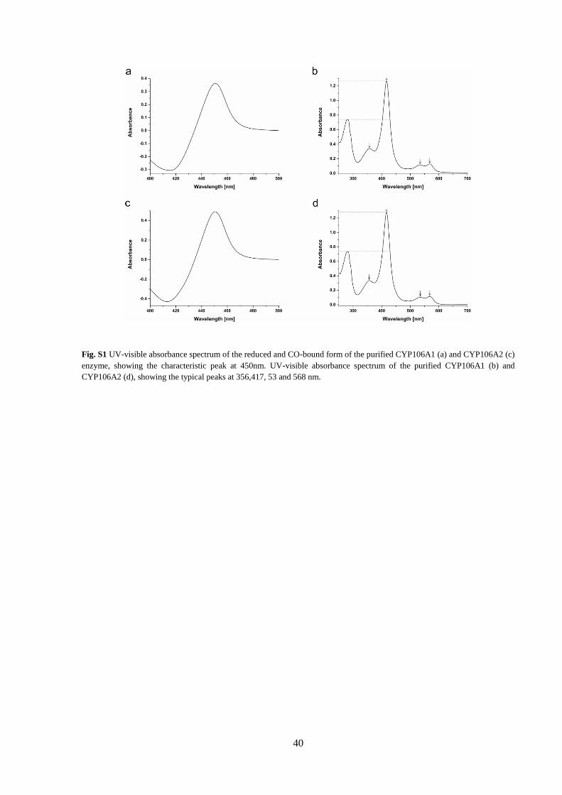
40
Fig. S1 UV-visible absorbance spectrum of the reduced and CO-bound form of the purified CYP106A1 (a) and CYP106A2 (c)
enzyme, showing the characteristic peak at 450nm. UV-visible absorbance spectrum of the purified CYP106A1 (b) and
CYP106A2 (d), showing the typical peaks at 356,417, 53 and 568 nm.

41
Fig. S2 Type I spectral shifts induced by the binding of corticosterone, cortisol and cortisone binding to CYP106A1 (a, c, e) and
to CYP106A2 (b, d, f). Increasing amounts of substrates dissolved in DMSO were titrated (10 µM in 50 mM potassium phosphate
buffer pH 7.4). The peak-to-through absorbance differences were plotted against the increasing concentrations of the substrate.
The mean values of three independent measurements were fitted by hyperbolic regression.

42

43
*The peaks labelled with an asterisk represent compounds which were not observed during the in vitro conversions, thus not treated as specific CYP106A enzyme
products.
Fig. S3 HPLC chromatograms of the in vivo conversion of androstenedione (a, b), corticosterone (c, d), 11-deoxycorticosterone
(e, f), 11-deoxycortisol (g, h), cortisol (I, j) and cortisone (k, l) by Bacillus megaterium MS941 strains, containing the plasmid
pSMF2.1B (CYP106A1) or pSMF2.1C (CYP106A2). The reactions were performed with resting cells in 50 mM potassium
phosphate buffer pH 7.4 at 30 °C for 24 hours with 200 µM substrate. Samples were collected at the indicated time points.

44
2.2. Kiss et al. (2015)
Identification of new substrates for the CYP106A1-mediated 11-oxidation and investigation of
the reaction mechanism
Flora Marta Kiss, Yogan Khatri, Josef Zapp, Rita Bernhardt
FEBS letters 2015 doi:10.1016/j.febslet.2015.07.011
Reprinted with the permission of Elsevier.

45
Identification of new substrates for the CYP106A1-mediated
11-oxidation and investigation of the reaction mechanism
Flora Marta Kiss1, Yogan Khatri1, Josef Zapp2, Rita Bernhardt1*
Abstract
CYP106A1 from Bacillus megaterium DSM319 was recently shown to catalyze steroid and terpene
hydroxylations. Besides producing hydroxylated steroid metabolites at positions 6β, 7β, 9α and 15β, the
enzyme displayed previously unknown 11-oxidase activity towards 11β-hydroxysteroids. Novel
examples for 11-oxidation were identified and confirmed by 1H- and 13C-NMR for prednisolone,
dexamethasone and 11β-hydroxyandrostenedione. However, only 11β-hydroxyandrostenedione formed
a single 11-keto product. The latter reaction was chosen to investigate the kinetic solvent isotope effect
on the steady-state turnover of the CYP106A1-mediated 11-oxidation. Our results reveal a large inverse
kinetic isotope effect (~0.44) suggesting the involvement of the ferric peroxoanion as a reactive
intermediate.
Keywords: CYP106A1, 11β-hydroxysteroid, 11-oxidation, kinetic solvent isotope effect, cytochrome
P450, ferric peroxoanion
Highlights:
Novel substrates identified for the CYP106A1-mediated 11-oxidation
Selective 11-oxidation of 11β-hydroxyandrostenedione observed
11-oxidation mechanism investigated via kinetic solvent isotope effect
Inverse kinetic solvent isotope effect, suggesting the ferric peroxoanion as reactive intermediate
Introduction
The CYP106A1 enzyme from Bacillus
megaterium DSM319 was recently isolated and
characterized by our group (Brill et al. 2014). Its
closest homologue, CYP106A2 from
B. megaterium ATCC13368 has been extensively
investigated and recognized as a highly potent
catalyst for both steroid and terpene
hydroxylations (Bernhardt 2006; Bleif et al. 2011;
Bleif et al. 2012; Schmitz et al. 2014;
Zehentgruber et al. 2010). Based on the 63 %
amino acid sequence identity between the
subfamily members, the functional
1Institute of Biochemistry, Saarland University, D-66123
Saarbruecken, Germany
2Institute of Pharmaceutical Biology, Saarland University,
D-66123 Saarbruecken, Germany
*Address correspondence to: Professor Dr. Rita Bernhardt
Email: [email protected]
subfamily members, the functional
characterization of CYP106A1 concerning tri-
terpene hydroxylation (Brill et al. 2014), followed
by the investigation of steroid hormone
transformation (Kiss et al. 2015; Lee et al. 2014)
was performed.
We have demonstrated the 6β, 7β, 9α and 15β
steroid-hydroxylase activity of CYP106A1, in
addition to the 11-oxidation of corticosterone and
cortisol (Kiss et al. 2015). The latter reaction was
also observed for the human CYP11B1,
converting cortisol to cortisone (Suhara et al.
1986); however, the reaction mechanism has never
been investigated. Since such 11-oxo steroid
formation is uncommon in P450 catalysis, we
were interested in studying the underlying
mechanism by evaluating the kinetic solvent
isotope effect (KSIE) in the CYP106A1-mediated
11-oxidation. The effect of hydrogen substitution
by deuterium on the catalytic activity provides

46
valuable information on proton involvement in the
P450 catalytic steps.
The catalytic cycle of a P450 (Fig. 1), based on
CYP101 (Denisov et al. 2005; Makris et al. 2002),
starts with the ferric resting state of the enzyme (1)
(Raag et al. 1991; Raag and Poulos 1989). Upon
substrate binding, the six-coordinated low-spin
complex is shifted to the five-coordinated high-
spin state (2) and an electron transfer from an
associated redox partner takes place (3) (Sligar
1976; Tsai et al. 1970). On subsequent binding of
a molecular oxygen, the oxy-ferrous state (4) is
formed (Champion et al. 1978; Macdonald et al.
1999), which is then reduced to a ferric
peroxoanion (5a) leading to the formation of a
hydroperoxo intermediate (5b), compound 0
(Cpd 0), on acceptance of a proton (Harris and
Loew 1996). The availability of a second proton
causes the release of a water molecule and
generates compound I (Cpd I) (6) (Groves 2006;
Rittle and Green 2010). Subsequently, the product
is released (7) regenerating the ferric heme, thus
allowing the next catalytic cycle to begin.
Alternatively, the activated oxygen can be
released through one of the “uncoupling”
pathways (shunts, depicted in dashed grey in
Fig. 1), which can abort the catalytic cycle, being
unproductive regarding substrate oxidation, yet
still consuming pyridine nucleotide,
NADP(H)/NADH.
In general, standard P450-mediated reactions
(e.g. hydroxylations) are considered to proceed
through the classical Cpd I-mediated H-rebound
mechanism, in which the reaction takes place with
the help of two consecutive proton uptakes
(Batabyal et al. 2013; Groves 2006; Liu and Ortiz
de Montellano 2000; Vidakovic et al. 1998).
However, the ferric peroxoanion (5a) can also
intervene in the P450 catalysis, which has recently
been observed in the CYP17A1-mediated lyase
reaction (Gregory et al. 2013).
In this study, we investigated the underlying
reaction mechanism of the CYP106A1-mediated
11-oxidation which has recently been described
using corticosterone and cortisol as substrates
(Kiss et al. 2015). Both steroid conversions
displayed several hydroxy-derivatives in addition
to the 11-keto-product. Therefore, further
11β-hydroxysteroid analogues were screened to
obtain a regio-selective C11-oxidation. The
chosen steroids, prednisolone, dexamethasone and
11β-hydroxyandrostenedione (11-OH-AD), were
previously identified as high-spin-shift-inducing
CYP106A1 substrates, yet, the reactions were not
investigated in detail and the products were not
characterized (Kiss et al. 2015).
Fig. 1 – Cytochrome P450 catalytic cycle. The numbers
(1-7) represent the actual state of the enzyme: 1 – Low-spin
substrate-free state. 2 – High-spin enzyme-substrate complex.
3 - High-spin ferrous state. 4 – Oxy-ferrous state. 5a - Ferric
peroxo intermediate. 5b – Ferric hydroperoxo
intermediate/compound 0. 6 – High-valent iron-oxo
state/compound I. 7 - Product oxidation and release. RH and
ROH illustrate the substrate and the product, respectively.
The three unproductive shunt-pathways are marked with
dashed grey arrows and the reduced oxygen products are
shown as outlets. (The cycle has been adapted from Makris et
al. (2002) and Denisov et al. (2005).)
At first, we performed in vitro transformation
of the substrates, followed by their in vivo turnover
using a CYP106A1-based B. megaterium whole-
cell system to obtain higher product yields
required for the structural elucidation by nuclear
magnetic resonance (NMR). The product
structures confirmed the 11-oxidation of all
substrates; however, only the 11-OH-AD
conversion led to a single 11-keto-product,
whereas prednisolone and dexamethasone
displayed several hydroxy-metabolites as well. As
a result, the regio-selective 11-oxidation of
11-OH-AD was chosen to study the reaction

47
mechanism employing the KSIE, a proven method
to distinguish the involvement of P450 reaction
intermediates Cpd I or the ferric peroxoanion
during P450 catalysis (Aikens and Sligar 1994;
Gregory et al. 2013; Khatri et al. 2014; Purdy et al.
2006; Schowen 1977; Vidakovic et al. 1998;
Xiang et al. 2000).
Materials and methods
Protein expression and purification
The expression and purification of the CYP106A1
enzyme was performed as described elsewhere
(Brill et al. 2014). The truncated bovine
adrenodoxin (Adx4-108) and the bovine
adrenodoxin reductase (AdR) were expressed and
purified as described by Uhlmann et al. (Uhlmann
et al. 1992) and Sagara et al. (Sagara et al. 1993),
respectively.
In vitro conversion and steady state kinetic
turnover
Prednisolone, dexamethasone and 11-OH-AD
were converted with a reconstituted system
containing the CYP106A1 enzyme, AdR and
Adx4-108 (in a ratio of 1:2:20), and a NADPH
regenerating system, as formerly described by
Kiss et al. (Kiss et al. 2015). The reaction was
performed at 30 °C, for 60 min using 200 µM
substrate. The final CYP106A1 concentration was
2 µM in the case of 11-OH-AD and 5 µM for both
prednisolone and dexamethasone.
The steady state kinetic turnover of 11-OH-AD
was performed in protiated and deuterated solvent
systems using 2 µM enzyme and 50 µM final
substrate concentration. To investigate the effect
of radical scavengers on the catalytic rate, the in
vitro conversions were performed with the
addition of ascorbate (20 mM), catalase (20 U) and
superoxide-dismutase (SOD) (3 U), individually
as well as in combination. To determine whether
hydrogen peroxide (H2O2) could be applied to
reconstitute the activity of CYP106A1, H2O2 and
cumene hydroperoxide were used in a final
concentration of 50 µM. The reactions were
stopped after 20 min and extracted twice by
addition of 250 μL ethyl acetate. After evaporating
the organic phases, the dried samples were
dissolved in methanol and subjected to high-
performance liquid chromatography (HPLC)
analysis. An ec MN Nucleodor C18 (3 µM,
4.0x125 mm) column was used, with a mobile
phase consisting of methanol, tetrahydrofuran and
water in an 8:19:72 ratio. The steroids were eluted
in an isocratic mode with a flow rate of
0.5 mL/min and detected at 240 nm. The
conversions were analyzed using the peak areas
(area %) of the HPLC chromatograms with the
help of the ChromPass/Galaxie Chromatography
Data System (Jasco, Gross-Umstadt,Germany).
In vivo conversion and product purification
The CYP106A1-based B. megaterium whole-cell
system (harboring the plasmid pSMF2.1B) was
used for biotransformation as described earlier
(Brill et al. 2014; Kiss et al. 2015). The substrate
was added in a final concentration of 400 µM to
the B. megaterium resting cells in 100 mM
potassium phosphate buffer (pH 7.4). The reaction
was stopped after 24 h and extracted twice with
ethyl acetate. The samples were dried and
resuspended in the HPLC mobile phase. The
product purification was performed with a
preparative HPLC column (ec MN Nucleodur C18
VP (5 µM, 8x250 mm) using gradient elution
(solvent A: 10 % acetonitrile (ACN), solvent B:
100 % ACN) with a flow rate of 2 mL/min.
NMR characterization of the metabolites
The NMR spectra were recorded in CD3OD or in
CDCl3 with a Bruker DRX 500 or a Bruker
Avance 500 NMR spectrometer at 300 K. The
chemical shifts were relative to CH3OD at δ 3.30
(1H NMR) and CD3OD at δ 49.00 (13C NMR) or
to CHCl3 at δ 7.26 (1H NMR) and CDCl3 at δ 77.00
(13C NMR) respectively, using the standard δ
notation in parts per million. The 1D NMR (1H and 13C NMR, DEPT135) and the 2D NMR spectra
(gs-HH-COSY, gs-NOESY, gs-HSQCED, and gs-
HMBC) were recorded using the BRUKER pulse
program library. All assignments were based on
extensive NMR spectral evidence. For the detailed
NMR data of the identified products see
Supplementary Material.

48
Results and discussion
Substrate conversions, product purification
and characterization by NMR
Since the previously identified substrates for the
CYP106A1 11-oxidation (corticosterone and
cortisol) gave rise to multiple product formation
(Kiss et al. 2015), other 11β-hydroxysteroid
analogues were investigated to identify a selective
11-keto product formation. The in vitro
conversion of the pharmaceutically relevant
glucocorticoids, prednisolone and
dexamethasone, also resulted in several products
(Table 1, Fig. 2B and 3B) in contrast to the
transformation of 11-OH-AD, where only a single
product was formed with a yield of 50 %
(Table 1, Fig. 4B).
Even though single product formation was only
observed with 11-OH-AD, we were interested in
characterizing all 11β-hydroxysteroid metabolites
by NMR, to identify novel 11-keto products and
other glucocorticoid derivatives of potential
pharmaceutical value. For the isolation and
purification of the reaction products, the steroid
bioconversions were performed via a CYP106A1-
based B. megaterium whole-cell system.
The in vivo turnover of prednisolone led to
~51 % conversion resulting in a main product, P1
(35 %) with identical retention time (~3.6 min) to
the one observed in vitro. Besides the major
product, multiple side-products (<5%) were also
detected, out of which P2 (~4.8 min) and P3
(~8.3 min) eluted at the same time as the
respective in vitro metabolites (Fig. 2 B, C).
Although both P1 and P2 were well resolved
during the purification, the NMR analysis of their
purified fractions showed a mixture of several
steroids, containing 15β-hydroxyprednisolone as a
major compound. Likewise, product P3, which
was identified as a 2:3 mixture of
15β-hydroxyprednisone and 1,2-dihydro-15β-
hydroxyprednisone displaying 11-oxidation
catalyzed by CYP106A1 (Scheme 1).
Fig. 2 - HPLC chromatograms of the CYP106A1
catalyzed prednisolone conversion. (A) Negative control,
containing prednisolone only. (B) In vitro prednisolone
conversion, using bovine Adx4-108 (100 µM), bovine AdR (10
µM) and CYP106A1 (5 µM). (C) In vivo conversion of
prednisolone using the CYP106A1-overexpressing
B. megaterium MS941 strain.
The unusual 1(2)-double bond hydrogenation
has not been observed in our previous studies
(Kiss et al. 2015), hence, it was proposed to be the
result of an uncharacterized enzyme present in the
B. megaterium MS941 strain. It has been reported
that 3-ketosteroid-dehydrogenases present in
Nocardia, Mycobacterium and Actinobacter sp.
can be responsible for both 1(2)-hydrogenase and
reductase reactions (Arinbasarova et al. 1996;
Goren et al. 1983; van der Geize et al. 2000).
However, these enzymes have not been described
so far for B. megaterium. Therefore, the origin of
Table 1 - In vitro conversion results showing relative conversion and product retention times, when applicable.
Substrate (RT/min) RC (%) Product RT (min)
Remark P1 P2 P3 P4
Prednisolone (10.5) 33 3.6 4.8 8.4 - Fig. 2B
Dexamethasone (20.4) 40 5.1 6.9 8.3 15.8 Fig. 3B
11-OH-AD (13.0) 50 10.5 - - - Fig. 4B
*RT, Retention time; RC, Relative conversion

49
the 1(2)-hydrogenase reaction remains unclear.
The above described prednisolone metabolites
were previously unidentified, and therefore, no
biotechnological applications are known to date.
Nonetheless, the 15β-hydroxyl group attached to
both prednisolone and prednisone structures
provides a new reaction site for further
functionalization, contributing to the development
of novel glucocorticoids possessing anti-
inflammatory/immunomodulatory activity with a
potentially reduced risk of side effects.
Using the CYP106A1-based whole-cell
system, nearly 50 % of dexamethasone was
converted in 24 h, yielding four products, P1 to P4,
which were all successfully isolated and
characterized. Each of the identified metabolites
showed identical retention times to the ones
observed in vitro, eluting at 5.1, 6.9, 8.3 and
15.8 min, respectively (Fig. 3 B, C). The major
product, P1 (37 %), and P2 (6 %) were identified
as 15β-hydroxy- and 6β-hydroxy-derivatives of
dexamethasone, respectively. The latter one also
known as the major human metabolite of
dexamethasone produced by CYP3A4 (Gentile et
al. 1996; Minagawa et al. 1986; Tomlinson et al.
1997). Besides the hydroxylated derivatives, two
minor 11-oxidized compounds were observed and
characterized as 11-ketodexamethasone (P4, 2 %)
and 15β-hydroxy-11-ketodexamethasone (P3,
3 %) (Scheme 2). The 11-keto compound was
previously identified as a specific glucocorticoid
receptor agonist, explaining the glucocorticoid
effect of dexamethasone in tissues expressing the
11β-hydroxysteroid dehydrogenase type 2
enzyme, responsible for 11-oxidation in
mammalian tissues (Rebuffat et al. 2004).
Fig. 3 - HPLC chromatograms of the CYP106A1-
catalyzed dexamethasone conversion. (A) Negative control,
containing dexamethasone only. (B) In vitro dexamethasone
conversion, using bovine Adx4-108 (100 µM), bovine AdR (10
µM) and CYP106A1 (5 µM). (C) In vivo conversion of
dexamethasone using the CYP106A1-overexpressing
B. megaterium MS941 strain.
The in vivo turnover of 11-OH-AD resulted in
~90 % conversion within 24 h and a single main
product along with traces of minor side products,
which were not observed in the in vitro system
(Fig. 4 C). The major product, P1 (~10.5 min),
showed identical retention time to that of the in
vitro reaction (Fig. 4 B). The NMR
characterization identified P1 as the 11-keto
derivative, adrenosterone, supporting the
previously observed 11-oxidase activity of
CYP106A1 during the transformation of
corticosterone and cortisol (Kiss et al. 2015)
(Scheme 3). Adrenosterone is a weak androgen
hormone found only in trace amounts in
mammals, but in higher quantities in fish, where it
Scheme 1 – Oxidative transformation of prednisolone catalyzed by CYP106A1.

50
acts as a precursor of 11-ketotestosterone, the
endogenous androgenic sex hormone (Blasco et al.
2009; Borg 1994). It is sold as a dietary
supplement, reducing body fat and increasing
muscle mass and was also proposed to be a
selective inhibitor of 11β-hydroxysteroid
dehydrogenase type I enzyme (Brooker et al.
2009).
Fig. 4 - HPLC chromatograms of the CYP106A1
catalyzed 11β-hydroxyandrostenedione conversion. (A)
Negative control, containing 11-OH-AD only. (B) In vitro 11-
OH-AD conversion, using bovine Adx4-108 (40 µM), bovine
AdR (4 µM) and CYP106A1 (2 µM). (C) In vivo conversion
of 11-OH-AD using the CYP106A1-overexpressing
B. megaterium MS941 strain.
Steady state kinetic turnover in protiated and
deuterated solvent systems
The CYP106A1-mediated conversion of
prednisolone and dexamethasone resulted in
multiple products, in contrast to 11-OH-AD,
where only the 11-keto product formation was
detected. Thus, the latter reaction was chosen as a
suitable model for the investigation of KSIE in
CYP106A1-mediated 11-oxidation. The steady
state turnover of 11-OH-AD by CYP106A1 was
investigated with a time-dependent reaction in the
presence of saturating substrate concentration. To
study the catalytic activity, an incubation time of
20 min was chosen, representing the linear phase
of the reaction (Fig. 5 A). The turnover was
carried out in a protiated buffer system, in which
an individual rate of adrenosterone formation of
0.36 ± 0.04 min-1 was observed. Interestingly,
upon H2O substitution for D2O, the conversion
demonstrated a significantly increased catalytic
rate of 0.81 ± 0.03 min-1, leading to a 2.25-times
faster product formation upon H/D exchange.
These rates correspond to an inverse KSIE (kH/kD)
of 0.44 (Fig. 5 B) suggesting that the 11-oxidation
reaction is mediated through the unprotonated
ferric peroxo intermediate, in contrast to
traditional hydroxylation reactions, which are
believed to proceed through the classical Groves
Scheme 2 – Oxidative transformation of dexamethasone catalyzed by CYP106A1.
Scheme 3 – 11-oxidation of 11β-hydroxyandrostenedione catalyzed by CYP106A1.

51
rebound mechanism with Cpd I as the reactive
intermediate (Batabyal et al. 2013; Groves 2006;
Liu and Ortiz de Montellano 2000; Vidakovic et
al. 1998). The detected large inverse KSIE could
be explained by the fact that inhibiting protonation
of the peroxo-ferric species in deuterated solvents
facilitates the productive oxidation rather than the
uncoupling via proton-dependent unproductive
pathways (peroxide shunt, oxidase shunt).
Forming the nucleophilic peroxoanion
intermediate (Fig. 1, (5a)) does not involve any
protonation event, thus the catalytic activity would
be expected to be the same in both H2O and D2O
buffer systems. However, the slower rate of Cpd I
formation in a deuterated solvent system in
combination with inhibited unproductive
pathways could result in the increase of the
peroxoanion intermediate, along with the product
formation (Gregory et al. 2013). The latter was
described for tyrosine hydroxylase (Frantom and
Fitzpatrick 2003) and putidamonooxin (Twilfer et
al. 2000), where, as a result of the slower rate of
uncoupling reactions compared with the
productive pathway in D2O, a large inverse KSIE
was observed. Concerning P450 enzymes, the
involvement of the ferric peroxoanion has only
been observed for the CYP17-catalyzed C17-C20
lyase reaction (Gregory et al. 2013), its
involvement in an oxidase reaction has not been
demonstrated so far.
To exclude the role of unproductive pathways
and the possible H2O2-mediated substrate
oxidation (reverse peroxide shunt) and to support
the participation of the ferric peroxo species in the
CYP106A1-catalyzed 11-oxidation, the in vitro
conversions were performed with the addition of
radical scavengers and in the presence of H2O2 or
cumene hydroperoxide. As scavenging agents
ascorbate (neutralizing the superoxide radical,
singlet oxygen and hydroxyl radicals), catalase
(decomposing hydrogen peroxide to water and
oxygen) and superoxide dismutase (SOD)
(scavenger of superoxide anion) were applied,
individually or in combination. No significant
decrease in the catalytic rate was observed in
comparison to the positive control, the 11-OH-AD
conversion in protiated environment (Fig. 5 C).
Using H2O2 or cumene hydroperoxide (CHP), no
product formation was detected. These results
suggest that the previously observed inverse KSIE
is not the result of an inhibited uncoupling reaction
but of the catalysis via a proton-independent
intermediate. Since steroid 11-dehydrogenation by
a P450 had never been investigated before, similar
P450 dependent keto-product formations
described in the literature have been considered
for possible parallels. Interestingly, in contrast to
our observations, the microsomal oxidation of
cyclohex-2-en-1-ol (Bellucci et al. 1996),
the CYP3A11 mediated hydroxy-Δ8-
tetrahydrocannabinol transformation (Matsunaga
et al. 2001) and the CYP2E1-catalyzed ethanol
oxidation (Wang et al. 2007) were all proposed to
proceed through the gem-diol or the combination
Fig. 5 - Investigation of the catalytic rate of 11-oxidation by CYP106A1. (A) Time dependent conversion of 11-OH-AD
using CYP106A1 (2 µM) and 11-OH-AD (50 µM). (B) Steady-state kinetic solvent isotope effects observed for 11-oxidase
CYP106A1 catalysis. The numbers inside the bars indicate the catalytic activity, while the error bars represent the standard
deviation of 5 independent reactions. (C) The effect of radical scavengers on the catalytic rate of 11-OH-AD oxidation. The
error bars represent the standard deviation of 3 independent measurements.
*SOD, superoxide-dismutase; RSC, radical scavengers in combination; CHP, cumene hydroperoxide

52
of gem-diol and double hydrogen abstraction
pathways involving Cpd I (Fig. 1, [6]) as a reactive
intermediate. However, the observed inverse
KSIE and the absent role of proton-dependent
uncoupling pathways in the CYP106A1 catalyzed
11-oxidation are not reconcilable with a proton-
dependent, Cpd I-mediated mechanism.
Therefore, we propose that the 11-oxidation
reaction of 11-OH-AD is mediated by a
nucleophilic attack of the ferric peroxoanion, as
illustrated in Scheme 4. To provide additional
evidence for the involvement of the ferric peroxo
species, the spectroscopic characterization of the
reactive intermediate could be a topic of further
research, as reviewed elsewhere (Krest et al. 2013;
Luthra et al. 2011; Sligar et al. 2005).
In conclusion, we present prednisolone,
dexamethasone and 11-OH-AD as novel
substrates for the 11-oxidation by CYP106A1.
The enzyme performed selective 11-oxidation on
11-OH-AD, resulting in a single 11-keto product,
while the conversion of prednisolone and
dexamethasone displayed 6β- and
15β-hydroxylated derivatives, as well as the
11-keto steroids. Since the hydroxy metabolites
can be developed into functionalized drugs with
improved properties (e.g.: higher specificity,
improved pharmacokinetics) (Bernhardt and
Urlacher 2014), the production of such novel
derivatives using CYP106A1 is considered a
promising approach for the pharmaceutical
industry. In addition, due to the selective
conversion of 11-OH-AD, the 11-oxidation
mechanism was also investigated. We observed an
inverse KSIE (~0.44), suggesting that the
11-oxidation takes place without the involvement
of proton uptake, proposing for the first time the
ferric peroxoanion as a reactive intermediate in a
P450-mediated oxidation reaction.
Author contributions
FMK carried out the experiments, analyzed and interpreted the
data and drafted the manuscript. YK and RB designed the project,
analyzed and interpreted the results, and assisted in drafting the
manuscript. JZ performed the NMR measurements and the
structure determination of the converted steroids. All authors read
and approved the final manuscript.
Acknowledgements
This work was generously supported by the People Programme
(Marie Curie Actions) of the European Union's 7th Framework
Programme (FP7/2007-2013), P4FIFTY - FP7 PEOPLE ITN
2011-289217 and by a grant from the Federal Ministry of
Education and Research (BMBF - 031A166A). The authors thank
Birgit Heider-Lips for the expression and purification of AdR and
Adx4-108 and Nicolas Souza Carmona for the language revision.
References Aikens J, Sligar SG (1994) Kinetic Solvent Isotope Effects
During Oxygen Activation by Cytochrome P-450Cam.
Journal of the American Chemical Society 116(3):1143-
1144
Arinbasarova AY, Karpov AV, Fokina VV, Medentsev AG,
Koshcheyenko KA (1996) Kinetic Characteristics of 1-en-
Dehydrogenation of 6-Alpha-Methylhydrocortisone by
Cells of Arthrobacter-Globiformis-193. Enzyme Microb
Technol 19(7):501-506
Batabyal D, Li H, Poulos TL (2013) Synergistic Effects of
Mutations in Cytochrome P450cam Designed To Mimic
CYP101D1. Biochemistry 52(32):5396-5402
Bellucci G, Chiappe C, Pucci L, Gervasi PG (1996) The
Mechanism of Oxidation of Allylic Alcohols to α,β-
Unsaturated Ketones by Cytochrome P450. Chemical
Research in Toxicology 9(5):871-874
Bernhardt R (2006) Cytochromes P450 as versatile biocatalysts.
J Biotechnol 124(1):128-145
Bernhardt R, Urlacher VB (2014) Cytochromes P450 as
promising catalysts for biotechnological application:
Scheme 4 - Proposed model for the 11-oxidation of 11-OH-AD by CYP106A1. The 11-oxidation proceeds throughh a
nucleophilic attack at the C11 hydroxyl group of 11-OH-AD (A) by the ferric peroxo species. A subsequent hydroxylation step
leads to the ‘gem-diol’ (B). The ‘gem-diol’ undergoes a dehydration reaction, abstracting the hydrogen and forming the 11-keto
product, adrenosterone (C).

53
chances and limitations. Applied microbiology and
biotechnology 98(14):6185-6203
Blasco M, Carriquiriborde P, Marino D, Ronco AE, Somoza GM
(2009) A quantitative HPLC–MS method for the
simultaneous determination of testosterone, 11-
ketotestosterone and 11-β hydroxyandrostenedione in fish
serum. Journal of Chromatography B 877(14–15):1509-
1515
Bleif S, Hannemann F, Lisurek M, Kries J, Zapp J, Dietzen M,
Antes I, Bernhardt R (2011) Identification of CYP106A2
as a regioselective allylic bacterial diterpene hydroxylase.
Chembiochem : a European journal of chemical biology
12(4):576-582
Bleif S, Hannemann F, Zapp J, Hartmann D, Jauch J, Bernhardt
R (2012) A new Bacillus megaterium whole-cell catalyst
for the hydroxylation of the pentacyclic triterpene 11-keto-
beta-boswellic acid (KBA) based on a recombinant
cytochrome P450 system. Applied microbiology and
biotechnology 93(3):1135-1146
Borg B (1994) Androgens in teleost fishes. Comparative
Biochemistry and Physiology Part C: Pharmacology,
Toxicology and Endocrinology 109(3):219-245
Brill E, Hannemann F, Zapp J, Bruning G, Jauch J, Bernhardt R
(2014) A new cytochrome P450 system from Bacillus
megaterium DSM319 for the hydroxylation of 11-keto-
beta-boswellic acid (KBA). Applied microbiology and
biotechnology 98:1701-1717
Brooker L, Parr MK, Cawley A, Flenker U, Howe C, Kazlauskas
R, Schänzer W, George A (2009) Development of criteria
for the detection of adrenosterone administration by gas
chromatography-mass spectrometry and gas
chromatography-combustion-isotope ratio mass
spectrometry for doping control. Drug Testing and
Analysis 1(11-12):587-595
Champion PM, Gunsalus IC, Wagner GC (1978) Resonance
Raman investigations of cytochrome P450CAM from
Pseudomonas putida. Journal of the American Chemical
Society 100(12):3743-3751
Denisov IG, Makris TM, Sligar SG, Schlichting I (2005)
Structure and Chemistry of Cytochrome P450. Chem Rev
105(6):2253-2278
Frantom PA, Fitzpatrick PF (2003) Uncoupled Forms of Tyrosine
Hydroxylase Unmask Kinetic Isotope Effects on Chemical
Steps. Journal of the American Chemical Society
125(52):16190-16191
Gentile D, Tomlinson E, Maggs J, Park B, Back D (1996)
Dexamethasone metabolism by human liver in vitro.
Metabolite identification and inhibition of 6-
hydroxylation. J Pharmacol Exp Ther 277(1):105-12
Goren T, Harnik M, Rimon S, Aharonowitz Y (1983) 1-Ene-
steroid reductase of Mycobacterium sp. NRRL B-3805. J
Steroid Biochem 19((6)):1789-97.
Gregory MC, Denisov IG, Grinkova YV, Khatri Y, Sligar SG
(2013) Kinetic Solvent Isotope Effect in Human P450
CYP17A1-Mediated Androgen Formation: Evidence for a
Reactive Peroxoanion Intermediate. Journal of the
American Chemical Society 135(44):16245-16247
Groves JT (2006) High-valent iron in chemical and biological
oxidations. Journal of Inorganic Biochemistry 100(4):434-
447
Harris DL, Loew GH (1996) Investigation of the proton-assisted
pathway to formation of the catalytically active, ferryl
species of P450s by molecular dynamics studies of
P450eryF. J Am Chem Soc 118(27):6377-6387
Khatri Y, Luthra A, Duggal R, Sligar SG (2014) Kinetic solvent
isotope effect in steady-state turnover by CYP19A1
suggests involvement of Compound 1 for both
hydroxylation and aromatization steps. FEBS Letters
588(17):3117-3122
Kiss F, Schmitz D, Zapp J, Dier TF, Volmer D, Bernhardt R
(2015) Comparison of CYP106A1 and CYP106A2 from
Bacillus megaterium – identification of a novel 11-oxidase
activity. Applied microbiology and biotechnology:1-20
Krest CM, Onderko EL, Yosca TH, Calixto JC, Karp RF, Livada
J, Rittle J, Green MT (2013) Reactive Intermediates in
Cytochrome P450 Catalysis. Journal of Biological
Chemistry 288(24):17074-17081
Lee G-Y, Kim D-H, Kim D, Ahn T, Yun C-H (2014) Functional
characterization of steroid hydroxylase CYP106A1
derived from Bacillus megaterium. Arch Pharm Res:1-10
Liu Y, Ortiz de Montellano PR (2000) Reaction Intermediates
and Single Turnover Rate Constants for the Oxidation of
Heme by Human Heme Oxygenase-1. Journal of
Biological Chemistry 275(8):5297-5307
Luthra A, Denisov IG, Sligar SG (2011) Spectroscopic features
of cytochrome P450 reaction intermediates. Archives of
Biochemistry and Biophysics 507(1):26-35
Macdonald IDG, Sligar SG, Christian JF, Unno M, Champion
PM (1999) Identification of the Fe−O−O Bending Mode in
Oxycytochrome P450cam by Resonance Raman
Spectroscopy. Journal of the American Chemical Society
121(2):376-380
Makris TM, Davydov R, Denisov IG, Hoffman BM, Sligar SG
(2002) Mechanistic Enzymology of Oxygen Activation by
the Cytochromes P450 Drug Metabolism Reviews
34(4):691-708
Matsunaga T, Tanaka H, Higuchi S, Shibayama K, Kishi N,
Watanabe K, Yamamoto I (2001) Oxidation Mechanism of
7-Hydroxy-Δ8-tetrahydrocannabinol and 8-Hydroxy-Δ9-
tetrahydrocannabinol to the Corresponding Ketones by
CYP3A11. Drug Metabolism and Disposition
29(11):1485-1491
Minagawa K, Kasuya Y, Baba S, Knapp G, Skelly J (1986)
Identification and quantification of 6 beta-
hydroxydexamethasone as a major urinary metabolite of
dexamethasone in man. Steroids 47(2-3):175-88
Purdy MM, Koo LS, Ortiz de Montellano PR, Klinman JP (2006)
Mechanism of O2 Activation by Cytochrome P450cam
Studied by Isotope Effects and Transient State Kinetics.
Biochemistry 45(51):15793-15806
Raag R, Martinis SA, Sligar SG, Poulos TL (1991) Crystal
structure of the cytochrome P-450CAM active site mutant
Thr252Ala. Biochemistry 30(48):11420-11429
Raag R, Poulos TL (1989) The structural basis for substrate-
induced changes in redox potential and spin equilibrium in
cytochrome P-450CAM. Biochemistry 28(2):917-922
Rebuffat AG, Tam S, Nawrocki AR, Baker ME, Frey BM, Frey
FJ, Odermatt A (2004) The 11-ketosteroid 11-
ketodexamethasone is a glucocorticoid receptor agonist.
Molecular and cellular endocrinology 214(1–2):27-37
Rittle J, Green MT (2010) Cytochrome P450 Compound I:
Capture, Characterization, and C-H Bond Activation
Kinetics. Science 330(6006):933-937
Sagara Y, Wada A, Takata Y, Waterman M, Sekimizu K,
Horiuchi T (1993) Direct expression of adrenodoxin
reductase in Escherichia coli and the functional
characterization. Biol Pharm Bull 16:627-630
Schmitz D, Zapp J, Bernhardt R (2014) Steroid conversion with
CYP106A2 - production of pharmaceutically interesting
DHEA metabolites. Microbial Cell Factories 13:81
Schowen R (1977) Solvent isotope effects on enzymic reactions.
In: Cleland WW, O’Leary MH, Northrop DB (eds) Isotope
Effects on Enzyme-Catalyzed Reactions. University Park
Press, Baltimore, pp 64-99
Sligar SG (1976) Coupling of spin, substrate, and redox
equilibriums in cytochrome P450. Biochemistry
15(24):5399-5406
Sligar SG, Makris TM, Denisov IG (2005) Thirty years of
microbial P450 monooxygenase research: Peroxo-heme
intermediates - The central bus station in heme oxygenase
catalysis. Biochemical and biophysical research
communications 338(1):346-354

54
Suhara K, Takeda K, Katagiri M (1986) P-45011β-dependent
conversion of cortisol to cortisone, and 19-
hydroxyandrostenedione to 19-oxoandrostenedione.
Biochemical and biophysical research communications
136(1):369-375
Tomlinson ES, Lewis DFV, Maggs JL, Kroemer HK, Park BK,
Back DJ (1997) In vitro metabolism of dexamethasone
(DEX) in human liver and kidney: The involvement of
CYP3a4 and CYP17 (17,20 LYASE) and molecular
modelling studies. Biochemical Pharmacology 54(5):605-
611
Tsai R, Yu CA, Gunsalus IC, Peisach J, Blumberg W, Orme-
Johnson WH, Beinert H (1970) Spin-state changes in
cytochrome P-450(cam) on binding of specific substrates.
Proceedings of the National Academy of Sciences of the
United States of America 66(4):1157-1163
Twilfer H, Sandfort G, Bernhardt F-H (2000) Substrate and
solvent isotope effects on the fate of the active oxygen
species in substrate-modulated reactions of
putidamonooxin. European Journal of Biochemistry
267(19):5926-5934
Uhlmann H, Beckert V, Schwarz D, Bernhardt R (1992)
Expression of bovine adrenodoxin in E. coli and site-
directed mutagenesis of/2 Fe-2S/cluster ligands.
Biochemical and biophysical research communications
188:1131-1138
van der Geize R, Hessels GI, van Gerwen R, Vrijbloed JW, van
der Meijden P, Dijkhuizen L (2000) Targeted Disruption
of the kstD Gene Encoding a 3-Ketosteroid Δ1-
Dehydrogenase Isoenzyme ofRhodococcus erythropolis
Strain SQ1. Applied and Environmental Microbiology
66(5):2029-2036
Vidakovic M, Sligar SG, Li H, Poulos TL (1998) Understanding
the Role of the Essential Asp251 in Cytochrome P450cam
Using Site-Directed Mutagenesis, Crystallography, and
Kinetic Solvent Isotope Effect. Biochemistry 37(26):9211-
9219
Wang Y, Yang C, Wang H, Han K, Shaik S (2007) A New
Mechanism for Ethanol Oxidation Mediated by
Cytochrome P450 2E1: Bulk Polarity of the Active Site
Makes a Difference. Chembiochem : a European journal of
chemical biology 8(3):277-281
Xiang H, Tschirret-Guth RA, Ortiz de Montellano PR (2000) An
A245T Mutation Conveys on Cytochrome P450eryF the
Ability to Oxidize Alternative Substrates. Journal of
Biological Chemistry 275(46):35999-36006
Zehentgruber D, Hannemann F, Bleif S, Bernhardt R, Lütz S
(2010) Towards preparative scale steroid hydroxylation
with cytochrome P450 monooxygenase CYP106A2.
Chembiochem : a European journal of chemical biology
11:713-721

55
FEBS letters
Supplementary material
Identification of new substrates for the CYP106A1-mediated 11-oxidation
and investigation of the reaction mechanism
Flora Marta Kiss1, Yogan Khatri1, Josef Zapp2, Rita Bernhardt1*
1Institute of Biochemistry, Saarland University, D-66123 Saarbruecken, Germany
2Institute of Pharmaceutical Biology, Saarland University, D-66123 Saarbruecken, German
*Address correspondence to: Professor Dr. Rita Bernhardt
Institute of Biochemistry, Saarland University, Campus B 2.2, D-66123 Saarbruecken, Germany
Tel: +49 681 302 305
Fax: +49 681 302 4739
Email: [email protected]

56
NMR data of the identified products of:
Prednisolone:
15β-Hydroxyprednisolone
Its NMR data matched perfectly with those of an
authentic sample (Schmitz et al., in preparation). 1H
NMR (CD3OD, 500 MHz): δ 1.06 (m, H-9), 1.11 (s,
3xH-18), 1.16 (m, H-7a), 1.52 (s, 3xH-19), 1.57 (dd,
13.8 and 3.3 Hz, H-12a), 1.61 (dd, 11.2 and 6.0 Hz,
H-14), 1.98 (dd, 15.5 and 7.5 Hz, H-16a), 1.99 (m,
H-12b), 2.39 (m, H-6a), 2.44 (m, H-7b), 2.52 (m, H-
8), 2.68 (tdd, 13.5, 5.0 and 2.0 Hz, H-6b), 2.73 (dd,
15.5 and 2.3 Hz, H-16b), 4.24 (d, 19.2 Hz, H-21a),
4.31 (ddd, 7.5, 6.0 and 2.3 Hz, H-15), 4.39 (q, 3.3
Hz, H-11), 4.62 (d, 19.2 Hz, H-21b), 6.01 (t, 2.0 Hz,
H-4), 6.22 (dd, 10.0 and 2.0 Hz, H-2), 7.48 (d, 10.0
Hz, H-1).
15β-Hydroxyprednisone
The NMR spectra of this prednisolone product
revealed a carbonyl, instead of an alcohol function at
C-11 (δC 208.01). A further hydroxyl group at
position 15β could be deduced from the results of the
2D NMR measurements. 1H NMR (CDCl3, 500
MHz): δ 0.97 (s, 3xH-18), 1.35 (m, H-7a), 1.48 (s,
3xH-19), 1.74 (m, H-8), 2.04 (m, H-9), 2.09 (d, 11.7
Hz, H-12a), 2.29 (dd, 11.6 and 6.0 Hz, H-14), 2.35
(dd, 15.5 and 7.5 Hz, H-16a), 2.39 (m, H-7b), 2.40
(m, H-6a), 2.58 (tdd, 13.5, 5.0 and 1.7 Hz, H-6b),
2.75 (dd, 15.5 and 2.3 Hz, H-16b), 2.80 (d, 11.7 Hz,
H-12b), 4.29 (d, 20.0 Hz, H-21a), 4.56 (ddd, 7.5, 6.0
and 2.3 Hz, H-15), 4.66 (d, 20.0 Hz, H-21b), 6.15 (t,
2.0 Hz, H-4), 6.22 (dd, 12.0 and 2.0 Hz, H-2), 7.68
(d, 10.2 Hz, H-1). 13C NMR (CDCl3, 125 MHz): δ
18.40 (CH3, C-18), 18.83 (CH3, C-19), 24.73 (CH,
C-8), 32.15 (CH2, C-6), 32.80 (CH2, C-7), 42.41 (C,
C-10), 47.07 (CH, C-16), 50.05 (CH2, C-12), 51.35
(C, C-13), 53.33 (CH, C-14), 60.90 (CH, C-9), 67.44
(CH2, C-21), 69.10 (CH, C-15), 87.36 (C, C-17),
124.74 (CH, C-4), 127.75 (CH, C-2), 154.98 (CH, C-
1), 166.19 (C, C-5), 186.29 (C, C-3), 208.01 (C, C-
11), 210.94 (C, C-20).
1,2-Dihydro-15β-hydroxyprednisone
Its 1H NMR spectrum showed some remarkable
differences to that of the substrate, prednisolone. The
resonances for the double bond between C-1 and C-
2, as well as for the hydroxyl group at C-11 were
absent. Signals of four additional methylene protons
appeared in the spectrum and the doublet resonances
of H-12a and H-12b showed an α-carbonyl induced
downfield shift. These observations led to the
structure of 1,2-dihydro-15β-hydroxyprednisone. 1H
NMR (CD3OD, 500 MHz): δ 0.84 (s, 3xH-18), 1.34
(m, H-7a), 1.47 (s, 3xH-19), 1.70 (m, H-1a), 1.99 (d,
12.0 Hz, H-12a), 2.15 (m, H-9), 2.21 (dd, 15.5 and
7.5 Hz, H-16a), 2.24 (m, H-2a), 2.31 (m, H-7b), 2.33
(m, 2H, H-8 and H-14), 2.52 (m, H-2b), 2.55 (m, 2H,
H-6a and H-6b), 2.72 (m, H-1b), 2.75 (dd, 15.5 and
2.3 Hz, H-16b), 2.96 (d, 12.0 Hz, H-12b), 4.20 (d,
19.5 Hz, H-21a), 4.42 (ddd, 7.5, 5.7 and 2.2 Hz, H-
15), 4.60 (d, 19.5 Hz, H-21b), 5.72 (br s, H-4).
Dexamethasone:
15β-Hydroxydexamethasone
Compared to the substrate, dexamethasone, the
NMR data of the conversion product showed
resonances for an additional hydroxyl group (δC
78.60; δH 3.67 dd). Its position at C-15 could be
deduced by vicinal couplings of the methine proton
with H-14 (δH 2.08 dd, 12.0 and 6.3 Hz,) and H-16
(δH 3.02 qd, 7.5 and 3.0 Hz). The β orientation of the
hydroxyl was confirmed by the NOESY effects of H-
15 to H-16 and H-14, all in a-position. 1H NMR
(CD3OD, 500 MHz): δ 0.93 (d, 7.5 Hz, 3H, Me-16),
1.17 (s, 3xH-18), 1.43 (dd, 13.7 and 2.5 Hz, H-12a),
1.57 (m, H-7a), 1.60 (s, 3xH-19), 2.08 (dd, 12.0 and
6.3 Hz, H-14), 2.20 (m, H-7b), 2.29 (m, H-12b), 2.40
(tdd, 14.0, 5.0 and 2.0 Hz, H-6a), 2.73 (m, H-6b),
2.80 (dtd, 29.5, 12.0 and 4.5 Hz, H-8), 3.02 (qd, 7.5
and 3.0 Hz, H-16), 3.67 (dd, 6.3 and 3.0 Hz, H-15),
4.22 (d, 19.2 Hz, H-21a), 4.25 (m, H-11), 4.60 (d,
19.2 Hz, H-21b), 6.09 (t, 2.0 Hz, H-4), 6.28 (dd, 10.0
and 2.0 Hz, H-2), 7.41 (d, 10.0 Hz, H-1). 13C NMR
(CD3OD, 125 MHz): δ 13.12 (CH3, Me-16), 20.21
(CH3, C-18), 23.67 (d, JCF=6.0 Hz, CH3, C-19),
27.66 (CH2, C-7), 32.15 (CH2, C-6), 32.27 (d,
JCF=19.3 Hz, CH, C-8), 38.12 (CH2, C-12), 48.92 (C,
C-13), 48.95 (CH, C-14), 49.47 (CH, C-16), 50.41
(d, JCF=22.5 Hz, C, C-10), 67.89 (CH2, C-21), 72.83
(d, JCF=37.5 Hz, CH, C-11), 78.60 (CH, C-15), 92.01
(C, C-17), 103.02 (d, JCF=176.0 Hz, C, C-9), 125.09
(CH, C-4), 129.81 (CH, C-2), 155.96 (CH, C-1),
171.16 (C, C-5), 189.07 (C, C-3), 211.95 (C, C-20).
6β-Hydroxydexamethasone
Selected 1H NMR data of this compound were
published by Matsuzaki et al. [1]. Our 1H NMR
results matched these data. 2D NMR measurements
led to the full assignments of the 1H and 13C NMR

57
spectra. 1H NMR (CD3OD, 500 MHz): δ 0.89 (d, 7.5
Hz, 3H, Me-16), 1.04 (s, 3xH-18), 1.22 (ddd, 12.0,
8.0 and 4.0 Hz, H-15d), 1.48 (dd, 13.8 and 2.0 Hz,
H-12a), 1.76 (m, H-7a), 1.77 (s, 3xH-19), 1.78 (m,
H-15b), 1.92 (ddd, 14.5, 4.5 and 2.3 Hz, H-7b), 2.23
(dd, 12.0 and 8.0, H-14), 2.30 (dt, 13.8 and 3.3 Hz,
H-12b), 2.69 (dtd, 29.5, 12.0 and 4.5 Hz, H-8), 3.09
(dqd, 11.3, 7.5 and 4.0 Hz, H-16), 4.25 (d, 19.2 Hz,
H-21a), 4.29 (m, H-11), 4.44 (dd, 3.8 and 2.2 Hz, H-
6), 4.59 (d, 19.2 Hz, H-21b), 6.16 (t, 2.0 Hz, H-4),
6.26 (dd, 10.0 and 2.0 Hz, H-2), 7.37 (d, 10.0 Hz, H-
1). 13C NMR (CD3OD, 125 MHz): δ 15.34 (CH3,
Me-16), 17.44 (CH3, C-18), 25.12 (d, JCF=5.5 Hz,
CH3, C-19), 31.86 (d, JCF=20.0 Hz, CH, C-8), 33.54
(CH2, C-15), 36.01 (CH2, C-7), 37.05 (CH, C-16),
37.62 (CH2, C-12), 45.07 (CH, C-14), 49.46 (C, C-
13), 50.33 (d, JCF=22.7 Hz, C, C-10), 68.08 (CH2, C-
21), 72.80 (d, JCF=38.3 Hz, CH, C-11), 73.07 (CH,
C-6), 92.09 (C, C-17), 102.42 (d, JCF=172.5 Hz, C,
C-9), 127.39 (CH, C-4), 128.95 (CH, C-2), 157.27
(CH, C-1), 167.85 (C, C-5), 189.38 (C, C-3), 212.69
(C, C-20).
11-Ketodexamethasone
Due to the low amount of obtained product, only 1H
NMR and HHCOSY could have been measured for
this sample. These revealed a C11-keto function as
the only structural difference from the substrate. 1H
NMR (CD3OD, 500 MHz): δ 0.74 (s, 3xH-18), 0.91
(d, 7.5 Hz, 3H, Me-16), 1.30 (m, H-15a), 1.55 (s,
3xH-19), 1.61 (m, H-7a), 1.78 (q, 12.0 Hz, H-15b),
1.89 (m, H-7b), 2.08 (dd, 11.7 and 1.5 Hz, H-12a),
2.73 (m, H-14), 3.09 (m, H-16), 2.45 (m, 2H, H-6a
and H-8), 2.70 (m, H-6b), 3.35 (dd, 11.7 and 6.5 Hz,
H-12b), 4.22 (d, 19.5 Hz, H-21a), 4.55 (d, 19.5 Hz,
H-21b), 6.15 (t, 2.0 Hz, H-4), 6.23 (dd, 10.3 and 2.0
Hz, H-2), 7.50 (d, 10.3 Hz, H-1).
15β-Hydroxy-11-ketodexamethasone
In contrast to dexamethasone, the NMR data of this
conversion product missed resonances for the
hydroxyl group at C-11. However, a supplemental
carbonyl resonance appeared as a doublet at δC
206.81 exhibiting a JCF = 27.5 Hz coupling with the
fluorine atom at C-9. In addition, a hydroxyl group
at 15β-position could be deduced from the
comparison of its NMR data with those of 15β-
hydroxydexamethasone and by means of 2D NMR. 1H NMR (CD3OD, 500 MHz): δ 0.91 (s, 3xH-18),
0.98 (d, 7.5 Hz, 3H, Me-16), 1.58 (s, 3xH-19), 1.66
(m, H-7a), 2.04 (dd, 11.7 and 1.5 Hz, H-12a), 2.22
(m, H-7b), 2.47 (ddd, 14.2, 4.5 and 2.3 Hz, H-6a),
2.63 (m, H-14), 2.67 (m, H-8), 2.72 (m, H-6b), 3.03
(qd, 7.5 and 3.0 Hz, H-16), 3.37 (dd, 11.7 and 6.5 Hz,
H-12b), 3.74 (dd, 6.3 and 3.0 Hz, H-15), 4.21 (d, 19.2
Hz, H-21a), 4.57 (d, 19.2 Hz, H-21b), 6.16 (t, 2.0 Hz,
H-4), 6.24 (dd, 10.3 and 2.0 Hz, H-2), 7.52 (d, 10.3
Hz, H-1). 13C NMR (CD3OD, 125 MHz): δ 13.08
(CH3, Me-16), 18.30 (CH3, C-18), 22.07 (d, JCF=5.5
Hz, CH3, C-19), 26.90 (CH2, C-7), 32.38 (CH2, C-6),
36.73 (d, JCF=21.0 Hz, CH, C-8), 48.34 (CH, C-14),
48.69 (CH2, C-12), 49.27 (d, JCF=23.0 Hz, C, C-10),
51.27 (CH, C-16), 52.52 (C, C-13), 68.16 (CH2, C-
21), 77.07 (CH, C-15), 91.04 (C, C-17), 101.59 (d,
JCF=186.5 Hz, C, C-9), 126.62 (CH, C-4), 129.89
(CH, C-2), 154.63 (CH, C-1), 167.88 (C, C-5),
188.42 (C, C-3), 206.81 (d, JCF=27.5 Hz, C, C-11),
211.70 (C, C-20).
11β-hydroxyandrostenedione:
Adrenosterone
(Androst-4-ene-3,11,17-trione)
Its 13C NMR data matched those in literature [2, 3]. 1H NMR (CDCl3, 500 MHz): δ 0.88 (s, 3xH-18),
1.31 (m, H-7a), 1.49 (s, 3xH-19), 1.65 (m, H-1a),
1.70 (td, 12.5 and 9.3 Hz, H-15a), 1.90 (ddd, 12.5,
11.0 and 6.0 Hz, H-14), 1.92 (d, 11.0 Hz, H-9), 2.07
(m, H-8), 2.11 (m, H-7b), 2.14 (m, H-15b), 2.28 (dt,
19.5 and 9.2 Hz, H-16a), 2.32 (m, H-2a), 2.33 (m, H-
12a), 2.35 (m, H-6a), 2.46 (m, H-6b), 2.47 (m, H-2b),
2.50 (d, 13.0 Hz, H-12b), 2.58 (ddd, 19.5, 9.2 and 1.3
Hz, H-16b), 2.77 (ddd, 13.5, 5.0 and 3.2 Hz, H-1b),
5.75 (s, H-4). 13C NMR (CDCl3, 125 MHz): δ 14.65
(CH3, C-18), 17.30 (CH3, C-19), 21.56 (CH2, C-15),
30.92 (CH2, C-7), 31.97 (CH2, C-6), 33.68 (CH2, C-
2), 34.71 (CH2, C-1), 35.93 (CH2, C-16), 36.80 (CH,
C-8), 38.30 (C, C-10), 49.83 (CH, C-14), 50.36 (C,
C-13), 50.40 (CH2, C-12), 63.34 (CH, C-9), 124.82
(CH, C-4), 167.80 (C, C-5), 199.50 (C, C-3), 207.49
(C, C-11), 216.77 (C, C-17).
References
1. Matsuzaki, K., Miyazaki, T. & Yasuda, K. (1991) Formation
of 6 beta-hydroxydexamethasone from dexamethasone by A6
cells, Endocrinol Jpn. 38, 131-135.
2. Al-Sanea, M. M., Abdel-Hafez, A. A., Omar, F. A. & Youssef,
A. F. (2008) Biotransformation studies of prednisone using
human intestinal bacteria Part I: Aerobic incubation Bull Pharm
Sci 31, 215-228.
3. Hanson, J. R. & Siverns, M. (1975) 13C nuclear magnetic
resonance spectra of some steroidal unsaturated ketones, J Chem
Soc Perkin 1 1956-1958.

58
2.3. Kiss et al. (2015)
Process development for the production of 15β-hydroxycyproterone acetate using Bacillus
megaterium expressing CYP106A2 as whole-cell biocatalyst
Flora Marta Kiss, Marie T. Lundemo, Josef Zapp, John M. Woodley, Rita Bernhardt
Microbial Cell Factories 2015 Mar doi: 10.1186/s12934-015-0210-z
Reprinted with the permission of BioMed Central.

59
Process development for the production of
15β-hydroxycyproterone acetate using Bacillus megaterium
expressing CYP106A2 as whole-cell biocatalyst
Flora M Kiss1,†, Marie T Lundemo3,†, Josef Zapp2, John M Woodley3, Rita Bernhardt1*
Abstract
Background: CYP106A2 from Bacillus megaterium ATCC 13368 was first identified as a regio- and
stereoselective 15β-hydroxylase of 3-oxo-Δ4-steroids. Recently, it was shown that besides
3-oxo-Δ4-steroids, 3-hydroxy-Δ5-steroids as well as di- and triterpenes can also serve as substrates for
this biocatalyst. It is highly selective towards the 15β position, but the 6β, 7α/β, 9α, 11α and 15α positions
have also been described as targets for hydroxylation. Based on the broad substrate spectrum and
hydroxylating capacity, it is an excellent candidate for the production of human drug metabolites and
drug precursors.
Results: In this work, we demonstrate the conversion of a synthetic testosterone derivative, cyproterone
acetate, by CYP106A2 under in vitro and in vivo conditions. Using a Bacillus megaterium whole-cell
system overexpressing CYP106A2, sufficient amounts of product for structure elucidation by nuclear
magnetic resonance spectroscopy were obtained. The product was characterized as
15β-hydroxycyproterone acetate, the main human metabolite. Since the product is of pharmaceutical
interest, our aim was to intensify the process by increasing the substrate concentration and to scale-up the
reaction from shake flasks to bioreactors to demonstrate an efficient, yet green and cost-effective
production. Using a bench-top bioreactor and the recombinant Bacillus megaterium system, both a
fermentation and a transformation process were successfully implemented. To improve the yield and
product titers for future industrial application, the main bottlenecks of the reaction were addressed. Using
2-hydroxypropyl-β-cyclodextrin, an effective bioconversion of 98 % was achieved using 1 mM substrate
concentration, corresponding to a product formation of 0.43 g/L, at a 400 mL scale.
Conclusions: Here we describe the successful scale-up of cyproterone acetate conversion from shake
flasks to bioreactors, using the CYP106A2 enzyme in a whole-cell system. The substrate was converted
to its main human metabolite, 15β-hydroxycyproterone acetate, a highly interesting drug candidate, due
to its retained antiandrogen activity but significantly lower progestogen properties than the mother
compound. Optimization of the process led to an improvement from 55 % to 98 % overall conversion,
with a product formation of 0.43 g/L, approaching industrial process requirements and a future large-
scale application.
Keywords: Cytochrome P450, CYP106A2, Bacillus megaterium, Whole-cell biocatalyst, Steroid
hydroxylase, Cyproterone acetate, Bioreactor, Cyclodextrin
1 Institute of Biochemistry, Saarland University, Campus B 2.2,
66123 Saarbruecken, Germany
2 Institute of Pharmaceutical Biology, Saarland University,
Campus B 2.2, 66123 Saarbruecken, Germany
3 CAPEC-PROCESS, Department of Chemical and Biochemical
Engineering, Technical University of Denmark, DK-2800
Lyngby, Denmark
† Equal contributors. * Corresponding author
Email: [email protected]

60
Background
Cytochrome P450 monooxygenases (P450s) are
hemeproteins that catalyze the oxidation of a wide
variety of substrates, from endogenous
compounds, such as steroids, vitamins and fatty
acids, to xenobiotics and drugs. They have
attracted interest in research due to their unique
ability to activate molecular oxygen, introducing
one atom into the substrate and reducing the
second one to water [1]. The fact that they are able
to introduce an oxygen atom into non-activated
hydrocarbons without extreme conditions, in
combination with their high regio- and
stereoselectivity on complex molecules, makes
their application particularly interesting to the
pharmaceutical industry. P450s represent a
suitable alternative over chemical synthesis,
especially in the hydroxylation of steroidal
pharmaceuticals, where the chemical methods are
either time- and labor-intensive, expensive and
complex or non-existent [2]. Steroid-based drugs
are one of the largest groups of marketed
pharmaceuticals [3]. There are about 300
approved steroid drugs to date and their number is
constantly growing due to the production of
diversely functionalized steroid cores, resulting in
often altered therapeutic activity [4]. Thus,
steroid-hydroxylating P450s could provide an
alternative for the production of drug precursors
and human drug metabolites.
The bacterial P450, CYP106A2 from Bacillus
megaterium (B. megaterium) ATCC 13368, is one
of the few cloned bacterial steroid hydroxylases
that has been studied in detail and was announced
to be a suitable biocatalyst for the production of
hydroxysteroids [5]. CYP106A2, also known as
15β-hydroxylase, converts mainly
3-oxo-Δ4-steroids [6-8] although recent studies
have shown that it can perform di- and triterpenoid
hydroxylation [9-11] and the conversion of
3-hydroxy-Δ5-steroids [12]. Moreover, as a result
of on-going screening of a natural substrate
library, the substrate range of this enzyme is
constantly extended. However, the native
substrate of CYP106A2, and thus its biological
function, is still unknown. Its natural electron
transfer protein is also unknown, yet the activity
was successfully demonstrated using megaredoxin
and megaredoxin reductase [7] and it is also
supported by the bovine adrenal redox partners as
well as by putidaredoxin and putidaredoxin
reductase [5,13,14].
In the past two decades CYP106A2 was
profoundly investigated as a biocatalyst, applying
the enzyme in whole-cell systems, efficiently
using both Escherichia coli (E. coli) [5,13] and
B. megaterium as expression hosts [9-12]. Whole-
cell systems, in which the P450 is expressed by a
microbial host, have the advantage of stabilizing
the enzyme under process conditions and
providing cofactor regeneration through cellular
metabolism, avoiding the need for the expensive
NADPH supply.
Since the transport of hydrophobic substances
across the outer membrane of E. coli was found to
be limiting [5], the attention was shifted to the
gram-positive B. megaterium as host. This spore-
forming, mainly aerobic bacterium became a long-
term participant of the biotechnological industry,
due to the fact that even the wild-type strains are
capable of producing high titers of proteins of
industrial interest [15,16]. Further attractive
characteristics include the ability to grow on a
variety of carbon sources, the GRAS status, high
plasmid stability and the lack of endotoxin and
extracellular protease production. These
characteristics make this organism highly favored
for industrial practice [17].
The endogenous CYP106A2 system in
B. megaterium ATCC 13368 was used for in vivo
transformation of the diterpene abietic acid
producing 12β-hydroxyabietic acid and
12α- hydroxyabietic acid [10]. As a next step, the
CYP106A2 gene was overexpressed in
combination with bovine adrenal redox partners in
B. megaterium MS941 for the hydroxylation of
11-keto-β-boswellic acid in the 15α position [9].
Another triterpenoid, dipterocarpol, was also
hydroxylated by CYP106A2 in B. megaterium
ATCC 13368 resulting in six products at a 1 L
scale [11]. Recently reported, the regioselective
hydroxylation of the 3-hydroxy-Δ5 steroid
dehydroepiandrosterone (DHEA) was achieved by
CYP106A2 expressed in the natural host

61
B. megaterium ATCC 13368 and the recombinant
B. megaterium MS941 [12].
In the present work, the conversion of a
synthetic testosterone derivative, cyproterone
acetate (CPA, 6-Chloro-1β,2β-dihydro-17-
hydroxy-3’H-cyclopropa(1,2)-pregna-1,4,6-
triene-3,20-dione acetate), was performed using a
recombinant B. megaterium MS941 system
overexpressing the CYP106A2 enzyme. The
synthetic antiandrogen, CPA, was converted to
15β-hydroxy cyproterone acetate (15β-OH-CPA,
15β-hydroxy-6-Chloro-1β,2β-dihydro-17-
hydroxy-3′H-cyclopropa(1,2)-pregna-1,4,6-
triene-3,20-dione acetate) (Scheme 1). CPA is a
synthetic derivative of 17α-hydroxyprogesterone,
an anti-androgenic compound with additional
progestogen and antigonadotropic properties
[18,19]. It has antagonistic properties towards the
androgen receptor, although it can also act as its
partial agonist. It is generally used as a treatment
for metastatic prostate cancer and for the control
of libido in severe hypersexuality and/or sexual
deviation in males, but it is also applied for the
treatment of hirsutism and acne in female patients
and in oral contraceptive pills. The main human
metabolite of CPA in both plasma and urine is the
15β-OH-CPA. It shows only 10 % of the
progestogenic potency of CPA but retains the anti-
androgen activity [19]. These characteristics
imply that the metabolite is potentially a better
option for the treatment of androgen-induced
problems, particularly in male patients.In 1982,
the B. megaterium ATCC 13368 strain was
already proposed for the bioconversion of
1α,2α-methylene steroids into their 15β-hydroxy
derivatives, in order to produce new anti-
androgenic steroids with minor progestogen
activity [20]. According to our latest information,
1 mg 15β-hydroxy metabolite costs 300 $, while
250 mg of the original compound costs
199 $ (Santa Cruz Biotechnology,
http://www.scbt.com/, 2014). Although we lack
detailed information about the production, the
price difference suggests an expensive procedure.
In the present work we perform process
development of the CPA bioconversion in shake
flasks and lab-scale bioreactors, thus providing an
improved model for a greener yet cost-effective
large-scale production of the 15β-hydroxy
metabolite. The reaction was successfully carried
out at 400 mL scale, although to further improve
the conversion rate the bottlenecks of the system
were identified. Working with P450s applied in
whole-cell systems, the following difficulties have
already been recognized [1,21]:
• NADP(H) depletion
• low substrate and product solubility
• problematic uptake of the substrate and efflux of
the product
• substrate or product inhibition/toxicity.
To find the bottleneck of our system, we
addressed each point separately. The cofactor
limitation was investigated by adding NADPH in
excess. Issues with solubility, toxicity or
inhibition, related to substrate or product were
investigated. Subsequently, substrate feeding
strategies were evaluated in an attempt to
overcome these effects. Moreover, the proposed
transport restriction was addressed by using
different membrane permeabilization methods
(freeze-thawing, ultrasonication, acetone
treatment). 2-hydroxypropyl-β-cyclodextrin
(HP-β-CD) was also applied to improve the
process performance since it was previously
Scheme 1 Cyproterone acetate conversion to its main human metabolite 15β-hydroxycyproterone acetate by
CYP106A2.

62
described to be successful as a solubilizing agent
and was used for improved substrate transport
across the cell membrane. By identifying the
limitations of the system, the aim was to be able to
improve the economic performance of the process
by increasing the reaction yield at higher substrate
concentrations.
Results
Purification and spectral characterization of
CYP106A2
The CYP106A2 protein was expressed and
purified using a recombinant E. coli C43 (DE3)
strain. The UV–vis absorbance spectra recorded
from 250 to 700 nm showed the characteristic
absorbance peaks at 356, 417, 534 and 567 nm in
the oxidized form. In the case of the reduced and
carbon monoxide-bound form, the peak at 450 nm
was observed, with no peak indicating inactive
P450 at 420 nm.
In vitro conversion, reaction kinetics and
inhibition studies
Using difference spectroscopy, the binding of
CPA to CYP106A2’s active site was studied in
vitro. CPA did not induce any spectral shift,
indicating that the steroid does not contribute to
the replacement of the axial water molecule,
hindering the determination of the dissociation
constant. The catalytic activity of CYP106A2
towards CPA was first tested in vitro. The activity
was reconstituted using bovine adrenal redox
partners (Adx4–108 and AdR) proven to be highly
efficient electron suppliers for CYP106A2
[14,22]. The CYP106A2-dependent conversion of
CPA was analyzed by high-performance liquid
chromatography (HPLC) and resulted in one main
product. Using 0.5 μM CYP106A2 and 400 μM
substrate, the conversion reached 48.2 ± 2.8 % in
60 minutes (Figure 1).
The in vitro conversions were also performed
with increasing substrate concentrations (50 μM-
1.2 mM) to study the potential inhibitory effect of
the substrate. Using 200 μM or higher substrate
concentrations, the product concentration never
exceeded 200 μM. These results suggest that the
enzyme is inhibited above a certain product
concentration, regardless of the amount of
substrate, since the reaction stops after 150 to 200
μM product was formed (Figure 2).
Figure 1 HPLC chromatogram of the in vitro conversion
of cyproterone acetate by CYP106A2. The reaction was
performed in 50 mM potassium phosphate buffer containing
20 % glycerol (pH 7.4) at 30 °C, using 0.5 μM CYP106A2,
10 μM Adx4–108 and 1 μM AdR. The reaction was stopped and
extracted twice by ethyl acetate directly after the addition of
the substrate (grey dotted line, 0 h) and after 1 h (black line,
1 h). Cyproterone acetate (400 μM) was converted to one
main product (P1).
Figure 2 In vitro conversion of cyproterone acetate using
increasing substrate concentrations. The reaction was
performed using substrate concentrations, in a range of 50 μM
to 1.2 mM for 60 min. Each bar represents the mean value of
three independent measurements, with the corresponding
standard deviation indicated by the error bars.
As a next step, the Michaelis-Menten
parameters for the CYP106A2-dependent CPA
conversion were determined. The catalytic activity
of the CPA conversion showed a Vmax of
61.65 ± 2.56 nmol product per nmol CYP106A2
per minute and a Km of 103.14 ± 11.99 μM (Figure
3).

63
Figure 3 Determination of the kinetic parameters for the
cyproterone acetate conversion catalyzed by CYP106A2.
The reaction kinetics were performed in 50 mM potassium
phosphate buffer containing 20 % glycerol (pH 7.4) at 30 °C
for 2 min using 0.5 μM CYP106A2, 10 μM Adx4–108 and
1 μM AdR. Cyproterone acetate was used in a concentration
range of 0 to 400 μM. The data shown are the result of four
independent measurements (R2>0.98).
To investigate potential product inhibition, in
vitro product inhibition experiments were
performed using a fixed amount of substrate with
increasing initial product concentrations (purified
by preparative HPLC). The results confirmed the
assumption that the product used in 200 μM or
higher concentrations strongly inhibits the
reaction. Using 200 μM 15β-OH-CPA, we could
observe only a 25 ± 1.9 % conversion, which was
roughly half of the product formation that was
observed in the control sample (47 ± 4.3 %)
containing only the substrate (400 μM). When
increasing the product concentration to 1200 μM
the conversion drastically decreased to 5 ± 0.12 %,
one tenth of the control conversion (Figure 4).
In vivo conversion, product localization and
catalyst reusability
Following the successful conversion of CPA by
the CYP106A2-overexpressing B. megaterium
strain (Figure 5), the product of the reaction was
purified on preparative HPLC and its structure was
identified by nuclear magnetic resonance (NMR)
spectroscopy. The resulting main product (P1),
15β-OH-CPA, was used in the above-mentioned
in vitro as well as in the in vivo product inhibition
studies. The whole-cell catalyst was further
characterized by examining the substrate and
product localization. Both substrate and product
were shown to be attached to the cell pellet
fraction (data not shown). Adding more cells after
4 h of conversion did not improve the reaction
yield, most likely since all remaining substrate
was already inside or attached to the original cells
(Figure 6). However, the addition of 3 times more
cells (150 g/L wet cell weight (WCW)) and 2
times more substrate (1 mM) only doubled the
product concentration thereby giving a lower
biocatalyst yield.
Figure 4 In vitro conversion of cyproterone acetate using
increasing initial product concentrations. The reactions
were performed using 400 μM substrate concentration and
initial product concentrations ranging from 0 to 1200 μM for
60 min. The data represents the mean of three independent
measurements with the corresponding standard deviation
shown by the error bars.
Figure 5 HPLC chromatogram of the cyproterone acetate
conversion using B. megaterium MS941 overexpressing
the CYP106A2 enzyme. The reaction was performed with
resting cells in 100 mM potassium phosphate buffer (pH 7.4)
at 30 °C, 150 rpm. Cyproterone acetate (400 μM) was added
to the cells in DMSO solution (2 % v/v). Samples were
collected directly at the point of substrate addition (grey
dotted line) and after 2 h (black line). The substrate was
converted to one main product (P1).

64
Figure 6 Effect of whole-cell catalyst addition, with and
without additional substrate, on 15β-hydroxycyproterone
acetate production. Reactions were performed in shake
flasks with 1 mM cyproterone acetate and an initial catalyst
concentration of 50 g/L WCW. After 4 h, additional 150 g/L
WCW was added to the reaction represented with , both
catalyst and substrate (1 mM) were added to the reaction
represented with and buffer was added to the control ()
to compensate for the change in volume.
In a further attempt to improve the biocatalyst
yield, the reusability of the whole-cell system was
investigated to decrease the cost contribution of
the catalyst and increase the economic potential of
a resting cell process. Removal of the product by
solvent extraction between batches was explored.
Washing with buffer did not have any effect on the
product in the cell pellet fraction. Exposing the
cells to the organic solvent ethyl acetate
completely destroyed the activity of the cells.
Furthermore, washing with decanol removed the
product from the cells, but at the same time
damaged the catalyst resulting in around 30 %
relative conversion compared to the first batch
(Figure 7).
Transport and cofactor dependence
The potential limitations of the whole-cell system
in terms of substrate transport across the cell
membrane and cofactor regeneration were ruled
out as seen in Figure 8. The reactions were
performed with resting whole-cells in shake flasks
and NADPH was added in 1 mM (stoichiometric
to substrate) and 2 mM final concentrations. The
influence of cofactor addition and the cell
permeabilization methods (sonication, acetone
treatment or freeze-thawing) on the reaction rate
was investigated. The initial rates measured in
these experiments did not show any significant
difference (data not shown). In addition, Figure 8
shows that no significant difference was observed
in the final product concentration either,
regardless of permeabilization treatment or
external addition of cofactor. The cofactor is
assumed to pass the cell membrane and enter the
cell, similarily to observations in E. coli where this
was monitored by a decreasing absorbance of the
supernatant at 340 nm (unpublished work, results
not shown). The results demonstrate that the
cofactor regeneration of the host B. megaterium
MS941 is sufficient to support the observed
biocatalytic reaction rates and that the natural
redox partners are sufficiently expressed to
transport electrons from the cofactor to the active
site of the overexpressed P450. The substrate
transport that has been shown to limit a
CYP106A2-catalyzed steroid transformation in
E. coli [5] was not limiting the reaction studied
here in B. megaterium, according to the tested
permeabilization methods, emphasizing the
suitability of this whole-cell catalyst.
Figure 7 Cell recycling by product extraction using
decanol. represents the control and the cells washed
with decanol after 4 h. Reactions were performed with resting
cells in shake flasks, using 1 mM cyproterone acetate and an
initial catalyst concentration of 100 g/L WCW. 1 mM
substrate was added when starting each reaction but only to
the washed cells at 4 h.
In vivo substrate and product inhibition
The influence of increasing substrate and product
concentrations on the reaction performance was
also investigated in vivo with the recombinant
B. megaterium MS941 strain overexpressing
CYP106A2. The substrate inhibition studies were

65
performed within a concentration range of 50 μM
to 1 mM. When using up to 200 μM substrate
concentration, complete conversion of the
substrate took place already within 2 h, while at
higher substrate concentrations the conversion
stopped at an approximate product concentration
of 300 μM (70 % conversion in the case of 400 μM
and 27 % in the case of 1000 μM CPA) (Figure 9),
showing a similar trend as the in vitro
experiments.
Figure 8 Effect of different cell permeabilization methods
and cofactor addition on 15β-hydroxycyproterone acetate
production. White bars represent the results without cofactor
addition, grey bars have 1 mM NADPH and the black bar
have 2 mM NADPH added. All reactions were run using
1 mM cyproterone acetate, 2 % DMSO and 100 g/L WCW
cells from the same fermentation batch. Error bars are 1σ.
Product removal strategies
To remove the product and push the reaction
equilibrium in the forward direction, thereby
enhancing the reaction performance, the use of
two water-immiscible solvents, diisononylphtalate
(C26H42O4) and hexadecane (C16H34) (selected to
be compatible with the whole-cell catalyst and
with oxygen requiring reactions) was investigated.
However, solubilization of the substrate in these
solvents, prior to addition to the aqueous phase
containing the whole-cell catalyst, did not improve
the reaction performance. The hydrophobicity of
the substrate hinders the partitioning to the
aqueous phase and thereby hampers the catalytic
reaction (results not shown). The negative results
could also be explained by analytical difficulties
and problems with homogenous sampling in a
solid–liquid 2-phase system.
Figure 9 In vivo conversions performed in shake flasks
with increasing substrate concentrations using resting
cells. The reactions were performed in 100 mM potassium
phosphate buffer (pH 7.4) at 30 °C, 150 rpm, with a substrate
concentration ranging from 50 μM to 1 mM.
Another approach to maintain a low
concentration of the dissolved product in the
aqueous phase is to avoid the use of a miscible co-
solvent and instead apply solid substrate. This
approach requires that the rate of dissolution of the
substrate into the aqueous phase is faster than the
reaction rate so that it does not limit the observed
reaction. However, this method was not successful
either, most likely due to the low solubility and
rate of dissolution of the substrate.
A third approach was to pre-solubilize the
substrate in an aqueous solution of HP-β-CD
((C6H9O5)7(C3H7O)4.5), in order to take advantage
of its multiple effects. Cyclodextrin (CD),
especially the derivatized forms, have been shown
to be useful in enhancing steroid conversions by
e.g. increasing the cell-wall permeability,
stimulating cell growth and efficiently solubilizing
hydrophobic substrates [2]. The complexation of
β-CD with substrate and/or product also leads to
lower amounts of free dissolved species and
thereby lower inhibitory effects of either substrate
or product on the catalyst, as suggested previously
for steroid biotransformations [23,24]. Using CD-
solubilized CPA, the transformations were
performed first in shake flasks with 1 mM final
concentration of the substrate. As a control, the
conversion was also performed with the substrate
dissolved in dimethyl sulfoxide (DMSO). The
CPA was added from the 45 % CD solution, not
exceeding 5 % of the reaction volume. During the

66
conversion, 250 μl reaction samples were taken at
the indicated time points and the product/substrate
ratio was analyzed by HPLC. Despite the slow
initial rate, a higher conversion was reached
within 4 h using CD as solubilizing agent,
compared to the control. After 24 h, the conversion
with CD showed 38 ± 0.05 % product formation,
while the control could only reach 27 ± 4.6 %
(Figure 10). Given the improved conversion, the
same strategy was applied in the bioreactor
(Figure 11). 98 % conversion of 1 mM substrate
was achieved on a 400 mL scale compared to a
final conversion of 55 % for the control without
CD.
Figure 10 In vivo cyproterone acetate conversion in shake
flasks, using 2-hydroxypropyl-β-cyclodextrin and DMSO
for substrate solubilization. The reactions were carried out
with resting cells, in 100 mM potassium phosphate buffer (pH
7.4) at 30 °C, 150 rpm. The substrate was pre-dissolved either
in a 45 % CD solution mixed overnight () or in DMSO ()
with a final concentration of 1 mM.
Influence of reaction mixing
In this study, catalyst-dependent parameters,
believed to be independent of the used scale, were
investigated in shake flasks. Shake flask
experiments are commonly used in research
laboratories, providing a simple and fast tool to
demonstrate the proof of concept. However, for
process development and scale-up studies, a more
controlled environment and a more easily scalable
configuration is preferred. When comparing batch
transformations between bioreactor and shake
flasks under identical conditions, the former
showed faster initial rates (Figure 12). This result
indicates that increased mixing enhances the
reaction rate, most likely due to increased mass
transfer of poorly water-soluble substrate but also
due to the increased aeration. These results also
suggest that for processes targeting scale-up and
industrial implementation, process development
should be performed with the intended final
reactor configuration, in this case a stirred tank
reactor instead of shake flasks. This especially
concerns reactions involving species with low
water solubility and gaseous components
(e.g. oxygen).
Figure 11 In vivo cyproterone acetate conversion in
bioreactor, using 2-hydroxypropyl-β-cyclodextrin and
DMSO for substrate solubilization. The reactions were
carried out in 400 mL final volume with resting cells
resuspended in 100 mM potassium phosphate buffer (pH 7.4)
at 30 °C. The substrate was either pre-dissolved, in a 45 %
CD solution mixed overnight () or in DMSO () with a
final concentration of 1 mM.
Catalyst stability
The stability of a biocatalyst is crucial for the
economic potential of a biocatalytic process. In
this study we examined the storage stability, due
to practical reasons, and more importantly, the
stability under process conditions, under which the
relevant information is collected. The dry cell
weight and the correctly folded P450 were
monitored during the process and it was found that
the cells remained intact but the stability of the
CYP106A2 was limited. One third of the correctly
folded P450 is degraded after 4 h of the reaction
and more than 50 % is lost after 24 h (Figure 13).
The storage stability of the catalyst was
determined by running resting cell
biotransformations with cells previously
resuspended in buffer and stored gently shaken at
4 °C. No significant loss in reaction performance

67
was found over 7 days of storage (data not shown).
The stability of the catalyst could potentially also
be affected by the mode of operation. However, no
significant difference was seen between growing
and resting cells in terms of conversion,
whole-cell stability or P450 stability.
Figure 12 Comparison of 1 mM cyproterone acetate
transformation in bioreactor and shake flask. The
reactions were carried out using the same batch of cells, in a
resting cell format (100 g/L WCW), and the substrate was
added dissolved in DMSO. The initial rate of the reaction
performed in the bioreactor () showed a higher initial rate
compared to the reaction performed in shake flasks (),
although the reaction in the shake flask resulted in a higher
final conversion.
Figure 13 Stability of the whole-cell catalyst under process
conditions. Reaction performed with resting cells in the
bioreactor at 1 mM cyproterone acetate concentration and a
cell density of 100 g/L WCW. Samples were collected at the
indicated time points. The dry cell weight () and the P450
content () were measured.
Discussion
Even though in the past decade significant
progress was made to develop efficient
biocatalysts for steroid transformations, there is
still a great demand for greener and cost-efficient
routes for valuable steroid production [2].
Microbial steroid transformations, using
heterologously expressed cytochrome P450
enzymes are considered to be a promising
approach, since the regio- and stereospecific
hydroxylations are known to be challenging for
synthetic steroid chemistry, suffering from low
predictability and specificity [1,25]. However, in
most cases the reactions performed at analytical
scale are not directly applicable for industrial
practice. For an industrial process, the economic
feasibility needs to be considered. For guidance, at
the current stage of process development, targets
for economic metrics such as space-time yield (for
a growing cell process), final product
concentration and biocatalyst yield (for a resting
cell process) can be used. For high-value products
or pharmaceuticals, such as the 15β-OH-CPA,
target values for product concentration of 20 g/L,
a space-time yield of 2 g/L/h (for a growing
cell process) and a biocatalyst yield of
10 g product/g dry cell weight (DCW) (for a
resting-cell process) can be used as reference [21].
These targets can only be achieved using higher
substrate concentrations, and problems associated
with higher concentrations such as low solubility
of the substrate, potential substrate or product
inhibition and toxicity most likely will arise.
In a previous study, Hannemann and co-
workers converted 11-deoxycorticosterone
to 15β-hydroxy-11-deoxycorticosterone using
CYP106A2, functionally expressed with bovine
adrenal redox partners in a growing whole-cell
E. coli host. According to our calculations, the
molar substrate concentration applied in this study
(0.5 mM) could result in a maximal final product
concentration of 0.17 g/L. The space-time yield
was reported to be 0.33 g/L/d, however, it has to
be taken into account that the substrate was not
fully converted after 24 h [13]. Nevertheless, the
focus of the study was to design a whole-cell
catalyst and demonstrate the applicability of a
screening assay. The whole-cell system was then
further improved by the coexpression of a cofactor
regeneration system and was used for the
15β-hydroxylation of progesterone and

68
testosterone [5]. This system was shown to be
limited by substrate transport across the cell
membrane and solubility of the substrate was
shown to be crucial for the reaction performance.
The productivity concerning testosterone
hydroxylation was reported to be 5.5 g/L/d using
0.5 mM initial substrate concentration and a
lyophilized cell extract on a 20 mL scale, however,
this value was extrapolated from a 30 min
reaction. Taking into account the 0.5 mM initial
substrate concentration the 96 % conversion could
only lead to a final product concentration of
0.15 g/L. In the same study experiments with
growing cells showed only 25 % selectivity
towards the 15β position, considerably lower than
resting cells (≥80 %). More recently, DHEA was
regioselectively hydroxylated to 7β-OH-DHEA
by CYP106A2 expressed in B. megaterium [12].
A benchmark 0.44 g/L total final product
concentration was obtained upon addition of
1.5 mM substrate in a repeated batch mode,
yielding 6 different products in 12 h, which is the
highest reported concentration of the studies
published so far. The selectivity towards the
7β-position was improved from 0.7 to 0.9 by
changing the host from B. megaterium strain
ATCC 13368 to MS941. The bottlenecks of the
reaction were discussed but never investigated.
In our study a systematic approach was taken
to identify catalyst- and reaction-related
limitations to enable an efficient process with
defined targets based on economic guidance. By
addressing the identified limitations we managed
to evaluate and improve the process performance.
This is a novel approach compared to previous
studies on steroid hydroxylation by CYP106A2.
We investigated the regio- and stereoselective
15β-oxidation of CPA, in shake flasks and
laboratory scale bioreactors. CYP106A2 was
overexpressed in B. megaterium MS941 and
proved to be a robust catalyst for the synthetic
biotransformation of CPA. The B. megaterium
host offers a protected environment for the
enzyme, enhancing its stability, besides the
cofactor regeneration provided by the cellular
metabolism, which, according to our studies, was
shown to be sufficient to not limit catalysis. The
substrate transport across the membrane of the
B. megaterium whole-cell catalyst turned out not
to be a limiting factor either, as was described in
the case of steroid hydroxylation by E. coli [5]. In
contrast, the low overall solubility of the reactants,
the limited stability of the P450, in combination
with product inhibition, are suggested to be the
main bottlenecks of this system.
The low solubility of substrate is a common
challenge to many P450-catalyzed reactions. In
the case of CPA the solubility is reported to be
0.64 mg/L [26] and 34 mg/L for the hydroxylated
product [27]. However, this should be put in
perspective to the aimed final product
concentration of 20 g/L. Low steroid solubility is
usually solved by the application of a water
immiscible co-solvent but this approach has
limitations at higher concentrations when the co-
solvent can damage the biocatalyst [28]. This can
partly be circumvented by the application of CDs.
These cyclic oligosaccharides are used in e.g. drug
delivery [29] and have also been applied in P450-
catalyzed reactions, mainly as a solubilizing agent
and to improve substrate transport across the cell
membrane, however at limited substrate
concentrations (50 mg/L) [23]. The effects of
HP-β-CD on steroid dehydrogenation of the gram-
positive bacteria Arthrobacter simplex was
investigated by Shen and coworkers [30]. Cells
pretreated with HP-β-CD showed double the
initial rate and reached final conversion 1 h faster
compared to the non-treated cells, although in the
end both reached the same concentration. This was
shown to be the result of the cell membrane
permeabilization by altered lipid and protein
profiles of the membrane. CDs have also been
applied in other fermentation and
biotransformation processes to avoid toxic and
inhibitory effects of the substrate or product, as
summarized by Singh et al. [31]. In this work,
using HP-β-CD, we were able to achieve 98 %
conversion of 1 mM CPA in a regio- and
stereoselective manner within 8 h, resulting in
0.43 g/L product. This matches the literature
benchmark for steroid conversion by CYP106A2
[12], yet the 8 h conversion time applied here
improves the space-time yield by 31 %. This new
approach of bottleneck identification, takes P450-
catalyzed reactions one step further towards

69
higher product titers and economic viability. In
our system, the time-dependent product inhibition
was shown to limit final achievable product
concentrations at higher substrate concentrations,
thereby decreasing the reaction yield. By
overcoming this problem, the main effect of CD
was believed to be the complexation of the product
in the lipophilic central cavity, thereby pulling it
out of the hydrophilic environment present in the
cell and pushing the equilibrium towards the
product formation.
Conclusion
Ultimately, with the help of HP-β-CD, we could
achieve a nearly complete conversion and a
product formation of 0.43 g/L at a 400 mL scale,
getting closer to industrial process requirements
and a future large-scale application. However, in
order to fully exploit the potential of the CD
process, further optimization studies should be
performed. Using CD to circumvent the identified
bottlenecks of solubility and product inhibition,
the stability of the P450 is still a challenge for an
economically feasible process. Considering that
the stability of the enzyme will make cell recycling
difficult, and that no significant differences could
be found between a growing and resting cell
process, the preferred mode of operation for
further process development would be growing
cells. As stated above, the targets for an
economically feasible growing cell process are a
final product concentration of 20 g/L and a space-
time yield of 2 g/L/h [21]. Although we could
demonstrate a successful process development
towards the suggested values by addressing the
limitations, there is still room for improvement.
Materials and methods
Reagents and chemicals
All used chemicals were from standard sources
and of highest grade available. Solvents of
analytical grade were used for HPLC, while
solvents used for large-scale extraction were of
reagent grade. CPA was obtained from
BIOTREND Chemikalien GmbH, Köln, Germany
(≥98 % (HPLC)).
Bacterial strains and plasmids
The in vivo conversions were performed using a
recombinant B. megaterium MS941 strain, a
variant of DSM319, lacking the major
extracellular protease gene nprM [32]. The host
organism was transformed with the plasmid
pSMF2.1C (CYP106A2 gene introduced within
the SpeI/MluI sites) by a polyethylene glycol-
mediated technique using protoplasts [9,16]. As
control, the wild type B. megaterium MS941 strain
was used (lacking the pSMF2.1 plasmid, but
naturally containing cytochrome P450 genes) to
confirm that the reaction is catalyzed by the
CYP106A2 enzyme. The wild type strain did not
display any activity towards the substrate (data not
shown) and no P450 was detected using CO
difference spectroscopy [33], indicating that the
P450 expression and catalytic activity reported is
assigned to the overexpressed CYP106A2.
Protein expression, purification and spectral
characterization
The expression and purification of the CYP106A2
protein was performed as described previously
[6,34]. For the reconstituted in vitro system, a
truncated form of bovine adrenodoxin (Adx4–108)
was used in combination with bovine
adrenodoxin-reductase (AdR), their expression
and purification was completed as described
elsewhere [35,36]. The characteristics of the
purified CYP106A2 were analyzed by UV-visible
absorbance spectroscopy. The spectrum was
recorded in a range of 200 to 700 nm with a double
beam spectrophotometer (UV-1800, UV-2101 PC,
Shimadzu Corporation, Kyoto, Japan) and
analyzed constantly during the purification
process, to determine the Q value (A417/A280),
which was in all cases above 1.5, suggesting a high
amount of correctly folded, active P450s. The
samples taken from the bacterial cultures during
cultivation or conversion were spun down and the
pellet kept frozen at -20 °C until measurement
when the samples were resuspended in 100 mM
potassium phosphate buffer, pH 7.4. The
concentration of the purified protein and the
protein expressed in the whole-cell system was
determined by CO difference spectroscopy

70
according to the method of Omura and Sato [33],
using an extinction coefficient of 91 mM−1 cm−1.
Substrate binding assay
The substrate binding spectrum was investigated
using a double-beam spectrophotometer (UV-
2101PC, Shimadzu, Japan) and tandem quartz
cuvettes. The analysis took place in 800 μL total
volume. One chamber of each cuvette contained
10 μM solution of CYP106A2 in 50 mM
potassium phosphate buffer pH 7.4, while the
other chamber was filled with buffer. CPA was
dissolved in DMSO at a stock concentration of
10 mM. The enzyme was titrated with the
substrate in a concentration range of 5 to 150 μM.
After each titration step the spectrum was recorded
in a range of 350 to 500 nm.
In vitro conversions and enzyme kinetics
The in vitro conversion of CPA was carried out
with a reconstituted system in a final volume of
250 μL at 30 °C for 30 min in 50 mM potassium
phosphate buffer (pH 7.4), containing 20 % (v/v)
glycerol. The reconstituted system contained
bovine AdR (1 μM), a truncated form of Adx4–108
(10 μM), CYP106A2 (0.5 μM), a NADPH-
regenerating system [MgCl2 (1 mM),
glucose-6-phosphate (5 mM), glucose-6-
phosphate dehydrogenase (1 U), and NADPH
(0.1 μM)], and CPA (200 μM). The reaction was
started by adding NADPH (1 mM) and incubated
at 30 °C. The assay was stopped by the addition of
250 μL of ethyl acetate, mixed vigorously, and
extracted twice. The combined organic phases
were evaporated and the residues were dissolved
in the HPLC mobile phase (60:40% ACN:H2O)
and subjected to analysis.
Reaction kinetics of CPA and CYP106A2 were
performed for 2 min as described above, using
bovine AdR (0.5 μM), bovine Adx4–108 (5 μM) and
CYP106A2 (0.25 μM). The substrate
concentration varied from 20 to 250 μM. Product
amounts were determined by plotting the amount
of product formed (nmol product/nmol P450/min)
against the substrate concentration (μM). Each
reaction was performed six times, the data
represents the mean of these independent results.
Data were fitted by hyperbolic regression with the
help of Origin (OriginLab Corporation,
Massachusetts, USA).
The substrate inhibition studies were
performed using a 20 mM CPA stock solution
dissolved in DMSO and added in a final
concentration of 200 to 1200 μM. Studying the
product inhibition, the substrate was added in a
final concentration of 400 μM, while the purified
product concentration ranged from 0 to 1200 μM.
The reactions took place for 60 min, then the
samples were extracted and subjected to HPLC
analysis as described before.
Heterologous expression in shake flasks
Pre-cultures were inoculated from a -80 °C
glycerol stock, using 25 mL complex TB medium
(24 g/L yeast extract, 12 g/L soytone, 2.31 g/L
KH2PO4 and 12.5 g/L K2HPO4) supplemented
with 10 mg/L tetracycline and incubated
overnight, at 150 rpm, 30 °C. The main culture,
containing 250 mL complex medium
(supplemented with the corresponding amount of
tetracycline) was inoculated with 1 % of the
culture volume from the pre-culture. The main
culture was incubated until an OD578 = 0.5, when
5 g/L xylose solution was added to induce
expression. After 24 h expression, the cells were
harvested (4500 x g, 4 °C, 15 min), the cell pellet
was washed and resuspended in 100 mM
potassium phosphate buffer (pH 7.4).
Heterologous expression at fermenter scale
The fermentation process was adapted from
Korneli and coworkers [37]. A -80 °C glycerol
stock was used to inoculate the first pre-culture
with LB medium which was used to inoculate a
second pre-culture with batch medium (3.52 g/L
KH2PO4; 6.62 g/L Na2HPO4*2H2O; 0.3 g/L
MgSO4*7 H2O; 25 g/L (NH4)2SO4; 1 g/L yeast
extract and trace elements as described [37]). The
first pre-culture was incubated for 8 h in a 100 mL
shake flask with 10 mL LB medium at 150 rpm,
37 °C. 100 mL batch medium supplemented with
5 g/L fructose was used for the cultivation of the
second pre-culture in a 1 L flask, inoculated from
the first pre-culture to an OD600 of 0.1. After 12 h
of cultivation, it served as inoculum for the
fermenter. The batch medium in the fermenter was

71
supplemented with 15 g/L fructose. The feed
medium consisted of 150 g/L fructose; 5 g/L
D-xylose; 9.9 g/L KH2PO4; 14.98 g/L Na2HPO4;
0.3 g/L MgSO4*7H2O, 25 g/L (NH4)2SO4. All
media preparations were supplemented with
10 mg/L tetracycline. The fed-batch fermentation
process was carried out in a 1 L Infors Multifors
fermentation vessel (Infors HT, Bottmingen,
Switzerland). Initial conditions were set to 37 °C;
pH 7.0; aeration 0.5 Lpm and pO2 setpoint 20 %
controlled by stirrer. The fermenter was inoculated
to a final OD600 of 0.7 and induced at an OD600 of
10 with 5 g/L xylose. The induction at a higher
OD600 in the fermenter, compared to shake flask,
is due to the higher possible final cell density
utilizing a fed-batch process. At the time of
induction, the temperature was decreased to 30 °C,
agitation was increased to 1 Lpm and a linear feed
was initiated. The pH was controlled during the
process with 5 M NaOH and 1 M H3PO4. Cells
were harvested by centrifugation and washed by
resuspension in 100 mM potassium phosphate
buffer, pH 7.4. 20 mM fructose was added to the
cells for direct use in biotransformation or stored
at 4 °C, gently shaking, at a cell density of 200 g/L
WCW.
Bioconversion in shake flasks
The small scale conversion of CPA was performed
with resting cells in a 15 mL culture volume using
100 mL baffled shake flasks. The catalyst
concentration was 100 g/L WCW, unless
otherwise stated. To obtain sufficient amount of
product (mg) for NMR structure characterization,
the conversion was scaled up to 50 mL, using
300 mL baffled shake flasks. CPA was added in
200 μM final concentration from a 20 mM DMSO
stock solution. The use of DMSO did not exceed
2 % of the culture volume. Every 4 h, 2 M fructose
solution was added as carbon source in a final
concentration of 20 mM. 250 μL samples were
taken at indicated time points to monitor the
conversion. The samples were extracted twice
using 250 μL ethyl acetate, the organic phases
collected and evaporated to dryness for the
subsequent analysis by reverse-phase HPLC. In
the case of product isolation, the reaction was
stopped and the steroids were extracted twice by
ethyl acetate. The organic phase was dried over
anhydrous MgSO4 and concentrated to dryness in
a rotavapor (Büchi R-114). The yellowish residue
was dissolved in the mobile phase of the HPLC
and filtered through a sterile syringe filter
(Rotilabo syringe filter, 0.22 μm, Carl Roth
GmbH, Karlsruhe, Germany). The product
purification was completed by preparative HPLC,
according to its retention time. The collected
fractions were evaporated to dryness and analyzed
by NMR spectroscopy. The in vivo substrate and
product inhibition was studied in shake flasks,
using resting cells, as described for the in vitro
studies.
HPLC analysis
The HPLC analysis was performed either on a
Jasco system consisting of a Pu-980 HPLC pump,
an AS-950 sampler, a UV-975 UV/Vis detector,
and an LG-980–02 gradient unit (Jasco, Gross-
Umstadt, Germany) or on a Dionex UltiMate 3000
HPLC equipped with a Photodiode Array Detector
(Dionex, Thermo Scientific, Sunnyvale, CA,
USA). A reversed-phase ec MN Nucleodor C18
(3 μM, 4.0x125 mm) column (Macherey-Nagel,
Betlehem, PA, USA) was used and kept at an oven
temperature of 40 °C. An isocratic gradient of
acetonitrile:water in a ratio of 60:40 was applied
using a flow rate of 0.8 mL/min. UV detection of
the substrate and product was accomplished at
282 nm. Product isolation was performed using
preparative reversed-phase HPLC (ec MN
Nucleodur C18 VP (5 μM, 8x250 mm), Macherey-
Nagel, Betlehem, PA, USA) with a flow rate of
2.5 mL/min. The results are presented as
conversion %, calculated from the product area
divided by the sum of substrate and product areas.
Regarding the product inhibition experiments,
when product was added initially to the system,
the data is presented as relative conversion
(subtracting the initially added product) or
calculated back to concentrations from
conversions.
NMR characterization of the main metabolite
The NMR spectrum was recorded in CDCl3 with a
Bruker DRX 500 NMR spectrometer at 300 K.
The chemical shifts were relative to TMS using the

72
standard δ notation in parts per million. The 1D
NMR (1H and 13C NMR, DEPT135) and the 2D
NMR spectra (gs-HH-COSY, gs-NOESY, gs-
HSQCED, and gs-HMBC) were recorded using
the BRUKER pulse program library. All
assignments were based on extensive NMR
spectral evidence.
The product (P1) was identified as
15β-hydroxycyproterone acetate (15β-OH-CPA)
(3.4 mg). In comparison to cyproterone acetate the
NMR spectra of its conversion product showed
signals for an additional secondary hydroxyl
group (δH 4.50 ddd, J = 7.5, 6.0 and 2.0 Hz; δC
68.86). Its position at C-15 could be deduced by
vicinal couplings of the methin proton with H-14
(δH 1.93 dd, J = 12.0 and 6.0 Hz), H-16α (δH 2.47
dd, J = 16.8 and 7.5 Hz) and H-16β (δH 3.03 dd, J
= 16.8 and 2.0 Hz) in the HHCOSY and with C-
13 (δC 47.00) and C-17 (δC 96.11) in HMBC. The
β orientation of the hydroxyl was obvious by the
NOESY effects of H-15 to H-16α and to H-9 (δH
1.53 m) and H-14, both in α position. In addition,
the coupling constants found for H-15α resembled
those for other closely related steroids, e.g.
15β-hydroxy-11-deoxycortisol [8]. Selected 1H
NMR data of 15β-hydroxycyproterone acetate
could be found in the literature [38] and matched
with our values. 1H NMR (CDCl3, 500 MHz): δ
0.89 dt (6.3 and 4.7 Hz, cPr-Ha), 1.01 s (3xH-18),
1.27 s (3xH-19), 1.29 ddd (9.0, 7.8 and 4.7 Hz,
cPr-Hb), 1.53 m (H-9), 1.62 m (2H, H-11β and H-
12β), 1.74 td (7.8 and 6.3 Hz, H-1), 1.93 dd (12.0
and 6.0 Hz, H-14), 1.98 m (H-11α), 2.03 m (H-2),
2.04 m (H-1 2α), 2.09 s (3x OCOCH3), 2.10 s
(3xH-21), 2.47 dd (16.8 and 7.5 Hz, H-16α), 2.73
td (12.0 and 2.3 Hz, H-8), 3.03 dd (16.8 and 2.0
Hz, H-16β), 4.50 ddd (7.5, 6.0 and 2.0 Hz, H-15),
6.20 brs (H-4), 6.44 d (2.3 Hz, H-7). 13C NMR
(CDCl3, 125 MHz): δ 12.30 (CH2, cPr), 16.95
(CH3, C-18), 20.74 (CH2, C-11), 21.11 (C,
OCOCH3), 22.86 (CH3, C-19), 25.24 (CH, C-2),
26.12 (CH, C-1), 26.35 (CH3, C-21), 31.96 (CH2,
C-12), 34.44 (CH, C-8), 38.84 (C, C-10), 43.13
(CH2, C-16), 47.00 (C, C-13), 47.98 (CH, C-9),
52.80 (CH, C-14), 68.86 (CH, C-15), 96.11 (C, C-
17), 120.47 (CH, C-4), 130.45 (C, C-6), 136.30
(CH, C-7), 152.32 (C, C-5), 170.51 (C, OCOCH3),
197.98 (C, C-3), 202.84 (C, C-20).
Transport and cofactor dependence
Transport limitation was examined by different
cell membrane permeabilization methods prior to
the biocatalytic reaction. Both mechanical and
chemical methods were applied (freeze-thawing,
acetone treatment and ultra-sonication). Frozen
cells were spun down and the pellet was kept at
−20 °C overnight. Acetone-treated cells were
incubated with 5 % acetone for 2 min while
vortexing and mechanical disruption was
performed by sonication for 2 min (amplitude
60 %, 0.5 s cycles) (UP400 S; Hielscher
Ultrasonic GmbH, Teltow, Germany). Following
the permeabilization treatment (acetone and
sonication) cells were spun down and the pellet
resuspended in 100 mM potassium phosphate
buffer pH 7.4. Cofactor was added to the untreated
control samples and to the permeabilized ones,
once and twice stoichiometric amounts relative to
substrate concentration.
Storage stability
To examine the storage stability of the whole-cell
catalyst, the transformation was performed with
resting cells after 1, 3 and 7 days of storage. The
cells were stored at 4 °C, gently shaking at a cell
density of 200 g/L WCW in 100 mM potassium
phosphate buffer, pH 7.4. 20 mM fructose was
added as carbon source at the time of harvest, after
1 and 3 days of storage and also at the start of the
reactions.
Cyclodextrin
As an alternative substrate feeding strategy, the
substrate was pre-dissolved in a 45 % (w/v)
solution of HP-β-CD in sterile-filtered MilliQ
water and stirred overnight using a magnetic
stirrer.
Optical density and dry cell weight
determination
To estimate the cell concentration, the optical
density at 600 nm (OD600) was monitored and the
gravimetric dry cell weight (g/L DCW) was
determined. Samples collected for dry cell weight

73
measurement were spun down, the supernatant
was discarded and the pellet was kept at −20 °C
until further analysis. Thereafter the pellets were
thawed and resuspended in the original sample
volume using 100 mM potassium phosphate
buffer, pH 7.4. Dry cell weight was measured in
triplicates by filtering the samples through a pre-
weighed 0.22 μm PES membrane filter (Frisenette,
Knebel, Denmark) applying vacuum. The filters
were washed with buffer, dried in a microwave
oven and weighed after equilibrating to room
temperature in a desiccator.
Bioconversion in bioreactor
Biocatalysis in bioreactors was performed in the
same vessels as the fermentation (Infors Multifors,
Infors HT, Bottmingen, Switzerland) in a working
volume of 400 mL. Set points applied were: 30 °C,
aeration 1 Lpm, pO2 30 % controlled by agitation,
pH 7.2 controlled with 5 M NaOH and 1 M H3PO4.
2 M stock solution of fructose was added at time
point 0, 4 and 8 h in a final concentration of
20 mM. CPA was dissolved in DMSO and added
in a final concentration of 1 mM, the DMSO
content not exceeding 2 % of the total volume. For
an accurate comparison of growing and resting
cells half of the fermentation volume was removed
after 16 h and this fraction was harvested by
centrifugation, washed and resuspended in
100 mM potassium phosphate buffer, pH 7.4.
Resuspended cells were transferred to a
bioreactor, parallel to the still growing cells.
Fructose and xylose were continuously fed, for
both resting and growing cells, at half the
volumetric rate compared to the fermentation due
to half the volume.
Abbreviations
AdR, Adrenodoxin reductase; Adx, Adrenodoxin;
B. megaterium, Bacillus megaterium; CD, Cyclodextrin; CPA,
Cyproterone acetate, 15β-OH-CPA, 15β-hydroxycyproterone
acetate; DHEA, Dehydroepiandrosterone; DMSO, Dimethyl
sulfoxide; DCW, Dry cell weight; HP-β-CD, 2-hydroxypropyl-β-
cyclodextrin; HPLC, High-performance liquid chromatography;
E. coli, Escherichia coli; NMR, Nuclear magnetic resonance;
P450, Cytochrome P450 enzymes; WCW, Wet cell weight.
Competing interests
The authors declare that they have no competing interests.
Authors’ contributions
FMK and MTL contributed equally to the biochemical and
biotechnological experiments and both drafted the manuscript. JZ
did the NMR measurement and the structure determination of the
produced hydroxysteroid in this work. JMW and RB participated
in the design of the project, interpretation of the results and
assisted in the manuscript drafting. All authors read and approved
the final manuscript.
Acknowledgements
This work was generously supported by the People Programme
(Marie Curie Actions) of the European Union’s 7th Framework
Programme (FP7/2007-2013), P4FIFTY - FP7 PEOPLE ITN
2011–289217. The authors thank Dr. Daniela Schmitz for helpful
discussions on the project, Wolfgang Reinle and Birgit Heider-
Lips for the excellent expression and purification of AdR and
Adx4–108.
References 1. Bernhardt R, Urlacher VB. Cytochromes P450 as promising
catalysts for biotechnological application: chances and
limitations. Appl Microbiol Biotechnol. 2014;98(14):6185–
203.
2. Donova MV, Egorova OV. Microbial steroid
transformations: current state and prospects. Appl Microbiol
Biotechnol. 2012;94(6):1423–47.
3. Bureik M, Bernhardt R. Steroid hydroxylation: microbial
steroid biotransformations using cytochrome P450 enzymes.
In: Schmid RD, Urlacher VB, editors. Modern Biooxidation:
Enzymes, Reactions and Applications. KGaA, Weinheim:
Wiley-VCH Verlag GmbH & Co; 2007. p. 155–76.
4. Tong W, Dong X. Microbial biotransformation: recent
developments on steroid drugs. Recent Pat Biotechnol.
2009;3(2):141–53.
5. Zehentgruber D, Hannemann F, Bleif S, Bernhardt R, Lütz
S. Towards preparative scale steroid hydroxylation with
cytochrome P450 monooxygenase CYP106A2.
ChemBioChem. 2010;11(5):713–21.
6. Lisurek M, Kang M, Hartmann RW, Bernhardt R.
Identification of monohydroxy progesterones produced by
CYP106A2 using comparative HPLC and electrospray
ionisation collision-induced dissociation mass spectrometry.
Biochem Biophys Res Commun. 2004;319(2):677–82.
7. Berg A, Gustafsson JA, Ingelman-Sundberg M.
Characterization of a cytochrome P-450-dependent steroid
hydroxylase system present in Bacillus megaterium. J Biol
Chem. 1976;251(9):2831–8.
8. Kiss FM, Schmitz D, Zapp J, Bernhardt R: Comparison of
CYP106A1 and CYP106A2 from Bacillus megaterium -
identification of a novel 11-oxidase activity. Appl Microbiol
Biotechnol 2015; In press.
9. Bleif S, Hannemann F, Zapp J, Hartmann D, Jauch J,
Bernhardt R. A new Bacillus megaterium whole-cell catalyst
for the hydroxylation of the pentacyclic triterpene 11-keto-β-
boswellic acid (KBA) based on a recombinant cytochrome
P450 system. Appl Microbiol Biotechnol. 2012;93(3):1135–
46.
10. Bleif S, Hannemann F, Lisurek M, von Kries JP, Zapp J,
Dietzen M, et al. Identification of CYP106A2 as a
Regioselective Allylic Bacterial Diterpene Hydroxylase.
ChemBioChem. 2011;12(4):576–82.
11. Schmitz D, Zapp J, Bernhardt R. Hydroxylation of the
triterpenoid dipterocarpol with CYP106A2 from Bacillus
megaterium. FEBS J. 2012;279(9):1663–74.
12. Schmitz D, Zapp J, Bernhardt R. Steroid conversion with
CYP106A2 - production of pharmaceutically interesting
DHEA metabolites. Microb Cell Fact. 2014;13:81.

74
13. Hannemann F, Virus C, Bernhardt R. Design of an
Escherichia coli system for whole cell mediated steroid
synthesis and molecular evolution of steroid hydroxylases. J
Biotechnol. 2006;124(1):172–81.
14. Virus C, Lisurek M, Simgen B, Hannemann F, Bernhardt R.
Function and engineering of the 15β-hydroxylase
CYP106A2. Biochem Soc Trans. 2006;34(6):1215–8.
15. Meinhardt F, Stahl U, Ebeling W. Highly efficient expression
of homologous and heterologous genes in Bacillus
megaterium. Appl Microbiol Biotechnol. 1989;30(4):343–
50.
16. Barg H, Malten M, Jahn M, Jahn D. Protein and vitamin
production in Bacillus megaterium. Methods Biotechnol.
2005;18:205–23.
17. Vary PS, Biedendieck R, Fuerch T, Meinhardt F, Rohde M,
Deckwer W, et al. Bacillus megaterium - from simple soil
bacterium to industrial protein production host. Appl
Microbiol Biotechnol. 2007;76(5):957–67.
18. Neumann F, Toepert M. Pharmacology of antiandrogens. J
Steroid Biochem. 1986;25(5):885–95.
19. Frith RG, Phillipou G. 15-Hydroxycyproterone acetate and
cyproterone acetate levels in plasma and urine. J
Chromatogr, Biomed Appl. 1985;338(1):179–86.
20. Petzoldt K, Steinbeck H, Elger W. 15 Hydroxylation of
1α,2α-methylene steroids. U.S. Patent 4,337,311, June 29,
1982.
21. Lundemo MT, Woodley JM. Guidelines for development and
implementation of biocatalytic P450 processes. Appl
Microbiol Biotechnol 2015; In press.
22. Ewen KM, Ringle M, Bernhardt R. Adrenodoxin—a
versatile ferredoxin. IUBMB Life. 2012;64(6):506–12.
23. Roglic U, Plazl I, Znidarsic-Plazl P. Batch and continuous
transformation of progesterone by Rhizopus nigricans pellets
in the presence of β-cyclodextrin. Biocatal Biotransform.
2007;25(1):16–23.
24. Singer Y, Shity H, Bar R. Microbial transformations in a
cyclodextrin medium. Part 2. Reduction of androstenedione
to testosterone by Saccharomyces cerevisiae. Appl Microbiol
Biotechnol. 1991;35(6):731–7.
25. Pellissier H, Santelli M. Chemical and biochemical
hydroxylations of steroids. A review. Org Prep Proced Int.
2001;33(1):1–58.
26. ChemSpider [http://www.chemspider.com/Chemical-
Structure.9496.html]
27. ChemSpider [http://www.chemspider.com/Chemical-
Structure.143151.html]
28. Laane C, Boeren S, Vos K, Veeger C. Rules for optimization
of biocatalysis in organic solvents. Biotechnol Bioeng.
1987;30(1):81–7.
29. Davis ME, Brewster ME. Cyclodextrin-based
pharmaceutics: past, present and future. Nat Rev Drug
Discovery. 2004;3(12):1023–35.
30. Shen Y, Liang J, Li H, Wang M. Hydroxypropyl-β-
cyclodextrin-mediated alterations in cell permeability, lipid
and protein profiles of steroid-transforming Arthrobacter
simplex. Appl Microbiol Biotechnol. 2014.
doi:10.1007/s00253-014-6089-5.
31. Singh M, Sharma R, Banerjee UC. Biotechnological
applications of cyclodextrins. Biotechnol Adv. 2002;20(5–
6):341–59.
32. Wittchen K, Meinhardt F. Inactivation of the major
extracellular protease from Bacillus megaterium DSM319 by
gene replacement. Appl Microbiol Biotechnol.
1995;42(6):871–7.
33. Omura T, Sato R. The carbon monoxide-binding pigment of
liver microsomes. I. Evidence for its hemoprotein nature. J
Biol Chem. 1964;239(7):2370–8.
34. Simgen B, Contzen J, Schwarzer R, Bernhardt R, Jung C.
Substrate Binding to 15β-Hydroxylase (CYP106A2) Probed
by FT Infrared Spectroscopic Studies of the Iron Ligand CO
Stretch Vibration. Biochem Biophys Res Commun.
2000;269(3):737–42.
35. Sagara Y, Wada A, Takata Y, Waterman MR, Sekimizu K,
Horiuchi T. Direct expression of adrenodoxin reductase in
Escherichia coli and the functional characterization. Biol
Pharm Bull. 1993;16(7):627–30.
36. Uhlmann H, Beckert V, Schwarz D, Bernhardt R. Expression
of bovine adrenodoxin in E. coli and site-directed
mutagenesis of /2 Fe-2S/ cluster ligands. Biochem Biophys
Res Commun. 1992;188(3):1131–8.
37. Korneli C, Bolten CJ, Godard T, Franco-Lara E, Wittmann
C. Debottlenecking recombinant protein production in
Bacillus megaterium under large-scale conditions-targeted
precursor feeding designed from metabolomics. Biotechnol
Bioeng. 2012;109(6):1538–50.
38. Bhargava AS, Seeger A, Guenzel P. Isolation and
identification of 15-β-hydroxycyproterone acetate as a new
metabolite of cyproterone acetate in dog, monkey and man.
Steroids. 1977;30(3):407–18.

75
3. Discussion and outlook
Cytochrome P450 monooxygenases are considered as unique biocatalysts due to their ability to
introduce atomic oxygen into non-activated hydrocarbons. They are amongst the most potent enzymes to
exploit as industrial biocatalysts, capable of performing regio- and stereo-selective hydroxylations of
complex molecules such as steroids [29]. Today, there are more than 300 approved steroid drugs, making
them the second largest marketed medical product group after antibiotics [115, 116]. The pharmaceutical
activity of steroidal drugs is strongly influenced by their functional groups, for instance a selectively
introduced hydroxyl group could result in distinct physiological functions [107]. Selective regio-
/stereochemistry is challenging to achieve by classical chemical approaches making P450s even more
interesting for the pharmaceutical industry. Bacterial P450s are of particular interest, since, being soluble
enzymes, they display higher expression levels, stability and often higher substrate specificity compared
to their eukaryotic counterparts [13]. The biosynthesis of novel steroid drugs and drug precursors by
bacterial species is a well-established tool in the pharmaceutical industry. Despite the fact that
microorganisms are widely used for steroid modifications, the responsible enzyme catalyzing the reaction
is usually not identified and/or characterized [106, 107]. The increasing number of sequenced bacterial
genomes enables the continuous identification of novel P450s, providing a variety of uncharacterized
catalysts with potential value for metabolite production, such as the CYP106A1 from the B. megaterium
strain DSM 319 [81, 92]. The in depth analysis of the bacterial P450s described in this work contributes
to the exploration of their so far unknown potentials, providing a detailed characterization of the
CYP106A subfamily towards their future application as industrial biocatalysts for steroid hydroxylation.
Our approach started off with a comparison of the well-studied CYP106A2 enzyme and its
subfamily member CYP106A1, sharing 63 % sequence identity (Chapter 2.1). Although CYP106A1
was recently identified by Brill et al. (2014) [81] and was shown to successfully convert steroids by Lee
et al. (2014) [82], its potential value for steroid transformations was not thoroughly investigated. We
focused on the differences between the subfamily members with respect to substrate binding, enzyme
activity and product pattern, using a library of 23 steroids, from which 13 novel substrates were identified
for CYP106A1 and 7 for CYP106A2. Difference spectroscopy was used to reveal the binding properties
of all tested steroids towards the enzymes. Out of 23, only 4 (β-estradiol, estrone, pregnenolone and
17α-hydroxypregnenolone) did not display a type I shift, all with similar structural characteristics, an
aromatic A-ring or a double bond between C5 and C6. However, due to the opposing results (type I shift)
with another 3-hydroxy-Δ5 steroid, dehydroepiandrostenedione, we could not draw an explicit conclusion
about such structures. Despite the lack of spin shift induction, pregnenolone and
17α-hydroxypregnenolone were converted by both enzymes, coinciding with previous results and
supporting that type I binding is an indication but not a necessity for conversion [100, 121-123]. The lack
of spin-shift induction could be explained by the absence of a hydrogen bond acceptor in the case of the
pregnenolone structure, which results in weaker binding to the heme iron and only partial displacement

76
of the water as sixth ligand. Regarding the aforementioned estrogens, neither type I shift nor a conversion
were detected; most probably due to the lower flexibility of the aromatic A-ring interfering with the
binding of the substrate [100]. 3-oxo-Δ4-steroids were the first and for long the only known substrates
described for CYP106A2. However, in our study we came across two exceptions,
19-hydroxyandrostenedione and adrenosterone, which despite the 3-oxo-Δ4-moiety were not subjects for
conversion. Since these steroids have a high structural similarity to the known substrate, androstenedione,
the additional hydroxyl group at C19 or the keto-function at C11 may account for the unsuccessful
conversion.
Subsequently, the substrates were narrowed down to those 6 (androstenedione, corticosterone,
11-deoxycorticosterone (DOC), 11-deoxycortisol (RSS), cortisol and cortisone) whose conversion
revealed differences between the enzymes concerning the product pattern or activity. Binding studies
demonstrated similar steroid affinities to both subfamily members as reflected by their dissociation
constants (KD). The strongest binding was observed for DOC (68 ± 2 µM with CYP106A1 and
50 ± 1 µM with CYP106A2) and androstenedione (77 ± 2 µM and 81 ± 10 µM, respectively) with both
enzymes, while functionalized substrates at C11 or C17 showed fairly weak interactions.
Previously established whole-cell biocatalysts [81, 88] were applied for the biotransformations
on a preparative scale, leading to a sufficient amount of products for structure elucidation by NMR.
Concerning C21-steroids such as, DOC, RSS and cortisone, both subfamily members favored the
15β-hydroxylation. However, the CYP106A1 conversions were shown to be less selective in all cases
producing additional mono- and dihydroxysteroids at 6β, 7β and 7β,15β positions. Such distinct behavior
was also observed regarding androstenedione, where CYP106A2 formed the 15β-hydroxy compound as
main product, while CYP106A1 produced only the 6β and 7β-hydroxy derivatives. In the literature, 6
and 7-substituted 1,4,6-androstatriene-3,17-diones have been described as suitable inhibitors of
CYP19A1, the enzyme responsible for the development of post-menopausal breast cancer [124, 125].
The 6β and 7β-hydroxyandrostenediones produced by CYP106A1 could serve as precursors for further
functionalization, the hydroxyl function being an easily accessible reaction site, thus, an important feature
for drug design and development.
Even though most steroid hydroxylations took place at the B- and D-rings, the transformation of
11β-hydroxysteroids produced unexpected results: CYP106A1 performed the 11-dehydrogenation of
both cortisol and corticosterone, whereas CYP106A2 demonstrated the same with corticosterone (yet still
producing the 15β-hydroxy derivative as main metabolite). Besides the unprecedented CYP106A
11-oxidase activity, the transformation of cortisol to cortisone is also remarkable, since both steroid
hormones have important roles as pharmaceutical agents: cortisone being widely used as a short-term
pain killer and anti-inflammatory drug and cortisol applied as an immunosuppressive drug against severe
allergic reactions. The production of cortisol at an industrial scale started in the 1940s, when it was proved
to be an efficient treatment for symptoms of rheumatoid arthritis. Over the decades its production was
reduced from a complex 31-step to an 11-step chemical synthesis in combination with microbial

77
bioconversion. Today, the industrial production of cortisone starts from stigmasterol through the
11α-hydroxylation of progesterone with Rhizopus nigricans, followed by the chemical modification to
cortisol and then to cortisone. Another possibility is the chemical conversion of diosgenin to cortexolone,
which is 11β-hydroxylated by Curvularia lunata to cortisol [35, 117]. However in each strategy the last
reaction step leading to cortisone is still accomplished by synthetic chemistry. Even though microbial
11-oxidations were described from the 1950s, involving e.g. Cunninghamella blakesleeana, Trichoteum
roseum and Absidia regnieri, none of these microbes were implemented for the last step of the industrial
cortisone production [126]. We propose that the identified CYP106A 11-oxidase activity could serve as
an alternative to the chemical reaction step. However, concerning the low selectivity of the reaction,
additional protein engineering and strain optimization must be applied to enhance the 11-keto formation.
To facilitate this, the homology model based on the CYP106A2 crystal structure [102] or the CYP106A1
crystal structure itself has to be attained. This could serve to define the substrate-interacting amino acids
and provide a better understanding of the connection between high spin-shift induction and conversion
of certain substrates. Additionally, based on our results, the steroid-hydroxylase and 11-oxidase function
of the CYP106A subfamily could be further exploited by screening a steroid library including only high-
value pharmaceuticals to target the efficient production of their hydroxy derivatives used as drugs or
human drug metabolites.
In summary, the substrate array of the CYP106A subfamily was extended, revealing 13 new
steroid substrates for CYP106A1 and 7 for CYP106A2. The latter was shown to be more selective
towards the 15β-hydroxylation, whereas CYP106A1 produced hydroxy-metabolites at positions 6β, 7β,
9α and 15β. In addition, a previously unknown 11-oxidase activity of the CYP106A subfamily was
observed during the conversion of 11β-hydroxysteroids. The selectively introduced hydroxyl groups in
the steroid backbone makes these derivatives valuable as drug precursors or drug metabolites, stressing
the advantage of the CYP106A subfamily over chemical approaches for the production of fine chemicals
and pharmaceuticals.
The 11-oxidase activity observed for the CYP106A subfamily is uncommon for P450 catalysis.
Such an 11-oxidation reaction was only suggested to occur for one other P450, CYP11B1, transforming
cortisol to cortisone, yet, the mechanism was never investigated [127]. In Chapter 2.2 we aimed to obtain
a better understanding of the 11-dehydrogenation mechanism focusing on the CYP106A1 enzyme.
Although CYP106A1 catalyzed the 11-oxidation of both corticosterone and cortisol, due to the multiple
product formation observed in both transformations, none of these were considered suitable to study the
kinetic solvent isotope effect (KSIE) on the reaction. Therefore, analogous substrates were probed with
hydroxyl function at C-11 (prednisolone, dexamethasone and 11β-hydroxyandrostenedione
(11-OH-AD)) to identify a suitable model reaction resulting in single product formation. The steroid
conversions were carried out first in vitro then in vivo using resting whole-cells. Single product formation
was only observed for 11-OH-AD, yet, all obtained product structures were elucidated in order to broaden

78
our understanding of the CYP106A1-mediated steroid transformations and to obtain functionalized
glucocorticoids of potential pharmaceutical importance. The main products of dexamethasone and
prednisolone were identified as the 15β-hydroxy compounds. Nevertheless, the 11-keto formation was
also detected in both cases. Additionally, the formation of the main human metabolite,
6β-hydroxydexamethasone, was also observed, while in the case of prednisolone a 1(2)-double bond
hydrogenation took place, speculated to be the outcome of an uncharacterized enzyme present in the
B. megaterium MS941 strain.
The conversion of 11-OH-AD yielded a single 11-keto product, adrenosterone, hence this
reaction was chosen to further investigate the mechanism using KSIE. The conversion was performed,
first in protiated, then in deuterated buffer system, displaying a 2.25 times faster product formation upon
H/D exchange. This corresponds to an inverse KSIE (kH/kD) of 0.44, suggesting the involvement of the
ferric peroxoanion in the 11-oxidation. Since the P450-mediated 11-oxidation of steroids was studied
here for the first time, similar alcohol-oxidations by P450s described in literature have been considered
for possible equivalents. For this purpose, the microsomal cyclohex-2-en-1-ol oxidation [128], the
CYP3A11-dependent hydroxy-Δ8-tetrahydrocannabinol conversion [129] and the CYP2E1 mediated
ethanol oxidation [130] were reviewed. In all these cases the gem-diol and the combination of gem-diol
and double hydrogen abstraction pathways were suggested for the mechanism, propounding Cpd I as
reactive intermediate. These propositions are analogous to standard P450-mediated hydroxylations,
which are believed to proceed through the Cpd I-mediated H-rebound mechanism [131-133]. However,
the involvement of Cpd I seems not to be reconcilable with our observations concerning the 11-oxidation,
which suggests the involvement of the ferric peroxoanion.
To exclude the influence of the unproductive pathways and the potential H2O2-mediated substrate
oxidation (reverse peroxide shunt), the in vitro conversions were performed with the addition of radical
scavengers and in the presence of H2O2 or cumene hydroperoxide. The presence of radical scavengers
did not alter the catalytic rate compared to the rate observed in the protiated system, while with H2O2 or
cumene hydroperoxide no conversion was detected. These results exclude the influence of a slower
uncoupling reaction rate in deuterated buffer on the KSIE, thus suggesting that the catalysis takes place
with the help of the ferric peroxo intermediate via a nucleophilic attack at C11 hydroxyl function.
To sum up, in Chapter 2.2, we present three novel substrates (prednisolone, dexamethasone and
11-OH-AD) for the 11-oxidation by CYP106A1. Single 11-keto product formation was found for
11-OH-AD, while prednisolone and dexamethasone displayed 6β- and 15β-hydroxylated derivatives,
besides the 11-keto product. Since hydroxysteroids are well-suited precursors for further steroid
functionalization, the production of such derivatives using CYP106A1 is considered to be a promising
approach for the pharmaceutical industry. The selective conversion of 11-OH-AD was chosen to further
investigate the 11-oxidation mechanism. An inverse KSIE was observed in the catalytic activity,
indicating for the first time the ferric peroxoanion as reactive intermediate in a P450-mediated oxidation.

79
The involvement of the ferric peroxo species was based on the experimental results obtained
from the KSIE in this study. However, further evidence could be provided by spectroscopic
characterization (e.g.: electron paramagnetic resonance, X-ray, raman resonance etc.) of the reactive
intermediate, also serving as an interesting topic for future research [134, 135]. As for further follow-up
experiments, a broader library of 11-hydroxysteroid analogues could be screened, targeting only
substrates with known and valuable 11-keto metabolites. Besides, by obtaining the CYP106A1 homology
model/crystal structure, computational substrate docking studies may help to define amino acids which
potentially influence enzyme activity/specificity. Consequently, via the production of rationally designed
mutants, the selectivity of the 11-oxidation could be improved. In lack of mechanistic and structural data,
the mutants could be produced by directed evolution. To provide a rapid preliminary screening method
of the forthcoming CYP106A1 mutants, suitable for the analysis of the cortisol-cortisone turnover, a
fluorimetric assay developed by Appel et al. (2005) [136] has been established (Appendix 5.1).
Finally, with the aim of establishing a model for potential industrial scale P450-biocatalysis, the
CYP106A2 based B. megaterium MS941 system was applied for the conversion of cyproterone acetate
(CPA) in a collaborative project with the Technical University of Denmark (Chapter 2.3). CPA is a
synthetic derivative of 17α-hydroxyprogesterone, an anti-androgen with additional progestogen and
antigonadotropic properties. It has been long used in the treatment of prostate cancer to control severe
hypersexuality in males, as well as for the treatment of hirsutism and acne in females and as a component
of oral contraceptive pills (Diane 35 or Bella Hexal 35 in Germany) [137-140].
The synthetic antiandrogen was first transformed under in vitro conditions using purified
CYP106A2 enzyme, followed by the in vivo conversion with a recombinant B. megaterium whole-cell
system, in which CYP106A2 was overexpressed with the autologous redox partners present in the MS941
strain. The single conversion product was then purified using reverse phase HPLC and the subsequent
NMR characterization confirmed the structure of 15β-hydroxycyproterone acetate (15β-OH-CPA), also
known as the main human metabolite. The product retained anti-androgen activity but only 10 %
progestogen potency of CPA, which makes it a potentially better option for the treatment of solely
androgen-induced problems, particularly in males [141]. To evaluate the potentially altered
pharmaceutical effect of 15β-OH-CPA, the cell growth and drug sensitivity of the human prostate
carcinoma cell line, LNCaP, was investigated using an XTT cell proliferation assay (Appendix 5.2).
Besides its potential pharmaceutical value, another motivation for its alternative process
development was the significant price difference between the substrate and the product, the latter being
375 fold more expensive (Santa Cruz Biotechnology, http://www.scbt.com/, 2015). This suggests a rather
pricy process, however, detailed information about the production is not available. Considering the
product’s pharmaceutical importance and presumed costly current production, our goal was to perform a
scale-up of the selective 15β-oxidation of CPA from shake flasks to laboratory scale bioreactors. To
improve the process performance, working towards a future large-scale production of 15β-OH-CPA, we

80
identified and addressed the limitations associated with the catalyst and the reaction. To recognize the
bottlenecks we reviewed and analyzed the already described difficulties associated with P450 whole-cell
applications [29, 40]:
Transcellular transport limitation
Cofactor (NADP(H)) limitation
Substrate/product inhibition and/or toxicity
Poor substrate/product solubility
B. megaterium MS941 has proven to be a stable host organism providing a protective
environment for the CYP106A2 enzyme. Following the conversion of CPA, both, substrate and product,
were found to be in the cell pellet fraction, either inside or attached to the cell membrane. The increase
of the biocatalyst concentration during the reaction time did not improve the yield, possibly due to the
fast substrate uptake into the cells. The recycling of the catalyst was also examined, on one hand to
improve the biocatalyst yield and on the other hand to decrease the cost contribution of the catalyst. The
removal of the attached product was attempted by washing the cells either with buffer or organic solvents
(ethyl acetate and decanol). Washing with buffer showed no effect on product elimination, while the
organic solvents damaged the catalyst resulting in no or limited further activity.
The transport limitation across the cell membrane was probed using different membrane
permeabilization methods (freeze-thawing, ultrasonication, acetone treatment) in combination with
stoichiometric amounts of cofactor addition. Neither the permeabilization nor the cofactor addition had
any influence on the conversion rates compared with the untreated control cells, which lead to the
conclusion that the B. megaterium cells show no hindrance of transcellular transport and that the cellular
cofactor regeneration is sufficient not to limit catalysis.
The inhibitory effects of substrate and product were investigated under in vitro and in vivo
conditions. The increasing substrate concentration had no effect on the initial rate. However, the reactions
seemed to stop, following an approximate 300 µM substrate consumption, consistent with the results of
the product inhibition studies, indicating hindered catalytic activity above a product concentration of
200 µM. The stability of the biocatalyst, an important factor in assessing the economic feasibility of the
bioprocess, was examined under process conditions as well as throughout the storage of the cells.
Monitoring the cell dry weight and the P450 concentration during the transformation, a significant loss
in the correctly folded P450 concentration was observed after 4 h of conversion, while the Bacillus cells
seemed to remain undamaged. During 7 days of storage, no substantial loss in activity was found
compared to conversion made with freshly harvested cells.
Concluding our preliminary studies, the main bottlenecks of the reaction were identified as:
limited P450 stability (under process conditions)
low overall solubility of the reactants
product inhibition

81
To identify the best mode of operation for the process, we performed the biotransformation using both,
resting and growing cells, keeping in mind that the instability of the CYP106A2 enzyme might be a
limiting factor in the process. Interestingly, our results showed no significant difference between growing
and resting cells in terms of conversion rate, whole-cell or P450 stability. The latter lead to the conclusion,
that the preferred operation mode, being economically favorable, time- and labor-saving, is the use of
growing cells.
The poor solubility of the reactants was addressed by alternative substrate feeding strategies, by
adding the solid substrate to the system, dissolving the substrate in water-immiscible solvents
(diisononylphtalate and hexadecane) or aqueous CD solution. The solid feeding of the substrate did not
succeed, most likely due to the low dissolution rate of the substrate. The use of water-immiscible organic
solvents did not improve the reaction performance either, owing to the strong hydrophobic character of
the solvents hampering the substrate partitioning to the aqueous phase, thus limiting the catalytic reaction.
Our ultimate approach was the application of the aqueous solution of a CD derivative, 2-HP-β-CD.
Functionalized CD derivatives were already described in the literature to enhance steroid conversion by
solubilizing hydrophobic substrates, increasing the cell-wall permeability, transcellular transport and
supporting cell growth [106]. Besides resolving solubility issues, CDs were shown to have an effect on
the potential substrate and/or product inhibition by forming a complex with the respective compound,
thus lowering its local concentration [55, 142]. The 2-HP-β-CD derivative was selected due to its highly
efficient steroid solubilizing capacity [143], its relatively low influence on cell growth, no denaturing
effect on proteins, no enzyme inhibition and reasonable price [144, 145].
The transformation of CPA was performed first in shake flasks and subsequently in the
bioreactor, applying CD as solubilizing agent, parallel to the organic solvent DMSO, used for the control
reactions. The shake flask conversions using CD showed only a moderate (~10 %) improvement in the
relative conversion compared to the DMSO control. Interestingly, applying the same strategy in the
bioreactor resulted in an almost complete (~98 %) substrate conversion on a 400 mL scale, representing
a 43 % improvement of the relative conversion compared to the control reaction. The undoubtable
improvement achieved in the bioreactor also highlights the added value of the reactor configuration
providing a controlled environment and easily scalable system. The increased mixing and aeration could
contribute to the enhancement of the reaction rate resulting in improved mass transfer of the poorly
soluble substrate. Based on these observations, we stress the advantage of process development in the
final reactor configuration, in our case, using a small-scale stirred tank reactor instead of shake flasks.
To conclude, in Chapter 2.3 we were able to present the successful process development of
15β-hydroxycyproterone acetate production based on a systematic strategy involving the identification
and a step by step analysis of the reaction bottlenecks. As a result, we were able to attain an almost
complete substrate conversion resulting in a 0.43 g/L product concentration at a 400 mL scale using
2-HP-β-CD as solubilizing agent. Although these results provide a stable basis for a possible industrial-
scale application of CYP106A2, to achieve the recommended target values for an economically feasible

82
industrial production (final product concentration of 20 g/L and space-time yield of 2 g/L/h) there is still
room for further optimization [40]. Future challenges could include the investigation of further,
chemically modified CD derivatives such as methyl-CD, permethyl-β-CD or other hydroxypropyl-CDs
which could increase the concentration of steroids in aqueous medium to 20 g/L or higher [146, 147].
Optimizing the transformation using the best possible CD candidate would allow us to reach higher
substrate concentrations, resulting in enhanced reaction yields and ultimately to get closer to the above
stated economic targets.

83
4. List of abbreviations
11-OH-AD 11β-hydroxyandrostenedione
15-OH-CPA 15β-hydroxycyproterone acetate
2-HP-β-CD 2-hydroxypropyl-β-cyclodextrin
B. megaterium Bacillus megaterium
CD Cyclodextrin
CPA Cyproterone acetate
Cpd 0 Compound 0
Cpd I Compound I
CPR Cytochrome P450 reductase
CTS Charcoal treated/stripped serum
DHT Dihydrotestosterone
DMSO Dimethyl sulfoxide
DOC 11-deoxycorticosterone
E. coli Escherichia coli
EDTA Ethylenediaminetetraacetic acid
FAD Flavin adenine dinucleotide
FBS Fetal bovine serum
FMN Flavin mononucleotide
HPLC High performance Liquid chromatography
KD Dissociation constant
KSIE Kinetic solvent isotope effect
NAD(P)H Nicotinamide adenine dinucleotide (phosphate)
NMR Nuclear magnetic resonance
P450 Cytochrome P450
RSS 11-deoxycortisol/Reichstein’s substance S
S. cerevisiae Saccharomyces cerevisiae
SRS Substrate recognition site

84
5. Appendix
5.1. Investigating the applicability of a fluorescence assay for the analysis of cortisol-cortisone
turnover by CYP106A1
To provide a suitable analysis for the cortisol-cortisone turnover and a rapid preliminary
screening method of the forthcoming CYP106A1 mutants with altered 11-oxidase activity, a fluorimetric
assay developed by Appel et al. (2005) [136] has been implemented. The assay is based on the formation
of a fluorescent charged resonance complex when cortisol is incubated with the mixture of sulfuric acid
and acetic acid. The strong fluorescence of cortisol was attributed to the carbonium ion formation at C11
and the C3 keto group protonation upon sulfuric acid addition. In the case of 11-deoxycortisol, only a
minor fluorescence was detected, resulting from the C3 keto group protonation, since the structure lacks
a functional group at C11.
Based on these results, our intention was to find out, whether any difference could be detected in
the fluorescence behavior of cortisol and cortisone, which could form the basis of the 11-oxidation
analysis. The steroids were incubated in isobutyl methyl ketone in a black, acid-resistant 96-well
microplate with an acid mixture (90:10 sulfuric / acetic acid) for fluorescence development. The
excitation (300-520 nm) and emission spectra (510-700 nm) of the steroids were measured with constant
emission (λem 530 nm) and excitation wavelengths (λex 490 nm), respectively, to detect the different
fluorescent behavior of cortisol and its CYP106A1-dependent product, cortisone.
Cortisone showed a significantly lower relative fluorescence intensity (R.F.U.) than cortisol
(Figure A1). Such behavior can be explained by the presence of the 11-oxo-function of cortisone, which
alters the intensity and characteristics of the resulting fluorescence spectra.
Figure A1 - Excitation (A) and emission (B) spectra of the fluorescent cortisol (blue) and cortisone
(green) complexes formed in the acidic medium in comparison to the steroid-free control (red). The
spectra were recorded with a constant emission wavelength of 530 nm and an excitation wavelength of
490 nm, respectively.
The differences between the substrate and product allow the detection of the changes in
conversion efficiency. This makes this assay suitable for the screening of mutants with improved

85
11-oxidation activity reflected by their decreasing fluorescence. Although the assay does not diminish
the potential selection of false positive mutants, it still serves as a fast and reliable first filter for the
evaluation of mutants followed by a more precise HPLC analysis.
5.2. XTT-based cell proliferation assay using the LNCaP cell line, cyproterone acetate and its
product
To get an insight into the possibly altered effect of the 15β-OH-CPA on tumor cells, the cell
growth and drug sensitivity of the human prostate carcinoma cell line, LNCaP, were probed using an
XTT cell proliferation assay kit (Sigma-Aldrich, Germany). The LNCaP cells have been isolated from a
metastatic lesion of human prostate cancer and have been applied for long as an experimental model to
assess new hormonal therapies [148]. The cell line has been considered to be androgen-dependent and
shown to be inhibited by antiandrogens in vitro [149].
The effect of CPA, 15β-OH-CPA and dihydrotestosterone (DHT), an androgen hormone with
high androgen receptor affinity found in the prostate, was tested on the LNCaP cells. The cell proliferation
was investigated in 96-well tissue culture plates. Half of the cells were cultivated in RPMI 1640 medium
containing 5 % fetal bovine serum (FBS), while the other half was fed with the same medium
supplemented with 5 % charcoal-treated serum (CTS), depleted of steroid hormones. 48 h after seeding
and attachment of the cells, the medium was replaced containing increasing concentrations (0.01 nM to
10 µM) of the corresponding steroid dissolved in DMSO. The untreated control was supplemented only
with DMSO. The cells were incubated for 7 days prior to the XTT assay.
The DHT treatment correlated with the earlier results from Horoszewicz et al. [148], which have
shown that DHT stimulates cell proliferation in CTS-containing media but shows a dose-dependent
suppression of cell proliferation in media with FBS. Surprisingly, CPA and 15β-OH-CPA had no
significant effect on the cell proliferation in either of the used media compared to the control. However,
since the absorbance readings of the assay were too low in all the cases, a clear conclusion about the
differences was not possible. Despite increasing the cell numbers per well and prolonging incubation
time with the XTT reagent, no improvement was achieved. Therefore, further studies are necessary to
evaluate possible differences in the action of CPA and its product, 15β-OH-CPA, which was proposed to
have great potential as an alternative antiandrogen drug.

86
6. References
1. Nelson, D. R. (2013) A world of cytochrome P450s, Philosophical Transactions of the Royal Society
of London B: Biological Sciences. 368.
2. Urlacher, V. B. & Eiben, S. (2006) Cytochrome P450 monooxygenases: perspectives for synthetic
application, Trends in biotechnology. 24, 324-330.
3. Werck-Reichhart, D. & Feyereisen, R. (2000) Cytochromes P450: a success story, Genome Biology.
1, reviews 3003.1–3003.9.
4. Bernhardt, R. (2006) Cytochromes P450 as versatile biocatalysts, Journal of Biotechnology. 124, 128-
145.
5. Brodie, B. B., Axelrod, J., Cooper, J. R., Gaudette, L., La Du, B. N., Mitoma, C. & Udenfriend, S.
(1955) Detoxication of drugs and other foreign compounds by liver microsomes, Science. 121, 603-
604.
6. Axelrod, J. (1955) The enzymatic demethylation of ephedrine Journal of Pharmacology and
Experimental Therapeutics. 114, 430-438.
7. Klingenberg, M. (1958) Pigments of rat liver microsomes, Archives of Biochemistry and Biophysics.
75, 376-386.
8. Garfinkel, D. (1958) Studies on pig liver microsomes. I. Enzymic and pigment composition of
different microsomal fractions, Archives of Biochemistry and Biophysics. 77, 493-509.
9. Omura, T. & Sato, R. (1962) A New Cytochrome in Liver Microsomes, Journal of Biological
Chemistry. 237, 1375-1376.
10. Ichikawa, Y., Hiwatashi, A. & Yamano, T. (1978) Behaviors on NAD(P)H-reduction and their
magnetic absorptions of multiple molecular forms of cytochromes P-450 and P-448, Medical journal
of Osaka University. 28, 205-214.
11. Palmer, G. & Reedijk, J. (1991) Nomenclature Committee of the International Union of Biochemistry
(NC-IUB). Nomenclature of electron-transfer proteins. Recommendations 1989, Biochimica et
Biophysica Acta. 1060, 599-611.
12. Nebert, D. W., Nelson, D. R., Adesnik, M., Coon, M. J., Estabrook, R. W., Gonzalez, F. J.,
Guengerich, F. P., Gunsalus, I. C., Johnson, E. F. & Kemper, B. (1989) The P450 superfamily:
updated listing of all genes and recommended nomenclature for the chromosomal loci, DNA. 8, 1-
13.
13. Hannemann, F., Bichet, A., Ewen, K. M. & Bernhardt, R. (2007) Cytochrome P450 systems--
biological variations of electron transport chains, Biochimica et Biophysica Acta. 1770, 330-344.
14. Bernhardt, R. (1996) Cytochrome P450: structure, function, and generation of reactive oxygen
species, Reviews of Physiology, Biochemistry and Pharmacology. 127, 137-221.
15. Gunsalus, I. C. & Sligar, S. G. (1978) Oxygen reduction by the P450 monoxygenase systems,
Advances in Enzymology and Related Areas in Molecular Biology. 47, 1-44.
16. Lambeth, J. D., Geren, L. M. & Millett, F. (1984) Adrenodoxin interaction with adrenodoxin
reductase and cytochrome P-450scc. Cross-linking of protein complexes and effects of adrenodoxin
modification by 1-ethyl-3-(3-dimethylaminopropyl)carbodiimide, Journal of Biological Chemistry.
259, 10025-10029.
17. Hanukoglu, I. (1996) Electron transfer proteins of cytochrome P450 systems, Advances in Molecular
and Cell Biology. 14, 29-55.
18. Graham, S. E. & Peterson, J. A. (1999) How Similar Are P450s and What Can Their Differences
Teach Us?, Archives of Biochemistry and Biophysics. 369, 24-29.
19. Presnell, S. R. & Cohen, F. E. (1989) Topological distribution of four-alpha-helix bundles,
Proceedings of the National Academy of Sciences of the United States of America. 86, 6592-6596.
20. Nelson, D. R., Koymans, L., Kamataki, T., Stegeman, J. J., Feyereisen, R., Waxman, D. J., Waterman,
M. R., Gotoh, O., Coon, M. J., Estabrook, R. W., Gunsalus, I. C. & Nebert, D. W. (1996) P450

87
superfamily: update on new sequences, gene mapping, accession numbers and nomenclature,
Pharmacogenetics. 6, 1-42.
21. Denisov, I. G., Makris, T. M., Sligar, S. G. & Schlichting, I. (2005) Structure and Chemistry of
Cytochrome P450, Chemical Reviews. 105, 2253-2278.
22. Poulos, T. L., Finzel, B. C. & Howard, A. J. (1987) High-resolution crystal structure of cytochrome
P450cam, Journal of molecular biology. 195, 687-700.
23. Peterson, J. A. & Graham, S. E. (1998) A close family resemblance: the importance of structure in
understanding cytochromes P450, Structure. 6, 1079-1085.
24. Gotoh, O. (1992) Substrate recognition sites in cytochrome P450 family 2 (CYP2) proteins inferred
from comparative analyses of amino acid and coding nucleotide sequences, Journal of Biological
Chemistry. 267, 83-90.
25. Pochapsky, T., Kazanis, S. & Dang, M. (2010) Conformational Plasticity and Structure/Function
Relationships in Cytochromes P450, Antioxidants and Redox Signaling. 13, 1273-1296.
26. Koshland, D. E. (1958) Application of a Theory of Enzyme Specificity to Protein Synthesis,
Proceedings of the National Academy of Sciences of the United States of America. 44, 98-104.
27. Makris, T. M., Davydov, R., Denisov, I. G., Hoffman, B. M. & Sligar, S. G. (2002) Mechanistic
Enzymology of Oxygen Activation by the Cytochromes P450 Drug Metabolism Reviews. 34, 691-
708.
28. Matsunaga, I., Yamada, A., Lee, D.-S., Obayashi, E., Fujiwara, N., Kobayashi, K., Ogura, H. &
Shiro, Y. (2002) Enzymatic Reaction of Hydrogen Peroxide-Dependent Peroxygenase Cytochrome
P450s: Kinetic Deuterium Isotope Effects and Analyses by Resonance Raman Spectroscopy,
Biochemistry. 41, 1886-1892.
29. Bernhardt, R. & Urlacher, V. B. (2014) Cytochromes P450 as promising catalysts for
biotechnological application: chances and limitations, Applied microbiology and biotechnology. 98,
6185-6203.
30. Urlacher, V. B. & Girhard, M. (2012) Cytochrome P450 monooxygenases: an update on perspectives
for synthetic application, Trends in biotechnology. 30, 26-36.
31. Julsing, M., Cornelissen, S., Buhler, B. & Schmid, A. (2008) Heme-iron oxygenases: powerful
industrial biocatalysts?, Current Opinion in Chemical Biology. 12, 177-186.
32. Ortiz de Montellano, P. R. (2010) Hydrocarbon Hydroxylation by Cytochrome P450 Enzymes,
Chemical Reviews. 110, 932-948.
33. Schulz, S., Girhard, M. & Urlacher, V. B. (2012) Biocatalysis: Key to Selective Oxidations,
ChemCatChem. 4, 1889-1895.
34. Chefson, A. & Auclair, K. (2006) Progress towards the easier use of P450 enzymes, Molecular
BioSystems. 2, 462-469.
35. Urlacher, V. (2014) Catalysis with Cytochrome P450 monooxygenases in Handbook of Green
Chemistry (Cabtree, R. & Anastas, P., eds), John Wiley & Sons.
36. Labinger, J. A. (2004) Selective alkane oxidation: Hot and cold approaches to a hot problem, Journal
of Molecular Catalysis. 220, 27-35.
37. Bornscheuer, U. T. & Buchholz, K. (2005) Highlights in Biocatalysis – Historical Landmarks and
Current Trends, Engineering in Life Sciences. 5, 309-323.
38. Grogan, G. (2011) Cytochromes P450: exploiting diversity and enabling application as biocatalysts,
Current Opinion in Chemical Biology. 15, 241-248.
39. O'Reilly, E., Kohler, V., Flitsch, S. L. & Turner, N. J. (2011) Cytochromes P450 as useful
biocatalysts: addressing the limitations, Chemical Communications. 47, 2490-2501.
40. Lundemo, M. T. & Woodley, J. M. (2015) Guidelines for development and implementation of
biocatalytic P450 processes, Applied microbiology and biotechnology. 99, 2465-2483.
41. Kelly, S. L. & Kelly, D. E. (2013) Microbial cytochromes P450: biodiversity and biotechnology.
Where do cytochromes P450 come from, what do they do and what can they do for us?,
Philosophical Transactions of the Royal Society B: Biological Sciences. 368, 20120476.

88
42. Sakaki, T. (2012) Practical Application of Cytochrome P450, Biological and Pharmaceutical
Bulletin. 35, 844-849.
43. van Beilen, J. B., Duetz, W. A., Schmid, A. & Witholt, B. (2003) Practical issues in the application
of oxygenases, Trends in biotechnology. 21, 170-177.
44. Cornelissen, S., Julsing, M. K., Volmer, J., Riechert, O., Schmid, A. & Bühler, B. (2013) Whole-
cell-based CYP153A6-catalyzed (S)-limonene hydroxylation efficiency depends on host
background and profits from monoterpene uptake via AlkL, Biotechnology and Bioengineering. 110,
1282-1292.
45. Chen, R. R. (2007) Permeability issues in whole-cell bioprocesses and cellular membrane
engineering, Applied microbiology and biotechnology. 74, 730-738.
46. Grant, C., Woodley, J. M. & Baganz, F. (2011) Whole-cell bio-oxidation of n-dodecane using the
alkane hydroxylase system of P. putida GPo1 expressed in E. coli, Enzyme and Microbial
Technology. 48, 480-486.
47. Julsing, M. K., Schrewe, M., Cornelissen, S., Hermann, I., Schmid, A. & Bühler, B. (2012) Outer
Membrane Protein AlkL Boosts Biocatalytic Oxyfunctionalization of Hydrophobic Substrates in
Escherichia coli, Applied and Environmental Microbiology. 78, 5724-5733.
48. Zehentgruber, D., Hannemann, F., Bleif, S., Bernhardt, R. & Lütz, S. (2010) Towards preparative
scale steroid hydroxylation with cytochrome P450 monooxygenase CYP106A2, Chembiochem. 11,
713-721.
49. Schewe, H., Kaup, B.-A. & Schrader, J. (2008) Improvement of P450BM-3 whole-cell biocatalysis
by integrating heterologous cofactor regeneration combining glucose facilitator and dehydrogenase
in E. coli, Applied microbiology and biotechnology. 78, 55-65.
50. Cornelissen, S., Liu, S., Deshmukh, A., Schmid, A. & Bühler, B. (2011) Cell physiology rather than
enzyme kinetics can determine the efficiency of cytochrome P450-catalyzed C–H-
oxyfunctionalization, Journal of Industrial Microbiology & Biotechnology. 38, 1359-1370.
51. Eastburn, S. D. & Tao, B. Y. (1994) Applications of modified cyclodextrins, Biotechnology
Advances. 12, 325-339.
52. Shen, Y., Liang, J., Li, H. & Wang, M. (2015) Hydroxypropyl-β-cyclodextrin-mediated alterations
in cell permeability, lipid and protein profiles of steroid-transforming Arthrobacter simplex, Applied
microbiology and biotechnology. 99, 387-397.
53. Brewster, M. E. & Loftsson, T. (2007) Cyclodextrins as pharmaceutical solubilizers, Advanced Drug
Delivery Reviews. 59, 645-666.
54. Fokina, V. V., Karpov, A. V., Sidorov, I. A., Andrjushina, V. A. & Arinbasarova, A. Y. (1997) The
influence of β-cyclodextrin on the kinetics of 1-en-dehydrogenation of 6α-methylhydrocortisone by
Arthrobacter globiformis cells, Applied microbiology and biotechnology. 47, 645-649.
55. Roglič, U., Plazl, I. & Žnidaršič-Plazl, P. (2007) Batch and continuous transformation of
progesterone by Rhizopus nigricans pellets in the presence of β-cyclodextrin, Biocatalysis and
Biotransformation. 25, 16-23.
56. Jadoun, J. & Bar, R. (1993) Microbial transformations in a cyclodextrin medium. Part 4. Enzyme vs
microbial oxidation of cholesterol, Applied microbiology and biotechnology. 40, 477-482.
57. Jadoun, J. & Bar, R. (1993) Microbial transformations in a cyclodextrin medium. Part 3. Cholesterol
oxidation by Rhodococcus erythropolis, Applied microbiology and biotechnology. 40, 230-240.
58. Singh, M., Sharma, R. & Banerjee, U. C. (2002) Biotechnological applications of cyclodextrins,
Biotechnology Advances. 20, 341-359.
59. Ro, D.-K., Paradise, E. M., Ouellet, M., Fisher, K. J., Newman, K. L., Ndungu, J. M., Ho, K. A.,
Eachus, R. A., Ham, T. S., Kirby, J., Chang, M. C. Y., Withers, S. T., Shiba, Y., Sarpong, R. &
Keasling, J. D. (2006) Production of the antimalarial drug precursor artemisinic acid in engineered
yeast, Nature. 440, 940-943.

89
60. Ajikumar, P. K., Xiao, W.-H., Tyo, K. E. J., Wang, Y., Simeon, F., Leonard, E., Mucha, O., Phon,
T. H., Pfeifer, B. & Stephanopoulos, G. (2010) Isoprenoid Pathway Optimization for Taxol
Precursor Overproduction in Escherichia coli. Science.1191652.330, 70-74.
61. Duport, C., Spagnoli, R., Degryse, E. & Pompon, D. (1998) Self-sufficient biosynthesis of
pregnenolone and progesterone in engineered yeast, Nature Biotechnology. 16, 186-189.
62. Szczebara, F. M., Chandelier, C., Villeret, C., Masurel, A., Bourot, S., Duport, C., Blanchard, S.,
Groisillier, A., Testet, E., Costaglioli, P., Cauet, G., Degryse, E., Balbuena, D., Winter, J., Achstetter,
T., Spagnoli, R., Pompon, D. & Dumas, B. (2003) Total biosynthesis of hydrocortisone from a
simple carbon source in yeast, Nature Biotechnology. 21, 143-149.
63. Petzoldt, K., Annen, K., Laurent, H. & Wiechert, R. (1982) Process for the preparation of 11-beta-
hydroxy steroids in Schering Aktiengesellschaft (Patent, U., ed) Germany.
64. Serizawa, N. & Matsuoka, T. (1991) A two component-type cytochrome P-450 monooxygenase
system in a prokaryote that catalyzes hydroxylation of ML-236B to pravastatin, a tissue-selective
inhibitor of 3-hydroxy-3-methylglutaryl coenzyme A reductase, Biochimica et Biophysica Acta.
1084, 35-40.
65. Watanabe, I., Nara, F. & Serizawa, N. (1995) Cloning, characterization and expression of the gene
encoding cytochrome P-450sca-in2 from Streptomyces carbophilus involved in production of
pravastatin, a specific HMG-CoA reductase inhibitor, Gene. 163, 81-85.
66. Sawada, N., Sakaki, T., Yoneda, S., Kusudo, T., Shinkyo, R., Ohta, M. & Inouye, K. (2004)
Conversion of vitamin D3 to 1α,25-dihydroxyvitamin D3 by Streptomyces griseolus cytochrome
P450SU-1, Biochemical and biophysical research communications. 320, 156-164.
67. Hayashi, K., Sugimoto, H., Shinkyo, R., Yamada, M., Ikeda, S., Ikushiro, S., Kamakura, M., Shiro,
Y. & Sakaki, T. (2008) Structure-Based Design of a Highly Active Vitamin D Hydroxylase from
Streptomyces griseolus CYP105A1, Biochemistry. 47, 11964-11972.
68. Katsumoto, Y., Fukuchi-Mizutani, M., Fukui, Y., Brugliera, F., Holton, T. A., Karan, M., Nakamura,
N., Yonekura-Sakakibara, K., Togami, J., Pigeaire, A., Tao, G.-Q., Nehra, N. S., Lu, C.-Y., Dyson,
B. K., Tsuda, S., Ashikari, T., Kusumi, T., Mason, J. G. & Tanaka, Y. (2007) Engineering of the
Rose Flavonoid Biosynthetic Pathway Successfully Generated Blue-Hued Flowers Accumulating
Delphinidin, Plant and Cell Physiology. 48, 1589-1600.
69. Holton, T. A., Brugliera, F., Lester, D. R., Tanaka, Y., Hyland, C. D., Menting, J. G. T., Lu, C.-Y.,
Farcy, E., Stevenson, T. W. & Cornish, E. C. (1993) Cloning and expression of cytochrome P450
genes controlling flower colour, Nature. 366, 276-279.
70. Schroer, K., Kittelmann, M. & Lütz, S. (2010) Recombinant human cytochrome P450
monooxygenases for drug metabolite synthesis, Biotechnology and Bioengineering. 106, 699-706.
71. Vary, P., Biedendieck, R., Fuerch, T., Meinhardt, F., Rohde, M., Deckwer, W.-D. & Jahn, D. (2007)
Bacillus megaterium - from simple soil bacterium to industrial protein production host, Applied
microbiology and biotechnology. 76, 957-967.
72. David, F., Westphal, R., Bunk, B., Jahn, D. & Franco-Lara, E. (2010) Optimization of antibody
fragment production in Bacillus megaterium: The role of metal ions on protein secretion, Journal of
Biotechnology. 150, 115-124.
73. Korneli, C., David, F., Biedendieck, R., Jahn, D. & Wittmann, C. (2013) Getting the big beast to
work—Systems biotechnology of Bacillus megaterium for novel high-value proteins, Journal of
Biotechnology. 163, 87-96.
74. Biedendieck, R., Borgmeier, C., Bunk, B., Stammen, S., Scherling, C., Meinhardt, F., Wittmann, C.
& Jahn, D. (2011) Chapter ten - Systems Biology of Recombinant Protein Production Using Bacillus
megaterium in Methods in Enzymology (Daniel Jameson, M. V. & Hans, V. W., eds) pp. 165-195,
Academic Press.
75. Jordan, E., Hust, M., Roth, A., Biedendieck, R., Schirrmann, T., Jahn, D. & Dubel, S. (2007)
Production of recombinant antibody fragments in Bacillus megaterium, Microbial Cell Factories. 6,
2.

90
76. Stammen, S., Müller, B. K., Korneli, C., Biedendieck, R., Gamer, M., Franco-Lara, E. & Jahn, D.
(2010) High-Yield Intra- and Extracellular Protein Production Using Bacillus megaterium, Applied
and Environmental Microbiology. 76, 4037-4046.
77. Korneli, C., Bolten, C. J., Godard, T., Franco-Lara, E. & Wittmann, C. (2012) Debottlenecking
recombinant protein production in Bacillus megaterium under large-scale conditions—targeted
precursor feeding designed from metabolomics, Biotechnology and Bioengineering. 109, 1538-
1550.
78. He, J. S. & Fulco, A. J. (1991) A barbiturate-regulated protein binding to a common sequence in the
cytochrome P450 genes of rodents and bacteria, Journal of Biological Chemistry. 266, 7864-7869.
79. Whitehouse, C. J. C., Bell, S. G. & Wong, L.-L. (2012) P450BM3 (CYP102A1): connecting the dots,
Chemical Society Reviews. 41, 1218-1260.
80. He, J., Ruettinger, R., Liu, H. & Fulco, A. (1989) Molecular cloning, coding nucleotides and the
deduced amino acid sequence of P-450BM-1 from Bacillus megaterium., Biochimica et Biophysica
Acta. 1009, 301-303.
81. Brill, E., Hannemann, F., Zapp, J., Bruning, G., Jauch, J. & Bernhardt, R. (2014) A new cytochrome
P450 system from Bacillus megaterium DSM319 for the hydroxylation of 11-keto-beta-boswellic
acid (KBA), Applied microbiology and biotechnology. 98, 1701-1717.
82. Lee, G.-Y., Kim, D.-H., Kim, D., Ahn, T. & Yun, C.-H. (2014) Functional characterization of steroid
hydroxylase CYP106A1 derived from Bacillus megaterium, Archives of Pharmacal Research. 1-10.
83. Berg, A., Gustafsson, J. & Ingelman-Sundberg, M. (1976) Characterization of a cytochrome P-450-
dependent steroid hydroxylase system present in Bacillus megaterium, The Journal of biological
chemistry. 251, 2831-2838.
84. Berg, A., Ingelman-Sundberg, M. & Gustafsson, J. (1979) Isolation and characterization of
cytochrome P-450meg, Acta Biologica et Medica Germanica. 38, 333-344.
85. Nguyen, K., Virus, C., Gunnewich, N., Hannemann, F. & Bernhardt, R. (2012) Changing the
regioselectivity of a P450 from C15 to C11 hydroxylation of progesterone, Chembiochem. 13, 1161-
1166.
86. Virus, C. & Bernhardt, R. (2008) Molecular evolution of a steroid hydroxylating cytochrome P450
using a versatile steroid detection system for screening, Lipids. 43, 1133-1141.
87. Bleif, S., Hannemann, F., Lisurek, M., Kries, J., Zapp, J., Dietzen, M., Antes, I. & Bernhardt, R.
(2011) Identification of CYP106A2 as a regioselective allylic bacterial diterpene hydroxylase,
Chembiochem. 12, 576-582.
88. Bleif, S., Hannemann, F., Zapp, J., Hartmann, D., Jauch, J. & Bernhardt, R. (2012) A new Bacillus
megaterium whole-cell catalyst for the hydroxylation of the pentacyclic triterpene 11-keto-beta-
boswellic acid (KBA) based on a recombinant cytochrome P450 system, Applied microbiology and
biotechnology. 93, 1135-1146.
89. Schmitz, D., Zapp, J. & Bernhardt, R. (2012) Hydroxylation of the triterpenoid dipterocarpol with
CYP106A2 from Bacillus megaterium, The FEBS journal. 279, 1663-1674.
90. He, J., Liang, Q. & Fulco, A. (1995) The molecular cloning and characterization of BM1P1 and
BM1P2 proteins, putative positive transcription factors involved in barbiturate-mediated induction
of the genes encoding cytochrome P450BM-1 of Bacillus megaterium., Journal of Biological
Chemistry. 270, 18615-18625.
91. Shaw, G. C., Hsueh, Y. H. & Kao, H. S. (2000) The basal-level expression of the cytochrome
P450(BM-1) gene is negatively affected by the bm1P1 gene of Bacillus megaterium, Current
Microbiology. 40, 47-50.
92. Eppinger, M., Bunk, B., Johns, M. A., Edirisinghe, J. N., Kutumbaka, K. K., Koenig, S. S. K., Huot
Creasy, H., Rosovitz, M. J., Riley, D. R., Daugherty, S., Martin, M., Elbourne, L. D. H., Paulsen, I.,
Biedendieck, R., Braun, C., Grayburn, S., Dhingra, S., Lukyanchuk, V., Ball, B., Ul-Qamar, R.,
Seibel, J., Bremer, E., Jahn, D., Ravel, J. & Vary, P. S. (2011) Genome Sequences of the
Biotechnologically Important Bacillus megaterium Strains QM B1551 and DSM319, Journal of
Bacteriology. 193, 4199-4213.

91
93. McAleer, W. J., Jacob, T. A., Turnbull, L. B., Schoenewaldt, E. F. & Stoudt, T. H. (1958)
Hydroxylation of progesterone by Bacillus cereus and Bacillus megaterium, Archives of
Biochemistry and Biophysics. 73, 127-130.
94. Berg, A., Ingelman-Sundberg, M. & Gustafsson, J. A. (1979) Purification and characterization of
cytochrome P-450meg, Journal of Biological Chemistry. 254, 5264-5271.
95. Berg, A. & Rafter, J. J. (1981) Studies on the substrate specificity and inducibility of cytochrome P-
450meg, Biochemical Journal. 196, 781-786.
96. Rauschenbach, R., Isernhagen, M., Noeske-Jungblut, C., Boidol, W. & Siewert, G. (1993) Cloning
sequencing and expression of the gene for cytochrome P450meg, the steroid-15β-monooxygenase
from Bacillus megaterium ATCC 13368, Molecular and General Genetics. 241, 170-176.
97. Virus, C., Lisurek, M., Simgen, B., Hannemann, F. & Bernhardt, R. (2006) Function and engineering
of the 15beta-hydroxylase CYP106A2, Biochemical Society Transactions. 34, 1215-1218.
98. Hannemann, F., Virus, C. & Bernhardt, R. (2006) Design of an Escherichia coli system for whole
cell mediated steroid synthesis and molecular evolution of steroid hydroxylases, Journal of
Biotechnology. 124, 172-181.
99. Agematu, H., Matsumoto, N., Fujii, Y., Kabumoto, H., Doi, S., Machida, K., Ishikawa, J. & Arisawa,
A. (2006) Hydroxylation of Testosterone by Bacterial Cytochromes P450 Using the Escherichia coli
Expression System, Bioscience, Biotechnology, and Biochemistry. 70, 307-311.
100. Schmitz, D., Zapp, J. & Bernhardt, R. (2014) Steroid conversion with CYP106A2 - production of
pharmaceutically interesting DHEA metabolites, Microbial Cell Factories. 13, 81.
101. Janocha, S. & Bernhardt, R. (2013) Design and characterization of an efficient CYP105A1-based
whole-cell biocatalyst for the conversion of resin acid diterpenoids in permeabilized Escherichia
coli, Applied microbiology and biotechnology. 97, 7639-7649.
102. Janocha, S. (2013) Umsatz von Harzsäuren durch die bakteriellen Cytochrome CYP105A1 und
CYP106A2 - Strukturelle Grundlagen und potentielle Anwendungen. Universität des Saarlandes,
Saarbrücken.
103. Lisurek, M., Kang, M., Hartmann, R. & Bernhardt, R. (2004) Identification of monohydroxy
progesterones produced by CYP106A2 using comparative HPLC and electrospray ionisation
collision-induced dissociation mass spectrometry, Biochemical and biophysical research
communications. 319, 677-682.
104. Lisurek, M., Simgen, B., Antes, I. & Bernhardt, R. (2008) Theoretical and experimental evaluation
of a CYP106A2 low homology model and production of mutants with changed activity and
selectivity of hydroxylation, Chembiochem. 9, 1439-1449.
105. Souness, G. W. & Morris, D. J. (1996) 11α- and 11β-Hydroxyprogesterone, Potent Inhibitors of
11β-Hydroxysteroid Dehydrogenase, Possess Hypertensinogenic Activity in the Rat, Hypertension.
27, 421-425.
106. Donova, M. & Egorova, O. (2012) Microbial steroid transformations: current state and prospects,
Applied microbiology and biotechnology. 94, 1423-1447.
107. Fernandes, P., Cruz, A., Angelova, B., Pinheiro, H. M. & Cabral, J. M. S. (2003) Microbial
conversion of steroid compounds: recent developments, Enzyme and Microbial Technology. 32,
688-705.
108. Lednicer, D. (2010) Introduction in Steroid Chemistry at a Glance pp. 2-9, John Wiley & Sons,
Ltd.
109. Callewaert, F., Boonen, S. & Vanderschueren, D. (2010) Sex steroids and the male skeleton: a tale
of two hormones, Trends in Endocrinology & Metabolism. 21, 89-95.
110. Rubtsov, A. V., Rubtsova, K., Kappler, J. W. & Marrack, P. (2010) Genetic and hormonal factors
in female-biased autoimmunity, Autoimmunity Reviews. 9, 494-498.
111. Garcia-Segura, L. M. & Balthazart, J. (2009) Steroids and neuroprotection: New advances,
Frontiers in Neuroendocrinology. 30, 5-9.

92
112. Waters, C. M. & Bassler, B. L. (2005) QUORUM SENSING: Cell-to-Cell Communication in
Bacteria, Annual Review of Cell and Developmental Biology. 21, 319-346.
113. Rupprecht, R. (2003) Neuroactive steroids: mechanisms of action and neuropsychopharmacological
properties, Psychoneuroendocrinology. 28, 139-168.
114. Baker, M. E. (2011) Origin and diversification of steroids: Co-evolution of enzymes and nuclear
receptors, Molecular and cellular endocrinology. 334, 14-20.
115. Bureik, M. & Bernhardt, R. (2007) Steroid Hydroxylation: Microbial Steroid Biotransformations
Using Cytochrome P450 Enzymes in Modern Biooxidation pp. 155-176, Wiley-VCH Verlag GmbH
& Co. KGaA.
116. Tong, W. Y. & Dong, X. (2009) Microbial biotransformation: recent developments on steroid drugs,
Recent Patents on Biotechnology 3, 141-153.
117. Carballeira, J. D., Quezada, M. A., Hoyos, P., Simeó, Y., Hernaiz, M. J., Alcantara, A. R. &
Sinisterra, J. V. (2009) Microbial cells as catalysts for stereoselective red–ox reactions,
Biotechnology Advances. 27, 686-714.
118. Choudhary, M. I., Erum, S., Atif, M., Malik, R., Khan, N. T. & Atta ur, R. (2011) Biotransformation
of (20S)-20-hydroxymethylpregna-1,4-dien-3-one by four filamentous fungi, Steroids. 76, 1288-
1296.
119. Choudhary, M. I., Sultan, S., Khan, M. T. H. & Rahman, A.-u. (2005) Microbial transformation of
17α-ethynyl- and 17α-ethylsteroids, and tyrosinase inhibitory activity of transformed products,
Steroids. 70, 798-802.
120. Janeczko, T., Dmochowska-Gładysz, J., Kostrzewa-Susłow, E., Białońska, A. & Ciunik, Z. (2009)
Biotransformations of steroid compounds by Chaetomium sp. KCH 6651, Steroids. 74, 657-661.
121. Ferrero, V. E. V., Di Nardo, G., Catucci, G., Sadeghi, S. J. & Gilardi, G. (2012) Fluorescence
detection of ligand binding to labeled cytochrome P450 BM3, Dalton Transactions. 41, 2018-2025.
122. Girhard, M., Klaus, T., Khatri, Y., Bernhardt, R. & Urlacher, V. (2010) Characterization of the
versatile monooxygenase CYP109B1 from Bacillus subtilis, Applied microbiology and
biotechnology. 87, 595-607.
123. Simgen, B., Contzen, J., Schwarzer, R., Bernhardt, R. & Jung, C. (2000) Substrate binding to
15beta-hydroxylase (CYP106A2) probed by FT infrared spectroscopic studies of the iron ligand CO
stretch vibration, Biochemical and Biophysical Research Communications. 269, 737-742.
124. Li, P. K. & Brueggemeier, R. W. (1990) 7-Substituted 1,4,6-androstatriene-3,17-diones as enzyme-
activated irreversible inhibitors of aromatase, Journal of steroid biochemistry. 36, 533-539.
125. Numazawa, M., Kamiyama, T., Tachibana, M. & Oshibe, M. (1996) Synthesis and
Structure−Activity Relationships of 6-Substituted Androst-4-ene Analogs as Aromatase Inhibitors,
Journal of medicinal chemistry. 39, 2245-2252.
126. Shirasaka, M., Takasaki, R. & Tsurutan, M. (1960) Microbiological 11β-Hydroxy Dehydrogenation
of Corticosterone, Nature. 186, 390-391.
127. Suhara, K., Takeda, K. & Katagiri, M. (1986) P-45011β-dependent conversion of cortisol to
cortisone, and 19-hydroxyandrostenedione to 19-oxoandrostenedione, Biochemical and biophysical
research communications. 136, 369-375.
128. Bellucci, G., Chiappe, C., Pucci, L. & Gervasi, P. G. (1996) The Mechanism of Oxidation of Allylic
Alcohols to α,β-Unsaturated Ketones by Cytochrome P450, Chemical Research in Toxicology. 9,
871-874.
129. Matsunaga, T., Tanaka, H., Higuchi, S., Shibayama, K., Kishi, N., Watanabe, K. & Yamamoto, I.
(2001) Oxidation Mechanism of 7-Hydroxy-Δ8-tetrahydrocannabinol and 8-Hydroxy-Δ9-
tetrahydrocannabinol to the Corresponding Ketones by CYP3A11, Drug Metabolism and
Disposition. 29, 1485-1491.
130. Wang, Y., Yang , C., Wang, H., Han , K. & Shaik, S. (2007) A New Mechanism for Ethanol
Oxidation Mediated by Cytochrome P450 2E1: Bulk Polarity of the Active Site Makes a Difference,
Chembiochem. 8, 277-281.

93
131. Groves, J. T. (2006) High-valent iron in chemical and biological oxidations, Journal of Inorganic
Biochemistry. 100, 434-447.
132. Liu, Y. & Ortiz de Montellano, P. R. (2000) Reaction Intermediates and Single Turnover Rate
Constants for the Oxidation of Heme by Human Heme Oxygenase-1, Journal of Biological
Chemistry. 275, 5297-5307.
133. Vidakovic, M., Sligar, S. G., Li, H. & Poulos, T. L. (1998) Understanding the Role of the Essential
Asp251 in Cytochrome P450cam Using Site-Directed Mutagenesis, Crystallography, and Kinetic
Solvent Isotope Effect, Biochemistry. 37, 9211-9219.
134. Krest, C. M., Onderko, E. L., Yosca, T. H., Calixto, J. C., Karp, R. F., Livada, J., Rittle, J. & Green,
M. T. (2013) Reactive Intermediates in Cytochrome P450 Catalysis, Journal of Biological
Chemistry. 288, 17074-17081.
135. Luthra, A., Denisov, I. G. & Sligar, S. G. (2011) Spectroscopic features of cytochrome P450
reaction intermediates, Archives of Biochemistry and Biophysics. 507, 26-35.
136. Appel, D., Schmid, R., Dragan, C.-A., Bureik, M. & Urlacher, V. (2005) A fluorimetric assay for
cortisol, Analytical and Bioanalytical Chemistry. 383, 182-186.
137. Neumann, F. & Töpert, M. (1986) Pharmacology of antiandrogens, Journal of steroid biochemistry.
25, 885-895.
138. Sciarra, F., Toscano, V., Concolino, G. & Di Silverio, F. (1990) Antiandrogens: Clinical
applications, The Journal of steroid biochemistry and molecular biology. 37, 349-362.
139. Wooltorton, E. (2003) Diane-35 (cyproterone acetate): safety concerns, CMAJ: Canadian Medical
Association Journal. 168, 455-456.
140. Pasquali, R. & Gambineri, A. (2014) Therapy Of Endocrine Disease: Treatment of hirsutism in the
polycystic ovary syndrome, European Journal of Endocrinology. 170, 75-90.
141. Frith, R. & Phillipou, G. (1985) 15-Hydroxycyproterone acetate and cyproterone acetate levels in
plasma and urine., Journal of Chromatography 338, 179-186.
142. Singer, Y., Shity, H. & Bar, R. (1991) Microbial transformations in a cyclodextrin medium. Part 2.
Reduction of androstenedione to testosterone by Saccharomyces cerevisiae, Applied microbiology
and biotechnology. 35, 731-737.
143. Lahiani-Skiba, M., Barbot, C., Bounoure, F., Joudieh, S. & Skiba, M. (2006) Solubility and
Dissolution Rate of Progesterone-Cyclodextrin-Polymer Systems, Drug Development and Industrial
Pharmacy. 32, 1043-1058.
144. Manosroi, A., Saowakhon, S. & Manosroi, J. (2008) Enhancement of androstadienedione
production from progesterone by biotransformation using the hydroxypropyl-β-cyclodextrin
complexation technique, The Journal of steroid biochemistry and molecular biology. 108, 132-136.
145. Ma, Y.-H., Wang, M., Fan, Z., Shen, Y.-B. & Zhang, L.-T. (2009) The influence of host–guest
inclusion complex formation on the biotransformation of cortisone acetate Δ1-dehydrogenation, The
Journal of steroid biochemistry and molecular biology. 117, 146-151.
146. Andrushina, V. A., Druzhinina, A. V., Yaderets, V. V., Stitsenko, T. S. & Voishvillo, N. E. (2011)
Hydroxylation of steroids by Curvularia lunata mycelium in the presence of methyl-β-
cyclodextrine, Applied Biochemistry and Microbiology. 47, 42-48.
147. Wang, M., Zhang, L., Shen, Y., Ma, Y., Zheng, Y. & Luo, J. (2009) Effects of hydroxypropyl-β-
cyclodextrin on steroids 1-en-dehydrogenation biotransformation by Arthrobacter simplex TCCC
11037, Journal of Molecular Catalysis B: Enzymatic. 59, 58-63.
148. Horoszewicz, J. S., Leong, S. S., Kawinski, E., Karr, J. P., Rosenthal, H., Chu, T. M., Mirand, E.
A. & Murphy, G. P. (1983) LNCaP Model of Human Prostatic Carcinoma, Cancer Research. 43,
1809-1818.
149. Bologna, M., Muzi, P., Biordi, L., Festuccia, C. & Vicentini, C. (1995) Finasteride dose-
dependently reduces the proliferation rate of the LnCap human prostatic cancer cell line in vitro,
Urology. 45, 282-290.