stereotactic radiosurgery for arteriovenous malformations · arteriovenous malformations ......
TRANSCRIPT
Arteriovenous malformations (AVMs) are congenital vascu-lar anomalies comprised of an abnormal number of bloodvessels that are abnormally constructed. The blood vesselsdirectly shunt blood from arterial input to the venous sys-tem without an intervening capillary network to dampenpressure. Both abnormal blood vessel construction and ab-normal blood flow lead to a risk of rupture and intracranialhemorrhage. In addition, patients with lobar vascular mal-formations may suffer from intractable vascular headachesor develop seizure disorders. The annual incidence of AVMrecognition is thought to be �10,000 patients per year inthe United States. However, the reliance on magnetic reso-nance imaging (MRI) has led to an increasing recognition ofthese vascular anomalies even in patients with minimalsymptoms. The decision making relative to management ofan AVM must be carefully evaluated based on several riskfactors. The options for management include observation,endovascular embolization alone or in preparation for otheradjuvant management, craniotomy and surgical removal,and stereotactic radiosurgery (SRS).1 All treatments may bedone in one or more stages.
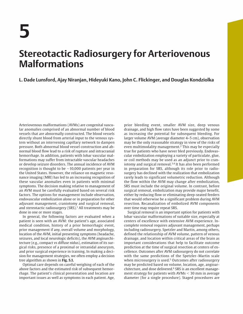
In general, the following factors are evaluated when apatient is seen with an AVM: the patient’s age, associatedmedical condition, history of a prior hemorrhagic event,prior management if any, overall volume and morphology,location of the AVM, initial presenting symptoms (headache,seizures, and local neurologic deficits), the AVM angioarchi-tecture (e.g., compact vs diffuse nidus), estimation of its sur-gical risks, presence of a proximal or intranidal aneurysm,and prior surgical experience in training. In making a deci-sion for management strategies, we often employ a decisiontree algorithm as shown in Fig. 5.1.
Optimal care depends on careful weighing of each of theabove factors and the estimated risk of subsequent hemor-rhage. The patient’s clinical presentation and location areimportant issues as well as symptoms in each patient. Age,
prior bleeding event, smaller AVM size, deep venousdrainage, and high flow rates have been suggested by someas increasing the potential for subsequent bleeding. Forlarger volume AVM (average diameter 4–5 cm), observationmay be the only reasonable strategy in view of the risks ofeven multimodality management.2 This may be especiallytrue for patients who have never bled previously. Endovas-cular embolization employing a variety of particulate, glue,or coil methods may be used as an adjunct prior to cran-iotomy and surgical removal.3,4 It has also been performedin preparation for SRS, although its role prior to radio-surgery has declined with the realization that embolizationrarely leads to significant volumetric reduction. Althoughthe flow within the AVM may change after embolization,SRS must include the original volume. In contrast, beforesurgical removal, embolization may provide major benefit,either by reducing flow or eliminating deep-seated feedersthat would otherwise be a significant problem during AVMresection. Recanalization of embolized AVM componentsover time may require repeat SRS.
Surgical removal is an important option for patients withlobar vascular malformations of suitable size, especially atcenters of excellence with extensive AVM experience. In-complete removal requires adjuvant management, perhapsincluding radiosurgery. Spetzler and Martin, among others,defined the relationship of AVM volume, pattern of venousdrainage, and location within critical areas of the brain asimportant considerations that help to facilitate outcomeprediction at the time of surgical resection at centers of ex-cellence. Outcomes after AVM radiosurgery do not correlatewith the same predictions of the Spetzler–Martin scalewhen microsurgery is used.5 Outcomes after radiosurgerymay be predicted based on volume, location, age, angioar-chitecture, and dose delivered.6 SRS is an excellent manage-ment strategy for patients with AVMs � 30 mm in averagediameter (for a single procedure). Staged procedures are
5Stereotactic Radiosurgery for ArteriovenousMalformations
L. Dade Lunsford, Ajay Niranjan, Hideyuki Kano, John C. Flickinger, and Douglas Kondziolka
c05 4/4/09 12:55 AM Page 29

used for larger vascular malformations or for those thatwere incompletely obliterated 3 years or more after aninitial procedure.
The chief benefit of radiosurgery management is riskreduction; the chief deficit of radiosurgery is the latencyinterval that is required to achieve complete obliteration ofthe AVM.7,8 The latency interval is generally 2 to 3 years, butin selected patients it may be longer. AVM radiosurgery hasbeen used for children not suitable for other managementstrategies, as well as for older patients who have significantmedical risk factors for surgical removal.
◆ History of Radiosurgery for Arteriovenous Malformations
Radiation to obliterate abnormal blood vessels in the brainis a procedure that was first considered in the late 1960s.Raymond Kjellberg, using the Harvard-affiliated protonfacility in Cambridge, Massachusetts, advocated proton Braggpeak stereotactic radiation during the 1970s and early1980s.9,10 More than 1000 AVM patients were treated, butthe dose-planning technique was quite rudimentary. Thetechnology of the Bragg peak proton facility was designedto provide a low exit dose based on the radiophysiologicalcharacteristics of this technology. The doses that were actu-ally used in this series of patients were quite low and do notcorrespond to doses that we now know may be effective inthe obliteration of AVMs. Although Kjellberg maintainedthat Bragg peak radiation stabilized AVM blood vessel wallsand reduced the subsequent risk of hemorrhage (in com-parison to age-related survivals from a life insurance table),only 20% of patients had complete obliteration of their AVMover time. Fabrikant et al, working at the Lawrence Livermore
National Laboratory, began to use helium ion beam to performmultisession AVM irradiation in the 1980s.11
In Stockholm, Lars Leksell and Ladislau Steiner initiatedwork with the first-generation Leksell Gamma Knife unit.12
The first patient was treated in March 1970, using the originalprototype 179 cobalt source photon beam unit designed byBorje Larsson and Lars Leksell. The target definition was basedon biplane angiography done during the procedure itself.Patients were observed for a period of time in preparation fora larger experience that began to emerge in Stockholm usingthe second-generation unit, which was built in 1975.
Linear accelerator (linac) technologies have been adaptedfor SRS. O.O. Betti, working in Paris and Buenos Aires,13
J.L. Barcia-Salorio in Spain,14,15 and Colombo in Vincenza,Italy,16–18 were pioneers in the application of photon radiationusing newer generation linacs. In addition, surgeons and radi-ation oncologists working at the Joint Center in Boston19 andin Gainesville, Florida,20 used modified linacs to treat a largenumber of vascular malformations. Most centers continue toevaluate SRS as part of an overall management plan that mayinclude embolization, microsurgical removal, or radiosurgeryalone or in combination. We will discuss our current view-point relative to the role of embolization subsequently.
◆ Radiosurgery for ArteriovenousMalformations at the University of Pittsburgh
History
Our first AVM patient was treated in August 1987,21 and wereported our initial experience in 227 patients in 1991.22
Confirming the work of others, we noted that the 2-year
Intr
acra
nia
l Ste
reot
acti
c R
adio
surg
ery
30
Fig. 5.1 Clinical algorithm for choosingmanagement options for patients withintracranial arteriovenous malformations(AVMs).
c05 4/4/09 12:55 AM Page 30

success rate in terms of complete obliteration was related tonidus volume and dose. In high-dose cases (AVM � l cc),obliteration rates of 100% were noted, which declined to85% for volumes of 1 to 4 cc and 58% for AVMs � 4 cc involume.
Our subsequent 20-year experience in Pittsburgh hasnow increased to more than 1100 patients who have under-gone Gamma Knife radiosurgery for their AVM using one ormore radiosurgical procedures. We have more recentlyanalyzed the outcome data of 906 patients who underwentradiosurgery between 1987 and 2004 (Tables 5.1, 5.2,
and 5.3). Our median patient age was 36 years, with a rangeof 3 to 80 years. Typical symptoms at presentation includedhemorrhage (46%), seizures (24%), and headache (18%).Eight percent had neurologic deficits (8%). An incidentalAVM was detected in 4% of patients. Prior managementstrategies included surgical removal or clot evaluation in7%. Twenty-one percent underwent one or more interven-tional procedures (embolization). The median target vol-ume was 3.4 cc (range 0.065–57.7 cc). The median margindose was 20 Gy (range 13–32 Gy). A single procedure wasperformed in 865 (95.5%) of patients, and repeat radio-surgery for incomplete nidus obliteration after 3 years wasneeded in 113 (12.5%) patients. Prospective volume stagedradiosurgery was performed in 41 (4.5%) patients.
At a median follow-up of 3 years, complete nidus oblit-eration was achieved in 78% (confirmed by angiography orMRI). In addition, 21% of patients achieved subtotal oblit-eration of the nidus. During the follow-up interval, 38bleeds (4.1%) occurred after the procedure. Seizure controlwas improved in 51% of patients who presented withseizures. Adverse radiation effects (AREs) resulting in neu-rologic deficits developed in 24 patients (2.6%) and thedetection of new T2 signal increase surrounding the AVMtarget in 108 patients (12%). We noted long-term compli-cations such as delayed cyst formation and encephaloma-lacia in 16 patients (1.7%). No patient in this AVM serieshas developed a radiation-related tumor at the time ofthis writing.
Stereotactic Radiosurgical Procedure
We perform intracranial radiosurgery using the LeksellGamma Knife (Elekta Instruments AB, Stockholm, Sweden),beginning with the model unit U, and proceeding to the B,C, 4-C, and in recent experience Perfexion model. Thepatient’s clinical studies and imaging are reviewed forsuitability for SRS. As noted previously, we evaluate thebleeding history, the age of the patient, existing comor-bidities, location, and clinical symptomatology. Patientswith lobar AVMs were placed prophylactically on anticon-vulsants for a period of 2 to 4 weeks around the time ofthe procedure. This has reduced the risk of a perioperativeseizure event from as high as 5% in year 1 of our 20-year
5 St
ereo
tact
ic R
adio
surg
ery
for
Art
erio
ven
ou
s M
alfo
rmat
ion
s
31
Table 5.1 Patient Demographics from Radiosurgery Experience atthe University of Pittsburgh, 1987–2004
Number of patients 906Patient ageMedian 36 yearsRange 3–80 years
GenderMale 474 (52%)Female 432 (48%)
Presenting symptomsHemorrhage 417 (46%)Seizures 213 (24%)Headache 164 (18%)Sensory motor deficit 74 (8%)Incidental 38 (4%)
Prior managementEmbolization 194 (21%)Surgery 63 (7%)
Table 5.2 Locations and Grades of Arteriovenous Malformationsfrom Radiosurgery Experience at the University of Pittsburgh,1987–2004 (N � 906)
AVM locations PercentageTemporal 18.50Frontal 18Parietal 17.50Thalamus/basal ganglia 16.0Occipital 11.50Cerebellar 6.30Brainstem 5.50Dural 2.70Corpus callosal 2.0Intraventricular 1.0Pineal 1.0
Spetzler–Martin gradeGrade I 2.1Grade II 24.4Grade III 42.4Grade IV 15.0Grade V 2.7Grade VI 13.4
Coexistence of aneurysm 77 (8.5)
Table 5.3 Radiosurgical Parameters of Arteriovenous MalformationsTreated at the University of Pittsburgh, 1987–2004
AVM volumeMedian 3.4 mLRange 0.065–57.7 mL
Radiosurgery doseMedian 20 GyRange 13–32 Gy
RadiosurgerySingle session 865/906 (95.5%)Prospective volume staged 41/906 (4.5%)Repeat radiosurgery 113/906 (12.5%)
c05 4/4/09 12:55 AM Page 31

experience to a risk of � 1% at the current time. We requirethat all women within the child-bearing age have a recentnegative pregnancy test, or we perform it on the day of theprocedure.
Patients are evaluated preoperatively by the neurosur-geon, the radiation oncologist, and the nursing team. On themorning of the procedure, the patients arrive at 6 o’clockand begin conscious sedation using oral lorazepam fol-lowed by intravenous (IV) conscious sedation (fentanyl andmidazolam) as needed. Scalp anesthetic injection using acombination of Marcaine and Xylocaine is injected at thesites of pin application. At the current time, we prefer to usetitanium pins with plastic inserts, to minimize the risk ofMRI artifacts. General anesthesia may be required for frameapplication in imaging in patients younger than 12 yearsof age.
Neurodiagnostic imaging follows using both IV paramag-netic contrast-enhanced three-dimensional (3D) volumetricMRI scan and a whole-head T2 fast spin echo imaging se-quence. Patients subsequently undergo biplane digital sub-traction angiography (DSA) as the next step. In patientswhere MRI scans are not possible (those with undeter-mined implants, pacemakers, old aneurysm clips, etc.), we
use contrast-enhanced stereotactic computed tomographyangiography (CTA). The development of axial imaging aspart of the imaging paradigm is critical for AVM management.This has allowed us to make superior 3D conformal plansmuch more carefully in comparison to using two-planeangiography alone.
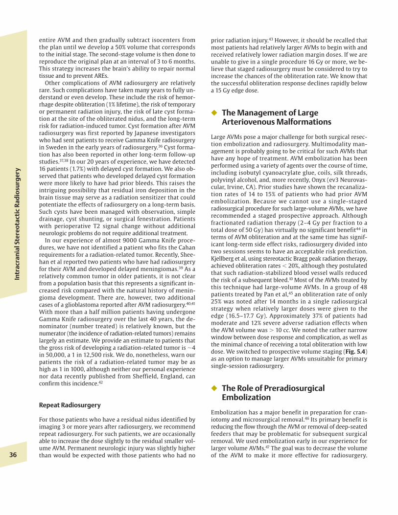
Imaging studies are placed in a dose-planning computersystem (Elekta Instruments), which currently has bothhigh speed and high resolution. We begin the planningusing MRI while the patient is undergoing angiography.Angiography is used to “fine-tune” or idealize the angio-graphic plan. In both the 4-C and Perfexion models cur-rently used, optimization of the dose planning is achievedby confining the sharpest fall-off isodose (usually the 50%)to the edge of the 3D defined volume. Each slice is lookedat serially to confirm excellent conformality (conformingthe edge dose to the 3D target volume). High selectivity(rapid fall-off of the radiation dose outside the target) isequally important (Fig. 5.2). We also assess the patient forthe presence of proximal and intranidal aneurysms. Westrive to reduce dose to draining veins when feasible. Typ-ical doses at the margin of the AVM are 18 to 25 Gy andnever below 16 Gy. Maximal doses are usually 36 to 50 Gy
Intr
acra
nia
l Ste
reot
acti
c R
adio
surg
ery
32
Fig. 5.2 Gamma Knife dose plan for a right hemisphere arteriove-nous malformation showing the 50% isodose line projecting on anteropos-terior and lateral views of angiograms and axial poster, as well as coro-
nal and sagittal reconstruction of magnetic resonance images. Thisplan was created using Leksell GammaPlan PFX.
c05 4/4/09 12:55 AM Page 32

at the margin of the AVM. Final dose selection dependson the volume and location, which also helps to estimateARE risks. We also consider the presence of preexistingneurologic conditions, the patient’s age, and prior bleed-ing history. Because the dose to the surrounding brain is acritical predictor of ARE, we must maximize conformalityand selectivity.
At the conclusion of the procedure, patients receive 20 to40 mg of methylprednisolone. This coupled with anticon-vulsants in lobar AVM patients tends to reduce the risk ofperioperative seizures. For those patients who have previ-ously undergone embolization in an attempt to reduce flowor to achieve volumetric reduction of the AVM, or for thosewho have had intracranial surgery or hematoma evacua-tion, Gamma Knife radiosurgery may be used as an adjuvantstrategy rather than a primary management. We often per-form radiosurgery once the patient has achieved stable neu-rologic improvement but almost never within the firstmonth after an ictal event, such as a bleed or embolization.For patients who have had intracranial hemorrhages, weprefer to wait between 1 and 3 months to see if there willbe a regional clot reabsorption. The AVM nidus should notbe compressed by clot at the time of the radiosurgery.Failure of radiosurgery can be traced in some ways to inade-quate planning, inadequate recognition of the 3D geometryof the AVM, reappearance of a component of the AVM previ-ously embolized, or reappearance of a component of theAVM that was previously compressed by intracerebralhematoma.
Follow-up imaging is recommended at regular intervals.Whenever possible, imaging is done at our center, but if thepatient lives a distance, we recommended the images bedone at an imaging site closer to home. Communicationwith the patient’s referring physician team is critical. Wenormally recommend MRI scans at 6 months and then an-nually to assess the effect of radiosurgery. If at the end of3 years MRI suggests complete obliteration, then we requestthat a repeat DSA be performed. If MRI clearly defines resid-ual nidus, angiography is delayed, and the patient is con-tacted to suggest the possible need for repeat radiosurgeryto achieve the final obliteration response. In such cases,repeat stereotactic imaging may include both repeat MRIand angiography.
For those patients who have large AVMs (determinedby multiplying the MRI X, Y, and Z dimensions of theAVM and dividing by 2, a rough approximation of an ellipsoid volume), we recommend consideration ofprospective staged SRS. Staged radiosurgery, with twostages separated usually by 3 months, is recommendedfor AVMs � 15 cm3. We also consider it for those AVMsbetween 10 and 15 cm3. For those AVM volumes 10 cm3
or smaller, we normally perform single-stage radio-surgery. The interval between stages varies from 3 to 6months based on the goal of some radiobiological repairof surrounding brain during the observation interval.However, we do not want to wait too long, as the goal ofprotection from bleeding cannot begin until the radio-surgical procedure has been completed for all volumetriccomponents of the AVM.
◆ Current Outcomes of StereotacticRadiosurgery of ArteriovenousMalformations
In the absence of treatment, the overall risk of a sponta-neous bleed from a brain AVM appears to range from 1 to5% per year, depending on various risk factors.23 In theFinnish population–based 24-year study, hemorrhage was arelatively constant lifetime risk, with an annual death riskof �1% and approximately a 4% risk of bleed per year.23
Recent work reported from the cooperative trial in Canadasuggested that an annual bleed rate may be as high as 5% (CWallace, personal communication). We performed an indi-vidualized analysis of the hemorrhage risk of AVM patientsbefore radiosurgery.24 Our findings demonstrated an overallcrude annual hemorrhage rate of 2.4% per year.
There were several factors associated with hemorrhagerisk, the primary one being a prior hemorrhage (an identifi-cation of a single draining vein on angiography), along withthe detection of a diffuse AVM nidus on angiography. Pol-lock et al constructed an estimation of bleed risks related tothese significant factors24 (Tables 5.4 and 5.5). For low-riskAVMs (no prior hemorrhage and no other risk factors, diffusenidus, or single draining vein), the annual risk rate was �1%per year. In contrast, the risk of a second hemorrhage forpatients with additional risk factors ranged from 2.0 to 3.7%for AVMs with a compact nidus up to 8.94% for those withhigh-risk features of the angioarchitecture (one drainingvein or diffuse morphology).
In talking with families, we often use a “rule of thumb” toestimate the lifetime bleeding risk related to a patient’s age.The age at which the risk of a bleed is greater than the risk ofmorbidity from radiosurgery may well depend on the loca-tion and size of the AVM. A simple lifetime analysis risk ratesuggests that a patient’s age subtracted from 105 will givethe total cumulative risk of that patient having a bleedingevent.25,26 Clearly, for a child, the risk is very large, whereasfor a senior citizen, the remaining lifetime risk of a bleed fora patient who has never bled before may be reasonably low.
Bleeding Risks after Stereotactic Radiosurgery for Arteriovenous Malformations
We have also analyzed the bleeding rate during the observa-tion interval (latency interval) after radiosurgery and before
5 St
ereo
tact
ic R
adio
surg
ery
for
Art
erio
ven
ou
s M
alfo
rmat
ion
s
33
Table 5.4 Estimated Annual Risk of First and Second Hemorrhage inAVM Patients
Estimated Risk of Intracranial Hemorrhage
AVM Characteristics First Bleed Second Bleed
Low-risk AVM (well-defined 1.00% 3.70%nidus and � 1 draining vein)
High-risk AVM (diffuse nidus or 2.20% 8.90%only 1 draining vein)
Abbreviation: AVM, arteriovenous malformation.
c05 4/4/09 12:55 AM Page 33

complete AVM obliteration.27 For this study, we evaluated312 patients with clinical angiographic outcomes followedfor an average of 4 years. Twenty-one patients suffered AVMbleeds at a median of 8 months after radiosurgery. The over-all total risk of postradiosurgery hemorrhage per patientwas 7.4%, after exclusion of additional bleeding risk factorssuch as untreated aneurysms. The actual hemorrhage ratefrom a patent AVM before complete obliteration was 4.8%per year during the first 2 years after radiosurgery, and5% per year for the third to fifth years after radiosurgery, ifthe AVM continued to be unobliterated. Our data have notprovided strong evidence to date that there is a protectivebenefit of radiosurgery that gradually emerges during theobservation years, even before complete obliteration hasoccurred. In contrast, studies from the University of Tokyoexperience reported by Maruyama et al28 as well as reinter-pretation of the outcome data from the Karolinska experi-ence29 of Steiner have suggested that there may be someprotective benefit to AVMs even before complete oblitera-tion of their nidus occurs.
Patients who have a proximal unsecured aneurysm havean increased risk of postradiosurgical hemorrhage. If theaneurysm is immediately proximal to the AVM, it will likelyclose as the AVM obliterates. We have not found that in-tranidal aneurysms increase the risk of bleeding during thelatency interval. For those patients with aneurysms morethan one arterial branch proximal to their AVM, we believethat the aneurysm requires a different management algo-rithm that should be determined based on those character-istics that guide whether surgery or endovascular is thebest management. Such aneurysms generally do not goaway at the time that the AVM is obliterated.
To date, in our experience, no patient has suffered a hem-orrhage after definitive high-resolution angiography hasconfirmed complete obliteration of their AVM. We have asingle patient with more than 50 aneurysm clips who hasundergone multiple surgical procedures. The AVM wasthought to be obliterated after radiosurgery, but the patientsuffered a subsequent hemorrhage. A new AVM was identi-fied on follow-up angiography (neither CT nor MRI scan was
feasible in this patient). The possibility of recanalization of apreviously treated AVM requiring additional treatment andmonitoring is important. For this reason, even in thosepatients who have angiographic complete closure of theirAVM, we recommend MRI scans at 2-year intervals to assessthe overall brain response to detect late ARE or delayed cystdevelopment.
Although the studies of both Maruyama et al and Karls-son et al28,29 provide some evidence of an overall reductionin bleeding rates during the latency interval, this hypothesisremains largely unproven at the present time. In addition,both ours and other outcome studies have shown that evenwith complete obliteration, the hemorrhage rate is not zero.For patients who have defined angiographic obliteration,we may safely estimate that the lifetime risk of a bleed isnow � 1%.
Arteriovenous Malformation Obliteration after Stereotactic Radiosurgery
In our study of 351 patients followed for 3 to 11 years by im-aging, and using dosages at the margin of 20 Gy (median),we documented AVM obliteration (Fig. 5.3) in 73% of thosepatients studied by angiography and in 86% of those patientsstudied by MRI alone.30 Furthermore, we believe that thereis approximately a 95% accuracy that MRI-detected oblitera-tion will be confirmed by follow-up angiography.31 There aresome patients who are unwilling to undergo repeat angiog-raphy even though this remains the gold standard for detec-tion of response. Even the presence of an early draining vein,without discernible nidus, is a sign of satisfactory response.To our knowledge, no patient has subsequently bled whenonly an early draining vein is seen. Follow-up angiographyafterwards at 6 months to 1 year in such cases invariablyshows loss of the early draining vein as well.
Failure of obliteration is multifactorial and as noted ear-lier may be related to dose, volume, inadequate recognitionof the 3D geometry, recanalization of the previously em-bolized component, or clot-compressed AVM that was
Intr
acra
nia
l Ste
reot
acti
c R
adio
surg
ery
34
Table 5.5 Estimated Cumulative Lifetime Risk of Hemorrhage According to History of Prior Hemorrhage and Whether Any High-Risk MorphologicRisk Features* Are Absent or Present
Lifetime Risk of Intracranial Bleed
Low Hemorrhage Risk AVMs High Hemorrhage Risk AVMs
Expected Survival (years) No Prior Bleed Prior Bleed No Prior Bleed Prior Bleed
15 77 46.0 90.5 75.1 99.725 67 40.4 86.1 68.9 99.235 78 34.8 80.4 61.9 98.245 79 28.7 72.4 53.4 95.955 80 22.0 61.2 43.0 90.465 83 16.4 49.5 33.2 81.575 86 10.4 34.1 21.9 64.385 91 5.8 20.3 12.6 43.0
* Increased risk diffuse morphology or one draining vein.Abbreviation: AVM, arteriovenous malformation.
Patient Age atDiagnosis (years)
c05 4/4/09 12:55 AM Page 34

subtotally treated. In our studies designed to detect the rea-sons for marginal failure, we noted that such persistence ofout-of-field residual AVM was seen in 18% of previously em-bolized patients but only 5% of nonembolized patients. Wehave also noted that the successful obliteration for thesame-volume AVMs may be slightly lower in women, ishigher in children, and occurs more completely and at afaster rate in children than in adults.
Adverse Effects of Radiosurgery
Early adverse effects are relatively rare and include headachefrom the frame application, nausea from conscious sedationmedications, and the relatively small risk of developing
seizures in patients with subcortical lobar AVMs.31–34 It is forthis reason that prophylactic anticonvulsants are used inthese patients but not in patients with deep-seated AVMs.Late AREs of radiosurgery are relatively rare. We evaluateddata from 85 AVM patients who developed symptomaticcomplications after Gamma Knife radiosurgery and comparedthem with 337 patients who had no complications and wereevaluated as part of another multiinstitutional study.35
Thirty-five of 85 patients were classified as having a perma-nent symptomatic sequela. We constructed various modelsto study the effects of AVM location and the volume of tissuereceiving 12 Gy or more (the 12 Gy volume) with the risk ofdeveloping permanent postradiosurgery ARE. AVM locationsin increasing order of risk as might be expected were frontal,temporal, interventricular, parietal, cerebellar, corpus callo-sum, occipital, medulla, thalamus, basal ganglia, and ponsand midbrain. We were able to construct statistical modelspredicting the risk of permanent radiation sequelae with a12 Gy volume. Such data are very important for subsequentplanning of additional patients relative to a risk/benefitanalysis. This database, though useful, was constructed witha relatively small number of complications, considering thelarge number of patients and the relatively large number ofvariables. It is likely that the risk predictions for some brain-stem locations are significantly overestimated. Certainly, therisk of complications is expected to be high for large-volumeAVMs in critical locations of the brain (Table 5.6). It is forthis reason that for larger volume AVMs, 15 cc or more, weclearly recommend staging. The goal of this is to increase theeventual obliteration rate while maintaining safety. We gen-erally divide the AVM into two volumes of approximatelyequal proportion. At the time of the first procedure (which isdone with angiography and MRI), we devise a plan for the
5 St
ereo
tact
ic R
adio
surg
ery
for
Art
erio
ven
ou
s M
alfo
rmat
ion
s
35
100
80
60
40
20
08 10 12 14 16
No Embolization With Embolization (n=297) (n=54)
18
Prescribed Marginal Dose (Gy)
% P
atie
nts
wit
h A
ng
iog
rap
hic
or
MR
Ob
liter
atio
n
20 22 24 26 28
Fig. 5.3 Graph showing a higher percentage of arteriovenousmalformation obliteration rates with higher margin doses. MR,magnetic resonance.
Table 5.6 Estimated Percentage Risk of Symptomatic Adverse Radiation Effects for Arteriovenous MalformationsMeasuring 1, 2, 3, and 4 cm in Average Diameter According to Location
AVM Location
Risk of Symptomatic Radiosurgery-Related Complication by AVM Diameter
1 cm 2 cm 3 cm 4 cm
Low-risk brain regionsFrontal lobe 0.04 0.07 0.11 1.48Temporal lobe 0.59 0.94 1.45 16.95
Mild-risk brain regionsIntraventricular 1.32 2.11 3.22 31.63Cerebellum 1.65 2.62 4.00 36.68Parietal lobe 2.61 2.55 3.88 35.99
Moderate-risk brain regionsCorpus callosum 3.73 5.88 8.80 57.32Occipital lobe 3.87 6.09 9.11 58.20
High-risk brain regionsMedulla 7.43 11.46 16.66 73.55Thalamus 12.36 18.51 25.98 83.00Basal ganglia 15.01 22.15 30.54 85.95Pons/midbrain 44.02 55.89 66.19 96.46
Abbreviation: AVM, arteriovenous malformation. *Marginal doses were chosen according to 3% guidelines from the integrated logistic formula.32 Because of limitedexperience, estimated brainstem risks are probably less than predicted in this table.
c05 4/4/09 12:56 AM Page 35
entire AVM and then gradually subtract isocenters fromthe plan until we develop a 50% volume that correspondsto the initial stage. The second-stage volume is then done toreproduce the original plan at an interval of 3 to 6 months.This strategy increases the brain’s ability to repair normaltissue and to prevent AREs.
Other complications of AVM radiosurgery are relativelyrare. Such complications have taken many years to fully un-derstand or even develop. These include the risk of hemor-rhage despite obliteration (1% lifetime), the risk of temporaryor permanent radiation injury, the risk of late cyst forma-tion at the site of the obliterated nidus, and the long-termrisk for radiation-induced tumor. Cyst formation after AVMradiosurgery was first reported by Japanese investigatorswho had sent patients to receive Gamma Knife radiosurgeryin Sweden in the early years of radiosurgery.36 Cyst forma-tion has also been reported in other long-term follow-upstudies.37,38 In our 20 years of experience, we have detected16 patients (1.7%) with delayed cyst formation. We also ob-served that patients who developed delayed cyst formationwere more likely to have had prior bleeds. This raises theintriguing possibility that residual iron deposition in thebrain tissue may serve as a radiation sensitizer that couldpotentiate the effects of radiosurgery on a long-term basis.Such cysts have been managed with observation, simpledrainage, cyst shunting, or surgical fenestration. Patientswith perioperative T2 signal change without additionalneurologic problems do not require additional treatment.
In our experience of almost 9000 Gamma Knife proce-dures, we have not identified a patient who fits the Cahanrequirements for a radiation-related tumor. Recently, Shee-han et al reported two patients who have had radiosurgeryfor their AVM and developed delayed meningiomas.39 As arelatively common tumor in older patients, it is not clearfrom a population basis that this represents a significant in-creased risk compared with the natural history of menin-gioma development. There are, however, two additionalcases of a glioblastoma reported after AVM radiosurgery.40,41
With more than a half million patients having undergoneGamma Knife radiosurgery over the last 40 years, the de-nominator (number treated) is relatively known, but thenumerator (the incidence of radiation-related tumors) remainslargely an estimate. We provide an estimate to patients thatthe gross risk of developing a radiation-related tumor is �4in 50,000, a 1 in 12,500 risk. We do, nonetheless, warn ourpatients the risk of a radiation-related tumor may be ashigh as 1 in 1000, although neither our personal experiencenor data recently published from Sheffield, England, canconfirm this incidence.42
Repeat Radiosurgery
For those patients who have a residual nidus identified byimaging 3 or more years after radiosurgery, we recommendrepeat radiosurgery. For such patients, we are occasionallyable to increase the dose slightly to the residual smaller vol-ume AVM. Permanent neurologic injury was slightly higherthan would be expected with those patients who had no
prior radiation injury.43 However, it should be recalled thatmost patients had relatively larger AVMs to begin with andreceived relatively lower radiation margin doses. If we areunable to give in a single procedure 16 Gy or more, we be-lieve that staged radiosurgery must be considered to try toincrease the chances of the obliteration rate. We know thatthe successful obliteration response declines rapidly belowa 15 Gy edge dose.
◆ The Management of LargeArteriovenous Malformations
Large AVMs pose a major challenge for both surgical resec-tion embolization and radiosurgery. Multimodality man-agement is probably going to be critical for such AVMs thathave any hope of treatment. AVM embolization has beenperformed using a variety of agents over the course of time,including isobutyl cyanoacrylate glue, coils, silk threads,polyvinyl alcohol, and, more recently, Onyx (ev3 Neurovas-cular, Irvine, CA). Prior studies have shown the recanaliza-tion rates of 14 to 15% of patients who had prior AVMembolization. Because we cannot use a single-stagedradiosurgical procedure for such large-volume AVMs, we haverecommended a staged prospective approach. Althoughfractionated radiation therapy (2–4 Gy per fraction to atotal dose of 50 Gy) has virtually no significant benefit44 interms of AVM obliteration and at the same time has signif-icant long-term side effect risks, radiosurgery divided intotwo sessions seems to have an acceptable risk prediction.Kjellberg et al, using stereotactic Bragg peak radiation therapy,achieved obliteration rates � 20%, although they postulatedthat such radiation-stabilized blood vessel walls reducedthe risk of a subsequent bleed.10 Most of the AVMs treated bythis technique had large-volume AVMs. In a group of 48patients treated by Pan et al,45 an obliteration rate of only25% was noted after 14 months in a single radiosurgicalstrategy when relatively larger doses were given to theedge (16.5–17.7 Gy). Approximately 37% of patients hadmoderate and 12% severe adverse radiation effects whenthe AVM volume was � 10 cc. We noted the rather narrowwindow between dose response and complication, as well asthe minimal chance of receiving a total obliteration with lowdose. We switched to prospective volume staging (Fig. 5.4)as an option to manage larger AVMs unsuitable for primarysingle-session radiosurgery.
◆ The Role of PreradiosurgicalEmbolization
Embolization has a major benefit in preparation for cran-iotomy and microsurgical removal.46 Its primary benefit isreducing the flow through the AVM or removal of deep-seatedfeeders that may be problematic for subsequent surgicalremoval. We used embolization early in our experience forlarger volume AVMs.47 The goal was to decrease the volumeof the AVM to make it more effective for radiosurgery.
Intr
acra
nia
l Ste
reot
acti
c R
adio
surg
ery
36
c05 4/4/09 12:56 AM Page 36

However, embolization can be effective only if it perma-nently reduces the nidus volume. Reduction in flow from anAVM does not provide improvement in radiosurgical out-come data. Our most recent analysis suggests that radiosur-gical embolization had a negative effect on AVM obliterationrates.48 Others have reported that up to 30% of patients whohad AVM embolization subsequently had an increase in thenidus volume when a subsequent angiogram was performedat the time of radiosurgical targeting.49 Twelve percent ofembolized AVMs showed recanalization within a year.50 Un-like surgery that removes an AVM nidus within a few weeksof embolization, radiosurgery takes several years to be fullyeffective. This latency interval, unfortunately, allows sufficienttime for embolized AVM components to recanalize, remodel,or even recruit new feeding blood vessels. In a recent reportusing both embolization and radiosurgery, permanentneurologic deficits ranged from 5 to 12% of patients, with amortality rate of 1.5 to 2.7% of patients.49–51 Complete
obliteration rates varied from 47 to 55%.49–51 In a study of47 patients who had radiosurgery and embolization incomparison to 47 matched patients who were treated withradiosurgery alone, nidus obliteration was achieved in 47%of the embolization group but in 70% of the radiosurgerygroup.52 The experience using adhesive agents such as Onyxis too early yet to define whether radiosurgery will have asignificant improvement in patients who have previouslyundergo liquid adhesive embolization.
◆ Treatment of Dural ArteriovenousMalformations
In contrast to the experience with intracranial AVMs, duralvascular malformations or dural arteriovenous fistulas(DAVFs, Fig. 5.5) may indeed benefit from embolizationand radiosurgery.53 The timing of the individual procedures
5 St
ereo
tact
ic R
adio
surg
ery
for
Art
erio
ven
ou
s M
alfo
rmat
ion
s
37
Fig. 5.4 Radiosurgery dose plans of staged radiosurgery for a large-volume arteriovenous malformation (AVM). (A) The medial part of thenidus was selected for stage 1 treatment. A margin dose of 18 Gy wasprescribed to this target (yellow line). The nidus volume for stage 2
radiosurgery (outlined in red) was also defined at the time of first radiosurgery. (B) Dose plan of second-stage radiosurgery showingcomplete coverage of the remaining AVM nidus.
c05 4/4/09 12:56 AM Page 37

is critical. In general, we prefer to do radiosurgery first in anewly diagnosed patient, at a time when the entire con-nection of the fistula can be defined. Most of these lesionsoccur in the region of the transverse or sigmoid sinus andmay be associated with pulsatile tinnitus.54 Others mayoccur in the cavernous sinus and are associated withdiplopia, impaired vision, or exophthalmos. Lesions of thesuperior sagittal sinus may cause papilledema, vision loss,and hydrocephalus. Those lesions that have cortical venousdrainage are prone to have intracranial hemorrhages,progressive deficits, or seizures. Because such patientshave an overall intracranial hemorrhage rate of �2% peryear, management of these lesions may be very impor-tant.54 We generally recommend early radiosurgeryfollowed the same day by embolization. Patients have theirframe placed and undergo MRI, followed by initial angiog-raphy, and are transferred to Gamma Knife for radiosurgerywith a femoral sheath catheter left in position. After com-pletion of the radiosurgical component of the procedure,they are transferred back to the interventional radiologysuite, where they undergo embolization. The combinationof radiosurgery and embolization provides both earlysymptom relief of the DAVF and long-term relief of thecondition by radiosurgery.
◆ Summary
AVM radiosurgery is a well-established management strat-egy for AVMs at the present time. With more than 25 yearsof experience using Gamma Knife technique, and withthousands of patients worldwide who have undergone thisprocedure, we understand the overall success rate, the com-plication rate, and the various alternative strategies. Webelieve that at the present time embolization has an impor-tant role prior to microsurgery, an important role afterradiosurgery for the management of DAVFs, and a relativelylimited role in the prospective management of AVMs within
the brain. Although outcome studies continue to be lookedat both in Canada and in the United States, we suspect thatmost patients with an intracranial AVM require strong con-sideration for intervention, providing the managementstrategy has an acceptable risk to the patient. It seemsunlikely that higher doses will be feasible. The ability toconform dose and to restrict dose (selectivity) seemed to bemaximized with the current imaging techniques as well asradiation delivery systems, including the new PerfexionGamma Knife. We may be able to increase the chance ofobliteration by developing specific radiation sensitizers de-livered by microcatheters immediately before radiosurgery.To date, brain protection technologies and drugs have beendisappointing. The greatest remaining issue for AVM radio-surgery is the risk of a latency interval hemorrhage, so ef-forts to reduce the latency interval would be important. Therecognition of late long-term side effects, including thepossible risk of radiation-related tumors or late cyst devel-opment, continues to be evaluated. Within the context of adisease that has a 1% annual mortality left untreated, thecurrent data have shown that SRS is a safe and effectivemanagement strategy that can be applied to both lobar anddeep-seated AVMs. Many patients with AVMs were nevertreatable by any technique prior to the development ofradiosurgery.
Acknowledgments Drs. Lunsford and Kondziolka are consult-ants with Elekta Instruments AB. Dr. Lunsford is a stockholderwith Elekta.
References
1. Deruty R, Pelissou-Guyotat I, Morel C, Bascoulergue Y, Turjman F.Reflections on the management of cerebral arteriovenous malforma-tions. Surg Neurol 1998;50(3):245–255, discussion 55–56
2. Han PP, Ponce FA, Spetzler RF. Intention-to-treat analysis of Spetzler–Martin grades IV and V arteriovenous malformations: natural historyand treatment paradigm. J Neurosurg 2003;98(1):3–7
Intr
acra
nia
l Ste
reot
acti
c R
adio
surg
ery
38
Fig. 5.5 (A) Axial T1-weighted contrast-enhanced and (B) T2-weighted magnetic resonance images showing a leftcerebellopontine (CP) angle dural arteriovenous malformation (AVM).
(C) Digital subtraction angiography showing the left meningohy-pophyseal trunk feed into the left CP angle dural AVM.
c05 4/4/09 12:56 AM Page 38

3. Ledezma CJ, Hoh BL, Carter BS, Pryor JC, Putman CM, Ogilvy CS.Complications of cerebral arteriovenous malformation embolization:multivariate analysis of predictive factors. Neurosurgery 2006;58(4):602–611
4. Raymond J, Iancu D, Weill A, et al. Embolization as one modality in acombined strategy for the management of cerebral arteriovenous mal-formations. Interventional Neuroradiol 2005;11(Suppl):57–62
5. Spetzler RF, Martin NA. A proposed grading system for arteriovenousmalformations. J Neurosurg 1986;65(4):476–483
6. Pollock BE, Flickinger JC. A proposed radiosurgery-based grading sys-tem for arteriovenous malformations. J Neurosurg 2002;96(1):79–85
7. Liscak R, Vladyka V, Simonova G, et al. Arteriovenous malformationsafter Leksell Gamma Knife radiosurgery: rate of obliteration and com-plications. Neurosurgery 2007;60(6):1005–1014, discussion 1015–1016
8. Pollock BE, Gorman DA, Coffey RJ. Patient outcomes after arteriove-nous malformation radiosurgical management: results based on a 5- to 14-year follow-up study. Neurosurgery 2003;52(6):1291–1296,discussion 1296–1297
9. Kjellberg RN. Stereotactic Bragg peak proton beam radiosurgery for cere-bral arteriovenous malformations. Ann Clin Res 1986;18(Suppl 47):17–19
10. Kjellberg RN, Hanamura T, Davis KR, Lyons SL, Adams RD. Bragg-peakproton-beam therapy for arteriovenous malformations of the brain. NEngl J Med 1983;309(5):269–274
11. Fabrikant JI, Levy RP, Steinberg GK, et al. Heavy-charged-particleradiosurgery for intracranial arteriovenous malformations. StereotactFunct Neurosurg 1991;57(1–2):50–63
12. Steiner L, Leksell L, Greitz T, Forster DM, Backlund EO. Stereotaxicradiosurgery for cerebral arteriovenous malformations: report of acase. Acta Chir Scand 1972;138(5):459–464
13. Betti OO, Munari C, Rosler R. Stereotactic radiosurgery with the linearaccelerator: treatment of arteriovenous malformations. Neurosurgery1989;24(3):311–321
14. Barcia-Salorio JL, Barcia JA, Soler F, Hernandez G, Genoves JM. Stereo-tactic radiotherapy plus radiosurgical boost in the treatment of largecerebral arteriovenous malformations. Acta Neurochir Suppl (Wien)1993;58:98–100
15. Barcia-Salorio JL, Soler F, Hernandez G, Barcia JA. Radiosurgical treat-ment of low flow carotid-cavernous fistulae. Acta Neurochir Suppl(Wien) 1991;52:93–95
16. Colombo F, Benedetti A, Pozza F, Marchetti C, Chierego G. Linear accel-erator radiosurgery of cerebral arteriovenous malformations. Neuro-surgery 1989;24(6):833–840
17. Colombo F, Cavedon C, Francescon P, et al. Three-dimensional angiog-raphy for radiosurgical treatment planning for arteriovenous malfor-mations. J Neurosurg 2003;98(3):536–543
18. Colombo F, Pozza F, Chierego G, Casentini L, De Luca G, Francescon P.Linear accelerator radiosurgery of cerebral arteriovenous malforma-tions: an update. Neurosurgery 1994;34(1):14–20
19. Loeffler JS, Alexander E III, Siddon RL, Saunders WM, Coleman CN,Winston KR. Stereotactic radiosurgery for intracranial arteriovenousmalformations using a standard linear accelerator. Int J Radiat OncolBiol Phys 1989;17(3):673–677
20. Friedman WA, Bova FJ, Mendenhall WM. Linear accelerator radio-surgery for arteriovenous malformations: the relationship of size tooutcome. J Neurosurg 1995;82(2):180–189
21. Altschuler EM, Lunsford LD, Coffey RJ, Bissonette DJ, Flickinger JC.Gamma Knife radiosurgery for intracranial arteriovenous malforma-tions in childhood and adolescence. Pediatr Neurosci 1989;15(2):53–61
22. Lunsford LD, Kondziolka D, Flickinger JC, et al. Stereotactic radio-surgery for arteriovenous malformations of the brain. J Neurosurg1991;75(4):512–524
23. Ondra SL, Troupp H, George ED, Schwab K. The natural history ofsymptomatic arteriovenous malformations of the brain: a 24-yearfollow-up assessment. J Neurosurg 1990;73(3):387–391
24. Pollock BE, Flickinger JC, Lunsford LD, Bissonette DJ, Kondziolka D.Factors that predict the bleeding risk of cerebral arteriovenous malfor-mations. Stroke 1996;27(1):1–6
25. Brown RD Jr. Simple risk predictions for arteriovenous malformationhemorrhage. Neurosurgery 2000;46(4):1024
26. Kondziolka D, McLaughlin MR, Kestle JR. Simple risk predictions forarteriovenous malformation hemorrhage. Neurosurgery 1995;37(5):851–855
27. Pollock BE, Flickinger JC, Lunsford LD, Bissonette DJ, Kondziolka D. He-morrhage risk after stereotactic radiosurgery of cerebral arteriovenousmalformations. Neurosurgery 1996;38(4):652–659, discussion 9–61
28. Maruyama K, Shin M, Tago M, Kishimoto J, Morita A, Kawahara N. Radio-surgery to reduce the risk of first hemorrhage from brain arteriovenousmalformations. Neurosurgery 2007;60(3):453–458, discussion 458–459
29. Karlsson B, Lax I, Soderman M. Risk for hemorrhage during the 2-yearlatency period following Gamma Knife radiosurgery for arteriovenousmalformations. Int J Radiat Oncol Biol Phys 2001;49(4):1045–1051
30. Flickinger JC, Kondziolka D, Maitz AH, Lunsford LD. An analysis of thedose-response for arteriovenous malformation radiosurgery and otherfactors affecting obliteration. Radiother Oncol 2002;63(3):347–354
31. Pollock BE, Kondziolka D, Flickinger JC, Patel AK, Bissonette DJ,Lunsford LD. Magnetic resonance imaging: an accurate method toevaluate arteriovenous malformations after stereotactic radiosurgery.J Neurosurg 1996;85(6):1044–1049
32. Flickinger JC, Kondziolka D, Lunsford LD, et al. Development of amodel to predict permanent symptomatic postradiosurgery injury forarteriovenous malformation patients. Arteriovenous MalformationRadiosurgery Study Group. Int J Radiat Oncol Biol Phys 2000;46(5):1143–1148
33. Flickinger JC, Kondziolka D, Maitz AH, Lunsford LD. Analysis of neuro-logical sequelae from radiosurgery of arteriovenous malformations:how location affects outcome. Int J Radiat Oncol Biol Phys 1998;40(2):273–278
34. Flickinger JC, Kondziolka D, Pollock BE, Maitz AH, Lunsford LD. Com-plications from arteriovenous malformation radiosurgery: multivari-ate analysis and risk modeling. Int J Radiat Oncol Biol Phys 1997;38(3):485–490
35. Flickinger JC, Kondziolka D, Lunsford LD, et al. A multi-institutionalanalysis of complication outcomes after arteriovenous malformationradiosurgery. Int J Radiat Oncol Biol Phys 1999;44(1):67–74
36. Hara M, Nakamura M, Shiokawa Y, et al. Delayed cyst formation afterradiosurgery for cerebral arteriovenous malformation: two casereports. Minim Invasive Neurosurg 1998;41(1):40–45
37. Izawa M, Hayashi M, Chernov M, et al. Long-term complications aftergamma knife surgery for arteriovenous malformations. J Neurosurg2005;102(Suppl):34–37
38. Pan HC, Sheehan J, Stroila M, Steiner M, Steiner L. Late cyst formationfollowing gamma knife surgery of arteriovenous malformations.J Neurosurg 2005;102(Suppl):124–127
39. Sheehan J, Yen CP, Steiner L. Gamma Knife surgery-induced menin-gioma: report of two cases and review of the literature. J Neurosurg2006;105(2):325–329
40. Berman EL, Eade TN, Brown D, et al. Radiation-induced tumor afterstereotactic radiosurgery for an arteriovenous malformation: casereport. Neurosurgery 2007;61(5):E1099
41. Kaido T, Hoshida T, Uranishi R, et al. Radiosurgery-induced braintumor: case report. J Neurosurg 2001;95(4):710–713
42. Rowe J, Grainger A, Walton L, Silcocks P, Radatz M, Kemeny A. Risk ofmalignancy after Gamma Knife stereotactic radiosurgery. Neuro-surgery 2007;60(1):60–65, discussion 65–66
43. Maesawa S, Flickinger JC, Kondziolka D, Lunsford LD. Repeated radio-surgery for incompletely obliterated arteriovenous malformations. JNeurosurg 2000;92(6):961–970
44. Karlsson B, Lindqvist M, Blomgren H, et al. Long-term results afterfractionated radiation therapy for large brain arteriovenous malforma-tions. Neurosurgery 2005;57(1):42–49
45. Pan DH, Guo WY, Chung WY, Shiau CY, Chang YC, Wang LW. GammaKnife radiosurgery as a single treatment modality for large cerebralarteriovenous malformations. J Neurosurg 2000;93(Suppl 3):113–119
46. Mathis JA, Barr JD, Horton JA, et al. The efficacy of particulate em-bolization combined with stereotactic radiosurgery for treatment oflarge arteriovenous malformations of the brain. AJNR Am J Neurora-diol 1995;16(2):299–306
47. Dawson RC III, Tarr RW, Hecht ST, et al. Treatment of arteriovenousmalformations of the brain with combined embolization and stereo-tactic radiosurgery: results after 1 and 2 years. AJNR Am J Neuroradiol1990;11(5):857–864
48. Pollock BE, Flickinger JC, Lunsford LD, Maitz A, Kondziolka D. Factorsassociated with successful arteriovenous malformation radiosurgery.Neurosurgery 1998;42(6):1239–1244, discussion 1244–1247
49. Miyachi S, Negoro M, Okamoto T, et al. Embolisation of cerebral arteri-ovenous malformations to assure successful subsequent radiosurgery.J Clin Neurosci 2000;7(Suppl 1):82–85
50. Gobin YP, Laurent A, Merienne L, et al. Treatment of brain arteriove-nous malformations by embolization and radiosurgery. J Neurosurg1996;85(1):19–28
5 St
ereo
tact
ic R
adio
surg
ery
for
Art
erio
ven
ou
s M
alfo
rmat
ion
s
39
c05 4/4/09 12:56 AM Page 39

51. Henkes H, Nahser HC, Berg-Dammer E, Weber W, Lange S, Kuhne D.Endovascular therapy of brain AVMs prior to radiosurgery. Neurol Res1998;20(6):479–492
52. Andrade-Souza YM, Ramani M, Scora D, Tsao MN, terBrugge K, SchwartzML. Embolization before radiosurgery reduces the obliteration rateof arteriovenous malformations. Neurosurgery 2007;60(3):443–451,discussion 451–452
53. Houser OW, Campbell JK, Campbell RJ, Sundt TM Jr. Arteriovenousmalformation affecting the transverse dural venous sinus—an acquiredlesion. Mayo Clin Proc 1979;54(10):651–661
54. Awad IA, Little JR, Akarawi WP, Ahl J. Intracranial dural arteriovenousmalformations: factors predisposing to an aggressive neurologicalcourse. J Neurosurg 1990;72(6):839–850
Intr
acra
nia
l Ste
reot
acti
c R
adio
surg
ery
40
c05 4/4/09 12:56 AM Page 40