stepwise polarisation of the drosophila follicular epithelium
TRANSCRIPT

Developmental Biology 338 (2010) 136–147
Contents lists available at ScienceDirect
Developmental Biology
j ourna l homepage: www.e lsev ie r.com/deve lopmenta lb io logy
Stepwise polarisation of the Drosophila follicular epithelium
Andre Franz 1, Veit Riechmann ⁎,2
Institut für Entwicklungsbiologie, Universität zu Köln, Gyrhofstr. 17, D-50931 Cologne, Germany
⁎ Corresponding author. Fax: +49 621 383 9652.E-mail address: [email protected]: http://www.zbio.org (V. Riechmann).
1 Present address: Institute for Genetics, University oCluster on Cellular Stress Responses in Aging-AssociateStrasse 47, D-50674 Cologne, Germany.
2 Present address: Heidelberg University, Medical FacCell and Molecular Biology, Ludolf-Krehl-Strasse 13-17,
0012-1606/$ – see front matter © 2009 Elsevier Inc. Adoi:10.1016/j.ydbio.2009.11.027
a b s t r a c t
a r t i c l e i n f oArticle history:Received for publication 13 August 2009Revised 13 October 2009Accepted 20 November 2009Available online 3 December 2009
Keywords:Epithelial polarityOogenesisMesenchymal–epithelial transitionBazooka/Par-3Par-6aPKCβ-Catenin/ArmadilloDiscs largeAdherens junctionsCrumbsStardust
The function of epithelial tissues is dependent on their polarised architecture, and loss of cell polarity is ahallmark of various diseases. Here we analyse cell polarisation in the follicular epithelium of Drosophila, anepithelium that arises by a mesenchymal–epithelial transition. Although many epithelia are formed bymesenchymal precursors, it is unclear how they polarise. Here we show how lateral, apical, and adherensjunction proteins act stepwise to establish polarity in the follicular epithelium. Polarisation starts with theformation of adherens junctions, whose positioning is controlled by combined activities of Par-3, β-catenin,and Discs large. Subsequently, Par-6 and aPKC localise to the apical membrane in a Par-3-dependent manner.Apical membrane specification continues by the accumulation of the Crumbs complex, which is controlled byPar-3, Par-6, and aPKC. Thus, our data elucidate the genetic mechanisms leading to the stepwise polarisationof an epithelium with a mesenchymal origin.
© 2009 Elsevier Inc. All rights reserved.
Introduction
Epithelia are sheets of adherent cells that separate differentcompartments of the animal body, and their polarisation is essentialfor epithelial structure and function. Epithelial polarity is manifestedin the formation of distinct membrane domains, a polarisedcytoskeleton, polarised membrane transport and highly elaboratedcell–cell junctions. Adherens junctions (AJs) are conserved junctionsmediating homotypic cell adhesion between neighbouring epithelialcells. Typically, the AJs form an adhesive belt, called zonula adherens,close to the apical membrane of the epithelium facing the externalenvironment. A main component of the AJs is the single-passtransmembrane protein E-cadherin, which mediates homophilicinteractions with its extracellular part, while its intracellular partis associated with cytoskeletal and signalling proteins. Armadillo(Arm)/β-catenin is one of these intracellular binding proteins with akey role in AJ formation (see Gumbiner, 2005; Knust and Bossinger,2002; Müller, 2000; Nelson, 2003; Tepass et al., 2001 for reviews).
erg.de (V. Riechmann).
f Cologne, Cologne Excellenced Diseases (CECAD), Zülpicher
ulty Mannheim, Department ofD-68167 Mannheim, Germany.
ll rights reserved.
The formation of AJs is an important hallmark for the establishmentof polarised membrane domains as they subdivide the cell cortex intoan apical and a basolateral region. Genetic screens in Drosophila andCaenorhabditis elegans identified several genes controlling the forma-tion of AJs and polarised membrane domains. The identity of thedifferent membrane domains is provided by the polarised localisationof distinct proteins. It has been shown for Drosophila that the lateralmembrane is specified by the PDZ containing proteins Scribble andDiscs large (Dlg) and by the WD-40 repeats containing protein Lethal(2) giant larvae (Lgl) (see Assemat et al., 2008 for review). Theformation of the apical membrane requires a complex containing thetransmembrane protein Crumbs (Crb) and the PDZ containing proteinStardust (Sdt). These two proteins are core components of thiscomplex, and they have been shown to bind to several transientcomponents, which contribute to cell polarisation (reviewed inBulgakova and Knust, 2009). The PDZ domain containing proteinPar-6 and the atypical protein kinase aPKC are two of these transientbinding partners. Par-6 and aPKC can bind Par-3, another protein withPDZ domainswhosefly homologue is called Bazooka (Baz). Dependingon the cell type Baz, Par-6 and aPKCmay localise to the apical cortex orto the AJs. Baz, Par-6, and aPKC are part of the apical Par signallingnetwork, and their binding capacities appear to be regulated in a celltype specific manner. All of the above mentioned polarity proteins arehighly conserved, and are thought to be required for the polarisation ofmost, if not all, types of epithelia (Assemat et al., 2008).
Functional analysis in Drosophila has led to a model of how theseproteins interact to polarise the epithelium of the blastoderm embryo.

137A. Franz, V. Riechmann / Developmental Biology 338 (2010) 136–147
In this model, apical and lateral membrane identity is provided by aregulatory hierarchy involving Baz, Crb, and the lateral proteins;Scribble, Dlg, and Lgl. The lateral proteins repress apical identity byantagonising Baz. At the apical membrane, Baz recruits Crb, whichcounteracts the activity of lateral proteins (Bilder et al., 2003;Tanentzapf and Tepass, 2003). Localisation of Baz to the apicolateralregion of the epithelial cells seems to be a critical first step in theinitiation of this regulatorymechanism. Baz protein uses the polarisedcytoskeleton of the embryo for its localisation to the apicolateralregion, where it controls the assembly of AJs (Harris and Peifer, 2004;Harris and Peifer, 2005). The mechanisms underlying epithelialpolarisation have been discovered through investigation of theembryonic epithelium, which develops de novo from a syncytiumby multiple invaginations and polarised growth of the plasmamembrane. The follicular epithelium of the Drosophila ovary has adifferent developmental origin and arises, like many other epithelia,by a mesenchymal–epithelial transition (Tepass et al., 2001). Incontrast to the embryonic epithelium, these cells arise from stem celldivisions, which generate mesenchymal precursors that migrate toand integrate into the newly formed epithelium. This raises thequestions whether establishment of polarity varies depending on thegenesis of the epithelium, and if different developmental programsare accompanied by distinct polarisation mechanisms.
The mesenchymal precursors of the follicular epithelium aregenerated by stem cells, which reside in the germarium, a structurelocated at the anterior tip of the ovaries. Within the germarium eggchambers (or follicles), the functional unit of oogenesis, are generatedby the encapsulation of a germ line cyst by a monolayer of epithelialcells (see scheme in Fig. 1a). The somatic epithelial stem cells arelocated in the middle region of the germarium (Margolis andSpradling, 1995) and give rise to follicle cell precursors, whichsubsequently migrate posteriorly towards the germ line cyst. In theposterior part of the germarium the follicle cells make contact withthe cyst, and form an epithelial sheet surrounding the germ line cells.After encapsulation of the cyst, newly formed egg chambers bud offfrom the germarium. The budding egg chamber represents stage 1 ofoogenesis and is enclosed by a cuboidal epithelium (Horne-Badovinacand Bilder, 2005, and references therein). Initially low, the frequencyof epithelial cell divisions in the germarium increases in response tothe growth of the germ line cyst until ceasing at stage 6 of oogenesis(Wang and Riechmann, 2007).
In comparison to the epithelium of the early embryo, we knowrelatively little about the mechanisms polarising the follicularepithelium. Analysis of agametic ovaries revealed that follicle cellcontact to the germ line is essential for correct epithelial formation(Margolis and Spradling, 1995; Goode et al., 1996). The finding thatexpression of proteins Egghead and Brainiac is required within thegerm line cyst for formation and polarisation of the epithelium led tothe idea that germ line signalling is instructive for correct epithelialformation (Goode et al., 1996). Although the exact nature of thissignal is unknown, the identification of Egghead and Brainiac asglycosyltransferases suggests that it is modified by lipid-linkedoligosaccharide chains (Wandall et al. 2005). Further analysis ofagametic ovaries has shown that the presence of the basementmembrane surrounding the germarium is sufficient to specify thebasal membrane of the follicle cells. The correct localisation of apicaland lateral markers is dependent on the germ line cyst indicating thatthe putative signal from the germ line is important for the polarisationof the apical and lateral membrane domains. The finding thatlocalisation of the apical protein, Crb, is completely abolished inagametic ovaries provides further support for a central role of thegerm line in epithelial polarisation (Tanentzapf et al., 2000).
Crb accumulation at the apical membrane is also dependent on AJsand the microtubule motor Dynein. Recent data show that mRNAsencoding Crb and Sdt are localised to the apical membrane in aDynein-dependent manner, suggesting that they are transported
along microtubules (Horne-Badovinac and Bilder, 2008; Li et al.,2008). Additionally, the apical localisation of Crb is dependent on arm,indicating a critical role for AJs in follicle cell polarisation (Tanentzapfet al., 2000). The polarisation of the lateral membrane involves thelateral exclusion of Baz by Par-1. Par-1 kinase localises to the lateralmembrane, where it inhibits the formation of Baz–aPKC complexes byBaz phosphorylation (Benton and St Johnston, 2003).
Although several reports demonstrate important roles for many ofthe known polarity regulators in the follicular epithelium, it remainslargely unknown how these proteins interact during epithelialpolarisation (Manfruelli et al., 1996; Goode and Perrimon, 1997;Bilder et al., 2000; Tanentzapf et al., 2000; Abdelilah-Seyfried et al.,2003; Benton and St Johnston, 2003). Elucidation of these interactionsrequires a careful analysis of the localisation of known polarityproteins during epithelial formation, based on which a functionalanalysis can be performed. Here, we examine the spatial distributionand functions of polarity regulators in the follicular epithelium, andpresent a model for its stepwise polarisation. In the first step, thelateral membrane and the AJs are specified by combined activities ofBaz, Arm, and Dlg. In the second step, apical identity is specified byBaz, Par-6, and aPKC, which regulate the apical localisation of the Crbcomplex in the third step of epithelial polarisation.
Materials and methods
Fly strains
Weused the following alleles and FRTs for our study:armYD35 FRT9-2(Peifer and Wieschaus, 1990), bazXI106 FRT9-2 (Wieschaus et al., 1984),FRT82B crb11A22 (Tepass and Knust, 1990), FRT42B apkcl(2)k406403
(Wodarz et al., 2000), par-6226 FRT9-2 (Petronczki and Knoblich,2001), sdtXP96 FRT19A (Wieschaus et al., 1984), and dlgM52 FRT 101(Woods and Bryant, 1991).
Antibodies
Primary antibodies: rabbit anti-GFPpreabsorbed (1:400), goat anti-GFP FITC-conjugated (1:200, Biozol), rabbit anti-Baz preabsorbed(1:500, A. Wodarz), mouse anti-Arm (1:400, Hybridoma Bank), ratanti-DE-cadherin (1:50, Hybridoma Bank), rabbit anti-Par-6 preab-sorbed (1:500), rabbit anti-aPKC (1:200, Santa Cruz), rat anti-Crb(1:250, E. Knust), rabbit anti-Sdt (1:250, E. Knust), mouse anti-Dlg(1:50, Hybridoma Bank).
Secondary antibodies (Invitrogen) with different fluorescent dyes(Alexa 488/568/633) were used in a 1:400 dilution.
Induction of homozygous follicle cells clones using the FRT/FLP system
Flies carrying the mutant allele on a chromosome with an FRT sitewere crossed to males carrying the respective wild type allele and aGFP reporter gene with the corresponding FRT site and an FLPrecombinase under the control of a heat shock promoter on anotherchromosome (Xu and Rubin, 1993). Flies were allowed to lay eggsover night and eggs developed for 24 hours. During the following4 days, eggs and larvae were treated with a heat shock at 37 °C for1 hour, respectively. After heat shock procedure larvae were kept at25 °C until hatching. Freshly enclosed females were collected andsupplied with some males. After 24 hours at 25 °C, females weredissected and ovaries prepared according to the staining protocol.
Immunohistology
Ovaries were fixed in 8% formaldehyde in PBS (phosphate-buffered saline) for 10 minutes followed by two washing steps in0.1% PBT (PBS+0.1% Triton X-100). Ovaries were blocked in 0.5%bovine serum albumin (BSA) diluted in 0.1% PBT before they were

138 A. Franz, V. Riechmann / Developmental Biology 338 (2010) 136–147
139A. Franz, V. Riechmann / Developmental Biology 338 (2010) 136–147
incubated with primary antibodies diluted in 0.1% PBT with 0.5% BSAover night on a shaker. After two washing steps with 0.1% PBT, ovarieswere incubated with 10% normal goat serum (NGS) in 0.1% PBT for 1–2 hours before secondary antibodies were applied for 2–3 hours.Subsequently, ovaries were washed in 0.1% PBT and then incubatedwith DAPI (1:1000 in 0.1% PBT) for 5 minutes. After 2 washes in 0.1%PBT, ovaries were transferred to a slide in mounting medium.Ovarioles were separated with fine needles before a cover slip wasadded and fixed with nail polish. Probes were then analysed using aLeica SP2/SP5 confocal microscope.
Processing of images
Confocal images were analysed and adjusted using Adobe Photo-shop C2 software. For projections of z-stacks, half of the egg chamberwas scanned from an optical section in the middle of the egg chamberto its top in 1 μm steps (ranging from 15 to 25 stacks, pinhole1). Thetop level for scanning was determined by the identification of acontinuous Arm distribution, which indicates the apicolateral regionof the follicular epithelium. Projections were then calculated usingLeica SP2 software (max mode).
Results
Localisation of Baz, Par-6, and aPKC during formation of the follicularepithelium
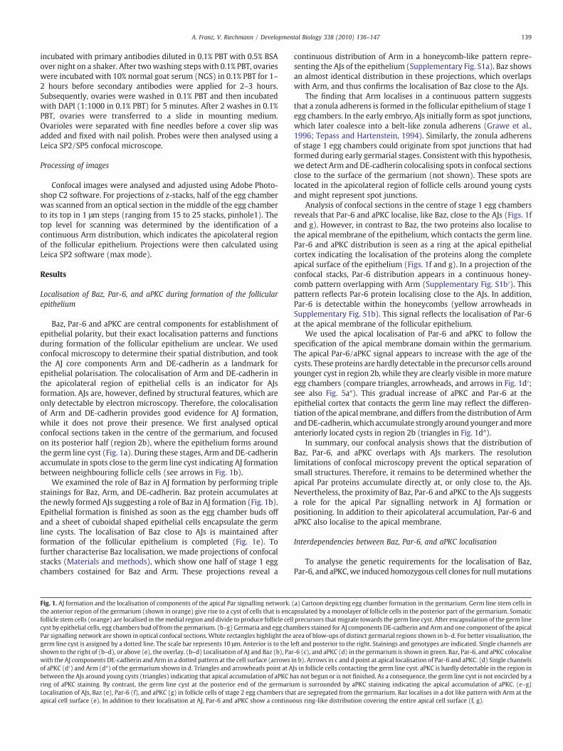
Baz, Par-6 and aPKC are central components for establishment ofepithelial polarity, but their exact localisation patterns and functionsduring formation of the follicular epithelium are unclear. We usedconfocal microscopy to determine their spatial distribution, and tookthe AJ core components Arm and DE-cadherin as a landmark forepithelial polarisation. The colocalisation of Arm and DE-cadherin inthe apicolateral region of epithelial cells is an indicator for AJsformation. AJs are, however, defined by structural features, which areonly detectable by electron microscopy. Therefore, the colocalisationof Arm and DE-cadherin provides good evidence for AJ formation,while it does not prove their presence. We first analysed opticalconfocal sections taken in the centre of the germarium, and focusedon its posterior half (region 2b), where the epithelium forms aroundthe germ line cyst (Fig. 1a). During these stages, Arm and DE-cadherinaccumulate in spots close to the germ line cyst indicating AJ formationbetween neighbouring follicle cells (see arrows in Fig. 1b).
We examined the role of Baz in AJ formation by performing triplestainings for Baz, Arm, and DE-cadherin. Baz protein accumulates atthe newly formed AJs suggesting a role of Baz in AJ formation (Fig. 1b).Epithelial formation is finished as soon as the egg chamber buds offand a sheet of cuboidal shaped epithelial cells encapsulate the germline cysts. The localisation of Baz close to AJs is maintained afterformation of the follicular epithelium is completed (Fig. 1e). Tofurther characterise Baz localisation, we made projections of confocalstacks (Materials and methods), which show one half of stage 1 eggchambers costained for Baz and Arm. These projections reveal a
Fig. 1. AJ formation and the localisation of components of the apical Par signalling network.the anterior region of the germarium (shown in orange) give rise to a cyst of cells that is encafollicle stem cells (orange) are localised in themedial region and divide to produce follicle celcyst by epithelial cells, egg chambers bud of from the germarium. (b–g) Germaria and egg chaPar signalling network are shown in optical confocal sections. White rectangles highlight thegerm line cyst is assigned by a dotted line. The scale bar represents 10 μm. Anterior is to the lshown to the right of (b–d), or above (e), the overlay. (b–d) Localisation of AJ and Baz (b), Parwith the AJ components DE-cadherin and Arm in a dotted pattern at the cell surface (arrows iof aPKC (d′) and Arm (d″) of the germarium shown in d. Triangles and arrowheads point at Abetween the AJs around young cysts (triangles) indicating that apical accumulation of aPKC hring of aPKC staining. By contrast, the germ line cyst at the posterior end of the germariuLocalisation of AJs, Baz (e), Par-6 (f), and aPKC (g) in follicle cells of stage 2 egg chambers thaapical cell surface (e). In addition to their localisation at AJ, Par-6 and aPKC show a continu
continuous distribution of Arm in a honeycomb-like pattern repre-senting the AJs of the epithelium (Supplementary Fig. S1a). Baz showsan almost identical distribution in these projections, which overlapswith Arm, and thus confirms the localisation of Baz close to the AJs.
The finding that Arm localises in a continuous pattern suggeststhat a zonula adherens is formed in the follicular epithelium of stage 1egg chambers. In the early embryo, AJs initially form as spot junctions,which later coalesce into a belt-like zonula adherens (Grawe et al.,1996; Tepass and Hartenstein, 1994). Similarly, the zonula adherensof stage 1 egg chambers could originate from spot junctions that hadformed during early germarial stages. Consistent with this hypothesis,we detect Arm and DE-cadherin colocalising spots in confocal sectionsclose to the surface of the germarium (not shown). These spots arelocated in the apicolateral region of follicle cells around young cystsand might represent spot junctions.
Analysis of confocal sections in the centre of stage 1 egg chambersreveals that Par-6 and aPKC localise, like Baz, close to the AJs (Figs. 1fand g). However, in contrast to Baz, the two proteins also localise tothe apical membrane of the epithelium, which contacts the germ line.Par-6 and aPKC distribution is seen as a ring at the apical epithelialcortex indicating the localisation of the proteins along the completeapical surface of the epithelium (Figs. 1f and g). In a projection of theconfocal stacks, Par-6 distribution appears in a continuous honey-comb pattern overlapping with Arm (Supplementary Fig. S1b′). Thispattern reflects Par-6 protein localising close to the AJs. In addition,Par-6 is detectable within the honeycombs (yellow arrowheads inSupplementary Fig. S1b). This signal reflects the localisation of Par-6at the apical membrane of the follicular epithelium.
We used the apical localisation of Par-6 and aPKC to follow thespecification of the apical membrane domain within the germarium.The apical Par-6/aPKC signal appears to increase with the age of thecysts. These proteins are hardly detectable in the precursor cells aroundyounger cyst in region 2b, while they are clearly visible inmorematureegg chambers (compare triangles, arrowheads, and arrows in Fig. 1d′;see also Fig. 5a″). This gradual increase of aPKC and Par-6 at theepithelial cortex that contacts the germ line may reflect the differen-tiation of the apicalmembrane, and differs from the distribution of ArmandDE-cadherin,which accumulate strongly aroundyounger andmoreanteriorly located cysts in region 2b (triangles in Fig. 1d″).
In summary, our confocal analysis shows that the distribution ofBaz, Par-6, and aPKC overlaps with AJs markers. The resolutionlimitations of confocal microscopy prevent the optical separation ofsmall structures. Therefore, it remains to be determined whether theapical Par proteins accumulate directly at, or only close to, the AJs.Nevertheless, the proximity of Baz, Par-6 and aPKC to the AJs suggestsa role for the apical Par signalling network in AJ formation orpositioning. In addition to their apicolateral accumulation, Par-6 andaPKC also localise to the apical membrane.
Interdependencies between Baz, Par-6, and aPKC localisation
To analyse the genetic requirements for the localisation of Baz,Par-6, and aPKC, we induced homozygous cell clones for nullmutations
(a) Cartoon depicting egg chamber formation in the germarium. Germ line stem cells inpsulated by a monolayer of follicle cells in the posterior part of the germarium. Somaticl precursors that migrate towards the germ line cyst. After encapsulation of the germ linembers stained for AJ components DE-cadherin and Arm and one component of the apicalarea of blow-ups of distinct germarial regions shown in b–d. For better visualisation, theeft and posterior to the right. Stainings and genotypes are indicated. Single channels are-6 (c), and aPKC (d) in the germarium is shown in green. Baz, Par-6, and aPKC colocalisen b). Arrows in c and d point at apical localisation of Par-6 and aPKC. (d) Single channelsJs in follicle cells contacting the germ line cyst. aPKC is hardly detectable in the region inas not begun or is not finished. As a consequence, the germ line cyst is not encircled by am is surrounded by aPKC staining indicating the apical accumulation of aPKC. (e–g)t are segregated from the germarium. Baz localises in a dot like pattern with Arm at theous ring-like distribution covering the entire apical cell surface (f, g).

140 A. Franz, V. Riechmann / Developmental Biology 338 (2010) 136–147
with the help of the FRT/FLP system. Cell clones were identified by theabsence of GFP. We started our analysis in egg chambers which hadalready segregated from the germarium (stage 2 and older). Thisanalysis revealed that the phenotypesweobtained arevariable and thatwe were unable to distinguish whether a phenotype reflects an earlyfunction of a gene during polarity establishment or a late function inpolarity maintenance. The analysis of these stages was furthercomplicated by the fact that mutant cell clones frequently formmultilayered epithelial, in which cell polarity cannot be determinedunambiguously. Therefore,we focused on cloneswithin the germariumand in stage 1 egg chambers, where cell polarity is established, andconsidered only egg chambers with monolayered epithelia. Byanalysing early stages, we observed highly penetrant phenotypes.Moreover,we focused our analysis on cell clones induced in the somaticstem cells, minimising the probability of protein perdurance after cloneinduction.
We first analysed interdependencies between Baz, Par-6, and aPKCregarding their recruitment to the membrane. In clones mutant forpar-6 and apkc, Baz protein localises in apicolateral spots resemblingAJs (Figs. 2a and b). This indicates that Baz recruitment to theapicolateral membrane occurs independently of par-6 and apkc. Bycontrast, bazmutant clones show complete absence of Par-6 and aPKCproteins from the membrane, suggesting that baz is required torecruit Par-6 and aPKC protein to the membrane (Figs. 2c and d).Similarly, aPKC protein is absent from the membrane in par-6mutantcells, and Par-6 protein is absent in apkc mutant cells, revealinginterdependencies between the two proteins in membrane recruit-ment (Figs. 2e and f). Thus, we found a genetic hierarchy, whichcontrols the membrane recruitment of members of the apical Parsignalling network. Here, Baz acts upstream of Par-6 and aPKC, as Bazis recruited to the membrane independently of par-6 and apkc. This isin contrast to Par-6 and aPKC, which depend on the presence of theother two proteins for their membrane recruitment.
The continuous distribution of Par-6, aPKC, and Baz along thezonula adherens prompted us to examine if the spatial distribution ofBaz is completely independent of par-6 and apkc, or if there aredifferences that are not visible in optical confocal sections. To this end,we analysed Baz distribution in confocal stacks of egg chambers thatare exclusively assembled by par-6 or apkcmutant cells. In contrast towild type egg chambers, where Baz is evenly distributed along the AJs,Baz protein accumulates in clumps in par-6 and apkcmutant epithelia(Figs. 2h and b″). Thus, Par-6 and aPKC are essential for the continuousdistribution of Baz along the apicolateral membrane. In summary, ourdata indicate that Baz does not need Par-6 and aPKC for itsrecruitment to the membrane, whereas correct Baz positioning atthe membrane is Par-6 and aPKC dependent.
Baz, Par-6, and aPKC are required for the belt-like distribution of AJs
The colocalisation of Baz, Par-6, and aPKC with Arm duringformation of the follicular epithelium indicates a role of the apical Parsignalling network in AJs assembly. Therefore, we compared thelocalisation of the AJ components Arm and DE-cadherin in germariawith homozygous clones mutant for baz. In germaria in whichsomatic stem cell clones mutant for baz have been induced, thefollicular epithelium forms around the germ line cyst, and AJsassemble as indicated by the colocalisation of Arm and DE-cadherin(Fig. 3b). However, we found ectopic AJs along the basolateralmembrane (see arrows). Moreover, in the absence of baz, theformation of a junctional belt is abolished, and aggregates of AJmaterial form instead (Fig. 3b′). Thus, baz is not required for theformation of AJs per se, but is critical for their positioning and for theirbelt-like distribution.
Analysis of par-6 and apkc mutant epithelia shows similar, albeitweaker, defects in AJs distribution. In both mutant conditions, AJsform occasionally at ectopic positions at the lateral membrane
(arrows in Figs. 3c and d). In addition, confocal stacks show that AJsform clumps and that their belt-like distribution is abolished (Figs. 3c′and d′). We therefore conclude that the apical Par signalling networkis not required for AJ assembly, but for AJ positioning and belt-likedistribution in the apicolateral region of the epithelium. The strongerdefects in baz mutants compared to par-6 and apkc mutants may beexplained by the fact that baz is necessary for Par-6 and aPKCrecruitment to the membrane. In baz mutants, Par-6 and aPKC areabsent from themembrane, and none of the Par proteins can provide acue for AJ positioning. This is in contrast to par-6 and apkc mutants,where Baz protein is still localised to apicolateral regions of theepithelium, and it might rescue aspects of AJ positioning here.
AJs are required for the belt-like distribution of Baz, Par-6, and aPKC
To test if the AJs also influence the localisation of Baz, Par-6, andaPKC, we generated follicle cell clones mutant for a null allele of arm(armYD35). It has been shown previously that in the absence of Armneither DE-cadherin nor DN-cadherin localises, indicating that AJs arenot formed (Tanentzapf et al., 2000). In arm mutants' cell clones, Bazlocalises, in addition to its normal apicolateral position, to ectopicspots at the basolateral membrane (arrows in Fig. 4a). Moreover,confocal stacks show that the belt-like distribution of Baz, Par-6, andaPKC in the apicolateral region of the epithelium is abolished and thatthe proteins accumulate in clumps instead (Figs. 4b, d, and f). Inaddition, neither aPKC nor Par-6 distribution appears as a ring inoptical sections of stage 1 egg chambers with arm mutant follicle cellclones, indicating that the apical accumulation of the two proteins isaffected (see arrowheads in Figs. 4c and e). We therefore concludethat Arm and AJs are essential for the belt-like distribution of Baz, Par-6, and aPKC in the apicolateral region of the epithelium, and for theaccumulation of aPKC and Par-6 at the apical membrane.
Taken together, our data suggest the following interactions duringthe formation of AJs in the follicular epithelium. Arm and DE-cadherinassociate independently of Baz to form AJs at the membrane, but theircorrect positioning at the membrane depends largely on Baz. The Bazprotein itself requires some positional cue from the AJs for its correctlocalisation, indicating a positive feedback in defining the region of AJformation. Baz protein also recruits Par-6 and aPKC to the apicolateralregion of the epithelium, and interactions between the apical Parproteins and the AJs mediate a belt-like distribution of all apicolateralproteins.
Baz, Par-6, and aPKC are required for the apical localisation of theCrb–Sdt complex
Next, we examined the role of Baz, Par-6, and aPKC for thelocalisation of the Crb–Sdt complex to the apical membrane. First, weexamined whether the apical Par proteins and the Crb–Sdt complexlocalise simultaneously or consecutively to the apical membrane.Fig. 5a shows a wild type germarium stained for Crb, aPKC, and Arm.Like Par-6, aPKC accumulates early, in region 2b at the apicalmembrane, while Crb protein is hardly detectable at the membraneat this stage (see arrows in Fig. 5a, apical localisation of Par-6 is shownFig. 1d). Crb is clearly detectable only during cyst budding (stage 1),and it localises to the apical cortex as revealed by its ring-likedistribution in confocal sections (see also Tanentzapf et al., 2000).Similarly, Sdt protein is only detectable at stage 1 at the apicalmembrane (Fig. 5b). Thus, the apical localisation of Par-6 and aPKCprecedes the localisation of Sdt and Crb. At stage 1 when epithelialformation is finished, all four proteins are present as rings in opticalconfocal sections indicating that they cover the entire apical surface ofthe epithelium. In addition to their apical localisation, Crb and Sdtaccumulate close to the AJs (arrowheads in Fig. 5).
Two recent studies show that the apical localisation of Sdt and Crbrequires Dynein. These data provide strong evidence that the two

Fig. 2. Interdependencies between Baz, Par-6, and aPKC localisation. Genetic mosaic germaria generated by the FRT/FLP system are shown. Cells harbouring wild type alleles aremarked by the presence of GFP, while homozygous mutant cell clones lack GFP. For better visualisation, the germ cell cyst is assigned by a dotted line in a–f. Stainings and genotypesare indicated. Single channels are shown to the right. Scale bars represent 10 μm. (a, b) Germaria with follicle stem cell clones mutant for par-6 and apkc are shown. Baz proteinlocalises in spots at the apicolateral level of the follicular epithelium. (c, d) bazmutant cell clones fail to recruit Par-6 and aPKC protein to themembrane. (e, f) Follicle cells mutant forpar-6 do not localise aPKC, and apkc mutant cells do not localise Par-6 protein at the membrane. (g, h, and b″) Projections of confocal z-stacks of stage 1 or 2 egg chambers. Eggchamber in b″ is identical with the one shown in b. (g) Wild type egg chamber stained for Baz protein. Baz is continuously localised along the sites of cell–cell contacts in theapicolateral region (see also Supplementary Fig. S1a). (h, b″) In epithelia derived from stem cell clones mutant for par-6 (h) and apkc (b″), continuous apicolateral distribution of Bazprotein is abolished. Baz instead localises randomly in clumps at the apical cell surface.
141A. Franz, V. Riechmann / Developmental Biology 338 (2010) 136–147
proteins and their encoding mRNAs are localised by apically directedtransport along microtubules (Horne-Badovinac and Bilder, 2008; Liet al., 2008). In light of our data, we tested the hypothesis that thelocalisation of Sdt and Crb to the apical cortexmay also depend on Baz,Par-6, and aPKC. Our analysis of stage 1mutant cell clones reveals that
the membrane localisation of Sdt and Crb is completely abolished inbaz, par-6, and apkc mutants (Figs. 6a–e). Thus, Crb and Sdt need, notonly Dynein, but also Baz, Par-6, and aPKC for their apical localisation.
Next, we tested if crb and sdt are essential for the localisation ofaPKC and Par-6. To this end, we focused on germaria with somatic

Fig. 3. Requirement of Baz, Par-6 and aPKC for the assembly of a zonula adherens. (a) Projection of z-stacks through a stage 1 wild type egg chamber stained for Arm protein.Continuous belt-like distribution of Arm along sites of apicolateral cell–cell contacts is visible. (b–d) Single optical confocal sections of germaria with follicle stem cell clones mutantfor baz (b), par-6 (c), and apkc (d). (b) Arm protein colocalises with DE-cadherin in bazmutant epithelia indicating AJ formation. AJs frequently localise to ectopic sites in bazmutantcells (arrows in b). (c, d) Arrows point at ectopic AJs in par-6 and apkc mutant epithelia. (b′, c′, d′) Projections of z-stacks through stage 1 egg chambers shown in b, c, and d. Thecontinuous belt-like distribution of AJs is disturbed, and clumps of AJ are formed at the apicolateral cell surface. The scale bars represent 10 μm.
142 A. Franz, V. Riechmann / Developmental Biology 338 (2010) 136–147
stem cell clones mutant for crb and sdt and on egg chambers derivedfrom these germaria. In the epithelia of these mutants, the membraneaccumulation of aPKC and Par-6 is slightly reduced, but both proteinsclearly localise to the apical membrane (Figs. 6f–i). Thus, aPKC andPar-6 can localise apically in the absence of the Crb complex, whereasapical Crb and Sdt localisation strictly depends on aPKC and Par-6.
Fig. 4. AJs are involved in the continuous apicolateral distribution of Baz, Par-6, and aPKC. Gand aPKC. Epithelia with armmutant cells are deformed and the egg chambers frequently logenotypes are indicated. Single channels are shown to the right. Scale bars represent 10 μmepithelium of the egg chamber originates from arm mutant follicle stem cell clones. Theunderneath the stage 1 egg chamber and is not visible in this focal plane. Inwild type egg chamform at the basolateral membrane in the stage 1 armmutant egg chamber (arrows). (c, e) Thefollicle cells, but the ring-like distribution in optical sections is lost (arrowheads point at gapaPKC is abolished in arm mutant follicle cells, and the proteins accumulate in clumps instea
This indicates that aPKC and Par-6 act upstream of the Crb complex inpolarising the apical membrane.
Taken together, our data suggest a stepwise polarisation of theapicolateral and apical regions of the follicular epithelium. Positiveinteractions between the apical Par signalling network and the AJsresult in the belt-like distribution of these components,whichpolarises
ermaria with follicle cell clones homozygous mutant for arm are stained for Baz, Par-6,ose their round shape (see also Müller, 2000 and Tanentzapf et al., 2000). Stainings and. (a) Optical confocal section of a germarium with a stage 1 egg chamber is shown. Thegerm line cyst of the egg chamber is marked by white dots. Germarial region 2b liesbers, Baz localisation is restricted to apicolateral spots (Fig. 1e), while ectopic Baz spotsbulk of Par-6 (c) and aPKC (e) protein localises to the apicolateral cortex in armmutant
s in the ring). (b, d, f) Projections of z-stacks. The belt-like distribution of Baz, Par-6, andd.

Fig. 5. Localisation of Crb, Sdt, and Dlg in the follicular epithelium. Wild type germaria are stained with the indicated antibodies. Single channels are shown below the overlay. Scalebar represents 10 μm. (a) The apical localisation of Crb is only faintly detectable in region 2b of the germarium, while aPKC accumulates at the apical membrane (arrows). In stage 1egg chambers, Crb localises to the entire apical cell surface resembling aPKC localisation. Crb colocalises also with Arm (arrowheads). (b) Sdt and Crb localise in an almost identicalpattern. Both proteins are hardly detectable in region 2b of the germarium, while they accumulate apically in stage 1 egg chambers. In contrast to Arm, both Crb and Sdt show acontinuous ring-like distribution along the apical cell surface contacting the germ line cyst. Arrowheads point at Crb and Sdt localisation close to the AJs. (c) Dlg (green) localises tothe lateral membrane domain when the follicular epithelium forms. Dlg localisation does not overlap with DE-cadherin (see arrow).
143A. Franz, V. Riechmann / Developmental Biology 338 (2010) 136–147
the membrane along the contact region to the germ line. The evendistribution of Baz, Par-6, aPKC and the AJs in the apicolateral plane is aprerequisite for the accumulation of Par-6 and aPKC at the apicalmembrane. The localisation of Par-6 and aPKC is the first sign of thespecification of the apical membrane, which occurs in region 2b of thegermarium. The Crb-Sdt complex localises to the apical cortex onlyafter the apical accumulation of Par-6 and aPKC, and the maintenanceof the complex is dependent on the presence of Baz, Par-6 and aPKC.
Dlg prevents the lateral localisation of apical and AJs proteins
We analysedwhether the lateral membrane domain influences theformation of AJs and apical membrane specification. Among theproteins that localise to the lateral membrane, and those involved intheir specification, are Scribble, Fasciclin 2, Par-1, Lgl, and Dlg(Szafranski and Goode, 2007; Benton and St Johnston, 2003; Bilderet al., 2000; Bilder and Perrimon, 2000; Goode and Perrimon, 1997;Woods and Bryant, 1991). We took the Dlg protein as an example of alateral protein. Fig. 5c shows that Dlg and DE-cadherin do notcolocalise (see arrow) when the epithelium is formed in region 2b ofthe germarium indicating that Dlg is excluded from the newly formedAJs.
It has previously been shown for the Drosophila embryo thatscribble, lgl, and dlg prevent the formation of AJs and the localisationof apical proteins along the lateral membrane (Bilder et al., 2000;Bilder and Perrimon, 2000). Consistent with this, Par-3, Crb, and DE-
cadherin mislocalise to the lateral membrane in hypomorphic dlgmutant egg chambers at mid-oogenesis stages (Goode et al., 2005). Totest if dlg acts in the germarium at the time of polarity establishment,we analysed early clones of a strong dlgmutant allele. In these clones,Baz, Par-6, aPKC, Crb, and Sdt all spread into the lateral membranerevealing a central role for dlg in establishment of follicle cell polarity(Figs. 7a–e). Further, we observed Arm spots at the lateral membraneindicating ectopic AJ formation (Fig. 7f). Thus, during formation of thefollicular epithelium, Dlg localises to the lateral membrane, where itprevents AJ formation and the localisation of Baz, Par-6, aPKC, Crb, andSdt.
Arm prevents the apical expansion of Dlg
The important role of Dlg in excluding more apically localisedproteins from the lateral membrane prompted us to analyse how Dlglocalisation is controlled. First, we examined the distribution of Dlg inmutants for baz, par-6, apkc, and crb. Fig. 7g shows that Dlg is stillconfined to the lateral membrane in baz mutant follicle cell clones.Moreover, loss of par-6, apkc, and crb also does not affect thelocalisation of Dlg (Supplementary Fig. S2). These results areconsistent with our previous data showing that Baz acts upstreamof these proteins as revealed by the findings that Par-6 aPKC, and Crbare all absent from the membrane in baz mutant follicle cells. Thus,neither the apical Par proteins nor the Crb complex excludes Dlg fromthe apical membrane domain.

Fig. 6. The Crb complex requires apical Par proteins for its membrane localisation, while aPKC and Par-6 localise apically in the absence of Crb and Sdt. Germaria and stage 1 to 3 eggchambers with homozygous clones mutant for baz, par-6, apkc, crb, and sdt. Stainings and genotypes are indicated. Higher magnifications of single channels are shown to the right.Scale bar represents 10 μm. (a–e) Follicle cells mutant for baz (a, b), apkc (c, d), and par-6 (e) fail to localise Crb and Sdt proteins to the cell membrane. Scale bar in a is valid for a–e.(f–i) Germaria with somatic stem cell clones mutant for crb (f, g) and sdt (h, i), and egg chambers derived from these germaria are shown. In the absence of the Crb and Sdt, aPKC andPar-6 accumulate at the apical cortex of epithelia of stage 1–3 egg chambers.
144 A. Franz, V. Riechmann / Developmental Biology 338 (2010) 136–147
We tested whether AJs play a role in the localisation of Dlg. In armmutant follicle cells, Dlg protein is clearly detectable at the apicalcortex in addition to its lateral accumulation (Fig. 7h). Fig. 7j shows amosaic egg chamber, which is partially assembled of wild type folliclecells. While Dlg is confined to the lateral membrane in the wild typecells of this egg chamber (arrowheads), the protein spreads apically inthe adjacent arm mutant cells (arrow). Hence, we conclude that thelateral restriction of Dlg in the epithelium depends on arm, suggestingthat Arm and the AJs act as a barricade, which prevents the apicalspreading of Dlg protein.
Discussion
Our clonal analysis defines distinct roles for polarity regulators inthe establishment of the membrane domains in the follicularepithelium. Baz, Par-6, and aPKC are essential for the specification ofthe apicalmembrane as revealed by the loss of all apicalmarkers in themutants (Supplementary Fig. S3). Arm prevents the spreading of Dlg(andmost likely other lateral proteins) into the apical domain, leadingto the “lateralisation” of the membrane in armmutant cells. Similarly,Dlg prevents the expansion of apical proteins into the lateral domain asloss of dlg function results in an “apicalised” membrane. Our data alsoindicate how positive and negative interactions between theseproteins lead to the formation of a zonula adherens. Moreover, wehave identified a genetic hierarchy in the localisation of apical proteins,in which Baz recruits Par-6 and aPKC to themembrane, and Baz, Par-6,and aPKC are essential for the apical accumulation of Crb and Sdt.
A model for the stepwise polarisation of the follicular epithelium
Integration of these data suggests a model of stepwise polarisationof the epithelial membrane (Fig. 8). In the first step, Baz, Arm, and Dlgsubdivide the membrane into a lateral domain, which is devoid of AJs,and an apicolateral region where AJs form. The positioning of AJs iscontrolled by inhibitory interactions at the lateral membrane, whereDlg prevents AJs formation. Here, Dlg could act by blocking the laterallocalisation of either Baz or Arm, or Dlg could inhibit both proteinssimultaneously. In wild type follicle cells, Arm protein is detectable atthe lateral membrane (Tanentzapf et al., 2000), from which Baz isexcluded (Fig. 1e), suggesting that Dlg prevents AJ formation byinhibiting Baz rather than Arm localisation. Moreover, Dlg has thecapacity to exclude the apical proteins Par-6, aPKC, Crb, and Sdt fromthe lateralmembrane. Due to the pivotal function of Dlg, it is crucial formembrane polarity that Dlg itself does not spread into apical regions.Our data suggest that apical expansion of Dlg is prevented by the AJs,which might act as a barricade for Dlg and other lateral proteins. Inaddition to lateral exclusion by Dlg, AJ positioning is controlled bypositive positional cues exchanged between Baz and Arm. Baz issupported by Par-6 and aPKC in providing positional information for AJformation, as revealed by ectopic AJ formation in par-6 and apkcmutant epithelia. However, Baz acts upstream of Par-6 and aPKC asboth need Baz for their recruitment to the membrane.
Baz, Arm, and DE-cadherin accumulate strongly at the AJs offollicle cells contacting the germ line cyst. This accumulation isalready detectable around the youngest cysts in region 2b of the

Fig. 7. Function and localisation of Dlg during polarisation of the follicular epithelium. (a–f) Localisation of Baz, Par-6, aPKC, Crb, and Sdt Arm in germaria and egg chambers with dlgmutant cell clones. Stainings and genotypes are indicated. Higher magnifications of the single channels are shown in the right panel. In dlg mutant cells, all apical and apicolateralmarker all spread into the lateral membrane. (g–j) Localisation of Dlg in baz (g, i) and arm (h, j) mutant cell clones. Right panel shows the Dlg channel in the same (i′, j′) or in highermagnification (g′, h′). (g, i) In follicle cells homozygous mutant for baz, the restriction of Dlg to the lateral cell membranes is not affected. (h, j) Dlg enters the apical cell cortex infollicle cells mutant for arm (arrow), while wild type cells retain Dlg lateral (arrowheads). Scale bars represent 10 μm. The scale bar in e is valid for (a–e, g, and h), while the scale barshown in j is also valid for i.
Fig. 8. Schemes for epithelial polarisation. Model for the stepwise polarisation of the follicular epithelium. See text for description. Thick arrow from Baz to Par-6/aPKC indicates thatBaz is necessary for membrane recruitment of Par-6 and aPKC. Thin arrow from Par-6/aPKC to Baz indicates the requirement of Par-6 and aPKC for the continuous apicolateraldistribution of Baz.
145A. Franz, V. Riechmann / Developmental Biology 338 (2010) 136–147

146 A. Franz, V. Riechmann / Developmental Biology 338 (2010) 136–147
germarium. On the contrary, Par-6 and aPKC accumulate in a gradualfashion at the side of the epithelial membrane, which is exposed to thegerm line (Fig. 1d′). At the beginning of epithelial formation, Par-6and aPKC are hardly detectable around anteriorly located cysts inregion 2b. Later, they appear as a faint ring around the cysts, andfinally, they strongly accumulate apically in budding egg chambers.This gradual increase of Par-6 and aPKC suggests that the specificationof the apical membrane occurs subsequently to AJ formation.Therefore, we propose that the specification of the apical membraneis the second polarisation step reflected by the accumulation of Par-6and aPKC at the apical membrane domain.
The third step in membrane polarisation is the localisation of Crband Sdt to the apicalmembrane. Immunohistological labellings ofwildtype germaria indicate that their localisation occurs subsequently tothe apical accumulation of Par-6 and aPKC (Fig. 5), and functionalanalysis shows that their localisation is dependent on baz, par-6, andapkc. By contrast, Par-6 and aPKC can localise apically in crb and sdtmutant cells. We conclude that the localisation of the Crb complex isdownstream of the apical Par signalling network. As a result of thisstepwise polarisation, the epithelium is subdivided into an apical andlateral membrane domain, which is separated by AJs.
Extrinsic cues for epithelial polarisation
It is important to note that the model proposed above considersonly regulatory mechanisms that are intrinsic to the follicularepithelium. Previous data have shown that contact of the folliclecells to the basement membrane is sufficient to establish the basalmembrane. A second external cue is provided by the germ line and isrequired for the differentiation of lateral versus apical membranedomains (Tanentzapf et al., 2000). This unknown germ line cue couldinduce AJ formation and apical membrane specification simulta-neously. Alternatively, AJs and the apical membrane could be inducedconsecutively with either the AJs or the apical membrane beinginduced first. Our finding that the apical markers localise to the germline contacting themembrane only after the AJs have been establishedsuggests that the apical membrane is specified after AJs formation.When the epithelium assembles around the youngest cysts in region2b Arm, DE-cadherin and Baz are already localised to spots at the cell–cell contact sites indicating newly established AJs. At this stage, Par-6and aPKC are not yet localised to the apical membrane. Therefore, wepropose that the germ line provides an initial cue that determineswhere the interactions between Arm and Baz take place, which resultsin AJ formation. The specification of the apical membrane domainoccurs in a subsequent step.
Differences between the polarisation of the embryonic and the follicularepithelium
The formation of the embryonic and the follicular epitheliumshows several differences. The developmental origin of the twoepithelia is different as the follicular epithelium forms through amesenchymal–epithelial transition, while the embryonic epitheliumforms de novo from a syncytium. Formation of the embryonicepithelium is a rapid process that takes around 40 minutes, duringwhich AJs and membrane domains are established. By contrast, eggchamber formation in the germarium has been estimated to last40 hours (Margolis and Spradling, 1995) indicating that epithelialformation and polarisation takes much longer in the ovary. Moreover,the cues polarising these epithelia differ. Polarisation of the follicularepithelium is triggered by cues from the basement membrane and thegerm line, while the embryonic epithelium appears to utilise itscytoskeleton for polarisation.
In the early embryo, Dynein is involved in the transport of Baz tothe apical cortex, where the protein is anchored (Harris and Peifer,2005). After its apical localisation, Baz is required for the formation of
the AJs leading to a model, in which Baz is clearly upstream of Armand determines if and where the AJs are formed (Harris and Peifer,2005; Harris and Peifer, 2004;Müller andWieschaus, 1996). The rapidand hierarchical polarisation mechanism in the embryo requires ahighly polarised microtubule cytoskeleton, which is present when theAJs are formed. By contrast, in the follicular epithelium themesenchymal precursor cells have to integrate into the plane of theepithelium after their migration towards the germ line, and thisprocess involves the reorganisation of their cytoskeleton. The need forcytoskeletal rearrangements and the longer period of epithelialformation suggest that different polarisation mechanisms act infollicle cells. Consistent with this idea, our data provide evidence foran interactive rather than a hierarchical polarisation mode in thefollicular epithelium, in which establishment of AJs and membranedomains involves mutual dependencies between Arm, Baz, and Dlg.However, our data do not exclude Dynein and microtubules roles inAJs formation, and it remains to be determined if the cytoskeletoncontributes to the earliest steps of follicle cell polarisation.
Taken together, our analysis identifies the genetic mechanismsleading to the stepwise polarisation of the follicular epithelium. Thiswork contributes substantially to our understanding of how epithelia,which arise from mesenchymal–epithelial transitions, are polarised.Our study now facilitates integration of newly identified regulators offollicle cell polarity into the framework of known determinants ofepithelial polarity.
Acknowledgments
We thank Arno Müller, Eli Knust, Andreas Wodarz, JürgenKnoblich, Norbert Perrimon, Thomas Klein, the Bloomington stockcentre and the Developmental Studies Hybridoma Bank for fly stocksand antibodies. We are grateful to Stefan Kölzer for excellent technicalassistance. We thank Eli Knust, Matthias Carl, Grainne Kerr, andanonymous referees for helpful suggestions to improve the manu-script. This work was supported by the Deutsche Forschungsge-meinschaft (DFG, SFB 572).
Appendix A. Supplementary data
Supplementary data associated with this article can be found, inthe online version, at doi:10.1016/j.ydbio.2009.11.027.
References
Abdelilah-Seyfried, S., Cox, D.N., Jan, Y.N., 2003. Bazooka is a permissive factor for theinvasive behavior of discs large tumor cells in Drosophila ovarian follicularepithelia. Development 130, 1927–1935.
Assemat, E., Bazellieres, E., Pallesi-Pocachard, E., Le Bivic, A., Massey-Harroche, D., 2008.Polarity complex proteins. Biochim. Biophys. Acta 1778, 614–630.
Benton, R., St Johnston, D., 2003. Drosophila PAR-1 and 14-3-3 inhibit Bazooka/PAR-3 toestablish complementary cortical domains in polarized cells. Cell 115, 691–704.
Bilder, D., Perrimon, N., 2000. Localization of apical epithelial determinants by thebasolateral PDZ protein Scribble. Nature 403, 676–680.
Bilder, D., Li, M., Perrimon, N., 2000. Cooperative regulation of cell polarity and growthby Drosophila tumor suppressors. Science 289, 113–116.
Bilder, D., Schober, M., Perrimon, N., 2003. Integrated activity of PDZ protein complexesregulates epithelial polarity. Nat. Cell. Biol. 5, 53–58.
Bulgakova, N.A., Knust, E., 2009. The Crumbs complex: from epithelial-cell polarity toretinal degeneration. J. Cell. Sci. 122, 2587–2596.
Goode, S., Perrimon, N., 1997. Inhibition of patterned cell shape change and cellinvasion by Discs large during Drosophila oogenesis. Genes Dev. 11, 2532–2544.
Goode, S., Melnick, M., Chou, T.B., Perrimon, N., 1996. The neurogenic genes eggheadand brainiac define a novel signaling pathway essential for epithelial morphogen-esis during Drosophila oogenesis. Development 122, 3863–3879.
Goode, S., Wei, J., Kishore, S., 2005. Novel spatiotemporal patterns of epithelial tumorinvasion in Drosophila discs large egg chambers. Dev. Dyn. 232, 855–864.
Grawe, F., Wodarz, A., Lee, B., Knust, E., Skaer, H., 1996. The Drosophila genes crumbsand stardust are involved in the biogenesis of adherens junctions. Development122, 951–959.
Gumbiner, B.M., 2005. Regulation of cadherin-mediated adhesion in morphogenesis.Nat. Rev. Mol. Cell. Biol. 6, 622–634.
Harris, T.J., Peifer, M., 2004. Adherens junction-dependent and-independent steps inthe establishment of epithelial cell polarity in Drosophila. J. Cell. Biol. 167, 135–147.

147A. Franz, V. Riechmann / Developmental Biology 338 (2010) 136–147
Harris, T.J., Peifer, M., 2005. The positioning and segregation of apical cues duringepithelial polarity establishment in Drosophila. J. Cell. Biol. 170, 813–823.
Horne-Badovinac, S., Bilder, D., 2005. Mass transit: epithelial morphogenesis in theDrosophila egg chamber. Dev. Dyn. 232, 559–574.
Horne-Badovinac, S., Bilder, D., 2008. Dynein regulates epithelial polarity and the apicallocalization of stardust A mRNA. PLoS Genet. e8, 4.
Knust, E., Bossinger, O., 2002. Composition and formation of intercellular junctions inepithelial cells. Science 298, 1955–1959.
Li, Z., Wang, L., Hays, T.S., Cai, Y., 2008. Dynein-mediated apical localization of crumbstranscripts is required for Crumbsactivity in epithelial polarity. J. Cell. Biol. 180, 31–38.
Manfruelli, P., Arquier, N., Hanratty, W.P., Semeriva, M., 1996. The tumor suppressorgene, lethal(2)giant larvae (1(2)g1), is required for cell shape change of epithelialcells during Drosophila development. Development 122, 2283–2294.
Margolis, J., Spradling, A., 1995. Identification and behavior of epithelial stem cells inthe Drosophila ovary. Development 121, 3797–3807.
Müller, H.A., 2000. Genetic control of epithelial cell polarity: lessons from Drosophila.Dev. Dyn. 218, 52–67.
Müller, H.A., Wieschaus, E., 1996. armadillo, bazooka, and stardust are critical for earlystages in formation of the zonula adherens and maintenance of the polarizedblastoderm epithelium in Drosophila. J. Cell. Biol. 134, 149–163.
Nelson,W.J., 2003. Adaptation of coremechanisms to generate cell polarity. Nature 422,766–774.
Peifer, M., Wieschaus, E., 1990. The segment polarity gene armadillo encodes afunctionally modular protein that is the Drosophila homolog of human plakoglobin.Cell 63, 1167–1176.
Petronczki, M., Knoblich, J.A., 2001. DmPAR-6 directs epithelial polarity and asymmetriccell division of neuroblasts in Drosophila. Nat. Cell. Biol. 3, 43–49.
Szafranski, P., Goode, S., 2007. Basolateral junctions are sufficient to suppress epithelialinvasion during Drosophila oogenesis. Dev. Dyn. 236, 364–373.
Tanentzapf, G., Tepass, U., 2003. Interactions between the crumbs, lethal giant larvaeand bazooka pathways in epithelial polarization. Nat. Cell. Biol. 5, 46–52.
Tanentzapf, G., Smith, C., McGlade, J., Tepass, U., 2000. Apical, lateral, and basalpolarization cues contribute to the development of the follicular epithelium duringDrosophila oogenesis. J. Cell. Biol. 151, 891–904.
Tepass, U., Hartenstein, V., 1994. The development of cellular junctions in the Drosophilaembryo. Dev. Biol. 161, 563–596.
Tepass, U., Knust, E., 1990. Phenotypic and developmental analysis of mutations at thecrumbs locus, a gene required for the development of epithelia in Drosophilamelanogaster. Roux's Arch. Dev. Biol. 199, 189–206.
Tepass, U., Tanentzapf, G., Ward, R., Fehon, R., 2001. Epithelial cell polarity and celljunctions in Drosophila. Annu. Rev. Genet. 35, 747–784.
Wandall, H.H., Pizette, S., Pedersen, J.W., Eichert, H, Levery, S.B., Mandel, U, Cohen, S.M.,Clausen, H., 2005. Egghead and brainiac are essential for glycosphingolipidbiosynthesis in vivo. J. Biol. Chem. 280, 4858–4863.
Wang, Y., Riechmann, V., 2007. The role of the actomyosin cytoskeleton in coordinationof tissue growth during Drosophila oogenesis. Curr. Biol. 17, 1349–1355.
Wieschaus, E., Nüsslein-Volhart, C., Jürgens, G., 1984. Mutations affecting thepattern of the larval cuticle in Drosophila melanogaster: III. Zygotic loci on theX chromosome and the fourth chromosome. Rox. Arch. Dev. Biol. 193,296–307.
Wodarz, A., Ramrath, A., Grimm, A., Knust, E., 2000. Drosophila atypical protein kinase Cassociates with Bazooka and controls polarity of epithelia and neuroblasts. J. Cell.Biol. 150, 1361–1374.
Woods, D.F., Bryant, P.J., 1991. The discs-large tumor suppressor gene of Drosophilaencodes a guanylate kinase homolog localized at septate junctions. Cell 66,451–464.
Xu, T., Rubin, G.M., 1993. Analysis of genetic mosaics in developing and adult Drosophilatissues. Development 117, 1223–1237.