stephanie hunstable, miranda williams, mandy boyd, anissa lara & cindy anderson macular...
TRANSCRIPT

Stephanie Hunstable, Miranda
Williams, Mandy Boyd, Anissa Lara
& Cindy Anderson
Macular DegenerationRegion 11
Description
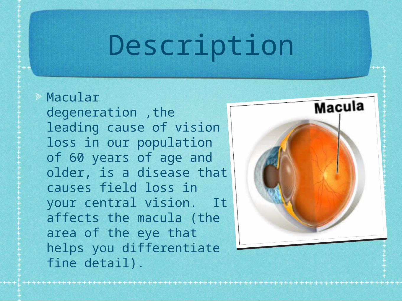
Macular degeneration ,the leading cause of vision loss in our population of 60 years of age and older, is a disease that causes field loss in your central vision. It affects the macula (the area of the eye that helps you differentiate fine detail).
What parts are affected and how?
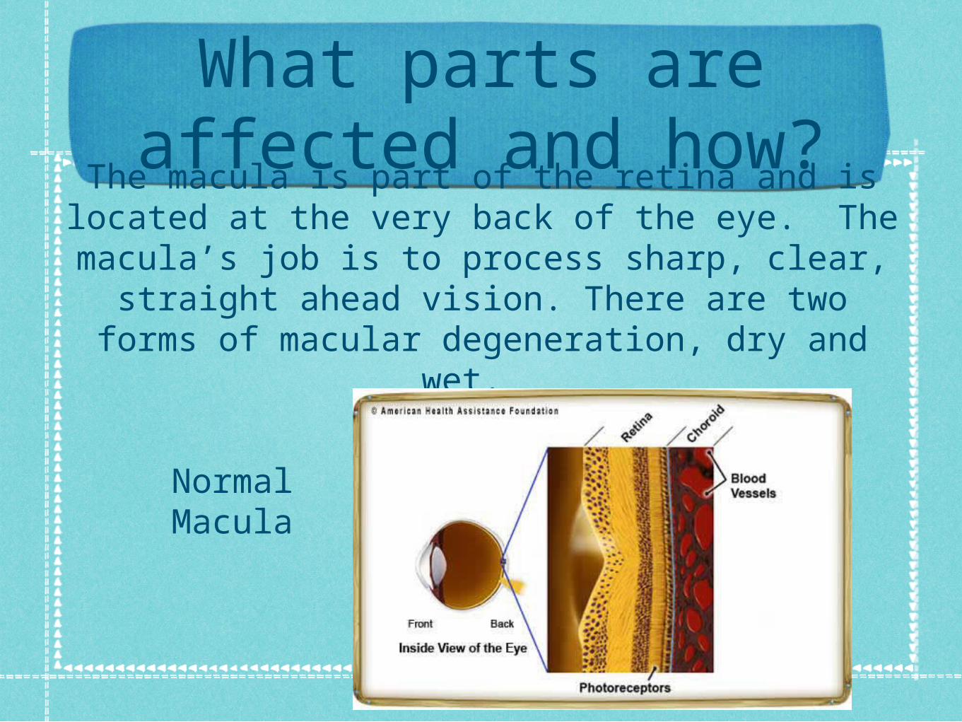
The macula is part of the retina and is located at the very back of the eye. The macula’s job is to
process sharp, clear, straight ahead vision. There are two forms of macular degeneration,
dry and wet.
Normal Macula

Symptoms and Signs
Straight lines become distorted or blurry
Dark or white areas appear in your central vision
Perception of color changes or decreases
Need brighter light to work
Hard to adapt to levels of low light
Printed words become blurry
Difficulty recognizing faces

Functional Implications
Makes reading and driving harder
More difficult to recognize faces
Central vision loss

Adult FormsIt occurs in 10% of people over 50 years of age and 33% of people over 75 years of age.
1.2 million people with macular degeneration will lose part of their central vision.
200,000 will have complete loss of central vision in both eyes
Adult forms of macular degeneration can be inherited or age-related macular degeneration (AMD). The most prevalent forms are “wet” and “dry”.

Wet Macular Degeneration
• Wet Macular Degeneration is not as common and only accounts for about 10 percent of cases. This occurs when abnormal blood vessels grow behind the macula, leaking fluid and blood, causing scaring on the macula. Wet Macular Degeneration can happen very quickly.
Eye withWet Macular
Degeneration
Normal Eye

Dry Macular Degeneration
• This is the most common form. Drusen are small white or yellow deposits that sometimes build up in the back of the eye. These “spots” can prevent the light-sensing cells, called photoreceptors, in the retina from getting enough oxygen or nutrients. When this occurs, the light-sensing cells can then become damaged, which can lead to degeneration of the macula.
Eye withDry Macular
Degeneration
Normal Eye

Juvenile FormsJuvenile forms of macular degeneration are all inherited. The most common forms are:
Stargardt’s (also called fundus flavimaculatus or macular dystrophy) is the most common form. Typical diagnoses occurs during the childhood or teen years.
Best disease (also called vitelliform macular degeneration) is the second most common form. Typical diagnoses usually happens between birth and 7 years of age.

Stargardt’s
Stargardt's disease is the most common form of inherited juvenile degeneration. It is characterized by a reduction of central vision, but it preserves peripheral vision.
The progression of vision loss is variable and can start with a visual acuity of 20/40 and decrease to 20/200 (legal Blindness). By age 50, approximately 50% of all those studied in clinical trials had visual acuities of 20/200 to 20/400. In late stages of this disease there may also be color vision impairment.
Mutations in the ABCR4 gene, which cause Stargardt's disease, produce a dysfunctional protein that cannot perform its transport function. As a result, photoreceptor cells degenerate and vision loss occurs.
Currently there is no treatment for Stargardt's disease, but one will hopefully be found in the near future.

Best Disease
Best disease (also known as vitelliform macular dystrophy) consists of a yellow cyst that forms beneath the macula. In this case, visual acuity could remain nearly normal, 20/30 to 20/50, for many years. Peripheral vision also typically remains unaffected. In many cases, the cyst will eventually rupture causing fluid and yellow deposits to spread throughout the macula creating further vision loss, to about 20/100 later in life. Best disease does not always affect both eyes equally.
This disease is passed through families by the autosomal dominant pattern of inheritance. Therefore, if an affected individual has one Best gene paired with one normal gene, there is a 50% chance that the affected parent will pass the disease-causing gene to each child.
Currently, there is no treatment for Best disease.
Congenital and Progressive
Juvenile Macular Degeneration is congenital meaning it is passed down genetically.
It is also progressive, meaning it gets worse over time.

Who is most at risk?
• Age -- Macular degeneration is the leading cause of severe vision loss in people over 60.
• Gender -- Women are more likely to develop it than men.
• Cigarette smoking• Family history of macular degeneration• Heart disease• High cholesterol• Light eye color• Long-term exposure to sunlight• Low levels of antioxidants in your blood• Carrying weight around your waist (belly fat)

Treatments to slow the progression
Take anti-oxidant vitamins
Use sunglasses with 100% UV protection
Wear a hat or visor
Frequently test using the Amsler grid
These treatments can help slow the loss of vision, but
it cannot be restored.

Amsler Grid

Amsler Grid with Macular Degeneration

More Aggressive Treatments
Drug Therapies
•For wet AMD, a type of medication called anti-vascular endothelial growth factor (anti-VEGF) can be injected into your eye to stop new blood vessels from growing. Two such drugs are approved to treat AMD:
•Pegaptanib (Macugen) & Ranibizumab (Lucentis)
Surgical and Other Procedures
•Surgical and other procedures may help some cases of wet macular degeneration.
•Photocoagulation (laser surgery) -- In photocoagulation, doctors use a laser to seal off blood vessels that have grown under the macula. Whether this procedure is used depends on where the blood vessels are located, how much fluid or blood has leaked out, and how healthy the macula is.
•Photodynamic therapy -- Often used to seal off blood vessels that are under the center of the macula. Using photocoagulation on that location would result in permanent central vision loss. With photodynamic therapy, the doctor gives you a drug that stays in the blood vessels under the macula. When a light is shined in your eye, the drug closes them off without damaging the rest of the macula. Photodynamic therapy slows vision loss but doesn’t stop it.
Progressive Research on Preventative
TreatmentWhat new research is being done to find treatments for macular degeneration?
• Researchers continue to explore how macular degeneration might be promoted by things found in the environment (like cigarette smoke), inherited genetic differences and diet. Many new treatments are also being explored, including: drugs to prevent or slow down the progress of the disease; gene therapies to replace ‘bad’ genes with ‘good’ ones; cells that can be transplanted into the retina to replace the sick ones; drugs to prevent the growth of new blood vessels under the macula; computer chips put in the retina to help simulate vision; and many more.
Case study
“Robert Marcus” a 12 year old with Stargardt’s Disease.

References
Macular Degeneration International. (n.d.). Understanding macular degeneration. Retrieved from http://www.maculardegeneration.org/enroll.html
Medline Plus. (2013, April). Macular degeneration. Retrieved from http://www.nlm.nih.gov/medlineplus/maculardegeneration.html
MD Support. (2002, October). Types of juvenile macular degeneration. Retrieved from http://www.mdsupport.org/library/juvenile.html
University of Maryland Medical Center. (2011). Macular degeneration. Retrieved from http://umm.edu/health/medical/altmed/condition/macular-degeneration
The Children's Corner. (n.d.). Macular degeneration. Retrieved from https://www.childrenscorner.org/about-macular-degeneration/quick-facts