stemi coordinator case study trinity health, minot · •admit to icu for close observation until...
TRANSCRIPT

STEMI Coordinator Case Study
Trinity Health, Minot

Case Study #1-EMS
• 59 year old female
• PMH: Smoker
• Patient developed CP at the casino at 1615.
• Called EMS
• Dispatched 1715, Patient was driven to the ambulance station by family.
• FMC 1734
2

EKG
• EKG at 1739
• Decision made to administer TNKase to the patient prior to transfer to Trinity
3

Thrombolytics
• Checklist reviewed-no contraindications
• TNKase administered at 1758
• Other meds administered Heparin bolus, fentanyl, Zofran
• Post EKG

Arrival at Trinity
• Arrive 1853
• Pain free, and ST elevation resolved
• EKG on arrival:

Cath Lab
• To Cath lab the following day at 1000
• Intervention at 1035
Before After
6Circumflex had 80% occlusion treated with a bare metal stent

Outcome
• Troponin peaked at 5.10
• EF was 55%
• Patient recovered from her heart attack, she was noted to have some abnormalities in her blood work, so a bare metal stent was placed.
• Clinic follow up the patient is still smoking and is having further work up for her blood issues.
7

Case Study #2-CAH
• 45 Year old male
• PMH: HTN, smoker, Moderate ETOH use
• Onset of symptoms 7-8 pm 12-25-16
8
Remember this?

• EMS Called
• Prolonged transport due to weather
• First EKG: 2200

• Repeat EKG at 2214

• Patient arrived at CAH at 2223
• Repeat EKG at 2227
• TNKase at 2237 (13 minutes)
• Other meds given-Heparin bolus/drip, Morphine, Plavix, Aspirin prior to arrival
11

Now what??

• Decision made to keep patient at the CAH until it is safe to transfer, possibly in the morning.
• Admit to ICU for close observation until transfer can be arranged.
• Started on a Nitro drip upon arrival to ICU
• 12/26/16 0121: Patient went into Vfib, shocked at 200J(Lopressor/Amiodarone given)
• 12/26/16 0221: Vfib once again, shocked at 200J, Amiodarone bolus given
• Urgent transfer arranged
13

Transport
• EMS Dispatched 0236
• Arrive at CAH 0251
• Depart scene 0311
• Department of Transportation Snow plow led the way to Trinity in Minot.
• Arrived at Trinity 0730

Arrival
• Admitted to ICU as EKG improved and the patient was completely pain free.
• Troponin on arrival was 15.4
• Peaked at 56.58
• Cath lab arrival time: 1504
• Wire: 1600

Cath Lab
Before After
16
RCA 80% occluded treated with a drug eluting stent

Outcome
• Patient recovered from his AMI, his EF at discharge was 55%.
• He returned in 2 weeks and had intervention to his Circ which was also severely diseased.
• Follow up appointments-patient struggling to quit smoking, requesting Chantix prescription.
Otherwise, no further chest pain, but some compliance issues with statin.

Sanford Health Bismarck
Case Studies

Scenario #1• 40 year old female presenting as a direct admission
from a Critical Access Hospital
• Patient had sudden onset of substernal ischemic chest pain with radiation to the jaw and shortness of breath.
• Initial evaluation included EKG, labwork, and vitals obtained in the ED of the Critical Access Hospital
• Based on symptoms and EKG results, patient received TNK and was transferred emergently to Sanford Health for further intervention.

Patient Information & History
• Demographics• 40 year old• Native American• Female
• Pertinent Medical History• Hypertension• Hyperlipidemia• Diabetes• Smoker
• Pertinent Home Medications• None

Initial EKG• EKG Showed an Acute Anterolateral Infarction
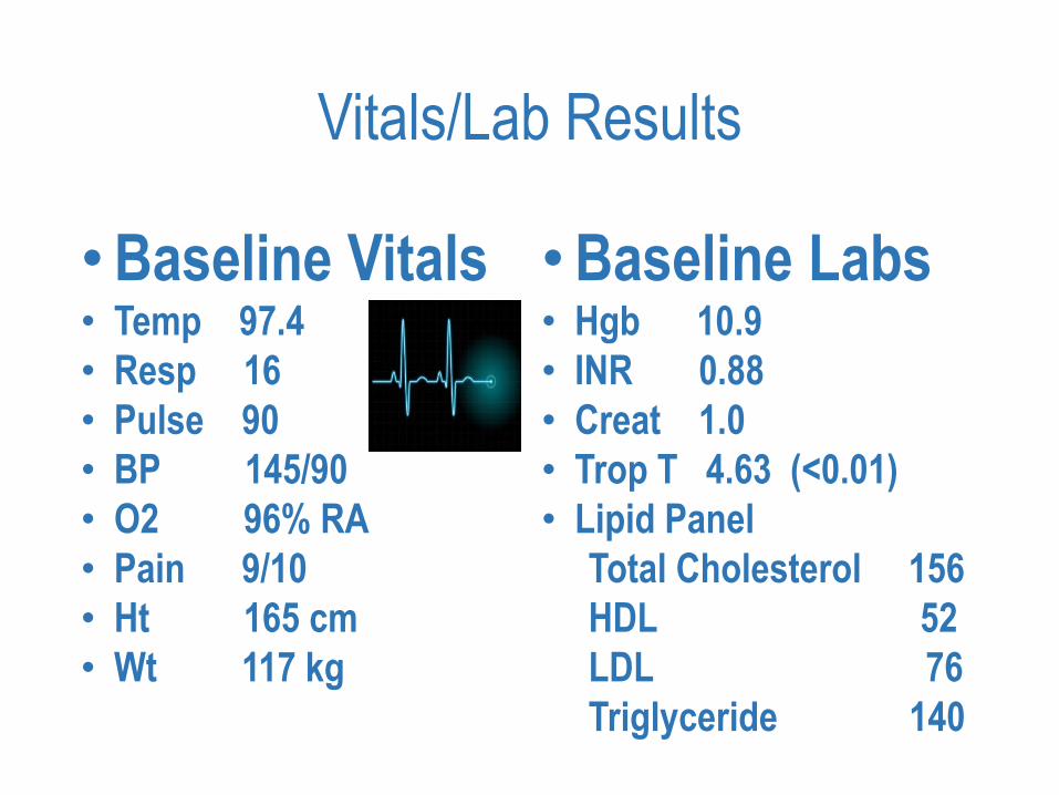
Vitals/Lab Results
•Baseline Vitals• Temp 97.4
• Resp 16
• Pulse 90
• BP 145/90
• O2 96% RA
• Pain 9/10
• Ht 165 cm
• Wt 117 kg
•Baseline Labs• Hgb 10.9
• INR 0.88
• Creat 1.0
• Trop T 4.63 (<0.01)
• Lipid Panel
Total Cholesterol 156
HDL 52
LDL 76
Triglyceride 140

Meds Received Within 24 Hours
• Heparin Bolus 4,000 units
• Heparin Drip 1,000 units/hr
• Aspirin 324 mg
• Clopidogrel 300 mg
• Metoprolol
• Tenecteplase (TNK)
• Morphine
• Atorvastatin
PCI
• Upon arrival from Critical Access Hospital and post lytics, patient was asymptomatic.
• Per Cardiologist, patient was clinically reperfused, but continued to have ST segment elevation in the anteroseptal leads.
• Plan was made to proceed with angiography and possible PCI.

PCI
• Access was gained through right radial site.
• Angiography demonstrated 90% obstruction in the proximal LAD with TIMI 2 flow distally.
• The decision was made to perform PCI to the vessel. Bare-metal stent was placed with excellent result of 0% residual stenosis and TIMI 3 flow in the artery.

Before

After

Critical Access Hospital Times
• Symptom Onset: Approx. 12:00
• Presentation to CAH ED: 14:31
• CAH EKG: 14:40
• Lytics Given: 14:50
• Departed CAH: 15:53

Sanford Bismarck Times
• STEMI Code Paged: 16:29—Prior to Patient’s Arrival
• Arrived Sanford Health ED 16:54
• EKG: 16:59
• ED Door Out: 17:17
• Arrived Cath Lab: 17:17
• Cath Lab Start Time: 17:31
• Balloon Time: 18:40
• Door to Balloon: N/A—Rescue PCI post failed lytics

Outcome
• After cath and PCI, patient was transferred to the ICU in stable condition for continued monitoring post lytics.
• She was discharged home 3 days later on:• ASA
• Clopidogrel
• Valsartan
• Metoprolol
• Atorvastatin
• Referral to cardiac rehab program
• Smoking cessation education

Scenario #2
• 88 year old male presenting via POV to the ED.
• Patient had symptoms of chest pain, shortness of breath, diaphoresis, nausea/vomiting, weakness, and overall not feeling well.
• Patient’s initial heart rate was in the 30s.
• Initial EKG showed diffuse ST elevation throughout all leads.
• Atropine was given and Dopamine drip was started.
• STEMI code was called and patient was transferred emergently to the cath lab in critical condition.

Patient Information & History
• Demographics• 88 year old• Caucasian• Male
• Pertinent Medical History• Hypertension• Hyperlipidemia• Type II Diabetes• CKD Stage III• Atherosclerotic Heart Disease• Former Smoker—Quit 1980s
• Pertinent Home Medications• Cozaar• Metoprolol• Glyberide• Januvia

Initial EKGDiffuse ST Elevation noted throughout—HR in the 30s—Atropine given and
EKG repeated

Repeat EKGDiffuse ST Elevation noted throughout all leads—Post Atropine

Vitals/Lab Results
• Baseline Vitals• Temp 96.8
• Resp 20
• Pulse 39
• BP 103/81
• O2 76% RA
• Ht 178 cm
• Wt 103 kg
• Baseline Labs• Hgb 12.5
• INR 0.90
• Creat 1.53
• Trop T <0.01 (<0.01)
• Lipid Panel
Total Cholesterol 211
HDL 38
LDL 149
Triglyceride 121

Meds Received Within 24 Hours
• ASA 324 mg
• Plavix 600 mg
• Statin
• Beta Blocker—Contraindicated due to low BP and cardiogenic shock
• Heparin Bolus—4,000 Units
• Eptifibatide

PCI
• Femoral Approach
• Angiography Revealed: • Left Main—Visible clot distally• LAD—90% Ostial Stenosis; 80% Proximal Stenosis; 90% Distal Stenosis• Left Circumflex—99% Proximal Stenosis with Visible Clot and TIMI-1 Flow• RCA—100% Occlusion with Collateral Vessels Present
• Decision was made to intervene on the Left Main• Patient developed significant ventricular arrhythmias—Amiodarone bolus and drip
started; A second pressor was added• The Left Main was stented into the Left Circumflex and Prox. LAD• Proximal LAD—Follow-up angiogram revealed stable distal LAD disease and TIMI 3 flow
in both arteries• The patient had intermittent sustained v-tach and v-fib during the procedure which
required CPR, shock X5, multiple boluses of epinephrine, neo-synephrine, bicarbonate, magnesium, and lidocaine. The patient was intubated during the procedure.
• Balloon pump/Impella were not able to be placed due to severe peripheral vascular disease.
• Patient was transferred to ICU in critical condition.

Before

After

Times
• Arrived Sanford Health ED 11:03
• EKG: 11:07
• STEMI Code Paged: 11:09
• ED Door Out: 11:24
• Arrived Cath Lab: 11:27
• Cath Lab Start Time: 11:35
• Balloon Time: 11:44
• Door to Balloon: 41 Minutes!

Outcome
• After cath and PCI, patient was transferred to the ICU in critical condition, intubated, on 2 pressors, and Amiodarone drip.
• Patient thought to have poor prognosis going forward and family opted for no CPR if needed. Family initially considering withdrawal of care.
• Patient slowly began to improve and was successfully extubated on day 7 and continued to improve from there.
• Patient doing well enough to discharged to a rehab facility after 12 day stay.• Patient discharged from rehab to home with home health after total
22 day hospital/rehab stay.

Questions??

DILEMMAWhere to go

Presentation
• 61 y/o male
• Developed nausea, diaphoresis and generalized weakness while playing golf at 1030
• Went home had lunch and at 1300 developed retrosternal CP as well as nausea and diaphoresis. Rated it at 9:10
• Presented to outlaying clinic at 1320

Patient History
• No known cardiac history
• Has lower back problem with an implanted stimulator for pain control and sleep apnea
• Father had bypass surgery in his 50’s
• Several uncles with heart disease
• Angio in 2006 for epigastric pain which showed no coronary disease
• Truck driver at the mines

Clinic findings
• EKG showed ST elevation in anterior, lateral and inferior leads.
• Troponin was negative

Treatment at Clinic
• Received 325 of aspirin
• Heparin not available
• Discussed transfer by Helicopter versus ground
• Discussed with cardiology going to CAH- 11 miles away (15 min) or Bismarck 82 miles (75min)away
• Transfer was made to Bismarck by ambulance which could leave immediately.

St. Alexius
• Ambulance met by cardiologist, who immediately gave 5000 units of heparin and without stopping went directly to cath lab.
• Heparin drip started in the lab.

Heart cath
• LM: no disease
• LAD: 99% prox. Stenosis with thrombus
• CX: mild luminary irregularities otherwise no disease
• RCA: 30% mid and distal stenosis, PDA and PLB free of disease
• LAD treated with a DES with good results


Treatment
• Placed in integrillin for 12 hours post procedure
• Brilinta BID X 1 year
• ASA 81 mg QD for lifetime
• Toprol, lisinopril and Statin
• EF 40%
• Echo showed mid to distal anteroseptalhypokinesia

Timeline
• FMC to EKG 1min
• Transport from clinic to hospital 116 min.
• Hospital to lab 7 min.
• Door to reperfusion 28 min.
• FMC to reperfusion 144 min (standard <120min)

CAH presentation
• 69 y/o female awoke at 0430 with N&V, abdominal discomfort and diarrhea.
• Aches and pains in upper back and bilateral arms. No SOB or CP
• Arrived at hospital at 1401.

Patient History
• On no medications other than ibuprofen
• Smokes <1/2 pack per day X 50 years
• Grandfather had an MI in his 40’s

Clinical findings
• EKG showed ST elevation in inferior leads
• Troponin 17.676 (nl 0.056)
• No changes in patient symptoms


Treatment
• Initially given ASA and protonix
• Followed by TNK, lovenox and plavix
• Transferred by helicopter

St. Alexius
• Discomfort subsided shortly before arrival
• EKG 2mm ST elevation remained in inferior leads with 1mm elevation in lateral leads
• Taken immediately to cath lab

Heart cath
• LM: no disease
• LAD: proximal 40% stenosis and 30% mid
• CX: Mild luminal irregularities otherwise the remained of the vessel noral
• RCA: Ruptured plaque in the proximal RCA with associated 80-90% stenosis. Focal 60-70% stenosis in mid RCA, PDA and RPL ok
• Both the proximal and mid RCA was stented.


Treatment
• ASA for life
• Plavix for minimum of 1 year
• Statin, beta blocker
• Cardiac rehab
• Smoking cessation
• EF 40%
• Entire inferior, basal and mid inferolateral wall akinetic

Timeline
• FMC to EKG 1 min.
• FMC to lytic 29 min
• DIDO 44 min

Case Studies By: Lynde Quirk, RN STEMI & Stroke Coordinator

Case Study 1.
46 year old Female
▪ Presents to regional clinic 2 days in a
row with complaints of neck pressure,
that goes into her shoulders, back, and
then into the back of her neck. This
pain has been going on for 3 days prior
to seeking attention in clinic.
▪ “Episodes” last 1-10 mins.
▪ Denies diaphoresis
▪ Denies nausea
▪ Increasing in frequency of episodes for
2 days.
▪ 1 pack per day smoker
▪ BP 128/84 ~ Pulse 79 ~ SpO2 97% ~
Resp 18 ~ Temp 98.6

Case Study 1.
Patient History:
▪ Breast Cancer in 2016 (last chemo in
June 2016)
▪ Radical bilateral mastectomy
▪ Was noted prior to that surgery to
have a “block” in carotid artery, and
was treated with Lovenox for 6 months.
▪ Abdominal Hernia
Patient Medications:
▪ Ibuprofen
▪ Multivitamin

Case Study 1.
First Clinic Visit:
▪ Sinus Rhythm EKG
▪ GI cocktail given
▪ Patient remained pain free since
administration of cocktail
▪ Within normal limit on all labs
(including troponin).
▪ Chest x-ray negative
▪ Patient discharged home on Protonix
40mg, and follow up with primary in 2
days.
▪ Diagnosis – Acid Reflux

Case Study 1.
Second Clinic Visit
▪ Patient reports “The pressure in my
neck is getting worse, and is now going
into my shoulders, back. These
“episodes” are getting more frequent.
Also she is now having some discomfort
in her chest.
▪ Patient denies nausea, diaphoresis and
shortness of breath.
▪ Patient then sent from clinic to ER.
ER Orders:
▪ EKG, IV, CBC, TRIP, CMP, Protime-INR,
APTT, aspirin 324mg, troponin and CPK.

Case Study 1.
▪ EKG completed within 8 Mins of
arrival to ER.
▪ Inferior STEMI

Case Study 1.
ER TimeLine:
▪ 1429 -Arrived in ED EKG at 1437
▪ Regional MD contacts Altru Cardiologist
▪ Cardiologist orders – TNKase and
Heparin per protocol, ASA
▪ 1445 - LifeFlight called
▪ 1451- ASA and Heparin given
▪ 1503 - Flight team Arrival
▪ 1509 - TNKase given (40 mins after
arrival)
▪ Patient converted to SR after TNKase was
infusing for 5 mins.
▪ 1529 – Troponin resulted, remains
negative
▪ 1530 – Patient discharged with flight
team
▪ Patient arrival to discharge 61 mins.

Case Study 1.
Altru Timeline
▪ 1610 - Patient lands at Altru
▪ 1615 – Patient arrived in Cath Lab
▪ 1629 – Lido time
▪ 1641 – Deployment of drug eluting stent to
mid Circ. (95% occluded)

Case Study 1.
▪ Patient Outcome:
▪ Discharged on day 2
▪ EF 55%
▪ Patient reports no pain or
discomfort since procedure
▪ Started on ASA , Lipitor, Toprolol
XL, Nicoderm CQ and Brilinta.
▪ Cardiac Rehab
▪ Smoking cession counseling
given
▪ Follow-up with primary
scheduled

Case Study 2.
▪ EMS dispatched for 75 year old
male unresponsive, but
breathing.
▪ BP 171/110 ~ P 139 ~ SpO2 96% ~ R 14
▪ On arrival patient was sitting on
the stairs on the third floor. Patient’s
eyes were open, but only
responded to painful stimuli on
right side; no response on the left
side. Will moan some with any
movement from paramedics.
Airway was patent, however he was
drooling. Pupils equal. Blood Sugar
156.

Case Study 2.
▪EMS report from wife:
▪ “We just finished eating
pizza, and were walking up
the stairs, and when I got to
the top I saw him sitting at
the bottom of the stairs just
how he is now. We did have
a couple beers tonight with
supper.”

Case Study 2.
▪ EMS Facts:
▪ Blood Sugar 156
▪ Last known well time 1910
▪ Stroke Code called to ER
▪ 2 18 gauge IV’s placed (blood
drawn)
▪ 12 lead EKG obtained and
transmitted
▪ (Sinus arrhythmia with some ST
elevations and some depressions)
▪ Patent starting to move right side in
route to hospital
▪ Wife denied falls
▪ Scene time 12 mins

Case Study 2.
▪ ER Timeline:
▪ 1939 Patient arrival
▪ Vitals ~ B/P 135/75, P 135, SpO2 90%,
Resp 20
▪ 1940 Dr. at EMS cot, decision to take to
CT on EMS Cot with ER MD.
▪ 1948 Back from CT
▪ 1955 Pt weakness completely resolved,
A&O x3, can speak in full sentences,
denies any pain or shortness of breath.
Patient keeps stating “I am fine, my
speech just feels a little weird.”
▪ 1956 Patient goes for CTA head and
neck
▪ 2000 Tele Neurologist examining patient
▪ 2001 EKG done

Case Study 2.
▪ 2002 ER MD at bedside; patient begins to
cough and have more difficulty breathing.
▪ 2008 Stroke Code canceled – STEMI code
called (3mm elevation v1, v2, AVR, 2mm st
depression lead 1, 3mm st depression AVF
and V6)

Case Study 2.
▪ 2009 No Pulse, CPR started
▪ 1 round epi given
▪ Compressions for 2 mins
▪ Intubated (lots of pink frothy
secretions from ET tube)
▪ 2013 Pulse back
▪ 2035 patient sent to Cath Lab
with cardiologist

Case Study 2.
▪Cardiologist Findings:
▪ Mild Coronary artery
disease, does not
contribute to the clinical
picture.
▪ Incidental finding of a
small aneurysm and fistula,
does not contribute to the
clinical picture.

Case Study 2.
▪Diagnosis?
▪Severe aortic stenosis
▪ Seen by cardiovascular
surgery, TAVR was
suggested. Patient
transferred out 2 days
later.