stem cells and diabetes - howard hughes medical...
TRANSCRIPT

HHMI Stem Cells – Teacher Materials Page 1 of 23
Stem Cells and Diabetes—Teacher Materials
Abstracts
This lesson consists of three distinct parts and a review game that may be done together or at different points in the year to learn about stem cell research focused on a possible treatment or cure for diabetes.
Pulse-Chase Primer
The Primer uses Meselson and Stahl’s classic experiment to provide students with a basic understanding of how a pulse-chase analysis allows scientists to examine cellular processes that take place over a period of time. It also provides students with the background information needed for the Pancreatic Beta Cell activity.
Pancreatic Beta Cells
This part of the lesson has three components that introduce students to stem cells via Dr. Doug Melton’s pulse-chase experiment:
• a Pulse-Chase Primer
• an interactive PowerPoint, Stem Cells and Diabetes: The Present
• an inquiry activity, Are adult pancreatic β cells formed by self-duplication or stem cell differentiation?
The Pulse-Chase Primer provides students with background information on the basics of a pulse-chase analysis. The interactive PowerPoint presents information about diabetes and offers an opportunity for questions and discussion on topics such as possible treatment options. This leads students to the question of how pancreatic β cells are replenished. Students learn about both type 1 and type 2 diabetes and how stem cell research may lead to a cure or treatment. They work through a modified version of Melton’s experiment and conclude that β cells arise from existing β cells and not from adult stem cells. After completing the activity, students can view the section of the HHMI DVD Potent Biology, during which Dr. Melton describes his experiment and how the results inform future stem cell and diabetes research.
Microarrays and Stem Cells
This part of the lesson consists of:
• an interactive PowerPoint, Stem Cells and Diabetes: The Future
• a hands-on activity, Microarray Stem Cell Activity.
The PowerPoint delves into gene expression, stem cells, embryonic development, germ layers, and the differentiation of stem cells into pancreatic β cells. Animations and images from the HHMI DVD Potent Biology are incorporated in the PowerPoint. An introductory reading with hyperlinks to a video clip and an animation provides students with background information needed to interpret the results of a paper microarray simulation. Students will position cDNA strips on mini-microarrays to discover which genes are expressing, to what degree they are expressing, and which are not. They use these findings to trace the differentiation of embryonic stem cells that give rise to pancreatic β cells and other cell types. The role of growth factors and proximity of other cell types is central to students understanding how researchers may direct the ultimate fate of stem cells. The value of this in treating diabetes is also discussed.
Stem Cell Differentiation Review Game
This game uses a modified Uno deck to review the material covered in the Pulse-Chase Primer, Pancreatic Beta Cells and Microarrays and Stem Cells. Students accumulate points and answer questions so that they can be the first to differentiate into a pancreatic β cell.
____________________________________________________________________________________________________
Appropriate Grade Level
High School Biology–honors, IB, and AP
Undergraduate Biology

HHMI Stem Cells – Teacher Materials Page 2 of 23
____________________________________________________________________________________________________
Goals
These activities have been designed to engage students in thinking about the following:
• experimental design • type 1 and type 2 diabetes and the pancreas
• data analysis • differentiation and gene expression
• model organisms • the properties of embryonic stem cells including pluripotency
• microarrays as a research tool • embryonic development and the germ layers ____________________________________________________________________________________________________
Time Requirements
• Pulse-Chase Primer: This activity can easily be completed as a homework assignment. If done in class, it will require one 45-minute period. This would include 25 minutes to read through the primer and answer the questions, and a 20-minute summary discussion.
• Pancreatic Beta Cells: The PowerPoint will require approximately 45 minutes. The inquiry activity, Are adult pancreatic β cells formed by self-duplication or stem cell differentiation? works best if done by students working in groups of two during class. This allows for questions and discussion. With a more advanced class, this can also be assigned as homework. The summary discussion that follows and watching Doug Melton’s pulse-chase segment on the Potent Biology DVD will require another 45 minutes. (Total: approximately 1.5 to 2 hours of class time)
• Microarrays and Stem Cells: This part of the lesson can easily be divided into segments that fit into a variety of schedules. The PowerPoint and introduction will require 45 minutes. The hands-on portion will require an additional 45 minutes. (Total: 1.5 hours)
• Stem Cell Differentiation Review Game: The time this game takes will vary. It depends on the number of individuals per team and how well they know the material.
____________________________________________________________________________________________________ Required Student Background Information
Students should have a basic understanding of:
• mitotic cell division • genes • transcription and translation
• DNA, RNA • feedback • growth and differentiation ____________________________________________________________________________________________________
Introducing the Activities — General Introduction
The following introduction is recommended as a way to discover what students know—or think they know—about stem cells.
• Pretend you are cutting some paper with scissors and nip the end of your finger. You cut off a little skin. Ask students “How will your body replace the skin you just cut off?” They should come up with mitotic cell division.
• Ask students, “If you donate blood during a blood drive, how does your body replace those cells?” You may need to remind students that mature red blood cells lack a nucleus. They should conclude that there are cells (adult stem cells) that can differentiate to form the various blood cells. Some of these cells are located in the blood while others are located in the bone marrow.
• As background, you could prompt discussion about adult stem cells by telling students, “You might know about leukemia and bone marrow transplants. Leukemia is a cancer of the blood. In a bone marrow transplant, doctors try to replace cancerous blood stem cells. Adult stem cells make bone marrow transplants possible. Adult stem cells also replace the blood a donor gives during a blood drive.”

HHMI Stem Cells – Teacher Materials Page 3 of 23
• Tell or remind students that adult stem cells are undifferentiated cells found among differentiated cells in a tissue or organ that can differentiate to form some or all of the specialized cells of the tissue or organ. Their primary function is maintenance and repair.
• Ask students, “What are some other types of stem cells?” They will very likely come up with embryonic stem cells. You can ask what they know about embryonic stem cells; such as what they do, where they are located, and any issues surrounding their use. This would be to get a sense of what students already know.
• Students may also mention umbilical cord blood as a source of multipotent stem cells. These stem cells are useful because they are less likely to be rejected by the recipient. Some doctors recommend saving a child’s umbilical cord blood for future use.
• Show the animation “Human Embryonic Development” on the Potent Biology DVD under the animations menu. It can also be accessed from the BioInteractive website, www.biointeractive.org/stemcells/human_emb_dev.html. Stop the animation at the key points shown in the chart that follows
Types/Locations of Stem Cells Function
Totipotent stem cells differentiate to form all of the cells of an embryo as well as the cells that form the placenta and other extraembryonic tissues. This type of stem cell can divide, differentiate, and ultimately form a complete organism. The zygote is totipotent as are the cells that result from the first few divisions.
Embryonic stem (ES) cells are derived from the inner cell mass (ICM) of the early embryo. These cells can differentiate into any of the three germ layers. They have the capacity to form all of the specialized cell types and organs of the body. However, they cannot develop into a fetus as they lack the ability to form extraembryonic tissue such as the placenta. At this stage, the cells are defined as pluripotent.
Adult stem cells are undifferentiated cells located in a tissue or an organ. They can differentiate to form some or all of the specialized cells of a tissue or an organ. Their primary function is maintenance and repair.
Embryonic Stem Cells
Totipotent Stem Cells
Adult Stem Cells

HHMI Stem Cells – Teacher Materials Page 4 of 23
Note: Unless otherwise noted, all of the video clips, animations, and lecture chapters referenced in this lesson are located both on the Potent Biology DVD and the BioInteractive website (http://www.hhmi.org/biointeractive/). When using the website, click on the drop down menus for Animation or Video and then click on Stem Cells. The animations and videos are listed. To access chapters from the lecture go to the Lectures drop down menu and click on Holiday Lectures and then on Stem Cells.
Pulse-Chase Primer
Materials
Each student should have a copy of the Pulse-Chase Primer.
Background
The Pulse-Chase Primer can be done as a stand-alone exercise or after the introductory PowerPoint when doing the Pancreatic Beta Cell activity.
Most students are not familiar with pulse-chase analysis. By working through the Meselson and Stahl pulse-chase analysis of DNA replication, students become familiar with the use of labeled molecules to follow reactions over a period of time. The Pulse-Chase Primer can easily be done as homework and followed with a summary discussion in class.
A basic understanding of pulse-chase is needed to complete the Pancreatic Beta Cell part of the lesson.
Answers to Questions for Pulse-Chase Primer
1. Using Figure 1 as a reference, indicate the location of the band for heavy DNA in Generation Zero in the centrifuge tube represented below.
2. If DNA replication is semiconservative, explain how the results of the centrifugation of the DNA from Generation One would be different than the results obtained from Generation Zero. Assume that each bacterium divided exactly once. Use the key provided below to illustrate the arrangement of light and heavy isotopes of nitrogen in the DNA molecules formed in Generation One and in Generation Two.

HHMI Stem Cells – Teacher Materials Page 5 of 23
Explanation: The original strand contains only heavy 15N. If replication is semiconservative, in Generation One, each of the two DNA molecules will consist of one strand of the original DNA and one strand of complementary DNA made using 14N available in the media. The original DNA serves as a template for replication. The DNA separates between the nitrogen bases. E. coli synthesizes new nucleotides. These nucleotides are what bond to the original strands that have separated.
In Generation Two, when the DNA molecules separate during replication, two of the new DNA molecules receive an original heavy strand of DNA. The other two DNA molecules consist entirely of DNA strands synthesized using 14N available in the media.
3. Using the tube on the left as the standard, sketch where the bands of DNA would collect in the tubes for Generations Zero, One, and Two if DNA replication is semiconservative.
4. Using the key provided in question 2 illustrate the location of light and heavy isotopes of nitrogen in the strands of DNA in Generations Zero, One, and Two if DNA replication is conservative.
Explanation:If replication is conservative, the original molecule serves as the template. During the chase, the only nitrogen available for the synthesis of new DNA molecules is light. Therefore, all of the newly synthesized molecules will be light DNA. The original DNA molecule will remain intact and composed entirely of heavy DNA. Analysis will show an increasing amount of light DNA from one generation to the next.
5. In the tubes below, illustrate the banding patterns Meselson and Stahl would have observed if the results of their experiment supported the conservative model of DNA replication.

HHMI Stem Cells – Teacher Materials Page 6 of 23
6. Using the key provided in question 2, illustrate the location of light and heavy isotopes of nitrogen in the stands of DNA in Generations Zero, One, and Two if DNA replication is dispersive.
Explanation: Answers will vary. The distribution of heavy and light nucleotides in the newly synthesized DNA will be random. However, the number of heavy DNA nucleotides cannot increase from one generation to the next. The number of light DNA nucleotides will increase.
7. In the tubes below, illustrate the banding patterns Meselson and Stahl would have observed if the results of their experiment supported the dispersive model of DNA replication.
8. Briefly explain the role of each of the following in a pulse-chase experiment.
(a) label: A label is a substance/molecule that is introduced into an experimental system. The substance/molecule can be seen or measured. It provides a way to follow metabolic processes and often involves radioisotopes.
(b) pulse: It is the time during which a label is introduced into an experimental system. During the pulse phase of the experiment, cells are exposed to a labeled compound. The labeled compound is incorporated into the molecule or pathway being studied.
(c) chase: The period of time during which a label is processed through or out of an experimental system. During the chase phase, an unlabeled form of a compound replaces the labeled compound. The reaction is monitored to see how long it takes the labeled form of the compound to be replaced by the unlabeled form.
9. In addition to a model organism, what else did Dr. Melton’s team need to have in place so that they would be able to determine how β cells are replaced?
Answers will vary. Students should suggest that Dr. Melton’s team needed a way to label pancreatic beta cells so that they could determine if new beta cells came from existing beta cells or from stem cells.

HHMI Stem Cells – Teacher Materials Page 7 of 23
Pancreatic Beta Cells
Materials
Per Class
• Stem Cells and Diabetes: the Present PowerPoint
• Potent Biology: Stem Cells, Cloning, and Regeneration DVD or access to the Internet
• printed copies of Pulse-Chase Primer and Are adult pancreatic beta cells formed by self-duplication or stem cell differentiation?
• printed copy of the notes pages version of the PowerPoint
Per student:
• 1 copy of the components of the activity – Are adult pancreatic beta cells formed by self-duplication or stem cell differentiation?
Note: It is recommended that students work in groups of two. _________________________________________________________________________________
Background Information
Professor Melton and his colleagues developed a method for distinguishing stem cell-derived beta cells from the progeny of pre-existing beta cells. Pre-existing beta-cells were defined as post-natal cells transcribing the insulin gene. The transgenic mice developed by Dr. Melton allowed heritable labeling of beta-cells with a tamoxifen-inducible Cre/Lox system (pulse). The label is the expression of the human placental alkaline phosphatase protein (HPAP), an enzyme that can be detected by a histochemical stain. The insulin promoter in the transgenic mice drives the expression of the tamoxifen-dependent Cre recombinase.
To create the transgenic mice, they inserted two pieces of special DNA into the genome of the mouse. One piece of DNA carried a gene that produces an enzyme called Cre that requires tamoxifen for activation. Cre recognizes the pairs of LoxP DNA sequences and splices out what’s between them.
The Cre gene was coupled with a copy of the insulin promoter normally coupled to the insulin gene. Since the insulin promoter is only active in pancreatic β cells, Cre is only expressed in the pancreatic β cells of the transgenic mice. Cre however is not active until it encounters tamoxifen.
The second piece of DNA carried the HPAP marker gene. The DNA was made so that the marker gene is in tandem with another gene directly upstream from it. That gene is constantly expressing but of no importance in this system other than its STOP codon prevents the marker gene from being transcribed. The upstream gene is flanked on both sides by the LoxP DNA sequences that are recognized by Cre.
A basic representation of how the recombination occurs is illustrated below.
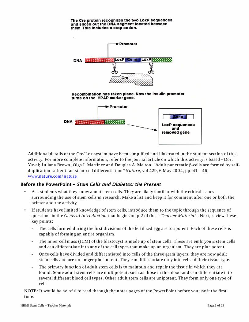
HHMI Stem Cells – Teacher Materials Page 8 of 23
Additional details of the Cre/Lox system have been simplified and illustrated in the student section of this activity. For more complete information, refer to the journal article on which this activity is based - Dor, Yuval; Juliana Brown; Olga I. Martinez and Douglas A. Melton “Adult pancreatic β-cells are formed by self-duplication rather than stem-cell differentiation” Nature, vol 429, 6 May 2004, pp. 41 – 46 www.nature.com/nature
Before the PowerPoint – Stem Cells and Diabetes: the Present
• Ask students what they know about stem cells. They are likely familiar with the ethical issues surrounding the use of stem cells in research. Make a list and keep it for comment after one or both the primer and the activity.
• If students have limited knowledge of stem cells, introduce them to the topic through the sequence of questions in the General Introduction that begins on p.2 of these Teacher Materials. Next, review these key points:
- The cells formed during the first divisions of the fertilized egg are totipotent. Each of these cells is capable of forming an entire organism.
- The inner cell mass (ICM) of the blastocyst is made up of stem cells. These are embryonic stem cells and can differentiate into any of the cell types that make up an organism. They are pluripotent.
- Once cells have divided and differentiated into cells of the three germ layers, they are now adult stem cells and are no longer pluripotent. They can differentiate only into cells of their tissue type.
- The primary function of adult stem cells is to maintain and repair the tissue in which they are found. Some adult stem cells are multipotent, such as those in the blood and can differentiate into several different blood cell types. Other adult stem cells are unipotent. They form only one type of cell.
NOTE: It would be helpful to read through the notes pages of the PowerPoint before you use it the first time.

HHMI Stem Cells – Teacher Materials Page 9 of 23
After the PowerPoint – Stem Cells and Diabetes: the Present
• Discuss why blood can be replaced and why an arm cannot. Show them the animation, Newt Limb Regeneration. Ask them what is present in a newt that enables it to regenerate a missing limb that is not present in humans. Follow this by asking why damaged nerve tissue such as occurs in spinal cord injury is not readily replaced. In all three examples, it is the presence or absence of adult stem cells. This prepares students for the pulse-chase β cell experiment.
• If you have not already assigned the Pulse-Chase Primer, this is a good time. It will provide the background necessary for understanding why researchers would use a pulse-chase analysis in the pancreatic beta cell investigation. The Pulse-Chase Primer can be assigned as homework.
• Before starting Are adult pancreatic beta cells formed by self-duplication or stem cell differentiation, view the following chapters from Lecture One of Potent Biology: Stem Cells, Cloning, and Regeneration?
#31 - Some cell types replenish by division, not by stem cells
#32 - A pulse-chase experiment on pancreatic cell replacement
Important: Do not show Chapters #33 and #34 as this would eliminate much of the inquiry component of the activity.
• The “Pulse-Chase Experiment Mechanism” portion of the activity is a good lesson in biotechnology and genetic engineering. It does require some basic DNA background. If students are not ready, this part of the activity can be omitted and the mechanism considered as a “black box.”
• At this point, assign the activity Are adult pancreatic β cells formed by self-duplication or stem cell differentiation?
After the Activity
• View the following chapters from Lecture One of Potent Biology: Stem Cells, Cloning, and Regeneration
Optional: Re-show #31 and #32.
#33 -New pancreatic beta cells are from division, not stem cells
#34 - In type I diabetes, no new beta cells can be made
Answers to Questions for Pancreatic Beta Cells
Activity: Are adult pancreatic beta cells formed by self-duplication or stem cell differentiation?
1. A, B, and C each represent one of the three outcomes predicted by Dr. Melton. For each one, indicate which outcome it represents and support your answer.
A New β cells are derived entirely through the mitotic division of existing β cells. The presence of about the same number of labeled β cells shows that cells labeled during the pulse phase of the experiment are dividing to produce new, labeled β cells. This is because through mitosis, all cells make two identical cells. If these cells are produced through mitosis, the percentage will stay approximately the same in the new group of cells. In this illustration, it is 100%.
B This illustrates that new beta cells are derived entirely from stem cells—cells not transcribing the insulin gene at the time of the pulse. There are no labeled β cells in the in the cross section because all of the β cells were replaced by stem cell derived cells. All of the β cells were formed after the pulse phase of the experiment.
C New β cells are derived by both the differentiation of stem cells and the mitotic division of existing β cells. The number of labeled cells is decreasing as stem cells differentiate to replace older, labeled β cells. These new β cells are not labeled since they were not producing insulin at the time of the pulse. There will be a small number of progeny of the labeled cells that persist.
2. Explain what would be expected to happen to the percentage of labeled beta cells in the islets if new beta cells are the result of the mitotic division of existing beta cells.

HHMI Stem Cells – Teacher Materials Page 10 of 23
The number of beta cells in the islets would remain approximately the same. There would be small fluctuations depending on the individual.
3. How would the results be different if the source of new beta cells involved both the mitotic division of existing cells and stem cells?
There would always be some labeled cells. However, the percentage would decrease over time since some new beta cells would result from the differentiation of unlabeled stem cells.
4. Explain what would happen over time if all new beta cells resulted from differentiation of stem cells.
The percentage of labeled cells would decrease since new beta cells would be produced entirely through the division and differentiation of stem cells. Eventually all of the beta cells would be unlabeled.
5. Which type of beta cell replacement is supported by the data? Explain.
This supports model A that states that new β cells are derived from exiting β cells and not from stem cells. The involvement of stems cells would cause the number of labeled β cells to decrease to the point where some of the islets would not contain any.
6. Using evidence from Dr. Melton’s pulse-chase analysis to support your answer, explain which cell type—adult stem cells or differentiated β cells—should be chosen to investigate as a possibility for cell-based therapy for type l diabetes.
Differentiated β cells should be selected.
7. (a) Identify a problem associated with the use of differentiated β cells in the research for possible cell-based therapies for type l diabetes.
Possible answers include but are not limited to: - There may be no β cells left in the patient since they are destroyed by the body’s own immune
system. - β cell transplants will require the use of immunosuppressant drugs. - β cells may be destroyed by the disease.
(b) Identify a problem associated with using adult stem cells in the research for possible cell-based therapies for type l diabetes.
Possible answers include but are not limited to: - Adult stem cells do not appear to be involved in β cell formation. - How would researchers obtain the adult stem cells?
Microarrays and Stem Cells
Materials
Per Class
• Stem Cells and Diabetes: The Future PowerPoint
• Potent Biology: Stem Cells, Cloning, and Regeneration DVD or access to the Internet
• Connect 4 Game
• Turkey baster
Per student
• a copy of the Microarray Stem Cell Activity
This includes: Microarray Background Information
What are some of the genes involved in the differentiation of ES cells to form pancreatic beta cells?
Analysis including the Stem Cell Differentiation Flowchart Gene Reference Sheet

HHMI Stem Cells – Teacher Materials Page 11 of 23
Microarray Summary Sheet Microarray Sheet for cell types 1 – 4 unshaded Microarray Template Sheet Microarray Sheet for cell types 5 – 8 shaded
• 1 set of short nucleotide sequences for cell types 1 - 4 in labeled sealable bags per two students
• 2 markers—1 red and 1 pink
Note: It is recommended that students work in groups of two.
Suggestions:
• You might want to enlarge and laminate the Microarray Template Sheets. Students could write on them with washable marker. The larger size provides more workspace. Laminating them makes it possible to wipe off student sequences so that the templates can be used multiple times.
• Use different colors of paper or cardstock for each of the cell type nucleotide sequences. For example, make the sequences for Cell Type 1 red, Cell Type 2 blue, Cell Type 3 green, and Cell Type 4 yellow. Place the sequences for each cell type in a small, labeled sealable bag. The different colors will make it easy to see that the proper sequences are in each bag and not mixed in with each other.
• Do not copy or print the Cell Types 1 – 4 Microarray Sheet and Cell Types 5 – 8 Microarray Sheet front and back. Marker tends to bleed through. Also, it makes the analysis of the eight microarrays more difficult.
• Do not staple the Gene Reference Sheet to the rest of the student materials. It should be a separate handout for easy reference.
• Have students work in groups of two or four. They can divide the work and share information amongst themselves. This will allow them to discuss their findings and work as a collaborative team when deciding the source of the various cell types. You could even have different teams determine the source of a particular cell type and ask each team to report their findings to the class. This will allow for discussion and validation.
Before the Microarray and Stem Cells Activity
• In class, work through the PowerPoint Stem Cells and Diabetes: The Future.
NOTE: It would be helpful to read trough the notes pages of the PowerPoint before you use it the first time.
• After students have read and answered the questions in the Microarray Background Information section, do the following demonstration to prepare students for what they will do next. Use a Connect 4 game to model how a microarray works. Use the video clip “Using a DNA Chip to Study Gene Expression” as a model for your own demonstration. Pretend that you are washing mRNA (or cDNA) from a hypothetical cell over the microarray. Dr. Melton uses a turkey baster to demonstrate the process. He then drops Connect 4 chips into the slots of the game. Explain to your students that each spot represents one gene. Red chips represent a gene that is expressing. Black chips represent genes that are not expressing. Tell students that some genes express in almost all cells most of the time. These are housekeeping genes that are essential to basic life processes. Other genes express only in specific cells. Designate one of the chips that is expressing as a housekeeping gene. This one could be labeled with a sticker or piece of colored tape to set it apart from the other chips. Provide students with scrap paper and have them make a sketch showing the location of the red and black chips and the housekeeping gene.
Take the chips out and simulate a different cell. Be sure that the housekeeping gene is in the same location and is expressing. Have students compare the microarray results of this cell type to their sketch of the first one. Ask them to describe what information they can get from the two microarray results.
Another strategy is to drop all black chips into the game. Have some of the chips painted or marked

HHMI Stem Cells – Teacher Materials Page 12 of 23
with a red sticker or on the side facing away from the students. Then shine your laser pointer on the Connect 4 chips and turn the game around so that the side with black, red, and a housekeeping gene now shows. This could simulate the multiple steps involved in the actual microarray process along with the fact that lasers and computers are involved in the actual analysis. This also could be done twice as above.
Next, share that some microarrays indicate more than just what genes are expressing. They also indicate to what degree the genes are expressing. This simple model does not show that. In the activity they will be doing, red represents a gene that is expressing to a high degree. Pink represents one that is expressing to a lesser degree. Genes that are not expressing will be left blank. This saves the students from having to color them in with black.
After the Microarray and Stem Cells Activity
• Ask students what the role of stem cells is in the growth and development of an organism such as themselves. Tell them the story about how early scientists thought that the sperm contained a very tiny, completely formed individual called a homunculus. When the sperm was placed inside a female, the homunculus would grow into a child. There were many arguments both for and against this idea. In chapter 5 of Lecture One, Dr. Melton discusses growth and development by using a sponge “grow animal” as an example of how a homunculus would grow into a child.
• Have students play Stem Cell Differentiation Game to review the facts and concepts introduced in the activities that makeup this lesson.
Answers to questions for Microarray Background Information 1. What information does the mRNA that is extracted from a sample provide researchers?
It tells researchers when a gene is turned on or expressed.
2. Explain how researchers are able to get embryonic stem cells grown in culture to spontaneously
differentiate.
The factors that allow stem cells to self-renew are removed.
3. State why fluorescently labeled nucleotides are used in the synthesis of cDNA.
When the fluorescently labeled cDNA hybridizes with the DNA on a microarray, a reader can then detect the presence and exact location of the cDNA. Special computer programs are used to calculate the fluorescence ratio for each spot. The calculated ratio reflects the relative expression of a given gene.
4. Explain why it is important to learn more about the molecular mechanisms involved in the
differentiation of embryonic stem cells to other cell types.
This knowledge would allow researchers to manipulate embryonic stem cells and encourage them to differentiate into the types of cells needed to treat disorders and injuries.
5. What information can microarrays provide scientists about embryonic stem cells?
Microarrays can provide researchers with information about which genes are being expressed in stem cells. This provides further information about how to either maintain stem cells in their differentiated state or how to initiate differentiation into specific cell types.

HHMI Stem Cells – Teacher Materials Page 13 of 23
Short Nucleotide Sequences for making sets of cell types 1-4 packets. Cut out individual sequences along the dashed lines. Place each cell type in a separate, labeled sealable storage bag. They can be used multiple times.
Cell Type One
ggtggcgtcgcc - 1 gactctgggagg - 1 ggaagcgttcgg - 1
ggtggcgtcgcc - 1 gataattgaaca - 1 aggggcggggct – 1
taatatttagat - 1 taatatttagat - 1 gataattgaaca – 1
gactctgggagg - 1 gactctgggagg - 1
Cell Type One
ggtggcgtcgcc - 1 gactctgggagg - 1 ggaagcgttcgg - 1
ggtggcgtcgcc - 1 gataattgaaca - 1 aggggcggggct – 1
taatatttagat - 1 taatatttagat - 1 gataattgaaca – 1
gactctgggagg - 1 gactctgggagg - 1
Cell Type One
ggtggcgtcgcc - 1 gactctgggagg - 1 ggaagcgttcgg - 1
ggtggcgtcgcc - 1 gataattgaaca - 1 aggggcggggct – 1
taatatttagat - 1 taatatttagat - 1 gataattgaaca – 1
gactctgggagg - 1 gactctgggagg - 1

HHMI Stem Cells – Teacher Materials Page 14 of 23
Cell Type Two
ggtggcgtcgcc - 2 gactctgggagg - 2 gactctgggagg - 2
ggtggcgtcgcc - 2 gactctgggagg - 2 gcgcacccgcgc - 2
gcgcacccgcgc - 2
Cell Type Two
ggtggcgtcgcc - 2 gactctgggagg - 2 gactctgggagg - 2
ggtggcgtcgcc - 2 gactctgggagg - 2 gcgcacccgcgc - 2
gcgcacccgcgc - 2
Cell Type Two
ggtggcgtcgcc - 2 gactctgggagg - 2 gactctgggagg - 2
ggtggcgtcgcc - 2 gactctgggagg - 2 gcgcacccgcgc - 2
gcgcacccgcgc - 2
Cell Type Two
ggtggcgtcgcc - 2 gactctgggagg - 2 gactctgggagg - 2
ggtggcgtcgcc - 2 gactctgggagg - 2 gcgcacccgcgc - 2
gcgcacccgcgc - 2

HHMI Stem Cells – Teacher Materials Page 15 of 23
Cell Type Three
ggtggcgtcgcc - 3 gactctgggagg – 3 gactctgggagg – 3
ggtggcgtcgcc - 3 gactctgggagg – 3
Cell Type Three
ggtggcgtcgcc - 3 gactctgggagg – 3 gactctgggagg – 3
ggtggcgtcgcc - 3 gactctgggagg – 3
Cell Type Three
ggtggcgtcgcc - 3 gactctgggagg – 3 gactctgggagg – 3
ggtggcgtcgcc - 3 gactctgggagg – 3
Cell Type Three
ggtggcgtcgcc - 3 gactctgggagg – 3 gactctgggagg – 3
ggtggcgtcgcc - 3 gactctgggagg – 3
Cell Type Three
ggtggcgtcgcc - 3 gactctgggagg – 3 gactctgggagg – 3
ggtggcgtcgcc - 3 gactctgggagg – 3
Cell Type Three
ggtggcgtcgcc - 3 gactctgggagg – 3 gactctgggagg – 3
ggtggcgtcgcc - 3 gactctgggagg – 3

HHMI Stem Cells – Teacher Materials Page 16 of 23
Cell Type Four
gtcgatgttagt - 4 cccgtcgttcct – 4 ctcttgcccctc - 4
ggagcctggggt - 4 cgccggcggcct - 4 ggcggccctctc - 4
cttgacagtttc - 4 cccgtcgttcct – 4 ggtggcgtcgcc - 4
gtcgatgttagt - 4 cccgtcgttcct – 4 cgccggcggcct – 4
Cell Type Four
gtcgatgttagt - 4 cccgtcgttcct – 4 ctcttgcccctc - 4
ggagcctggggt - 4 cgccggcggcct - 4 ggcggccctctc - 4
cttgacagtttc - 4 cccgtcgttcct – 4 ggtggcgtcgcc - 4
gtcgatgttagt - 4 cccgtcgttcct – 4 cgccggcggcct – 4
Cell Type Four
gtcgatgttagt - 4 cccgtcgttcct – 4 ctcttgcccctc - 4
ggagcctggggt - 4 cgccggcggcct - 4 ggcggccctctc - 4
cttgacagtttc - 4 cccgtcgttcct – 4 ggtggcgtcgcc - 4
gtcgatgttagt - 4 cccgtcgttcct – 4 cgccggcggcct – 4

HHMI Stem Cells – Teacher Materials Page 17 of 23
Answers to questions for What are some of the genes involved in the differentiation of ES cells to form pancreatic beta cells?
1-2 The complementary DNA sequences for the Microarray Template Sheet are recorded below. This will
serve as your reference for Cell Types 1 – 4.
Note: The cDNA is the top sequence in each spot and is underlined.
Brn4
cgctgagcagcg ttggagtaagag
IsL1
ctgagaccctcc
LeftyA
cagctacaatca
Ins
gcgactcgtcgc gtcgatgttagt aacctcattctc gactctgggagg
attataaatcta
Nanog
gagaacggggag
NeuroD1/Beta2
cctcggacccca
Ngn3
gcggccgccgga
Nkx2.2
taatatttagat ctcttgcccctc ggagcctggggt cgccggcggcct
Nkx6-1
ccgccgggagag ccttcgcaagcc
Oct-4
gggcagcaagga
Pax4
cgcgtgggcgcg
Pax6
ggcggccctctc ggaagcgttcgg cccgtcgttcct gcgcacccgcgc
gaactgtcaaag
Pdx-1
ctattaacttgt
SOX2
tccccgccccga
TDGF1
ccaccgcagcgg
ACTA1
cttgacagtttc gataattgaaca aggggcggggct ggtggcgtcgcc

HHMI Stem Cells – Teacher Materials Page 18 of 23
Completed microarrays for cell types 1 – 4:
Brn4 IsL1 LeftyAIns
Nanog NeuroD1/Beta2 Ngn3 Nkx2.2
Nkx6-1 Oct-4 Pax4 Pax6
Pdx-1 SOX2 TDGF1 ACTA1
Cell Type 1
Brn4 IsL1 LeftyAIns
Nanog NeuroD1/Beta2 Ngn3 Nkx2.2
Nkx6-1 Oct-4 Pax4 Pax6
Pdx-1 SOX2 TDGF1 ACTA1
Cell Type 2
Brn4 IsL1 LeftyAIns
Nanog NeuroD1/Beta2 Ngn3 Nkx2.2
Nkx6-1 Oct-4 Pax4 Pax6
Pdx-1 SOX2 TDGF1 ACTA1
Cell Type 3
Brn4 IsL1 LeftyAIns
Nanog NeuroD1/Beta2 Ngn3 Nkx2.2
Nkx6-1 Oct-4 Pax4 Pax6
Pdx-1 SOX2 TDGF1 ACTA1
Cell Type 4

HHMI Stem Cells – Teacher Materials Page 19 of 23
The microarray results for the eight cell types have been recorded in the table below. A short summary for each gene is included. Students should refer to their Gene Reference Sheets when determining the source of each cell type. There is not enough information provided for students to place cell types 3 and 5.
Cell Type Summary
1 (a) LeftyA controls the differentiation of stem cells into cells of the endoderm and mesoderm.
(b) Nanog is expressed in stem cells.
(c) Oct-4 helps maintain an undifferentiated phenotype.
(d) SOX2 is important in self-renewal.
(e) TDGF1 is important in maintaining pluripotency.
(f) ACTA1 is a housekeeping gene.
2 (a) LeftyA controls the differentiation of stem cells into cells of the endoderm and mesoderm.
(b) Pax6 codes for a transcription factor important in the development of the central nervous system and islet alpha cells of the pancreas.
(c) ACTA1 is a housekeeping gene.
3 (a) LeftyA controls the differentiation of stem cells into cells of the endoderm and mesoderm.
(b) ACTA1 is a housekeeping gene
4 (a) Ins is the insulin gene
(b) NeuroD1/beta2 helps regulate the formation of the pancreas and the expression of the insulin gene.
(c) Ngn3 regulates which cells differentiate into cells of the endocrine pancreas.
(d) Nkx2.2 encodes a transcription factor needed to initiate β cell differentiation.
(e) Nkx6-1 codes for the most β cell specific transcription factor.
(f) Pax4 is involved in the differentiation of β cells.
(g) Pdx-1 is an insulin promoter.
(h) ACTA1 is a housekeeping gene.
5 (a) IsL1 is a transcription factor that encodes the formation of islets in the pancreas. (b) NeuroD1/beta2 helps regulate the formation of the pancreas and the expression of the
insulin gene. (c) Ngn3 regulates which cells differentiate into cells of the endocrine pancreas. (d) ACTA1 is a housekeeping gene.
6 (a) IsL1 is a transcription factor. It encodes the formation of the islets in the pancreas.
(b) Ngn3 regulates which cells differentiate into cells of the endocrine pancreas.
(c) Nkx2.2 encodes a transcription factor needed to initiate β cell differentiation.
(d) Nkx6-1 codes for the most β cell specific transcription factor.
(e) Pax4 is involved in the differentiation of β cells.
(f) Pax6 codes for a transcription factor important in the development of islet alpha cells of the pancreas.
(g) ACTA1 is a housekeeping gene.

HHMI Stem Cells – Teacher Materials Page 20 of 23
7 (a) Nanog is expressed in stem cells.
(b) SOX2 is important in self-renewal.
(c) TDGF1 is important in maintaining pluripotency.
(d) ACTA1 is a housekeeping gene.
8 (a) Brn4 is expressed in glucagons-producing islets cell lines. (b) IsL1 is a transcription factor that encodes the formation of islets in the pancreas. (c) NeuroD1/beta2 helps regulate the formation of the pancreas and the expression of the
insulin gene. (d) ACTA1 is a housekeeping gene.
Analysis Answer key
1. Record the number of each cell type by the appropriate cell on the Stem Cell Differentiation Flowchart. Note: This will be possible for only 6 of the 8 cell types. Two cell types do not readily correlate to any particular cell type shown of the flowchart.
2. Two of the cell types you analyzed were impossible to place on the Stem Cell Differentiation Flowchart. Explain what additional steps researchers would need to take in order to know where these cells should be positioned on the flowchart.
Find out what genes are expressing in islet cells that secrete somatostatin or polypeptide or the mesoderm. Place sequences from these genes on a new microarray. Run the analysis and see if any of these genes are expressing in the unidentified cells.
Embryonic stem cell
From Stem Cell to Pancreatic Islet Stem Cell Differentiation Flowchart
___
Mesoderm cells
Endoderm cells ___
Embryonic stem cells ___
Ectoderm cells ___
Islet cell secretes pancreatic polypeptide
Islet cell secretes somatostatin
Islet beta cell secretes insulin
___
Islet cell secretes glucagon
___
7
8
4
6
2 1

HHMI Stem Cells – Teacher Materials Page 21 of 23
3. Pluruipotent stem cells sometimes divide to form more pluripotent stem cells. Other times they divide to form one or two cells that differentiate to become specialized tissues. Identify at least two factors that influence whether or not a stem cell differentiates after dividing.
Possible answers include but are not limited to:
- transcription factors present
- growth factors
- cytoplasmic factors
- signals from other cells
4. If scientists were to manipulate cultures of pluripotent stem cells to differentiate to form insulin-producing β cells, explain what factors would need to be regulated and why.
The answer should be the same as the response to question 2. Whatever factors influence stem cells in vivo will influence them in vitro.
5. Stem cells initially differentiate into cells that form the early ectoderm, mesoderm, and endoderm. Identify two or more tissues that develop from each of these germ layers.
Possible answers include but are not limited to: Ectoderm: skin and nervous system Mesoderm: Muscles and blood Endoderm: pancreas and gut tube.
6. Individuals receiving either islet or pancreas transplants must take immunosuppressant drugs. Would this be necessary if the source of the transplanted β cells was pluripotent stem cells manipulated in culture? Support your answer.
Possible answers include but are not limited to:
- Yes. They would trigger an immune response like any other foreign material.
- Yes. These cells would present antigens that would stimulate the production of antibodies.
7. The microarrays associated with each step provide information that can be used to eventually direct the differentiation process in culture.
Describe what type of information the microarrays provide and how it can be used to direct embryonic stem cells to differentiate to become β cells. (Note: Emphasize that a basic understanding of how this works is the purpose of this activity.)
Possible answers include but are not limited to:
Microarrays reveal which genes are expressing, to what degree they are expressing, and which are
HHMI Stem Cells – Teacher Materials Page 22 of 23
not. Researchers can then provide the appropriate transcription, growth, and cytoplasmic factors at the appropriate times. This should allow them to direct the differentiation process and obtain β cells needed for research or the treatment of diabetes.
8. Researchers are actively investigating how to deprogram and reprogram already differentiated cells so that they would become pluripotent stem cells. Such cells are referred to as induced pluripotent stem cells (iPS cells). If the technique is perfected, iPS cells could be created from a patient’s skin cells and then used to replace diseased or damaged tissue. Explain why this would be better than using embryonic stem cells obtained from a different source.
Possible answers include but are not limited to:
-This is better because these cells come from the same individual they are replacing the damaged or diseased tissue. The antigens displayed are those of the individual and should not stimulate an immune response.
-Better because the tissues produced from the individual’s iPS cells will have the same genetic information (and antigens) as other cells.
-No embryos are involved.
Note: To further the discussion of iPS cells, view the following chapters from Lecture Three of the 2008 Holiday Lecture DVD, Making Your Mind: Molecules, Motion, and Memory.
#39 - Using stem cells to create new motor neurons in mice
#40 - Using skin cells to make patient-specific stem cells
Thomas M. Jessell, Ph.D. explains that it is now possible to deprogram skin cells. They form iPS cells which when exposed to the right set of factors, become motor neurons.
References
Dor, Yuval; Juliana Brown; Olga I. Martinez and Douglas A. Melton “Adult pancreatic β-cells are formed by self-duplication rather than stem-cell differentiation” Nature, vol 429, 6 May 2004, pp. 41 - 46 www.nature.com/nature
HHMI Bulletin
“Sources of Renewal” by Robin Mejia, May 2008 pp 33 -36, 56
“Researchers Create Insulin-Producing Cells from Adult Pancreatic Cells” Research News. August 27, 2008
HHMI Holiday Lecture. Potent Biology: Stem Cells, Cloning, and Regeneration, DVD December 2006
Lester, Linda. Stem Cells in Endocrinology. Humana Press; 1 edition (February 22, 2005) ISBN-10: 1588294072 and ISBN-13: 978-1588294074
National Institutes of Health Snapshots. DNA Chips: A Genetics Lab in the Palm of Your Hand. http://www.sciencenetlinks.com/lessons.php?DocID=339
National Institutes of Health Resource for Stem Cell Research. Stem Cells and Diabetes. http://stemcells.nih.gov/info/scireport/chapter7.asp
NCBI A Science Primer http://www.ncbi.nlm.nih.gov/About/primer/microarrays.html

HHMI Stem Cells – Teacher Materials Page 23 of 23
Raj R. Rao and Steven L. Stice. “Gene Expression Profiling of Embryonic Stem Cells Leads to Greater Understanding of Pluripotency and Early Developmental Events” Biology of Reproduction 71, 1772–1778 (2004)
Zanta, Carolyn, Using Microarrays to Study Genes Involved in Cancer. UIUC-HHMI Biotechnology Education and Outreach Program (BEOP) www.life.uiuc.edu/hughes/footlocker
_______________________________________________________________________
Author: HHMI:
Mary Colvard Dennis WC. Liu, Ph.D. Cobleskill-Richmondville High School (retired) Satoshi Amagai, Ph.D. Cobleskill, NY Jennifer Bricken [email protected]
Field Testers:
Bender, Charles Guilderland Central School Guilderland, NY
Black, Pat Niskayuna High School Niskayuna, NY
Stephanie Branley Ravena-Coeymans-Selkirk HS Ravena, NY
Chien, Annie Schenectady City School District Schenectady, NY
Cole, Deb Shaker High School North Colonie Central Schools Latham, NY
Graves, Donna Bethlehem Central School District Delmar, NY
Hoffman, Kathy Shaker High School North Colonie Central Schools Latham, NY
McHale-Sullivan, Kelly Voorheesville CSD Voorheesville, NY
Raymond, Becky Deposit Central School Deposit, NY
Ryan, Kelly Shaker High School North Colonie Central Schools Latham, NY