stem cell recruitment of newly formed host cells via a successful
TRANSCRIPT

Stem Cell Recruitment of Newly Formed Host Cells via aSuccessful Seduction? Filling the Gap betweenNeurogenic Niche and Injured Brain SiteNaoki Tajiri1, Yuji Kaneko1, Kazutaka Shinozuka1, Hiroto Ishikawa1, Ernest Yankee2, Michael McGrogan2,Casey Case2, Cesar V. Borlongan1*
1 Center of Excellence for Aging and Brain Repair, Department of Neurosurgery and Brain Repair, University of South Florida Morsani College of Medicine,Tampa, Florida, United States of America, 2 Sanbio Inc, Mountain View, California, United States of America
Abstract
Here, we report that a unique mechanism of action exerted by stem cells in the repair of the traumatically injuredbrain involves their ability to harness a biobridge between neurogenic niche and injured brain site. This biobridge,visualized immunohistochemically and laser captured, corresponded to an area between the neurogenicsubventricular zone and the injured cortex. That the biobridge expressed high levels of extracellular matrixmetalloproteinases characterized initially by a stream of transplanted stem cells, but subsequently contained only fewto non-detectable grafts and overgrown by newly formed host cells, implicates a novel property of stem cells. Thetransplanted stem cells manifest themselves as pathways for trafficking the migration of host neurogenic cells, butonce this biobridge is formed between the neurogenic site and the injured brain site, the grafted cells disappear andrelinquish their task to the host neurogenic cells. Our findings reveal that long-distance migration of host cells fromthe neurogenic niche to the injured brain site can be achieved through transplanted stem cells serving as biobridgesfor initiation of endogenous repair mechanisms. This is the first report of a stem cell-paved “biobridge”. Indeed, todate the two major schools of discipline in stem cell repair mechanism primarily support the concept of “cellreplacement” and bystander effects of “trophic factor secretion”. The present novel observations of a stem cellseducing a host cell to engage in brain repair advances basic science concepts on stem cell biology and extracellularmatrix, as well as provokes translational research on propagating this stem cell-paved biobridge beyond cellreplacement and trophic factor secretion for the treatment of traumatic brain injury and other neurological disorders.
Citation: Tajiri N, Kaneko Y, Shinozuka K, Ishikawa H, Yankee E, et al. (2013) Stem Cell Recruitment of Newly Formed Host Cells via a SuccessfulSeduction? Filling the Gap between Neurogenic Niche and Injured Brain Site. PLoS ONE 8(9): e74857. doi:10.1371/journal.pone.0074857
Editor: Marcel Daadi, Stanford University School of Medicine, United States of America
Received June 18, 2013; Accepted August 6, 2013; Published September 4, 2013
Copyright: © 2013 Tajiri et al. This is an open-access article distributed under the terms of the Creative Commons Attribution License, which permitsunrestricted use, distribution, and reproduction in any medium, provided the original author and source are credited.
Funding: Financial support for this study was through Sanbio Inc. CVB is additionally funded by the James and Esther King Biomedical ResearchFoundation 1KG01-33966, NIH 5U01NS055914-04 and NIH 1R01NS071956-01A1, Celgene Cellular Therapeutics, KMPHC and NeuralStem Inc. Thefunders had no role in study design, data collection and analysis, decision to publish, or preparation of the manuscript.
Competing interests: The authors have read the journal's policy and have the following competing interest: CVB has patents and patent applications onstem cells, their therapeutic applications and mechanisms of action. In particular, the patents and patent applications relate to the use of stem cells fortreating human disorders, including the present study's target disease indication of traumatic brain injury. EY, MM and CC are employed by the funder ofthis study, Sanbio Inc. CVB is supported by National Institutes of Health, National Institute of Neurological Disorders and Stroke 1R01NS071956-01, Jamesand Esther King Foundation for Biomedical Research Program, SanBio Inc., Celgene Cellular Therapeutics, KMPHC and NeuralStem Inc. Senior authorCVB is a PLOS ONE Editorial Board member. Patents: 1995 Sertoli cells as neurorecovery inducing cells for neurodegenerative disorders. (US patent:6,036,951) Inventors: Sanberg, P.R., Borlongan, C.V. & Cameron, D.F. 1995 Sertoli cells as transplantation facilitator for cell transplantation. (US patent:5,942,437) Inventors: Sanberg, P.R., Borlongan, C.V. & Cameron, D.F. 1996 Method and media for enhancing cryopreservation of cells (US patent:6,037,175) Inventors: Sanberg, P.R., Borlongan, C.V., & Cameron, D.F. 1999 Melatonin as a protective agent against stroke. (US patent: 6,075,045)Inventors: Nishino, H. & Borlongan, C.V. 2001 Method of attenuating cognitive deficits with methanesulfonyl fluoride. (Patent pending) Inventors:Borlongan, C.V. & Moss, D. 2006 Multipotent progenitor cells rescue against stroke. (IP disclosure at MCG) Inventors: Carroll, J.E., Hess, D.C., &Borlongan, C.V. 2009 Human amnion as a source of stem cells for CNS therapy. (US Patent Pending) Inventors: Borlongan, C.V., & Parollini, O. 2009Delta opioid peptide for stroke therapy. (US Patent Pending) Inventors: Borlongan, C.V., Su, T.-P., Wang, Y. 2010 Methods for enhancing neuroprotectionvia administration of stem cells and blood brain barrier permeabilizers. US patent: 7,674,457 B2 Inventors: Borlongan, C.V., and Sanberg, P.R. This doesnot alter the authors' adherence to all the PLOS ONE policies on sharing data and materials.
* E-mail: [email protected]
Introduction
Initially employed for in-depth examination of celldevelopment [1], stem cells have become a cornerstone for
regenerative medicine in establishing cell-based therapies forneurological disorders [2,3]. A fundamental gap in ourknowledge about the mechanism underlying stem cell therapyremains unresolved. Functional recovery has been observed in
PLOS ONE | www.plosone.org 1 September 2013 | Volume 8 | Issue 9 | e74857

experimental models of neurological disorders despite few oreven absent survival of transplanted stem cells within theinjured brain site [4,5]. The original concept of direct cellreplacement has been challenged by the view that stem cellsafford indirect rescue of the injured tissue via secretion oftherapeutic molecules [6,7].
Stem cells exist even in adulthood [8], and possess thecapacity to self-renew and differentiate into multiple lineages[9], contribute to normal homeostasis [10], and exerttherapeutic benefits either endogenously [11–14] or followingtransplantation in injured organs, i.e., brain [15–21]. Thesubventricular zone (SVZ) of the lateral ventricles and thesubgranular zone of the hippocampus dentate gyrus (DG) arethe two major stem-cell niches in the adult brain [22,23],although quiescent neural stem cells (NSCs) have beendetected in other brain regions [24]. Induction of stem cellsafter injury corresponds to a new frontier in regenerativemedicine [2,3,11–21]. Indeed, laboratory studies on stem cellshave recently been translated into limited clinical trials for braindisorders [25–27]. Despite these scientific advances andclinical applications, much work remains to understand thestem cell-mediated repair mechanisms in brain injury.
The present study provides evidence of a novel therapeuticfeature of stem cells involving their ability to harness abiobridge between neurogenic niche and injured brain site in atraumatic brain injury (TBI) model. This biobridge expressedhigh levels of extracellular matrix metalloproteinases (ECM)characterized initially by a stream of transplanted stem cells,but subsequently replaced by newly formed host cells. Thetransplanted stem cells serve as migratory cues for hostneurogenic cells, guiding their exodus from the neurogenic sitetowards the injured brain site. Our findings reveal that long-distance migration of host cells from the neurogenic niche tothe injured brain site can be achieved through transplantedstem cells serving as biobridges for initiation of endogenousrepair mechanisms.
Materials and Methods
SummaryThis study was designed to evaluate potential therapeutic
value of intracerebral transplantation of cultured Notch-inducedhuman bone marrow-derived mesenchymal stromal cells(MSCs) (referred to as SB623, supplied by SanBio Inc.) [26,28]in an animal model of TBI. Transplantation was carried out at 7days after TBI with functional readouts of behavioral andhistological deficits conducted during the subsequent threemonth period after TBI. We characterized locomotor andneurological performance at baseline (prior to TBI), then at 7days after TBI (prior to transplantation), and monthly thereafterup to three months after TBI. Following completion ofbehavioral testing at one month or three months after TBI,animals were euthanized by transcardial perfusion and brainsharvested to histologically characterize the extent of braindamage. The stem cell engraftment and host tissueendogenous repair mechanisms (e.g., increased host cellsurvival in peri-TBI lesion area) were examined byimmunohistochemical analyses. A total of 40 animals identified
at baseline (prior to TBI surgery) as exhibiting normalbehaviors (EBST: 50-60% bias swing activity; Rotorod: 60seconds staying time on rotating rod; Bederson: at most 0-0.5mean neurologic score), received TBI surgery as describedbelow. Only TBI animals reaching the criterion of behavioralimpairment (EBST: at least 75% bias swing activity; Rotorod:30 seconds or less staying time on rotating rod; Bederson: atleast 2.5 mean neurologic score) were randomly assigned toeither SB623 transplants (n=20) or vehicle infusion (n=20) onDay 7 post-TBI. All animals were monitored monthly post-grafting for behavioral outcomes. Randomly selected animalswere euthanized at one month (n=10 per group) post-TBI, andthe remaining animals euthanized at three months post-TBI bytranscardial perfusion with 4% paraformaldehyde. For outcomemeasures, transplant outcome were evaluated using thefollowing parameters: 1) locomotor behavior via elevated bodyswing test (EBST) and Rotorod; (2) neurological performancevia a Bederson-modified neurological examination; 3) lesionvolume via hematoxylin and eosin (H&E) histologic stains; 4)graft survival via immunohistochemistry using specific antibodyshown to detect human cells, and; 5) mechanism-basedimmunohistochemial analyses of neuroprotection and/orregeneration using antibodies directed against the graftedhuman cells and host cells.
SubjectsThe University of South Florida Institutional Animal Care and
Use Committee approved all procedures used in this study.Animals had free access to food and water, and all werehoused under normal conditions (20°C, 50% relative humidity,and a 12 hour light/dark cycle).
TBI surgeryAll surgical procedures were conducted under aseptic
conditions. Adult male Sprague-Dawley (SD) rats (8-weeks old)were anesthetized with 1.5% isofluorane and checked for painreflexes. Under deep anesthesia, animals underwent themoderate TBI model. Each animal was placed in a stereotaxicframe (anesthesia maintained via gas mask) with 1-2%isoflurane. After exposing the skull, a 4.0 mm craniectomy wasperformed over the left frontoparietal cortex (center at −2.0 mmanteroposterior (AP) and +2.0 mm mediolateral (ML) tobregma) [29]. A pneumatically operated metal impactor(diameter = 3.0 mm) impacted the brain at a velocity of 6.0 m/sreaching a depth of 1.0 mm below the dura mater layer andremained in the brain for 150 milliseconds. The impactor rodwas angled 15° to the vertical to be perpendicular to thetangential plane of the brain curvature at the impact surface. Alinear variable displacement transducer (Macrosensors,Pennsauken, NJ) connected to the impactor measured velocityand duration to verify consistency. After controlled corticalimpact injury, the incision was sutured after bleeding ceased.An integrated heating pad and rectal thermometer unit withfeedback control allowed maintenance of body temperature atnormal limits. All animals were monitored until recovery fromanesthesia. In addition, animals were weighed and observeddaily for the next three consecutive days following TBI surgery,weighed twice a week thereafter, and monitored daily for health
Stem Cell-Paved Biobridges
PLOS ONE | www.plosone.org 2 September 2013 | Volume 8 | Issue 9 | e74857

status and any signs that indicate problems or complicationsthroughout the study.
Grafting proceduresAll surgical procedures were conducted under aseptic
conditions. Animals were anesthetized with 1.5% isofluoraneand checked for pain reflexes. Once deep anesthesia wasachieved (by checking for pain reflexes), hair was shavedaround the area of surgical incision (skull area) with enoughborder to prevent contaminating the operative site, followed bytwo surgical germicidal scrubs of site, and draping with steriledrapes. The animal was then fixed to a stereotaxic apparatus(Kopf Instruments). A 26-gauge Hamilton syringe was thenlowered into a small burred skull opening (transplantcoordinates were adjusted to correspond with the cortical areaadjacent to the core injury site: 0.5 mm anterior and 1.0 mmlateral to bregma and 2.0 mm below the dural surface [29]).Within this single needle pass, 3 deposits of the test article(100,000 cells in 3 µl per deposit or a total of 300,000 cells in 9µl of Plasmalyte A for 3 deposits) were made. The target areawas the medial cortex which corresponded to the peri-injuredcortical area, based on previously established target sites forsimilar stereotaxic implants. Each deposit consisted of 100,000viable cells in 3 µl volume infused over a period of 3 minutes.Following an additional 2-minute absorption time, the needlewas retracted and the wound closed stainless steel wound clip.A heating pad and a rectal thermometer allowed maintenanceof body temperature at about 37°C throughout surgery andfollowing recovery from anesthesia.
Behavioral and neurological testsAll investigators testing the animals were blinded to the
treatment condition. Animals were subjected to elevated bodyswing test (EBST), neurological exam, and Rotorod. EBSTinvolved handling the animal by its tail and recording thedirection of the swings. The test apparatus consisted of a clearPlexiglas box (40 x 40 x 35.5 cm). The animal was gentlypicked up at the base of the tail, and elevated by the tail untilthe animal’s nose was at a height of 2 inches (5 cm) above thesurface. The direction of the swing, either left or right, wascounted once the animals head moved sideways approximately10 degrees from the midline position of the body. After a singleswing, the animal was placed back in the Plexiglas box andallowed to move freely for 30 seconds prior to retesting. Thesesteps were repeated 20 times for each animal. Intact ratsdisplay a 50% swing bias, that is, the same number of swingsto the left and to the right. A 75% swing bias indicated 15swings in one direction and 5 in the other during 20 trials. Wehave previously utilized the EBST, and noted that unilaterallylesioned animals display >75% biased swing activity at onemonth after a nigrostriatal lesion or unilateral hemisphericinjury; asymmetry is stable for up to six months [3,26]. Aboutone hour after the EBST, a modified Bederson-Neurologicalexam was conducted following the procedures previouslydescribed [3,26] with minor modifications. Neurologic score foreach rat was obtained using 3 tests which include (1) forelimbretraction, which measured the ability of the animal to replacethe forelimb after it was displaced laterally by 2 to 3 cm, graded
from 0 (immediate replacement) to 3 (replacement after severalseconds or no replacement); (2) beam walking ability, graded 0for a rat that readily traversed a 2.4-cm-wide, 80-cm-long beamto 3 for a rat unable to stay on the beam for 10 seconds; and(3) bilateral forepaw grasp, which measured the ability to holdonto a 2-mm-diameter steel rod, graded 0 for a rat with normalforepaw grasping behavior to 3 for a rat unable to grasp withthe forepaws. The scores from all 3 tests, which were doneover a period of about 15 minutes on each assessment day,were added to give a mean neurologic deficit score (maximumpossible score, 9 points divided by 3 tests = 3). After an hour ofcompletion of the neurological exam, the animals were thensubjected to the Rotorod test. The Rotorod test involvedplacement of the animal on an accelerating Rotorod(Accuscan, Inc.) that used a rotating treadmill that acceleratesfrom 4 rpm to 40 rpm over a 60-second period. The totalnumber of seconds maintained on the Rotorod was recordedand used as index of motor coordination. We have previouslyshown that TBI model animals exhibited significantly shortertime staying on the Rotorod compared to sham operated ornormal controls. Animals were subjected to this battery of testsat baseline (prior to TBI), then at 7 days after TBI (prior totransplantation) and monthly thereafter up to three monthspost-TBI.
HistologyPerfusion. Brain section preparation was designed to
identify extent of brain damage and host cell survival. Atscheduled intervals (1 month or 3 months) after TBI, randomlyselected rats were euthanized (n=10 per group), and perfusedby transcardial perfusion with 4% paraformaldehyde. Thebrains were dissected, post-fixed for overnight in 4%paraformaldehyde, then subsequently immersed in 30%sucrose. Each forebrain was cut into 40 µm thick coronal tissuesections with anterior–posterior coordinates correspondingfrom bregma 5.2 mm to bregma -8.8 mm per animal andsubsequently processed for measurements of brain damageand analyses of cell survival in the peri-TBI lesion area (seebelow).
Measurements of brain damage. At least 4 coronal tissuesections per brain were processed for H&E staining. Everysixth coronal tissue sections per brain were collected beginningat AP -2.20 and ending at AP +0.20 anterior to the bregma, andrandomly selected for measurement of cortical core and peri-injury area [29]. The indirect lesion area, in which the intactarea of the ipsilateral hemisphere was subtracted from the areaof the contralateral hemisphere, was calculated to revealcerebral damage. The lesion volume was presented as avolume percentage of the lesion compared to the contralateralhemisphere.
Analyses of cell survival in peri-TBI lesionarea. Randomly selected high powerfield corresponding to theperi-injured cortical area was used to quantitatively count hostcells surviving in this region.
ImmunohistochemistryFree floating sections were processed for immunofluorescent
microscopy. Briefly, 40 µm cryostat sectioned tissues were
Stem Cell-Paved Biobridges
PLOS ONE | www.plosone.org 3 September 2013 | Volume 8 | Issue 9 | e74857

examined at 4X magnification and digitized using a PC-basedImage Tools computer program. Brain sections were blind-coded and Abercrombie’s formula was used to calculate thetotal number of immunopositive cells [3,26]. Cell engraftmentindex for SB623 was assessed using monoclonal humanspecific antibody (HuNu) that did not cross-react with rodentproteins. Additional brain sections were processed formechanism-based immunohistochemical analyses of braintissue samples focusing on cell proliferation (Ki67), migration(doublecortin or DCX) and immature neural marker (nestin).
ZymographyA separate cohort of animals consisting of TBI plus SB623
cells, TBI plus vehicle, and control-sham operated age-matched adult SD rats (n=3 per group) was subjected to thesame experimental paradigm as above, but tissues wereprocessed for zymography, a process involving electrophoreticseparation of proteins for assessment of proteolytic activity[30,31]. The tissue corresponding to the biobridge formed bythe migrating cells from the SVZ to the impacted cortex waslaser captured. After extraction, the tissue was placed incryotubes and flash frozen in liquid nitrogen. The tubes werestored in a -80°C freezer until homogenization. The sampleswere homogenized in 450 µL of cold working buffer containing50 mM Tris-HCl (pH 7.5), 75 mM NaCl, and 1 mM PMSF. Thetissue was processed with a homogenizer for 10 minutes andcentrifuged at 4°C for 20 minutes at 13000 rpm. Thesupernatants were separated, frozen and kept at -80°C untiluse. The total protein concentration was assessed by theBradford method. On the day of the zymography, the volumeequivalent to 50 µg of total protein was loaded into fresh madegelatin zymography gels. The gels were thenelectrophoretically separated under non-reducing conditionsand 100 V. After electrophoresis the zymogram gels werewashed in 125 ml 2.5% Triton twice for 20 minutes. The gelswere then incubated in activation buffer (ZymogramDevelopment Buffer, Bio-Rad, Hercules, CA) for 20 hours at37°C. The next day, the gels were stained with CoomassieBlue R-250 Staining Solution (Bio-Rad) for 3 hours anddestained for 25 minutes with Destain Solution (Bio-Rad). Thegelatinolytic activity of the samples was assessed bydensitometric analysis (Gel-Pro v 3.1, Media Cybernetics,Carlsbad, CA) of the bands as a relative comparison to astandard band of recombinant enzyme. To minimize inter-gelvariability, all gels had a control lane loaded with 0.5 ngrecombinant enzyme, which was used as a standard opticaldensity and enzyme amount (in ng). The lytic bands identifiedin the zymogram gels were subjected to molecular weightidentification with the use of pre-stained standard proteinmarker (Bio-Rad). Membranes were blocked with blotting gradeblocker non-fat dry milk (Bio-Rad). After washing with 0.1%tween 20- tris-buffered saline (TTBS), the membranes wereincubated with anti-matrix metalloproteinase (MMP)-9monoclonal mouse antibody overnight at 4°C. Membraneswere washed again in TTBS, incubated with secondaryantibody (goat anti-mouse IgG, horseradish peroxidaseconjugated antibody, Calbiochem) for one hour and finallydeveloped with horseradish peroxidase development solution
(ECL advance detection kit, Amersham). The membranes wereexposed to autoradiography films (Hyblot CL, DenvilleScientific Inc.). The density of the sample bands for thezymograms was expressed as maximal optical density relativeto the standard band.
Cell migration assayUsing a transwell assay, primary rat neuronal cells, PRNCs,
(embryos at Day 18; BrainBits) (1×10⁵ cells/well) seeded ontothe upper chamber of a Boyden chamber (Costar Transwellassay, Corning, NY, USA) supplemented with NbActive4(BrainBits) in the absence of antibiotics. The chamber wasplaced in a 24-well plate containing confluent SB623 cells(1×10⁵ cells/well) and starved with serum-free DMEM/F-12medium in the presence or absence of Cyclosporine-A (aknown MMP-9 inhibitor; 104 ng/mL in dimethyl sulfoxide;Sigma-Aldrich Inc., St Louis, MO, USA) for 24 h in the cellincubator. Next, the upper chamber was removed and wipedclean, then the lower side of the filter was washed and fixed in4% paraformaldehyde. For quantification, migratory cells thatreached the lower chamber and attached to the lower side ofthe filter were counted from five randomly captured microscopicfields (X400) and averaged for each treatment condition. Thismigratory assay was performed in triplicates.
Results
Adult male SD rats were initially evaluated in motor andneurological tests (all performed by two investigators blinded tothe treatment condition throughout the study) to confirm that allanimals included here were displaying normal behaviors atbaseline (i.e., prior to brain insult). Animals were exposed toexperimental TBI, then seven days later subjected to the samebehavioral tests to confirm the typical TBI-induced motor andneurological impairments, and thereafter (also at 7 days post-TBI) assigned in a random fashion to receive either stereotaxictransplants of either SB623 cells [26,32] or vehicle infusion intothe cortex (see Methods). At one month and three monthspost-TBI, transplanted animals displayed significantly improvedmotor and neurological functions coupled with significantlyreduced damage to the cortical core and peri-injured corticalareas compared to traumatically injured animals that receivedvehicle only (Figure 1). These behavioral and histologicalimprovements were achieved with modest graft survival of0.60% and 0.16% at one month and three months post-TBI,respectively. Based on the robust functional recovery despitelack of graft persistence, we next examined the status of thehost tissue. At one month post-TBI, immunofluorescent andconfocal microscopy revealed a surge of endogenous cellproliferation (Ki67) and immature neural differentiation (nestin)in the peri-injured cortical areas and SVZ, with a stream ofmigrating cells (DCX) along the corpus callosum (CC) of thetransplanted animals, while those that received vehicle alonedisplayed limited cell proliferation, neural differentiation, andscattered migration in the peri-injured cortical areas and almostabsent expression of newly formed cells in the SVZ (Figure 2).At three months post-TBI, the brains from transplanted animalsexhibited a much more massive cell proliferation and neural
Stem Cell-Paved Biobridges
PLOS ONE | www.plosone.org 4 September 2013 | Volume 8 | Issue 9 | e74857

differentiation encasing the peri-injured cortical areas (CTX)accompanied by a solid stream of neuronally labeled cells(nestin as well as DCX) migrating not just along but across theCC from the SVZ to the impacted CTX (Figure 3). In contrast,the brains from vehicle-infused animals while producing amuch more elevated cell proliferation, showed that newlyformed cells were “trapped” within the SVZ and the CC andonly sporadic cells were able to reach the impacted CTX(Figure 3). Quantitative analyses of Ki67, nestin and DCXimmunolabeled cells in SVZ, DG, CC and CTX revealedstatistically significant differences between transplanted andvehicle-infused animals (Figure S1). We next focused on thebiobridge formed by the migrating cells from the SVZ to theimpacted cortex and laser captured this corresponding tissue ina separate cohort of animals consisting of TBI plus SB623cells, TBI plus vehicle, and control-sham operated age-matched adult SD rats (n=3 per group) using the sameexperimental paradigm as above. Zymographic assaysrevealed two-fold and nine-fold upregulation of the MMP-9expression/activity in TBI transplanted animals at one monthand three months post-transplantation compared to vehicle-infused TBI animals or control-sham operated animals (Figure4A). Parallel in vitro studies provided further evidence thatSB623 cells promoted cell migration via an ECM-mediatedmechanism (Figure 4B).
Discussion
The present results revealed that SB623 transplantsremodeled the traumatically injured brain by harnessing abiobridgebetween SVZ and the peri-injured cortex (Figure 5).This new mechanism of stem cell therapy opens the possibilityof creating similar biobridges between neurogenic and non-neurogenic sites to facilitate injury-specific migration of cellsacross tissues that otherwise are non-conducive barriersagainst cell motility.
A Phase I/IIa transplantation study of SB623 cells in chronicstroke patients is underway. The clinical product entailsallogeneic SB623 cells. In cell culture and animal models ofbrain disorders, SB623 cells have been shown to attenuatebehavioral and histological deficits associated with stroke,spinal cord injury, and Parkinson’s disease [33–35]. Thepresent study is designed to extend the utility of SB623 in TBI.The US FDA recently approved a limited clinical trial oftransplanting SB623 cells in TBI, in part based on the databeing reported here. Thus, a major human implication impetusin this study was to provide the preclinical basis for initiating aclinical trial of SB623 in TBI.
The novel finding of SB623-facilitated migration ofendogenous cells via a biobridge implicates the active role of
MMPs and ECMs in stroke pathology [36,37] and theirincreasingly recognized role as therapeutic targets for stroke[38,39]. A variety of stem cells, including those derived fromumbilical cord blood, peripheral blood, and adult brain, havebeen demonstrated to alter levels and functions of MMPs andECMs [40–42], which would suggest their potential to similarlyserve as biobridges as seen with the present Notch-inducedSB623 MSCs.
Although neurogenic niches in the adult brain, such as theSVZ, have now been documented to exist and demonstrated tobe critical in the repair of the stroke brain [43–47], a key limitingfactor for endogenous repair is the successful migration ofthese newly formed host cells to reach the ischemic brain area.Our present results suggest that SB623 cell transplantationboosted endogenous repair mechanisms by guiding themigration of new cells from the neurogenic SVZ, across a non-neurogenic brain area, and eventually reaching the site ofinjury. The fundamental mechanism of action of SB623 cellsinvolves their capacity to form biobridges consisting of MMPsand ECMs which serve as a gateway to ferry the newly formedcells from the neurogenic niche into the ischemic tissue.Although the grafted SB623 pioneered the formation of thesebiobridges, they subsequently relinquished these biobridges tothe endogenous stem cells, altogether facilitating the host brainremodeling process. Our findings directly advance the conceptof a biobridge mechanism as a robust stem cell-mediated brainrepair strategy in TBI, and provide pivotal guidance on thetranslational applications of cell therapy in TBI patients. Futurestudies require closely monitoring the long-term efficacy andsafety of SB623 cell therapy in chronic TBI animals in order tofurther optimize the conduct of the clinical trial of these cells inTBI patients.
A basic knowledge gap in functional restoration after stemcell transplantation is the elusive demonstration of integrationof grafted cells into the recipient brain tissue and theirsubsequent interaction with host cells. The cellular interactionbetween the transplanted cell and host cell becomes extremelyessential when graft survival is mediocre, indicating that forrobust and stable therapeutic benefits an endogenous repairmechanism must be set in motion by the graft, in particularfinding a way for the host cells to reach their destination evenacross non-neurogenic and injured tissues. MMPs have beenimplicated in recovery in chronic brain injury [32], with MMPinhibition abrogating neurogenic migration from SVZ intodamaged tissues and retarding neurovascular remodeling [48].Stem cells may serve as biobridges expressing MMP profilesthat recapitulate the neurovascular unit abetting the transplant-mediated host cell migration towards injured brain areas inaffording functional recovery in TBI.
Stem Cell-Paved Biobridges
PLOS ONE | www.plosone.org 5 September 2013 | Volume 8 | Issue 9 | e74857
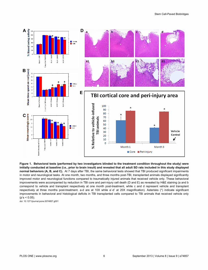
Figure 1. Behavioral tests (performed by two investigators blinded to the treatment condition throughout the study) wereinitially conducted at baseline (i.e., prior to brain insult) and revealed that all adult SD rats included in this study displayednormal behaviors (A, B, and C). At 7 days after TBI, the same behavioral tests showed that TBI produced significant impairmentsin motor and neurological tasks. At one month, two months, and three months post-TBI, transplanted animals displayed significantlyimproved motor and neurological functions compared to traumatically injured animals that received vehicle only. These behavioralimprovements were accompanied by reduction in TBI core and peri-injury cell death (D and E) as revealed by H&E staining (a and bcorrespond to vehicle and transplant respectively at one month post-treatment, while c and d represent vehicle and transplantrespectively at three months post-treatment. a-d are at 10X while a’-d’ at 20X magnification). Asterisks (*) indicate significantimprovements in behavioral and histological deficits in TBI transplanted cells compared to TBI animals that received vehicle only(p’s < 0.05).doi: 10.1371/journal.pone.0074857.g001
Stem Cell-Paved Biobridges
PLOS ONE | www.plosone.org 6 September 2013 | Volume 8 | Issue 9 | e74857

Figure 2. The biobridge between SVZ and impacted cortex consists of highly proliferative, neurally committed, andmigratory cells. At one month post-TBI, confocal microscopy revealed a surge of proliferative Ki67 positive cells and immatureneurally nestin labeled cells in the peri-injured cortical areas (A) and subventricular zone (A’), with a stream of migrating cells (DCX)along the corpus callosum (B) in TBI animals that received the stem cell transplants. In contrast those that received vehicle alonedisplayed limited cell proliferation (C), neural differentiation (C’), and scattered migration in the peri-injured cortical areas (D) andalmost absent expression of newly formed cells in the SVZ (C’).doi: 10.1371/journal.pone.0074857.g002
Stem Cell-Paved Biobridges
PLOS ONE | www.plosone.org 7 September 2013 | Volume 8 | Issue 9 | e74857

Figure 3. At three months post-TBI, the brains from vehicle-infused animals displayed a disparate pattern of cell fate inthat the newly formed Ki67 positive and nestin labeled cells were sequestered within the corpus callosum (A) and the SVZ(B) and only sporadic cells were able to reach the impacted cortex (A’ and B’), with likely resident DCX cells seen aroundthe impacted cortex (C). In contrast, at three months post-TBI, the brains from transplanted animals exhibited a much moremassive cell proliferation and neural differentiation encasing the peri-injured cortical areas accompanied by a solid stream of nestin(D, D’) and DCX labeled cells (E) migrating not just along, but across the corpus callosum from the SVZ to the impacted cortex.doi: 10.1371/journal.pone.0074857.g003
Stem Cell-Paved Biobridges
PLOS ONE | www.plosone.org 8 September 2013 | Volume 8 | Issue 9 | e74857

Figure 4. Laser-captured biobridge, corresponding to the brain tissue between SVZ and impacted cortex, expressed highlevels of MMP-9 gelatinolytic activities at one month and three months post-TBI in animals transplanted with SB623 whichwere significantly higher than those TBI animals that received vehicle only or sham-operated animals (*p’s< 0.05 vs.vehicle or sham; Panel A). Although vehicle-infused TBI animals also showed a significantly upregulated MMP-9 gelatinolyticactivity at one month post-TBI (**p< 0.05 vs. sham), the level of this neurovascular proteinase activity reverted back to control-shamlevels at three months post-TBI. Each bar represents the mean ± standard deviation from n=3 per treatment group for each timepoint. Next, to further reveal that SB623 cells promoted cell migration via an ECM-mediated mechanism, primary rat cortical cellswere either grown alone or co-cultured with SB623 in the presence or absence of the MMP-9 inhibitor Cyclosporine-A (Panel B).Migratory cell assay (see inset) revealed significantly enhanced migration of primary rat cortical cells into the chamber thatcontained SB623, which was significantly suppressed by treatment with the inhibitor (*p< 0.05 vs. all other treatment conditions).The absence of SB623 and inhibitor in the cell culture condition, the treatment of the inhibitor alone, and the combined treatment ofSB623 and inhibitor did not significantly differ in the resulting cell migratory potential.doi: 10.1371/journal.pone.0074857.g004
Stem Cell-Paved Biobridges
PLOS ONE | www.plosone.org 9 September 2013 | Volume 8 | Issue 9 | e74857
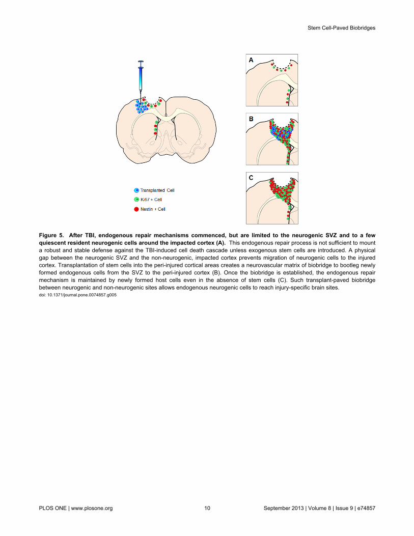
Figure 5. After TBI, endogenous repair mechanisms commenced, but are limited to the neurogenic SVZ and to a fewquiescent resident neurogenic cells around the impacted cortex (A). This endogenous repair process is not sufficient to mounta robust and stable defense against the TBI-induced cell death cascade unless exogenous stem cells are introduced. A physicalgap between the neurogenic SVZ and the non-neurogenic, impacted cortex prevents migration of neurogenic cells to the injuredcortex. Transplantation of stem cells into the peri-injured cortical areas creates a neurovascular matrix of biobridge to bootleg newlyformed endogenous cells from the SVZ to the peri-injured cortex (B). Once the biobridge is established, the endogenous repairmechanism is maintained by newly formed host cells even in the absence of stem cells (C). Such transplant-paved biobridgebetween neurogenic and non-neurogenic sites allows endogenous neurogenic cells to reach injury-specific brain sites.doi: 10.1371/journal.pone.0074857.g005
Stem Cell-Paved Biobridges
PLOS ONE | www.plosone.org 10 September 2013 | Volume 8 | Issue 9 | e74857

Supporting Information
Figure S1. Quantifications of Ki67, nestin and DCX labeledcells are shown in panels A, B, and C, respectively.Asterisks (*) indicate significant increase in the number ofphenotypically labeled cells counted per high-power field view(28,800 µm2) selected at random in the region of interest in TBIanimals transplanted with SB623 cells compared to TBIanimals that received vehicle only (p’s < 0.05).
(TIF)
Author Contributions
Conceived and designed the experiments: EY MM CC CVB.Performed the experiments: NT YK KS HI CVB. Analyzed thedata: NT EY MM CC CVB. Contributed reagents/materials/analysis tools: EY MM CC CVB. Wrote the manuscript: NT EYMM CC CVB.
References
1. Joyner AL, Skarnes WC, Rossant J (1989) Production of a mutation inmouse En-2 gene by homologous recombination in embryonic stemcells. Nature 338: 153-156. doi:10.1038/338153a0. PubMed: 2563902.
2. Yasuhara T, Matsukawa N, Hara K, Yu G, Xu L et al. (2006)Transplantation of human neural stem cells exerts neuroprotection in arat model of Parkinson’s disease. J Neurosci 26: 12497-12511. doi:10.1523/JNEUROSCI.3719-06.2006. PubMed: 17135412.
3. Yasuhara T, Hara K, Maki M, Mays RW, Deans RJ et al. (2008)Intravenous grafts recapitulate the neurorestoration afforded byintracerebrally delivered multipotent adult progenitor cells in neonatalhypoxic-ischemic rats. J Cereb Blood Flow Metab 28: 1804-1810. doi:10.1038/jcbfm.2008.68. PubMed: 18594556.
4. Borlongan CV, Hadman M, Sanberg CD, Sanberg PR (2004) Centralnervous system entry of peripherally injected umbilical cord blood cellsis not required for neuroprotection in stroke. Stroke 35: 2385-2389. doi:10.1161/01.STR.0000141680.49960.d7. PubMed: 15345799.
5. Pastori C, Librizzi L, Breschi GL, Regondi C, Frassoni C et al. (2008)Arterially perfused neurosphere-derived cells distribute outside theischemic core in a model of transient focal ischemia and reperfusion invitro. PLOS ONE 3: e2754. doi:10.1371/journal.pone.0002754.PubMed: 18648648.
6. Redmond DE Jr, Bjugstad KB, Teng YD, Ourednik V, Ourednik J et al.(2007) Behavioral improvement in a primate Parkinson’s model isassociated with multiple homeostatic effects of human neural stemcells. Proc Natl Acad Sci U S A 104: 12175-12180. doi:10.1073/pnas.0704091104. PubMed: 17586681.
7. Lee JP, Jeyakumar M, Gonzalez R, Takahashi H, Lee PJ et al. (2007)Stem cells act through multiple mechanisms to benefit mice withneurodegenerative metabolic disease. Nat Med 13: 439-447. doi:10.1038/nm1548. PubMed: 17351625.
8. Ma DK, Marchetto MC, Guo JU, Ming GL, Gage FH et al. (2010)Epigenetic choreographers of neurogenesis in the adult mammalianbrain. Nat Neurosci 13: 1338-1344. doi:10.1038/nn.2672. PubMed:20975758.
9. Hong SH, Rampalli S, Lee JB, McNicol J, Collins T et al. (2011) Cellfate potential of human pluripotent stem cells is encoded by histonemodifications. Cell Stem Cell 9: 24-36. doi:10.1016/j.stem.2011.06.002.PubMed: 21726831.
10. Kim Y, Sharov AA, McDole K, Cheng M, Hao H et al. (2011) Mouse B-type lamins are required for proper organogenesis but not byembryonic stem cells. Science 334: 1706-1710.
11. Borlongan CV (2011) Bone marrow stem cell mobilization in stroke: a‘bonehead’ may be good after all! Leukemia 25: 1674-1686. doi:10.1038/leu.2011.167. PubMed: 21727900.
12. Barha CK, Ishrat T, Epp JR, Galea LA, Stein DG (2011) Progesteronetreatment normalizes the levels of cell proliferation and cell death in thedentate gyrus of the hippocampus after traumatic brain injury. ExpNeurol 231: 72-81. doi:10.1016/j.expneurol.2011.05.016. PubMed:21684276.
13. Jaskelioff M, Muller FL, Paik JH, Thomas E, Jiang S et al. (2011)Telomerase reactivation reverses tissue degeneration in agedtelomerase-deficient mice. Nature 469: 102-106. doi:10.1038/nature09603. PubMed: 21113150.
14. Wang L, Chopp M, Teng H, Bolz M, Francisco MA et al. (2011) Tumornecrosis factor α primes cerebral endothelial cells for erythropoietin-induced angiogenesis. J Cereb Blood Flow Metab 31: 640-647. doi:10.1038/jcbfm.2010.138. PubMed: 20700128.
15. Andres RH, Horie N, Slikker W, Keren-Gill H, Zhan K et al. (2011)Human neural stem cells enhance structural plasticity and axonaltransport in the ischaemic brain. Brain 134: 1777-1789. doi:10.1093/brain/awr094. PubMed: 21616972.
16. Liu Z, Li Y, Zhang RL, Cui Y, Chopp M (2011) Bone marrow stromalcells promote skilled motor recovery and enhance contralesional axonalconnections after ischemic stroke in adult mice. Stroke 42: 740-744.doi:10.1161/STROKEAHA.110.607226. PubMed: 21307396.
17. Mazzocchi-Jones D, Döbrössy M, Dunnett SB (2009) Embryonic striatalgrafts restore bi-directional synaptic plasticity in a rodent model ofHuntington's disease. Eur J Neurosci 30: 2134-2142. doi:10.1111/j.1460-9568.2009.07006.x. PubMed: 20128850.
18. Lee HS, Bae EJ, Yi SH, Shim JW, Jo AY et al. (2010) Foxa2 and Nurr1synergistically yield A9 nigral dopamine neurons exhibiting improveddifferentiation, function, and cell survival. Stem Cells 28: 501-512.PubMed: 20049900.
19. Hargus G, Cooper O, Deleidi M, Levy A, Lee K et al. (2010)Differentiated Parkinson patient-derived induced pluripotent stem cellsgrow in the adult rodent brain and reduce motor asymmetry inParkinsonian rats. Proc Natl Acad Sci U S A 107: 15921-15926.PubMed: 20798034.
20. Yasuda A, Tsuji O, Shibata S, Nori S, Takano M et al. (2011)Significance of remyelination by neural stem/progenitor cellstransplanted into the injured spinal cord. Stem Cells 29: 1983-1994.doi:10.1002/stem.767. PubMed: 22028197.
21. Mezey E (2011) The therapeutic potential of bone marrow-derived stemcells. J Cell Biochem 112: 2683-2687. doi:10.1002/jcb.23216. PubMed:21678464.
22. Sanai N, Nguyen T, Ihrie RA, Mirzadeh Z, Tsai HH et al. (2011)Corridors of migrating neurons in the human brain and their declineduring infancy. Nature 478: 382-386. doi:10.1038/nature10487.PubMed: 21964341.
23. Carlén M, Meletis K, Göritz C, Darsalia V, Evergren E et al. (2009)Forebrain ependymal cells are Notch-dependent and generateneuroblasts and astrocytes after stroke. Nat Neurosci 12: 259-267. doi:10.1038/nn.2268. PubMed: 19234458.
24. Robel S, Berninger B, Götz M (2011) The stem cell potential of glia:lessons from reactive gliosis. Nat Rev Neurosci 12: 88-104. doi:10.1038/nrn2978. PubMed: 21248788.
25. Seol HJ, Jin J, Seong DH, Joo KM, Kang W et al. (2011) Geneticallyengineered human neural stem cells with rabbit carboxyl esterase cantarget brain metastasis from breast cancer. Cancer Lett 311: 152-159.doi:10.1016/j.canlet.2011.07.001. PubMed: 21868150.
26. Yasuhara T, Matsukawa N, Hara K, Maki M, Ali MM et al. (2009) Notch-induced rat and human bone marrow stromal cell grafts reduceischemic cell loss and ameliorate behavioral deficits in chronic strokeanimals. Stem Cells Dev 18: 1501-1514. doi:10.1089/scd.2009.0011.PubMed: 19301956.
27. Pollock K, Stroemer P, Patel S, Stevanato L, Hope A et al. (2006) Aconditionally immortal clonal stem cell line form human corticalneuroepithelium for the treatment of ischemic stroke. Exp Neurol 199:143-155. doi:10.1016/j.expneurol.2005.12.011. PubMed: 16464451.
28. Dezawa M, Kanno H, Hoshino M, Cho H, Matsumoto N et al. (2004)Specific induction of neuronal cells from bone marrow stromal cells andapplication for autologous transplantation. J Clin Invest 113:1701-1710. doi:10.1172/JCI20935. PubMed: 15199405.
29. Paxinos G, Watson C (2007) The Rat Brain in Stereotaxic Coordinates:Hard Cover Edition. Academic Press.
30. Hawkes SP, Li H, Taniguchi GT (2010) Zymography and reversezymography for detecting MMPs and TIMPs. Methods Mol Biol 622:257-269. doi:10.1007/978-1-60327-299-5_16. PubMed: 20135288.
31. Machado LS, Kozak A, Ergul A, Hess DC, Borlongan CV et al. (2006)Delayed minocycline inhibits ischemia-activated matrixmetalloproteinases 2 and 9 after experimental stroke. BMC Neurosci 7:56. doi:10.1186/1471-2202-7-56. PubMed: 16846501.
Stem Cell-Paved Biobridges
PLOS ONE | www.plosone.org 11 September 2013 | Volume 8 | Issue 9 | e74857

32. Zhao BQ, Tejima E, Lo EH (2007) Neurovascular proteases in braininjury, hemorrhage and remodeling after stroke. Stroke 38: 748-752.doi:10.1161/01.STR.0000253500.32979.d1. PubMed: 17261731.
33. Aizman I, Tate CC, McGrogan M, Case CC (2009) Extracellular matrixproduced by bone marrow stromal cells and by their derivative, SB623cells, supports neural cell growth. J Neurosci Res 87: 3198-3206. doi:10.1002/jnr.22146. PubMed: 19530164.
34. Tate CC, Fonck C, McGrogan M, Case CC (2010) Humanmesenchymal stromal cells and their derivative, SB623 cells, rescueneural cells via trophic support following in vitro ischemia. CellTransplant 19: 973-984. doi:10.3727/096368910X494885. PubMed:20350349.
35. Dao M, Tate CC, McGrogan M, Case CC (2013) Comparing theangiogenic potency of naive marrow stromal cells and Notch-transfected marrow stromal cells. J Transl Med 11: 81. doi:10.1186/1479-5876-11-81. PubMed: 23531336.
36. Park KP, Rosell A, Foerch C, Xing C, Kim WJ et al. (2009) Plasma andbrain matrix metalloproteinase-9 after acute focal cerebral ischemia inrats. Stroke 40: 2836-2842. doi:10.1161/STROKEAHA.109.554824.PubMed: 19556529.
37. del Zoppo GJ, Frankowski H, Gu YH, Osada T, Kanazawa M et al.(2012) Microglial cell activation is a source of metalloproteinasegeneration during hemorrhagic transformation. J Cereb Blood FlowMetab 32: 919-932. doi:10.1038/jcbfm.2012.11. PubMed: 22354151.
38. Tejima E, Guo S, Murata Y, Arai K, Lok J et al. (2009) Neuroprotectiveeffects of overexpressing tissue inhibitor of metalloproteinase TIMP-1. JNeurotrauma 26: 1935-1941. doi:10.1089/neu.2009.0959. PubMed:19469687.
39. Chen J, Cui X, Zacharek A, Cui Y, Roberts C et al. (2011) White matterdamage and the effect of matrix metalloproteinases in type 2 diabeticmice after stroke. Stroke 42: 445-452. doi:10.1161/STROKEAHA.110.596486. PubMed: 21193743.
40. Barkho BZ, Munoz AE, Li X, Li L, Cunningham LA et al. (2008)Endogenous matrix metalloproteinase (MMP)-3 and MMP-9 promote
the differentiation and migration of adult neural progenitor cells inresponse to chemokines. Stem Cells 26: 3139-3149. doi:10.1634/stemcells.2008-0519. PubMed: 18818437.
41. Sobrino T, Pérez-Mato M, Brea D, Rodríguez-Yáñez M, Blanco M et al.(2012) Temporal profile of molecular signatures associated withcirculating endothelial progenitor cells in human ischemic stroke. JNeurosci Res 90: 1788-1793. doi:10.1002/jnr.23068. PubMed:22513751.
42. Lin CH, Lee HT, Lee SD, Lee W, Cho CW et al. (2013) Role ofHIF-1alpha-activated Epac1 on HSC-mediated neuroplasticity in strokemodel. Neurobiol Dis 58C: 76-91.
43. Ekdahl CT, Kokaia Z, Lindvall O (2009) Brain inflammation and adultneurogenesis: the dual role of microglia. Neuroscience 158: 1021-1029.doi:10.1016/j.neuroscience.2008.06.052. PubMed: 18662748.
44. Hassani Z, O’Reilly J, Pearse Y, Stroemer P, Tang E et al. (2012)Human neural progenitor cell engraftment increases neurogenesis andmicroglial recruitment in the brain of rats with stroke. PLOS ONE 7:e50444. doi:10.1371/journal.pone.0050444. PubMed: 23185625.
45. Trueman RC, Klein A, Lindgren HS, Lelos MJ, Dunnett SB (2013)Repair of the CNS using endogenous and transplanted neural stemcells. Curr Top Behav Neurosci 15: 357-398. PubMed: 22907556.
46. Wang X, Mao X, Xie L, Sun F, Greenberg DA et al. (2012) Conditionaldepletion of neurogenesis inhibits long-term recovery afterexperimental stroke in mice. PLOS ONE 7: e38932. doi:10.1371/journal.pone.0038932. PubMed: 22723908.
47. Ducruet AF, Zacharia BE, Sosunov SA, Gigante PR, Yeh ML et al.(2012) Complement inhibition promotes endogenous neurogenesis andsustained anti-inflammatory neuroprotection following reperfusedstroke. PLOS ONE 7: e38664. doi:10.1371/journal.pone.0038664.PubMed: 22761695.
48. Zhao BQ, Wang S, Kim HY, Storrie H, Rosen BR et al. (2006) Role ofmatrix metalloproteinases in delayed cortical responses after stroke.Nat Med 12: 441-445. doi:10.1038/nm1387. PubMed: 16565723.
Stem Cell-Paved Biobridges
PLOS ONE | www.plosone.org 12 September 2013 | Volume 8 | Issue 9 | e74857