stem cell reports · neonatal mammalian hearts do enclose a true cardiac- ... heart following ar....
TRANSCRIPT

Stem Cell Reports
ReportDo Neonatal Mouse Hearts Regenerate following Heart Apex Resection?
Ditte Caroline Andersen,1,2,* Suganya Ganesalingam,1,3 Charlotte Harken Jensen,1
and Søren Paludan Sheikh1,3,*1Laboratory of Molecular and Cellular Cardiology, Department of Clinical Biochemistry and Pharmacology, Odense University Hospital,
Winsloewparken 213rd, 5000 Odense C, Denmark2Clinical Institute, University of Southern Denmark, 5000 Odense C, Denmark3Institute of Molecular Medicine, University of Southern Denmark, 5000 Odense C, Denmark
*Correspondence: [email protected] (D.C.A.), [email protected] (S.P.S.)
http://dx.doi.org/10.1016/j.stemcr.2014.02.008
This is an open access article under the CC BY-NC-ND license (http://creativecommons.org/licenses/by-nc-nd/3.0/).
SUMMARY
The mammalian heart has generally been considered nonregenerative, but recent progress suggests that neonatal mouse hearts have a
genuine capacity to regenerate following apex resection (AR). However, in this study, we performed AR or sham surgery on 400 neonatal
mice from inbred and outbred strains and found no evidence of complete regeneration. Ideally, new functional cardiomyocytes, endo-
thelial cells, and vascular smooth muscle cells should be formed in the necrotic area of the damaged heart. Here, damaged hearts were
9.8% shorter and weighed 14% less than sham controls. In addition, the resection border contained a massive fibrotic scar mainly
composed of nonmyocytes and collagen disposition. Furthermore, there was a substantial reduction in the number of proliferating car-
diomyocytes in AR hearts. Our results thus question the usefulness of the AR model for identifying molecular mechanisms underlying
regeneration of the adult heart after damage.
INTRODUCTION
A key question in cardiovascular biology is to what degree
the heart is able to regenerate after tissue damage from
either cardiac stem cells or cardiomyocyte division. Cardio-
vascular disease including myocardial infarct is currently
one of the leading causes of death worldwide, and the gen-
eral view is that this is mainly caused by a genuine inability
of themammalian heart to regenerate upon damage (Vieira
and Riley, 2011). Yet, this dogma was recently challenged
by exciting data suggesting that the mouse heart retains
regenerative ability up to 1 week after birth (Porrello
et al., 2011), and without being reproduced by others, it
has now been accepted as an established principle that
neonatal mammalian hearts do enclose a true cardiac-
regenerative potential following apex resection (AR)
(Aguirre et al., 2013; Garbern and Lee, 2013). As a minimal
requirement, complete cardiac regeneration should
include the restoration of the functional continuity of car-
diomyocytes, as well as blood supply in the necrotic area of
the damaged heart with no sign of scar formation. Indeed,
urodele amphibians and zebrafish have been shown to
possess a high capacity to repair the heart following dam-
age such as AR that meets these minimal criteria (Garbern
et al., 2013). Accordingly, the zebrafish heart is regenerated
in 60 days following AR, with full recovery of the myocar-
dium (Poss et al., 2002). The mammal and zebrafish heart
anatomy/physiology diverge substantially (Garbern et al.,
2013). It was therefore a breakthrough in regenerativemed-
icine of the mammalian heart when Porrello et al. in 2011
showed that the neonatal mouse heart (1 day old) holds an
406 Stem Cell Reports j Vol. 2 j 406–413 j April 8, 2014 j ª2014 The Author
STEMCR 103
intrinsic capability to regenerate completely following
resection of 10% of the heart apex (Porrello et al., 2011).
As in the zebrafish heart (Jopling et al., 2010), the regener-
ative response in mice was primarily accomplished
through reentry of cardiomyocytes into the cell cycle
(Porrello et al., 2011). Interestingly, this ability was only
transient and lost by postnatal day 7 (P7) (Porrello et al.,
2011), a scenario the authors most recently suggested is
caused by the homeobox transcription factor Meis1 inhib-
iting cardiomyocyte proliferation (Mahmoud et al., 2013).
Remarkably, the repairing response seems to be faster
in mice (21 days) (Porrello et al., 2011) than in teleost
fish (60–180 days) (Lafontant et al., 2012; Poss et al.,
2002). Furthermore, the regenerated neonatal mouse
heart reportedly showed no signs of major scarring after
21 days (Porrello et al., 2011), which is in contrast to the
mammalian adult heart that lacks substantial regenerative
capacity (Garbern et al., 2013; Vieira and Riley, 2011). In
addition, urodele amphibians and teleost fish show sub-
stantial scarring up until 60–180 days postinjury (Lafont-
ant et al., 2012; Oberpriller and Oberpriller, 1974; Poss
et al., 2002).
The reported availability of the neonatal mouse heart
regeneration model is thus extremely valuable to re-
searchers in order to identify factors that may be used for
improving regeneration of the adult heart in the large
group of patients suffering from cardiac infarcts. We thus
originally set out to identify factors enabling regeneration
of the heart. However, our data do not yield evidence of a
complete regenerative response in the neonatal mouse
heart following AR.
s
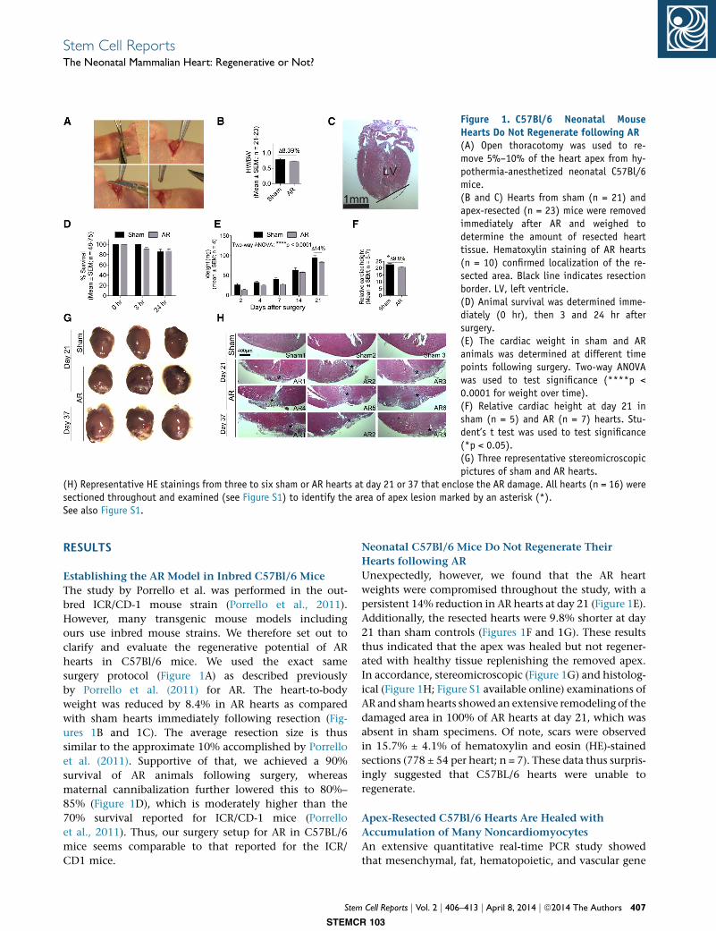
Figure 1. C57Bl/6 Neonatal MouseHearts Do Not Regenerate following AR(A) Open thoracotomy was used to re-move 5%–10% of the heart apex from hy-pothermia-anesthetized neonatal C57Bl/6mice.(B and C) Hearts from sham (n = 21) andapex-resected (n = 23) mice were removedimmediately after AR and weighed todetermine the amount of resected hearttissue. Hematoxylin staining of AR hearts(n = 10) confirmed localization of the re-sected area. Black line indicates resectionborder. LV, left ventricle.(D) Animal survival was determined imme-diately (0 hr), then 3 and 24 hr aftersurgery.(E) The cardiac weight in sham and ARanimals was determined at different timepoints following surgery. Two-way ANOVAwas used to test significance (****p <0.0001 for weight over time).(F) Relative cardiac height at day 21 insham (n = 5) and AR (n = 7) hearts. Stu-dent’s t test was used to test significance(*p < 0.05).(G) Three representative stereomicroscopicpictures of sham and AR hearts.
(H) Representative HE stainings from three to six sham or AR hearts at day 21 or 37 that enclose the AR damage. All hearts (n = 16) weresectioned throughout and examined (see Figure S1) to identify the area of apex lesion marked by an asterisk (*).See also Figure S1.
Stem Cell ReportsThe Neonatal Mammalian Heart: Regenerative or Not?
RESULTS
Establishing the AR Model in Inbred C57Bl/6 Mice
The study by Porrello et al. was performed in the out-
bred ICR/CD-1 mouse strain (Porrello et al., 2011).
However, many transgenic mouse models including
ours use inbred mouse strains. We therefore set out to
clarify and evaluate the regenerative potential of AR
hearts in C57Bl/6 mice. We used the exact same
surgery protocol (Figure 1A) as described previously
by Porrello et al. (2011) for AR. The heart-to-body
weight was reduced by 8.4% in AR hearts as compared
with sham hearts immediately following resection (Fig-
ures 1B and 1C). The average resection size is thus
similar to the approximate 10% accomplished by Porrello
et al. (2011). Supportive of that, we achieved a 90%
survival of AR animals following surgery, whereas
maternal cannibalization further lowered this to 80%–
85% (Figure 1D), which is moderately higher than the
70% survival reported for ICR/CD-1 mice (Porrello
et al., 2011). Thus, our surgery setup for AR in C57BL/6
mice seems comparable to that reported for the ICR/
CD1 mice.
Stem
STEMC
Neonatal C57Bl/6 Mice Do Not Regenerate Their
Hearts following AR
Unexpectedly, however, we found that the AR heart
weights were compromised throughout the study, with a
persistent 14% reduction in AR hearts at day 21 (Figure 1E).
Additionally, the resected hearts were 9.8% shorter at day
21 than sham controls (Figures 1F and 1G). These results
thus indicated that the apex was healed but not regener-
ated with healthy tissue replenishing the removed apex.
In accordance, stereomicroscopic (Figure 1G) and histolog-
ical (Figure 1H; Figure S1 available online) examinations of
AR and shamhearts showed an extensive remodeling of the
damaged area in 100% of AR hearts at day 21, which was
absent in sham specimens. Of note, scars were observed
in 15.7% ± 4.1% of hematoxylin and eosin (HE)-stained
sections (778 ± 54 per heart; n = 7). These data thus surpris-
ingly suggested that C57BL/6 hearts were unable to
regenerate.
Apex-Resected C57Bl/6 Hearts Are Healed with
Accumulation of Many Noncardiomyocytes
An extensive quantitative real-time PCR study showed
that mesenchymal, fat, hematopoietic, and vascular gene
Cell Reports j Vol. 2 j 406–413 j April 8, 2014 j ª2014 The Authors 407
R 103
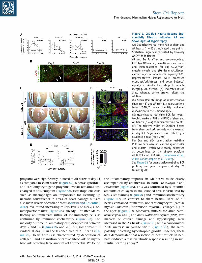
Figure 2. C57Bl/6 Hearts Become Sub-stantially Fibrotic following AR andShow Signs of Hypertrophy(A) Quantitative real-time PCR of sham andAR hearts (n = 4) at indicated time points.Statistical significance tested by two-wayANOVA is indicated.(B and D) Paraffin- and cryo-embeddedC57Bl/6 AR hearts (n = 4–8) were sectionedand immunostained for (B) CD45/non-muscle myosin and (D) desmin/collagen;cardiac myosin; nonmuscle myosin/CD31.Representative images were processed(contrast/brightness and color balance)equally in Adobe Photoshop to enablemerging. An asterisk (*) indicates lesionarea, whereas white arrows reflect theAR line.(C) Sirius Red stainings of representativesham (n = 5) and AR (n = 11) heart sectionsfrom C57Bl/6 mice identify collagendisposition in the lesioned apex.(E) Quantitative real-time PCR for hyper-trophic markers (ANP and BNP) of sham andAR hearts (n = 4) at indicated time points.(F) The relative width of C57Bl/6 heartsfrom sham and AR animals was measuredat day 21. Significance was tested by aStudent’s t test (*p < 0.05).For (A) and (E), quantitative real-timePCR raw data were normalized against B2Mand b-actin, which were stably expressedas determined by the qBase+ platform(M:0.579 and CV:0.202) (Hellemans et al.,2007; Vandesompele et al., 2002).See Figure S2 for quantitative real-time PCRprofiling on gene programs at day 21following AR.
Stem Cell ReportsThe Neonatal Mammalian Heart: Regenerative or Not?
programs were significantly induced in AR hearts at day 21
as compared to sham hearts (Figure S2), whereas epicardial
and cardiomyocyte gene programs overall remained un-
changed at this endpoint (Figure S2). Hematopoietic cells
such as macrophages are responsible for cleaning up
necrotic constituents in areas of heart damage but are
also main drivers of cardiac fibrosis (Santini and Rosenthal,
2012). We found increasing mRNA levels of Cd45, a he-
matopoietic marker (Figure 2A), already 3 hr after AR, re-
flecting an immediate influx of inflammatory cells as
confirmed by immunohistochemistry (Figure 2B). The
majority of these inflammatory cells disappeared between
days 7 and 14 (Figures 2A and 2B), but some were still
evident at day 21 in the lesioned area of AR hearts (Fig-
ure 2B). Heart fibrosis is characterized by deposition of
collagen I and a transition of cardiac fibroblasts to myofi-
broblasts secreting large amounts of fibronectin. We found
408 Stem Cell Reports j Vol. 2 j 406–413 j April 8, 2014 j ª2014 The Author
STEMCR 103
the inflammatory response in AR hearts to be clearly
accompanied by an increase in both Pro-collagen I and
Fibronectin (Figure 2A). This was confirmed by substantial
amounts of collagen in the lesioned area as visualized by
Sirius Red staining (Figure 2C) and immunohistochemistry
(Figure 2D). In contrast to sham hearts, 100% of AR
hearts contained numerous noncardiomyocytes (cardiac
myosin�/desmin�/nonmuscle myosin+, collagen I+) in
the apex (Figure 2D). Moreover, mRNAs for Atrial Natri-
uretic Peptide (ANP) and Brain Natriuretic Peptide (BNP), two
markers of cardiac damage and hypertrophy, were
increased in the AR hearts (Figure 2E) with a concomitant
7.5% increase in cardiac width (Figure 2F), the latter
possibly indicating hypertrophic growth. Together, these
data demonstrated that resection of heart muscle in neo-
nates induced a massive fibrotic response resulting in sub-
stantial scarring at day 21.
s

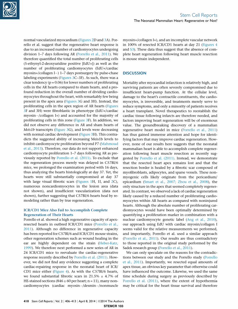
Figure 3. The Apex-Resected Zone inC57Bl/6 Hearts Reveals Limited Vascu-larization and Numbers of ProliferatingCardiomyocytes(A) Cryosectioned C57Bl/6 AR hearts (n =4–8) were immunostained for aSMA, CD31,and NG2/CD31. Representative images wereprocessed (contrast/brightness and colorbalance) equally in Photoshop to enablemerging. An asterisk (*) indicates lesionarea, whereas a number sign (#) refers tonondamaged myocardium.(B) Quantitative real-time PCR for Cd31 ofsham and AR hearts (n=4) at indicatedtime points. Quantitative real-time PCR rawdata were normalized against the stablyexpressed B2M and b-actin genes (seeFigure 2).(C–F) Paraffin-embedded C57Bl/6 AR andsham hearts (n = 5) from EdU pulse-chaselabeled animals were sectioned and immu-nostained for EdU/cardiac myosin/collagenI/DAPI to enable identification of prolifer-ating cells in total and proliferatingcardiomyocytes specifically. Images weretaken in four different zones (white boxesin C and areas magnified in F) of each heartand used for cell number quantification(D and E) as described in ExperimentalProcedures.(G) Quantitative real-time PCR for Meis1b ofsham and AR hearts (n = 4) at indicatedtime points. Quantitative real-time PCR rawdata were normalized against the stablyexpressed B2M and b-actin genes (see Fig-ure 2).(H) Meis1b is downregulated with cardiacdevelopment. Hearts were microdissectedfrom C57Bl/6 mice at indicated time pointsand used for quantitative real-time PCR.Primers used were previously described byMahmoud et al. (2013). Each biologicalexperiment (n = 3–6) included betweenone and six hearts. qPCR raw data werenormalized against Gapdh and b-actin,which were stably expressed as determined
by the qBase+ platform (M:0.306, CV: 0.106) (Hellemans et al., 2007; Vandesompele et al., 2002). Statistical significance was testedusing a one-way ANOVA with a Dunnett’s multiple comparison posttest. E11.5, embryonic day 11.5.
Stem Cell ReportsThe Neonatal Mammalian Heart: Regenerative or Not?
The Damaged Apex Comprises Modest
Vascularization, but Few Proliferating
Cardiomyocytes
Ideally, cardiac regeneration should be able to restore the
functional continuity of cardiomyocytes, as well as blood
supply (endothelial and smooth muscle cells) in the
necrotic area of the damaged heart. We did encounter
blood vessels with CD31+ endothelia, and surrounding
Stem
STEMC
smooth muscle cells (aSMA+ and NG2+) in the lesioned
area (Figures 2D and 3A). These vessels may either be preex-
isting vessels developed in the border zone prior to lesion,
but they may also represent newly formed vascularity in
the damaged area because both Cd31 (Figure 2B) and
numerous other vascular genes (Figure S2) were increased
in AR hearts as compared with sham controls. Yet, the
vascularization was still incomplete as compared with
Cell Reports j Vol. 2 j 406–413 j April 8, 2014 j ª2014 The Authors 409
R 103
Stem Cell ReportsThe Neonatal Mammalian Heart: Regenerative or Not?
normal vascularizedmyocardium (Figures 2D and 3A). Por-
rello et al. suggest that the regenerative heart response is
due to an increased number of cardiomyocytes undergoing
division 1–7 days following AR (Porrello et al., 2011). We
therefore quantified the total number of proliferating cells
(5-ethynyl-2-deoxyuridine positive [EdU+]) as well as the
number of proliferating cardiomyocytes (EdU+/cardiac
myosin+/collagen I�) 1–7 days postsurgery by pulse-chase
labeling experiments (Figures 3C–3F). As such, there was a
clear tendency (p = 0.06) for lower numbers of proliferating
cells in the AR hearts compared to sham hearts, and a pro-
found reduction in the overall number of dividing cardio-
myocytes throughout the heart, with remarkably few being
present in the apex area (Figures 3G and 3H). Instead, the
proliferating cells in the apex region of AR hearts (Figures
3F and 3H) were fibroblastic in phenotype (EdU+/cardiac
myosin�/collagen I+) and accounted for the majority of
proliferating cells in this zone (Figure 3F). In addition, we
did not observe any difference in AR and sham hearts of
Meis1b transcripts (Figure 3G), and levels were decreasing
with normal cardiac development (Figure 3H). This contra-
dicts the suggested ability of increasing Meis1b levels to
inhibit cardiomyocyte proliferation beyond P7 (Mahmoud
et al., 2013). Therefore, our data do not support enhanced
cardiomyocyte proliferation 1–7 days following AR as pre-
viously reported by Porrello et al. (2011). To exclude that
the regeneration process merely was delayed in C57Bl/6
mice, we prolonged the examination period with 16 days,
thus analyzing the hearts histologically at day 37. Yet, the
hearts were still substantially compromised at day 37
with large visual fibrotic scars (Figures 1H, 1I, and 2C),
numerous noncardiomyocytes in the lesion area (data
not shown), and insufficient vascularization (data not
shown), further suggesting that C57Bl/6 hearts heal by re-
modeling rather than by true regeneration.
ICR/CD1 Mice Also Fail to Accomplish Complete
Regeneration of Their Hearts
Porrello et al. showed a high regenerative capacity of apex-
resected hearts in outbred ICR/CD1 mice (Porrello et al.,
2011). Although no difference in regenerative capacity
has been reported for C57Bl/6 and ICR/CD1mouse strains,
other regeneration schemes such as wound healing in the
ear are highly dependent on the strain (Heber-Katz,
1999). We therefore next performed a new series of AR in
24 ICR/CD1 mice to reevaluate the cardiac-regenerative
response recently described by Porrello et al. (2011). How-
ever, we did not find any evidence suggesting a complete
cardiac-repairing response in the neonatal heart of ICR/
CD1 mice either (Figure 4). As with the C57Bl/6 hearts,
we found substantial fibrotic scars in 21.5% ± 4.7% of
HE-stained sections (846 ± 60 per heart; n = 11), many non-
cardiomyocytes (cardiac myosin�/desmin�/nonmuscle
410 Stem Cell Reports j Vol. 2 j 406–413 j April 8, 2014 j ª2014 The Author
STEMCR 103
myosin+/collagen I+), and an incomplete vascular network
in 100% of resected ICR/CD1 hearts at day 21 (Figures 4
and S3). These data thus suggest that the absence of com-
plete heart regeneration following heart muscle resection
is mouse strain independent.
DISCUSSION
Mortality after myocardial infarction is relatively high, and
surviving patients are often severely compromised due to
insufficient heart-pump function. At the cellular level,
damage to the heart’s contractile constituents, the cardio-
myocytes, is irreversible, and treatments merely serve to
reduce symptoms, and only a minority of patients receives
a heart transplant. Novel therapeutics to reestablish the
cardiac tissue following infarcts are therefore needed, and
factors improving heart regeneration will be of enormous
value. The groundbreaking discovery of a mammalian
regenerative heart model in mice (Porrello et al., 2011)
has thus gained immense attention and hope for identi-
fying factors that may improve cardiac regeneration. How-
ever, none of our results here suggests that the neonatal
mammalian heart is able to accomplish complete regener-
ation following heart muscle resection as recently sug-
gested by Porrello et al. (2011). Instead, we demonstrate
that the resected heart apex remains lost and that the
resection border is healed by a fibrotic scar composed of
myofibroblasts, adipocytes, and sparse vessels. These non-
myogenic cells likely originate from the pericardium/
epicardium (Smart et al., 2007; Zhou et al., 2008), the
only structure in the apex that seemed completely regener-
ated. In contrast, we observed a lack of cardiac regeneration
likely caused by a reduced number of proliferating cardio-
myocytes within AR hearts as compared with noninjured
hearts. Although the absolute number of proliferating car-
diomyocytes would have been optimally determined by
quantifying a proliferation marker in combination with a
nuclear cardiomyocyte genetic label (Ang et al., 2010),
our approach using EdU with cardiac myosin/collagen I
seems valid for the relative measurements we performed,
and importantly, Porrello et al. used a similar approach
(Porrello et al., 2011). Our results are thus contradictory
to those reported in the original study performed by the
Sadek research group (Porrello et al., 2011).
We can only speculate on the reasons for the contradic-
tions between our study and the Porrello study (Porrello
et al., 2011). Importantly, we resected equal amounts of
apex tissue, an obvious key parameter that otherwise could
have influenced the outcome. Likewise, we used the same
time schedule during surgery as previously described by
Porrello et al. (2011), where the extent of hypothermia
may be critical for the heart tissue survival and therefore
s
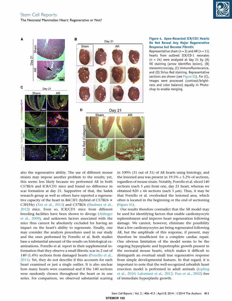
Figure 4. Apex-Resected ICR/CD1 HeartsDo Not Reveal Any Major RegenerativeResponse but Become FibroticRepresentative sham (n = 3) and AR (n = 11)hearts from outbred ICR/CD-1 neonates(n = 24) were analyzed at day 21 by (A)HE staining (arrow identifies lesion), (B)stereomicroscopy, (C) immunofluorescence,and (D) Sirius Red staining. Representativesections are shown (see Figure S3). For (C),images were processed (contrast/bright-ness and color balance) equally in Photo-shop to enable merging.
Stem Cell ReportsThe Neonatal Mammalian Heart: Regenerative or Not?
also the regenerative ability. The use of different mouse
strains may impose another problem to the results; yet,
this seems less likely because we performed AR in both
C57Bl/6 and ICR/CD1 mice and found no difference in
scar formation at day 21. Supportive of that, the Sadek
research group as well as others have reported a regenera-
tive capacity of the heart in B6C3F1 (hybrid of C57Bl/6 3
C3H/He) (Xin et al., 2013) and C57Bl/6 (Haubner et al.,
2012) mice. Even so, ICR/CD1 mice from different
breeding facilities have been shown to diverge (Aldinger
et al., 2009), and unknown factors associated with the
mice thus cannot be absolutely excluded for having an
impact on the heart’s ability to regenerate. Finally, one
may consider the analysis procedures used in our study
and the ones performed by Porrello et al. Both studies
base a substantial amount of the results on histological ex-
aminations. Porrello et al. report in their supplemental in-
formation that they observe a small fibrotic scar in 2 out of
140 (1.4%) sections from damaged hearts (Porrello et al.,
2011). Yet, they do not describe if this accounts for each
heart examined or just a single outlier. It is also unclear
how many hearts were examined and if the 140 sections
were randomly chosen throughout the heart or in one
series. For comparison, we observed substantial scarring
Stem
STEMC
in 100% (31 out of 31) of AR hearts using histology, and
the lesioned area was present in 19.5% ± 5.2% of sections,
regardless ofmouse strain. Notably, Porrello et al. sliced 140
sections (each 5 mm) from one, day 21 heart, whereas we
obtained 820 ± 66 sections (each 5 mm). Thus, it may be
that Porrello et al. overlooked the lesioned area, which
often is located in the beginning or the end of sectioning
(Figure S1).
Our results therefore contradict that the AR model may
be used for identifying factors that enable cardiomyocyte
replenishment and improve heart regeneration following
damage. We cannot, however, eliminate the possibility
that a few cardiomyocytes are being regenerated following
AR, but the amplitude of this response, if present, may
therefore be insufficient for a complete cardiac repair.
One obvious limitation of the model seems to be the
ongoing hyperplastic and hypertrophic growth present in
the neonatal mouse hearts, which makes it difficult to
distinguish an eventual small true regenerative response
from simple developmental features. In that regard, it is
important to note that the well-recognized zebrafish heart
resection model is performed in adult animals (Jopling
et al., 2010; Lafontant et al., 2012; Poss et al., 2002) free
of immediate hyperplastic growth.
Cell Reports j Vol. 2 j 406–413 j April 8, 2014 j ª2014 The Authors 411
R 103
Stem Cell ReportsThe Neonatal Mammalian Heart: Regenerative or Not?
Yet, from our results, we cannot exclude that other car-
diac regeneration models such as the left anterior-descend-
ing (LAD) artery ligation in the neonatal myocardium
(Haubner et al., 2012; Mahmoud et al., 2013; Xin et al.,
2013) do unravel a true and complete regenerative poten-
tial of the heart. Still, we find it questionable that a full
cardiac regenerative response is accomplished in only
21 days (Mahmoud et al., 2013; Porrello et al., 2011),
and in one study (Haubner et al., 2012), after only 7 days
following LAD damage of the mouse heart. For compari-
son, Jesty et al. recently showed that cryo-injury to the
neonatal heart is associated with scar formation even after
94 days (Jesty et al., 2012), a scenario that is also seen up
until 60–180 days after cardiac injury in simpler organisms
like teleost fish (Jopling et al., 2010; Lafontant et al., 2012).
However, in a previous study, a genetic model of cardio-
myocyte ablation showed that the cardiomyocyte pool
is restored in the fetal mouse heart by enhanced prolifera-
tion of remaining cardiomyocytes (Drenckhahn et al.,
2008). One may speculate if a similar scenario could take
place after neonatal mouse heart LAD damage, whereas
the AR model lacks an intact 3D heart architecture, a
feature that likely is important to some extent for mamma-
lian regeneration to be accomplished. In summary, we
thus believe that additional clarifying data are required
from the scientific community on this controversial
matter to firmly establish whether the mammalian heart
is regenerative, otherwise our data substantiate the view
that it is not.
EXPERIMENTAL PROCEDURES
A detailed version of our Experimental Procedures can be
found in the Supplemental Experimental Procedures. All animal
experiments were approved by the Danish Council for Supervi-
sion with Experimental Animals (#2011/561-1966). AR was
performed at P1 as previously described by Porrello et al.
(2011). Briefly, neonates were anesthetized by hypothermia,
and the apex was resected until left ventricle chamber expo-
sure, after which the thoracic wall and skin were sutured. Sham
mice underwent the exact same procedure without resecting
the apex of the heart. For pulse-chase labeling experiments,
mice were injected 1 day after surgery with EdU, and the
number of proliferating cardiomyocytes and cells in total was
counted after 7 days. Relative quantitative PCR (qPCR) and his-
tology were performed as previously described (Andersen et al.,
2009). All analyses comprised at least four independent experi-
ments, and statistical significance (p < 0.05) was tested as
indicated.
SUPPLEMENTAL INFORMATION
Supplemental Information includes Supplemental Experimental
Procedures and three figures can be found with this article online
at http://dx.doi.org/10.1016/j.stemcr.2014.02.008.
412 Stem Cell Reports j Vol. 2 j 406–413 j April 8, 2014 j ª2014 The Author
STEMCR 103
AUTHOR CONTRIBUTIONS
D.C.A. conceived and designed the study; performed collection,
analysis, and interpretation of data; prepared and approved the
manuscript; and provided funding. S.G. performed data collec-
tion and analysis. C.H.J. designed the study; performed data
collection, analysis, and interpreted the data; and participated in
the discussion and approval of the manuscript. S.P.S. performed
interpretation of data, approved the manuscript, and provided
funding.
ACKNOWLEDGMENTS
Wewould like to thank Charlotte Nielsen, Tonja L. Jørgensen, and
Anette Kliem (LMCC, Odense University Hospital) for technical
assistance on this study and Dr. Bruce Conklin (Gladstone Insti-
tute, CA) for language editing. The work was supported by
The Novo Nordisk Foundation, The Danish National Research
Council (#09-073648), The Lundbeck Foundation (#R48-A4785),
Lægeforeningen (#2011-3271/480853-109), Tømrermester Alfred
Andersen og Hustru’s Fond, Hertha Christensens Foundation,
Eva and Henry Frænkels Foundation, and Department of Clinical
Biochemistry and Pharmacology/Odense University Hospital.
Received: August 19, 2013
Revised: February 24, 2014
Accepted: February 25, 2014
Published: April 3, 2014
REFERENCES
Aguirre, A., Sancho-Martinez, I., and Izpisua Belmonte, J.C. (2013).
Reprogramming towardheart regeneration: stem cells and beyond.
Cell Stem Cell 12, 275–284.
Aldinger, K.A., Sokoloff, G., Rosenberg, D.M., Palmer, A.A., and
Millen, K.J. (2009). Genetic variation and population substructure
in outbred CD-1 mice: implications for genome-wide association
studies. PLoS ONE 4, e4729.
Andersen, D.C., Petersson, S.J., Jørgensen, L.H., Bollen, P., Jensen,
P.B., Teisner, B., Schroeder, H.D., and Jensen, C.H. (2009). Charac-
terization of DLK1+ cells emerging during skeletal muscle remod-
eling in response to myositis, myopathies, and acute injury. Stem
Cells 27, 898–908.
Ang, K.L., Shenje, L.T., Reuter, S., Soonpaa,M.H., Rubart, M., Field,
L.J., and Galinanes, M. (2010). Limitations of conventional
approaches to identify myocyte nuclei in histologic sections of
the heart. Am. J. Physiol. Cell Physiol. 298, C1603–C1609.
Drenckhahn, J.D., Schwarz, Q.P., Gray, S., Laskowski, A., Kiriazis,
H., Ming, Z., Harvey, R.P., Du, X.J., Thorburn, D.R., and Cox, T.C.
(2008). Compensatory growth of healthy cardiac cells in the pres-
ence of diseased cells restores tissue homeostasis during heart
development. Dev. Cell 15, 521–533.
Garbern, J.C., and Lee, R.T. (2013). Cardiac stem cell therapy and
the promise of heart regeneration. Cell Stem Cell 12, 689–698.
Garbern, J.C., Mummery, C.L., and Lee, R.T. (2013). Model systems
for cardiovascular regenerative biology. Cold Spring Harb Perspect
Med 3, a014019.
s
Stem Cell ReportsThe Neonatal Mammalian Heart: Regenerative or Not?
Haubner, B.J., Adamowicz-Brice, M., Khadayate, S., Tiefenthaler,
V., Metzler, B., Aitman, T., and Penninger, J.M. (2012). Complete
cardiac regeneration in a mouse model of myocardial infarction.
Aging (Albany, N.Y. Online) 4, 966–977.
Heber-Katz, E. (1999). The regenerating mouse ear. Semin. Cell
Dev. Biol. 10, 415–419.
Hellemans, J., Mortier, G., De Paepe, A., Speleman, F., and Vande-
sompele, J. (2007). qBase relative quantification framework and
software for management and automated analysis of real-time
quantitative PCR data. Genome Biol. 8, R19.
Jesty, S.A., Steffey, M.A., Lee, F.K., Breitbach, M., Hesse, M., Rein-
ing, S., Lee, J.C., Doran, R.M., Nikitin, A.Y., Fleischmann, B.K.,
and Kotlikoff, M.I. (2012). c-kit+ precursors support postinfarction
myogenesis in the neonatal, but not adult, heart. Proc. Natl. Acad.
Sci. USA 109, 13380–13385.
Jopling, C., Sleep, E., Raya, M., Martı, M., Raya, A., and Izpisua Bel-
monte, J.C. (2010). Zebrafish heart regeneration occurs by cardio-
myocyte dedifferentiation and proliferation. Nature 464, 606–609.
Lafontant, P.J., Burns, A.R., Grivas, J.A., Lesch, M.A., Lala, T.D.,
Reuter, S.P., Field, L.J., and Frounfelter, T.D. (2012). The giant danio
(D. aequipinnatus) as a model of cardiac remodeling and regenera-
tion. Anat. Rec. (Hoboken) 295, 234–248.
Mahmoud, A.I., Kocabas, F., Muralidhar, S.A., Kimura, W., Koura,
A.S., Thet, S., Porrello, E.R., and Sadek, H.A. (2013).Meis1 regulates
postnatal cardiomyocyte cell cycle arrest. Nature 497, 249–253.
Oberpriller, J.O., and Oberpriller, J.C. (1974). Response of the adult
newt ventricle to injury. J. Exp. Zool. 187, 249–253.
Stem
STEMC
Porrello, E.R., Mahmoud, A.I., Simpson, E., Hill, J.A., Richardson,
J.A., Olson, E.N., and Sadek, H.A. (2011). Transient regenerative
potential of the neonatal mouse heart. Science 331, 1078–1080.
Poss, K.D., Wilson, L.G., and Keating, M.T. (2002). Heart regenera-
tion in zebrafish. Science 298, 2188–2190.
Santini, M.P., and Rosenthal, N. (2012). Myocardial regenera-
tive properties of macrophage populations and stem cells.
J. Cardiovasc. Transl. Res. 5, 700–712.
Smart, N., Risebro, C.A., Melville, A.A., Moses, K., Schwartz, R.J.,
Chien, K.R., and Riley, P.R. (2007). Thymosin beta-4 is essential
for coronary vessel development and promotes neovascularization
via adult epicardium. Ann. N Y Acad. Sci. 1112, 171–188.
Vandesompele, J., De Preter, K., Pattyn, F., Poppe, B., Van Roy, N.,
De Paepe, A., and Speleman, F. (2002). Accurate normalization of
real-time quantitative RT-PCR data by geometric averaging of mul-
tiple internal control genes. Genome Biol. 3, H0034.
Vieira, J.M., and Riley, P.R. (2011). Epicardium-derived cells: a new
source of regenerative capacity. Heart 97, 15–19.
Xin,M., Kim, Y., Sutherland, L.B., Murakami,M., Qi, X., McAnally,
J., Porrello, E.R., Mahmoud, A.I., Tan, W., Shelton, J.M., et al.
(2013). Hippo pathway effector Yap promotes cardiac regenera-
tion. Proc. Natl. Acad. Sci. USA 110, 13839–13844.
Zhou, B., Ma, Q., Rajagopal, S., Wu, S.M., Domian, I., Rivera-
Feliciano, J., Jiang, D., von Gise, A., Ikeda, S., Chien, K.R., and
Pu, W.T. (2008). Epicardial progenitors contribute to the
cardiomyocyte lineage in the developing heart. Nature 454,
109–113.
Cell Reports j Vol. 2 j 406–413 j April 8, 2014 j ª2014 The Authors 413
R 103