state of the veterinary technician “where we are and...
TRANSCRIPT

State of the Veterinary Technician “Where We Are and Where We Are Going”
Dr. Danny Walker, D.V.M.
A historical perspective of how the role of the veterinary technologist evolved into its
current status over the past sixty years will be reviewed. Some of the national and state
legislation which governs the scope of practice of the veterinary technologist and other
professionals involved in the health care of animals will be discussed and compared to
legislation affecting other types of health care professionals. The effect of how legislation has
economically impacted the client, the veterinarian and the veterinary technologist will be
presented. Future “Demand versus Supply” estimations for the next decade in regard to the
veterinary technology profession will be shared. Educational perspectives related to the
expansion of the veterinary technologist’s role to meet these future demands will be identified.
The importance of the need for the veterinary technologist to acquire a new “mindset” in
regard to continuing education, and professional association membership for job security and
for professional status enhancement within the animal health care industry will emphasized.
HEPATIC LIPIDOSIS – MANAGING & FEEDING THE ANORECTIC CAT Margie Scherk DVM, Dip ABVP (feline practice)
catsINK, Vancouver, Canada
Introduction Hepatic lipidosis (HL) is a common sequel to inappetence in cats regardless of the cause of appetite disruption. It is the most common form of liver disease diagnosed in cats in North America. Treating the caloric deficiency must be the first priority. Diagnostics are required to define the underlying cause(s) to enable treatment and correction of the same where feasible. When cytology is utilized, it is important to interpret findings in light of collection method. Aspiration will result in lipid being harvested in any inappetant cat, regardless of cause; passive fine needle biopsy provides a greater opportunity to evaluate cells in the liver. Successful therapy of lipidosis requires aggressive nutritional support feeding adequate amounts of a balanced diet to reverse the catabolic state. Good quality, biologically available protein is necessary and this nutrient should not be restricted unless a patient is showing evidence of hepatic encephalopathy. Early treatment with Vitamin K1 allows fine needle or exploratory biopsy as well as large bore tube placement, which is key to recovery. Fluid therapy for tissue perfusion, oxygen delivery, waste scavenging and the correction of electrolyte imbalance, especially hypokalemia, is essential. Addressing concurrent or underlying problems that have contributed to the inappetence is very important. Drugs used in the treatment of feline lipidosis include antiemetics if needed, taurine, arginine, S-adenosyl-L-methionine, L-carnitine, ursodeoxycholic acid, silibinin and Vitamins B and K1.
Etiology And Pathophysiology Anything causing a significant decrease in dietary intake or cellular starvation can result in lipidosis in cats. Thus, uncontrolled diabetic cats often have concurrent lipidosis despite polyphagia. It is unlikely that one pathogenesis explains HL in all affected individuals, however alterations in fat metabolism are key. There are five types of fat in the liver: triglycerides (TG), phospholipids, lipoproteins, cholesterol and cholesterol esters. Lipid vacuolation in lipidosis is predominantly composed of triglycerides. They accumulate in the liver when the rate of hepatic synthesis exceeds their dispersal. Hepatic TGs are produced from fatty acids from systemic circulation (dietary lipids and adipose stores) and from de novo synthesis within the liver. Over nutrition with carbohydrates or protein results in hepatic fat accumulation, as these excess nutrients are stored as triglycerides. Systemically ill cats generally develop some degree of hepatocellular fatty vacuolation despite an increased rate of very low-density lipoprotein (VLDL) secretion. A 2004 study (Blanchard) showed that TG, VLDL, low density lipoprotein (LDL) and high density lipoprotein (HDL) levels are all increased in cats with lipidosis suggesting that VLDL secretion is enhanced, VLDL and LDL catabolism is reduced and lipoprotein exchange is impaired. Fatty vacuolation is not problematic until the degree of vacuolation is morphologically severe. In a normal feline liver, neutral fat comprises < 5% of total organ weight. In contrast, the liver of a cat with HL may double or triple in weight due to fat accumulation. Over-nutrition augments hepatic fat accumulation; feeding a high proportion of dietary carbohydrates may have an

inhibitory influence on mitochondrial fat oxidation favouring hepatic fat accumulation. Feline obesity predisposes to development of HL: in inappetant obese cats, release of fatty acids from their ample peripheral adipose stores challenges the liver’s capacity for fat utilization and dispersal. The metabolic balance of lipolysis and storage of TGs is influenced by blood glucose concentrations as well as diverse hormonal and neural mechanisms. Hormone sensitive lipase (HSL, promoting adipocyte lipolysis) and lipoprotein lipase (LPL, promoting fat uptake into adipocytes) directly influence adipocyte fat metabolism. Adipocyte LPL activity promotes fat uptake in the well-fed individual. In starvation, LPL activity declines, while that of HSL increases. Thus, lipolysis exceeds fat uptake. A previously obese individual undergoing starvation is at increased risk for lipolysis. Norepinephrine, epinephrine, growth hormone, glucagon, corticosteroids, and thyroxin increase HSL activity, whereas insulin inhibits it. Since this effect is enhanced in the absence of insulin, occult or overt hepatic TG accumulation in unregulated diabetics is common. While LPL promotes fat uptake into adipocytes in the well-fed condition, during starvation, LPL activity declines and HSL increases creating a hormonal balance favouring hepatocellular fat accumulation. Therefore, an obese individual undergoing starvation (anorexia) has increased risk for peripheral fat mobilization and excessive hepatic TG retention. Catecholamine release associated with stress or illness may result in NEFA release contributing to the development of lipidosis. (Brown) Fatty acids may undergo beta-oxidation, be used for TG synthesis, be converted to phospholipids, be used in the formation of cholesterol esters, or be packaged with apoproteins for dispersal as lipoproteins. The most important route for TG dispersal is via formation of VLDL. This requires intact lipid transport through subcellular compartments, particle combination with apoprotein, formation of a secretory particle and a vesicle and out of the hepatocyte and into the perisinusoidal space. Impairment at any one of these steps will prevent mobilization of hepatic fat. An imbalance between essential lipoprotein components will also interfere with fat dispersal. Brown et al showed that cats with HL have a higher concentration of nonesterified fatty acids (NEFA) compared to cats with cholangiohepatitis suggesting that hormone sensitive lipase is unimpaired. Catecholamines associated with stress or illness may result in NEFA release (as well as HSL) contributing to the development of lipidosis. In 2013, Mazaki-Tovi reported that adiponectin levels are elevated in cats with lipidosis or other liver disease, increased concentration of leptin was found only in cats with HL. Adiponectin concentrations correlated with ALT activity whereas leptin levels correlated with alkaline phosphatase activity. The essential interaction of fatty acids and L-carnitine at mitochondrial membranes influences both the intraorganelle activation of fatty acids and their availability for beta-oxidation. Carnitine may improve fatty acid oxidation but not in a cat being fed a diet low in n-3 fatty acids (Ibrahim). In addition, dietary L-carnitine appears to protect against ketosis during weight gain (Blanchard, 2002).

Deficiency of hepatocellular GSH in cats with HL may reflect intramitochrondrial GSH insufficiency where it is needed for redox-balance to sustain continued energy production. Finding hepatic GSH deficiency in cats with HL implicates dysfunction of the transsulfuration pathway with diminished SAMe availability. Low GSH levels are common in liver disease in cats (Center 2002)
Clinical Findings And Diagnostics Cats of any age may be affected by HL. Domestic shorthair cats are commonly affected but this may reflect breed popularity. Obese middle-aged females are most often reported, but any inappetant cat may develop lipidosis. The period of inappetence or anorexia may be as short as 2 to 7 days. Initial clinical features include inappetence, weight loss, vomiting, lethargy, diarrhea or constipation and weakness. Unless hepatic encephalopathy is present (rare), cats generally remain bright and alert. Physical findings include dehydration, variable icterus, an unkempt appearance, and palpable liver margins. Although fat may become depleted on the limbs and dorsal trunk, abdominal fat stores are preserved. Rarely, skin fragility has been reported (Trotman, Daniel). Hematologic features include a nonregenerative anemia and a stress leukogram reflecting the primary illness causing anorexia. Poikilocytosis is common and may reflect altered RBC membrane lipids, metabolism or oxidative stress affecting cell membrane flexibility. Biochemical changes reflect cholestasis, and to a lesser degree, altered hepatocellular membrane permeability and viability. Most cats have a markedly increased ALP activity with lesser magnitudes of increase in transaminases. Occasionally, a cat will have high transaminases with only modest ALP activity. Finding high GGT activity in a cat with HL increases suspicion of underlying pancreatitis, pancreatic neoplasia, cholangiohepatitis, major bile duct obstruction, cholelithiasis or choledochitis, or biliary tree neoplasia. Hyperbilirubinemia and high serum bile acid concentrations are common. Hypokalemia reflects inappetence and is significantly associated with failure to survive if uncorrected. Hypochloremia may reflect vomiting. Hypophosphatemia reflects intercompartmental shifts in phosphate and most commonly heralds onset of a refeeding syndrome. Urinalysis typically reports urobilinogen with bilirubin pigmenturia and bile crystalluria. Because most cases of lipidosis are secondary to something causing inappetence, ultrasound is helpful to look for the underlying problem. Evaluate the liver, pancreas, stomach, small and large bowel to determine if there is triaditis (inflammation of the liver, pancreas and bowel), disease of the gall bladder and biliary tree, etc. In one study, 38% of cats with HL had concurrent histopathologic acute pancreatitis and the recovery rate of cats with acute necrotizing pancreatitis plus lipidosis was 20% rather than >80% of cats with HL alone. Ultrasonographically, the liver characteristically shows a hyperechoic parenchyma, however, in a recent report, it was concluded that statistical evaluation of ultrasonographic criteria did not yield clinically acceptable accuracy for discrimination among the 7 categories of diffuse liver diseases (including normal liver) in either cats or dogs. Liver aspirates demonstrate profound, but nonspecific, hepatocellular lipid vacuolation with more than 80% of hepatocytes markedly affected. Aspiration cytology is adequate for a presumptive diagnosis of HL, hepatic lymphoma, and overt hepatic sepsis but is unreliable for detection of necroinflammatory disorders as they
cannot rule out, or definitively confirm, cholangitis or cholangiohepatitis. Harvesting cored samples using a passive collection, rather than active aspiration, may improve the yield of the samples. Surgical or laparoscopic biopsy of the liver is usually not necessary and purposely avoided to minimize anaesthetic and hemorrhagic complications. However, if other organs are affected, surgery should be considered to get multiple organ biopsies (and to place a g- or e-tube) if the patient is stable enough and once coagulation factors and electrolyte abnormalities are corrected. On gross inspection, the liver is yellow-tan in colour, tissues are friable, and biopsy specimens float in formalin. Histopathology of hepatic biopsies reveals profound hepatocellular vacuolation. However, lipid globules can only be confirmed on frozen sections stained with oil red O; paraffin embedment of tissue results in lipid extraction.
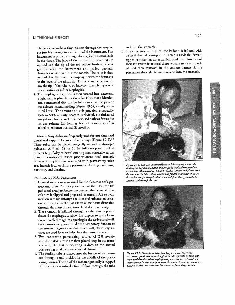
Therapy The most important treatment is provision of a balanced, not protein-restricted feline diet delivering adequate energy (goal feeding of 40-60 kcal/kg ideal weight/day). Lack of nutrients promotes lipolysis and glycogenolysis. This fuels the already imbalanced TG dispersal mechanisms. Protein is essential for this process, in order to make lipoproteins VLDL, thus protein restriction is contraindicated unless encephalopathy (HE) is present. Dietary management of IHL cats should consist of a higher protein, lower carbohydrate diet. Arginine is needed for normal detoxification of nitrogen. Cage resting cats with HL may reduce muscle release of ammonia, thus may be beneficial. Protein should only be restricted in patients with irrefutable signs of hepatic encephalopathy in order to reduce the production of false neurotransmitters. Carbohydrates as a source of calories are problematic as these cats are already prone to insulin resistance. If HE is present, then lactulose (0.25-2 ml/kg to effecting soft stools), plus metronidazole (7.5 mg/kg PO q12h) are indicated. Feeding is best achieved using a large bore feeding tube to avoid development of food aversion that may follow forced oral alimentation. Use of large bore tubes is associated with improved survival. Esophagostomy tubes are preferred as these are placed quickly and have fewer complications than gastrostomy tubes. However, initial feeding may be via a nasoesophageal tube while correcting of hydration and initial electrolyte abnormalities. The large bore tube may be placed once the patient is stable enough to tolerate anaesthesia and bleeding tendencies have been circumvented by administration of vitamin K1. The number of feedings per day is determined based on the volume of food tolerated per feeding. Carbohydrate supplementation is not advised (e.g., dextrose infusion) because of its inhibitory influence on fatty acid oxidation. Appetite stimulants including cyproheptadine (1 mg/cat PO BID), mirtazapine (2-3 mg/cat PO q72h) may help jump-start a cat’s appetite, but one must be wary not to lose sight of total calories consumed. If a cat is eating but not enough, supportive feeding (assisted syringe feeding or tube feeding) must be considered. A cat eating small amounts of baby food will not meet his caloric needs until he eats two – three jars/day. Meat baby food is not balanced, but is sufficient for several weeks. There are several diets specifically designed for the assisted feeding of cats (Royal Canin Recovery, Hill’s a/d, Eukanuba Maximum Calorie), liquid balanced enteral diets

for cats (Clinicare, Rebound) Additionally, we can make a slurry from any canned food. In order to minimize loss of caloric density, blend with one of the liquid diets rather than water. There are numerous options for assisted feeding. In general, this author starts with syringe assisted feeding until the cat is stable enough to allow the brief anaesthetic required for the placement of an esophageal tube. Because liver disease is known to be present, three doses of Vitamin K1 are given SC (1.0 mg/kg q12h SC) prior to tube placement, biopsies or any other procedure that might result in bleeding. Placement of esophageal tubes is discussed elsewhere. Suffice it to say that the instrumentation for this procedure is very basic requiring only the following: 14-16 Fr red rubber feeding tube/urinary catheter, Carmalt or other long curved forceps, a scalpel blade, suture and bandaging materials and a multiple use injection port (prn adaptor). A brief anaesthetic is required. Several protocols are appropriate including propofol, despite hepatic metabolism, whose use did not increase morbidity or fatalities in cats with primary lipidosis. Calculating how much to feed requires that you know the patient’s current weight as well as their healthy weight and the caloric densities (kcal/ml) of the diet you are intending to use (see Table 1). Use 50 kcal/kg as a rough guide to determine calories needed. Start by feeding 1/3-1/2 of the calories needed for the current, inappetant weight. On day two, feed 2/3-3/4 of this number and on day three, feed the full calories needed for the current weight. For weight gain, gradually increase to the calories needed for the cat’s healthy weight. Example: 3.4 kg sick cat BCS 3/9, healthy weight 4.0 kg BCS 5/9 % weight change = (4 – 3.4) / 4 = 15% 4.0 kg X 50 kcal/kg/day = 200 kcal by day 3 200 kcal = 95 ml Iams Maximum Calorie
OR 154 ml of Hill’s a/d or Royal Canin Recovery or PVD CN Day 1 feed 35 ml of Max Cal or 50 ml of the other diets Day 2 feed 70 ml of Max Cal or 100 ml of the other diets Day 3 feed 95 ml Max Cal or 154 ml of the other diets. With surgically placed tubes there is a delay in how quickly one can start to use them. The esophageal tube requires only a 2-3 hour delay to ensure full recovery from anaesthesia whereas gastrostomy and jejunostomy tubes require a longer wait of 10-12 hours. This is to ensure that some healing has begun to reduce the chance of food leaking from around the tube through the stoma into the peritoneal cavity. Any tube may clog. Should this occur, a cola-type soft drink, pancreatic enzymes or meat tenderizer can be instilled into the tube, left to incubate for about 10 minutes before attempting to flush tepid water through the tube to check for patency. Cats can eat with any of these tubes in place. It is recommended to avoid offering food for a week to reduce the likelihood of them developing aversion to the food offered. Once a cat is eating well with tube in place the question becomes when one can remove the tube. Weigh the cat and, as long as he/she is eating well, avoid using the tube (for nutrients) for a week then reweigh the kitty. If the weight is stable (or increased), then it is safe to remove the tube. Because of stoma formation (except with nasoesophageal tubes), removal does not require
anaesthesia. Remove the suture (purse-string or stay sutures) and pull the tube out. In the case of a gastrostomy tube, straighten out the bulb/balloon by inserting a straight probe through the tube while concurrently pulling the tube out. Suturing is not required for any of the skin openings. Cleanse any minimal serous discharge that may occur for 2-3 days. With the exception of nasoesophageal tubes, which should be only used short term (< 5 days), tubes may be left in place as long as a patient requires nutritional support. The longest that the author has had a gastrostomy tube in a cat was 18 months. If red rubber tubes are used rather than silicone or polyurethane, they may discolour and weaken over time. Esophageal tubes are easily changed. Simply remove the existing tube as described and, slip a new tube in through the stoma. A local anaesthetic will be needed to place a new purse-string suture. (Resource: Kitty Kollar e-tube collar: www.kittykollar.com) How often should you feed? The number of feedings per day, (and hence timing), is determined based on the volume of food tolerated per feeding. Start with 6 ml and increase by 6 ml increments to about 36-48 for most cats. In the uncommon case of the patient who cannot tolerate even 6 ml boluses despite antiemetic therapy, trickle feeding may be instituted. Trickle feeding is a technique in which liquefied food is syringed into an empty fluid bag and administered gravitationally or by pump assistance via an intravenous line attached to the large bore feeding tube or by use of a large syringe filled with food and syringe pump. Care must be taken to renew food and delivery tubing and syringe at 12-hour intervals to avoid bacterial contamination. A promotility agent may be warranted. Table 1: Caloric densities, for feeding volume calculations ReboundTM: 1 kcal/ml ClinicareTM: 1 kcal/ml Royal Canin/MediCal RecoveryTM: 1.23 kcal/ml Hill’s a/dTM: 1.3 kcal/ml Iams Maximum CalorieTM: 2.1 kcal/ml PVD CNTM: 1.2 kcal/ml
Vomiting must be controlled. Although this can be achieved medically (see drug doses), it may also be alleviated by reducing meal volume, increasing the number of meals per day, using a trickle feeding approach with a syringe or fluid pump, and providing an opportunity for exercise (to stimulate enteric motility). Trickle feeding refers to placing liquefied food into an empty fluid bag and administering it gravitationally or by pump assistance via an intravenous line attached to the large bore feeding tube or by use of a large syringe filled with food and syringe pump. Care must be taken to renew food and delivery tubing and syringe at 8-hour intervals to avoid bacterial contamination. A promotility agent may be warranted as well. Table 2: Select Anti-emetics for use in the Cat

Generic Name Product™ Dose (feline)
Chlorpromazine Thorazine, Largactil 0.5 mg/kg q8h IM Prochlorpromazine Compazine 0.1 mg/kg q6h IM Diphenhydramine Benadryl 2.0-4.0 mg/kg q8h PO 2.0 mg/kg q8h IM Dimenhydrinate Dramamine, Gravol 8.0 mg/kg q8h PO Metoclopramide Reglan 1-2 mg/kg constant rate
infusion IV over 24hours Ondansentron Zofran 0.1-0.15 mg/kg slow push IV
q6-12 hours prn Dolasetron Anzemet 0.6 mg/kg IV, SC q24h Mirtazapine Remeron 3 mg PO q72h Maropitant Cerenia 0.5-1 mg/kg SC, IV or PO q 24 hr
Fluid support is needed for rehydration as well as for maintenance of this state. It is best provided intravenously using non- lactate, non-glucose containing fluids. Lactate intolerance is suspected in HL and steady glucose infusion may potentiate hepatic TG accumulation. Dehydration impairs hepatic circulation, compromises normal detoxifi-cation processes, azotemia enhances ammonia production, and constipation augments absorption of colonic toxins. Aggressive attention to correction of hypokalemia is essential; hypokalemia is a negative predictor for survival. Use the customary sliding scale for fluid potassium supplementation. In the hypokalemic state, potassium (K) shifts from inside the cell to extracellular fluid. This parallels an increase in extracellular pH, which causes increases in intracellular ammonia trapping. In cats deficient in arginine, this becomes more severe. Hypokalemia also alters the threshold of response of neuroreceptors. Hypophosphatemia may precede initiation of nutritional support but more commonly reflects a refeeding phenomenon within 12 hours of initial food ingestion. Hypophosphatemia is treated using potassium phosphate, delivered at 0.01-0.03 nmol/kg/hr. Monitor serum phosphate every 6 hours, and discontinue supplementation when serum phosphorus levels reach and sustain > 2 mg/dL (> 0.65 mmol/L). Avoid iatrogenic hyperkalemia by adjusting KCl supplements to account for potassium delivered with the potassium phosphate. Water-soluble vitamins should be added to intravenous fluids (1 to 2 mL Vitamin B complex per litre of fluids). Cobalamin
(Vitamin B12) deficiency should be considered in HL cats with suspected intestinal or pancreatic disease. Thiamine (vitamin B1) deficiency is also suspected in some cats with HL showing sluggish pupillary light responses, vestibular signs, and abnormal postural reactions, or severe neck ventroflexion. Because a vasovagal response may result from injected thiamine, the oral route is preferred (50 to 100 mg per cat per day for 1 week in those demonstrating consistent clinical signs). Other differentials for neck ventroflexion in cats include hypophosphatemia, hypokalemia, hepatic encephalopathy, hyperthyroidism, neuromuscular weakness (e.g. myasthenia gravis), and disorders causing cervical vertebral or muscular pain.

Cats with HL are often deficient in vitamin K1. Lack of dietary intake, altered intestinal bacterial flora subsequent to antimicrobial treatment, and impaired vitamin K epoxidase cycle associated with hepatic dysfunction are underlying causes. Treatment with vitamin K1 is recommended (0.5-1.5 mg/kg SC at 12-hour intervals X 3 doses). Vitamin K treatment must precede insertion of feeding appliances, jugular venipuncture, cystocentesis, hepatic aspiration, or hepatic biopsy. Supplementation with 250 to 500 mg L-carnitine per day has been recommended yet there is no conclusive evidence that outcome is improved through using it. L-carnitine is an essential cofactor for fatty acid oxidation; supplementation may assist in preventing hepatocellular accumulation of free fatty acids and help remove toxic acetyl groups from the mitochondria. It is surmised that in HL a “relative” hepatocellular carnitine deficiency exists despite plasma or muscle carnitine concentrations. Owing to solubility issues, only medical grade liquid carnitine should be used. Taurine supplementation is also recommended (250 to 500 mg/day), as this essential amino acid is obligatory for bile acid conjugation in the cat and cats with HL waste taurine in their urine. Taurine supplementation increases water solubility of bile acids, reduces their cellular toxicity, and facilitates their circulation and renal elimination. Because bile cannaliculi are narrowed, consider using ursodeoxycholic acid. This bile acid has a number of hepatoprotective effects and provides IgA to proximal duodenum. It requires taurine for conjugation. S-adenosyl-L-methionine (SAMe) is a naturally occurring substance in the body. It initiates transmethylation, transsulfuration and aminopropylation. The first pathway contributes to cell membrane fluidity and carnitine synthesis among other actions. Transsulfuration is the process by which glutathione (GSH) is produced; glutathione is an important component of the antioxidant defence system detoxifying xenobiotics and protecting against oxidative injury. Via aminopropylation, SAMe may have anti-inflammatory and analgesic properties as well as assist in protein synthesis. All of these actions could be beneficial in supporting resolution of lipidosis. Nevertheless, no studies have been done to date that look at whether incorporation of SAMe improves response to therapy in cats with hepatic lipidosis. Cats with lipidosis or necroinflammatory liver diseases have been shown to have low liver tissue concentrations of glutathione. Table 3: Drugs, Dosages and Indications
Drug Dose Range Frequency Route Indications Chlorpromazine
0.5 mg/kg TID IM Vomiting
Prochlorpromazine
0.1 mg/kg QID IM Vomiting
Dimenhydrinate
8.0 mg/kg TID PO Vomiting
Metoclopramide
1-2 mg/kg Over 24 hours
CRI IV Vomiting
Ondansentron 0.1-0.15 mg/kg
Q6-12 h prn Slow push IV
Vomiting

Dolasetron 0.6 mg/kg SID PO, SC, IV Vomiting Mirtazapine 3 mg Q72h PO Vomiting Maropitant 1 mg/kg SID < 5
days SC Vomiting
Vitamin K1 0.5-1.0 mg/kg Q12h X 3 SC Lipidosis L-carnitine 250-500 mg SID PO/tube Lipidosis Arginine 250 mg SID PO/tube Lipidosis Taurine 250-500 mg SID PO/tube Lipidosis Ursodeoxycholic acid
15 mg/kg Q12-24h PO/tube Cholestasis, lipidosis
S-adenosyl-L-methionine
20-40 mg/kg SID PO/tube Glutathione donor, lipidosis
Milk thistle (silymarin)
5-15mg/kg SID PO/tube Hepatoprotective antioxidant
PROGNOSIS Outcome depends on identification of concurrent/underlying problem and the ability to correct or attenuate it. Refractory hypokalemia is a negative prognostic indicator, thus hypokalemia must be addressed early on and aggressively. Should serum potassium levels not respond to standard therapy, serum magnesium should be evaluated. Correction of hypomagnesaemia may make serum potassium levels more responsive to KCl therapy.
SUMMARY Effective treatment of hepatic lipidosis requires commitment to the client as well as client education in discussing the likelihood of an underlying primary disease, the importance of supportive care, without which the condition has a poor prognosis, and encouragement because HL rarely reoccurs. Treatment may require weeks to months of assisted alimentation and metabolic support as well as concurrent management of underling medical conditions. A recovery rate exceeding 85% can be achieved if the primary disease can be identified and ameliorated and the patient survives the initial 72-hours of critical supportive care. REFERENCES Armstrong PJ, Blanchard G: Hepatic lipidosis in cats. Vet Clin North Am Small Anim Pract.
2009;39(3):599-616. Blanchard G, Paragon BM, Milliat F, et al. Dietary L-carnitine supplementation in obese
cats alters carnitine metabolism and decreases ketosis during fasting and induced hepatic lipidosis J Nutr. 2002;132(2):204-10.
Blanchard G, Paragon BM, Sérougne C, et al. Plasma lipids, lipoprotein composition and profile during induction and treatment of hepatic lipidosis in cats and the metabolic effect of one daily meal in healthy cats J Anim Physiol Anim Nutr (Berl). 2004;88(3-4):73-87.
Brenner K, Kukanich KS, Smee NM. Refeeding syndrome in a cat with hepatic lipidosis. J Feline Med Surg. 2011; 13(8):614-7.
Brown B, Mauldin GE, Armstrong J, et al. Metabolic and hormonal alterations in cats with hepatic lipidosis. J Vet Intern Med. 2000;14(1):20-6
Center SA, Warner K, Corbett J, et al. Proteins invoked by vitamin K absence and clotting

times in clinically ill cats J Vet Intern Med. 2000;14(3):292-7. Center SA: Investigations of the effect of L-carnitine on weight reduction, body condition,
and metabolism in obese dogs and cats. In Recent Advances in Canine and Feline Nutrition, vol 3. 2001, 113-122.
Center SA, Warner KL, Erb HN. Liver glutathione concentrations in dogs and cats with naturally occurring liver disease Am J Vet Res. 2002;63(8):1187-97.
Center SA: Feline hepatic lipidosis. Vet Clin North Am Small Anim Pract. 2005; 35:225-69. Daniel AGT, Lucas SRR, Júnior AR, et al. Skin fragility syndrome in a cat with
cholangiohepatitis and hepatic lipidosis. J Feline Med Surg. 2010;12(2):151-5. Feeney DA, Anderson KL, Ziegler LE, et al: Statistical relevance of ultrasonographic criteria
in the assessment of diffuse liver disease in dogs and cats. Am J Vet Res. 2008;69(2):212-21. Griffin B. Feline Hepatic Lipidosis: Treatment Recommendations Compend Contin Educ
Vet. 2000; 22(10): 910-921. Ibrahim WH, Bailey N, Sunvold GD, et al. Effects of carnitine and taurine on fatty acid
metabolism and lipid accumulation in the liver of cats during weight gain and weight loss. Am J Vet Res. 2003;64(10):1265-77.
Mazaki-Tovi M, Abood SK, Segev G, et al. Alterations in adipokines in feline hepatic lipidosis. J Vet Intern Med. 2013; 27(2):242-9.
Trotman TK, Elizabeth Mauldin E, Hoffmann V, et al. Skin fragility syndrome in a cat with feline infectious peritonitis and hepatic lipidosis Vet Dermatol. 2007;18(5):365-9.
Posner LP, Asakawa M, HN. Use of propofol for anesthesia in cats with primary hepatic lipidosis: 44 cases (1995-2004). J Am Vet Med Assoc. 2008;232(12):1841-3.
Zoran DL. The carnivore connection to nutrition in cats J Am Vet Med Assoc. 2002;221(11):1559-67.
Appendix Instructions for placing an esophagostomy tube from Ogilvie GK, Moore AS: Feline Oncology:
A Comprehensive Guide to Compassionate Care, Trenton, 2001, Veterinary Learning Systems, p 120-121.

MANAGING THOSE DARNED DIABETIC CATS – IMPROVING OUTCOMES Margie Scherk DVM, Dip ABVP (feline practice)
catsINK, Vancouver, Canada
Diabetes mellitus is one of the two most common endocrine disorders in cats. It is a heterogeneous group of disorders in which insulin production is reduced or in which tissue cells are resistant to the effects of insulin, resulting in impaired glucose homeostasis. From a clinical perspective, regardless of the cause, diabetes mellitus (DM) can be challenging to diagnose and treat in the cat because of their stress- induced hyperglycemia. The prevalence of this condition has increased over time from 8 out of 10,000 (in 1970) to 124 of 10,000 (in 1999) cats seen at veterinary teaching hospitals (Prahl). The frequency of occurrence also appears to vary with geographic location (0.21% in Swedish cats [Sallander]; 0.43% in the United Kingdom [McCann]; 0.74% of Australian cats [Lederer 2009]), with British and Australian Burmese being significantly over-represented at 3.7 and 3 fold, respectively. Fasting glucose concentrations are higher and glucose tolerance is lower in Burmese cats in Australia, New Zealand and the UK compared to matched non-Burmese cats [Lederer 2005]. It appears to be inherited as an autosomal, not fully penetrant trait in these Burmese. Pathophysiology Review Insulin is secreted after a meal, to facilitate utilization and storage of glucose, fat and amino acids in three primary tissues: liver, muscle and fat. A mild insulin deficiency results in decreased transfer of ingested nutrients into tissues causing mild to moderate hyperglycemia. Severe insulin deficiency not only hampers tissue uptake of ingested fuels, but also results in marked compensatory glucose overproduction along with excessive mobilization of the body's protein and fat stores. Combined with glucagon excess (relative or absolute), this results in an increased delivery of fatty acids to the liver, their oxidation to ketone bodies (betahydroxy-butyrate, acetoacetate, and acetone), and a clinical state of ketoacidosis. Because there is no insulin available to deliver the glucose into the cells, cells starve and polyphagia with concurrent weight loss occurs. Unabsorbed glucose (hyperglycemia) spills into the urine drawing water with it. This causes polyuria and compensatory polydypsia. Classification And Differentiation Between Type 1 And Type 2 Diabetes In human diabetes, Type 1 refers to a condition of insulin dependency seen in people who are generally lean, young and prone to ketogenesis. It is caused by immune-mediated beta cell depletion, causing an absolute insulin deficiency. Type 2 DM usually occurs in the older human, often obese but less prone to the development of ketoacidosis. The underlying problem is one of insulin receptor and post receptor defects, interfering with insulin uptake by tissues. This insulin resistance and associated hyperglycemia, causes the beta cells to produce more insulin, thus this state is one of a relative insulin deficiency. Type 2 may be controlled, at least initially, with weight loss, diet and oral hypoglycemic agents. Generally, diabetes is a disorder of the older, often overweight cat, similar to the Type 2 human patient. Risk factors include body weight > 7 kg, older age (> 10 years), male gender, neutered. Henso showed that increased body condition score (BCS) in nondiabetic cats is associated with increased circulating concentrations of IAPP and insulin. Obese cats appear to have a defect in

insulin secretion along with lower tissue sensitivity to insulin. Unlike human Type 2, however, by the time the diagnosis of diabetes is made, most cats are insulin dependent although not prone to ketogenesis. In addition to these differences, cats may also develop diabetes secondary to endocrinopathies (acromegaly or hyperadrenocorticism), or drug therapy (glucocorticoids and progestins). Inflammation is another recognized predisposing factor for susceptible individuals to develop diabetes. Franchini has shown at a molecular level that the inflammation induced by bacterial or viral infection can, via molecules recognized by toll-gate receptors, damage endocrine pancreatic tissue. It remains unclear whether pancreatitis is a significant co-morbidity (Forcada) or whether it may be a) a source of inflammation no different than other sites or b) pancreatitis develops as a result of beta cell apoptosis. Additionally, in cats pancreatic islet amyloid deposits are believed to interfere with insulin secretion, and that oral hypoglycemics (such as the secretagogue sulfonylureas) may actually increase islet amyloid polypeptide (IAPP) deposition. IAPP is co-secreted with insulin. Islet amyloidosis occurs in 90% of humans with Type 2 DM. (O'Brien) Thus, feline diabetes shares several similarities with the disease in humans. Impaired beta-cell function, decreased beta-cell mass, insulin resistance that is often related to obesity, and pancreatic amyloid deposition, are among these common features. (Zini March 2010) Unlike humans, DM does not predispose cats to hypertension. Diagnosis In the stressed patient, epinephrine release causes hyperglycemia and glucosuria. Therefore, even in cats with history and clinical findings of polyuria/polydypsia, polyphagia, weight loss, hyperglycemia and glucosuria, it is essential to differentiate between this stress response and diabetes. This can be done through verifying that the hyperglycemia and glucosuria are persistent over time. However, because stress recurs a better option is to request that a fructosamine level be run on the previously collected sample. Fructosamine measures the protein bound glucose levels over the preceding 10 - 20 days. It can be affected by protein metabolism as well, hence hyperthyroidism, with more rapid muscle turnover, may result in artificially lower fructosamine values. Urine ketone measurement is routinely performed in cats with diabetes mellitus to identify impending or established ketoacidosis. The urinary ketone dipstick test has a low sensitivity as it quantifies the less abundant ketone acetoacetate. Beta-hydroxybutyrate (beta-OHB) is the predominant serum ketone. Determination of plasma beta-OHB concentration was shown to be a useful method to distinguish between diabetic and non-diabetic sick cats. (Zeugswetter) Therapy And Management Of The Diabetic Cat Good glycemic control soon after diagnosis is associated with increased probability of remission. Some believe that it should be the goal of insulin therapy. (Roomp, Marshall) In a study published in 2010, clinical remission of diabetes was evaluated. Ninety cats with newly diagnosed diabetes were followed until death or remission. Remission was defined as normoglycemia without insulin for > 4 weeks. Likelihood of remission was found to be greater in older cats and in cats with higher body weight. Remission was less likely in cats with increased serum cholesterol and was of shorter duration when serum glucose was higher, i.e.,

less well regulated. (Zini, Nov 2010). More recently, Gostelow et al perfomed a literature review on feline remission. They found that the quality of the studies was lacking and biased especially regarding lack of randomization and blinding, small sample size and, most tellingly, a lack of consensus in criteria for defining remission as well as even the diagnosis of diabetes. Insulin Choice: There are many types of insulin available: they are derived from several sources and have several durations of action. In the United States, the FDA has eliminated any animal sourced insulin from the market. Insulins are currently produced from human recombinant technology. However, beef-pork and beef sourced insulins may be better suited to cats because of closer structural similarity to feline insulin. Speed Of Onset And Duration: The speed , onset of action and duration differs between insulins. 1. Regular (fast) - rapid onset of action (0.5h), max. effect (1-5h), end effect (8h) 2. NPH (intermediate) - onset of action (1.5h), max. effect (4-12h), end effect (24h) 3. Lente - onset of action (2.5h), max. effect (7-15h), end effect (24h) 4. Semilente - onset of action (1.5h), max. effect (5-10h), end effect (16h) 5. Combination: 70% NPH: 30% regular - onset of action (0.5h), max. effect (4-8h), end effect (24h) 6. Ultralente (long acting) - onset of action (4h), max. effect (10-30h), end effect (36h) 7. Synthetic insulin analogues: glargine and detemir (ultra-long acting) once a day in humans These values are for comparison only and reflect human metabolism. Insulin responses vary with the individual. Every cat is different and will respond differently to the insulin they take
in the management of diabetes. It is ALWAYS advisable to start with an insulin that is licenced for veterinary use. Vetsulin™, is a 40 U/ml porcine lente zinc insulin specifically registered for veterinary use. It has been available for over 15 years as Caninsulin around the world and is known as Vetsulin™ in the United States. Its peak activity is ~3h and duration of 6-10h. It is very effective for the treatment of feline diabetes. Protamine zinc insulin (PZI) is a long-acting, beef-pork insulin that was considered by many to be the insulin of choice for cats because of its molecular similarity to feline insulin. Since November 2009, an FDA approved recombinant human protamine zinc insulin preparation, ProZinc™, has come on the veterinary market. Like Vetsulin/Caninsulin, it is a 40 unit/ml (U 40) insulin. Humulin N and Novolin N are recombinant human NPH insulins (100 U/ml) that have an intermediate duration of action. They do not work well in most cats. Glargine (Lantus™) is a long-acting human recombinant DNA insulin analogue that has been modified by replacing one amino acid (asparagine) with another (glycine) as well as adding 2 arginine amino acids to the c-terminal end of the molecule. This changes the pH solubility making it microprecipitate at the site of subcutaneous injection that are slowly absorbed. This

should result in fewer troughs and a slower, smoother glycemic effect, however this does not appear to occur in all cats. Because the formation of microcrystals and slow absorption are dependent on the acidity of the product, glargine cannot be mixed or diluted. Interestingly, in cats with diabetic ketoacidosis, glargine may be used in place of regular (Toronto) insulin if given IM or IV. By these routes, it has a similar action profile to that of regular insulin. In fact, in some resistant diabetic cats, one might consider using it by both the IM and SC routes BID with 70% of the dose given SC and 30% of the dose given IM. Detemir (Levemir™) is another long acting human rDNA analogue. It is modified from insulin by adding an acylated fatty acid chain. This allows reversible binding to plasma proteins, resulting in a slow release into plasma. In cats, its action and duration are similar to those of glargine. The dose required may be less than that of glargine (~30% less in the Gilor study). Remission rates and time to remission are similar. Newer Insulin Analogues: Rapid-acting insulin analogues lispro, aspart, and glulisine act by blocking the formation of insulin dimers and hexamers. This allows larger amounts of the active monomeric insulin to be immediately available for postprandial use when given at mealtime. Studies in dogs and cats have yet to been reported. Insulin degludec (Tresiba™) is a new-generation, ultra-long-acting analogue not yet available in North America or Europe. It forms large soluble multi-hexamers at the injection site. Studies in dogs and cats have yet to been reported but, due to its extremely long action in humans (given once daily or three times a week), it might provide reliable once-a-day or once-every-other-day therapy in cats. A concept not used in veterinary medicine but that may help with some difficult diabetics is that of combining insulins to have one that provides basal control and another covering mealtime glycemic needs (basal-bolus therapy). In humans, this approach is taken using premixed combinations of a short-acting and a longer-acting or ultra-long-acting insulin analogues. While not yet studied in cats (or dogs), this effect might be achieved through giving SC glargine concurrently with an IM dose BID or by using SC Vetsulin or ProZinc concurrently with SC glargine or detemir BID. The insulins must not be mixed in one syringe. Concentration: It is critical to know the concentration of the insulin you are using and to match the syringes to that strength. For correct dosing, insulin should be administered using syringes specifically calibrated for the strength of insulin used. For example, most insulin is 100 Units/ml (U100) and micro-fine or ultra-fine U100 syringes should be used with these. With U-100 insulin, when only small amounts of insulin are needed, using a 3/10cc or 5/10cc U-100 allows even the tiniest dose to be measured more accurately. The advantage of using a 40 unit/ml insulin is that it is more making it easier to more accurately dose small amounts of insulin. The specific U-40 syringes should be prescribed with this product. As the use of U100 syringes for a more dilute U40 insulin risks miscommunication and tragic consequences.
While there are guidelines in choosing the starting dose of insulin for a patient, the maximum dose for that patient is the dose that he/she needs to resolve the clinical signs of excessive urination and drinking, lethargy and weakness. The majority of cats require twice daily injections, regardless of the type of insulin selected. Client Counselling Once the cat has been determined to be diabetic, client counselling is very important. Initially, most clients are intimidated at the thought of administering insulin injections. Booking a discharge or demonstration appointment with the nurse-technologist works well, as nurses are often more patient than veterinarians are at explaining and guiding the learning client. At this appointment, review the pertinent facts about insulin storage (refrigerator), handling (gently), re suspension (gentle figure 8's), drawing up into the syringe, administration (upon exhalation of client, walk through the door of the tent, OR pull the tent over the needle, think canvas, practise on a cat using saline), single use only of insulin syringes for sterility and sharpness sake. Show the client how to keep a diary, recording date, time of insulin administration, dose administered, activity level, BM, amount urinated (# and size of clumps of clumping litter), amount eaten, and amount drunk (by difference, measure amount left in bowl the next morning). Counsel on diet to be fed, as determined by the veterinarian. Lower carbohydrate, higher protein diets may be more effective for glycemic control, however this remains controversial. There is no scientific consensus on carbohydrates: to date there is no clear evidence that carbohydrates either cause or are contraindicated in the treatment of feline diabetes (Farrow, Coradini, Sallander, Slingerland, Owens , Hoenig). The native diet for a cat (bird or mouse) is high protein, moderate fat, low carbohydrate, it is reasonable to feed this macronutrient profile for any cat. Cats should have free access to food all the time, rather than feeding twice daily. Some cats refuse to eat the diets we recommend. For those patients and for clients unwilling/unable to offer those diets, here is a website which lists the protein and carbohydrate proportions of grocery store brands: http://www.sugarcats.net/sites/jmpeerson/. Other helpful websites for clients to use for information, support and encouragement (including teaching techniques) follow: www.petdiabetes.com, www.felinediabetes.com, www.sugarcats.com and www.cat-dog-diabetes.com/cats-diabetes-mellitus.asp Cats with comorbidities that require a different diet should be fed the diet appropriate for their concurrent disease. Insulin dose can be regulated with consistent feeding of any diet. Similarly, should a diabetic cat need prednisolone for a concurrent problem (e.g., asthma or inflammatory bowel disease [IBD]), treat the underlying problem as needed and regulate the insulin to that corticosteroid dose. If the antiinflammatory action can be provided through a non glucocorticoid agent, (e.g., chlorambucil for IBD, an NSAID for arthritis), then that can be attempted. Monitoring urine parameters at home is justified for:

Cats with transient diabetes- to identify when/if glucosuria recurs Cats on oral hypoglycemics to determine if glucosuria resolves Cats previously or currently ketoacidotic - to monitor for ketones Follow-Up Care And Monitoring At the discharge time, book an appointment for a blood glucose curve and re-evaluation for 14 days later. Let the client know that you will call daily for the first 3 - 4 days, to be supportive and available for questions, to find out how the kitty is doing, and to ascertain that they are observing the parameters you need diarized for evaluation. Let them know that it is unlikely that the initial dose will be the perfect one, and that, as they approach the "right" dose for this cat, there will initially be a marked reduction in urine output and drinking, however, after 3-4 days, these amounts will increase again as the cat's glucose homeostasis re-equilibrates. The timeline for care that the author uses is: Diagnose diabetes mellitus by confirming with fuctosamine; start insulin, diet and diary; 10-14 days later: in-clinic BG curve, adjust dose, teach ear prick BGs, add BID BG
monitoring to diary for practice; Another 10-14 days later: in-clinic BG curve, fructosamine, adjust dose; Subsequent BG curves are performed at home, follow-up by email, phone or fax to adjust
dose; Recheck cat q4-6 months (exam, fructosamine, U/A) as long as he/she is stable. At the blood glucose (BG) curve appointment, hospitalize the cat with food and water, after weighing him/her and ascertaining what time the insulin was administered and what dose the client gave. Measure BG immediately, to get a starting level. Using a 25G needle works well, as a mere drop or two of blood are needed for the portable glucometers. Plot the values on a graph for easier interpretation. Submit a serum fructosamine as well to determine how the average glycemic control has been over the past 10-20 days. Continue measuring the BG every 1 (-1.5) hours over a 12 hour period. Ear sampling and a calm, reassuring manner will help to minimize the stress (and its associated BG elevations) somewhat. Nevertheless, the readings generally will be higher than what is occurring at home, therefore it is imperative to read the client's diary and take the clinical signs into consideration when adjusting the insulin dose. Once the blood glucose goes up for two consecutive measurements, the curve can be stopped. (Note this does NOT apply in the case of a cat in diabetic ketoacidosis.) Use of the marginal ear vein is an accurate and easy technique for the measurement of BG. It is a useful technique in the clinic and, if the concept is introduced to clients with confidence and compassion, many are willing to perform curves at home. In general, these curves are more accurate as the cat’s stress level is lower. Additionally, it is valuable for clients to be able to measure a spot glucose if their cat “doesn’t look right” before deciding to give insulin or not. The goals of performing a BG curve are to determine 1) Whether the insulin is being absorbed 2) The glucose nadir (level and time to reach it) 3) The duration of insulin effect

4) The degree (delta) of insulin effect, and 5) To assess the fluctuations of glucose levels in this individual patient!
When using glargine, the protocol for regulation and curving is somewhat different. The following recommendations come from Dr. Jacquie Rand:
Measure glucoses every two hours for a minimum of 12 hours daily for the first three days in order to determine whether hypoglycemia is occurring as well as to assess how long the insulin is lasting in the individual. After this initial three day period, dose adjustments are based on the pre-insulin BG (vs. nadir as with other types of insulin).
If at a 7 day hospital recheck, the pre-insulin BG concentration is > 290 mg/dl (16 mmol/L), increase the dose by 1.0 U/cat. A 12h curve should be done on the following day to make sure that hypoglycemia is not occurring at this increased dose.
Do not change the dose if the pre-insulin BG concentration is 220-290 mg/dl (12-16 mmol/L).
The dose should be decreased by 0.5-1.0 U/cat if the pre-insulin BG concentration is < 180 mg/dL (10 mmol/L). I f biochemical hypoglycemia is present, the dose should be decreased by 1.0 U/cat. If clinical signs of hypoglycemia are present, the glargine dose should be decreased by 50%.
If a BG drops below normal range (< 80mg/dl or < 4.4 mmol/l), the staff person should notify the veterinarian after offering the cat some palatable food, as he/she may wish to administer dextrose intravenously to avoid a hypoglycemic crisis. Signs of hypoglycemia include weakness, lethargy, trembling, head tilt, ataxia, coma and death. If a hypoglycemic cat is offered food and doesn't eat right away, or if signs are severe, then corn syrup should be rubbed on the oral buccal mucosa while preparing to administer an intravenous dose of 50% dextrose.

The "Somogyi effect" is rebound hypoglycemia-induced hyperglycemia. If the cat's BG drops too low, the body reacts by releasing catecholamines (epinephrine), glucagon, glucocorticoids and growth hormone. This causes a rapid release of glucose into the serum causing this rebound to occur. It is important to not be tempted to increase the insulin dose in these individuals, as this would accentuate the problem and eventually cause a hypoglycemic crisis. "Spot checks" of BG levels should be avoided as they can be misleading and can mask a rebound effect, and be misinterpreted as needing more insulin. Over the next month or two, by performing blood glucose curves, measuring serum fructosamine and reassessing the cat clinically and historically (diary) every 2 weeks, the insulin dose suitable for this patient will be determined. Thereafter, it is advisable to see the stable diabetic cat every 4 - 6 months for a fructosamine. Consider, also, on these rechecks, to collect a sterile urine sample for urinalysis, as diabetic cats are more prone to bacterial urinary tract infections than non-diabetic individuals. If a diabetic patient becomes ill, then a glucose curve should be run as well as any other tests appropriate to their condition. Update on glucometers: In a study comparing AlphaTRAK, Ascensia ELITE and reference hexokinase methods for determining serum glucose, the AlphaTRAK meter results did not differ from the reference method, however results from the Ascensia ELITE were significantly lower. The superior performance of the AlphaTRAK meter supports its use to monitor blood glucose levels in cats. (Zini, 2009) Useful resources Cook A. A Protocol for Diabetic Management. Veterinary Team Brief Supplement, 2013: www.Veterinaryteambrief.com/diabeticmanagement Schermerhorn T. The Role of the Blood Glucose Curve. Clinician’s Brief. November 2010, 23-5: www.cliniciansbrief.com/column/patient-support/role-glucose-curve Sparkes A, Cannon M, Church D, et al. ISFM consensus guidelines on the practical management of diabetes mellitus in cats. J Feline Med Surg. 2015 17(3):235-50: jfm.sagepub.com/content/17/3/235.full.pdf+html Complete references are available from author on request

BLOOD GLUCOSE CURVES MADE EASY Margie Scherk DVM, Dip ABVP (feline practice)
catsINK, Vancouver, Canada Blood glucose curves can be very helpful to determine the dose and type of insulin for a given cat. They are not difficult to interpret when simple rules are followed. It is very important to get a reading every hour. Cats should have food available at all times. 1. Start by looking at the shape of whole curve. Identify the nadir (lowest BG value), time to nadir, starting and highest BG value, duration (Figure 1). Figure 1. Elements of a blood glucose curve
Check if the blood glucose (BG) level decreases for a reasonable period of time. This indicates that cells see and respond to the insulin? If the curve merely wobbles around the starting level (e.g., curve D in Figure 2), then either:
The cells do not see/respond to this insulin; The client is not administering the insulin correctly. This could be technique (intra-fur,
intradermal resulting in poor absorption) or lack of comprehension (giving air, wrong dose);
The insulin is damaged (dropped, client wiped vial with alcohol, bacteria introduced into vial);
Counter-regulatory phase of Somogyi response to overdose. 2. Time between BG at time zero (just before insulin is given) to time at which BG level is the same = duration of action. This value tells you how long the insulin lasts in this individual. If duration is 9-12h, then BID administration is appropriate;

If duration is 6-8h, then TID administration is appropriate. 3. Time to reach nadir indicates how rapidly insulin is being absorbed and taking effect. If peak insulin effect is between 2-4h after administration, be suspicious of Somogy overswing (too much insulin). This will be followed by a rapid increase in BG with the curve exceeding the starting BG. 4. BG level at nadir indicates maximum effect of insulin. IMPORTANT In order to determine nadir, one must have hourly BG readings. In fact, to really identify the nadir, we would need even more frequent readings, however with less than hourly measurements, we could easily miss a Somogyi, both at nadir as well as the overswing. 5. Glucose differential/delta is the difference between starting BG and nadir BG. If this difference is small (<7 mmol/L; 126 mg/dL), it is easy to decrease the starting BG without dropping the BG too low at nadir. This is a safe insulin to use for this patient. If the difference is large, it becomes difficult to increase the dose without risking hypoglycemia at peak effect. 6. Goal range for BG (not to be confused with differential) of 5.5-12 mmol/L; 100-215 mg/dL throughout the day provides good glycemic control and normalizes fructosamine levels. Spot checking should only be done to determine whether a lethargic, wobbly cat is hypoglycemic (and needs glucose) or hyperglycemic (and needs insulin) before rushing the cat to the clinic. Using spot checks (i.e., anything less than hourly measurements) does not provide useful information and can result in making inappropriate recommendations. Fructosamine reflects glycemic control, or time that BG is above ideal range over approximately the preceding 10-14 days. It is elevated if too little insulin is given but will also be elevated during Somogyi overswing, i.e., when too much insulin is being given. Glucosuria will occur under both situations as well. Figure 2. Examples of blood glucose curves A Ideal curve => continue dose and type of insulin B Short duration => give insulin more often or change type of insulin C Somogyi overswing: rapid drop in glucose with counter-regulatory overcorrection => decrease insulin dose or change type of insulin D Poor response due to client misunderstanding, poor technique, damaged insulin, attempt to correct from Somogyi response, extremely low dose => Client education, recheck curve. If no change, change insulin.

Useful resources Cook A. A Protocol for Diabetic Management. Veterinary Team Brief Supplement, 2013: www.Veterinaryteambrief.com/diabeticmanagement Schermerhorn T. The Role of the Blood Glucose Curve. Clinician’s Brief. November 2010, 23-5: www.cliniciansbrief.com/column/patient-support/role-glucose-curve Sparkes A, Cannon M, Church D, et al. ISFM consensus guidelines on the practical management of diabetes mellitus in cats. J Feline Med Surg. 2015 17(3):235-50: jfm.sagepub.com/content/17/3/235.full.pdf+html
RESPECTFUL CAT HANDLING vs. CAT WRANGLING: Part 1 - FROM THE CAT’S POINT OF VIEW Margie Scherk, DVM, DABVP (feline practice)
Vancouver, BC, Canada INTRODUCTION In many clinics, some veterinarians and other team members do not enjoy working with cats because they believe that cats are unpredictable and feel anxious about getting hurt. By understanding why cats feel that they need to defend themselves, by learning to identify the warning cues, managing the interactions in a positive manner, and making relatively minor changes to what the cat is exposed to, this fear can be reduced. The basis for working cooperatively with cats is being empathic to their nature and behaviors and trying to imagine what their experience is like. Cats are a species with a social structure different from ours. We need to look at cats differently, slow down and adjust our interactions. Minor modifications to the physical facility help reduce the strangeness and threats that cats experience in the veterinary clinic. The goal of these two presentations is to look at how to change the experience for cats thereby removing some of the obstacles to routine feline veterinary care. This benefits cats and their human companions with the resulting side effect of clinic growth. WHY CATS RESPOND THE WAY THEY DO In the wild, the number of feral cats living together depends on the availability of resources. These are food, water, privacy and safety, toileting areas, and availability of sexual partners. Mice and small birds are single portions; they are not large enough to be shared. After weaning, cats are responsible for feeding themselves. The resource density determines the number of cats living in a given area. In order to reduce conflict and the potential for physical harm associated with fighting, cats have developed an impressive repertoire of signals to maintain distance and protect resources within their territory. This results in little competition and a social structure that does not require sharing or taking turns. Stress is minimal unless resources become scarce. Aggressive communication signals developed in order to keep distance between individuals and to prevent contact with outsiders. Cats need to avoid physical injury in order to be able to hunt and protect themself. When resources are plentiful, a colony will develop consisting of related female cats with their young, who they jointly defend and nurse. Males are relegated to the periphery and vie for breeding privileges; only one mature tom usually lives with the group. Many of the behaviours cats show in a clinic situation stem from the fact that while they are predators of mice and small birds, they are prey relative to almost all other larger animals, including larger birds. When they feel threatened, they rely on “fight or flight” and will try to escape situations that they view as dangerous. When they can’t flee, they fight (self-defense) or freeze. From the perspective of a cat, humans are, (and what we do is), dangerous. As a result, we see frightened and defensive cats every day. Cats try to avoid physical confrontation through by using intimidating sounds and postures. This small creature feels more threatened than we do; it is important to refrain from becoming frightened ourselves.

Reading and understanding the cues and signals that cats use is important to detecting incipient fear. This allows us to respond respectfully as well as redirect the progression of an emotion and reshape experiences. We can learn to avoid using signals that are hostile (e.g., scruffing, making shushing/hissing sounds, looking into their faces) when we know how cats communicate. FELINE SIGNALING: READING THEIR CUES Tactile Sense Touch is very important to cats. They rub against each other (allorubbing), against us, and against inanimate objects. Whether a full-body rub or rubbing a flank, tail, cheek or other body part, rubbing is believed to be an affiliative behavior seen between members of the same social group, feline or human. Rubbing is not only tactile, but is also a means of depositing the colony (family) scent. Cats often rub against us; unfortunately, we often misinterpret it as a request to be fed. Allogrooming (mutual grooming) may precede a playful attack, follow a stressful interaction, and appear to be conciliatory or may simply be grooming. Kneading and treading occurs in adults either as a kitten-regressive behavior or as a component of sexual interaction. The neck bite/scruffing is used by cats in three contexts: for transportation of young kittens, for restraint during copulation, and for dominance in a fight. Our use of scruffing fits most closely with the last and does not promote shaping safe, respectful cooperation. (See AAFP and ISFM Feline Friendly Handling Guidelines.)
Olfactory Cues The role of smell and scent in feline communication is something we human beings are ill-equipped to appreciate. It has been estimated that the size of the olfactory epithelium in cats can be up to 20 cm2, whereas humans have only 2 to 4 cm2 of olfactory epithelium. While olfactory signals may be left by several methods, the one that is most problematic for people is urine spraying. This is a potent and important method of communication that we fail to appreciate. Other forms of olfactory messaging are cheek marking an object or individual, scratching to leave scent from glands below the footpads, and midden, (i.e., leaving a deposit of feces uncovered in a strategic place). All of these have several advantages over visual cues. The message persists over time and in the absence of the sender, allowing for remote communication without the potential for conflict that direct interaction risks. This is especially useful at night and in areas with poor visibility. These signals help cats spread out over space as well as time-share territory. The disadvantage of this form of communication is that the sender cannot change the message once it has been deposited; it cannot be altered or removed and no adjustments can be made in response to the recipient’s reaction. So, urine marking in the home is an attempt to signal to the other cats when “I was ‘here’” and to establish a routine so that the cats can keep a distance by time-sharing the same space without needing to come into conflict. Every time we remove the urine, we interfere with this communication! Because of our less well-developed olfactory sense, we fail to “read” the signals a patient may be giving us and are unable to fathom the overwhelming olfactory messages from previous patients and substances used in the hospital that the clinic experience presents to cats.
Visual Cues: Body Language (Posture, Face, Tail)
Body language and facial expression are extremely effective at maintaining or increasing distance between individuals potentially competing for resources. This requires having an unobstructed view, adequate ambient light, and, unlike olfactory cues, that the two individuals are in the same space at the same time. Body posture cues the big picture of emotional state but facial expression (eyes, ears, whiskers, mouth, visibility of teeth) provides the finer details and changes more rapidly. In a clinic setting, for us to appreciate the mental/emotional state of an individual, to avoid provoking them and getting hurt, it is extremely important to watch and interpret facial changes. As a species that generally leads a solitary existence, survival depends on speed, stealth, self-reliance, and outsmarting others. As a consequence, cats may “bluff”. When they act aggressively, they are generally hiding fear; “stoicism” hides vulnerability; subtle changes in behavior mask pain or significant illness. Body postures communicate confidence and physical prowess that may not be present. Keeping a threat at a distance may eliminate the need for a physical confrontation. The arched back “Halloween cat” typifies this façade of confidence. Making oneself smaller, on the other hand, to minimize threat and evade attention is portrayed by a crouch and withdrawal. In these postures, the weight remains on all four paws so that flight or chase remains possible. A cat feeling less fearful does not need to be on his or her feet. However, an extremely fearful threatened cat will roll exposing his or her abdomen with all four feet ready for self-defense. This cat may be screaming while showing all of its weapons (nails and teeth). Cats have extremely mobile ears. When the ears are forward, a cat is listening and is generally relaxed or alert but not emotionally aroused. Turned laterally, flat “airplane ears” indicate that the cat is more fearful or feels threatened. When ears are back and tight to the head, the cat is feeling very threatened and frightened. This cat will have a partially or fully open mouth and be hissing, spitting, yowling, or screaming. Cats will protect themselves if we fail to reduce the level of perceived threat. Ears turned back but erect indicates the most reactive and aggressive state. In this case, the mouth will be closed and the cat will be emitting a low growl with or without swallowing. This is the cat to be apprehensive of. Vocalization This form of communication requires the direct presence of the recipient. It has the benefit of being easy to adapt from moment to moment. As with other signals, cats have a well-developed repertoire of sounds to convey a need or wish to increase the distance between individuals. The sounds made for encouraging socialization are a trill/chirrup, purr, puffing, prusten, chatter, miaow, and sexual calling. The cat that is open-mouth screaming is highly aroused but is probably less aggressive than the cat that is close-mouthed growl/wah-wah/mowling.
Cats use a combination of these different signals in any situation. We have to learn to look for all of them and interpret them together.

Figure 1. Interpreting a cat’s body posture.
Figure 2. Interpreting a cat’s ear position and facial expression.

RESPECTFUL CAT HANDLING vs. CAT WRANGLING: Part 2 - PUTTING PURRSPECTIVE INTO YOUR PRACTICE
Margie Scherk, DVM, DABVP (feline practice) Vancouver, BC, Canada
INTRODUCTION Making the clinic environment more “feline friendly” requires imagining how a cat perceives it. The exercise becomes one of identifying potential threats and removing or reducing their significance. Reducing Threats In The Hospital Setting It is important to reduce exposure to true predators (dogs, people, other cats) and to other perceived threats. Visual barriers in the seating/waiting area help to prevent cats from seeing dogs. Covering the carriers with a towel will also help so that cats don’t see each other. Using chairs or ledges, keep kennels off the floor. If possible, have a separate cat-only waiting area. Reserve at least one examination room only for cats in order to reduce the smells of predators and to be able to furnish it with necessary items for examining cats as well as to strive to achieve cat comfort. Looking over our clinic/hospital environment, what can we do to reduce the stress and threat level of the physical and social environment? What things or events assault the five senses of a cat? How can we make positive changes to these? Table 1 shows a chart that can be completed by the clinic team. For example: Scary smells include alcohol, disinfectants, odours of other carried animals; this can be remediated by wiping the area to which alcohol had sparingly been applied with a damp cloth and using venipuncture sites far from the nose (medial saphenous) when possible. Disinfectant should be allowed to evaporate before a cat is placed/replaced in a kennel. Carry and examine all patients in their own, fresh towel rather than have their smells embed themselves in your clothing. Table 1. Chart For Evaluating Perceived Threats To Cats In Hospital Setting Sense Threat Reduce threat by Smell
Hearing
Sight
Taste
Touch
Handling (Examination, Hospitalization, Diagnostics, And Treatments) The goal is to handle our patients respectfully and provide an appeasing environment to build positive, long-term relationships. This is achieved by reducing threat and, thus, the cat’s need to
react defensively. Avoid doing things in a way that use threatening feline body language or tone. The aggressive cat is upright, stiff-legged, and large; sit down to examine cats. Never stare a frightened cat in the face: examine cats from behind and, other than for ophthalmic evaluation, avoid direct frontal facial viewing. Using a sideways glance with hooded eyelids indicates a desire to cooperate. A slow blink is a reassuring signal to a cat similar to a human smile. The aggressive cat growls and uses low tones; use light, upper register tones, perhaps chirruping as cats do when they are relaxed with conspecifics. Shushing a cat to try to calm her as we might a child is the equivalent to hissing at her. Short repetitive sounds should be avoided, since these may resemble spitting rhythms. Purrs, chuffing, trills, and chirrups are welcoming sounds. When cats feel secure and safe, even just able to hide their faces in an elbow or a towel, they allow most procedures. Try to keep all four of their paws on the floor and avoid changing their body position as much as possible. A comprehensive examination, blood and urine collection, body temperature and blood pressure evaluation can all be done without changing the cat’s position. Examine her in the base of her own carrier if the lid can be removed. Don’t hang a cat’s forelimbs over the edge of a table for jugular venipuncture. For the frightened individual, additional lack of support under the paws is not reassuring. Reaching into a kennel to pick up a patient blocks the light; to the cat you appear as a looming, frightening stranger (smells, sounds, visual input). Instead, approach the opening of a kennel from the side so that some light still enters. Do not block every chance for escape; if the possibility to have some control over her environment and situation exists, she will be much more cooperative. Because cats rely on flight and fight for survival and are not reliant on others, when it comes to restraint, the mantra holds true: Less is more! Cats inherently resist intimate handling and restraint. By restraining them, we take away their sense of control and cause them to react. It is very easy to condition negative emotional responses. Scruffing is strongly discouraged as it is an act of dominance that cats may resent. Similarly, stretching is an inappropriate, disrespectful and unnecessary way to apply restraint. Every future experience builds on the previous negative (or positive) experience. Cat bags, masks, and gloves all carry the scents of similarly terrified patients plus other sundry smells (anal gland secretion, pus, blood, halitosis, etc.) A towel is all that is needed to wrap a cat in, in order to protect the handler. Remember, a cat would rather flee than attack. Train all staff in respectful cat interactions and handling. An excellent and comprehensive resource is the American Association of Feline Practitioners (AAFP) and International Society of Feline Medicine (ISFM)’s Feline Friendly Handling Guidelines, downloadable at: www.isfm.net/wellcat/UK/FFHG.pdf. It is well worth reviewing and refining cat examination techniques as a clinic team for a consistent approach, the goal being to make them less threatening. Because value is “perceived worth” and because every visit is a valuable opportunity to educate the client, talk to the client and the cat throughout the entire procedure. Source and provide feline friendly medications, being sure to follow up one or more times with the client to find out how the patient is doing and if the client needs a refresher course on how to administer the medications. Be sure to send home an exam report with home care instructions for the client to refer to. Schedule recheck appointments or the next wellness visit before the client leaves the
practice. The AAFP has created the Cat Friendly Practice program through which any interested clinic can raise its cat care IQ. (catfriendlypractice.catvets.com) Meeting Environmental Needs Improves Health Recently, it has been recognized that emotional well-being is highly dependent on meeting the environmental needs of cats. These include those relating to the indoor and outdoor physical environment, as well as a cat’s social interactions, human and otherwise. In the AAFP and ISFM Feline Environmental Needs Guidelines, five pillars are described that form the basis of a healthy feline environment (Ellis, 2013). These pillars are:
1. A safe space 2. Multiple and separated resource stations (food, water, toileting areas, scratching areas,
play areas, perches, resting and sleeping areas) 3. Opportunity for play and expression of predatory behaviors 4. Positive, consistent and predictable interactions with humans 5. An environment that respects the importance of a cat’s sense of smell
When these are not met, cats become stressed to varying degrees. Some may express illness (such as inflammatory bowel disease, lower urinary tract inflammation), while others will manifest their distress through inappropriate elimination. Other Considerations As cats age, they tolerate less time in the clinic. Siamese cats are especially prone to becoming depressed. Three days may be as long as a cat can stand the anxieties and indignities of hospitalization, even with daily visits from the owner. Consider capping intravenous catheters and send patients home, having them return for outpatient care. Even for in-hospital care, capping catheters off overnight (administering the overnight dose via the subcutaneous route) allows greater ease of movement, avoids alarms, which keeps patients awake. In either case, administer the overnight fluid volume subcutaneously. Because cats “see” the world in overlapping clouds of smells, we should strive to provide familiar smells and reduce foreign, medicinal smells. Client-worn shirts or toys from home are helpful in cages. Feline facial pheromone may help to reduce stress. Because cats’ sense of hearing is tuned more finely than ours, a quiet and reassuring environment is desirable. Cats should not be exposed to the sounds of predators, namely barking dog, but strange machines (faxes, printers, phones, dishwashers, centrifuges, etc.) should also be addressed. Reducing noises should be addressed when using certain induction agents as some enhance hearing (e.g., ketamine). Avoid changing a cat’s diet during hospitalization as is likely to result in inappetence and possibly the development of an aversion. If a change in diet is required for therapeutic reasons, try to make that change gradually in the safety of the home territory. Taking a thorough history is especially important given cats’ tendency to hide illness. Listening carefully to clients and their concerns is extremely important. Often clients detect changes that represent real problems. This is probably more common than the client who is blissfully unaware of significant health problems. By asking open-ended questions, we elicit a more detailed history than using only specific questions. For example, asking, “Have you noticed any changes in the

contents of the litter box?” will probably evoke a yes or no answer. Whereas: “What does his stool look like?” followed by: “Would you describe it as hard pellets, moist logs, cowpie, or colored water? What colour is it? When did you first notice this?” will probably provide more useful answers. “Is there anything else?” is a very effective question. Schedule a recheck appointment to evaluate the effect of any medical or nutritional therapy. Reassessing important variables (e.g., body weight, body condition score, previously abnormal laboratory results) and updating the patient history allows us to provide better care for our feline patients. Care of the client is essential to providing complete patient care. It is only through listening to, educating, and working with the client that we are able to offer the very best veterinary care. Examples of practical applications
1. If a cat is uncooperative, a comprehensive physical examination can usually be done using only a towel as a protective barrier. Facing the cat away from you is less threatening for her. Confining the cat between your legs as you sit on the floor provides adequate persistent firm restraint that is reassuring rather than frightening.
2. Swaddling a cat’s forelimbs and torso may help with blood and urine collection, placing the cat in lateral recumbency for cystocentesis and using the medial saphenous vein. This vein is also a superb choice for catheter placement and administration of intravenous medications. If the cat is allowed to have her front end in a sternal position while the back end is in lateral recumbency, she may struggle less.
3. Allow the client to be with the kitty as much as, and whenever, possible. 4. Recognize that a persistently elevated systolic value above 160 or 170 mm Hg probably
represents true hypertension rather than the stress response. If in doubt, repeat the value later during the visit.
5. Feliway™ (Ceva Animal Health), a synthetic analog of a feline facial pheromone, may have a calming effect on cats. Spray (or wipe) it into kennels and carriers and even on your clothing before handling an anxious cat. Let the substance evaporate for a few minutes before placing the cat into the sprayed space. Feliway diffusers plugged into treatment and hospitalization areas as well as reception and consultation rooms can help patients relax.
6. Elevated blood glucose and glucosuria may be a result of persistent stress. A diagnosis of diabetes, therefore, should be confirmed by finding an elevated serum fructosamine.
RECOMMENDED READING 1. Buffington CAT. Cat Mastery – e book from iTunes 2. AVMA. US pet ownership and demographics sourcebook. Schaumburg, Ill: AVMA, 2007. 3. Volk JO, Felsted KE, Thomas JG, et al. Executive summary of the Bayer veterinary care
usage study. J Am Vet Med Assoc 2011;238:1275–1282. 4. Volk JO, Felsted KE, Thomas JG, et al. Executive summary of phase 2 of the Bayer
veterinary care usage study. J Am Vet Med Assoc 2011;239(10):1311-1316. 5. The domestic cat: The biology of its behaviour. 2nd ed. Turner DC, Bateson P (eds.).
Cambridge, U.K.: Cambridge University Press, 2000. 6. Crowell-Davis SL, Curtis TM, Knowles RJ. Social organization in the cat: a modern
understanding. J Feline Med Surg 2004:6:19–28.

7. Hide Perch Go and Cat Sense: www.spca.bc.ca/welfare/professional-resources/catsense/ 8. Gourkow N, Fraser D. The effect of housing and handling practices on the welfare,
behaviour and selection of domestic cats (Felis sylvestris catus) by adopters in an animal shelter. Anim Welfare 2006;15:371-377.
9. Rodan I, Sundahl E, Carney H, et al. AAFP and ISFM feline-friendly handling guidelines. J
Feline Med Surg 2011;13:364-375. 10. Ellis SL, Rodan I, Carney H, et al. AAFP and ISFM Feline Environmental Needs Guidelines
J Feline Med Surg 2013 15: 219-230
Assessment and Treatment of the Acute Equine Colic Lee Butler, DVM
This discussion will consist of the following: taking a proper history, basic physical
examination, determining location of pain within the horse’s abdomen, and
ranking severity of the patient’s condition. It will also cover basic in house
diagnostics including but not limited to blood work evaluation, abdominocentesis,
and ultrasonography in order to determine proper treatment plan. Short and long
term prognoses will be discussed in relation to diagnostics and physical
examination findings. There will also be a comparison of treatment options in
house versus referral in addition to discussing when a patient should be referred
for surgery.

Bovine Herd Health Lee Butler, DVM
This session will cover bovine herd health in regards to bovine respiratory
disease complex including individual and environmental factors. BRD is the most
common and costly disease affecting beef cattle. This session will also cover
cow/calf herd health topics in regards to infectious diseases, associated clinical
signs, treatment options, and vaccination protocols. There will also be some
discussion about toxins beef cattle are exposed to and ways to prevent and
treatment options.
Small Animal
REPRODUCTIVE AND PEDIATRIC EMERGENCIES Laura Bahorich, VMD
Canine and feline reproductive and pediatric difficulties are commonly encountered emergencies due to a combination of factors, including small animal anatomy and owner ignorance. Reproductive emergencies are considered all illnesses involving the reproductive system, with those occurring most frequently discussed here. Pediatric emergencies refer to life-threatening illnesses in small animal patients under six months of ages. An important part of understanding how to treat these types of emergencies is understanding why they occur, which is a matter of the varying physiology in pregnant and young animals. Pregnant animals live in a state of relative anemia, decreased lung capacity, and increased risk of hypotension, impairing their ability to adapt to stress and illness. Not surprisingly, these risks translate to the fetuses as well. Knowing that these differences exist allows proper handling and care to avoid unnecessary stress, anesthetics, and other therapies that could harm mother and offspring alike. Special considerations for pregnant animals are simple to employ and help maximize successful treatment of all involved. Examples include providing preemptive oxygen and ventilatory support, avoiding positioning in dorsal recumbency, and minimizing the use of medications that could lead to hypotension or bradycardia, Appropriate treatment of reproductive emergencies begins with a thorough understand of normal small animal pregnancy and parturition. A normal gestation length is 63 to 65 days (from the day of first breeding if multiple attempts occurred) in both dogs and cats. However, variation exists with large litters often arriving several days prematurely. Premature animals often lack fur on the dorsal aspects of their paws and carry a higher risk of mortality. If the exact time of breeding is unknown, radiographs should be performed, as a full-term fetus demonstrates ossification of its radius, ulna, tibia, and fibula. Normal labor is initiated by the fetus, and successful parturition requires a live fetus, as secretions from the fetal adrenal gland causes prostaglandin release in the placenta, which then promotes uterine contractions and antagonizes the ovarian progesterone needed to maintain pregnancy. Stage I of labor begins with the onset of uterine contractions and ends with dilation of the cervix. However, as the cervix is not palpable via vaginal exam in dogs and cats, behavioral changes are often used to determine this stage instead. Stage I typically lasts between 6 to 12 hours, although may last up to 24 hours without issue. Typical behavior changes include restlessness, panting, and nesting. Stage II of labor begins with full dilation of the cervix and ends with complete expulsion of a fetus. Stage III begins following expulsion of a fetus and ends with expulsion of the placenta. Stage II and III repeat until all fetuses are delivered. Dystocia, put simply, means ‘difficult birth’ and refers to the inability to expel the conceptus from the uterus through the birth canal. Recognizing dystocia is critical to appropriate care, as rapid treatment is needed for survival of mother and offspring. A small animal patient meeting any of the following criteria is considered to be experiencing dystocia: (1) greater than 24 hours of Stage I labor, (2) greater than 60 minute of active labor without delivery of conceptus, (3) greater than 4 hours between delivery of conceptus, (4) greater than 36 hours to deliver the entire litter, (5) obstruction of a fetus within the birth canal/obvious radiographic abnormality, (6)
Small Animal
history of previous dystocia, or (7) gestation period greater than 72 days from day of first breeding. Dystocias may result from many different maternal and fetal factors, including uterine inertia, birth canal narrowing, history of previous Caesarean section, malpresentation of the fetus, oversized fetus, or fetal death. Uterine inertia, one of the most common causes of dystocia, is failure of sufficient uterine contractions to expel the conceptus. There are several categories of uterine inertia, including primary inertia due to maternal factors (breed, systemic illness, electrolyte imbalances) versus secondary to fetal obstruction within the birth canal. Uterine inertia can be further divided into complete, where no conceptus is delivered, versus incomplete, meaning a portion of the litter is delivered normally prior to the uterine fatiguing. Treatment of uterine inertia may be medical (oxytocin or calcium gluconate) or surgical (Caesarean section) based on careful consideration of maternal and fetal factors. Medical management is best reserved for patients with normal labwork, radiographs, and fetal heart rates greater than 180 beats per minute. Surgical intervention is warranted when uterine inertia is unresponsive to oxytocin or if imagining identifies evidence of fetal death, oversize, obstruction, or distress (heart rate less than 140 beats per minute). As dystocia is known to recur with all future litters, ovariohysterectomy at the time of first Caesarean section is recommended. Another commonly occurring reproductive emergency is pyometra, or uterine infection. Pyometra is a life-threatening condition due to the risks of a patient developing septicemia or uterine rupture as a result of the uterine infection. A typical history for patients with pyometra includes an intact female with a previous heat cycle three weeks prior. Clinical signs may include polyuria/polydipsia, gastrointestinal upset, and lethargy. Vaginal discharge may or may not be present. Definitive diagnosis involves visualization of fluid-distended uterine horns via abdominal radiographs or ultrasound. Treatment of pyometra is emergency ovariohysterectomy and perioperative antibiotics. Medical management is infrequently performed given the high risk of side effects, pain associated with treatment, and risk of recurrence infection following all subsequent heat cycles. Eclampsia is another life-threatening reproduction-related illness and refers to severe hypocalcemia in a lactating dam. More specifically, eclampsia is defined as a total calcium less than 7 milligrams per deciliter (mg/dL) or ionized calcium less than 0.8 mg/dL. Eclampsia occurs due to calcium loss secondary to milk production. Additional risk factors include large litters, first litters, toy breed dogs, and poor prenatal nutrition. Clinical signs may include restlessness, ataxia, paresis, diffuse muscle tremors, or seizures depending on the severity of hypocalcemia. Treatment involves calcium supplementation, first immediate intravenous administration of calcium gluconate following by maintenance therapy with oral calcium carbonate. Diet change to a puppy or kitten diet is encouraged. Offspring greater than three weeks of age are weaned, whereas, litters less than three weeks old are prevented from nursing for 24 hours and then alternated between bottle feeding and nursing for a ten day period. In shifting the focus from damn to offspring, discussing pediatric emergencies continues to demand special consideration of the known differences in these patients’ physical exam findings and physiology when compared to adults. Many pediatric ‘normals’ fall outside the adult ranges included on complete blood count and serum chemistry profiles. In addition, urinalysis and radiographic differences are expected and important factors in assessing hydration status of a
Small Animal
pediatric patient. True abnormalities reported in the history of a sick pediatric may include crying, lethargy, weakness, gastrointestinal upset, decreased nursing, seizures, and/or weight loss. Examination of a pediatric patient should include assessment for problems such as congenital abnormalities, oral or skin ulceration (indicative of sepsis), or umbilical and inguinal hernias. TPR of a sick pediatric should always include a blood glucose level, as hypoglycemia is a very common. Pediatric dogs and cats have a very high risk of developing hypoglycemia due to several factors, including their greater glucose requirements versus adults combined with inefficient glucose synthesis and decreased glycogen stores within the liver. Causes of hypoglycemia include gastrointestinal disturbances (vomiting, diarrhea, anorexia), inadequate feeding, overfeeding if diarrhea ensues, and sepsis. Treatment is initiated with intravenous dextrose boluses to achieve normoglycemia and followed by a 2.5% to 5% dextrose constant rate infusion for several hours to avoid rebound hypoglycemia. Frequent feeding and treatment of any underlying gastrointestinal disease is also recommended. Dehydration occurs rapidly in pediatric patients due to their higher fluid requirements and increased water loss. Diagnosis of dehydration is made throughout a combination of fitting history of gastrointestinal upset and exam findings such as dry mucous membranes. Treatment of dehydration clearly involves rehydration, however, venous access is a challenging and essential aspect of fluid therapy in pediatric patients. An intravenous catheter is preferred, but when small patient size prevents such placement, an intraosseous catheter as a suitable second choice. Intraosseous catheters should be removed as soon as adequate rehydration allows for placement of an IV catheter, ideally within six hours, to reduce the risk of infection. Rehydration is best achieved with crystalloids given at an initial dose of 30 milliliters per kilogram followed by a 90 milliliter per kilogram per day constant rate infusion. Lactated ringers are the fluid of choice for pediatric patients as lactate is the preferred metabolic fuel in a hypoglycemic neonate. Although less common than other pediatric emergencies, sepsis is the most life-threatening and often occurs simultaneously with hypoglycemia and dehydration. Sepsis is systemic inflammation in response to an infection. In pediatric patients, the infection most often results from wounds such as tail docking or umbilical ligation, gastrointestinal upset, pneumonia, or failure of passive transfer. Clinical signs include crying, anorexia, oral or skin ulceration, and as in adults, hypoglycemia and hypotension. Treatment involves fluid resuscitation, dextrose supplementation, and broad-spectrum antibiotics. Unlike other causes of pediatric emergency room visits, sepsis carries a guarded prognosis even with rapid and aggressive care.

Small Animal
TRANSFUSION MEDICINE Laura Bahorich, VMD
Blood products are an essential and life-saving aspect of small animal emergency medicine. The keys to successful treatment of an anemic or bleeding patient include knowing when a blood transfusion is indicated, which blood product is required, and how to safely obtain and administer these blood products. In small animal medicine, there are several types of commercially available blood products. In-hospital blood donors can be used for collection of whole blood. Canine blood donors must be young, healthy, and weight at least 25 kilograms. Complete blood count, serum chemistry panel, thyroid hormone levels, and urinalysis must all be normal. They should be blood types and negative for fecal parasites and blood borne infections, including heartworm, tickborne infections, hemotropic Mycoplasma, Brucella, and Leishmania. In suitable donors, a total of 450 milliliters of whole blood is aseptically collected from the jugular vein using a closed collection system. Feline donors should also be young, healthy, and weigh at least 5 kilograms. In addition to normal baseline bloodwork and blood typing, they must be negative for infections including hemotropic Mycoplasma, feline leukemia virus, feline immunodeficiency virus, and Toxoplasma. Heavy sedation is recommended to allow safe and aseptic collection of 50 milliliters of whole blood from a jugular vein. Blood typing is a classification based on the presence or absence of antigenic substances on the surface of red blood cells. Identifying a patient’s blood type is recommended prior to every blood transfusion, although this testing displays limited accuracy in patients whose blood is auto-agglutinating. Unlike dogs, cats possess naturally occurring antibodies against other blood types and should never receive a transfusion without appropriate typing, as life-threatening reactions can occur. There are six dog erythrocyte antigens (DEAs), but as DEA 1.1 is the most immunogenic, it is also the most clinically important blood type. In simplest terms, canine blood typing divides patients into two groups: DEA 1.1 positive or negative. A Dal blood type is also present in some Dalmatians, however, due to limited availability of cage-side Dal typing tests, transfusion of exclusively Dalmatian blood to other Dalmatians is recommended. There are three feline blood types: A, B, and AB. Type A is the most common in domestic cats. Type B is the predominant blood type among Abyssinians, Persians, and Devon Rex breeds. Type AB is extremely rare with limited AB blood products available for clinic use. Crossmatch testing simulates the in vitro response of the recipient’s immune system to the donor’s blood components. Major crossmatching tests donor red blood cells against recipient plasma. Minor crossmatching tests donor plasma against recipient red blood cells. Crossmatching is recommended in patients that have received a blood transfusion greater than five days earlier, as there is sufficient time for anti-erythrocyte antibodies to be produced. It is also recommended in patients with auto-agglutination, as accurate blood typing cannot be performed. Despite these advantages, crossmatch testing cannot predict the risk of an immediate hypersensitivity reaction to the donor white blood cells or platelets present in whole blood. A blood transfusion is generally recommended in an acutely anemic patient with a packed cell volume (PCV) less than 20% or a chronically anemic animal with a PCV less than 10%. Ultimately, the need to transfuse should be guided by a patient’s clinical signs, including lethargy, tachycardia, tachypnea, and anorexia. Blood administration ideally occurs 3 to 4 hours
Small Animal
via a blood administration set and gravity delivery. Excessively rapid or prolonged transfusions increase a patient’s risk of developing a transfusion reaction. Delivery via a volumetric or syringe pump has been shown to result in significant lysis of canine red blood cells. The same has not been demonstrated with feline red blood cell transfusions. Blood products are typically warmed to room temperature prior to administration to avoid recipient hypothermia, although this is not essential when the need for transfusion is emergent. Appropriate testing prior to and close monitoring during a blood transfusion minimize the risk of a transfusion reaction, which occur in only 2% of small animal patients. It is the result of destruction of donor red blood cells by the recipient’s immune system. Common signs of a transfusion reaction may include pyrexia, tachycardia, gastrointestinal upset, and urticaria. Immediate transfusion reactions result from intravascular hemolysis, causing hemoglobinemia and hemoglobinuria. Delayed reactions are the result of extravascular hemolysis of donor red blood cells and cause hyperbilirubinemia hours to days following a transfusion. Treatment of a transfusion reaction involves immediate discontinuation of the transfusion and initiation of medications to stop hypersensitivity (such as antihistamines, corticosteroids, epinephrine, and others based on the patient’s need). Blood product choice is guided by an individual patient’s needs, hydration status, and source of blood loss. However, in veterinary medicine, blood product availability also weighs heavily into this decision. Whole blood remains a commonly used product amongst general practitioners as it can be obtained on an as needed basis from in-house donors. Whole blood contains all components of blood, including red blood cells, plasma, platelets, and white blood cells, and it requires no special processing following collection. It is best used in patients with acute blood loss or concurrent anemia and hypovolemia, as the dose is twice that of packed red blood cells, which results in significant blood volume expansion. Centrifugation of whole blood results in separation into two components: packed red blood cells and plasma. Packed red blood cells are the treatment of choice for anemic, normovolemic patients. Plasma contains all coagulation factors, albumin, and immunoglobulins. It is available in fresh and frozen preparations and is used to treat patients with coagulopathies, severe inflammatory conditions (pancreatitis, vasculitis, disseminated intravascular coagulation), and severe hypoalbuminemia. Cryoprecipitate is made via specific thawing and centrifugation of fresh frozen plasma. It contains von Willebrand factor, coagulation factors V, VIII, XIII, and fibrinogen. It is often reserved for patients with known Hemophilia or von Willebrand Disease prior to elective surgical procedures. Platelet concentrates are a specialized blood product that is collected from donor animals via plateletpheresis. It is used to treat patients with thrombocytopenia or thrombocytopathia, but is of limited use due to its very short shelf life (days) and even shorter half-life within the recipient (hours). Concentrated albumin is a human albumin product with limited uses in hypoalbuminemic canine patients. Repeat administration is not recommended due to rapid development of anti-albumin antibodies. Use in cats has not been studied. Lastly, autologous blood transfusions refer to a whole blood transfusion where the donor and recipient is the same patient. In veterinary medicine, autologous blood transfusions are most commonly perioperative red blood cell salvage procedures in a hemorrhaging patient, i.e. hemoabdomen or hemothorax. Although this eliminates the risk of immune reactions against foreign blood components, its safe use is limited by the need for specialized equipment to anticoagulated and filter the blood. In addition, as autologous transfusions are contraindicated in

Small Animal
patients with possible blood borne neoplasia or infection, there remains a very limited veterinary patient population for which this blood product is clinically useful.

Small Animal
RECOGNITION AND TREATMENT OF ANAPHYLAXIS Laura Bahorich, VMD
Anaphylaxis is defined in human and veterinary medicine as a severe, life-threatening, systemic hypersensitivity reaction. More specifically, anaphylaxis is traditionally a Type I, or Immunoglobulin E (IgE)-dependent, reaction. IgE are antibodies synthesized by the immune system in response to allergen exposure. IgE bind to receptors on the surfaces of mast cells and basophils, which leads to release of mediators such as histamine, heparin, proteases, leukotrienes, platelet activating factor, and many others. Anaphylaxis affects multiple organ systems, but especially targets the dermal, gastrointestinal, cardiovascular, respiratory, and neurologic systems where concentrations of mast cells are highest. While these definitions are helpful in describing what anaphylaxis is, the challenge remains determining when it is occurring. In human medicine, anaphylaxis is highly probable when one of the following three criteria is met: (1) acute onset of illness with skin involvement and either respiratory compromise or hypotension (allergen exposure unknown); or (2) acute onset of at least two of the following: skin signs, respiratory distress, hypotension, or persistent gastrointestinal signs following exposure to a likely allergen; or (3) acute hypotension following exposure to a known allergen. In veterinary medicine, allergen exposure is typically unknown; therefore, diagnosis of anaphylaxis is based upon clinical signs and exclusion of other differentials. True prevalence of anaphylaxis is unknown in both human and veterinary medicine, but is estimated to be between 0.05-2% lifetime prevalence in people. This combination of vague clinical signs and infrequent occurrence often leads to delayed or misdiagnosis. Ultimately, delayed recognition carries life-threatening consequences, as anaphylaxis can be fatal within one hour without appropriate treatment. Anaphylaxis is a potentially fatal illness due to the rapidity and volume with which vasoactive mediators are released and the widespread location of their receptors throughout most major organ systems. Clinical signs of anaphylaxis are species-specific and dependent upon distribution of mast cells throughout the body. In any species, however, the most severe signs result from histamine and leukotriene release, which causes severe vasodilation, increased vascular permeability, decreased cardiac contractility, and decreased venous return, or in a simpler sense, complete cardiovascular collapse. Portal hypertension is also common, leading to hypoxic liver damage. As histamine receptors are also present within the gastrointestinal tract, skin, airways, and myocardium, a wide range of signs can result, including vomiting, diarrhea/hematochezia, ileus, pruritis, rhinitis, bronchoconstriction, laryngeal edema, and coronary vasoconstriction, among others. Other complications include coagulopathies such as disseminated intravascular coagulation (DIC), hemolysis, rhabdomyolysis, and acute kidney injury. In dogs, the largest mast cell populations reside within the gastrointestinal tract and liver. As a result, canine cases of anaphylaxis typically involve severe GI signs (vomiting, diarrhea, and prolonged anorexia) and liver damage secondary to portal hypertension. In cats, respiratory distress is the most common sign. In humans experiencing anaphylaxis, respiratory and cardiac signs predominate. In some cases, biphasic reactions occur with recurrence of signs 72 hours after initial recovery.
Small Animal
There are many reported causes of anaphylaxis in small animals, the most common including medications (-lactam antibiotics, non-steroidal anti-inflammatory drugs, and chemotherapeutics), insects (especially Hymenoptera stings), and foods. Non-immune mediated triggers such as cold exposure and exercise have also been reported. When no trigger is identified, idiopathic anaphylaxis is diagnosed. In humans, biochemical markers such as serum histamine, tryptase, and platelet activating factor levels aid in diagnosing anaphylaxis. Limited test availability and lack of veterinary-specific research renders these markers of minimal value in small animal medicine. One recent canine study describes a correlation between alanine transaminase (ALT) elevation and gallbladder wall edema with anaphylaxis in dogs. The ALT elevation results from liver hypoxia. Gallbladder wall edema is visualized via a scanning abdominal ultrasound and is the result of portal hypertension. The study suggests that dogs experiencing acute onset of illness and concurrent ALT elevation and gallbladder wall edema have a high likelihood that their signs are a result of anaphylaxis, but this is as close to a diagnostic tool as we currently have in veterinary medicine. The mainstay of anaphylaxis treatment is epinephrine. Epinephrine acts to increased system blood pressure through vasoconstriction. It also increases heart rate and cardiac contractility and promotes bronchodilation, thereby counteracting the effects of histamine. Other therapies are based upon clinical signs but typically include intravenous fluid resuscitation, antihistamines, antacids such as H1 blockers, and other gastroprotectants. Broad-spectrum antibiotics are used in cases of severe gastrointestinal upset to avoid bacterial translocation. Albuterol is indicated to treat bronchoconstriction, and corticosteroid use is reserved only for cases of severe laryngeal edema. Plasma transfusion is recommended in coagulopathic patients. Additional vasopressors such as dopamine or norepinepherine are warranted in patients with hypotension refractory to epinephrine and fluid therapy. Most animals experiencing anaphylaxis present to the hospital in a life-threatening state of hypotension and hypovolemic shock, and despite stabilization of clinical signs within hours, often require days to a week of supportive care prior to discharge. This care is intensive and typically involves frequent blood pressure, clotting time, renal value, packed cell volume, serum protein and platelet level monitoring in addition to routine evaluation of vital parameters. Prognosis is unpredictable and depends on the severity of signs, which again varies between species and route of allergen exposure. Animals with parenteral exposure to the inciting trigger display the most significant illness and therefore the most guarded prognosis. Ultimately, anaphylaxis is such a challenging condition to diagnose and treat due to its sudden onset of life-threatening signs and highly variable clinical manifestation. Recognition of anaphylaxis begins with its remaining a top differential for any patient presenting with acute onset of critical illness.

Technician
CPR: BASIC LIFE SUPPORT
Amy Walsh, DVM
Early descriptions of mechanical ventilation date back to the sixteenth century, but our modern idea of cardiopulmonary resuscitation- chest compressions, endotracheal intubation, and artificial ventilation- evolved between 1950 and 1960. With the founding of the International Liaison Committee on Resuscitation (ILCOR) in 1992, and the publication of the first international CPR guidelines in 2000, physicians were given a means to study and perform CPR in a consistent manner. These changes have led to a measurable improvement in the rates of survival to discharge. Despite the frequent use of veterinary species in the study of CPR, there were no veterinary-specific guidelines for CPR until the publication of the Reassessment Campaign on Veterinary Resuscitation (RECOVER) in 2012. Prior to this point, veterinarians had used a combination of recommendations from human medicine (ILCOR and AHA guidelines), extrapolation from studies in various species (including humans), and personal or anecdotal experience. The leaders of the RECOVER initiative mirrored the ILCOR process, and performed a rigorous, systematic review of CPR literature; the result of their efforts was published in a special issue of JVECC, available online to everyone at a acvecc-recover.org. The 101 RECOVER guidelines are intended to provide a standardized format for CPR in small animal medicine. CPR certification courses based on the guidelines are available for both Basic and Advanced Life through veritasdvm.com. The published guidelines were subdivided into five domains: Preparedness and Prevention, Basic Life Support, Advanced Life Support, and Post-Resuscitative Care. Providing high-quality CPR, having a team that is comfortable performing CPR, and regular training are all important in improving outcomes; these items all fall within the first two domains and will be the focus of this lecture. Preparedness and prevention
A team trained in CPR cannot operate efficiently without the proper tools. Having a crash cart (or drawer, or box) that is well-stocked with the necessary items, regularly inventoried and restocked, and located in a central area of the hospital allows all the tools needed for a successful code to be on-hand and within reach. Supplies for intubation, ventilation, vascular access, as well as an oxygen source, suction, and resuscitation drugs should all be readily available. Monitoring equipment and a defibrillator (if one is available) should also be stored nearby. Becoming comfortable with CPR involves training, hands-one practice, and frequent discussions about how to improve one’s skills. The current guidelines recommend refreshing CPR skills every six months. Debriefing sessions, held after each CPR attempt, are another important recommendation. These sessions allow members of the resuscitation team to talk through what went well and provide feedback about areas for improvement; these discussions can often be more productive if the staff (and not the attending veterinarian) is allowed to lead. Basic life support
Technician
Rapid recognition of an arrest and immediate initiation of CPR are important in outcome. Performing an ABC survey (airway, breathing, circulation), and obtaining a brief history from the owner (for out-of-hospital arrests) should not delay chest compressions more than a few seconds in an apneic, unresponsive patient. Chest compressions require no special equipment, and are the first task performed during CPR. Cardiac output (the amount of blood pumped by the heart per minute) is 30% of normal during high-quality CPR. Performing high-quality CPR involves proper body mechanics, proper depth of compression, allowing full recoil of the chest wall, and minimal interruption of chest compressions for intubation, monitoring of pulses/EKG, or changing of compressors. The person compressing the chest should rotate out every two minutes to prevent fatigue. Because the guidelines call for minimal interruption of chest compressions, intubation should be performed in lateral recumbency, while the patient is being compressed. Use of a laryngoscope allows for visualization of the tube as it passes through the larynx and prevents esophageal intubation. Ventilation can be performed with an appropriately-sized ambu bag, or with an anesthesia machine (using oxygen alone and no inhalant anesthetics). Inhaled oxygen concentrations can vary from 21% (room air) to 100%. Rescue breathing should be performed at a rate of ten breaths per minute, with a one second inspiratory time. If intubation is not possible, mouth-to-snout breathing is achieved by extending the neck, using one hand to hold the mouth closed and make a seal, and breathing in through the nares. A single rescuer can provide CPR by alternating thirty chest compressions and two quick mouth-to-snout breaths. Outcome Overall survival statistics for veterinary CPR are grim. Less than 10% of patients who require CPR survive to be discharged from the hospital. Patients who arrest under anesthesia have better odds of survival (50%***), but any patient who arrests may have permanent neurologic deficits. Providing regular hospital-wide CPR training, having mock codes (with a stuffed animal or manikin), and allowing CPR participants to debrief after a code are all important in building a team that is comfortable and confident in their CPR skills.

Technician
TOXICITIES: THE BASICS, THE BAD ONES, THE BIZARRE
Amy Walsh, DVM
A toxicant can be defined as a material that interferes with normal biologic processes and causes adverse health effects when it contacts or enters the body via ingestion, inhalation, or dermal contact. In veterinary medicine, and particularly in emergency practice, toxicities are frequently encountered due to the curious nature of our patients and occasionally due to either malicious intent or good-intentioned ignorance. Being able to recognize common toxicities, understanding the therapies administered when treating a poisoned patient, and being familiar with expert resources are all essential tools for creating a successful outcome. General approach
In simplest terms, the goals of treating a patient who has been exposed to a toxin include decontamination, reducing absorption, counteracting or antagonizing the toxin, treating specific symptoms, and monitoring for changes (both expected and unexpected). The purpose of decontamination is to reduce the amount of toxicant in or on the body. Bathing, irrigation (for ocular and oral exposures), and/or emptying the gastrointestinal tract may be necessary. Inducing vomiting is indicated for many exposures, but the suspected toxin should meet the following criteria: 1) likely to still be in the stomach, 2) not causing clinical signs (sedation), and 3) not be a hydrocarbon, acid, alkali, or corrosive agent. Some toxins, such as ethylene glycol, are so rapidly absorbed that vomiting is not a useful means of decontamination. Administration of activated charcoal can reduce the amount of toxin taken into the bloodstream by adsorbing certain chemicals. It does not adsorb heavy metals or alcohols (including xylitol and ethylene glycol), and is contraindicated in salt ingestion (paintballs, homemade play-doh, table salt) as it can worsen signs. Patients who are sedate or who have a decreased gag reflex should not receive activated charcoal by mouth, as they are at risk for aspiration. If a toxin is known, there may be a drug that can reverse or counteract its effects. Sedatives often have reversal agents; opiates can be reversed with naloxone, benzodiazepines with flumazenil. Vitamin K1 directly counteracts the effects of anticoagulant rodenticides, and fomepazole can prevent kidney failure in patients who have ingested ethylene glycol if it is administered at the appropriate time. The use of intravenous lipid emulsion (ILE) has gained popularity in recent years for the treatment of lipid-soluble toxins. For certain drugs, this therapy has proven very helpful in reducing both the severity and the duration of signs. Symptomatic and supportive care should be administered based on the known or suspected exposure, and the organ system(s) affected. Monitoring for expected changes (kidney failure, liver failure, electrolyte abnormalities, seizures, high blood pressure) should also be tailored to the individual patient and her exposure. Common toxicities- the basics
Methylxanthines are stimulants and include caffeine, theobromine (found in chocolate), and theophylline. The signs of toxicity vary with the amount ingested, and can range from GI upset, increased thirst and urination, and excitability, to hypertension, tachycardia, seizures and death.
Technician
Dogs are often guilty of gorging themselves, and their bodies eliminate these chemicals far more slowly than humans do. This difference in both the quantity ingested and the decreased rate of elimination can lead to prolonged clinical signs (days). There are several online and spreadsheet calculators that can estimate the level of exposure for a given patient; some offer a wide variety of chocolate subtypes (72%, bakers, white, milk). Inducing emesis, administering multiple doses of activated charcoal, IV fluids, frequent walks (to empty the bladder), and treatment of high blood pressure, rapid heart rate, and agitation are often needed. Non-steroidal anti-inflammatory drugs (NSAIDs) are commonly prescribed for both humans and pets, and represent one of the most frequently reported exposures by the ASPCA APCC. The most common adverse effects include GI ulceration and acute kidney injury, although some can also cause liver toxicity and CNS signs. Veterinary-specific NSAIDs may be formulated as a chewable flavored tablet and can be very attractive to pets who “counter surf.” NSAIDs intended for human use are readily available over-the-counter, and exposures occur either due to a pet gaining access (chewing up a bottle of Advil) or inappropriate administration by a well-intentioned owner. Emesis, multiple doses of activated charcoal, GI supportive care, and IV fluid diuresis are often required for patients who are at risk for acute kidney injury. Some patients require 72 or more hours in the hospital. Marijuana is a drug used for both recreational and medicinal purposes, and has been legalized in some states. Common signs of intoxication include urinary incontinence, dilated pupils, sedation, and an exaggerated flinching reaction when the head is approached. Other signs include vomiting, low body temperature, low heart rate, and low blood pressure. Signs typically develop one to three hours after ingestion and can last for several days. Most exposures will recover with minimal intervention, but in cases where baked goods prepared with marijuana butter have been consumed, close monitoring is warranted. In one case series spanning five years the only patients who died from marijuana ingestion had been exposed to marijuana butter. Activated charcoal can be administered to patients who are not symptomatic. Supportive care may involve IV fluids, heat support, rotating the patient every four hours, and monitoring for agitation and excessive sedation. In cases of marijuana butter ingestion, ILE therapy has shown some promise in reducing the severity of signs. Baclofen is a centrally acting muscle relaxant. It has very limited use in veterinary medicine, and exposure most often results from ingestion of an owner's pill. Signs are variable, but can include dilated pupils, hypersalivation, disorientation, agitation, sedation, and low heart rate. Severe cases may develop respiratory depression and seizures. Onset of signs can occur anywhere from minutes to hours after ingestion. The recent use of ILE in these cases has greatly impacted prognosis, as it may prevent the need for ventilator support.
Common toxicities- the bad and ugly
Rodenticides can be classified by mechanism of action and can be remembered with the first three letters of the alphabet. A is for anticoagulants- warfarin derivatives and inhibit blood clotting within hours to days after ingestion. B is for bromethalin- a neurotoxin that can lead to seizures and coma. C is for cholecalciferol (or vitamin D3)- this leads to a rise in blood calcium and kidney damage or failure.

Technician
Recent legislation has changed the types of rodenticides available to for sale to the public; in 2008 the EPA banned the production of certain anticoagulant rodenticides for home use. Some manufacturers have shifted to a bromethalin product. While there is no known antidote for bromethalin, the quantity that must be ingested for a patient to develop clinical signs is often much larger than for an anticoagulant-type product. Early and aggressive decontamination is the best course of therapy; once clinical signs develop, bromethalin is nearly 100% lethal. Ethylene glycol is found in most commercial antifreezes. Other less well-known sources of this toxin include brake fluid, portable basketball goal bases, snow globes, spackle and caulking products, pen ink, paints, and solar heating systems. The initial signs of intoxication include excessive thirst and urination, lethargy, “drunken” behavior, and weakness. These signs are typically present during the first thirty minutes to twelve hours after exposure. There is a significant difference between dogs and cats in terms of exposure required; cats develop toxicity at much lower doses and require treatment with high-dose fomepizole within three hours to prevent acute kidney failure and death. Dogs can be treated with either fomepizole or ethyl alcohol.
Uncommon toxicities- the bizarre Macadamia nut ingestion is an uncommon condition, and has only been reported in dogs. Signs include depression, inability to use the rear legs, and high body temperature; these signs are evident within twelve hours of ingestion, and typically resolve within 24-48 hours. Minimal, if any, intervention is needed. Potential complications include gastroenteritis, pancreatitis, and bowel obstruction.
Dihydrogen monoxide is the chemical name for water. It is most often seen in dogs who have been swimming in fresh water, often retrieving toys or drinking large amounts as they play. Signs include staggering, vomiting large amounts of water, frequent urination, and weakness. Seizures, coma, and death can also occur in severe cases. Laboratory testing will show a very dilute urine and decreases in all electrolytes. These patients will worsen with administration of fluids- theirs is not a problem of too little salt, it is a problem of having too much water. Many patients will be able to urinate the excess water, more severely affected animals may require administration of a diuretic. Glow jewelry contains dibutyl phthalate, a chemical which causes uncoupling of oxidative phosphorylation. Fortunately, most products do not contain enough of this compound to be of concern, even if the entire contents are ingested. Ingestion is typically limited to a very small amount, however, due to the extremely bitter taste. Cats appear to be attracted to glow in the dark jewelry, and represent the majority of exposures. Clinical signs include excessive drooling, hyperactivity, and aggressive behavior. Recovery occurs within minutes, with or without intervention.
Technician
TALES FROM THE DARK SIDE: REAL-LIFE CASES FROM THE ER
Amy Walsh, DVM
Working in a veterinary emergency setting provides an ever-changing caseload. Presenting complaints may range from an ear infection to life-threatening trauma in the space of half an hour. The cases that stand out in memory typically involve unusual illnesses, improvisation, excellent teamwork, and an appreciation for just how crazy this job can be at times. This lecture will cover a few interesting case examples (some meant for education and others for entertainment) as well as some "life hacks" that might just save a life. Case 1: A 4 month old poodle puppy presents with respiratory distress immediately after administration of oral heartworm preventative. Case 2: Sophie, a young dachshund with an acutely painful ear. Case 3: In which we learn how Dr. Walsh became superstitious about moving patients during anesthetic recovery. Case 4: That's not a tick, Ma'am. Case 5: the only time I've ever called poison control about a chocolate toxicity. The lecture will end with a variety of tricks and tips, including alternate uses for Foley catheters and TB syringes, and a tongue-in-cheek method for determining patient survival.