state-of-the-art mri protocol for comprehensive assessment...
TRANSCRIPT

State-of-the-art MRI Protocol for Comprehensive Assessment of Vocal TractStructure and Function
Sajan Goud Lingala1, Asterios Toutios1, Johannes Toger1, Yongwan Lim1,Yinghua Zhu1, Yoon-Chul Kim2, Colin Vaz1, Shrikanth Narayanan1, Krishna Nayak1
1Electrical Engineering, University of Southern California, Los Angeles, CA, USA2Samsung Medical Center, Seoul, South Korea
[email protected], [email protected]
AbstractMagnetic Resonance Imaging (MRI) provides a safe and flex-ible means to study the vocal tract, and is increasingly usedin speech production research. This work details a state-of-the-art MRI protocol for comprehensive assessment of vocaltract structure and function, and presents results from repre-sentative speakers. The system incorporates (a) custom upperairway coils that are maximally sensitive to vocal tract tissues,(b) graphical user interface for 2D real-time MRI that provideson-the-fly reconstruction for interactive localization, and cor-rection of imaging artifacts, (c) off-line constrained reconstruc-tion for generating high spatio-temporal resolution dynamic im-ages at (83 frames per sec, 2.4 mm2), (d) 3D static imaging ofsounds sustained for 7 sec with full vocal tract coverage andisotropic resolution (resolution: 1.25 mm3), (e) T2-weightedhigh-resolution, high-contrast depiction of soft-tissue bound-aries of the full vocal tract (axial, coronal, sagittal sweeps withresolution: 0.58 x 0.58 x 3 mm3), and (f) simultaneous audiorecording with off-line noise cancellation and temporal align-ment of audio with 2D real-time MRI. A stimuli set was de-signed to capture efficiently salient, static and dynamic, artic-ulatory and morphological aspects of speech production in 90-minute data acquisition sessions.Index Terms: MRI system for speech production, constrainedreconstruction, rapid real-time MRI, structural and functionalcharacterization of vocal tract
1. IntroductionSpeech production involves a complex coordination of severalupper and lower airway organs. Magnetic Resonance Imaging(MRI) is increasingly used in speech production research. Incontrast to modalities such as electro-magnetic articulography(EMA), X-rays, and ultrasound, MRI provides a safe and flex-ible means to study the vocal tract with excellent soft-tissuecontrast, capability to view deep structures, and flexibility toview in any imaging plane [1, 2, 3]. MRI however has beenchallenged by slow imaging speed, which places a challengingtrade-off amongst the achievable spatio-temporal resolutions,slice coverage, and signal to noise. For instance, early use ofMRI in speech production research utilized dynamic imagingin “cine” mode, which utilized several repetitions of a speechtask to synthesize retrospectively a dynamic cine loop [2]. Ad-vances in rapid non-Cartesian imaging have enabled real-timedepiction of articulatory dynamics up to a native temporal res-olution of 78 ms/frame, and has enabled several insights inspeech science and language production [1, 4]. In this work,we put together improved upper-airway MRI acquisition and
reconstruction imaging tools that improves MRI imaging trade-offs [5, 6], and propose a state-of-the art 90-minute protocol forcomprehensive assessment of the vocal tract structure and func-tion. The proposed system synergistically combines advancesin custom coil design, sparse-sampling, constrained reconstruc-tion, and simultaneous audio acquisition to enable a) rapid 2Dreal time MRI at 2.4 mm2/pixel, and 12 ms/frame, b) 3D imag-ing of the full-vocal tract with isotropic 1.25 mm3 resolution in7 sec, and also utilizes c) conventional T2-weighted sequencesto provide high soft-tissue contrast images. The stimuli in theproposed protocol was designed to capture efficiently salient,static and dynamic, articulatory and morphological aspects ofspeech production.
2. MRI protocol for comprehensive vocaltract imaging
2.1. Upper-airway custom coils
All our experiments are performed on a 1.5 T GE Signa Excitescanner with high performance gradients (40 mT/m amplitude,and 150 mT/m/ms slew rate). We utilize custom designed coilsfor upper-airway imaging. The coil geometry has eight coil el-ements with four on either side of the jaw. The rationale forchoosing custom coils is the superior sensitivity to all upper air-way regions of interests including tongue, lips, and also deepstructures such as velum, epiglottis, and glottis. We have shownthat the custom coil provides a signal to noise ratio boost of 2-6fold in various upper-airway regions in comparison to coil ar-rays developed for other body parts (eg. head coils, head andneck coils) [5]. The coil geometry is designed in a way thatthere is an opening near the mouth so that the microphone canbe positioned in close proximity to the mouth (see Fig 1). Thecoils are available in two sizes for adults and children respec-tively.
2.2. Rapid 2D real-time MRI to evaluate dynamics of vocaltract
Real-time 2D imaging was performed via a custom real-timeinteractive imaging platform (RT-Hawk, Heart Vista Inc, LosAltos, CA) [7]. A multi-shot short spiral readout spoiled gra-dient echo pulse sequence (flip angle: 150, slice thickness: 6mm; readout time: 2.5msec, repetition time (TR): 6.004 msec,spatial resolution: 2.4 mm2) was implemented.
Copyright © 2016 ISCA
INTERSPEECH 2016
September 8–12, 2016, San Francisco, USA
http://dx.doi.org/10.21437/Interspeech.2016-559475
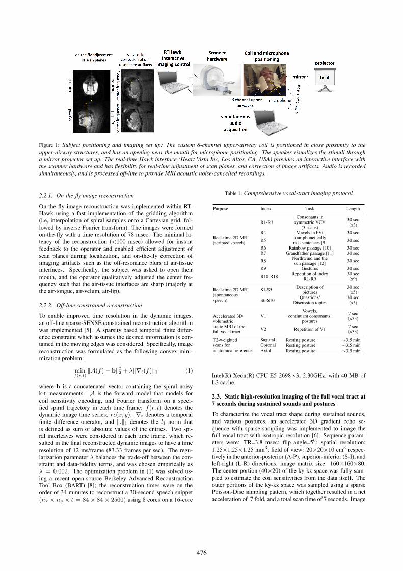
Figure 1: Subject positioning and imaging set up: The custom 8-channel upper-airway coil is positioned in close proximity to theupper-airway structures, and has an opening near the mouth for microphone positioning. The speaker visualizes the stimuli througha mirror projector set up. The real-time Hawk interface (Heart Vista Inc, Los Altos, CA, USA) provides an interactive interface withthe scanner hardware and has flexibility for real-time adjustment of scan planes, and correction of image artifacts. Audio is recordedsimultaneously, and is processed off-line to provide MRI acoustic noise-cancelled recordings.
2.2.1. On-the-fly image reconstruction
On-the fly image reconstruction was implemented within RT-Hawk using a fast implementation of the gridding algorithm(i.e, interpolation of spiral samples onto a Cartesian grid, fol-lowed by inverse Fourier transform). The images were formedon-the-fly with a time resolution of 78 msec. The minimal la-tency of the reconstruction (<100 msec) allowed for instantfeedback to the operator and enabled efficient adjustment ofscan planes during localization, and on-the-fly correction ofimaging artifacts such as the off-resonance blurs at air-tissueinterfaces. Specifically, the subject was asked to open theirmouth, and the operator qualitatively adjusted the center fre-quency such that the air-tissue interfaces are sharp (majorly atthe air-tongue, air-velum, air-lip).
2.2.2. Off-line constrained reconstruction
To enable improved time resolution in the dynamic images,an off-line sparse-SENSE constrained reconstruction algorithmwas implemented [5]. A sparsity based temporal finite differ-ence constraint which assumes the desired information is con-tained in the moving edges was considered. Specifically, imagereconstruction was formulated as the following convex mini-mization problem:
minf(r,t)
‖A(f)− b‖22 + λ‖∇t(f)‖1 (1)
where b is a concatenated vector containing the spiral noisyk-t measurements. A is the forward model that models forcoil sensitivity encoding, and Fourier transform on a speci-fied spiral trajectory in each time frame; f(r, t) denotes thedynamic image time series; rε(x, y). ∇t denotes a temporalfinite difference operator, and ‖.‖1 denotes the l1 norm thatis defined as sum of absolute values of the entries. Two spi-ral interleaves were considered in each time frame, which re-sulted in the final reconstructed dynamic images to have a timeresolution of 12 ms/frame (83.33 frames per sec). The regu-larization parameter λ balances the trade-off between the con-straint and data-fidelity terms, and was chosen empirically asλ = 0.002. The optimization problem in (1) was solved us-ing a recent open-source Berkeley Advanced ReconstructionTool Box (BART) [8]; the reconstruction times were on theorder of 34 minutes to reconstruct a 30-second speech snippet(nx × ny × t = 84 × 84 × 2500) using 8 cores on a 16-core
Table 1: Comprehensive vocal-tract imaging protocol
Purpose Index Task Length
Real-time 2D MRI(scripted speech)
R1-R3Consonants in
symmetric VCV(3 scans)
30 sec(x3)
R4 Vowels in bVt 30 sec
R5 four phoneticallyrich sentences [9] 30 sec
R6 Rainbow passage [10] 30 secR7 Grandfather passage [11] 30 sec
R8 Northwind and thesun passage [12] 30 sec
R9 Gestures 30 sec
R10-R18 Repetition of indexR1-R9
30 sec(x9)
Real-time 2D MRI(spontaneousspeech)
S1-S5 Description ofpictures
30 sec(x5)
S6-S10 Questions/Discussion topics
30 sec(x5)
Accelerated 3Dvolumetricstatic MRI of thefull vocal tract
V1Vowels,
continuant consonants,postures
7 sec(x33)
V2 Repetition of V1 7 sec(x33)
T2-weightedscans foranatomical reference
Sagittal Resting posture ∼3.5 minCoronal Resting posture ∼3.5 minAxial Resting posture ∼3.5 min
Intel(R) Xeon(R) CPU E5-2698 v3; 2.30GHz, with 40 MB ofL3 cache.
2.3. Static high-resolution imaging of the full vocal tract at7 seconds during sustained sounds and postures
To characterize the vocal tract shape during sustained sounds,and various postures, an accelerated 3D gradient echo se-quence with sparse-sampling was implemented to image thefull vocal tract with isotropic resolution [6]. Sequence param-eters were: TR=3.8 msec; flip angle=50; spatial resolution:1.25×1.25×1.25 mm3; field of view: 20×20×10 cm3 respec-tively in the anterior-posterior (A-P), superior-inferior (S-I), andleft-right (L-R) directions; image matrix size: 160×160×80.The center portion (40×20) of the ky-kz space was fully sam-pled to estimate the coil sensitivities from the data itself. Theouter portions of the ky-kz space was sampled using a sparsePoisson-Disc sampling pattern, which together resulted in a netacceleration of 7 fold, and a total scan time of 7 seconds. Image
476

Figure 2: Demonstration of the speech stimuli of (a) producing consonants in symmetric VCV phrases, and (b) producing scriptedsentences on two different subjects. The horizontal dotted arrows in the first column correspond to image cuts along which the temporalprofiles are shown in the second column. The proposed system depicts the articulatory dynamics of the various speech stimuli with goodtemporal fidelity. The improved temporal resolution (83 frames/sec) shows flexibility in adapting to capture fast arbitrary articulatorymotion in different speech tasks, and also adapts to subject specific speech rates (eg. compare speech rate in second row v/s first row).The improved spatial resolution (2.4mm2/pixel) ensures improved capture and depiction of dynamics of small structures such as theepiglottis.
reconstruction was achieved off-line by a sparse-SENSE con-strained reconstruction convex minimization problem, whichutilizes sparsity based spatial finite difference constraints:
minf(r,t)
‖A(f)−b‖22+λ∥∥∥√|∇x(f)|2 + |∇y(f)|2 + |∇z(f)|2
∥∥∥1
(2)where the first term denotes data-consistency, where A mod-els for Fourier under-sampling and coil sensitivity encoding,and the sparsity based total variation regularization term penal-izes rapidly changing pixel intensities (corresponding to under-sampling artifacts), while preserving high-resolution edge in-formation at various air-tissue interfaces. λ balances the trade-off between the regularization and data-fidelity terms, and waschosen empirically as λ = 0.2. (2) was solved using the BARTtool box [8]; the reconstruction time was ≈45 seconds on a 16-core Intel(R) Xeon(R) CPU E5-2698 v3; 2.30GHz, with 40 MBof L3 cache.
2.4. T2-weighted MRI for structural characterization ofvocal tract
High-resolution, high-contrast T2-weighted fast spin-echobased sequence was considered to provide images with softtissue contrast. The rationale for this sequence was to clearlyidentify soft-tissue boundaries and is included as a means to aidsegmentation (manual or automatic) of the vocal organs. Se-quence parameters were: TR: 4600ms, TE:120ms; slice thick-ness: 3 mm; in-plane resolution: 0.58x0.58 mm2; in-plane fieldof view: 30x30 cm2; number of averages: 1; echo train length:25; scan time: 3.5 minutes. The sequence was run to obtainfull sweeps of the vocal tract in the axial, sagittal, and coronalorientations.
2.5. Audio acquisition
Audio was recorded concurrently with MRI acquisition insidethe MRI scanner while subjects are imaged, using a fiberop-tic microphone (Optoacoustics Ltd., Moshav Mazor, Israel) andcustom recording and synchronization setup [13]. Speech in the
recorded audio was then enhanced, using a customized denois-ing method [13], in order to reduce the effect of loud scannernoise.
2.6. 90-minute protocol to study structural and functionalaspects of the vocal tract
Table 1 shows a comprehensive 90-minute protocol with theproposed sequences, where the stimuli set was designed to cap-ture efficiently salient, static and dynamic, articulatory and mor-phological aspects of speech production. Rapid real-time 2DMRI in the mid-sagittal view was used to obtain recordings ofscripted speech and spontaneous speech, where the stimuli inscripted speech was repeated twice.
The scripted speech contained producing consonantsin symmetric vowel-consonant-vowel context, vowels inter-spersed between consonant b and t in b-V-t context, four phonet-ically rich sentences [9], and three commonly used passages inlinguistic studies [10, 11, 12]. Several gestures such as clench-ing, wide opening of mouth and yawning, swallowing, sus-tained sound production of the sounds “eee-aah-uuw-eee”, trac-ing of palate with tongue tip, singing “la” at highest, lowestnote, were also acquired and repeated with 2D real-time MRI.Spontaneous speech tasks involved describing contexts in fiverandomly shown pictures, and discussion of five general topics(eg. “what is your favorite restaurant”). All the 2D real timescans involved pause time of ≈30 seconds between stimuli toallow enough recovery for the subject prior to the next task, andalso to avoid gradient heating.
With the accelerated volumetric protocol, we acquire tworepetitions of 33 stimuli which contained producing sustainedsounds of the vowels, continuant consonants, and several pos-tures such as normal breathing with mouth closed, clenching ofteeth, sticking of tongue out as far as possible, pulling back thetongue in as far as possible, tongue tip raise to the middle of thepalate, and holding breath. In the accelerated volumetric proto-col, a recovery time of ≈5-10 seconds was given to the subjectbetween the stimuli. The final set of scans involve acquisition of
477
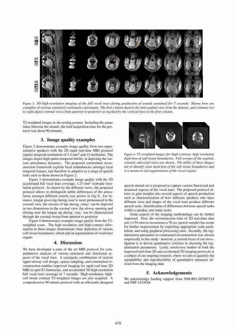
Figure 3: 3D high-resolution imaging of the full vocal tract during production of sounds sustained for 7 seconds: Shown here areexamples of various sustained continuant consonants. The first column depicts the mid-sagittal view from the dataset, and columns twoto eight depict coronal views from anterior to posterior as marked by the vertical lines in the first column.
T2-weighted images in the resting posture. Including the pausetimes between the stimuli, the total acquisition time for the pro-tocol was about 90 minutes.
3. Image quality examplesFigure 2 demonstrates example image quality from two repre-sentative speakers with the 2D rapid real-time MRI protocol(spatio-temporal resolutions of 2.4 mm2 and 12 ms/frame). Theimages depict high spatio-temporal fidelity in depicting the var-ious articulatory dynamics. The proposed constrained recon-struction framework exploits local redundancies amongst localtemporal frames, and therefore is adaptive to a range of speechtasks such as those shown in Figure 2.
Figure 3 demonstrates example image quality with the 3Daccelerated full vocal tract coverage, 1.25 mm3 isotropic reso-lution protocol. As shown by the different views, the proposedprotocol allows to distinguish subtle differences of the articu-lators amongst different sounds (see arrows in Fig.3). For in-stance, tongue grooving during /asa/ is more pronounced in thecoronal view, the closure of lips during /ama/ can be depictedin two dimensions in the coronal view, the airway opening andclosing near the tongue tip during /ara/ can be characterizedthrough the coronal sweep from anterior to posterior.
Figure 4 demonstrates example image quality from the T2-weighted scans. The high resolution and high-contrast infor-mation in these images demonstrate clear depiction of varioussoft-tissue boundaries, which aids in segmentation of vocal tractorgans.
4. DiscussionWe have developed a state of the art MRI protocol for com-prehensive analysis of various structural and functional as-pects of the vocal tract. A synergetic combination of customupper-airway coil design, sparse-sampling, and constrained re-construction enables improved imaging for rapid real time 2DMRI at upto 83 frames/sec, and accelerated 3D high-resolutionfull vocal tract coverage in 7 seconds. High-resolution, high-soft tissue contrast T2-weighted images are also acquired. Acomprehensive 90 minute protocol with an efficiently designed
speech stimuli set is proposed to capture various functional andstructural aspects of the vocal tract. The proposed protocol al-
Figure 4: T2-weighted images for high contrast, high resolutiondepiction of soft-tissue boundaries. Full sweeps of the sagittal,coronal, and axial views are shown . The utility of these imagesare to identify clear depiction of the soft-tissue boundaries andis a means to aid segmentation of the vocal organs.
lows to gain insights into several aspects of speech productionsuch as characterization of how different speakers who havedifferent sizes and shapes of the vocal tract produce differentspeech tasks, identification of differences between speech taskswithin a speaker, and many more.
Some aspects of the imaging methodology can be furtherimproved. First, the reconstruction time of 2D real-time datasets (≈34 mins to reconstruct a 30 sec speech sample) has scopefor further improvement by exploiting appropriate code paral-lelism, and using graphical processing units. Secondly, the reg-ularization parameter in constrained reconstruction was chosenempirically in this study - however, a current focus of our inves-tigation is to devise quantitative criterion in choosing the reg-ularization parameters. Lastly, test/re-test studies of both theimproved real-time 2D and accelerated 3D imaging protocols isa subject of our ongoing research, where we aim to quantify therepeatability and reproducibility of quantitative measures de-rived from the imaging data.
5. AcknowledgementsWe acknowledge funding support from NIH-R01-DC007124and NSF-1514544.
478

magnetic resonance imaging,” IEEE Signal Processing Magazine,vol. 25, no. 3, pp. 123–132, 2008.
[2] A. D. Scott, M. Wylezinska, M. J. Birch, and M. E. Miquel,“Speech mri: morphology and function,” Physica Medica, vol. 30,no. 6, pp. 604–618, 2014.
[3] S. G. Lingala, B. P. Sutton, M. E. Miquel, and K. S. Nayak,“Recommendations for real-time speech mri,” Journal of Mag-netic Resonance Imaging, vol. 43, no. 1, pp. 28–44, 2016.
[4] S. Narayanan, K. Nayak, S. Lee, A. Sethy, and D. Byrd, “An ap-proach to real-time magnetic resonance imaging for speech pro-duction,” The Journal of the Acoustical Society of America, vol.115, no. 4, pp. 1771–1776, 2004.
[5] S. G. Lingala, Y. Zhu, Y.-C. Kim, A. Toutios, S. Narayanan, andK. S. Nayak, “A fast and flexible mri system for the study of dy-namic vocal tract shaping,” Magnetic resonance in medicine, vol.early view, Jan 17, no. doi: 10.1002/mrm.26090, 2016.
[6] Y.-C. Kim, S. S. Narayanan, and K. S. Nayak, “Accelerated three-dimensional upper airway mri using compressed sensing,” Mag-netic Resonance in Medicine, vol. 61, no. 6, pp. 1434–1440, 2009.
[7] J. M. Santos, G. A. Wrigh, and J. M. Pauly, “Flexible real-time magnetic resonance imaging framework,” in Engineering inMedicine and Biology Society, 2004. IEMBS’04. 26th Annual In-ternational Conference of the IEEE, vol. 1. IEEE, 2004, pp.1048–1051.
[8] M. Uecker, F. Ong, J. I. Tamir, D. Bahri, P. Virtue, J. Y. Cheng,T. Zhang, and M. Lustig, “Berkeley advanced reconstructiontoolbox,” in Proceedings of the 23rd Annual Meeting ISMRM,Toronto, 2015, p. 2486.
[9] J. S. Garofolo, L. F. Lamel, W. M. Fisher, J. G. Fiscus, and D. S.Pallett, “Darpa timit acoustic-phonetic continous speech corpuscd-rom. nist speech disc 1-1.1,” NASA STI/Recon Technical Re-port N, vol. 93, 1993.
[10] G. Fairbanks, “The rainbow passage,” Voice and articulation drill-book, vol. 2, 1960.
[11] A. E. Aronson and J. R. Brown, Motor speech disorders. WBSaunders Company, 1975.
[12] I. P. Association, Handbook of the International Phonetic Associ-ation: A guide to the use of the International Phonetic Alphabet.Cambridge University Press, 1999.
[13] E. Bresch, J. Nielsen, K. Nayak, and S. Narayanan, “Synchro-nized and noise-robust audio recordings during realtime magneticresonance imaging scans,” The Journal of the Acoustical Societyof America, vol. 120, no. 4, pp. 1791–1794, 2006.
6. References[1] E. Bresch, Y.-C. Kim, K. Nayak, D. Byrd, and S. Narayanan,
“Seeing speech: Capturing vocal tract shaping using real-time
479