stamen = filament + anther angiosperms: production of male gametophyte meiosis inside anther male...
Post on 19-Dec-2015
238 views
TRANSCRIPT

Stamen = filament + anther
Angiosperms: Production of Male Gametophyte
Meiosis inside anther male spores
Details follow

Meiosis in lily anther 4 haploid daughter cells, also called “pollen tetrads”
Angiosperms: Production of Male Gametophyte
HaploidHaploid
HaploidHaploid
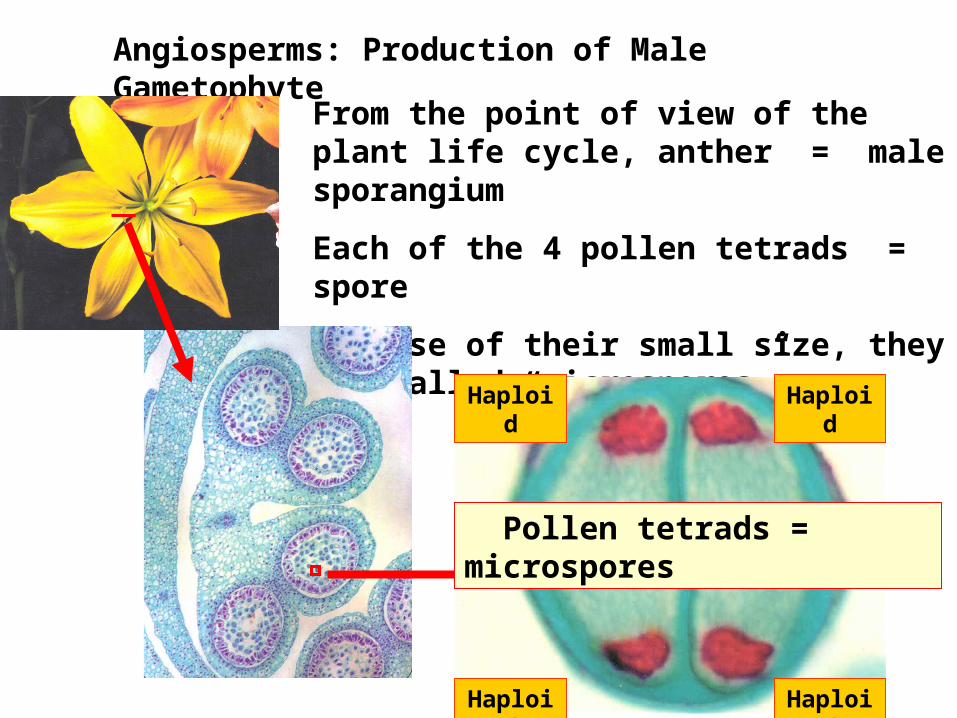
From the point of view of the plant life cycle, anther = male sporangium
Each of the 4 pollen tetrads = spore
Because of their small size, they are called “microspores”.
Angiosperms: Production of Male Gametophyte
HaploidHaploid
HaploidHaploid
Pollen tetrads = microspores
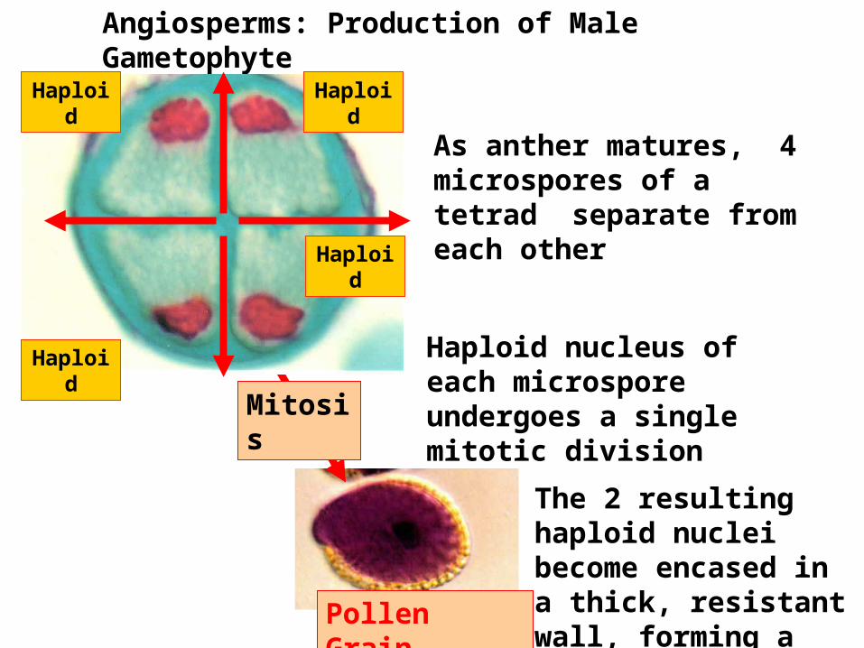
As anther matures, 4 microspores of a tetrad separate from each other
Angiosperms: Production of Male Gametophyte
Haploid nucleus of each microspore undergoes a single mitotic division
Pollen Grain
Mitosis
Haploid
Haploid
HaploidHaploid
The 2 resulting haploid nuclei become encased in a thick, resistant wall, forming a pollen grain.

From the point of view of the angiosperm life cycle, a pollen grain is an immature male gametophyte, since it has been produced by the mitotic division of a spore.
Angiosperms: Production of Male Gametophyte
Pollen Grain
Mitosis
Haploid
Haploid
HaploidHaploid
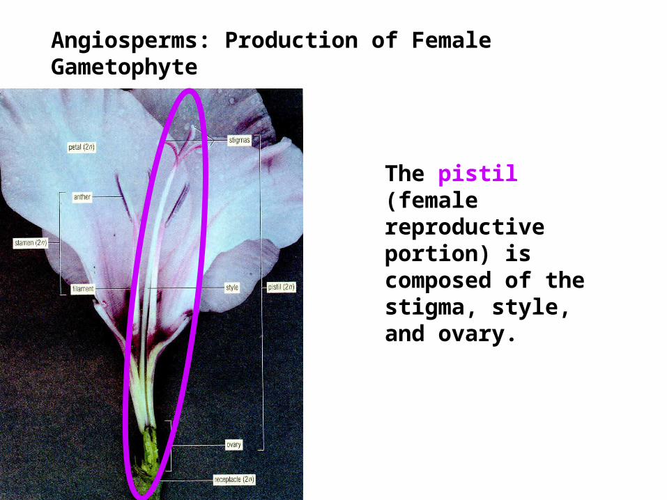
The pistil (female reproductive portion) is composed of the stigma, style, and ovary.
Angiosperms: Production of Female Gametophyte
Angiosperms: Production of Female Gametophyte
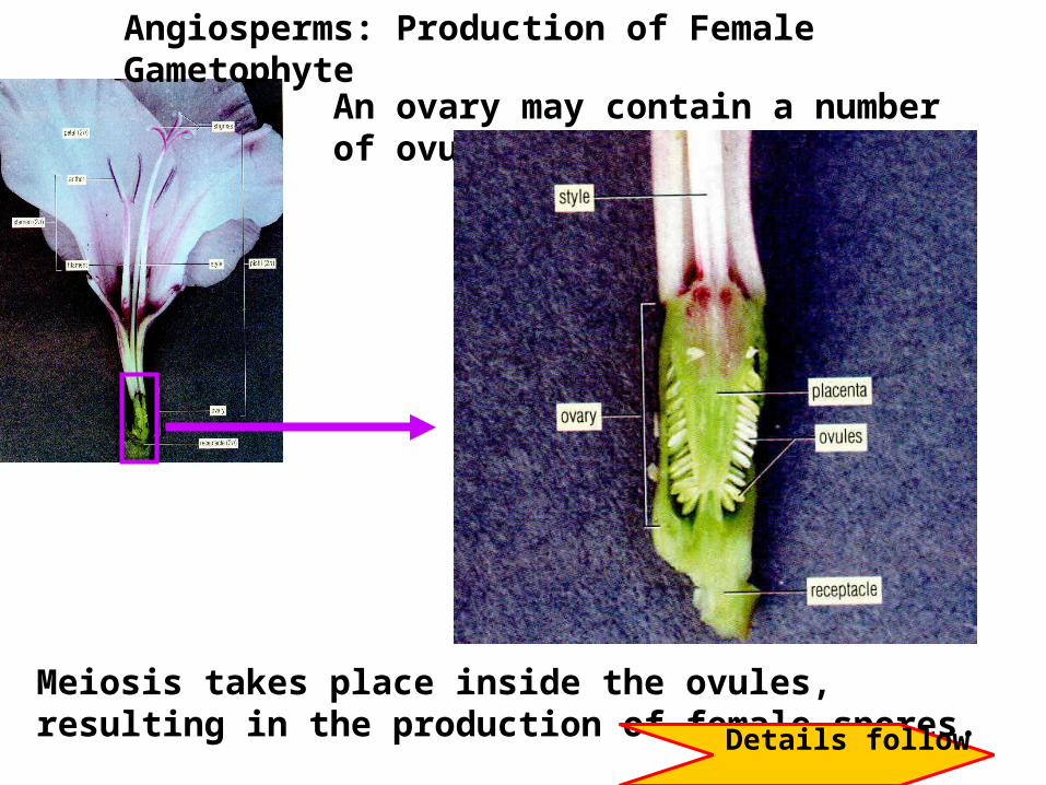
An ovary may contain a number of ovules.
Meiosis takes place inside the ovules, resulting in the production of female spores. Details follow

Angiosperms: Female Gametophyte
Only one of the haploid spores resulting from meiosis in the ovule matures. It undergoes 2 rounds of mitosis to form the “embryo sac”, which has 8 haploid nuclei.
Embryo sac = female gametophyte

To complete the life cycle, the gametes produced by the male and female gametophyte must unite, restoring the diploid sporophyte.
Female gametophyte = embryo sac
Immature male gametophyte = pollen grain
Alternation of Generations: Angiosperms

Fertilization and Embryo Formation
Pollen grain landing on stigma of ovary
pollen tube growth

2 haploid cells of pollen grain are called the “generative cell” and the “tube cell”
Fertilization and Embryo Formation
Pollen tube growing from a pollen grain
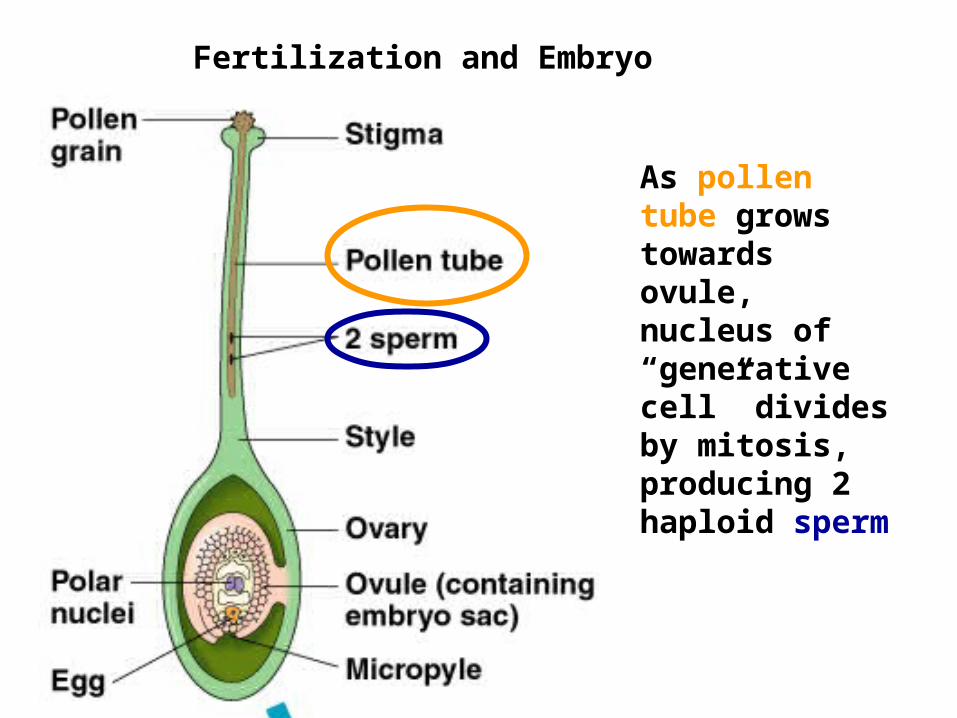
Fertilization and Embryo Formation
As pollen tube grows towards ovule, nucleus of “generative cell” divides by mitosis, producing 2 haploid sperm

Fertilization and Embryo Formation
The pollen grain, along with the pollen tube containing 2 sperm, is the mature male gametophyte.
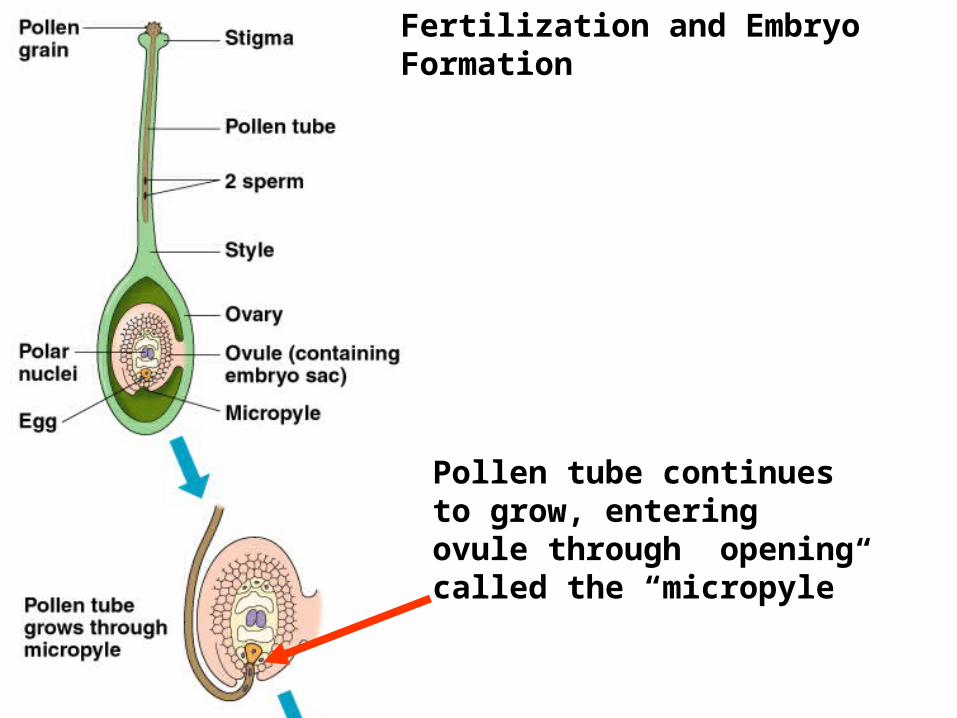
Fertilization and Embryo Formation
Pollen tube continues to grow, entering ovule through opening called the “micropyle”

Fertilization and Embryo Formation
One of the sperm fertilizes the egg, producing a diploid zygote. This zygote will divide and differentiate, forming the sporophyte plant. The angiosperm life cycle has been completed.
The other sperm will fuse with the 2 central haploid nuclei in the embryo sac, producing a triploid nucleus.
These events are called “double fertilization”.

Fertilization and Embryo Formation
Tissue that develops from the triploid nucleus = “endosperm”. Energy stored in this tissue nourishes the developing embryo.
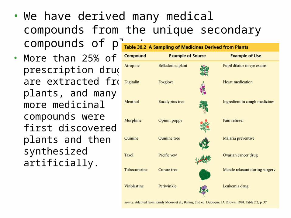
• We have derived many medical compounds from the unique secondary compounds of plants.
• More than 25% of prescription drugs are extracted from plants, and many more medicinal compounds were first discovered in plants and then synthesized artificially.

Evolutionary Trends in Plant Life Cycles
Angiosperms demonstrate an evolutionary trend in which the gametophyte is further reduced in size, and increasingly dependent upon the sporophyte.

Developing zygote, endosperm, and other tissues of the ovule eventually become a seed
Development of the Young Dicot Sporophyte
Corn
Bean
Example follows

Development of the Young Dicot Sporophyte
developing ovules
ContinuedLongitudinal section through Capsella ovary

Development of the Young Dicot Sporophyte
Suspensor
Continued
endosperm
Developing embryo proper
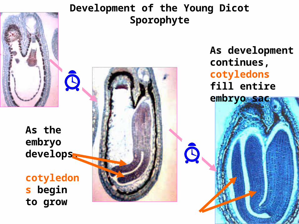
Development of the Young Dicot Sporophyte
As the embryo develops, cotyledons begin to grow
As development continues, cotyledons fill entire embryo sac

Development of the Young Dicot Sporophyte
Here is a longitudinal section of an ovary with a number of well-developed ovules inside.
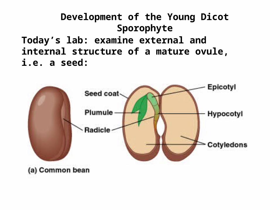
Development of the Young Dicot Sporophyte
Today’s lab: examine external and internal structure of a mature ovule, i.e. a seed:
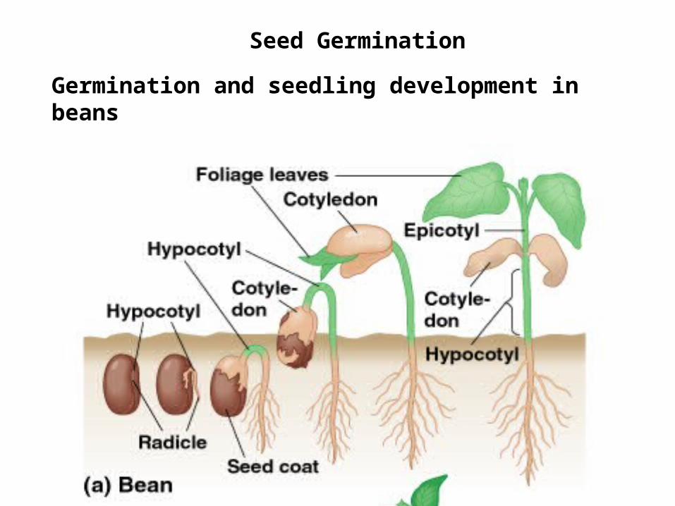
Seed Germination
Germination and seedling development in beans
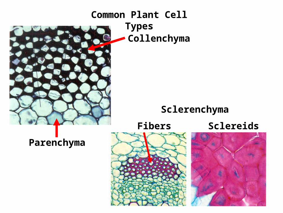
Common Plant Cell Types
Sclerenchyma
Fibers Sclereids
Collenchyma
Parenchyma

Common Plant Cell Types
Vessel elements & tracheids: important in xylem tissue
sieve tube members & companion cells: important in phloem tissue
cork cells: important in bark tissue

Primary vs. Secondary Growth
Secondary growth = growth in girth (width), e.g. Tilia stem cross-section
Primary growth= growth in length, e.g. in seed germination
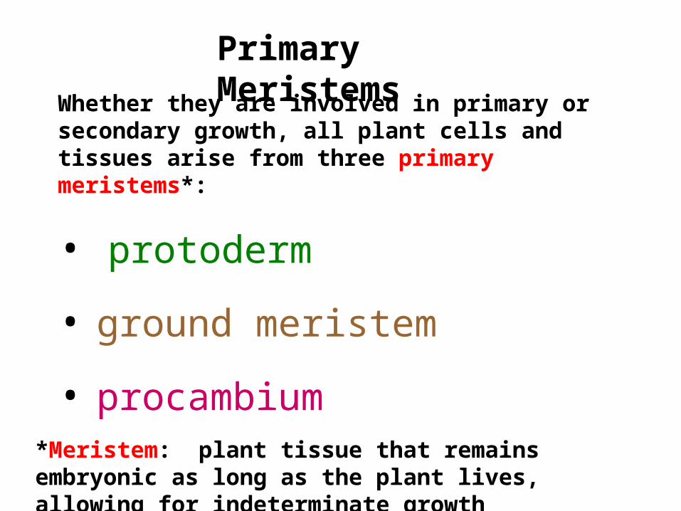
Whether they are involved in primary or secondary growth, all plant cells and tissues arise from three primary meristems*:
• protoderm
• ground meristem
• procambium
Primary Meristems
*Meristem: plant tissue that remains embryonic as long as the plant lives, allowing for indeterminate growth

Primary & Secondary Growth in a Woody Stem
Primary meristems
Protoderm
Ground meristem
Procambium
Primary Tissues
Epidermis
Pith
Ground
Cortex
Primary phloem
Primary xylem
Lateral Meristem
Secondary Tissues
Vascular Cambium
Cork cambium cork
2o phloem
2o xylem
Periderm
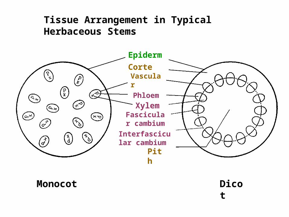
Tissue Arrangement in Typical Herbaceous Stems
EpidermisCortexVascular bundle
Pith
Interfascicular cambium
Fascicular cambium
Phloem
Xylem
Monocot Dicot

Secondary Growth in a Woody Dicot
vascular cambium produces 2o xylem (= wood) to the inside, 2o phloem to the outside
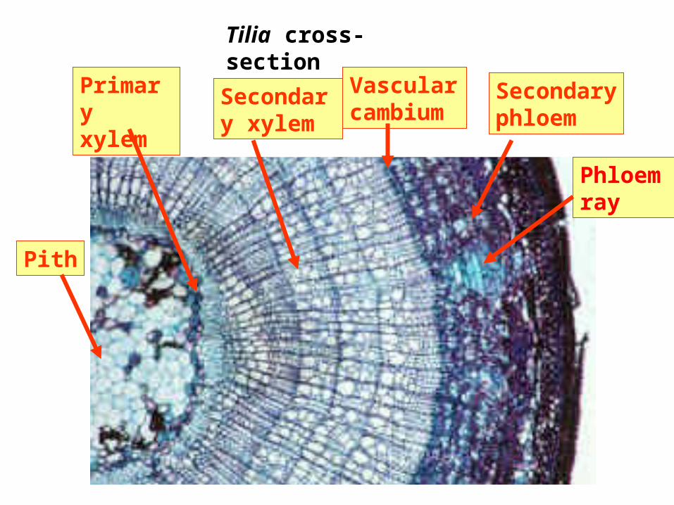
Tilia cross-section
Vascular cambium
Secondary xylem
Primary xylem
Pith
Secondary phloem
Phloem ray

Cell Types in Secondary Phloem Ray of Bark
Fibers
Sieve tube members Companion cells
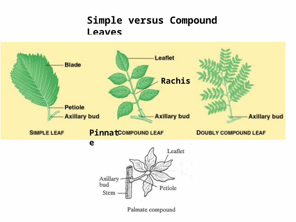
Simple versus Compound Leaves
Rachis
Pinnate

Generalized Leaf Anatomy

Typical Dicot Leaf X-Section
Palisade Parenchyma
Spongy Parenchyma
Vascular bundles
Epidermis
Cuticle
Stoma
Guard Cells
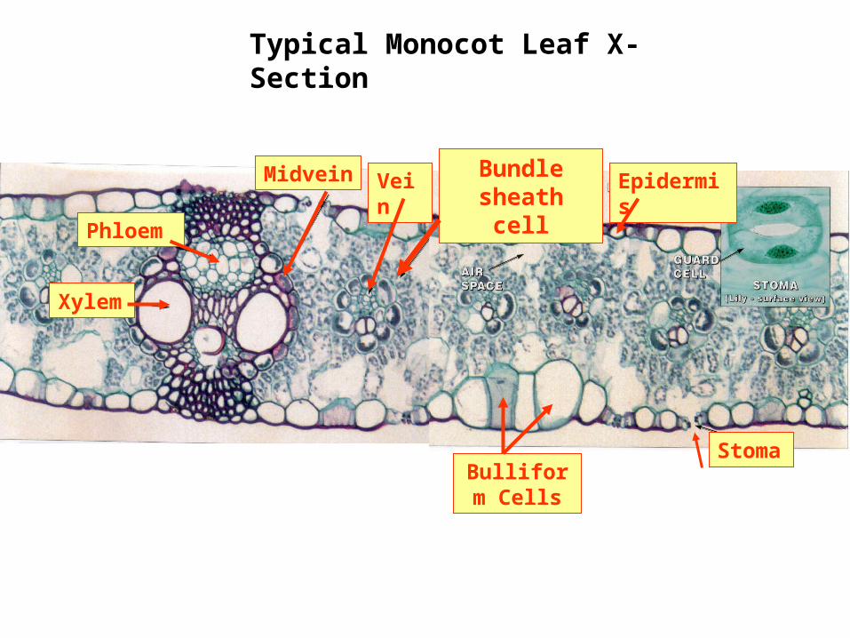
Typical Monocot Leaf X-Section
Xylem
Phloem
Bulliform Cells
Stoma
EpidermisMidvein Vein Bundle sheath cell

Leaf Stomata: Allow Gas Exchange
Stomata in Zebrina leaf epidermis
Guard cells with
chloroplasts
Stoma
Subsidiary cells

Bulliform Cells

Let’s see some TRICHOMES!
