stabilized arginine biosynthesis in streptococcus gordonii by coaggreg
TRANSCRIPT
JOURNAL OF BACTERIOLOGY, May 2008, p. 3646–3657 Vol. 190, No. 100021-9193/08/$08.00�0 doi:10.1128/JB.00088-08Copyright © 2008, American Society for Microbiology. All Rights Reserved.
Regulation of Gene Expression in a Mixed-Genus Community:Stabilized Arginine Biosynthesis in Streptococcus gordonii by
Coaggregation with Actinomyces naeslundii�
Nicholas S. Jakubovics,1 Steven R. Gill,2,4 Stacey E. Iobst,4M. M. Vickerman,2,3 and Paul E. Kolenbrander1*
National Institute of Dental and Craniofacial Research, National Institutes of Health, Building 30, Room 310, Bethesda,Maryland 208921; Department of Oral Biology,2 and Department of Periodontics and Endodontics,3
University at Buffalo School of Dentistry, Buffalo, New York; and Institute for Genomic Research,9712 Medical Center Drive, Rockville, Maryland 208504
Received 17 January 2008/Accepted 8 March 2008
Interactions involving genetically distinct bacteria, for example, between oral streptococci and actinomyces,are central to dental plaque development. A DNA microarray identified Streptococcus gordonii genes regulatedin response to coaggregation with Actinomyces naeslundii. The expression of 23 genes changed >3-fold incoaggregates, including that of 9 genes involved in arginine biosynthesis and transport. The capacity of S.gordonii to synthesize arginine was assessed using a chemically defined growth medium. In monoculture,streptococcal arginine biosynthesis was inefficient and streptococci could not grow aerobically at low arginineconcentrations. In dual-species cultures containing coaggregates, however, S. gordonii grew to high cell densityat low arginine concentrations. Equivalent cocultures without coaggregates showed no growth until coaggre-gation was evident (9 h). An argH mutant was unable to grow at low arginine concentrations with or withoutA. naeslundii, indicating that arginine biosynthesis was essential for coaggregation-induced streptococcalgrowth. Using quantitative reverse transcriptase PCR, the expression of argC, argG, and pyrAb was strongly(10- to 100-fold) up-regulated in S. gordonii monocultures after 3 h of growth when exogenous arginine wasdepleted. Cocultures without induced coaggregation showed similar regulation. However, within 1 h aftercoaggregation with A. naeslundii, the expression of argC, argG, and pyrAb in S. gordonii was partially up-regulated although arginine was plentiful, and mRNA levels did not increase further when arginine wasdiminished. Thus, A. naeslundii stabilizes S. gordonii expression of arginine biosynthesis genes in coaggregatesbut not cocultures and enables aerobic growth when exogenous arginine is limited.
It is now widely accepted that most bacteria in nature existin multispecies biofilm communities (48), where intergenericinteractions are commonplace (24). The outcome of these in-teractions plays a critical role in determining the success of anindividual species in a mixed population. Dental plaque bio-films are particularly complex, harboring combinations of�700 species or phylotypes (1). From the earliest stages ofplaque formation, microcolonies containing two or more gen-era of bacteria can be detected (24, 38), and therefore, signif-icant potential for competitive or cooperative interactions ex-ists through all phases in the development of dental biofilms.Specific cell-cell recognition, known as coaggregation, isthought to be fundamental for bringing together geneticallydistinct bacteria in dental plaque (21, 22).
Streptococcus spp. are key mediators of interactions in dentalplaque since they produce multiple cell surface adhesins andreceptors that bind both the salivary pellicle and numerousoral bacteria (17, 19). Streptococcus spp. are consistently iden-tified as the predominant microorganisms in nascent dentalplaque, comprising 60 to 80% of the total bacterial population
on tooth surfaces for at least 24 h after tooth cleaning (8, 30,36, 37). Actinomyces spp. constitute about one-third of non-streptococcal cells in early plaque biofilms (37) and are oftenjuxtaposed to Streptococcus spp. (38). A number of coaggrega-tion interactions between Actinomyces spp. and Streptococcusspp. have previously been described. For example, type 2 fim-briae of Actinomyces naeslundii genospecies 2 are necessary forthe recognition of streptococci that bear receptor polysaccha-rides (3, 34). Strong evidence that coaggregation between type2 fimbriated actinomyces and receptor polysaccharide-bearingstreptococci occurs in natural oral biofilms has been obtainedwith the use of specific antibodies against a known adhesin-receptor pair of antigens (38). These antigens were shown tobe colocalized in plaque formed on enamel surfaces held in themouths of human volunteers, indicating that the antigens in-teract in vivo (38). Such interactions are likely to initiate as wellas stabilize biofilm communities in vivo and in vitro. Hence, ithas been shown that the presence of Streptococcus gordoniiDL1 on a saliva-conditioned surface in vitro significantly en-hances the retention of a coaggregating partner, Actinomycesnaeslundii T14V, in a biofilm under flowing saliva (39).
By promoting contact between distinct species of bacteria,coaggregation enhances the exchange of signals and metabo-lites between cells. Communication between Streptococcusoralis and A. naeslundii mediated by signaling molecule auto-inducer 2 leads to the mutualistic growth of each species in a
* Corresponding author. Mailing address: National Institutes ofHealth/NIDCR, Building 30, Room 310, 30 Convent Drive, MSC 4350,Bethesda, MD 20892-4350. Phone: (301) 496-1497. Fax: (301) 402-0396. E-mail: [email protected].
� Published ahead of print on 21 March 2008.
3646
Dow
nloa
ded
from
http
s://j
ourn
als.
asm
.org
/jour
nal/j
b on
23
Oct
ober
202
1 by
121
.183
.74.
70.
flow cell biofilm with saliva as the sole nutrient source, condi-tions under which neither species can grow alone (41). Otherstudies have identified specific changes in gene expression thatoccur in response to cell-cell communication between oralbacteria. For example, the sensing of arginine deiminase(ArcA) on the surface of Streptococcus cristatus leads to thedown-regulation of the Porphyromonas gingivalis fimbrial genefimA (56, 57). In a different system, a diffusible signal fromVeillonella atypica mediates the up-regulation of S. gordoniiamyB, encoding �-amylase (12). Coaggregation per se is notessential for signaling in a closed vessel since a coaggregation-deficient V. atypica mutant is able to elicit a response from S.gordonii. However, in biofilms formed under flowing saliva withwild-type (coaggregating) strains, only S. gordonii cells juxta-posed to V. atypica cells switch on the transcription of amyB(12). Therefore, in an open flowing system, akin to the humanmouth, coaggregation is important for minimizing the dilutionof diffusible signals and maximizing the productive exchange ofsignals between senders and responders.
Despite the clear evidence that cell-cell communication oc-curs between oral bacteria, no reports of systematic searchesfor genes regulated in response to coaggregation exist. DNAmicroarrays are an extremely powerful tool for investigatingtranscriptional changes in response to stimuli, and it has beendemonstrated that microarrays can be employed to investigategene expression in one bacterial species in the presence ofanother (33, 46). Here, we describe the application of a re-cently designed DNA microarray (55) to assess changes in S.gordonii gene expression in response to coaggregation with A.naeslundii. From this analysis, we show that coaggregation hasa major impact on the expression of genes encoding enzymesfor arginine biosynthesis. We therefore undertook an investi-gation into arginine biosynthesis in S. gordonii. Our data indi-cate that S. gordonii synthesizes arginine inefficiently and thatcoaggregation with A. naeslundii enables arginine biosynthesisunder conditions that prohibit the growth of S. gordonii inmonoculture. We propose that coaggregation enables S. gor-donii cells to switch from quiescence to active growth whenarginine is scarce, as it is in human saliva.
MATERIALS AND METHODS
Strains and growth media. Streptococcus gordonii DL1 (Challis) and Actino-myces naeslundii MG1 (ATCC 43146) were routinely cultured in Todd-Hewittbroth (THB; Difco, Detroit, MI) or on THB medium solidified with 1.5%(wt/vol) Bacto agar, aerobically at 37°C and 5% CO2. The construction of the S.gordonii PK3337 argH::aphA3 strain is described below. This strain was subcul-tured in the presence of 250 �g/ml kanamycin. Chemically defined medium(CDM) was based on FMC (52) with the following modifications: L-leucine andL-isoleucine were each at a final concentration of 40 mg/liter rather than 100mg/liter in FMC; L-arginine and L-histidine were at 100 mg/liter rather than 200mg/liter in FMC; 0.1 mM CaCl2 (not present in FMC) was included, and themedium was adjusted to pH 7.3. All reagents for CDM were purchased fromSigma-Aldrich (St. Louis, MO). For some experiments, arginine was omitted orincluded at a different concentration. TYEG medium contained 1% (wt/vol)Bacto tryptone, 0.5% (wt/vol) yeast extract, 0.3% (wt/vol) K2HPO4, and 0.2%(wt/vol) D-glucose, adjusted to pH 7.5 before autoclaving.
Single- and mixed-species cultures. For experiments involving growth inCDM, cells were first cultured in TYEG medium for 16 h anaerobically under a90% N2-5% H2-5% CO2 atmosphere. Cells were harvested, resuspended inCDM, and adjusted to �5 � 109 CFU/ml. For monocultures, 300 �l of cellsuspension was added to 14.7 ml CDM in a capped 15-ml glass tube (finalconcentration, 1 � 108 CFU/ml). To induce coaggregation in dual-species cul-tures, 300 �l of S. gordonii cells was combined with 300 �l of A. naeslundii cells,
vortex mixed for 10 s, and adjusted to 15 ml with CDM. Vortex mixing densesuspensions of cells is a routine protocol for inducing coaggregation of organismswith complementary adhesins and receptors (23). For mixed-species cultureswithout induced coaggregation, 300 �l of S. gordonii cells and 300 �l of A.naeslundii cells were added to 14.4 ml CDM and mixed by gentle inversion.Cultures were incubated aerobically at 37°C.
Numbers of CFU were determined using 0.5-ml culture samples. Chains ofstreptococci, clumps of actinomyces, and coaggregates were disrupted by soni-cation in a Sonopuls ultrasonic homogenizer (Bandelin Electric, Berlin, Ger-many) equipped with a BR30 cup booster on 50% power for 1 min. Samples wereserially diluted, and triplicate 20-�l portions of each dilution were dropped ontosolid THB medium. Plates were incubated at 37°C and 5% CO2 for 24 h toenumerate S. gordonii CFU or for 48 h to quantify A. naeslundii CFU. Since A.naeslundii colonies were not visible after incubation for 24 h, this procedure wassuitable for the specific enumeration of S. gordonii CFU from mixed-genuscultures. For the determination of A. naeslundii CFU in dual-species cultures,serial dilutions of the culture were dropped onto solidified THB medium sup-plemented with 128 mg/liter mupirocin and 2.5 mg/liter metronidazole (29), andplates were incubated at 37°C and 5% CO2 for 48 h. Turbidities of S. gordoniimonocultures were measured using a Klett-Summerson colorimeter (Klett Man-ufacturing Co., Inc., New York, NY) with a 660-nm filter.
Fluorescence microscopy. S. gordonii and A. naeslundii cells were visualizedusing Alexa Fluor-conjugated immunoglobulin G. Antibodies against S. gordoniiDL1 (38) were labeled with Alexa 568, and anti-A. naeslundii T14V antibodies(5) were conjugated with Alexa 633 using Alexa Fluor labeling kits (MolecularProbes, Eugene, OR) according to the manufacturer’s instructions. Cells wereharvested, washed in phosphate-buffered saline (PBS), and resuspended in PBScontaining 1% (wt/vol) bovine serum albumin and 10 �g of each antibody per ml.Cells were incubated at 25°C for 20 min, harvested, resuspended in PBS, andexamined by epifluorescence microscopy.
Effects of arginine concentration on S. gordonii gene expression. S. gordoniimonocultures were prepared in CDM supplemented with arginine to a finalconcentration of 5 mM as described above and incubated anaerobically at 37°Cto mid-exponential phase (140 to 160 Klett units). Cultures were divided into twoaliquots, and cells were harvested by centrifugation at 3,500 � g for 7 min. Onealiquot was resuspended in CDM containing 5 mM arginine, and the other wasresuspended in CDM without arginine. Tubes were incubated anaerobically at37°C for a further 30 min before RNA extraction. Results are shown as thegeometric means and standard deviations for three independent cultures.
RNA extraction and purification. Prior to the extraction of RNA from bacte-rial cells, intracellular RNA was stabilized by the addition of 2 volumes ofRNAprotect (Qiagen, Valencia, CA) to 1 volume of sample from an exponen-tially growing culture, vortex mixing for 5 s, and incubation for 5 min at 20°C.Cells were harvested, the supernatant was discarded, and the pellet was stored at�70°C for up to 48 h. Total RNA was extracted using Trizol reagent (Invitrogen,Carlsbad, CA) according to the manufacturer’s instructions, with an extra step toensure efficient disruption of gram-positive cells: following resuspension of thepellet in 1 ml Trizol, cells were mixed with lysing matrix B (Qbiogene, MorganIrvine, CA), homogenized in a FastPrep bead beater (Qbiogene), and incubatedat 20°C for 10 min. Extracted RNA was treated with RQ1 DNase I (Promega,Madison, WI) at 37°C for 1 h and repurified on Qiagen RNeasy MinElutecolumns. To ensure that RNA had not degraded during extraction and to de-termine the concentration of RNA, an aliquot of each sample was analyzed bycapillary electrophoresis, using an RNA 6000 Nano chip in an Agilent 2100bioanalyzer system (Agilent Technologies, Santa Clara, CA). Purified RNA wasstored at �70°C.
Determination of extracellular arginine in CDM cultures. Cell- and protein-free extracts were prepared from CDM cultures of S. gordonii or A. naeslundiiincubated aerobically at 37°C for 3 h. Cells were pelleted by centrifugation at15,000 � g for 10 min at 25°C. An aliquot (500 �l) of the supernatant wastransferred to a Microcon YM-3 centrifugal filter device (Millipore, Billerica,MA) and centrifuged at 14,000 � g for 100 min at 25°C. Arginine concentrationswere determined by Scientific Research Consortium (St. Paul, MN). Briefly, thisprocess involved deproteinization in 13.5% (wt/vol) 5-sulfosalicylic acid hydrateand filtration through a 0.2-�m-pore-size membrane. Arginine was separatedfrom the mixture on a dedicated high-performance liquid chromatography aminoacid analyzer and detected by a colorimetric assay using ninhydrin reagent at131°C. Three independent extracts of each monoculture and a coaggregateculture were analyzed, and arginine in uninoculated CDM was measured as acontrol.
Microarray hybridization and data analysis. Labeling of RNA and microarrayhybridization were performed according to the standardized protocols availableat http://pfgrc.tigr.org/protocols.shtml. Briefly, aminoallyl-labeled cDNA was
VOL. 190, 2008 COAGGREGATION-INDUCED GENE REGULATION IN S. GORDONII 3647
Dow
nloa
ded
from
http
s://j
ourn
als.
asm
.org
/jour
nal/j
b on
23
Oct
ober
202
1 by
121
.183
.74.
70.
synthesized using Powerscript reverse transcriptase (Clontech, Mountain View,CA) and random hexamer primers (Invitrogen), with 5-(3-aminoallyl)-dUTP(Sigma) included in the reaction. Following the removal of unincorporatednucleotides on Qiagen MinElute columns, the cDNAs were labeled with Cy3 orCy5 dye (Amersham Biosciences, Piscataway, NJ) and repurified on QiagenMinElute columns. Labeling efficiencies were quantified using a NanoDropND-1000 spectrophotometer (NanoDrop Technologies, Wilmington, DE). La-beled cDNAs were denatured at 95°C for 5 min prior to use in hybridizationreactions. Glass microarray slides containing 70-mer oligonucleotide probes for2,195 S. gordonii open reading frames (ORFs) or loci (55) were prehybridized at42°C for 1 h in 5� SSC (1� SSC is 0.15 M NaCl plus 0.015 M sodium citrate),0.1% (wt/vol) sodium dodecyl sulfate (SDS), and 1% (wt/vol) bovine serumalbumin. Slides were washed extensively in deionized water, washed once inisopropyl alcohol, and dried by centrifugation. Slides were hybridized with mix-tures of Cy3- and Cy5-labeled cDNAs from two different populations (i.e.,monocultured S. gordonii and S. gordonii-A. naeslundii coaggregate cultures) inhybridization buffer (40% formamide, 5� SSC, 0.1% [wt/vol] SDS, 0.6 mg/mlsheared salmon sperm DNA) at 42°C for 16 h. Following hybridization, slideswere washed twice for 5 min at 25°C in each of three wash buffers: low-stringencybuffer (2� SSC, 0.1% [wt/vol] SDS, 0.1 mM dithiothreitol [DTT] preheated to55°C), medium-stringency buffer (0.1� SSC, 0.1% [wt/vol] SDS, 0.1 mM DTT),and high-stringency buffer (0.1� SSC, 0.1 mM DTT). Slides were rinsed severaltimes in deionized water and air dried.
Images of microarray slides were produced using an Axon Genepix 4000Bscanner (Molecular Devices, Sunnyvale, CA), and normalized spot intensitieswere determined with the associated Genepix Pro software. Data analysis wasperformed using the TM4 suite of applications (43). For statistical rigor, RNAwas extracted from three independent sets of cultures and flip-dye replicateswere performed for two of the three pairs of RNA preparations. Data fromflip-dye replicate pairs were compared, and spots with inconsistent expressionlevels (varying by �2 standard deviations from the average intensity) wereexcluded from further analysis. For other spots, the geometric means of thefluorescence intensities were calculated. Data from six in-slide replicates perORF/locus were combined, and the expression of each gene across three inde-pendent experiments was analyzed by significance analysis of microarrays (53),using a � value of 0.50.
Prediction of operon structure and regulatory motifs. Promoters and termi-nators in the S. gordonii genome sequence were detected using the BProm andFindTerm modules of the fgenesB gene prediction program in Molquest soft-ware (Softberry Inc., Mount Kisco, NY). This software gives output scores from�1 to �25 to estimate the likelihood that a predicted promoter or terminator isfunctional; a higher score indicates that the prediction is more likely to becorrect. The distribution of prediction scores was skewed, and �90% of putativepromoters and terminators in the S. gordonii genome were predicted with scoresbelow 10. We used an arbitrary range of values from 2.5 to 3.9 to indicate apromoter or terminator predicted with low confidence, and elements with scoresof �4.0 were considered likely to be functional. Promoter/operator elementscontaining ARG box motifs were identified by searching the S. gordonii genomewith a position weight matrix derived from known Bacillus subtilis AhrC recog-nition elements (see Fig. 6A), using the Virtual Footprint software program(http://prodoric.tu-bs.de/).
Disruption of argH in S. gordonii PK3337. Routine cloning procedures wereperformed as described by Sambrook et al. (44). The argH gene, encodingarginosuccinate lyase, was replaced with the aphA3 kanamycin resistance deter-minant by allelic exchange mutagenesis. Sequences of primers used for mutagen-esis are given in Table 1. Primers argHF1 and argHR1 were employed to amplifya 579-bp fragment comprising 30 bp of the 5 end of argH and 549 bp upstream.A 606-bp fragment including 148 bp of the 3 end of argH and 458 bp down-stream was PCR amplified using primers argHF2 and argHR2. A second PCRwas performed using equal amounts of the amplified products as templates andargHF1 and argHR2 primers and resulted in a 1,168-bp product comprising theends of the argH gene with the surrounding sequence and a central EcoRI site.This fragment was cloned in pGEM-T, generating pGEM-argH. A 937-bp regionharboring the aphA3 kanamycin resistance determinant was amplified fromstreptococcal integration plasmid pSF151 (50) by using primers aphA3F2 andaphA3R2 and ligated into the unique EcoRI site in pGEM-argH. The 2,086-bpargH::aphA3 insert was amplified from the vector by using primers argHF1 andargHR2 and used for the transformation of S. gordonii DL1 to generate thePK3337 �argH strain. The correct allelic exchange was verified by PCR ampli-fication and DNA sequencing. To ensure that phenotypic effects of the genedisruption were not due to the presence of the aphA3 kanamycin resistance gene,a similar �argH mutant was constructed by allelic exchange with the ermAM
erythromycin resistance determinant. This strain behaved similarly to thePK3337 argH::aphA3 strain in all assays (data not shown).
Q-RT-PCR. For the determination of relative mRNA concentrations by quan-titative reverse transcription-PCR (Q-RT-PCR), RNA extracted from bacterialcells was reverse transcribed with Superscript II reverse transcriptase (Invitro-gen) in accordance with the manufacturer’s instructions. The reaction was ter-minated by heating to 70°C for 15 min, and RNA was degraded from RNA-DNAhybrid molecules by incubation with 2 U RNase H (Invitrogen) for 15 min at37°C. cDNA was cleaned using MinElute columns (Qiagen). Primers for Q-RT-PCR were designed using Primer3 (42) and are listed in Table 1. Reactionmixtures contained 0 to 10 ng cDNA template, 12.5 �l Power Sybr green PCR
TABLE 1. Primers used in this study
Targetgenea Primer Primer sequence (5 to 3)b
argH argHF1 AGGTGTGCCGGTTGCTTTAGATGargHR1 ACGTCCGCCCCACAGTTTATG
argH argHF2 AACTGTGGGGCGGACGTAGGAATTCTCGTCGCTGATTG
argHR2 TTTCGCTCCGTCTCCTTGTAAT
aphA3c aphA3F2 TTAGAATTCAAGGAACAGTGAATTGGAG
aphA3R2 CGACGAATTCGATAAGCTTTTTAGACATCTAAATC
16S rRNA 16SSgF1 AGACACGGCCCAGACTCCTACgened 16SSgR1 CTCACACCCGTTCTTCTCTTACAA
16S rRNA 16SAnF1 CGGGGTTGTGGGGCTGTCCTGgenee 16SAnR1 CACCCACTACGCCACGCCTTCC
spxB 0292F AGCACAAGGAGCTGTTGGAT0292R GGAAGTGGACGGTGTTGAGT
argG 0175F AAACGATCAGGTCCGTTTTG0175R GATTTCTTCCTCCCGAGACC
arcD 1590F GCGCGTGGTATCCAAGTTAT1590R AGTCCTTGTTCACCCCAGTG
bfbF 1582F TATCCGGCTACTTGCAATCC1582R GCTCGCTAAAGTCCACCTTG
bfbC 1576F ATTTTGGCGCCTATGACATC1576R CCCAAGAAGGCTCCTATTCC
argC 1569F AAAGAGCCTGCTGAAGACCA1569R AGGGAATCAAGGCCAACTCT
argD 1566F GCTTTTCAGGACCATCCAAA1566R AATCCCCTGCTGAATTTCCT
pyrAb 1104F CGCTAAGATTCCACGCTTTC1104R TAGCCATGACTTCCCCTGTC
amyB 1075F GACAGCGAAAACGGAAACTATGAC
1075R CCAATCGGAAGCCCTGTAT
a Genes are from S. gordonii unless otherwise indicated.b EcoRI sites, included in some primers to facilitate cloning, are underlined.c The aphA3 kanamycin resistance gene is carried on chimeric vector pSF151
(50).d The structural gene for S. gordonii 16S rRNA. There are four copies of this
gene on the S. gordonii chromosome that are identical throughout the regionencompassed by primers 16SSgF1 and 16SSgR1.
e The structural gene for A. naeslundii 16S rRNA. The A. naeslundii chromo-some harbors three copies of the template for 16S rRNA with identical se-quences over the region amplified by primers 16SAnF1 and 16SAnR1.
3648 JAKUBOVICS ET AL. J. BACTERIOL.
Dow
nloa
ded
from
http
s://j
ourn
als.
asm
.org
/jour
nal/j
b on
23
Oct
ober
202
1 by
121
.183
.74.
70.
master mix (Applied Biosystems, Foster City, CA), and forward and reverseprimers, each at 300 nM, in a final reaction volume of 25 �l. Q-RT-PCR wasperformed in an MX3005P thermocycler (Stratagene, La Jolla, CA), using thefollowing thermocycle program: 95°C for 10 min, 40 cycles of 30 s at 95°C, 56°Cfor 1 min, and 72°C for 30 s, and a dissociation curve consisting of incubation at95°C for 1 min and 56°C for 30 s, with an incremental temperature increase to95°C. Sybr green fluorescence data were collected following the 56°C primerannealing step in each of the 40 amplification cycles and throughout the disso-ciation curve. The presence of a single sharp peak in the dissociation curveindicated that one specific product had been amplified. The sizes of productsfrom two representative reactions with each pair of primers were estimated byagarose gel electrophoresis. To determine the PCR amplification efficiency ofeach primer pair, 10-fold dilutions from 10 ng to 10�5 ng of a representativesample (cDNA derived from S. gordonii DL1 monoculture incubated for 3 h inCDM) were used as a template for Q-RT-PCR in three independent reactions.Only primer pairs that amplified a specific product of the predicted size with areaction efficiency of �80% were included in this study.
The relative quantity of S. gordonii-derived cDNA in each sample was esti-mated by Q-RT-PCR using primers 16SSgF1 and 16SSgR1, which are specific forthe S. gordonii 16S rRNA coding sequence. To compensate for variations in theefficiencies of RNA extraction and cDNA synthesis between samples, the ex-pression of all other genes was normalized to the measured expression level of S.gordonii 16S rRNA genes. Control cDNA synthesis reactions without reversetranscriptase were performed for three independent preparations from S. gor-donii monoculture cells incubated in CDM for 3 h. None of these gave significantlevels of amplification in Q-RT-PCRs with any primer pair, indicating that RNApreparations were not contaminated with DNA. Negative controls without tem-plate were included in each Q-RT-PCR and did not amplify DNA.
Nucleotide sequence accession number. Data from these experiments havebeen deposited in the GEO database under accession number GSE9478.
RESULTS
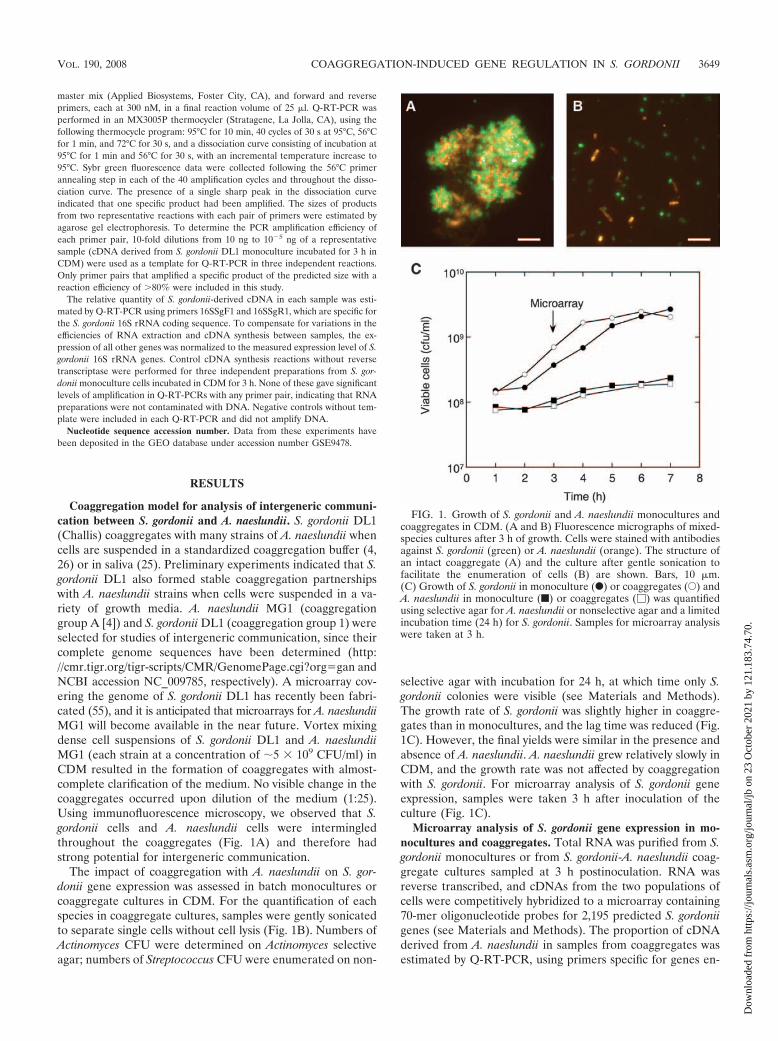
Coaggregation model for analysis of intergeneric communi-cation between S. gordonii and A. naeslundii. S. gordonii DL1(Challis) coaggregates with many strains of A. naeslundii whencells are suspended in a standardized coaggregation buffer (4,26) or in saliva (25). Preliminary experiments indicated that S.gordonii DL1 also formed stable coaggregation partnershipswith A. naeslundii strains when cells were suspended in a va-riety of growth media. A. naeslundii MG1 (coaggregationgroup A [4]) and S. gordonii DL1 (coaggregation group 1) wereselected for studies of intergeneric communication, since theircomplete genome sequences have been determined (http://cmr.tigr.org/tigr-scripts/CMR/GenomePage.cgi?orggan andNCBI accession NC_009785, respectively). A microarray cov-ering the genome of S. gordonii DL1 has recently been fabri-cated (55), and it is anticipated that microarrays for A. naeslundiiMG1 will become available in the near future. Vortex mixingdense cell suspensions of S. gordonii DL1 and A. naeslundiiMG1 (each strain at a concentration of �5 � 109 CFU/ml) inCDM resulted in the formation of coaggregates with almost-complete clarification of the medium. No visible change in thecoaggregates occurred upon dilution of the medium (1:25).Using immunofluorescence microscopy, we observed that S.gordonii cells and A. naeslundii cells were intermingledthroughout the coaggregates (Fig. 1A) and therefore hadstrong potential for intergeneric communication.
The impact of coaggregation with A. naeslundii on S. gor-donii gene expression was assessed in batch monocultures orcoaggregate cultures in CDM. For the quantification of eachspecies in coaggregate cultures, samples were gently sonicatedto separate single cells without cell lysis (Fig. 1B). Numbers ofActinomyces CFU were determined on Actinomyces selectiveagar; numbers of Streptococcus CFU were enumerated on non-
selective agar with incubation for 24 h, at which time only S.gordonii colonies were visible (see Materials and Methods).The growth rate of S. gordonii was slightly higher in coaggre-gates than in monocultures, and the lag time was reduced (Fig.1C). However, the final yields were similar in the presence andabsence of A. naeslundii. A. naeslundii grew relatively slowly inCDM, and the growth rate was not affected by coaggregationwith S. gordonii. For microarray analysis of S. gordonii geneexpression, samples were taken 3 h after inoculation of theculture (Fig. 1C).
Microarray analysis of S. gordonii gene expression in mo-nocultures and coaggregates. Total RNA was purified from S.gordonii monocultures or from S. gordonii-A. naeslundii coag-gregate cultures sampled at 3 h postinoculation. RNA wasreverse transcribed, and cDNAs from the two populations ofcells were competitively hybridized to a microarray containing70-mer oligonucleotide probes for 2,195 predicted S. gordoniigenes (see Materials and Methods). The proportion of cDNAderived from A. naeslundii in samples from coaggregates wasestimated by Q-RT-PCR, using primers specific for genes en-
FIG. 1. Growth of S. gordonii and A. naeslundii monocultures andcoaggregates in CDM. (A and B) Fluorescence micrographs of mixed-species cultures after 3 h of growth. Cells were stained with antibodiesagainst S. gordonii (green) or A. naeslundii (orange). The structure ofan intact coaggregate (A) and the culture after gentle sonication tofacilitate the enumeration of cells (B) are shown. Bars, 10 �m.(C) Growth of S. gordonii in monoculture (F) or coaggregates (E) andA. naeslundii in monoculture (f) or coaggregates (�) was quantifiedusing selective agar for A. naeslundii or nonselective agar and a limitedincubation time (24 h) for S. gordonii. Samples for microarray analysiswere taken at 3 h.
VOL. 190, 2008 COAGGREGATION-INDUCED GENE REGULATION IN S. GORDONII 3649
Dow
nloa
ded
from
http
s://j
ourn
als.
asm
.org
/jour
nal/j
b on
23
Oct
ober
202
1 by
121
.183
.74.
70.
coding 16S rRNA from S. gordonii and A. naeslundii. Theseanalyses indicated that cDNA from A. naeslundii comprisedapproximately 10 to 15% of the total cDNA (data not shown),which was in accordance with the proportion of A. naeslundiicells in coaggregate populations at 3 h (Fig. 1C). No significanthybridization of cDNA derived from A. naeslundii monocul-tures to the S. gordonii microarray was observed, and therefore,the presence of A. naeslundii RNA in samples from mixedcultures should not affect microarray analyses.
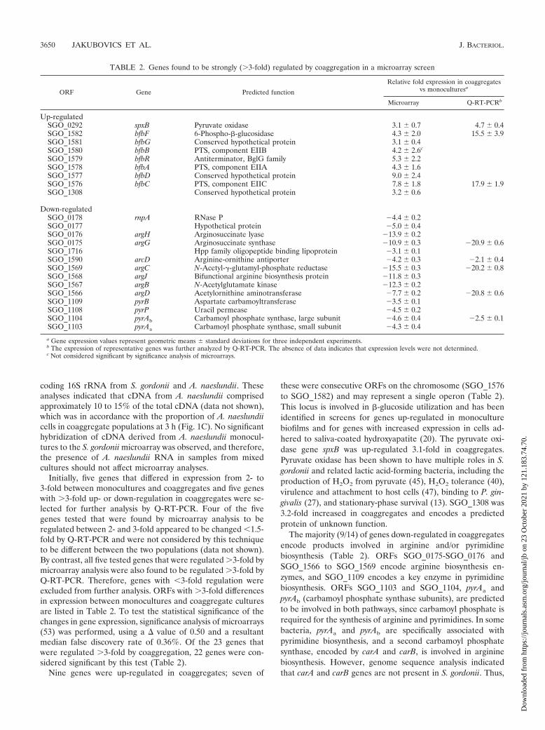
Initially, five genes that differed in expression from 2- to3-fold between monocultures and coaggregates and five geneswith �3-fold up- or down-regulation in coaggregates were se-lected for further analysis by Q-RT-PCR. Four of the fivegenes tested that were found by microarray analysis to beregulated between 2- and 3-fold appeared to be changed �1.5-fold by Q-RT-PCR and were not considered by this techniqueto be different between the two populations (data not shown).By contrast, all five tested genes that were regulated �3-fold bymicroarray analysis were also found to be regulated �3-fold byQ-RT-PCR. Therefore, genes with �3-fold regulation wereexcluded from further analysis. ORFs with �3-fold differencesin expression between monocultures and coaggregate culturesare listed in Table 2. To test the statistical significance of thechanges in gene expression, significance analysis of microarrays(53) was performed, using a � value of 0.50 and a resultantmedian false discovery rate of 0.36%. Of the 23 genes thatwere regulated �3-fold by coaggregation, 22 genes were con-sidered significant by this test (Table 2).
Nine genes were up-regulated in coaggregates; seven of
these were consecutive ORFs on the chromosome (SGO_1576to SGO_1582) and may represent a single operon (Table 2).This locus is involved in �-glucoside utilization and has beenidentified in screens for genes up-regulated in monoculturebiofilms and for genes with increased expression in cells ad-hered to saliva-coated hydroxyapatite (20). The pyruvate oxi-dase gene spxB was up-regulated 3.1-fold in coaggregates.Pyruvate oxidase has been shown to have multiple roles in S.gordonii and related lactic acid-forming bacteria, including theproduction of H2O2 from pyruvate (45), H2O2 tolerance (40),virulence and attachment to host cells (47), binding to P. gin-givalis (27), and stationary-phase survival (13). SGO_1308 was3.2-fold increased in coaggregates and encodes a predictedprotein of unknown function.
The majority (9/14) of genes down-regulated in coaggregatesencode products involved in arginine and/or pyrimidinebiosynthesis (Table 2). ORFs SGO_0175-SGO_0176 andSGO_1566 to SGO_1569 encode arginine biosynthesis en-zymes, and SGO_1109 encodes a key enzyme in pyrimidinebiosynthesis. ORFs SGO_1103 and SGO_1104, pyrAa andpyrAb (carbamoyl phosphate synthase subunits), are predictedto be involved in both pathways, since carbamoyl phosphate isrequired for the synthesis of arginine and pyrimidines. In somebacteria, pyrAa and pyrAb are specifically associated withpyrimidine biosynthesis, and a second carbamoyl phosphatesynthase, encoded by carA and carB, is involved in argininebiosynthesis. However, genome sequence analysis indicatedthat carA and carB genes are not present in S. gordonii. Thus,
TABLE 2. Genes found to be strongly (�3-fold) regulated by coaggregation in a microarray screen
ORF Gene Predicted function
Relative fold expression in coaggregatesvs monoculturesa
Microarray Q-RT-PCRb
Up-regulatedSGO_0292 spxB Pyruvate oxidase 3.1 0.7 4.7 0.4SGO_1582 bfbF 6-Phospho-�-glucosidase 4.3 2.0 15.5 3.9SGO_1581 bfbG Conserved hypothetical protein 3.1 0.4SGO_1580 bfbB PTS, component EIIB 4.2 2.6c
SGO_1579 bfbR Antiterminator, BglG family 5.3 2.2SGO_1578 bfbA PTS, component EIIA 4.3 1.6SGO_1577 bfbD Conserved hypothetical protein 9.0 2.4SGO_1576 bfbC PTS, component EIIC 7.8 1.8 17.9 1.9SGO_1308 Conserved hypothetical protein 3.2 0.6
Down-regulatedSGO_0178 rnpA RNase P �4.4 0.2SGO_0177 Hypothetical protein �5.0 0.4SGO_0176 argH Arginosuccinate lyase �13.9 0.2SGO_0175 argG Arginosuccinate synthase �10.9 0.3 �20.9 0.6SGO_1716 Hpp family oligopeptide binding lipoprotein �3.1 0.1SGO_1590 arcD Arginine-ornithine antiporter �4.2 0.3 �2.1 0.4SGO_1569 argC N-Acetyl-�-glutamyl-phosphate reductase �15.5 0.3 �20.2 0.8SGO_1568 argJ Bifunctional arginine biosynthesis protein �11.8 0.3SGO_1567 argB N-Acetylglutamate kinase �12.3 0.2SGO_1566 argD Acetylornithine aminotransferase �7.7 0.2 �20.8 0.6SGO_1109 pyrB Aspartate carbamoyltransferase �3.5 0.1SGO_1108 pyrP Uracil permease �4.5 0.2SGO_1104 pyrAb Carbamoyl phosphate synthase, large subunit �4.6 0.4 �2.5 0.1SGO_1103 pyrAa Carbamoyl phosphate synthase, small subunit �4.3 0.4
a Gene expression values represent geometric means standard deviations for three independent experiments.b The expression of representative genes was further analyzed by Q-RT-PCR. The absence of data indicates that expression levels were not determined.c Not considered significant by significance analysis of microarrays.
3650 JAKUBOVICS ET AL. J. BACTERIOL.
Dow
nloa
ded
from
http
s://j
ourn
als.
asm
.org
/jour
nal/j
b on
23
Oct
ober
202
1 by
121
.183
.74.
70.
in silico, the arginine and pyrimidine biosynthesis pathwaysappear to be linked in this organism.
SGO_1716 was down-regulated 3.1-fold in coaggregates. ThisORF is located immediately upstream of and in the same direc-tion as the hppH gene, encoding hexa/heptapeptide permeaselipoprotein HppH (18). The expression of hppH was down-regu-lated 2.6-fold 0.2-fold in coaggregates, and it is possible thathppH and SGO_1716 form an operon. The polypeptide encodedby SGO_1716 has a secretion signal sequence containing a con-sensus motif for lipid modification, LAAC (15), and shares 61.2%amino acid identity with HppH. It is therefore likely thatSGO_1716 is also involved in the uptake of short peptides. Twoother genes encoding membrane transporters were down-regu-lated �3-fold in coaggregates: those for uptake of arginine(SGO_1590, arcD) and pyrimidines (SGO_1108, pyrP) (Table 2).SGO_0177 and SGO_0178, encoding a hypothetical protein andRNase P, respectively, were down-regulated four- to fivefold incoaggregates. These ORFs may be cotranscribed with argGHsince they are immediately downstream of argH and are orientedin the same direction.
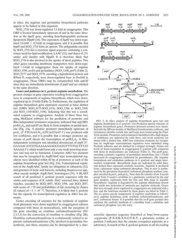
Genes and pathways in S. gordonii arginine metabolism. Thegreatest changes in gene expression resulting from coaggregationwere in components of arginine biosynthesis, which were down-regulated up to 15-fold (Table 2). Furthermore, the regulation ofarginine biosynthesis gene expression occurred at three distinctloci (ORFs SGO_0175-SGO_0176, SGO_1566 to SGO_1569,and SGO_1103-SGO_1104), indicating that this was a coordi-nated response to coaggregation. Analysis of these three loci,using MolQuest software for the prediction of promoter andRho-independent terminator sequences, indicated that argCJBD,pyrRAaAb, and argGH are likely to form complete or partial oper-ons (Fig. 2A). A putative promoter immediately upstream ofpyrAa (5-TTGAAA-N15-GTCAATAAT-3) was predicted withlow confidence, and it is possible that a stronger promoter up-stream of pyrR directs the transcription of pyrRAaAb. A Rho-independent terminator was identified downstream of argH (5-GAAAACATCGTGAAAAAGAGCGATGTTTTTGCTTTATAAAAA-3), which would form only a very weak stem-loop struc-ture and may not be functional. Consensus ARG box elementsthat may be recognized by ArgR/AhrC-type transcriptional reg-ulators were identified within 60 bp of promoters at each of thearginine biosynthesis gene loci (Fig. 2A). Transcriptional regula-tors of the ArgR/AhrC family are ubiquitous in eubacteria (32),and genomes of lactic acid bacteria, including Streptococcus spp.,often encode multiple ArgR/AhrC homologues (35). A BLASTsearch of all predicted S. gordonii protein sequences with theamino acid sequence of B. subtilis AhrC returned three distinctmatches, encoded by SGO_0697, SGO_1598, and SGO_2057,with scores of �70 and probabilities of hits occurring by chance(E values) of �1 � 10�14. Therefore, it is likely that S. gordoniihas the capacity for transcriptional regulation at ARG box ele-ments.
Genes encoding all enzymes for the synthesis of argininefrom glutamate were down-regulated in coaggregated culturescompared with those in monocultures, with the exception ofthe gene encoding an ornithine carbamoyltransferase (EC2.1.3.3) for the conversion of ornithine to citrulline (Fig. 2B).Ornithine carbamoyltransferase is evolutionarily related to as-partate carbamoyltransferase (28), involved in pyrimidine bio-synthesis, and these enzymes can be distinguished by a char-
acteristic signature sequence described at http://www.expasy.org/prosite (F-X-E/K-X-S-G/T-R-T; a glutamate residue atposition 3 indicates that the enzyme recognizes aspartate as asubstrate). A search of the S. gordonii genome in all six reading
FIG. 2. In silico analysis of arginine biosynthesis gene loci andarginine metabolism in S. gordonii. (A) Predicted operon structure ofarginine biosynthesis genes. Promoters (forward arrows) were identi-fied with the BProm module of MolQuest bioinformatics software, andterminators (double verticle line and loop) were found using the Find-Term module. Promoters and terminators identified with a high degreeof confidence are indicated by solid lines, and weak elements areshown with a dashed line. ARG box consensus elements for recogni-tion by ArgR-type transcriptional regulators were identified usingProdoric software and are marked by a striped rectangle. Genes andlevels of down-regulation in coaggregated S. gordonii cells comparedwith those in monocultures are indicated (Table 2). The pyrR gene,encoding a putative regulator of pyrimidine metabolism, was �3-folddecreased in coaggregates (indicated by parentheses). (B) Argininebiosynthesis and catabolism pathways in S. gordonii and major inter-mediary compounds. Biosynthetic steps are indicated by solid lines,and dashed lines represent catabolic reactions. Arginine is producedfrom L-glutamate using carbamoyl phosphate. This in turn is synthe-sized by the glutamine-dependent carbamoyl phosphate synthase com-plex encoded by pyrAa and pyrAb. Note that the transfer of an N-acetylgroup from N-acetyl-L-ornithine to L-glutamate, catalyzed by the argJgene product, represents both the first and fifth steps in argininebiosynthesis. Arginine is degraded by the arginine dihydrolase systemthat yields two molecules of ammonia per arginine molecule. Genesthat were strongly down-regulated in coaggregates are indicated with abox, and the functions encoded by these genes are listed in Table 2.The arc genes encode components of arginine catabolism as follows:arcA, arginine deiminase; arcB, ornithine carbamoyltransferase; andarcC, carbamate kinase. It is possible that the arcB gene product alsocatalyzes the anabolic synthesis of L-citrulline from L-ornithine andcarbamoyl phosphate (see the text).
VOL. 190, 2008 COAGGREGATION-INDUCED GENE REGULATION IN S. GORDONII 3651
Dow
nloa
ded
from
http
s://j
ourn
als.
asm
.org
/jour
nal/j
b on
23
Oct
ober
202
1 by
121
.183
.74.
70.
frames using the amino acid sequence of Lactococcus lactisanabolic ornithine carbamoyltransferase (ArgF) returned twosequences with BLAST scores of �100, ORFs SGO_1109 andSGO_1592. SGO_1109 is located within a cluster of pyrimidinebiosynthesis genes and encodes an amino acid sequence con-taining the signature for aspartate carbamoyltransferase en-zymes (PyrB). On the other hand, SGO_1592 encodes ArcB,an ornithine carbamoyltransferase enzyme associated with thecatabolic arginine dihydrolase system (10). Therefore, there isno clear anabolic ornithine carbamoyltransferase encodedwithin the S. gordonii genome. Although no reports documentthat ArcB also has the capacity to catalyze the anabolic con-version of ornithine to citrulline for arginine biosynthesis in S.gordonii, we suggest that this might occur.
Extracellular arginine concentration is a stimulus for thecoordinated regulation of arginine biosynthesis genes in S.gordonii. Amino acid biosynthesis genes in bacteria are com-monly regulated in response to the prevailing product concen-tration. The coordinated expression of arginine biosynthesisgenes in response to exogenous arginine in E. coli was de-scribed over 40 years ago (31). Using Q-RT-PCR, the expres-sion of S. gordonii arginine biosynthesis genes at three distinctloci (Fig. 2) was determined in cells cultured in arginine-re-plete (5 mM arginine) CDM, shifted to CDM without arginine,and incubated for a further 30 min. Under these conditions,the expression levels of argC, argG, and pyrAb were up-regu-lated 220-fold 1.8-fold, 130-fold 1.4-fold, and 9.8-fold 0.3-fold, respectively, in cells shifted to medium with no argi-nine compared with those of the genes in control cells resus-pended in medium with high arginine concentrations. As acontrol, levels of amyB mRNA, encoding �-amylase, weremonitored in each culture. The expression of amyB is increasedin S. gordonii cells apposed to Veillonella atypica cells (12) butis not affected by coaggregation with A. naeslundii. Levels ofamyB mRNA were not significantly regulated by arginine (1.5-fold 0.1-fold up-regulated in medium with no arginine).
The above data suggested that a major part of the responseof S. gordonii to coaggregation might be caused by modulationof the arginine concentration in the external milieu by A.naeslundii cells. Therefore, the concentration of arginine in thebulk medium was determined following the growth of S. gor-donii and A. naeslundii for 3 h in monoculture and coaggregateCDM (initial concentration, 0.5 mM arginine) cultures. In mo-noculture, S. gordonii consumed 57% 6.0% of the availableextracellular arginine during this period, whereas A. naeslundiicells removed only 4.4% 3.0% of the arginine. In coaggre-gate cultures, arginine was depleted by 64% 3.6%. It shouldbe noted that these measurements of global arginine concen-trations will not have detected any local variation that mayhave occurred in arginine levels within or around S. gordoniicells in coaggregates.
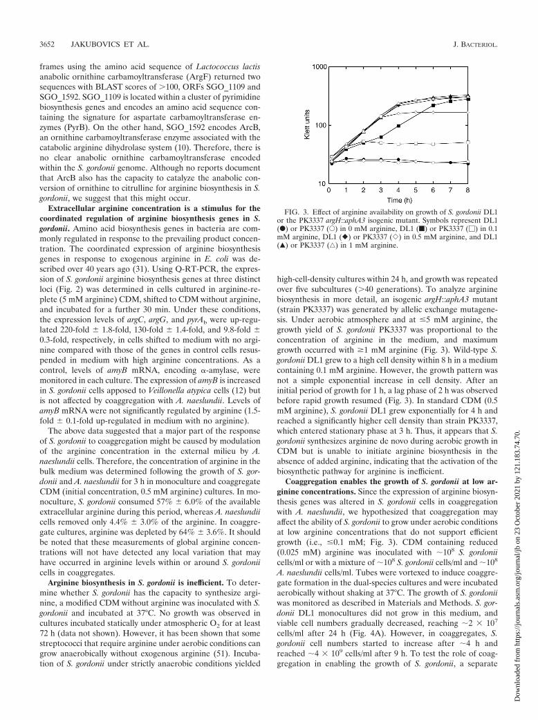
Arginine biosynthesis in S. gordonii is inefficient. To deter-mine whether S. gordonii has the capacity to synthesize argi-nine, a modified CDM without arginine was inoculated with S.gordonii and incubated at 37°C. No growth was observed incultures incubated statically under atmospheric O2 for at least72 h (data not shown). However, it has been shown that somestreptococci that require arginine under aerobic conditions cangrow anaerobically without exogenous arginine (51). Incuba-tion of S. gordonii under strictly anaerobic conditions yielded
high-cell-density cultures within 24 h, and growth was repeatedover five subcultures (�40 generations). To analyze argininebiosynthesis in more detail, an isogenic argH::aphA3 mutant(strain PK3337) was generated by allelic exchange mutagene-sis. Under aerobic atmosphere and at �5 mM arginine, thegrowth yield of S. gordonii PK3337 was proportional to theconcentration of arginine in the medium, and maximumgrowth occurred with �1 mM arginine (Fig. 3). Wild-type S.gordonii DL1 grew to a high cell density within 8 h in a mediumcontaining 0.1 mM arginine. However, the growth pattern wasnot a simple exponential increase in cell density. After aninitial period of growth for 1 h, a lag phase of 2 h was observedbefore rapid growth resumed (Fig. 3). In standard CDM (0.5mM arginine), S. gordonii DL1 grew exponentially for 4 h andreached a significantly higher cell density than strain PK3337,which entered stationary phase at 3 h. Thus, it appears that S.gordonii synthesizes arginine de novo during aerobic growth inCDM but is unable to initiate arginine biosynthesis in theabsence of added arginine, indicating that the activation of thebiosynthetic pathway for arginine is inefficient.
Coaggregation enables the growth of S. gordonii at low ar-ginine concentrations. Since the expression of arginine biosyn-thesis genes was altered in S. gordonii cells in coaggregationwith A. naeslundii, we hypothesized that coaggregation mayaffect the ability of S. gordonii to grow under aerobic conditionsat low arginine concentrations that do not support efficientgrowth (i.e., �0.1 mM; Fig. 3). CDM containing reduced(0.025 mM) arginine was inoculated with �108 S. gordoniicells/ml or with a mixture of �108 S. gordonii cells/ml and �108
A. naeslundii cells/ml. Tubes were vortexed to induce coaggre-gate formation in the dual-species cultures and were incubatedaerobically without shaking at 37°C. The growth of S. gordoniiwas monitored as described in Materials and Methods. S. gor-donii DL1 monocultures did not grow in this medium, andviable cell numbers gradually decreased, reaching �2 � 107
cells/ml after 24 h (Fig. 4A). However, in coaggregates, S.gordonii cell numbers started to increase after �4 h andreached �4 � 109 cells/ml after 9 h. To test the role of coag-gregation in enabling the growth of S. gordonii, a separate
FIG. 3. Effect of arginine availability on growth of S. gordonii DL1or the PK3337 argH::aphA3 isogenic mutant. Symbols represent DL1(F) or PK3337 (E) in 0 mM arginine, DL1 (f) or PK3337 (�) in 0.1mM arginine, DL1 (�) or PK3337 (�) in 0.5 mM arginine, and DL1(Œ) or PK3337 (‚) in 1 mM arginine.
3652 JAKUBOVICS ET AL. J. BACTERIOL.
Dow
nloa
ded
from
http
s://j
ourn
als.
asm
.org
/jour
nal/j
b on
23
Oct
ober
202
1 by
121
.183
.74.
70.
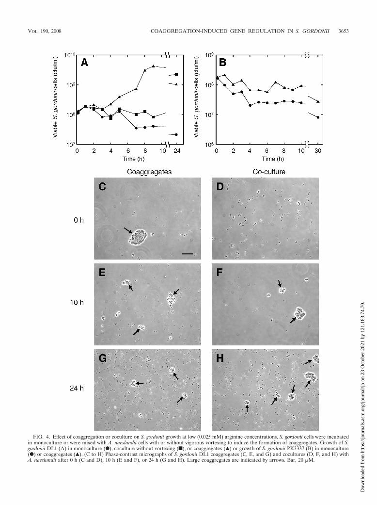
FIG. 4. Effect of coaggregation or coculture on S. gordonii growth at low (0.025 mM) arginine concentrations. S. gordonii cells were incubatedin monoculture or were mixed with A. naeslundii cells with or without vigorous vortexing to induce the formation of coaggregates. Growth of S.gordonii DL1 (A) in monoculture (F), coculture without vortexing (f), or coaggregates (Œ) or growth of S. gordonii PK3337 (B) in monoculture(F) or coaggregates (Œ). (C to H) Phase-contrast micrographs of S. gordonii DL1 coaggregates (C, E, and G) and cocultures (D, F, and H) withA. naeslundii after 0 h (C and D), 10 h (E and F), or 24 h (G and H). Large coaggregates are indicated by arrows. Bar, 20 �M.
VOL. 190, 2008 COAGGREGATION-INDUCED GENE REGULATION IN S. GORDONII 3653
Dow
nloa
ded
from
http
s://j
ourn
als.
asm
.org
/jour
nal/j
b on
23
Oct
ober
202
1 by
121
.183
.74.
70.
coculture containing S. gordonii cells and A. naeslundii cellsthat were not vortexed was set up and did not initially formcoaggregates (compare Fig. 4C and D). Coaggregation oc-curred gradually during extended coculture incubation, and by6 h, small clumps of �50 cells were observed (data not shown).After 10 h and 24 h, there was little difference between thenumber of coaggregates present in the coculture and that inthe culture that was vortexed to induce coaggregation (Fig. 4Eto H). No growth of S. gordonii was observed in coculturesduring the early period of uninduced formation of small coag-gregates, up to 9 h after inoculation. However, after prolongedincubation (24 h), high numbers of S. gordonii cells were de-tected in these cultures (�109 cells/ml), equivalent to the levelsof S. gordonii cells in mixed-species cultures that were initiallyvortexed to induce coaggregates.
To investigate whether the S. gordonii arginine biosyntheticpathway was required for growth in coaggregates, the S. gor-donii PK3337 argH::aphA3 strain was incubated in monocul-ture or in coaggregates with A. naeslundii as described above(Fig. 4B). The density of S. gordonii PK3337 cells decreasedgradually in both monocultures and coaggregate cultures, in-dicating that these cells were not growing but were losingviability. Therefore, an intact arginine biosynthesis pathway isrequired for coaggregation-mediated growth of S. gordonii atlow arginine concentrations.
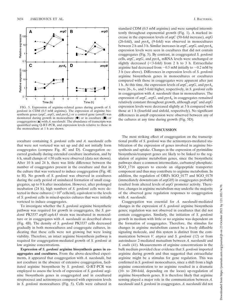
Expression of S. gordonii arginine biosynthesis genes in co-aggregates and cocultures. From the above-described experi-ments, it appeared that coaggregation with A. naeslundii, butnot coculture in the absence of extensive coaggregation, facil-itated arginine biosynthesis by S. gordonii. Q-RT-PCR wasemployed to assess the levels of expression of S. gordonii argi-nine biosynthesis genes in coaggregated and in coculturedstreptococci and actinomyces compared with expression levelsin S. gordonii monocultures (Fig. 5). Cells were cultured in
standard CDM (0.5 mM arginine) and were sampled intermit-tently throughout exponential growth (Fig. 1). A marked in-crease in the expression levels of argC (50-fold increase), argG(26-fold), and pyrAb (9-fold) was observed in monoculturesbetween 2 h and 3 h. Similar increases in argC, argG, and pyrAb
expression levels were seen in cocultures that did not containcoaggregates (Fig. 5). By contrast, in coaggregated S. gordoniicells, argC, argG, and pyrAb mRNA levels were unchanged orslightly decreased (�3-fold) from 2 h to 3 h. Extracellulararginine had decreased from �0.5 mM initially to �0.2 mM by3 h (see above). Differences in expression levels of S. gordoniiarginine biosynthesis genes in monocultures or coculturescompared with those in coaggregates were apparent after just1 h. At this time, the expression levels of argC, argG, and pyrAb
were 26-, 6-, and 5-fold higher, respectively, in S. gordonii cellsin coaggregation with A. naeslundii than in monocultures. Theexpression of argC, argG, and pyrAb in coaggregates remainedrelatively constant throughout growth, although argC and argGexpression levels were decreased slightly at 3 h compared withthose at 1 h (fourfold and sixfold, respectively). No significantdifferences in amyB expression were observed between any ofthe cultures at any time during growth (Fig. 5D).
DISCUSSION
The most striking effect of coaggregation on the transcrip-tional profile of S. gordonii was the actinomyces-mediated sta-bilization of the expression of genes involved in arginine bio-synthesis and uptake. Changes in the expression of pyrimidinebiosynthesis/transport genes are likely to be linked to the reg-ulation of arginine metabolism genes, since the biosyntheticpathways share a common intermediate, carbamoyl phosphate.SGO_1716 appears to encode an oligopeptide transportercomponent and thus may contribute to arginine metabolism. Inaddition, the regulation of ORFs SGO_0177 and SGO_0178(immediately downstream of argGH) in coaggregates probablyresulted from altered levels of argG promoter activity. There-fore, changes in arginine metabolism may underlie the majorityof the observed gene regulation responses to coaggregationwith A. naeslundii.
Coaggregation was essential for A. naeslundii-mediatedchanges in the expression of S. gordonii arginine biosynthesisgenes; regulation was not observed in cocultures that did notcontain coaggregates. Similarly, the initiation of S. gordoniigrowth in medium with little or no arginine was dependent onthe formation of coaggregates. Therefore, the stimulus forchanges in arginine metabolism cannot be a freely diffusiblesignaling molecule, and this system is distinct from the com-munication between V. atypica and S. gordonii (12) or fromautoinducer 2-mediated mutualism between A. naeslundii andS. oralis (41). Measurements of arginine concentrations in thebulk medium provided clear evidence that S. gordonii importedarginine during growth and thus suggested that extracellulararginine might be a stimulus for gene regulation. This wasconfirmed in S. gordonii monocultures, since a shift from a higharginine concentration to no arginine resulted in a dramatic(10- to 200-fold, depending on the locus) up-regulation ofarginine biosynthesis genes. It is therefore likely that argininesensing played a major role in the communication between A.naeslundii and S. gordonii in coaggregates. A. naeslundii did not
FIG. 5. Expression of arginine-related genes during growth of S.gordonii in CDM (0.5 mM arginine). The expression of arginine bio-synthesis genes (argC, argG, and pyrAb) or a control gene (amyB) wasmonitored during growth in monoculture (F) or in coculture (f) orcoaggregation (Œ) with A. naeslundii. The abundance of transcripts wasquantified using Q-RT-PCR, and expression levels relative to those inthe monoculture at 1 h are shown.
3654 JAKUBOVICS ET AL. J. BACTERIOL.
Dow
nloa
ded
from
http
s://j
ourn
als.
asm
.org
/jour
nal/j
b on
23
Oct
ober
202
1 by
121
.183
.74.
70.
significantly modulate the global arginine concentration in themedium, and this is consistent with the absolute requirementfor coaggregation to initiate changes in the S. gordonii argininebiosynthesis pathway.
It appears that coaggregation provides a local microenviron-ment in which arginine uptake and/or biosynthesis is facilitatedin S. gordonii. Thus, S. gordonii cells may acquire argininedirectly from A. naeslundii by a contact-dependent mechanism.For example, S. gordonii produces an extracellular arginineaminopeptidase (14) that could potentially scavenge arginineresidues from proteins on the cell wall of A. naeslundii. How-ever, A. naeslundii did not supply enough arginine to supportthe aerobic growth of an S. gordonii argH mutant in CDMcontaining 0.025 mM arginine (Fig. 4B). Alternatively, reducedO2 tension in the center of coaggregates might enhance theefficiency of arginine biosynthesis in S. gordonii. In this case,rapid up-regulation of arginine biosynthesis genes in cells pro-tected from O2 would enable them to accumulate an intracel-lular pool of arginine that could be utilized once externalarginine became scarce. This hypothesis would fit with theobservation that S. gordonii arginine biosynthesis genes areinitially up-regulated following coaggregation and then declineslightly as streptococci start to grow outside the coaggregates(Fig. 5). It would be interesting to determine whether en-hanced S. gordonii arginine biosynthesis is dependent upon themetabolic activity of A. naeslundii cells in coaggregates or in-deed whether oral bacteria other than A. naeslundii inducesimilar gene regulation in S. gordonii. Studies to resolve theseissues are under way.
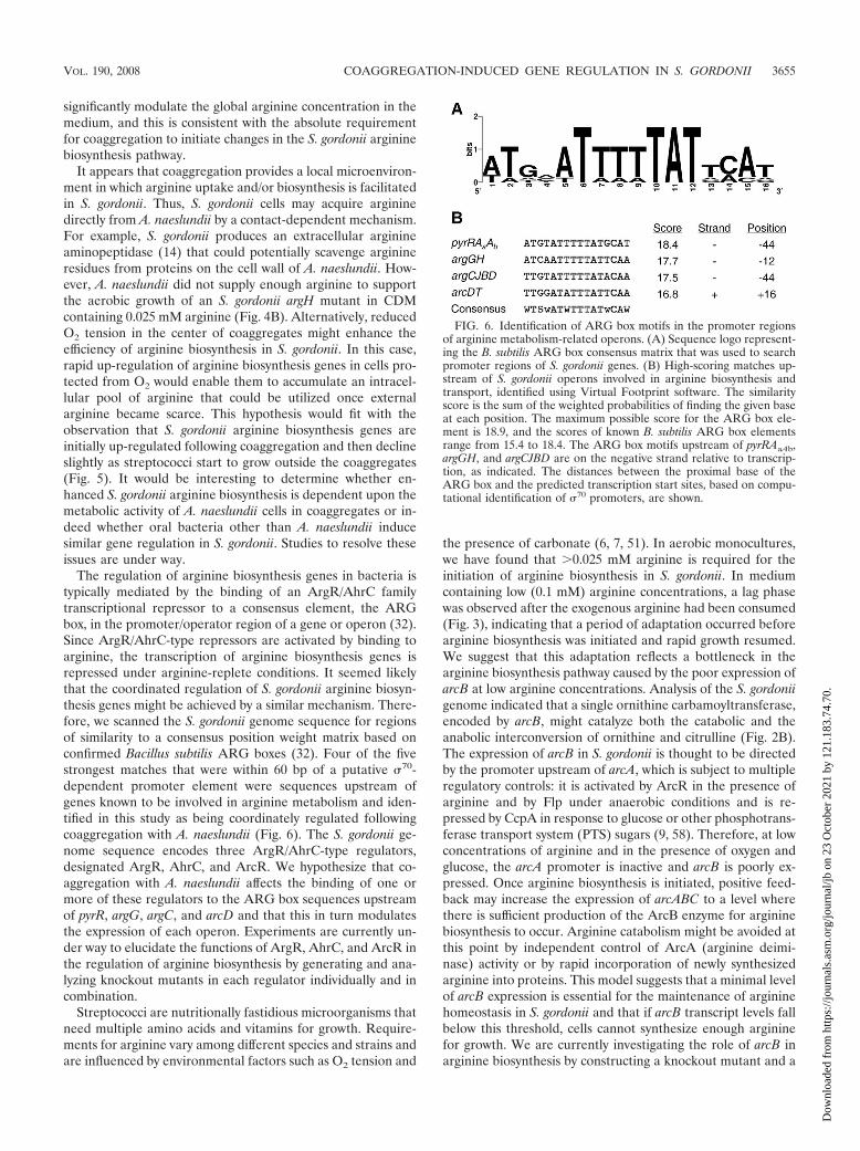
The regulation of arginine biosynthesis genes in bacteria istypically mediated by the binding of an ArgR/AhrC familytranscriptional repressor to a consensus element, the ARGbox, in the promoter/operator region of a gene or operon (32).Since ArgR/AhrC-type repressors are activated by binding toarginine, the transcription of arginine biosynthesis genes isrepressed under arginine-replete conditions. It seemed likelythat the coordinated regulation of S. gordonii arginine biosyn-thesis genes might be achieved by a similar mechanism. There-fore, we scanned the S. gordonii genome sequence for regionsof similarity to a consensus position weight matrix based onconfirmed Bacillus subtilis ARG boxes (32). Four of the fivestrongest matches that were within 60 bp of a putative �70-dependent promoter element were sequences upstream ofgenes known to be involved in arginine metabolism and iden-tified in this study as being coordinately regulated followingcoaggregation with A. naeslundii (Fig. 6). The S. gordonii ge-nome sequence encodes three ArgR/AhrC-type regulators,designated ArgR, AhrC, and ArcR. We hypothesize that co-aggregation with A. naeslundii affects the binding of one ormore of these regulators to the ARG box sequences upstreamof pyrR, argG, argC, and arcD and that this in turn modulatesthe expression of each operon. Experiments are currently un-der way to elucidate the functions of ArgR, AhrC, and ArcR inthe regulation of arginine biosynthesis by generating and ana-lyzing knockout mutants in each regulator individually and incombination.
Streptococci are nutritionally fastidious microorganisms thatneed multiple amino acids and vitamins for growth. Require-ments for arginine vary among different species and strains andare influenced by environmental factors such as O2 tension and
the presence of carbonate (6, 7, 51). In aerobic monocultures,we have found that �0.025 mM arginine is required for theinitiation of arginine biosynthesis in S. gordonii. In mediumcontaining low (0.1 mM) arginine concentrations, a lag phasewas observed after the exogenous arginine had been consumed(Fig. 3), indicating that a period of adaptation occurred beforearginine biosynthesis was initiated and rapid growth resumed.We suggest that this adaptation reflects a bottleneck in thearginine biosynthesis pathway caused by the poor expression ofarcB at low arginine concentrations. Analysis of the S. gordoniigenome indicated that a single ornithine carbamoyltransferase,encoded by arcB, might catalyze both the catabolic and theanabolic interconversion of ornithine and citrulline (Fig. 2B).The expression of arcB in S. gordonii is thought to be directedby the promoter upstream of arcA, which is subject to multipleregulatory controls: it is activated by ArcR in the presence ofarginine and by Flp under anaerobic conditions and is re-pressed by CcpA in response to glucose or other phosphotrans-ferase transport system (PTS) sugars (9, 58). Therefore, at lowconcentrations of arginine and in the presence of oxygen andglucose, the arcA promoter is inactive and arcB is poorly ex-pressed. Once arginine biosynthesis is initiated, positive feed-back may increase the expression of arcABC to a level wherethere is sufficient production of the ArcB enzyme for argininebiosynthesis to occur. Arginine catabolism might be avoided atthis point by independent control of ArcA (arginine deimi-nase) activity or by rapid incorporation of newly synthesizedarginine into proteins. This model suggests that a minimal levelof arcB expression is essential for the maintenance of argininehomeostasis in S. gordonii and that if arcB transcript levels fallbelow this threshold, cells cannot synthesize enough argininefor growth. We are currently investigating the role of arcB inarginine biosynthesis by constructing a knockout mutant and a
FIG. 6. Identification of ARG box motifs in the promoter regionsof arginine metabolism-related operons. (A) Sequence logo represent-ing the B. subtilis ARG box consensus matrix that was used to searchpromoter regions of S. gordonii genes. (B) High-scoring matches up-stream of S. gordonii operons involved in arginine biosynthesis andtransport, identified using Virtual Footprint software. The similarityscore is the sum of the weighted probabilities of finding the given baseat each position. The maximum possible score for the ARG box ele-ment is 18.9, and the scores of known B. subtilis ARG box elementsrange from 15.4 to 18.4. The ARG box motifs upstream of pyrRAaAb,argGH, and argCJBD are on the negative strand relative to transcrip-tion, as indicated. The distances between the proximal base of theARG box and the predicted transcription start sites, based on compu-tational identification of �70 promoters, are shown.
VOL. 190, 2008 COAGGREGATION-INDUCED GENE REGULATION IN S. GORDONII 3655
Dow
nloa
ded
from
http
s://j
ourn
als.
asm
.org
/jour
nal/j
b on
23
Oct
ober
202
1 by
121
.183
.74.
70.
complemented strain in which arcB is under the control of aheterologous promoter.
A number of S. gordonii genes unrelated to arginine biosyn-thesis were up-regulated in response to coaggregation with A.naeslundii. For example, seven genes in the biofilm-related bfblocus were strongly (three- to ninefold) up-regulated. UsingQ-RT-PCR to monitor gene expression, coaggregation wasessential for this response; in cocultures of S. gordonii and A.naeslundii that were not vortexed to induce coggregation, theexpression levels of bfb genes were equivalent to those in S.gordonii monocultures (data not shown). The bfb locus is oneof several gene clusters encoding PTS components and asso-ciated �-glucosidases that have been shown to be inducibleusing an S. gordonii in vivo expression technology screeningsystem (20). Increased promoter activity upstream of bfb geneswas reported by 25% of in vivo expression technology clonesretrieved from monoculture biofilms (20). The disruption ofany one of three genes in this locus, bfbF (�-glucosidase), bfbB(PTS EIIB), and/or bfbR (antiterminator), resulted in strainsdeficient in monospecies biofilm formation on plastic surfacesin microtiter wells. Furthermore, several distinct clones withreporter gene insertions in the bfb locus were recovered froma model of adhesion to saliva-coated hydroxyapatite, indicatingthat bfb genes are up-regulated by adherence to a surface (20).Our results confirm that adhesion is a trigger for increasedexpression of bfb genes and demonstrate that coaggregation isan excellent model for investigating biofilms and surface con-tact. Other genes up-regulated in response to coaggregationwere SGO_1308, encoding a hypothetical 154-amino-acid pro-tein, and spxB, encoding pyruvate oxidase. Recently, it hasbeen shown that SpxB is required for the binding of S. gordoniito P. gingivalis (27), and this protein may have a similar func-tion in adherence to A. naeslundii. Alternatively, the up-regu-lation of spxB expression may enhance the ability of S. gordoniito compete with neighboring species by increasing H2O2 pro-duction.
In summary, we have shown that coaggregation with A.naeslundii stabilizes arginine metabolism in S. gordonii andreduces dependence on extracellular arginine. This is likely tobe important in early dental plaque since the concentration offree arginine in saliva is extremely low, in the order of 0.004mM to 0.03 mM (2, 49, 54). Even within a mature 48-h dentalplaque biofilm, free arginine is present only at around 0.2 mM(16). S. gordonii can obtain arginine by degrading host salivarypolypeptides such as proline-rich proteins (11). However, it isnot clear that this can provide sufficient arginine for metabo-lism and growth, particularly in dense plaque biofilms wherediffusion of proteins is limited. These data indicate that coag-gregation may be fundamental for the successful colonizationof oral surfaces by streptococci and may act as a switch tostimulate growth in multispecies biofilm environments.
ACKNOWLEDGMENTS
This research was supported in part by the Intramural ResearchProgram of the National Institute of Dental and Craniofacial Re-search, National Institutes of Health. Microarray fabrication and anal-ysis were supported by Public Health Service grant DE11090 from theNational Institute of Dental and Craniofacial Research awarded toM.M.V.
REFERENCES
1. Aas, J. A., B. J. Paster, L. N. Stokes, I. Olsen, and F. E. Dewhirst. 2005.Defining the normal bacterial flora of the oral cavity. J. Clin. Microbiol.43:5721–5732.
2. Brand, H. S., G. G. Jorning, R. A. Chamuleau, and L. Abraham-Inpijn. 1997.Effect of a protein-rich meal on urinary and salivary free amino acid con-centrations in human subjects. Clin. Chim. Acta 264:37–47.
3. Cisar, J. O., S. H. Curl, P. E. Kolenbrander, and A. E. Vatter. 1983. Specificabsence of type 2 fimbriae on a coaggregation-defective mutant of Actino-myces viscosus T14V. Infect. Immun. 40:759–765.
4. Cisar, J. O., P. E. Kolenbrander, and F. C. McIntire. 1979. Specificity ofcoaggregation reactions between human oral streptococci and strains ofActinomyces viscosus or Actinomyces naeslundii. Infect. Immun. 24:742–752.
5. Cisar, J. O., A. E. Vatter, and F. C. Mcintire. 1978. Identification of viru-lence-associated antigen on surface fibrils of Actinomyces viscosus T14. In-fect. Immun. 19:312–319.
6. Cowman, R. A., M. M. Perrella, B. O. Adams, and R. J. Fitzgerald. 1975.Amino acid requirements and proteolytic activity of Streptococcus sanguis.Appl. Microbiol. 30:374–380.
7. Cowman, R. A., M. M. Perrella, and R. J. Fitzgerald. 1974. Influence ofincubation atmosphere on growth and amino acid requirements of Strepto-coccus mutans. Appl. Microbiol. 27:86–92.
8. Diaz, P. I., N. I. Chalmers, A. H. Rickard, C. Kong, C. L. Milburn, R. J.Palmer, Jr., and P. E. Kolenbrander. 2006. Molecular characterization ofsubject-specific oral microflora during initial colonization of enamel. Appl.Environ. Microbiol. 72:2837–2848.
9. Dong, Y., Y. Y. Chen, and R. A. Burne. 2004. Control of expression of thearginine deiminase operon of Streptococcus gordonii by CcpA and Flp. J.Bacteriol. 186:2511–2514.
10. Dong, Y., Y. Y. Chen, J. A. Snyder, and R. A. Burne. 2002. Isolation andmolecular analysis of the gene cluster for the arginine deiminase system fromStreptococcus gordonii DL1. Appl. Environ. Microbiol. 68:5549–5553.
11. Drobni, M., T. Li, C. Kruger, V. Loimaranta, M. Kilian, L. Hammarstrom,H. Jornvall, T. Bergman, and N. Stromberg. 2006. Host-derived pentapep-tide affecting adhesion, proliferation, and local pH in biofilm communitiescomposed of Streptococcus and Actinomyces species. Infect. Immun. 74:6293–6299.
12. Egland, P. G., R. J. Palmer, Jr., and P. E. Kolenbrander. 2004. Interspeciescommunication in Streptococcus gordonii-Veillonella atypica biofilms: signal-ing in flow conditions requires juxtaposition. Proc. Natl. Acad. Sci. USA101:16917–16922.
13. Goffin, P., L. Muscariello, F. Lorquet, A. Stukkens, D. Prozzi, M. Sacco, M.Kleerebezem, and P. Hols. 2006. Involvement of pyruvate oxidase activityand acetate production in the survival of Lactobacillus plantarum during thestationary phase of aerobic growth. Appl. Environ. Microbiol. 72:7933–7940.
14. Goldstein, J. M., D. Nelson, T. Kordula, J. A. Mayo, and J. Travis. 2002.Extracellular arginine aminopeptidase from Streptococcus gordonii FSS2.Infect. Immun. 70:836–843.
15. Hayashi, S., and H. C. Wu. 1990. Lipoproteins in bacteria. J. Bioenerg.Biomembr. 22:451–471.
16. Higham, S. M., and W. M. Edgar. 1989. Human dental plaque pH, and theorganic acid and free amino acid profiles in plaque fluid, after sucroserinsing. Arch. Oral Biol. 34:329–334.
17. Hsu, S. D., J. O. Cisar, A. L. Sandberg, and M. Kilian. 1994. Adhesiveproperties of viridans streptoccocal species. Microb. Ecol. Health Dis. 7:125–137.
18. Jenkinson, H. F., R. A. Baker, and G. W. Tannock. 1996. A binding-lipopro-tein-dependent oligopeptide transport system in Streptococcus gordoniiessential for uptake of hexa- and heptapeptides. J. Bacteriol. 178:68–77.
19. Jenkinson, H. F., and R. J. Lamont. 1997. Streptococcal adhesion and col-onization. Crit. Rev. Oral Biol. Med. 8:175–200.
20. Kilic, A. O., L. Tao, Y. Zhang, Y. Lei, A. Khammanivong, and M. C. Her-zberg. 2004. Involvement of Streptococcus gordonii �-glucoside metabolismsystems in adhesion, biofilm formation, and in vivo gene expression. J.Bacteriol. 186:4246–4253.
21. Kolenbrander, P. E. 1993. Coaggregation of human oral bacteria: potentialrole in the accretion of dental plaque. J. Appl. Bacteriol. 74(Suppl.):79S–86S.
22. Kolenbrander, P. E. 2000. Oral microbial communities: biofilms, interac-tions, and genetic systems. Annu. Rev. Microbiol. 54:413–437.
23. Kolenbrander, P. E. 1989. Surface recognition among oral bacteria: multi-generic coaggregations and their mediators. Crit. Rev. Microbiol. 17:137–159.
24. Kolenbrander, P. E., R. J. Palmer, Jr., A. H. Rickard, N. S. Jakubovics, N. I.Chalmers, and P. I. Diaz. 2006. Bacterial interactions and successions duringplaque development. Periodontol. 2000 42:47–79.
25. Kolenbrander, P. E., and C. S. Phucas. 1984. Effect of saliva on coaggrega-tion of oral Actinomyces and Streptococcus species. Infect. Immun. 44:228–233.
26. Kolenbrander, P. E., and B. L. Williams. 1981. Lactose-reversible coaggre-gation between oral actinomycetes and Streptococcus sanguis. Infect. Immun.33:95–102.
3656 JAKUBOVICS ET AL. J. BACTERIOL.
Dow
nloa
ded
from
http
s://j
ourn
als.
asm
.org
/jour
nal/j
b on
23
Oct
ober
202
1 by
121
.183
.74.
70.
27. Kuboniwa, M., G. D. Tribble, C. E. James, A. O. Kilic, L. Tao, M. C.Herzberg, S. Shizukuishi, and R. J. Lamont. 2006. Streptococcus gordoniiutilizes several distinct gene functions to recruit Porphyromonas gingivalisinto a mixed community. Mol. Microbiol. 60:121–139.
28. Labedan, B., A. Boyen, M. Baetens, D. Charlier, P. Chen, R. Cunin, V.Durbeco, N. Glansdorff, G. Herve, C. Legrain, Z. Liang, C. Purcarea, M.Roovers, R. Sanchez, T. L. Toong, M. Van de Casteele, F. van Vliet, Y. Xu,and Y. F. Zhang. 1999. The evolutionary history of carbamoyltransferases: acomplex set of paralogous genes was already present in the last universalcommon ancestor. J. Mol. Evol. 49:461–473.
29. Lewis, R., D. McKenzie, J. Bagg, and A. Dickie. 1995. Experience with anovel selective medium for isolation of Actinomyces spp. from medical anddental specimens. J. Clin. Microbiol. 33:1613–1616.
30. Li, J., E. J. Helmerhorst, C. W. Leone, R. F. Troxler, T. Yaskell, A. D.Haffajee, S. S. Socransky, and F. G. Oppenheim. 2004. Identification of earlymicrobial colonizers in human dental biofilm. J. Appl. Microbiol. 97:1311–1318.
31. Maas, W. K. 1994. The arginine repressor of Escherichia coli. Microbiol. Rev.58:631–640.
32. Makarova, K. S., A. A. Mironov, and M. S. Gelfand. 2001. Conservation ofthe binding site for the arginine repressor in all bacterial lineages. GenomeBiol. 2:research0013.1–research0013.8.
33. Mashburn, L. M., A. M. Jett, D. R. Akins, and M. Whiteley. 2005. Staphy-lococcus aureus serves as an iron source for Pseudomonas aeruginosa duringin vivo coculture. J. Bacteriol. 187:554–566.
34. McIntire, F. C., L. K. Crosby, A. E. Vatter, J. O. Cisar, M. R. McNeil, C. A.Bush, S. S. Tjoa, and P. V. Fennessey. 1988. A polysaccharide from Strep-tococcus sanguis 34 that inhibits coaggregation of S. sanguis 34 with Actino-myces viscosus T14V. J. Bacteriol. 170:2229–2235.
35. Nicoloff, H., F. Arsene-Ploetze, C. Malandain, M. Kleerebezem, and F. Bringel.2004. Two arginine repressors regulate arginine biosynthesis in Lactobacillusplantarum. J. Bacteriol. 186:6059–6069.
36. Nyvad, B., and M. Kilian. 1990. Comparison of the initial streptococcalmicroflora on dental enamel in caries-active and in caries-inactive individu-als. Caries Res. 24:267–272.
37. Nyvad, B., and M. Kilian. 1987. Microbiology of the early colonization ofhuman enamel and root surfaces in vivo. Scand. J. Dent. Res. 95:369–380.
38. Palmer, R. J., Jr., S. M. Gordon, J. O. Cisar, and P. E. Kolenbrander. 2003.Coaggregation-mediated interactions of streptococci and actinomyces de-tected in initial human dental plaque. J. Bacteriol. 185:3400–3409.
39. Palmer, R. J., Jr., K. Kazmerzak, M. C. Hansen, and P. E. Kolenbrander.2001. Mutualism versus independence: strategies of mixed-species oral bio-films in vitro using saliva as the sole nutrient source. Infect. Immun. 69:5794–5804.
40. Pericone, C. D., S. Park, J. A. Imlay, and J. N. Weiser. 2003. Factorscontributing to hydrogen peroxide resistance in Streptococcus pneumoniaeinclude pyruvate oxidase (SpxB) and avoidance of the toxic effects of theFenton reaction. J. Bacteriol. 185:6815–6825.
41. Rickard, A. H., R. J. Palmer, Jr., D. S. Blehert, S. R. Campagna, M. F.Semmelhack, P. G. Egland, B. L. Bassler, and P. E. Kolenbrander. 2006.Autoinducer 2: a concentration-dependent signal for mutualistic bacterialbiofilm growth. Mol. Microbiol. 60:1446–1456.
42. Rozen, S., and H. Skaletsky. 2000. Primer3 on the WWW for general usersand for biologist programmers. Methods Mol. Biol 132:365–386.
43. Saeed, A. I., N. K. Bhagabati, J. C. Braisted, W. Liang, V. Sharov, E. A.Howe, J. Li, M. Thiagarajan, J. A. White, and J. Quackenbush. 2006. TM4microarray software suite. Methods Enzymol. 411:134–193.
44. Sambrook, J., E. F. Fritsch, and T. Maniatis. 1989. Molecular cloning: alaboratory manual, 2nd ed. Cold Spring Harbor Laboratory, Cold SpringHarbor, NY.
45. Sedewitz, B., K. H. Schleifer, and F. Gotz. 1984. Purification and biochemicalcharacterization of pyruvate oxidase from Lactobacillus plantarum. J. Bacte-riol. 160:273–278.
46. Simionato, M. R., C. M. Tucker, M. Kuboniwa, G. Lamont, D. R. Demuth,G. D. Tribble, and R. J. Lamont. 2006. Porphyromonas gingivalis genesinvolved in community development with Streptococcus gordonii. Infect. Im-mun. 74:6419–6428.
47. Spellerberg, B., D. R. Cundell, J. Sandros, B. J. Pearce, I. Idanpaan-Heik-kila, C. Rosenow, and H. R. Masure. 1996. Pyruvate oxidase, as a determi-nant of virulence in Streptococcus pneumoniae. Mol. Microbiol. 19:803–813.
48. Stoodley, P., K. Sauer, D. G. Davies, and J. W. Costerton. 2002. Biofilms ascomplex differentiated communities. Annu. Rev. Microbiol. 56:187–209.
49. Syrjanen, S. M., L. Alakuijala, P. Alakuijala, S. O. Markkanen, and H.Markkanen. 1990. Free amino acid levels in oral fluids of normal subjectsand patients with periodontal disease. Arch. Oral Biol. 35:189–193.
50. Tao, L., D. J. LeBlanc, and J. J. Ferretti. 1992. Novel streptococcal-integra-tion shuttle vectors for gene cloning and inactivation. Gene 120:105–110.
51. Terleckyj, B., and G. D. Shockman. 1975. Amino acid requirements ofStreptococcus mutans and other oral streptococci. Infect. Immun. 11:656–664.
52. Terleckyj, B., N. P. Willett, and G. D. Shockman. 1975. Growth of severalcariogenic strains of oral streptococci in a chemically defined medium. In-fect. Immun. 11:649–655.
53. Tusher, V. G., R. Tibshirani, and G. Chu. 2001. Significance analysis ofmicroarrays applied to the ionizing radiation response. Proc. Natl. Acad. Sci.USA 98:5116–5121.
54. Van Wuyckhuyse, B. C., H. E. Perinpanayagam, D. Bevacqua, R. F. Raub-ertas, R. J. Billings, W. H. Bowen, and L. A. Tabak. 1995. Association of freearginine and lysine concentrations in human parotid saliva with caries expe-rience. J. Dent. Res. 74:686–690.
55. Vickerman, M. M., S. Iobst, A. M. Jesionowski, and S. R. Gill. 2007. Ge-nome-wide transcriptional changes in Streptococcus gordonii in response tocompetence signaling peptide. J. Bacteriol. 189:7799–7807.
56. Xie, H., G. S. Cook, J. W. Costerton, G. Bruce, T. M. Rose, and R. J. Lamont.2000. Intergeneric communication in dental plaque biofilms. J. Bacteriol.182:7067–7069.
57. Xie, H., X. Lin, B. Y. Wang, J. Wu, and R. J. Lamont. 2007. Identification ofa signalling molecule involved in bacterial intergeneric communication. Mi-crobiology 153:3228–3234.
58. Zeng, L., Y. Dong, and R. A. Burne. 2006. Characterization of cis-acting sitescontrolling arginine deiminase gene expression in Streptococcus gordonii. J.Bacteriol. 188:941–949.
VOL. 190, 2008 COAGGREGATION-INDUCED GENE REGULATION IN S. GORDONII 3657
Dow
nloa
ded
from
http
s://j
ourn
als.
asm
.org
/jour
nal/j
b on
23
Oct
ober
202
1 by
121
.183
.74.
70.