spontaneous rupture of the esophagus: report of a
TRANSCRIPT
Henry Ford Hospital Medical Journal
Volume 15 | Number 3 Article 2
9-1967
Spontaneous Rupture of the Esophagus: Report ofa Successfully Managed CaseEduardo Arciniegas
Follow this and additional works at: https://scholarlycommons.henryford.com/hfhmedjournal
Part of the Life Sciences Commons, Medical Specialties Commons, and the Public HealthCommons
This Article is brought to you for free and open access by Henry Ford Health System Scholarly Commons. It has been accepted for inclusion in HenryFord Hospital Medical Journal by an authorized editor of Henry Ford Health System Scholarly Commons.
Recommended CitationArciniegas, Eduardo (1967) "Spontaneous Rupture of the Esophagus: Report of a Successfully Managed Case," Henry Ford HospitalMedical Journal : Vol. 15 : No. 3 , 191-197.Available at: https://scholarlycommons.henryford.com/hfhmedjournal/vol15/iss3/2

Henry Ford Hosp. Med. Journal Vol. 15, No. 3, 1967
Spontaneous Rupture of tlie Esophagus Report of a Successfully Managed Case
Eduardo Arciniegas, M.D.*
Spontaneous rupture of the esophagus should be sitspecled in any patient developing severe chest pain after vomiting. The diagnosis is substantiated by the presence of suhcutaneotis emphysema in the suprasternal region on physical examination and of mediastinal air on the plain chest roentgenogram. Further radiological confirmation may be obtained by oral administration of a water-soluble radio-opaqtie material. Immediate surgical repair of the esophageal tear is tnaitdatory.
Spontaneous rupture of the esophagus is a relatively uncommon but very serious accident which demands prompt diagnosis and immediate surgical treatment.
The following case, which was treated successfully, illustrates some of the clinical features and problems encountered in the management of this catastrophe.
W. C. was a 52-year-old male admitted at 9:30 p.m. on December 28, 1966, because of severe retrosternal pain of one and one-half hours' duration which had its onset as he was straining at stool. He felt nauseated, vomited twice, and immediately developed severe retrosternal pain. This he described as "ripping", radiating to the back and aggravated by breathing. He had taken his previous meal seven hours earlier and he denied any alcoholic intake during the preceding several days. His past health had been excellent and he had no history of complaints related to the gastro-intestinal tract.
Physical examination revealed a thin, dyspneic male in severe distress. His breathing was rapid and shallow. Pulse was 120 per tninute and regular. Blood pressure was 130/80. There was subcutaneous emphysema in the suprasternal notch area. The trachea was in the mid-line. Percussion was dull in the lower half of the left hemi-thorax and hyper-resonant in its upper half. The vocal vibrations were diminished-to-absent in the same area. The heart sounds were normal. The abdomen was boardlike
*Froin the Division of Thoracic Surgery
191

Arciniegas
and tender to palpation in the epigastrium. The bowel sounds were hypoactive. The blood count and the electrocardiogram were normal. The chest x-ray (Fig. 1) showed a large hydropneumothorax and a suggestion of mediastinal air.
126 38 »
Figure 1
Chest film showing a left hydropneumothorax and air in the supper mediastinum.
Under local anesthesia, closed intercostal tube drainage was done and several hundred cc's of greyish fluid mixed with food particles were evacuated. A diagnosis of spontaneous rupture of the esophagus was made. While arrangements for immediate surgery were being made, this was confirmed by a gastrografin swallow. This showed very clearly the escape of contrast medium into the left pleural cavity through a rent in the left side of the distal esophagus (Fig. 2). Administration of intravenous fluids and antibiotics was started; and at 1:30 a.m., five and one-half hours after the onset of the symptoms, a left thoracotomy was performed. The left pleural cavity contained an estimated 1500 cc's of greyish fluid and some food particles. The inflammatory reaction was quite marked in the parietal and visceral pleurae. The mediastinal pleura was frayed and partly necrotic up to the level of the aortic arch. Most of this was debrided and the distal
192

Spontaneous Rupture of the Esophagus
Figure 2 Gastrografin esophagogram. Contrast medium is flowing from the distal esophagus into the left pleural cavity.
esophagus mobilized. It presented a three centimeter linear rent just above the esophageal hiatus. The edges of the rent were clean, sharp and surprisingly free of reaction. When inspected the esophageal lumen showed no evidence of esophagitis or additional tears. No bleeding was present. A naso-gastric tube was passed into the stomach by the anesthetist and the tear was closed using an inverting mucosal layer of 5-0 silk and an outer muscular layer of 3-0 silk. After the pleural cavity was thoroughly rinsed with warm sterile saline, the thoracotomy was closed. One chest tube was left for drainage.
Immediately after surgery the patient's condition was greatly improved. A chest film showed good lung expansion. Continuous nasogastric suction, intravenous fluids and antibiotics were provided. The postoperative course was marked by persistent fever which was thought to be secondary to residual mediastinitis. On the fourth postoperative night, it was also complicated by a bout of upper gastro-mediastinal bleeding. 'Vlthough this ceased spontaneously, it required several blood transfusions. On the tenth postoperative day, gastrografin swallow showed no evidence of extravasation of the contrast medium.
On the eleventh postoperative day, a chest wall abcess was noted. When this was opened, it was found to connect with an empyema cavity. Two chest tubes were
193

Arciniegas
inserted. Suspicion of esophagopleural fistula was confirmed by the oral administration of methylene blue, which drained promptly through the chest tubes. A decompression gastrostomy and a feeding jejunostomy were performed under local anesthesia on the same day. At this time exploration of the duodenum revealed a one-centimeter ulcer crater in the posterior wall of its first portion.
Over the next several weeks the patient was maintained on jejunostomy feedings and adequate antibiotic coverage. By the time of his discharge on February 26, 1967, the fistula had closed and he was on full oral feedings. A chest x-ray showed good expansion of the lungs (Fig. 3) and an upper GI series was normal (Fig. 4). When seen in the outpatient clinic on June 30th he was doing very well.
Discus.sion
Since the first report of spontaneous rupture of the esophagus by Boerhaave in 1724', this lesion has been recognized with increasing frequency, the reported cases
Figure 3
Chest x-ray prior to discharge. The lungs are well expanded.
194
Spontaneous Rupture of the Esophagus
Figure 4
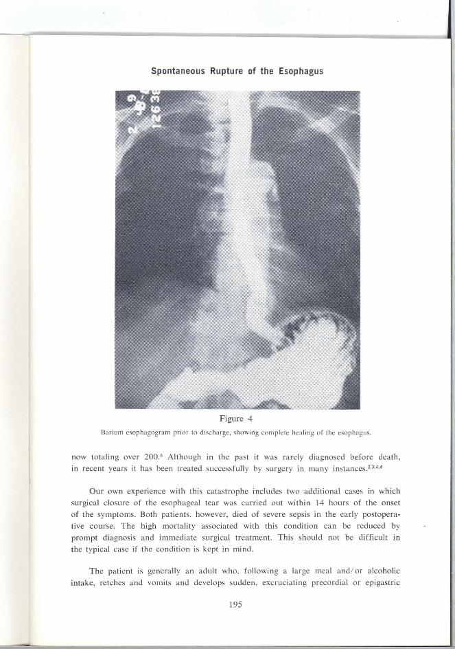
Barium esophagogram prior to discharge, showing complete healing of the esophagus.
now totaling over 200.' Although in the past it was rarely diagnosed before death,
in recent years it has been treated successfully by surgery in many instances.
Our own experience with this catastrophe includes two additional cases in which surgical closure of the esophageal tear was carried out within 14 hours of the onset of the symptoms. Both patients, however, died of severe sepsis in the early postoperative course. The high mortality associated with this condition can be reduced by prompt diagnosis and immediate surgical treatment. This should not be difficult in the typical case if the condition is kept in mind.
The patient is generally an adult who, following a large meal and/or alcoholic
intake, retches and vomits and develops sudden, excruciating precordial or epigastric
195
Arciniegas
pain. More rarely, as in our patient, there may be no history of overeating or drinking. Subcutaneous emphysema and hydrothorax or pneumothorax, or both, are almost invariably seen.
The tear involves all layers of the esophageal wall. When the esophageal mucosa alone is torn, massive bleeding may occur (as described originally by Mallory and Weiss'). In either instance experienced, the tear is caused by the sudden distention of the distal esophagus by a large bolus of fluid or food that is forcibly thrust into it from the stomach during a bout of violent vomiting. When the rupture is complete, it generally measures only a few centimeters. It is almost always located within a short distance from the esophagogastric junction, in the left side of the esophagus. This is thought to be an area of anatomical weakness.* The mediastinal pleura overlying the esophagus is also torn, thus establishing an esophago-pleural communication through which air, food and gastric juices are dumped into the pleural cavity and into the mediastinum. This gives rise to severe pleuritis and mediastinitis, loss of fluids and plasma into the chest, lung collapse, mediastinal shift and cardio-respiratory embarrassment.
The severity and location of the pain are suggestive of acute myocardial infarction and the pain and associated abdominal muscular spasm are consistent with perforated peptic ulcer. The condition may also mimic acute pancreatitis, acute cholecystitis and dissecting aortic aneurysm. However, when a typical history is obtained, the presence of subcutaneous emphysema on physical examination and mediastinal air on the chest fi lm make the diagnosis of rupture of the esophagus certain. No time should be wasted in other radiologic or laboratory procedures. A drink of a water-soluble radio-opaque material may be given to confirm the diagnosis and to determine the location of the tear. Barium should not be used, as its spillage will worsen the mediastinitis. Esophagoscopy is not ordinarily indicated.'
Once the diagnosis is made, immediate thoracotomy is mandatory as the mortality increases rapidly with each hour and anpproaches 75% within 36 hours." Primary two-layered closure of the esophagus is the treatment of choice and should be carried out whenever feasible as well as removal of the necrotic mediastinal pleura for ample mediastinal drainage, and tube drainage of the pleural cavity. The incidence of dehiscence of the esophageal repair is reported as high as 50%.^ Our case illustrates this problem. Under such circumstances, re-suture should not be attempted. Instead, the empyema should be drained and the tear allowed to heal while maintaining the patient's nutrition by means of a feeding jejunostomy.
Summary
A case is presented of spontaneous rupture of the esophagus that was managed successfully. The factors involved in the production of the lesion and its clinical and radiographic features are also discussed.
196
Spontaneous Rupture of the Esophagus
REFERENCES
1. Derbes, V. J., and Mitchell, R. E., Jr.: Hermann Boerhaave's Atrocis, nec Descripti Prius, Morbi Historia: the first translation of the classic case report of rupture of the esophagus, with annotations, Bull Med Libr Assoc 43:217-40, Apr 1955.
2. Barrett, N. R.: Report of a case ot spontaneous perforation of the oesophagus successfully treated by operation, Brit J Surg 35:216-8, Oct 1947.
3. Carter, J. P.; Alrich, E. M.; and Drash, E. C : Spontaneous rupture of the esophagus: report of two patients treated surgically with survival, Surgery 30:500-5, Sept 1951.
4. Derbes, V. J., and Mitchell, R. E., Jr.: Rupture of the esophagus. Surgery 39:688-709, Apr 1956: 39:865-88, May 1956.
5. Briggs, J. N.; Hamel, N. C: and Schulkins, T. A.: Spontaneous rupline of the esophagus, IVestern J Surg 69:351-4, Nov-Dec 1961.
6. Bowers, C. R., and Ferguson, D. H.: Rupture of the esophagus: emetic dehiscence of the normal esophagus: three new cases, Amer J Stirg 111:175-9, Feb 1966.
7. Weiss, S., and Mallory, G. K.: Lesions of the cardiac orifice of the stomach produced by vomiting, JAMA 98:1353-5, 16 Apr 1932.
8. Mackler, S. A.: Spontaneous rupture of the esophagus, Surg Gytiec Obstet 95:345-56, Sep 1952.
197