spine - american academy of orthopaedic surgeons · papers, posters & scientific exhibits spine...
TRANSCRIPT
PAPERS, POSTERS & SCIENTIFIC EXHIBITS SPINE
951 u The FDA has not cleared the drug and/or medical device for the use described in this presentation (i.e. the drug or medical device is being discussed for an off label use). For full information refer to page 14. An alphabetical faculty financial disclosure list can be found starting on page 19.
PAPERSPAPER NO. 76
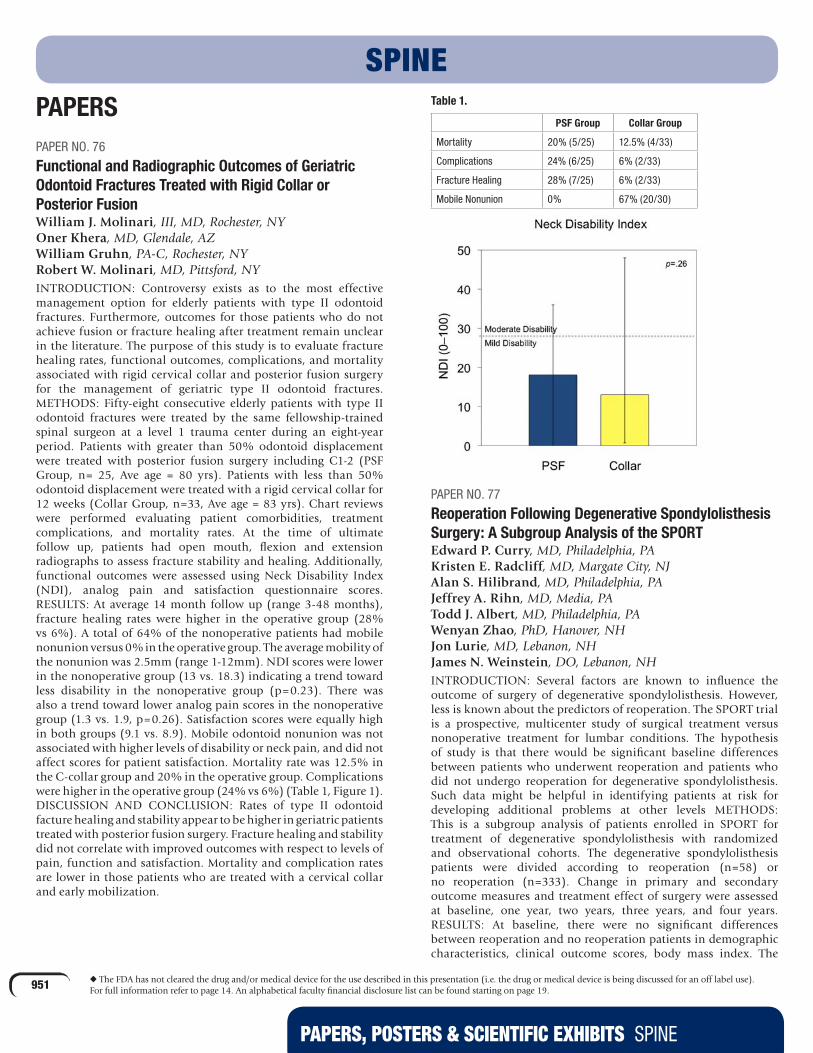
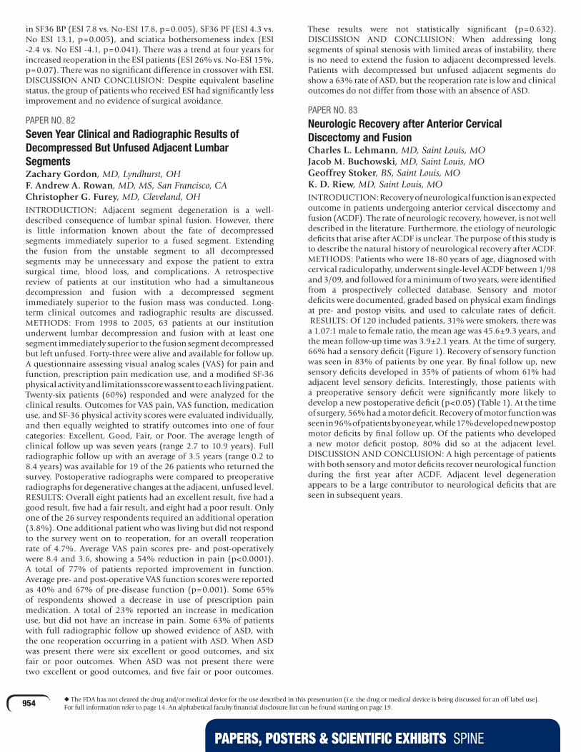
Functional and Radiographic Outcomes of Geriatric Odontoid Fractures Treated with Rigid Collar or Posterior FusionWilliam J. Molinari, III, MD, Rochester, NY Oner Khera, MD, Glendale, AZ William Gruhn, PA-C, Rochester, NY Robert W. Molinari, MD, Pittsford, NYINTRODUCTION: Controversy exists as to the most effective management option for elderly patients with type II odontoid fractures. Furthermore, outcomes for those patients who do not achieve fusion or fracture healing after treatment remain unclear in the literature. The purpose of this study is to evaluate fracture healing rates, functional outcomes, complications, and mortality associated with rigid cervical collar and posterior fusion surgery for the management of geriatric type II odontoid fractures.METHODS: Fifty-eight consecutive elderly patients with type II odontoid fractures were treated by the same fellowship-trained spinal surgeon at a level 1 trauma center during an eight-year period. Patients with greater than 50% odontoid displacement were treated with posterior fusion surgery including C1-2 (PSF Group, n= 25, Ave age = 80 yrs). Patients with less than 50% odontoid displacement were treated with a rigid cervical collar for 12 weeks (Collar Group, n=33, Ave age = 83 yrs). Chart reviews were performed evaluating patient comorbidities, treatment complications, and mortality rates. At the time of ultimate follow up, patients had open mouth, flexion and extension radiographs to assess fracture stability and healing. Additionally, functional outcomes were assessed using Neck Disability Index (NDI), analog pain and satisfaction questionnaire scores.RESULTS: At average 14 month follow up (range 3-48 months), fracture healing rates were higher in the operative group (28% vs 6%). A total of 64% of the nonoperative patients had mobile nonunion versus 0% in the operative group. The average mobility of the nonunion was 2.5mm (range 1-12mm). NDI scores were lower in the nonoperative group (13 vs. 18.3) indicating a trend toward less disability in the nonoperative group (p=0.23). There was also a trend toward lower analog pain scores in the nonoperative group (1.3 vs. 1.9, p=0.26). Satisfaction scores were equally high in both groups (9.1 vs. 8.9). Mobile odontoid nonunion was not associated with higher levels of disability or neck pain, and did not affect scores for patient satisfaction. Mortality rate was 12.5% in the C-collar group and 20% in the operative group. Complications were higher in the operative group (24% vs 6%) (Table 1, Figure 1).DISCUSSION AND CONCLUSION: Rates of type II odontoid facture healing and stability appear to be higher in geriatric patients treated with posterior fusion surgery. Fracture healing and stability did not correlate with improved outcomes with respect to levels of pain, function and satisfaction. Mortality and complication rates are lower in those patients who are treated with a cervical collar and early mobilization.
SPINETable 1.
PSF Group Collar Group
Mortality 20% (5/25) 12.5% (4/33)
Complications 24% (6/25) 6% (2/33)
Fracture Healing 28% (7/25) 6% (2/33)
Mobile Nonunion 0% 67% (20/30)
PAPER NO. 77
Reoperation Following Degenerative Spondylolisthesis Surgery: A Subgroup Analysis of the SPORTEdward P. Curry, MD, Philadelphia, PA Kristen E. Radcliff, MD, Margate City, NJ Alan S. Hilibrand, MD, Philadelphia, PA Jeffrey A. Rihn, MD, Media, PA Todd J. Albert, MD, Philadelphia, PA Wenyan Zhao, PhD, Hanover, NH Jon Lurie, MD, Lebanon, NH James N. Weinstein, DO, Lebanon, NHINTRODUCTION: Several factors are known to influence the outcome of surgery of degenerative spondylolisthesis. However, less is known about the predictors of reoperation. The SPORT trial is a prospective, multicenter study of surgical treatment versus nonoperative treatment for lumbar conditions. The hypothesis of study is that there would be significant baseline differences between patients who underwent reoperation and patients who did not undergo reoperation for degenerative spondylolisthesis. Such data might be helpful in identifying patients at risk for developing additional problems at other levels METHODS: This is a subgroup analysis of patients enrolled in SPORT for treatment of degenerative spondylolisthesis with randomized and observational cohorts. The degenerative spondylolisthesis patients were divided according to reoperation (n=58) or no reoperation (n=333). Change in primary and secondary outcome measures and treatment effect of surgery were assessed at baseline, one year, two years, three years, and four years.RESULTS: At baseline, there were no significant differences between reoperation and no reoperation patients in demographic characteristics, clinical outcome scores, body mass index. The

PAPERS, POSTERS & SCIENTIFIC EXHIBITS SPINE
952 u The FDA has not cleared the drug and/or medical device for the use described in this presentation (i.e. the drug or medical device is being discussed for an off label use). For full information refer to page 14. An alphabetical faculty financial disclosure list can be found starting on page 19.
reoperation patients had a significantly lower likelihood of having pseudoclaudication (74% vs. 88%, p=0.007) or asymmetric depressed reflexes (12% vs. 28%, p=0.015). There was no difference between reoperation and no-reoperation patients in type of procedure, levels of fusion, decompression level, or number of levels decompressed. There was a significantly increased percentage of complications of the index surgery in the reoperation patients. By the final four year follow up, there was no significant difference between reoperation and no reoperation groups in SF36 BP, SF36 PF, SF36 PCS, SF36 MCS, ODI, Sciatica Bothersomeness Index, Low Back Bothersomeness, Percent Satisfaction, Leg Pain Bothersomeness Index. The most common reasons for reoperation were recurrent stenosis/progressive spondylolisthesis (5%), new condition (2.4%), infection (2.4%), pseudoarthrosis/fusion exploration (1.1%). The overall rate of reoperation at four years was 14.8% for patients with degenerative spondylolisthesis in the SPORT.DISCUSSION AND CONCLUSION: The overall incidence of reoperation at four years was 14.8% for degenerative spondylolisthesis. Type of fusion, number of levels addressed, and instrumentation were not associated with a change in incidence of reoperation in patients with degenerative spondylolisthesis. Preoperative symptoms of pseudoclaudication and asymmetric depressed reflexes were associated with reduced risk of reoperation for degenerative spondylolisthesis. There was no significant difference in the outcome of patients who underwent reoperation for degenerative spondylolisthesis versus patients who did not undergo reoperation.
PAPER NO. 78
The Limited Benefit of Coronal Cobb Angle Correction in the Setting of Adult Spinal DeformityVirginie Lafage, PhD, New York, NY Frank J. Schwab, MD, New York, NY Benjamin Blondel, MD, New York, NY Justin S. Smith, MD, Charlottesville, VA Jason Demakakos Keith H. Bridwell, MD, Saint Louis, MO Steven D. Glassman, MD, Louisville, KY Christopher I. Shaffrey, MD, Charlottesville, VAINTRODUCTION: Adult spinal deformity (ASD) can cause pain and disability due to global malalignment. The extent of coronal plane deformity correction needed for clinical benefit remains controversial but Cobb angle has been described as a key parameter for evaluation of ASD patients. The aim of this study was to evaluate the amount of Cobb angle correction needed to achieve incremental clinical benefit and likelihood of reaching minimal clinically important difference (MCID) using health related quality of life (HRQL) scores.METHODS: Baseline and two year radiographic and HRQL data (ODI, SRS-22r and SF-12) were retrospectively analyzed for patients consecutively enrolled in a multi-center, prospective ASD database. Patients were divided into three groups based on postoperative Cobb angle improvement: 35°. Pre and post-op results for each group and changes in HRQL scores for each improvement subdivision were analyzed using paired t-test and one-way ANOVA.RESULTS: Sixty patients with thoraco-lumbar or lumbar curve >50° were included. A significant improvement for HRQOL scores was found between pre-op and last follow up data across the study population (p<0.05). A correction of <25° resulted in a significant improvement in HRQL scores (p35° also did not improve the likelihood to reach MCID for SRS pain but increased it for appearance (88%), activity (82%) and SF-12 PCS (82%).
DISCUSSION AND CONCLUSION: This prospective study offers clinically important data on the mixed benefit of limited coronal deformity correction in the setting of ASD. To improve patient reported pain, a Cobb correction of >25° but <35° appears sufficient with no added benefit to further deformity correction. Findings from this study add to the importance of pre-operative planning and patient counseling (risk vs. benefit) in terms of need for correction of coronal deformity.
PAPER NO. 79
A New Spine Surgery Specific Score to Predict Morbidity and Mortality in Elective Spine SurgeryAbhishek Manu, Coimbatore, India Ashok Thomas, Coimbatore, India Janardhan Yerramshetty, PhD, Coimbatore, India Ajoy P P. Shetty, Coimbatore, India S. Rajasekaran, PhD, Coimbatore, IndiaINTRODUCTION: Spine surgery is associated with significant postoperative complications which are probably caused by both magnitude of the surgery and associated patient comorbidities. Objectives of this study were to identify various pre-operative comorbidities and factors reflecting magnitude of surgeries that are associated with postoperative complications and to formulate a scoring system specific to spine surgery.METHODS: Pre-operative comorbidities and surgical factors (Table 1) along with mortality, major morbidity (life threatening complications that required intervention) and minor morbidity (mild and not life threatening complications) were documented in 1,217 elective spine surgeries for model development and testing. The maximum time period for assessing morbidity was one month. Using cross-tabulations and Chi-square analysis, groupings were identified for each significant factor based on major morbidity. Using risk stratification analysis, a weightage score was assigned to each group in all significant factors. Summation of all scores gave a final morbidity score for each patient. Using ROC and discriminant analyses, the developed score was tested to see whether it can do any better than chance at predicting morbidity and mortality in patients. The score was also validated on 200 randomly chosen patients from the same sample. Finally, the commonly used Charlson’s comorbidity index (CCI), an indicator of mortality, was also assessed in 665 patients and was compared to this study’s score.RESULTS: There were six deaths, 52 major morbidities and 142 minor morbidities. Chi-square analysis indicated that all individual factors had significant effect on post-operative major and minor morbidity. The ROC curves constructed from the final scores of all patients showed that the areas under the curves of Ganga spine scores for minor and major morbidities were 0.78 and 0.80 (nearly 80% chance that the score of a morbid case will be greater than no-morbid case) and significant (p<0.001), whereas, the CCI index produced an area of 0.64 (p<0.01). Based on discriminant analysis, an overall 74% of major morbid and 71% of minor morbid cases were correctly classified by Ganga score compared to 67% by CCI. Validation on randomly chosen 200 cases resulted in an overall correct classification of 82.8% major morbid and 74% of minor morbid cases indicating a stable and reliable model. Only age, ASA, BMI and ambulatory status came out as significant factors contributing to mortality. ROC analysis produced a significant curve area of 0.94 and an overall correct classification of 88% cases.DISCUSSION AND CONCLUSION: The study was able to determine important factors contributing to post-operative complications and was able to generate a score based on these factors, which performed better than CCI. While intra-operative factors emerged as predominant contributors to morbidity (Table

PAPERS, POSTERS & SCIENTIFIC EXHIBITS SPINE
953 u The FDA has not cleared the drug and/or medical device for the use described in this presentation (i.e. the drug or medical device is being discussed for an off label use). For full information refer to page 14. An alphabetical faculty financial disclosure list can be found starting on page 19.
1), pre-operative comorbidities emerged as significant factors in identifying mortality. Even though the number of deaths were much less (six cases), the model used for morbidity performed even better at discriminating mortality, which is also an important and required factor that can help a surgeon in making better and patient condition-based decisions.
Table 1: Strength of association (Kendall’s Tau-b) of individual factors with major & minor morbidity
VariableTau-b (major
morbidity)
Tau-b (minor
morbidity)
Critical Levels
Magnitude of surgery factors
Blood loss 0.21 0.32 > 1400 ml
Duration of surgery 0.18 0.26 > 240 min
Instrumentation 0.17 0.29 > 5 levels
Pre-operative factors
Age 0.15 0.11 > 70 years
ASA 0.14 0.13 > 3rd group
Ambulatory status 0.087 0.19 >= 3rd group
BMI 0.060 0.082 < 18.5
PAPER NO. 80
The Fate of Asymptomatic Cervical Stenosis: A Prospective Cohort StudyK. D. Riew, MD, Saint Louis, MO Michael P. Kelly, MD, St Louis, MO Moon Soo Park, MD, Anyang-Si, Republic of Korea Carie R. Kennedy, BS, RN, St Louis, MOINTRODUCTION: The natural history of asymptomatic patients who have cervical stenosis is not well-defined and the treatment of such patients remains controversial. As a result, there is variability in treatment recommendations for such patients. Some surgeons favor prophylactic decompression in order to prevent a catastrophic neurological injury, while others favor observation. Unfortunately, there are few long-term natural history studies that can provide a scientific rationale for treatment of such individuals. METHODS: We prospectively enrolled adult patients with spinal canal AP diameter measuring < 9mm on an MRI with no cord signal, no symptoms of myelopathy who were willing to participate in a long-term study with an endpoint of surgical intervention or neurologic injury. Exclusion criteria: Age over 80 years, pathologic or traumatic compression, instability requiring stabilization. Enrolled patients were recommended to have repeat physical exams and MRIs annually. Anyone who developed myelopathy, cord signal change or recalcitrant radiculopathy had operative intervention. Our null hypothesis is that a high percentage of asymptomatic persons with a spinal canal size ≤9 mm per MRI will become paralyzed or need operative intervention within a short period of time. RESULTS: We enrolled 73 patients. The space available for the spinal cord was between 3-9 mm. Follow up was between 1-12 years (mean 3.7 years; cumulative follow-up of 218.3 patient-years), with 48 patients having a minimum two-year follow-up (mean 4.6 years, 197.8 patient-years follow up). No patient became para or tetraplegic. The cumulative follow up of these patients represents 218.3 patient-years without irreversible neurologic deterioration. Only eight patients eventually required surgery (one arthroplasty, two laminaplasties, five anterior corpectomy/discectomies). Of these, three had myelopathy, two myeloradiculopathy and three radiculopathy. DISCUSSION AND CONCLUSION: Few prospective natural history studies with long-term follow up regarding the cervical spine exist. To our knowledge, this is the largest prospective
study with the longest follow up regarding the natural history of patients with cervical spinal stenosis without cord signal change or myelopathy. With over 218 patient-years of follow up, no one suffered a catastrophic neurologic event or even a minor neurologic deficit. This suggests that such events are relatively rare and that most patients can be managed safely with repeat exams, MRIs and education regarding the signs and symptoms of myelopathy. However, further follow up is needed to determine if such a regimen is safe long-term.
PAPER NO. 81
Do Epidural Steroid Injections Affect Outcomes for Lumbar Stenosis and Degenerative Spondylolisthesis Patients?Kristen E. Radcliff, MD, Margate City, NJ Christopher Kepler, MD, Philadelphia, PA Alan S. Hilibrand, MD, Philadelphia, PA Wenyan Zhao, PhD, Hanover, NH Jonathan Lurie, MD, Lebanon, NH Jeffrey A. Rihn, MD, Media, PA Todd J. Albert, MD, Philadelphia, PA James N. Weinstein, DO, Lebanon, NHINTRODUCTION: The SPORT trial is a prospective, multicenter study of surgical treatment versus nonoperative treatment for lumbar stenosis and degenerative spondylolisthesis. The hypothesis of this subgroup analysis was that patients who received epidural steroid injections (ESI) during treatment would have improved outcome and a lower rate of crossover to surgery compared to patients who did not receive ESI.METHODS: The study population includes patients enrolled in SPORT for treatment of spinal stenosis or degenerative spondylolisthesis who did not receive ESI prior to enrollment in the study. Patients who received epidural steroid injections during the first three months of the study were compared to patients who did not receive epidural injections during the first three months of the study (No ESI). Change in primary and secondary outcome measures and treatment effect of surgery were assessed at baseline, one year, two years, three years, and four years.RESULTS: In the spinal stenosis cohort, there were 69 ESI patients and 207 No-ESI patients. There were no significant differences in demographic factors, baseline clinical outcome scores, or operative details but there was an increased preference for nonsurgical treatment among the ESI patients (p<0.001). There was an average 26 minute increase in operative time and an increased length of stay by 0.9 days in the ESI patients. Averaged over four years, there was significantly less improvement in SF36 PF in surgically treated ESI patients (ESI 14.8 vs. No-ESI 22.5, p=0.025). Averaged over four years, there was significantly less improvement in SF36 BP (ESI 7.3 vs. No-ESI 16.7, p=0.007) and SF36 PF (ESI 5.5 vs. No-ESI 15.2, p=0.009). Of the spinal stenosis patients assigned to surgical treatment at enrollment, there was increased crossover associated with ESI to nonsurgical treatment (ESI 32% vs. No-ESI 11%). There was also increased crossover of spinal stenosis ESI patients assigned to nonsurgical treatment to surgery (ESI 58% vs. No-ESI 32%). In the spondylolisthesis cohort, there were 118 No-ESI patients and 45 ESI patients. There were significant differences in baseline SF36 BP, SF36 MCS, and leg pain bothersomeness index favoring No ESI patients. There were no other significant differences in baseline demographic factors or clinical outcome scores, but there was an increased preference for nonsurgical treatment among the ESI patients (p<0.001). Averaged over four years, there was significantly less improvement in the nonsurgically treated patients
PAPERS, POSTERS & SCIENTIFIC EXHIBITS SPINE
954 u The FDA has not cleared the drug and/or medical device for the use described in this presentation (i.e. the drug or medical device is being discussed for an off label use). For full information refer to page 14. An alphabetical faculty financial disclosure list can be found starting on page 19.
in SF36 BP (ESI 7.8 vs. No-ESI 17.8, p=0.005), SF36 PF (ESI 4.3 vs. No ESI 13.1, p=0.005), and sciatica bothersomeness index (ESI -2.4 vs. No ESI -4.1, p=0.041). There was a trend at four years for increased reoperation in the ESI patients (ESI 26% vs. No-ESI 15%, p=0.07). There was no significant difference in crossover with ESI.DISCUSSION AND CONCLUSION: Despite equivalent baseline status, the group of patients who received ESI had significantly less improvement and no evidence of surgical avoidance.
PAPER NO. 82
Seven Year Clinical and Radiographic Results of Decompressed But Unfused Adjacent Lumbar SegmentsZachary Gordon, MD, Lyndhurst, OH F. Andrew A. Rowan, MD, MS, San Francisco, CA Christopher G. Furey, MD, Cleveland, OHINTRODUCTION: Adjacent segment degeneration is a well-described consequence of lumbar spinal fusion. However, there is little information known about the fate of decompressed segments immediately superior to a fused segment. Extending the fusion from the unstable segment to all decompressed segments may be unnecessary and expose the patient to extra surgical time, blood loss, and complications. A retrospective review of patients at our institution who had a simultaneous decompression and fusion with a decompressed segment immediately superior to the fusion mass was conducted. Long-term clinical outcomes and radiographic results are discussed.METHODS: From 1998 to 2005, 63 patients at our institution underwent lumbar decompression and fusion with at least one segment immediately superior to the fusion segment decompressed but left unfused. Forty-three were alive and available for follow up. A questionnaire assessing visual analog scales (VAS) for pain and function, prescription pain medication use, and a modified SF-36 physical activity and limitations score was sent to each living patient. Twenty-six patients (60%) responded and were analyzed for the clinical results. Outcomes for VAS pain, VAS function, medication use, and SF-36 physical activity scores were evaluated individually, and then equally weighted to stratify outcomes into one of four categories: Excellent, Good, Fair, or Poor. The average length of clinical follow up was seven years (range 2.7 to 10.9 years). Full radiographic follow up with an average of 3.5 years (range 0.2 to 8.4 years) was available for 19 of the 26 patients who returned the survey. Postoperative radiographs were compared to preoperative radiographs for degenerative changes at the adjacent, unfused level.RESULTS: Overall eight patients had an excellent result, five had a good result, five had a fair result, and eight had a poor result. Only one of the 26 survey respondents required an additional operation (3.8%). One additional patient who was living but did not respond to the survey went on to reoperation, for an overall reoperation rate of 4.7%. Average VAS pain scores pre- and post-operatively were 8.4 and 3.6, showing a 54% reduction in pain (p<0.0001). A total of 77% of patients reported improvement in function. Average pre- and post-operative VAS function scores were reported as 40% and 67% of pre-disease function (p=0.001). Some 65% of respondents showed a decrease in use of prescription pain medication. A total of 23% reported an increase in medication use, but did not have an increase in pain. Some 63% of patients with full radiographic follow up showed evidence of ASD, with the one reoperation occurring in a patient with ASD. When ASD was present there were six excellent or good outcomes, and six fair or poor outcomes. When ASD was not present there were two excellent or good outcomes, and five fair or poor outcomes.
These results were not statistically significant (p=0.632).DISCUSSION AND CONCLUSION: When addressing long segments of spinal stenosis with limited areas of instability, there is no need to extend the fusion to adjacent decompressed levels. Patients with decompressed but unfused adjacent segments do show a 63% rate of ASD, but the reoperation rate is low and clinical outcomes do not differ from those with an absence of ASD.
PAPER NO. 83
Neurologic Recovery after Anterior Cervical Discectomy and FusionCharles L. Lehmann, MD, Saint Louis, MO Jacob M. Buchowski, MD, Saint Louis, MO Geoffrey Stoker, BS, Saint Louis, MO K. D. Riew, MD, Saint Louis, MOINTRODUCTION: Recovery of neurological function is an expected outcome in patients undergoing anterior cervical discectomy and fusion (ACDF). The rate of neurologic recovery, however, is not well described in the literature. Furthermore, the etiology of neurologic deficits that arise after ACDF is unclear. The purpose of this study is to describe the natural history of neurological recovery after ACDF.METHODS: Patients who were 18-80 years of age, diagnosed with cervical radiculopathy, underwent single-level ACDF between 1/98 and 3/09, and followed for a minimum of two years, were identified from a prospectively collected database. Sensory and motor deficits were documented, graded based on physical exam findings at pre- and postop visits, and used to calculate rates of deficit. RESULTS: Of 120 included patients, 31% were smokers, there was a 1.07:1 male to female ratio, the mean age was 45.6±9.3 years, and the mean follow-up time was 3.9±2.1 years. At the time of surgery, 66% had a sensory deficit (Figure 1). Recovery of sensory function was seen in 83% of patients by one year. By final follow up, new sensory deficits developed in 35% of patients of whom 61% had adjacent level sensory deficits. Interestingly, those patients with a preoperative sensory deficit were significantly more likely to develop a new postoperative deficit (p<0.05) (Table 1). At the time of surgery, 56% had a motor deficit. Recovery of motor function was seen in 96% of patients by one year, while 17% developed new postop motor deficits by final follow up. Of the patients who developed a new motor deficit postop, 80% did so at the adjacent level.DISCUSSION AND CONCLUSION: A high percentage of patients with both sensory and motor deficits recover neurological function during the first year after ACDF. Adjacent level degeneration appears to be a large contributor to neurological deficits that are seen in subsequent years.

PAPERS, POSTERS & SCIENTIFIC EXHIBITS SPINE
955 u The FDA has not cleared the drug and/or medical device for the use described in this presentation (i.e. the drug or medical device is being discussed for an off label use). For full information refer to page 14. An alphabetical faculty financial disclosure list can be found starting on page 19.
PAPER NO. 84
The Role of the Progressive Ankylosis Protein (ANK) in Spine FusionMartin Quirno, MD, New York, NY Kirk A. Campbell, MD, New York, NY Andrew Yoo, BA, New York, NY Thomas J. Errico, MD, New York, NY Thorsten Kirsch, PhD, New York, NYINTRODUCTION: Spine fusion follows a well-orchestrated cascade of cellular events that when altered, such as in ankylosing spondylitis, results in a significant increase in trabecular bone formation. Increased bone formation ultimately resulting in spontaneous spine fusion in patients with ankylosis spondylitis is caused by chronic inflammation. Chronic inflammation can
lead to increased recruitment of osseous precursor cells and the differentiation of these cells along the osteogenic lineage. However, the mechanisms of how inflammation stimulates bone formation during spine fusion are not well understood. A better understanding of these mechanisms may lead to the development of novel therapeutic strategies for the treatment of ankylosis spondylitis or the improvement of bone formation during spine fusion. The progressive ankylosis protein (ANK) transports intracellular pyrophosphate to the extracellular milieu. Lack of ANK function in ank/ank mice results in an ankylosis spondylitis phenotype. We hypothesized that the ank/ank mouse model represents a new animal model for the understanding of the mechanisms of how inflammation stimulates bone formation and ultimately spine fusion.METHODS: We utilized a well-established spinal fusion model using iliac crest allograft in six-week-old ANK-deficient (ank/ank) mice and wild type (WT) littermates. Mice were followed with weekly x-rays and groups were euthanized at two and five weeks (n = 5 in each group). Bone fusion including the size and levels fused was analyzed.RESULTS: Clinically the ank/ank mice were less mobile and had a prominent kyphosis, especially after five weeks from surgery. Unlike the WT mice, ank/ank mice were unable to stand on their hind limbs. Radiographically the ank/ank mice showed a marked increase in trabecular bone formation from L2-L5 when compared to WT littermates after two weeks. After five weeks the iliac crest allograft was no longer distinguished in the ank/ank mice and a large trabecular bone mass covering at least three levels was evident (Fig. 1A). The fusion mass in the WT mice was markedly smaller (Fig. 1B).DISCUSSION AND CONCLUSION: In this study we show for the first time that ANK plays a crucial role in spine fusion. Lack of ANK resulted in a larger and more robust spine fusion extending more levels. Despite our previous findings showing that the lack of ANK function delays the differentiation of early osteoblasts into mature osteoblasts causing an osteoporotic bone phenotype in ank/ank mice, bone formation during spine fusion was increased in ank/ank mice similar to patients with ankylosis spondylitis. These findings suggest that an abnormal/chronic inflammatory response in these mice results in increased bone formation during spine fusion. Therefore, the ank/ank mice provide a novel animal model to study the mechanisms of how inflammatory responses cause increased bone formation during spine fusion. The understanding of these mechanisms may provide insights into novel therapeutic strategies for the treatment of ankylosis spondylitis or the improvement of bone formation during spine fusion.

PAPERS, POSTERS & SCIENTIFIC EXHIBITS SPINE
956 u The FDA has not cleared the drug and/or medical device for the use described in this presentation (i.e. the drug or medical device is being discussed for an off label use). For full information refer to page 14. An alphabetical faculty financial disclosure list can be found starting on page 19.
PAPER NO. 85
Cervical Spondylotic Mielopathy - Minimal 10 Years Follow Up StudyManuel Ribeiro Da Silva, Porto, Portugal Nuno Neves, MD, Porto, Portugal Rui Matos, MD, Porto, Portugal Pedro C. Rodrigues, Porto, Portugal Joana Freitas, MD, Gaia, Portugal Manuel Santos Carvalho, Porto, Portugal Artur Antunes, MD, Vila Nova De Gaia, Portugal Rui A. Pinto, MD, Porto, PortugalINTRODUCTION: Cervical spondylotic mielopathy (CSM) is the most common cause of spinal cord dysfunction in the adult population. Treatment implies surgical decompression as soon as possible after the diagnosis. In this study the authors present the long term results of minimal 10 years follow up study of 99 patients that underwent anterior decompression and arthrodesis surgery for CSM.METHODS: Patients that underwent surgery for CSM between January 1990 and December 1994 were evaluated for sex, age, number of levels operated, functional evaluation with Nurick Scale pre operatively, one year after surgery and at the final the revision that took place in 2007 and 2008, evidence of consolidation and complications. All the patients were operated by anterior approach. T-Student Test was performed with SPSS for statistical analysis.RESULTS: Ninety-nine patients were evaluated during the study, 73 male, 26 female, with a mean age of 56, six years (42-86) and mean follow up time of 14,4 years. Three patients died in the immediate post op period, one in the first year, eight during the 15 year evaluation period. Sixteen patients were operated for one level, 22 for two levels, 36 for three levels and 22 for four levels (mean on 2,7±1,0 levels for patient). Pre op Nurick was 3,8±0,9. There was a significant improvement in neurological condition after one year surgery (Nurick 2,2±1,1; p<0,001), and between pre op and final evaluation (2,3±1,2; p<0,001). The degradation between the first year and the final evaluation was statistically significant (p=0,004). There was a strong correlation between age and the number of operated levels (r=0,391, p=0,01), age and initial neurologic status (r=0,238, p=0,05), initial neurological status and number of operated levels (r=0,251, p=0,05) and sex and number of operated levels, with women being operated on for more levels (r=0,208, p=0,05). There was also e stronger neurological deterioration between year one and year 15 in young patients when compared to older ones (r=0,250, p=0,05). There is a strong clinical relation between first year recuperation and final recuperation (r=0,838, p=0,01). There was a 100% rate of consolidation.DISCUSSION AND CONCLUSION: Surgical treatment for decompression and arthrodesis is considered the best option for the treatment of CSM in terms of improvement of pain, alignment and neurological function. A significant neurological improvement comes from surgery, and despite a significant clinical deterioration between the first year and the final evaluation, the benefits of surgery are still evident 15 years after, with a better neurological status when compared to the pre operative period.
PAPER NO. 86
Congenital Stenosis and Symptomatic Adjacent Segment Disease in the Cervical SpineJason Eubanks, MD, Willoughby Hills, OH Jon Belding, MD, MS, BA, Cleveland, OH Andrew A. Rowan, MD, MS, San Francisco, CA Gable B. Moffitt, BS, Cleveland Heights, OH Vinay Cheruvu, Kent, OH Justin Hohl, MD, Sandy, UT Alan S. Hilibrand, MD, Philadelphia, PA Henry H. Bohlman, MD, Cleveland, OH James Kang, MD, Pittsburgh, PAINTRODUCTION: Symptomatic adjacent segment disease (ASD) after anterior cervical arthrodesis can be seen in up to 25% of patients at 10 year follow up. Some debate exists as to whether this degeneration represents the natural history of the adjacent disc or whether the increased biomechanical stresses placed by the fusion accelerate this degenerative cascade. Congenital stenosis has been established as an important risk factor in the development of myelopathy. Further, MRI studies have suggested that congenitally stenotic spines experience greater pathological changes in the intervertebral discs and ensuing cord compression than spines with a normal canal diameter. The current study hypothesized that patients with congenital stenosis would have an increased prevalence of symptomatic adjacent segment disease after anterior arthrodesis than patients with normal canal diameters.METHODS: A retrospective review was performed on 497 patients undergoing a one to four level anterior cervical decompression and fusion by a single surgeon. Radiographs were evaluated for bony congenital stenosis by measuring the space available for the cord (SAC) and the Pavlov Ratio (PAV) using the stenosis parameters described by Kang et al. Radiographic ASD was measured according to the criteria established by Hilibrand et al. and correlated with clinically symptomatic ASD evaluated through chart review. Clinical outcome scores were graded on the Robinson and Odom criteria. Statistical analysis was performed using student t-tests and a linear regression model comparing symptomatic adjacent segment disease among patients with and without congenital stenosis.RESULTS: Congenital stenosis was observed in 87 (17.5%) patients. There were 239 men and 255 women in the study cohort. The average length of follow up was 46.6 months. There were 227 single level fusions (8 C3/C4, 22 C4/C5, 110 C5/C6, 47 C6/C7), 155 two level fusions (18 C3-C5, 53 C4-C6, 84 C5-C7), 84 three level fusions (23 C3-C6, 61 C4-C7), 26 four level fusions (C3-C7) and a smattering of non-contiguous, multi-level fusions. In the 87 patients with congenital stenosis, 35 had stenosis at C6 or C7. Pan-cervical stenosis (four or more levels) was apparent in 27 patients. Of the 497 patients, 188 (37.5%) developed ASD. Neither age (p=0.78), nor gender (p=0.86), nor congenital stenosis (p=0.62) correlated with the presence of clinically symptomatic ASD. Overall, clinical results demonstrated excellent or good Robinson scores in 86.7% of patients, whereas excellent or good Odom scores were reported in 90.1% of patients.DISCUSSION AND CONCLUSION: Congenital stenosis appears in 17.5% of patients undergoing anterior cervical decompression and fusion. Despite a predominance of excellent to good surgical outcomes, symptomatic ASD is common, occurring in 37.5% of patients at a mean follow up of 47 months. Contrary to our hypothesis, bony congenital stenosis does not appear to be a predictor of symptomatic ASD. ASD may represent more the natural history of the degenerating disc rather than the end product of underlying biomechanics in the congenitally stenotic cervical

PAPERS, POSTERS & SCIENTIFIC EXHIBITS SPINE
957 u The FDA has not cleared the drug and/or medical device for the use described in this presentation (i.e. the drug or medical device is being discussed for an off label use). For full information refer to page 14. An alphabetical faculty financial disclosure list can be found starting on page 19.
canal or the change in forces created by surgical arthrodesis.
PAPER NO. 87
Does Opioid Pain Medication Use Affect the Outcome of Patients with Lumbar Disk Herniation?Roman Isaac, MD, Philadelphia, PA Kristen E. Radcliff, MD, Margate City, NJ Alan S. Hilibrand, MD, Philadelphia, PA Todd J. Albert, MD, Philadelphia, PAINTRODUCTION: The SPORT trial is a prospective, multicenter study of surgical treatment versus nonoperative treatment for lumbar intervertebral disk herniation (IDH). The purpose of this study was to review the results of patients who received opioid pain medications during treatment compared to patients who did not receive opioid medications.METHODSThe study population includes patients enrolled in SPORT for treatment of IDH in combined randomized and observational cohorts. Patients who were receiving opioid pain medications at baseline (Opioid) were compared to those who were not (No-Opoid). Outcome measures were assessed at baseline, one year, two years, three years, and four years. The difference in improvement between surgical and nonoperative treatment (treatment effect) was determined at each follow period for each group.RESULTS: There were 520 patients in the Non-Opioid group and 542 patients in the Opioid group. At baseline, there was a significantly higher percentage of patients in the opioid medication group (p<0.05) on disability, with compensation claims, and actively smoking. Among the opioid medication group there were significantly (p<0.001) worse baseline scores for SF36 BP, SF36 PF, SF36 MCS, SF36 PCS, ODI, Sciatica Frequency Index, Sciatica Bothersomeness Index, Back Pain Bothersomeness Index, and percent dissatisfaction with current treatment. There was an increased percentage of patients in the opioid medication group with the perception of worsening symptoms (p<0.001). There was a statistically significant increase incidence of any neurological deficit (p<0.001), motor weakness (p=0.008), decreased sensation (p<0.001), and received surgery (p<0.001) in the opioid medication group. At four years follow up, there were no significant differences in primary or secondary outcome measures or treatment effect of surgery between opioid and non-opioid medication patients. There was significantly less crossover to nonsurgical treatment in the opioid patients versus the non-opioid patients (11% vs. 19%, p=0.0108). There was significantly increased crossover to surgery in the opioid pain medication patients (45% versus 31%, p=0.0045).DISCUSSION AND CONCLUSION: Despite treatment with stronger pain medications, patients who were treated with opioids had significantly worse baseline pain and quality of life. At final follow up, there was no long term improvement in outcome associated with opioid pain medication use. Opioid medication use was associated with increased crossover to surgery. FDA Device Status: No off label usage is discussed.
PAPER NO. 88
Direct Reduction and Transforaminal Lumbar Interbody Fusion for High Grade Isthmic SpondylolisthesisWael Koptan, MD, Cairo, Egypt Fady S. Sedra, MSc, MBBS, Hounslow, United Kingdom Yasser H. El Miligui, MD, FRCS, Cairo, Egypt Mohammad M. El-Sharkawi, MD, Assiut, EgyptINTRODUCTION: Several controversies exist over the most appropriate approach for managing high grade spondylolisthesis.
The classic interbody fusions are associated with a considerable degree of complications. The aim of this work is to determine the safety and efficacy of unilateral transforaminal lumbar interbody fusion (TLIF) in managing high grade isthmic spondylolisthesis.METHODS: The study was conducted between 2000 and 2008 and included 44 patients with high grade isthmic spondylolisthesis (Meyerding grades III and IV). The mean age was 24y (range 17 - 38y). All patients had severe back and radicular symptoms that failed to conservative treatment. Eighteen were at L4/5 and 26 at L5/S1. Limited decompression, pedicle screw instrumentation were performed; 21 had additional unilateral TLIF and direct reduction (Group 1) and 23 had an indirect reduction and posterolateral fusion using autograft bone (Group 2). Patients were followed up for an average of 4.5y (range 3 - 7y).RESULTS: Between Groups 1 and 2, Group 1 had significantly better ODI improvement (averaged 63% and 58% respectively); VAS improved significantly more (averaged 93% and 89% respectively); better anterolisthesis correction (averaged 56% and 48% respectively); better improvement in disc space height (averaged 46% and 37% respectively). None in Group 1 had an implant failure and its overall fusion rate was 94%; Group 2 had two patients with implant failure requiring revision and an overall fusion rate of 86%. Both groups had a similar complication rate of 4% including transient foot drop (one patient in each group) that spontaneously recovered.DISCUSSION AND CONCLUSION: Direct instrumented reduction and TLIF is an efficient option to treat high grade isthmic spondylolisthesis. It provided immediate stability and better clinical and radiological outcomes.
PAPER NO. 89
Early Diagnosis of Lumbar Spondylolysis in Young Athletes Using Magnetic Resonance ImagingAtsushi Kobayashi, MD, Maebashi Gunma, Japan Tsutomu Kobayashi, MD, Gunma, Japan Hiroshi Higuchi, MD, Maebashi-Shi, Japan Kazuo KATOU, MD, Gunma, JapanINTRODUCTION: Lumbar spondylolysis is a defect of the pars interarticularis and is considered to be a stress fracture. Active spondylolysis is a common cause of low back pain in young athletes. The early stage of spondylolysis is very difficult to diagnose using plain radiographs, and they often do not visualize defects seen on computed tomography (CT) scans. In a recent study, the usefulness of signal changes of the pedicle on magnetic resonance imaging (MRI) for the early diagnosis of spondylolysis was evaluated. However, only a few reports indicated the onset frequency of spondylolysis with MRI in cases without spondylolysis using plain radiographs. In addition, there are no validated examination findings for active spondylolysis. The purpose of this study was to evaluate the usefulness of MRI for the early diagnosis of active spondylolysis and frequency of spondylolysis in cases without spondylolysis using plain radiographs, and to evaluate whether there is any clinical assessment test assisting in the early detection of active spondylolysis.METHODS: A prospective cohort design was employed. We investigated 200 patients who presented to our clinic because of low back pain between May 2009 and May 2011. The inclusion criteria in this study were active in sports, and no or unclear spondylolysis on plain radiographs. There were 144 male and 56 female patients. The mean age was 14.1 years (range, 10-18 years). All were examined using plain radiographs and MRI. Only patients who showed a stress reaction on MRI were examined by CT. Stress reactions were diagnosed when there was a high signal intensity in the region of

PAPERS, POSTERS & SCIENTIFIC EXHIBITS SPINE
958 u The FDA has not cleared the drug and/or medical device for the use described in this presentation (i.e. the drug or medical device is being discussed for an off label use). For full information refer to page 14. An alphabetical faculty financial disclosure list can be found starting on page 19.
the pars interarticularis on short tau inversion (STIR) axial and sagittal images on MRI. We assessed the presence or absence of low back pain at the time of lumbar spine extension, flexion, knock, or the right/left kemp test as physical examination findings on the initial consultation of each case. For statistical analysis, Mann-Whitney’s U test and logistic regression analysis were used.RESULTS: Because the right and left pars interarticularis were studied separately, 129 pars interarticularis in 97 subjects (48.5%) showed evidence of active spondylosis as defined by MRI. CT was performed in 92 subjects. Based on CT images, these pars defects were organized into various categories as follows: 52 non-lysis, 37 prefissure, 22 fissure, 10 progressive, and no pseudoarthrosis. The affected vertebral level was L3 in seven patients, L4 in 29 patients, and L5 in 61 patients. We could not identify a significant factor for physical examination findings assisting in the early detection of active spondylosis. In addition, a case showing pain at the time of lumbar spine extension described as diagnostic for lumbar spondylolysis was noted in a group without (90.3%) and in a group with (88.7%) spondylolysis, and both frequencies were high.DISCUSSION AND CONCLUSION: The progression of active spondylolysis to pseudoarthrosis has been associated with an increased incidence of spondylolisthesis. Moreover, the early detection and treatment of acute spondylolysis are associated with improved fracture healing and are important in preventing progression to established pseudoarthrosis. These results suggest that MRI is useful for the early diagnosis of active spondylolysis, and there is high rate of active spondylolysis based on MRI in young athletes with low back pain in cases without spondylolysis using plain radiographs. Because there was no significant factor for physical examination findings in assisting in the early detection of active spondylosis, even if spondylolysis cannot be detected on plain radiographs, it is considered that MRI should be employed for young athletes with low back pain.
PAPER NO. 90
Evaluation of a Center of Excellence Program for Spine SurgeryNelson F. SooHoo, MD, Los Angeles, CA Ateev Mehrotra, MD, Pittsburgh, PA Elizabeth Sloss, PhD, Arlington, VA Peter Hussey, Arlington, VA Susan Lovejoy, Pittsburgh, PAINTRODUCTION: Medicare and many private health plans are encouraging patients to seek care at hospitals which are designated as centers of excellence. Few evaluations of whether the quality of care at hospitals designated as centers of excellence is better have been conducted. The Blue Cross Blue Shield Association, whose member plans insure one in three Americans, has established an initiative to designate hospitals as centers of excellence for spine surgery. The objective of our study was to test the hypothesis that hospitals designated as a spine surgery center of excellence would provide higher-quality care as demonstrated by a lower rate of complications and readmissions and, therefore, lower costs.METHODS: Claims from approximately 54 million enrollees were retrospectively analyzed. We identified individuals in 2007-9 who underwent one of three types of spine surgery, one or two level cervical fusion, one or two level lumbar fusion, or lumbar discectomy and/or decompression. Our main outcomes were the composite rate of complication, rates of readmission within 30 days of discharge, and costs within 90 days following the procedure.RESULTS: In our sample 29,775 patients had a one or two level cervical fusion, 27,689 patients had a one or two level lumbar fusion, and 29,338 patients had a lumbar discectomy and/or
decompression. Of the three types of surgery, 33.8%, 33.3%, and 40.2%, respectively, were performed at a hospital designated by Blue Cross/Blue Shield as a center of excellence for spine surgery. Designated spine surgery hospitals had a larger number of beds and were more likely to be an academic center. There were no significant differences in the risk of complications between patients having cervical fusion, lumbar fusion, or lumbar discectomy/decompression at a designated hospital and patients having the same type of spine surgery at one of the other hospitals. There were also no significant differences in 90-day index hospitalization costs.DISCUSSION AND CONCLUSION: On average, hospitals designated as spine surgery centers of excellence in this program had similar rates of complications, readmissions, and costs to other hospitals. Our results emphasize that it is important to empirically evaluate whether centers of excellence provide higher-quality care.
PAPER NO. 286
Ferromagnetic Interactions and Radiofrequency Induced Heating of Small Arms Ballistics in MRI (1.5T,3T,7T)Russell D. Dedini, MD, San Francisco, CA Robert T. McClellan, MD, San Francisco, CA Murat Pekmezci, MD, San Francisco, CAINTRODUCTION: This study was designed to test the safety of retained bullets in magnetic resonance imaging (MRI) scanners. Although limited prior studies suggest retained bullets are generally safe in 1.5 tesla (T) scanners it is not known if they are safe in increasingly common 3T and 7T MRI scanners. The purpose of this study is to analyze the motion as well as heat generation by retained bullets in different magnetic field strengths.METHODS: Thirty-nine commercially available bullets and shotgun pellets were tested in 1.5, 3.0, and 7 Tesla MRI scanners. The parameters that were measured include the ex-vivo translational force, rotational force (RF) and RF induced heating. Translational force in dynes was calculated using a previously described deflection angle technique and the equation F=mg*sinØ/cosØ where “F” is the translational force, “m” is the mass of the object in grams, “g” is the gravitational acceleration constant (980 cm/sec2) and “Ø” is the deflection angle from the vertical measured on a protractor. Qualitative assessment of rotational torque was made using a previously described 0-4 point scale to describe the force and speed with which each sample aligned its long axis within the static magnetic field. RF-induced heating was measured using a custom temperature probe and Fast Spin Echo T2 weighted sequences designed to simulate worst-case scenario exposure to the FDA-approved limit of radiofrequency induced excitation (3.2 watts/kilogram specific absorption rate).RESULTS: Of 33 bullets and six shotgun pellets tested, only a single armor-piercing bullet manufactured with a hardened steel core and two steel pellets were found to be ferromagnetic, with translational forces in excess of that considered safe in all three magnetic field strengths. Only a single armor-piercing bullet demonstrated unsafe rotational torque. RF-induced heating of 0.6°C was measured during the T2-weighted worst-case sequence.DISCUSSION AND CONCLUSION: Common, commercially available bullets manufactured in the United States are safe in MRI scanners up to 7 Tesla in strength. In contrast, armor-piercing bullets with steel cores and stainless steel shotgun pellets are not safe. While RF-induced heating occurs, it is minimal, and likely clinically insignificant. These findings suggest MRI should not be used when bullets known to contain steel are located very near to critical anatomic structures and underscores the importance of

PAPERS, POSTERS & SCIENTIFIC EXHIBITS SPINE
959 u The FDA has not cleared the drug and/or medical device for the use described in this presentation (i.e. the drug or medical device is being discussed for an off label use). For full information refer to page 14. An alphabetical faculty financial disclosure list can be found starting on page 19.
obtaining forensic information on retained projectiles whenever possible.
PAPER NO. 287
Complications After Anterior Lumbar Spine Exposure by a Spine Surgeon With and Without an Access SurgeonMicah Smith, MD, Redwood City, CA Kevin A. Rahn, MD, Fort Wayne, IN Robert M. Shugart, MD, Fort Wayne, IN Christopher D. Belschner, PA-C, Fort Wayne, IN Kary Stout, BS, Fort Wayne, IN Ivan Cheng, MD, Redwood City, CAINTRODUCTION: The anterior approach to the lumbar spine continues to be a useful technique for many pathologies. It seems, however, that anterior lumbar surgery often places a spine surgeon in an anatomic territory with which they are unfamiliar. Many spine surgeons utilize the service of a general surgeon or vascular surgeon to perform the surgical exposure. A review of the English-language literature reveals a lack of direct comparison of anterior spine surgery performed with and without the assistance of an access surgeon. Purpose: To compare perioperative parameters and complications in anterior lumbar spine surgery with the exposure performed by a spine surgeon versus an access surgeon. Study Design/Setting: A retrospective cohort study of consecutive patients. Patient Sample: 96 consecutive patients who underwent anterior lumbar spine surgery between L3-S1 by two spine surgeons. Outcome Measures: Estimated blood loss, operative time, length of hospital stay and complications METHODS: A retrospective review was completed on 96 consecutive patients who underwent anterior spine surgery between levels L3 and S1 from 1995 to 2008. Patient and surgery characteristics including demographics, comorbidities, perioperative parameters and complications were noted. In the first 56 consecutive patients, a general surgeon completed the exposure with an additional patient who later had the exposure performed by a general surgeon due to extensive prior abdominal surgeries. In the next 39 patients, the orthopaedic surgeon completed the exposure.RESULTSThe group without the assistance of an access surgeon for the exposure had statistically lower values with respect to estimated blood loss (204mL vs 420mL, p=0.0007), operative time (2.8 vs 3.9 hours, p=0.0003) and length of hospital stay (3.5 vs 4.7 days, p=0.0006). In the “with assistance” group, 82% of patients (47/57) experienced a complication versus 28% (11/39) in the “without assistance” group (p<0.000001). Major complications noted in the “with assistance” group included deep venous thrombus (one), retrograde ejaculation (two), iliac vein bleeding requiring repair (one) and dyspareunia (one). The most common minor complication was an ileus that occurred in 33 (58%) of the patients in the “with assistance” group and in one patient (2.6%) in the “without assistance” group.
Table. Post-operative complications comparing surgery with an access surgeon versus without.
Post-operative ComplicationWith Assistance
n (%)Without
Assistance n (%)
Ileus 33 (58%) 1 (2.6%)
Deep venous thrombus 1 (1.8%) 0
Retrograde Ejaculation 2 (3.5%) 0
Iliac Vein Bleeding 1 (1.8%) 0
Peritoneal rent requiring repair 2 (3.5%) 0
Dyspareunia 1 (1.8%) 0
Incisional Hernia 1 (1.8%) 0
Scrotal/Penile Swelling 1 (1.8%) 0
DISCUSSION AND CONCLUSION: The approach for anterior lumbar spine surgery is safe, and a trained spine surgeon who is knowledgeable and familiar with the anatomy can safely perform the exposure without the assistance of an access surgeon.
PAPER NO. 288
Coumadin is Associated with Increased Blood Loss and Transfusion in Lumbar Surgery Despite Preoperative CorrectionErnest Young, MS, Cleveland Heights, OH Kasra Ahmadinia, MD, Cleveland, OH Nicholas U. Ahn, MD, Shaker Heights, OHINTRODUCTION: Blood loss in lumbar surgery is an established concern. Previous studies have demonstrated that blood loss is increased with addition of levels decompressed and/or fused, use of instrumentation, male sex and advanced age. Prior studies have demonstrated that prior use of anticoagulants, even when stopped, can lead to increased surgical blood loss in orthopaedic procedures; however, the relationship has not been evaluated in spine surgery. METHODS: A total of 256 consecutive patients who underwent lumbar decompression were retrospectively divided into two groups including patients who were taking coumadin prior to surgery, and a control group with no anticoagulation history. We verified that patients in the anticoagulation group were reversed prior to surgery in accordance with accepted guidelines: INR < 1.5 and greater than one week off of anticoagulants. Operative and hospital records were obtained to determine intraoperative blood loss (EBL), length of hospital stay and postoperative transfusion requirements. RESULTS: Linear regression was used to evaluate the effect of pre-operative anticoagulation use on EBL as well as hospital stay. Confounding factors, such as age, sex, race, number of levels decompressed and number of co-morbidities were corrected for. A pre-operative history of coumadin led to a significant increase in blood loss (200cc/level; p = 0.01), transfusion requirements (0.21units/level; p = 0.05) and hospital stay (0.43days/level; p = 0.02). DISCUSSION AND CONCLUSION: Our results indicate that even with adequate reversal, patients on anticoagulants prior to lumbar surgery have increased blood loss, tranfusion requirements and hospital stay. This is particularly important given the increasing number of anticoagulated patients. The results of this study will allow the surgeon to better counsel patients regarding blood loss and will allow the surgeon to be better prepared for blood loss in patients with a history of anticoagulation use.
PAPER NO. 289
Timing of Thromboembolic Chemoprophylaxis in Postoperative Spinal Trauma PatientsLloydine Jacobs, MD, Pittsburgh, PA Justin Hohl, MD, Sandy, UT Joon Y. Lee, MD, Pittsburgh, PAINTRODUCTION: There is a paucity of literature available addressing the safety and efficacy of chemoprophylactic agents in postoperative spinal trauma patients. Because of this, there is a large degree of variability regarding administration of these agents.

PAPERS, POSTERS & SCIENTIFIC EXHIBITS SPINE
960 u The FDA has not cleared the drug and/or medical device for the use described in this presentation (i.e. the drug or medical device is being discussed for an off label use). For full information refer to page 14. An alphabetical faculty financial disclosure list can be found starting on page 19.
Most surgeons agree that patients with substantial risk factors for development of thromboembolic complications should receive postoperative chemoprophylaxis, but there remains no consensus regarding the optimal time for initiation of these agents. The purpose of this study was to review the timing of thromboembolic chemoprophylaxis in postoperative spinal trauma patients as well as the risk of hemorrhagic and thromboembolic events.METHODS: A retrospective analysis of 252 spinal trauma surgical patients was conducted at a level 1 trauma center from 2009-2010. Data collected included patient demographics (age, gender, body mass index), use of postoperative thromboembolic chemoprophylaxis, time to initiation of chemoprophylaxis, the incidence of bleeding complications (epidural hematoma, wound drainage and wound hematoma) and the incidence of thromboembolic complications (deep venous thrombosis (DVT) and pulmonary embolism (PE)).RESULTS: The study population consisted of 163 males (65%) and 89 females (35%). The average age was 50 years and the average BMI was 27.3. All patients received sequential compression devices or thromboembolic deterrent stockings with or without chemoprophylaxis postoperatively. A total of 204 patients (80%) received chemoprophylaxis. The average time to initiation of chemoprophylaxis was 2.5 days. No patients developed epidural hematoma (0%), nine patients (3.5%) developed wound drainage requiring surgical irrigation and debridement and one patient developed a superficial wound hematoma (0.3%) requiring surgical drainage. Postoperative DVT was diagnosed in 13 patients (6.3%) during their hospital stay. Five patients developed PE (1.9%). The average time to initiation of chemoprophylaxis in patients who developed thromboembolic complications was three days, while the average time to initiation in patients without evidence of thromboembolism was 2.5 days. All patients that developed DVT and PE were treated with both mechanical thromboembolic prophylactic devices in addition to chemoprophylaxis prior to diagnosis.DISCUSSION AND CONCLUSION: The incidence of hemorrhagic and thromboembolic complications in this series was similar to that reported in the literature. The results of this study suggest that initiating thromboembolic chemoprophylaxis in postoperative spinal trauma patients at 2.5 days postoperatively may be a safe protocol. Although this timing of chemoprophylaxis did not lead to any epidural hematomas, there was a significant rate of wound drainage that may be attributable to chemoprophylaxis. In 252 spinal trauma patients who had postoperative thromboembolic chemoprophylaxis initiated 2.5 days after surgery there were no epidural hematomas, but 3.5% of patients had draining wounds that required surgical irrigation and debridement. Larger studies are necessary to further evaluate the optimal timing for initiation of chemoprophylaxis.
PAPER NO. 290
The Seasonality of Postoperative Infection in Spine SurgeryJordan Gruskay, Philadelphia, PA Jeremy S. Smith, MD, Irvine, CA Christopher Kepler, MD, Philadelphia, PA Kristen E. Radcliff, MD, Margate City, NJ James Harrop, MD, Philadelphia, PA Alexander Vaccaro, MD, PhD, Gladwyne, PAINTRODUCTION: Seasonality of infection rates is a well-known phenomenon. Previous studies have found an association with the summer months and an increased rate of infection. The elevated temperature and humidity of these months is often
cited as being responsible for this increase. The “July Effect,” a hypothesis that the inexperience of new housestaff at the beginning of an academic year leads to an increase in wound complications, has also been considered. Finally, an increase in trauma-related admissions in the summer months likely results in an increased incidence of postoperative infections. Previous studies have revealed mixed results concerning perioperative spinal wound infections in the summer months. This study seeks to determine whether there is significant variation in postoperative spinal infection rates over the course of the year.METHODS: All spine surgery cases at a single tertiary referral institution in a five-year period between January 2005 and December 2009 were reviewed. A total of 8,120 cases were included. Patients presenting with a contaminated wound or active infection were excluded. Postoperative infections presenting within 30 days for simple decompressions and one year for spinal fusion were identified by the Division of Infectious Diseases using Center for Disease Control (CDC) guidelines. Infection rates were calculated on a monthly and seasonal basis and compared.RESULTS: A statistically significant increase in infection rate was present on both a seasonal and monthly basis (p = 0.03 and 0.024 respectively) comparing the periods of seasonal change from spring to summer. A significant decrease in infection rate was seen on a seasonal basis during the change from fall to winter (p = 0.04). The seasonal rate of infection was highest in the summer (4.1%) and decreased to the lowest point in the spring (2.8%) (p = 0.03).DISCUSSION AND CONCLUSION: At this tertiary referral institution, spine surgery cases performed during the summer and fall months were associated with a significantly higher incidence of surgical wound infection compared to the winter and spring. Infection rates reached their peak during the summer months (July, August and September) and fell to their low point in the spring (April, May, June). This data supports the existence of a seasonal effect on perioperative spinal infection rates, which may be explained by seasonal variation in weather patterns and in housestaff experience.
PAPER NO. 291
Risk Factors for Heterotopic Ossification in Patients with Spinal Cord Injury: A Case-Control StudyMustafa Citak, MD, Bochum, Germany Eduardo M. Suero, MD, New York, NY Manuel Backhaus, MD, Bochum, Germany Renate C. Meindl, SR, MD, Bochum, Germany Thomas A. Schildhauer, MD, Bochum, GermanyINTRODUCTION: Spinal cord injured patients have a high risk of developing heterotopic ossification (HO), with an incidence varying from 1% to 50%. Although the incidence of HO after spinal cord injury is relatively high, the exact etiopathogenesis is still unknown. Therefore, we designed a case-control study to analyze the risk factors associated with the development of HO in patients with traumatic spinal cord injury.METHODS: Patients who were treated for a traumatic spinal cord injury (SCI) in our hospital, and who subsequently developed HO, were identified by querying the electronic database at our hospital from January 2002 through December 2010. One-hundred and thirty-two patients met the inclusion and exclusion criteria and were included in the study. From the same database, we randomly selected a control group of patients over the age of 18, who were treated for traumatic SCI and who did not develop HO. Our primary outcome measures were the risk of developing HO according to whether the patient had suffered from (1) a complete spinal cord lesion (ASIA A); (2) tetraplegia or

PAPERS, POSTERS & SCIENTIFIC EXHIBITS SPINE
961 u The FDA has not cleared the drug and/or medical device for the use described in this presentation (i.e. the drug or medical device is being discussed for an off label use). For full information refer to page 14. An alphabetical faculty financial disclosure list can be found starting on page 19.
paraplegia; (3) cervical, thoracic or lumbar injury; (4) severe chest trauma; and (5) the time interval between injury and surgery. Secondary risk factors explored were: patient age; sex; presence and number of co-morbities; length of hospital and intensive care unit (ICU) stay; associated traumatic injuries; type of surgical procedure; presence of spasticity 13, pressure ulcers, deep venous thrombosis and urinary tract infection; and pulmonary complications, such as pneumonia and necessity of tracheostomy.RESULTS: Level of injury (paraplegia or tetraplegia) and time interval between injury and surgery did not influence the development of HO. However, patients with a complete lesion had an approximately six-fold increased risk of developing HO. Patients with lumbar lesions had a lower risk of developing HO. An approximately two-fold increased risk of HO development was found in patients with associated thoracic trauma. No differences in age, gender and mean length of hospital or ICU stay were found between the cases and controls. Patients with associated spasticity, pneumonia, presence of tracheostomy and urinary tract infection had a higher risk of developing HO. Fewer comorbidities were found in patients with HO. However, smokers had approximately a three-fold increased risk of developing HO. Associated head/brain injuries, upper limb injuries, lower limb injuries, abdominal trauma, pelvic trauma, pressure ulcers and deep vein thrombosis, as well as the type of surgery, did not influence the development of HO.DISCUSSION AND CONCLUSION: This is the largest study to date evaluating the risk factors for developing HO following traumatic spinal cord injury. Based on our data, we believe that inflammatory processes play an important role in developing HO. Interestingly, patients with associated pneumonia, thoracic trauma and necessity of tracheostomy had a higher risk of HO development. Smokers also had a higher risk of HO development. All those factors are associated with inflammatory reactions of the lung, which might be a key factor for HO development. It is also known that lung disease may lead to new bone formation. Various possible mechanisms for this effect have been proposed, including nerve stimulation, secretion of growth factors and overproduction of prostaglandin E2. Our study adds valuable new information to the literature and could guide further clinical and laboratory studies analyzing the development of HO in SCI patients.
PAPER NO. 292
uSubsidence and Osteolysis in Patients undergoing ALIF with and without rhBMP-2 Graft AugmentationEugene Carragee, MD, Redwood City, CA Michael S. Wildstein, MD, Charleston, SCINTRODUCTION: Fusion with rhBMP-2 has been associated with increasing rates of rhBMP-2 associated complications which were not reported in the original industry-sponsored trials. These complications have included osteolysis, implant subsidence and migration, inflammatory cyst formation, radiculitis and retrograde ejaculation. Few controlled trials unassociated with the device manufacturer have been reported.METHODS: Consecutive patients having unilateral transpedicular screw instrumentation and anterior lumbar interbody fusion (ALIF) with a femoral ring allograph for degenerative conditions or isthmic spondylolisthesis: comparing outcomes with (after 6/2003) and without (before 6/2003) rhBMP-2 in (ALIF) were compared using a protocol follow up by independent examiner and blinded radiographic review.RESULTS: Early subsidence, endplate erosion, loss of distraction, loosening of pedicle screw constructs and reoperation were greater in the rhBMP-2 group (p < 0.01). In 25 males there were three episodes of retrograde ejaculation in the rhBMP-2 group, two of
which resolved; in 29 males there were no episodes in the control group. (p = 0.09) Final pain scores, Oswestry Disability Index, medication intake and occupational duties were all trended worse in the rhBMP-2 group (p 0.1 - 0.05). Reoperation rate was higher in the rhBMP-2 group (p = 0.05) and occurred predominantly in women. The incidence of solid fusion was higher in the rhBMP-2 group at one year (p = 0.06), two years (0.17) but not at five years. Earlier return to work (RTW) was not different in either group, but there was a trend to earlier RTW in the rhBMP-2 group who did heavy labor (p = 0.21). Satisfaction with surgery was higher in the control group compared with the rhBMP-2 group (p=0.02).DISCUSSION AND CONCLUSION: This is one of the few controlled studies of rhBMP-2 reported by nonindustry-sponsored authors. In our experience, the use of rhBMP-2 in single or double level ALIF with unilateral posterior instrumentation has a greater loss of initial alignment, implant subsidence and reoperations than use of the same construct without rhBMP-2. This effect was greater in especially in women and may relate to early osteolysis in less dense bone. The addition of rhBMP-2 had a modest benefit with earlier radiographic fusion, however, this extremely expensive additional intervention did not improve ultimate clinical outcomes or patient satisfaction. Retrograde ejaculation risk in men may be increased with rhBMP-2 use in a retroperitoneal approach to the lower lumbar spine. The value added for use of this additional, expensive drug/implant was overall negative. Value may be improved if use is selectively directed to subjects with more metabolic morbidity (e.g. predisposing to union) or local pathologic risk of non-union (e.g. revision or avascular necrosis of bone) or longer constructs.
PAPER NO. 293
Chronic Antiplatelet Use is Associated with Increased Blood Loss in Lumbar Surgery Despite Adherence to ProtocolsErnest Young, MS, Cleveland Heights, OH Nicholas U. Ahn, MD, Shaker Heights, OHINTRODUCTION: Preoperative use of antiplatelets is of particular concern in lumbar surgery in which blood loss tends to be significant. In order to minimize this risk, platelet inhibitors such as aspirin and clopidogrel bisulfate are discontinued seven days prior to surgery. This study was performed to determine if blood loss and transfusion requirements during lumbar surgery reach normal levels if these guidelines are followed.METHODS: We retrospectively reviewed the records of 490 consecutive subjects who had undergone lumbar decompression with or without fusion and instrumentation. Patients with underlying coagulopathies or neoplastic disease were excluded from this study. Age, sex, intraoperative blood loss, transfusion requirements, chronic use of antiplatelets, number of levels decompressed, number of levels fused and use of instrumentation were recorded from patient files. Patients were separated into groups based on the current use of antiplatelets and into a control group with no history of antiplatelet therapy. Multivariate ANOVAs correcting for the above variables, and Chi-Square tests were used to determine differences in blood loss and the need for transfusion in patients who were taking chronic antiplatelets.RESULTS: Significant differences between patients using clopidogrel bisulfate were seen in mean blood loss per decompressed level (772cc vs. 964cc, p=0.02), per fused level (932cc vs. 1089cc, p=0.05) and per instrumented level (926cc vs. 1126cc, p=0.02). Additionally, differences between patients using aspirin and those not were seen in mean blood loss per fused level (924cc vs. 1054cc, p<0.01) and per instrumented level (914cc vs. 1084cc, p<0.01). Clopidogrel bisulfate and aspirin were associated with increased requirement
PAPERS, POSTERS & SCIENTIFIC EXHIBITS SPINE
962 u The FDA has not cleared the drug and/or medical device for the use described in this presentation (i.e. the drug or medical device is being discussed for an off label use). For full information refer to page 14. An alphabetical faculty financial disclosure list can be found starting on page 19.
for intraoperative transfusion (OR 3.35, p<0.01, OR 2.28, p<0.01).DISCUSSION AND CONCLUSION: The chronic use of antiplatelet medications appears to increase surgical blood loss and transfusion requirements in patients undergoing lumbar surgery despite adherence to protocols and measures to stop the usage of these drugs prior to surgery. This increases the potential complications in this patient population. Extra care should be taken in these patients and surgeons should be aware of the potential for increased blood loss and transfusion requirements.
PAPER NO. 294
Prospective, Randomized Study of Surgical Site Infections with One Year Follow UpRichelle C. Takemoto, MD, New York, NY Pedro Ricart Hoffiz, MD, MS, Valhalla, NY Tate Andres, BS, New York, NY Jeffrey A. Goldstein, MD, New York, NY Jeffrey M. Spivak, MD, New York, NY John A. Bendo, MD, New York, NY Thomas J. Errico, MD, New York, NY Baron Lonner, MD, New York, NYINTRODUCTION: Drains that are left in place for a prolonged period of time have a higher rate of bacterial contamination. However in spinal surgery if a drain has a significantly high output, it may be left in place for a longer period of time. Spinal surgery has a higher incidence of infection than other orthopaedic procedures and it has been shown that the use of a drain can help prevent infections. Given the significant consequences of an infection following spine surgery and the lack of data with regards to the use of antibiotics and drains, the purpose of our study was to compare infection rates in patients who were treated with antibiotics for 24 hours versus the duration of time the drain is in place.METHODS: A total of 363 patients who underwent thoracolumbar spine surgery requiring a post-operative drain were enrolled and randomized into two groups: one group receiving 24 hours of perioperative antibiotics (24) and one group receiving antibiotics for the duration (DUR) that the drain was in place. Data collected included demographics, medical co-morbidities, type of spine surgery and surgical site infection. These patients were prospectively followed for one year.RESULTS: A total of 24/172 (12.2%) in the 24 group developed a surgical site infection while 23/144 (13.8%) in the DUR group were found to have a surgical site infection. The differences between each group were not significant (p=0.754). There were no significant differences between the groups with respect to demographics, surgical time, type of surgery, drain output or length of stay. Five patients in the 24 group developed delayed infections whereas one patient in the DUR group developed a late infection. This was not significant (p=0.321).DISCUSSION AND CONCLUSION: Continuing peri-operative antibiotics for the entire duration a drain is in place after spine surgery does not confer additional protection against infection than 24 hour antibiotic administration in patients with one year follow up.
PAPER NO. 295
Incidence and Risk Factors for Venous Thromboembolism after Spine SurgeryKatsuhito Yoshioka, MD, Atlanta, GA Takeshi Oshima, Kanazawa, Japan Hideki Murakami, MD, Kanazawa, Japan Satoru Demura, MD, Kanazawa, Japan Yoshiyasu Fujimaki, MD, Kanazawa, Japan Satoshi Kato, MD, Kanazawa, Japan Takashi Ota, MD, Kanazawa, Ishikawa, Japan Kazuya Shinmura, MD, Ishikawa, Japan Hiroyuki Tsuchiya, MD, Kanazawa, JapanINTRODUCTION: Previously reported incidence and risk factors for venous thromboembolism (VTE) include various kinds of disease and different types of spine surgery. However, there has been no comparative study on VTE after spine surgery. The goal of this study is to demonstrate the incidence and identify the associated risk factors of VTE after spine surgery.METHODS: Four groups of patients were included in this study: 1) 82 patients with spine tumor treated with total en bloc spondylectomy or piecemeal excision with stabilization were defined as the tumor group; 2) 90 patients with lumbar and/or thoracic degenerative disease treated with spinal fusion were defined as the fusion group; 3) 79 patients with lumbar spinal stenosis treated with decompression without fusion were defined as the decompression group; 4) 89 patients with cervical degenerative disease treated with decompression and/or fusion were defined as the cervical group. All 340 patients received mechanical prophylaxis, including compression stocking and intermittent pneumatic compression devices, and all were examined with duplex ultrasonography assessments of both lower extremities and with lung perfusion scintigraphy seven to 10 days after surgery. If VTE was suspected, the patient underwent multidetector CT venography. The incidence of VTE was evaluated in relation to the tumor, operative time, intraoperative blood loss, obesity (body mass index), duration of bed rest and neurologic deficit. Statistical analysis was performed using the χ2 test, the Mann-Whitney U-test and the Fisher exact test.RESULTS: The overall incidence of VTE was 14.1% (48/340 patients). Distal thrombi were identified in 35, of whom 88.6% (31/35 patients) had thrombi in the soleus vein (seven patients bilateral, 15 in the left, nine in the right). Proximal thrombi were identified in six patients. Some 2.9% (10/340 patients) showed pulmonary embolism (PE), and in six of these 10, no deep venous thrombosis (DVT) was found. In the tumor group, 6.1% (5/82 patients) showed PE. The incidence of symptomatic PE was 0.59% (two/340 patients). Statistically significant was found in the existence of a tumor (P=0.023), operative time (P=0.020) and intraoperative blood loss (P=0.005). The incidence of VTE was 24.4% (20/82 patients) in the tumor group, 13.3% (12/90 patients) in the fusion group, 15.2% (12/79 patients) in the decompression group and 4.5% (four/89 patients) in the cervical group. The cervical group has a low risk of VTE (P<0.05). Operative time, intraoperative blood loss, obesity, duration of bed rest and neurologic deficit were not risk factors in any of groups. The incidence of VTE in the tumor group with more than 1000ml intraoperative blood loss indicated at the high rate of 33.3% (eight/24 patients).DISCUSSION AND CONCLUSION: This study shows that surgery for spine tumor has a high risk of VTE, especially associated with intraoperative blood loss. Cervical spine surgery has a low risk of VTE when compared with other groups studied. The decompression group indicated the same level incidence of VTE as the fusion group. No DVT was found in six of 10 patients with PE. This result

PAPERS, POSTERS & SCIENTIFIC EXHIBITS SPINE
963 u The FDA has not cleared the drug and/or medical device for the use described in this presentation (i.e. the drug or medical device is being discussed for an off label use). For full information refer to page 14. An alphabetical faculty financial disclosure list can be found starting on page 19.
indicates that screening for VTE using only duplex ultrasonography assessments is less than satifactory. Lung perfusion scintigraphy or multidetector CT venography is also needed for the screening of PE high risk patients such as spine tumor surgery associated with intraoperative blood loss.
PAPER NO. 296
The Correlation Between Frequency of Surgical Site Infections and Surgery Case OrderJordan Gruskay, Philadelphia, PA Christopher Kepler, MD, Philadelphia, PA Jeremy S. Smith, MD, Irvine, CA Kristen E. Radcliff, MD, Margate City, NJ Alexander Vaccaro, MD, PhD, Gladwyne, PAINTRODUCTION: Postoperative wound infection is the most common complication following spinal surgery. The incidence reported in the literature varies from 0.5% to 20%. The addition of instrumentation, use of preoperative prophylactic antibiotics, length of procedure and intraoperative blood loss have all been found to influence infection rate. No previous study, however, has attempted to correlate the case order with infection risk after surgery.METHODS: We reviewed all spine surgery cases at this institution between January 2005 and December 2009. Subjects were classified into four categories: fusion, laminectomy, tumor or preoperative infection. Postoperative infections were identified by the Division of Infectious Diseases. Hospital-acquired infections were defined as those occurring postoperatively within 30 days for simple decompressions and one year for fusions per CDC guidelines. Case order was determined, with each procedure labeled one to five depending on the number of previous cases in the room. Variables such as the American Society of Anesthesiologists (ASA) score, number of operative levels, wound class, age, sex and length of surgery were also tracked. A step-down binary regression was used to analyze each variable as a potential risk factor for infection.RESULTS: A total of 6,666 surgical procedures were analyzed including 5,023 fusions and 1,643 decompressions. Patients with tumor, traumatic injury or a active infection were excluded. Decompression cases had a 2.4% incidence of infection. Longer surgical time (OR 1.02 (1.009-1.022), p<0.001), and higher case order (OR 1.88 (1.20-2.93), p=0.005) were found to be significant risk factors for lumbar decompressions. Fusion cases had a 3.5% incidence of infection. Analysis of cervical fusions showed that the posterior approach (OR 15.4 (6.6-35.8), p<0.001) and revision cases (OR 2.2 (1.18-4.1), p=0.013 were significant risk factors for infection. For lumbar fusion cases, longer surgical time (OR: 1.002 (1.001-1.004), p=0.021), higher ASA score (OR: 1.68 (1.14-2.48), p=0.009) and older age (OR 1.024 (1.01-1.04), p=0.005) were all significant risk factors for infection.DISCUSSION AND CONCLUSION: Decompressive procedures performed later in the day carry a higher risk for postoperative infection although no similar trend was shown for fusion procedures, possibly due to the high inherent risk of infection and a tendency to perform complicated fusion cases early in the day. Our results identify potential modifiable risk factors contributing to infection rates in spinal procedures. Specific risk factors, although not defined in this study, might be related to contamination of the operative theater, cross contamination between healthcare providers over the course of the day, use of flash sterilization and mid-day shift changes. Although this data is limited to spinal procedures, our findings have potential implications for infection control in all surgical disciplines.
PAPER NO. 297
How Much Can Preoperative Embolization Decrease Blood Loss During Palliative Surgery for Spinal Metastasis?Satoshi Kato, MD, Kanazawa, Japan Hideki Murakami, MD, Kanazawa, Japan Satoru Demura, MD, Kanazawa, Japan Yoshiyasu Fujimaki, MD, Kanazawa, Japan Hiroyuki Hayashi, MD, Kanazawa, Japan Kei Inoue, PhD, Kanazawa, Japan Takashi Ota, MD, Kanazawa, Ishikawa, Japan Kazuya Shinmura, MD, Ishikawa, Japan Hiroyuki Tsuchiya, MD, Kanazawa, JapanINTRODUCTION: Palliative posterior decompression and instrumentation surgeries for spinal metastasis are effective in improving quality of life by providing good pain control and neurologic improvement. Preoperative transarterial embolization is usually performed because excessive blood loss is one of the main complications during the surgery. Hitherto, there have been some studies carried out to evaluate the effectiveness of preoperative transarterial embolization in devascularizing theses tumors. However, none of these studies have measured the effect of transarterial embolization on intraoperative blood loss in a single operative procedure. The purpose of this study was to evaluate the effectiveness of preoperative transarterial embolization in palliative posterior decompression and instrumentation surgery for spinal metastasis.METHODS: Forty-five patients were included in this study. Between 2000 and 2010, the patients underwent a palliative posterior decompression and instrumentation as the primary surgery for spinal metastasis in thoracolumbar spine. Not all of the patients had bleeding diathesis. One to three vertebral levels were decompressed by laminectomy and aggressive debulking of vertebral tumor. Five to seven vertebrae were stabilized using posterior instrumentation. Preoperative transarterial embolization was carried out on 23 patients (embolization group). In the embolization group, the primary tumors were in lung (seven patients), breast (four patients), kidney (four patients), prostate (two patients) and the others (six patients). The surgeries were carried out within three days after the embolization. The embolic materials used were gelatin sponge, polyvinyl alcohol foam and metallic coils. The other 22 patients did not have the embolization (no embolization group). In the no embolization group, the primary tumor were in lung (eight patients), prostate (three patients), colon (three patients), breast (two patients), thyroid (two patients), kidney (one patient) and the others (three patients).RESULTS: Angiography and embolization was performed in all 23 patients in the embolization group without causing neurologic deficit, skin or muscle necrosis. The pain and neurologic symptoms in all 45 patients were relieved after their surgery. The average intraoperative blood loss was 520 ml (range, 140-1380 ml) in the embolization group. This is significantly lower than 1059 ml (100- 3260 ml) in the no embolization group (p<0.05). In the embolization group, the intraoperative blood loss was not correlated with tumor vascularization degree, embolization degree and time between embolization and surgery.DISCUSSION AND CONCLUSION: The intraoperative blood loss, after preoparative transarterial embolization, was measured to be about the half of the intraoperative blood loss measured without preoperative arterial embolization.

PAPERS, POSTERS & SCIENTIFIC EXHIBITS SPINE
964 u The FDA has not cleared the drug and/or medical device for the use described in this presentation (i.e. the drug or medical device is being discussed for an off label use). For full information refer to page 14. An alphabetical faculty financial disclosure list can be found starting on page 19.
PAPER NO. 298
MRI Findings and Decision Making on Implant Removal in Deep Wound Infection after Lumbar Spinal InstrumentationMasahiro Kanayama, MD, Hakodate, Japan Daisuke Togawa, MD, Hakodate City Hokkaido, Japan Tomoyuki Hashimoto, MD, Hakodate, Japan Keiichi Shigenobu, MD, Hakodate, Japan Fumihiro Oha, MD, Hakodate, JapanINTRODUCTION: The objectives of this study were to retrospectively review the treatment of deep wound infection after instrumented lumbar fusion, and thereby to advocate appropriate strategy for deep wound infection.METHODS: A total of 1,219 consecutive patients who underwent instrumented lumbar fusion were reviewed retrospectively. Type of surgery was posterior lumbar interbody fusion (PLIF) in 837 patients, posterior lumbar fusion (PLF) in 247, transforaminal lumbar interbody fusion (TLIF) in 113 and posterior fusion in 22 patients. There were 15 deep wound infections (1.2%) requiring surgical treatment. MRI images were used to evaluate the presence or absence of osteomyelitis of instrumented vertebra (Figure: diffuse low signal intensity of instrumented vertebra in T1-weighted images) and intervertebral abscess (Figure: high signal intensity of cage-inserted disc space in T2-weighted images).RESULTS: Of 15 deep wound infections, nine patients (60%) underwent implant removal. All of them showed evidence of spondylodiscitis in MRI. Two patients developed spinal instability, but were successfully treated by redoing spinal reconstruction. Six patients (40%) underwent debridement and wound irrigation without implant removal. Four of them did not show evidence of spondylodiscitis in MRI, and finally achieved a solid fusion. However, the remaining two patients with MRI findings of spondylodiscitis lost fixation stability due to screw loosening, and showed progressive destruction of instrumented vertebra.DISCUSSION AND CONCLUSION: The current study showed that inappropriate retaining of spinal implant frequently complicated postoperative wound infection. Once evidence of osteomyelitis of instrumented vertebra or intervertebral abscess was found in MRI, all the spinal implant should be removed. Otherwise, loss of fixation stability due to screw loosening and progressive vertebral bone destruction might eliminate feasibility of future spinal reconstruction.
PAPER NO. 299
Risks of Spinal Surgery in Dialysis-dependent Patients: Analysis of a National Administrative DatabaseHirotaka Chikuda, MD, PhD, Tokyo, Japan Hideo Yasunaga, MD, PhD, Tokyo, Japan Katsushi Takeshita, MD, Tokyo, Japan Hiroshi Kawaguchi, MD, Tokyo, JapanINTRODUCTION: Spine surgery in dialysis-dependent renal failure patients remains a clinical challenge. Impact of dialysis-dependency on perioperative risks following spine surgery has not been fully understood.METHODS: We analyzed abstracted data from the Diagnosis Procedure Combination database, a national administrative database in Japan. The survey is conducted annually for the six-month period between July 1 and December 31. The data of years 2007 and 2008 were used. We included all patients who had undergone any combination of laminectomy, laminoplasty, discectomy and/or spinal arthrodesis. We compared the rates of in-hospital death and major complications between non-dialysis and dialysis-dependent patients. We further performed multivariate logistic regression analyses to adjust for confounding items.RESULTS: We identified 51,648 eligible patients (30,743 men and 20,905 women; mean age, 62 years), including 869 (1.7%) dialysis-dependent patients. Dialysis-dependent patients had a significantly higher in-hospital mortality than non-dialysis patients (3.57% vs 0.35%; p<0.001). Rates of cardiac events, sepsis and respiratory complications were also significantly increased in dialysis-dependent patients. After adjustment, dialysis-dependent patients reMEd at 10-fold higher risk of in-hospital death (odd ratio, 9.81; 95% confidence interval 5.96 to 16.29; p<0.001). Dialysis-dependent patients were also at higher risk of postoperative major complications (odd ratio, 2.43; 95% confidence interval 1.88 to 3.15; p<0.001).DISCUSSION AND CONCLUSION: Our analysis of data on >50,000 patients revealed that dialysis-dependent patients had 10-fold higher risk of in-hospital death than non-dialysis patients. These patients were also more likely to have major complications such as cardiac events, sepsis and respiratory complications.
PAPER NO. 300
Prophylactic Operative Site Powdered Vancomycin and Postoperative Deep Spinal Wound InfectionRobert W. Molinari, MD, Pittsford, NY William J. Molinari, III, MD, Rochester, NY Oner Khera, MD, Glendale, AZINTRODUCTION: Deep surgical wound infection after spinal surgery can result in higher postoperative morbidity, mortality, and healthcare costs. While prophylaxis in the form of perioperative intravenous antibiotic use has demonstrated to reduce the incidence of deep infection, other intraoperative adjunctive measures have not been sufficiently evaluated in the existing literature. Powered forms of antibiotics which are deposited directly into the spinal surgical wound prior to closure may be successful means to reduce postoperative deep spinal wound infection. Directly depositing the powdered form of the antibiotic into the operative site theoretically achieves the highest levels of antibiotic concentration in the spinal wound. The purpose of this study is to evaluate the effect of intraoperative powdered vancomycin on the rates of postoperative deep spinal wound infection.METHODS: During the period from 2005-2010, 1,512 consecutive adult spinal surgery cases were performed by the same fellowship-

PAPERS, POSTERS & SCIENTIFIC EXHIBITS SPINE
965 u The FDA has not cleared the drug and/or medical device for the use described in this presentation (i.e. the drug or medical device is being discussed for an off label use). For full information refer to page 14. An alphabetical faculty financial disclosure list can be found starting on page 19.
trained spinal surgeon at a level one trauma center. One gram of powdered vancomycin was placed in all surgical sites prior to wound closure. A total of 849 cases were uninstrumented, 443 cases were instrumented posterior thoracic or lumbar, 27 were instrumented anterior thoracic or lumbar, 146 were instrumented anterior cervical and 47 were instrumented posterior cervical. A retrospective operative data base and medical record review was performed to evaluate for evidence of postoperative wound infection.RESULTS: Thirteen of the 1,512 patients (0.86%) were identified as having evidence of postoperative deep wound infection. All 13 patients had reoperation for wound irrigation, debridement and reclosure. Staph aureus and MRSA were the most commonly identified organisms (10/13 cases). The rate of deep wound infection was 0.90% (6/663) for instrumented spinal surgeries, and 0.82% (7/849) for uninstrumented surgeries. Deep infection occurred in only 0.93% (3/324) of multilevel instrumented posterior spinal fusions, 0.73% (1/73) of open posterior lumbar interbody fusion procedures, and 0.81% (1/81) of single-level instrumented posterior fusions. Deep infection was not observed in any patient who had uninstrumented spinal fusion (0/162). Increased rates of complications related to powered vancomycin use were not identified in this series. (Tables 1& 2) DISCUSSION AND CONCLUSION: In this series of 1,512 consecutive spinal surgeries, the use of 1 gram of powdered intraoperative vancomycin placed in the wound prior to wound closure appears to be associated with a low rate deep spinal wound infection in both instrumented and uninstrumented cases. Rates of deep infection for instrumented fusion surgeries appear to be among the lowest reported in the existing literature. Further investigation of this technique using the case-controlled methodology with larger surgical subpopulations is needed.
Table 1. Rates of Deep Wound Infection
Uninstrumented spinal surgeries 0,82% (7/849)
Uninstrumented spinal fusion 0% (0/162)
Instrumented spinal surgeries 0.90% (6/663)
Multilevel instrumented psf 0.935 (3/324)
Open PLIF procedures 1.37% (1/73)
Single-level instrumented psf 1.23% (1/81)
Single-level posterior decompression 0.40% (1/249)
Multi-level posterior decompression 1.00% (6/600)
Anterior cervical fusion 0% (0/146)
Posterior cervical fusion 2.13% (1/47)
Table 2. Deep Infection Rates by Study Year
2005 1.14% (2/175)
2006 1.50% (3/200)
2007 1.18% (3/254)
2008 0.36% (1/275)
2009 0.98% (3/306)
2010 0.33% (1/302)
PAPER NO. 451
Dynamic Somatosensory Evoked Potentials; Electrophysiological Effects on the Spinal Cord with Cervical ExtensionYuichiro Morishita, MD, PhD, Iizuka, Japan Takeshi Maeda, Iizuka, Japan Tsuneaki Takao, MD, Iizuka, Japan Hiroaki Sakai, MD Tetsuo Hayashi, MD, Fukuoka, Japan Masatoshi Naito, MD, Fukuoka, Japan Keiichiro Shiba, MD, Iizuka, JapanINTRODUCTION: It is well known that static and dynamic spinal cord compressed factors may play an important role in the pathogenesis of cervical spondylotic myelopathy (CSM). Recently, kinematic MRI studies have been performed to evaluate dynamic alterations of the spinal canal and cord in patients with CSM. However, it is difficult to make objective clinical assessments of CSM based on radiological findings alone. Many authors have reported somatosensory evoked potentials (SSEPs) to be useful for neurophysiological study detecting objective functional abnormalities of the spinal cord. When the clinical presentations of CSM are equivocal, neurophysiological investigations such as SSEPs may be useful to evaluate clinical assessment. However, to the best of our knowledge, few consecutive studies have reported the dynamic effects on the spinal cord function using SSEPs. The purpose of this prospective study was to evaluate the efficacy of analyzing the dynamic SSEPs in order to predict the dynamic effects on the spinal cord.METHODS: A total of 40 subjects, including 20 CSM patients and 20 healthy volunteers, were prospectively examined using dynamic SSEP. Twenty CSM patients demonstrated cervical myelopathy due to cervical cord compression at the level of C4-5 segment evaluated with MRI. Twenty healthy volunteers presented none of the neurological deficit consciously. Both Institutional Review Board approval and informed consent was obtained from all patients. Median nerve SSEPs were performed with three different postures, including cervical spine neutral (neut), 10 minutes after 20° extensive (ext-10), and 20 minutes after 20° extensive (ext-20) postures. To confirm the reproducibility of the SSEPs, each measurement was carried out at least three times. N20 latencies and amplitudes were evaluated between each posture for CSM patients and healthy volunteers.RESULTS: In CSM subjects, N20 latencies were 20.94 ± 1.2 ms (neut), 20.99 ± 0.99 ms (ext-10), and 21.15 ± 1.26 ms (ext-20), respectively. The latencies tended to be deteriorated with cervical spine extension, however, no significant differences were observed. With respect to N20 amplitudes, they were 1.71 ± 0.63 µV (neut), 1.43 ± 0.5 µV (ext-10), and 1.41 ± 0.63 µV (ext-20), respectively. The amplitudes were significantly deteriorated with cervical spine extension (p<0.05, p<0.05, respectively). In healthy volunteers, N20 latencies were 19.11 ± 1.2 ms (neut), 19.27 ± 1.23 ms (ext-10), and 19.27 ± 1.18 ms (ext-20), respectively. The latencies tended to be deteriorated with cervical spine extension, however, no significant differences were observed. With respect to N20 amplitudes, they were 3.5 ± 1.17 µV (neut), 3.32 ± 1.18 µV (ext-10), and 3.32 ± 1.19 µV (ext-20), respectively. The amplitudes tended to be deteriorated with cervical spine extension, however, no significant differences were observed.DISCUSSION AND CONCLUSION: Median nerve SSEPs deteriorated with cervical spine extension in both CSM patients and healthy volunteers. These results suggested that cervical spine extension might lead to electrophysiological dysfunction of the

PAPERS, POSTERS & SCIENTIFIC EXHIBITS SPINE
966 u The FDA has not cleared the drug and/or medical device for the use described in this presentation (i.e. the drug or medical device is being discussed for an off label use). For full information refer to page 14. An alphabetical faculty financial disclosure list can be found starting on page 19.
spinal cord without clinical neurological manifestation. Moreover, N20 amplitudes were significantly deteriorated with cervical spine extension in CSM patients. The dynamic SSEP is a useful neurophysiological diagnostic technique to detect the effects of dynamic factors on pathogenesis of CSM.
PAPER NO. 452
Preoperative Autologous Blood Donation Results in More Transfusions in Idiopathic Scoliosis SurgeryLukas P. Zebala, MD, Saint Louis, MO Lawrence G. Lenke, MD, Saint Louis, MO Keith H. Bridwell, MD, Saint Louis, MO Jacob M. Buchowski, MD, Saint Louis, MO Brenda Sides, MA, Saint Louis, MOINTRODUCTION: Preoperative autologous blood donation (PABD) overuse in adolescent idiopathic scoliosis (AIS) surgery increases morbidity and healthcare cost. Our purpose was to evaluate the outcomes of PABD in adolescent idiopathic scoliosis surgery.METHODS: We prospectively enrolled 86 single-center AIS fusion patients from 2006-2010. Patients were divided into PABD (n=32) or no donation (NPABD, n=54)) as chosen by parents. Variables analyzed included demographic, radiographic, surgical and postop data, # PABD units, preop/postop hematocrit (Hct), and # intraop/postop transfusion units. Wasted PABD was calculated (PABD units - autologous units transfused). Continuous (t-test/Wilcoxon rank test) and categorical (Fischer’s exact/chi-square) data were analyzed. Univariate/multivariate logistic regressions were run. Twenty-eight PABD/NPABD pairs were matched on preop Hct (within one unit) to assess (McNemar’s test) if PABD patients are transfused at a higher Hct.RESULTS: Baseline data was similar between groups (Table 1). More PABD than NPABD patients received intraop (56% vs. 33%, p=0.04), postop (56% vs. 15%, p<0.0001) and total (81% vs. 41%, p=0.0003) transfusions. Transfusion was not related to age, gender, ethnicity or height. Weight influenced transfusion as 40% of patients <56kg and only 20% > 56kg were transfused (p=0.04). Increased # PABD decreased preop Hct (r=-0.27, p=0.01). Univariate logistic regression revealed transfusion was related to preop Hct (OR=0.78, p=0.01), drain output (OR=1.1, p=0.02), # fusion levels (OR=1.4, p=0.01) and PABD (OR=7.4, p=0.0001). Multivariate logistic regression showed PABD (OR=7.7, p=0.001), lower preop Hct (OR=0.75, p=0.03) and # fusion levels (OR=1.6, p=0.003) increased transfusion. Independent predictors of transfusion were EBL (r=0.67, p<0.0001) and PABD (r=0.47, p<0.0001). In the matched analysis, PABD patients were more likely to have transfusion at a given preop (p=0.008) or postop Hct (p=0.005). The # PABD units had a strong relationship with wasted PABD (r=0.76, p<0.0001).DISCUSSION AND CONCLUSION: PABD lowers baseline Hct and PABD patients are 7.7 times more likely to be transfused. Transfusion triggers differed between groups; PABD patients were more likely to get blood at a given Hct. More PABD was wasted with increased # PABD units. A total of 81% of PABD patients received a perioperative transfusion compared to 41% of NPABD patients. Thinner patients were more likely to receive a transfusion. PABD significantly reduced preoperative hematocrit and a lower preoperative hematocrit and PABD correlated with increased transfusions. At a given hematocrit, PABD patients had a greater chance of receiving a transfusion than NPABD patients. Wasted units of donated blood correlated with increased number of PABD units.
PAPER NO. 453
Evaluation of the Thoracolumbar Injury Classification System in 458 Consecutively Treated PatientsAndrei Joaquim, SR, MD, Campinas, Brazil Michael D. Daubs, MD, Salt Lake City, UT Brandon Lawrence, MD, Salt Lake Cty, UT Darrel S. Brodke, MD, Salt Lake City, UT Alpesh A. Patel, MD, Maywood, ILINTRODUCTION: The Thoracolumbar Injury Classification System (TLICS) system has been developed to improve injury classification and guide surgical decision-making, yet validation of this new system remains sparse. The objective of this study is to evaluate the use of the TLICS in a large, consecutive series of patients.METHODS: We performed a retrospective analysis of 458 consecutive patients treated for thoracic or lumbar spine trauma from 2000 to 2010 at a single, tertiary medical center. Clinical and radiological data were evaluated, classifying the injuries by ASIA status, the Magerl/AO classification, and the TLICS system.RESULTS: A total of 310 patients (67.6%) were treated conservatively (Group 1) and 148 patients (32.3%) were surgically (Group 2) treated. All patients in Group 1 were ASIA E, except one (ASIA C). In this group, 304 patients (98%) had an AO type A fracture. The TLICS score ranged from 1 to 7 (mean 1.53, median 1). Some 307/310 (99%) patients matched TLICS treatment recommendation (TLICS ≤ 4), except three with distractive injuries (TLICS 7) initially misdiagnosed. Nine patients (2.9%) were converted to surgical management. In Group 2, 105 (70.9%) were ASIA E while 43 (29%) had neurological deficits (ASIA A-D). A total of 103 patients (69.5%) were classified as AO type A, 36 (24.3%) as type B, and nine (6%) as type C. The TLICS score ranged from 2 to 10 (mean 4.29, median of 2). Sixty-nine patients (46.6%) matched the TLICS recommendation; all discordant patients (53.4%) were treated for stable burst fractures (TLICS=2). No neurological complications occurred in either group.DISCUSSION AND CONCLUSION: The TLICS recommendation matched treatment in 301/310 patients (97.1%) in the conservative group. However, in the surgical group, 53.4% of patients did not match TLICS recommendations; all were burst fractures without neurological injury (TLICS=2). The TLICS system can be used to effectively classify thoracolumbar injuries and guide conservative treatment. Inconsistencies, however, remain in the treatment thoracolumbar burst fractures.

PAPERS, POSTERS & SCIENTIFIC EXHIBITS SPINE
967 u The FDA has not cleared the drug and/or medical device for the use described in this presentation (i.e. the drug or medical device is being discussed for an off label use). For full information refer to page 14. An alphabetical faculty financial disclosure list can be found starting on page 19.
PAPER NO. 454
Influence of the Artery of Adamkiewicz Interruption on Neurologic FunctionHideki Murakami, MD, Kanazawa, Japan Satoru Demura, MD, Kanazawa, Japan Yoshiyasu Fujimaki, MD, Kanazawa, Japan Satoshi Kato, MD, Kanazawa, Japan Takashi Ota, MD, Kanazawa, Ishikawa, Japan Kazuya Shinmura, MD, Ishikawa, Japan Hiroyuki Tsuchiya, MD, Kanazawa, JapanINTRODUCTION: The most important feeding artery of the thoracolumbar spinal cord is the great anterior radiculomedullary artery, also called the artery of Adamkiewicz. The artery of Adamkiewicz supplies the lower two thirds of the spinal cord via the anterior spinal artery. In textbooks of the spine, the potential risk of neurologic deficits after sacrificing the artery of Adamkiewicz is described repeatedly. It is naturally believed among spine surgeons that interruption of the artery of Adamkiewicz during surgeries is absolutely contraindicated. However, this is a recommendation based on past experiences and there is no evidence. On the other hand, total en bloc spondylectomy (TES) for spine tumors requires interruption of bilateral segmental arteries corresponding to the levels of tumor-containing vertebrae. It is necessary to sacrifice the artery of Adamkiewicz during the surgery of TES, when the tumor, by chance, exists at the level of the artery of Adamkiewicz. Thus there is a possibility that the TES procedure for thoracolumbar vertebral tumors can cause postoperative paraplegia as a result of spinal cord ischemia brought about by interruption of the bilateral segmental arteries including the artery of Adamkiewicz. The purpose of this study is to assess neurologic function after interruption of the artery of Adamkiewicz in TES.METHODS: We have performed 211 cases of TES between 1990 and 2010. All cases except for few emergency cases received preoperative embolization before TES. The artery of Adamkiewicz was verified by angiography of the segmental arteries for the preoperative embolization. In this study, we defined the artery of Adamkiewicz as a spinal branch (radiculomedullary artery) of the segmental artery which supplied the anterior spinal artery extending to lumbar enlargement. There were 18 patients in which the artery of Adamkiewicz was found at the levels of resected vertebrae. TES on up to three vertebrae was performed in the 18 cases and up to three pairs of segmental arteries including the artery of Adamkiewicz were interrupted. Neurologic function was analyzed retrospectively according to Frankel grading system.RESULTS: Of the 18 patients the Frankel grade before surgery
was C in one, D in five, and E in 12. At follow up, the Frankel grade was D in one, and E in 17. There was no case of neurologic deterioration or paralysis at all, although ligation of the artery of Adamkiewicz was performed during TES surgery in our series of 18 cases. One case with a Frankel grade of C improved to E. Of five cases with D before surgery, four cases became E and one case became D at follow up. Transiently paralyzed patients in early postoperative period of TES were not found in this series. The number of excised vertebrae by TES was one in 10 cases, two in three cases, and three in five cases. Even in the eight cases with interruption of two or three pairs of segmental arteries vertebrae, there was no neurologic deterioration after the surgery.DISCUSSION AND CONCLUSION: On the basis of our results of TES on up to three vertebrae, interruption of the artery of Adamkiewicz for TES does not adversely affect neurologic function. We advocate strongly that our surgeons are allowed to sacrifice up to three pairs of segmental arteries, even including the artery of Adamkiewicz, if necessary. Description of the artery of Adamkiewicz in textbooks should be rewritten.
PAPER NO. 455
uProphylactic Intrathecal Steroids has a Protective Effect in a Spinal Cord Injury ModelMartin Quirno, MD, New York, NY Kirk A. Campbell, MD, New York, NY Andrew Yoo, BA, New York, NY Ying Zhang, MD, New York, NY Thorsten Kirsch, PhD, New York, NY Thomas J. Errico, MD, New York, NYINTRODUCTION: Many strategies have been developed to avoid spinal cord injury in high-risk spinal deformity surgery. Current monitoring strategies, through intraoperative conduction studies, can only report an injury when it happens and some injuries fail to be captured. This catastrophic complication is usually irreversible. The purpose of this study was to determine if prophylactic intrathecal steroid injection had a protective effect on rats with mechanically induced spinal cord injury.METHODS: We utilized a standardized rat spinal cord injury model by introducing a small arterial fogarty catheter through a T10 laminotomy and compressing the cord through inflation. The goal was to induce an incomplete spinal injury. Three different study groups were established: group 1 was treated with prophylactic intrathecal methylprednisone injection of prior to SCI; group 2 was treated with intrathecal normal saline (NS) prior to SCI; and group 3 was only treated with intrathecal methylprednisone injection and no SCI. Rats were followed twice weekly by a pair of blinded evaluators for a period of eight weeks utilizing the Basso-Beattie-Bresnahan (BBB) standardized spinal cord clinical classification.RESULTS: Post-operatively all rats that underwent SCI had a

PAPERS, POSTERS & SCIENTIFIC EXHIBITS SPINE
968 u The FDA has not cleared the drug and/or medical device for the use described in this presentation (i.e. the drug or medical device is being discussed for an off label use). For full information refer to page 14. An alphabetical faculty financial disclosure list can be found starting on page 19.
36 hour spinal shock phase where they were all unable to move both hind legs. Group 3 rats (without SCI) recovered from surgery without any signs of cord pathology. Following the initial 36 hours, group 1 (Steroids and SCI) had a significant improvement in their BBB score and had full recovery to pre-operative levels after only five weeks. This deferred from the group 2 controls (NS and SCI) in which they only recovered to approximately half their pre-operative status after eight weeks.DISCUSSION AND CONCLUSION: Prophylactic intrathecal treatment of rats prior to spinal cord injury has a protective effect against myelin damage and leads to faster clinical recovery. Similarly, the prophylactic treatment of patients in high-risk spinal deformity surgery with high concentrations of intrathecal steroid has the potential to fully restore pre-operative physiological cord functions in the case of spinal cord injury. This significant change in surgical protocol can have a large impact on the life of the few who develop this complication.
PAPER NO. 456
Comparative Analysis of Clinical Outcomes in Patients with OVCF: Conservative Treatment vs. Balloon KyphoplastySi Young Park, MD, PhD, Seoul, Republic of Korea Seung B. Han, MD, Seoul, Republic of Korea Woong Kyo Jeong, Seoul, Republic of Korea Dae-Hee Lee, MD, Seoul, Republic of KoreaINTRODUCTION: Most osteoporotic vertebral compression fractures (OVCF) can be treated conservatively. Recently, kyphoplasty has become a common treatment for painful OVCF and has shown numerous benefits. In spite of being a simple procedure, numerous complications related to kyphoplasty have been reported. Moreover, there is limited evidence to support its superiority.METHODS: We prospectively enrolled 259 patients who had acute painful OVCF confirmed by MRI. All patients were treated conservatively (CV) in the initial three weeks. Kyphoplasty (KP) was performed in 98 patients who complained of sustained back pain and disability in spite of conservative treatment for the initial three weeks. Participants were stratified according to demographic data and radiographic data. VAS and ODI score were assessed at one week and at one, three, six and 12 months RESULTS: A total of 238 patients (89 of 98 in the KP and 149 of 161 in CV) completed the one-year follow up. Risk factors for failure of three weeks of conservative treatment were older age, severe osteoporosis, overweight and larger collapse rates. At one month, better clinical results were observed in KP. However, there were not significant differences in outcome measures between the two groups after first month.DISCUSSION AND CONCLUSION: In summary, both treatments of OVCF showed successful clinical results at the end of the one-
year follow-up period. Kyphoplasty showed better outcomes in the first month only. Given these results, in the case of a patient with OVCF that has no risk factors for failure of conservative treatment, kyphoplasty might not be indicated. Rather, a trial of conservative treatment for three weeks can be beneficial.
PAPER NO. 457
Incidence of Undiagnosed Cervical Myelopathy in Patients with Odontoid FracturesChristopher Kepler, MD, Philadelphia, PA Julie L. Shaner, BA, Philadelphia, PA Todd J. Albert, MD, Philadelphia, PA Alan S. Hilibrand, MD, Philadelphia, PA Alexander Vaccaro, MD, PhD, Gladwyne, PA Kristen E. Radcliff, MD, Margate City, NJINTRODUCTION: The most common mechanism leading to odontoid fracture is a fall from standing height and this injury is found predominantly in elderly patients. Despite extensive study on outcomes and treatment method for odontoid fractures, no studies have identified risk factors for falls in this population which may predispose patients to odontoid fractures. The purpose of this study was to identify patients presenting with odontoid fracture who had undiagnosed subaxial cervical stenosis with spinal cord compression, defined factors which predispose patients to gait instability and falls.METHODS: This retrospective, cohort study included consecutive patients who presented to a single institution from 2006-2010 with an acute, displaced odontoid fracture. Patients were excluded who had a history of known risk factors for gait instability or falls including dementia, stroke, seizure disorder, Parkinson’s disease or severe vision disorders. Additionally, patients with subacute odontoid fractures were excluded. Charts were reviewed for demographic information, co-morbidities and mechanism of injury (fall from low energy fall vs. MVA vs. other). All CT and, when available, MRI images were evaluated for subaxial stenosis and spinal cord compression by a musculoskeletal radiologist excluding findings at the level of the odontoid fracture or other associated acute fractures. Cervical myelopathy was defined as the presence of abnormal clinical examination findings associated with corticospinal tract dysfunction including: Babinski’s sign on plantar stroke, positive Hoffman’s reflex, the presence of clonus and hyperreflexia in more than one extremity.RESULTS: After excluding patients for subacute fracture and confounding comorbidities, 110 patients were eligible, including 78 after acute falls from standing (FFS), 14 after MVA and 18 after other mechanism of injury. Of the 78 patients presenting after FFS, 14 patients (18%) had evidence of moderate to severe stenosis at subaxial levels on CT scan. An additional seven patients (9%) had evidence of moderate to severe stenosis on MRI for a total of 21 total patients with radiographic evidence of stenosis (27%). Of the patients with stenosis, five patients (6%) had compressive pathology which abutted the spinal cord and 13 patients (17%) had evidence of spinal cord compression at subaxial levels. Seventeen patients (22%) had abnormal clinical examination findings in the setting of radiographic evidence of subaxial stenosis consistent with the presence of myelopathy including 13 patients (17%) with subaxial spinal cord abutment or compression.DISCUSSION AND CONCLUSION: We found that 22% of patients presenting with acute odontoid fracture after a fall from standing have concomitant radiographic evidence of stenosis and abnormal examination findings consistent with myelopathy. We hypothesize that undiagnosed cervical myelopathy may be an important

PAPERS, POSTERS & SCIENTIFIC EXHIBITS SPINE
969 u The FDA has not cleared the drug and/or medical device for the use described in this presentation (i.e. the drug or medical device is being discussed for an off label use). For full information refer to page 14. An alphabetical faculty financial disclosure list can be found starting on page 19.
etiology for falls in patients who present with odontoid fractures. Identification and treatment of myelopathy may be important to prevention of primary falls and reduction of subsequent fractures after odontoid fracture.
PAPER NO. 458
Incidence of Undiagnosed Neurological Disorders in Hip Fracture PatientsEdward P. Curry, MD, Philadelphia, PA Kristen E. Radcliff, MD, Margate City, NJ Roman Trimba, BS, Brooklyn, NY Jeffrey B. Walker, BS, Media, PA Todd J. Albert, MD, Philadelphia, PA Javad Parvizi, MD, Philadelphia, PA Alan S. Hilibrand, MD, Philadelphia, PAINTRODUCTION: Hip fractures are a common cause of morbidity and mortality in elderly patients. Cervical myelopathy is a common condition in elderly patients that can result in significant ataxia. The hypothesis of this study was that undiagnosed neurological dysfunction, most likely cervical spondylotic myelopathy, would be a risk factor for falls resulting in hip fracture in elderly patients. The purpose of this study was to identify the incidence of undiagnosed cervical myelopathy in patients who fall and develop hip fractures compared to age-matched control patients who underwent total hip arthroplasty.METHODS: Prospective, case control study of consecutive patients who presented with displaced fractures involving the proximal femur (femoral neck, intertroch, subtroch) or pelvis (pubic ramus) after fall from standing. Exclusion criteria included cognitive impairment, known diagnosis of cervical myelopathy, previous cervical spine surgery, inability to comply with examination or refusal to participate. The control group of this study was age-matched elderly patients who underwent total hip arthroplasty. Myelopathy was considered the most likely neurological disorder based on clinical history elements (Japanese Orthopaedic Association Score <15) and pathological reflexes. Comparison of the incidence of myelopathy in the study population compared to the control population was performed using Fisher’s exact test.RESULTS: There were a total of 117 patients (68 hip fractures and 49 hip arthroplasties) who were eligible for enrollment in the study. Fifty-five patients were excluded including 14 elective patients and 41 hip fractures. The final study population included 28 hip fractures and 35 elective patients. There were 5/28 patients with cervical myelopathy among the fracture patients (18%) compared to 0/35 (0%) patients in the total hip arthroplasty group (p=0.01).DISCUSSION AND CONCLUSION: Hip fracture is a complex, multifactorial process and the majority of patients (60%) were not cognitively intact. However, 18% of the cognitively intact hip fracture patients manifested symptoms and signs consistent with a neurological disorder likely cervical spondylotic myelopathy. Undiagnosed neurological disorders may predispose patients to falls and development of fragility fractures. Screening for cervical myelopathy should be part of the workup of all hip fracture patients and may reduce the risk of subsequent fractures.
PAPER NO. 459
Cervical Spine Clearance Protocols in Level I, II and III Trauma Centers in the State of CAMurat Pekmezci, MD, San Francisco, CA Robert G. Dionisio, BS, San Francisco, CA Robert T. McClellan, MD, San Francisco, CAINTRODUCTION: Cervical spine clearance protocols have been developed to standardize the clearance of C-spine following high energy blunt trauma and prevent neurological deficits secondary to missed unstable spine injuries. The guidelines on cervical spine clearance are evolving as new imaging techniques become more available. Ideally each trauma center should develop and update their cervical spine clearance protocols, however this is not mandatory. The purpose of this study is to evaluate the cervical spine clearance practice in trauma centers in the State of CA.METHODS: Level I (n=15), II (n=30) and III (n=11) trauma centers in the State of CA were identified through the Trauma Managers Association of CA website. The trauma managers in these centers are contacted via e-mail and phone calls. If the center has an official cervical spine clearance protocol, these protocols were evaluated to understand their current practice.RESULTS: Overall, only 50% of all trauma centers in CA had a cervical spine clearance protocol. Specifically, 80% of Level I, 47% of Level II and only 18% of Level III trauma centers had an official cervical spine clearance protocol. Of the centers which has an official c-spine clearance protocol, 67% of Level I, 57% of Level II centers use Nexus criteria with/without painless ROM to clear asymptomatic patients. Again, 67% of Level I, 64% of Level II centers use multi-detector computed tomography scans as the first line of imaging in symptomatic patients. Fifty-eight percent of Level I and 28% of Level II centers prefer MRI in addition to CT scans, whereas 25% of Level I and 21% of Level II centers prefer CT scan only to clear cervical spine of obtunded patients. Only two Level III centers had protocols, however they were not clear with regard to the algorithm that should be followed.DISCUSSION AND CONCLUSION: This study showed that only 50% of the trauma centers in CA have an official cervical spine clearance protocol and only 30% of the centers have protocols that follow current recommendations. Policies should be made to improve the quality and use of official cervical spine protocols in all trauma centers.
PAPER NO. 460
Emergent Surgical Management of Spinal Metastases and Neurologic DeteriorationChristopher G. Furey, MD, Cleveland, OH Sanford E. Emery, MD, MBA, Morgantown, WV Jung U. Yoo, MD, Portland, ORINTRODUCTION: Patients with spinal metastases with neural compression and neurologic deterioration pose a great challenge to the spine surgeon. While other palliative options are available, including stereotactic radiosurgery, surgery is the best option in cases of progressive neurologic deterioration. Numerous factors must be considered when creating a treatment plan, including the patient’s medical condition, prognosis, tumor location and type, as well as the patient and family’s desires. The goal of this study was to evaluate the outcomes of emergent surgery performed on patients with progressive neurologic deterioration due spinal metastases. Additionally, multiple clinical and radiographic factors were analyzed in attempts to identify prognostic factors for neurologic improvement following surgery.

PAPERS, POSTERS & SCIENTIFIC EXHIBITS SPINE
970 u The FDA has not cleared the drug and/or medical device for the use described in this presentation (i.e. the drug or medical device is being discussed for an off label use). For full information refer to page 14. An alphabetical faculty financial disclosure list can be found starting on page 19.
METHODS: A total of 46 patients with neurologic deterioration due to spinal metastatic disease underwent emergent surgery over a six-year period (2004-2009). All 46 patients were unable to ambulate at the time of presentation. Pre-operatively 29 patients were ASIA B, 12 were ASIA C and five were ASIA D. Primary tumor was renal cell in 12 patients, lung in 11, breast in nine, prostate in eight, gastrointestinal in four, and unknown in two. Some 33 patients had undergone radiation therapy at some point prior to surgery. The location of tumor was in the thoracic spine in 40 patients and the lumbar spine in six. The location of spinal cord compression was anterior in 26 patients, posterior in seven cases and circumferential in 13. Surgical management included anterior decompression and structural grafting in six patients, posterior decompression and instrumented fusion in 30 patients, and a circumferential approach in 10 patients. Logistic regression analysis was used to evaluate multiple clinical and radiographic factors for their relationship to neurologic improvement.RESULTS: The mean survival post-operatively was 8.9 months (range: two weeks - 60 months). A total of 36 patients (78%) had neurologic improvement of at least one ASIA grade, seven (15%) did not improve, and three (7%) regressed a single grade. Some 33 patients (72%) regained ambulation post-operatively. Factors significantly associated with neurologic improvement included age < 65 years, less severe neurologic deficit (ASIA C or D), neurologic deficits present less than one week, neurologic deterioration occurring less than 48 hours prior to surgery, pre-surgical hospitalization less than 48 hours, and post-operative radiation therapy. Factors not significantly associated with neurologic improvement included tumor location, type of primary malignancy and surgical approach. Survival rates were significantly better in those patients who regained ambulation. Major peri-operative complications occurred in six patients (13%) and two patients (4%) required additional surgery within one week. Four patients (9%) died within six weeks of surgery. Inability to ambulate, lung cancer and an anterior transthoracic surgical approach were significant risk factors for early mortality.DISCUSSION AND CONCLUSION: Surgery is effective in arresting and reversing progressive neurologic deficits in patients with neural compression due to metastatic spinal disease. Patients with less severe neurologic deficits and those with neurolgic compromise of shorter duration prior to surgery had greater likelihood of neurologic recovery. Regaining the ability was associated with longer life spans. Complication rates and re-operation rates were not insignificant and were associated with worse neurologic recovery and shorter survival. Although palliative, emergent surgical management of patients with progressive neurologic deficits due to spinal metastases is effective in improving neurologic function.
PAPER NO. 461
Are Spine Injuries Sustained in Battle Truly Different?James A. Blair, MD, San Antonio, TX Jeanne C. Patzkowski, MD, San Antonio, TX Andrew Schoenfeld, MD, Canutillo, TX Jessica D. Cross, MD, Fort Sam Houston, TX Eric S. Grenier, MD, Fort Sam Houston, TX Ronald A. Lehman, MD, Potomac, MD Joseph R. Hsu, MD, San Antonio, TXINTRODUCTION: The severity and prognosis of combat-related injuries to the spine and spine injuries sustained unrelated to direct combat has not been previously compared. Differences may have implications on tactics, treatment strategies and directions for future research.METHODS: The Joint Theater Trauma Registry (JTTR) was queried
using ICD-9 codes to identify all individuals who sustained battle and nonbattle injuries to the neck, back, spinal column or spinal cord in Operation Iraqi Freedom (OIF) or Operation Enduring Freedom (OEF) from October 2001 to December 2009. Medical records of all identified service members were individually reviewed. Demographic information including sex, age, military rank, date of injury and final disposition were obtained for all patients. Spinal injuries were categorized according to anatomic location, associated neurological involvement, precipitating mechanism of injury (MOI) and concomitant wounds. These data points were compared for the groups battle spine injuries (BSI) and non battle spine injuries (NBSI).RESULTS: A total of 502 service members sustained a total of 1,837 battle injuries to the spinal column, including 1,687 fractures (92%); compared to 92 service members sustaining 269 nonbattle spinal column injuries, with 241 (90%) fractures. Ninety-one BSI service members (18% of patients) sustained spinal cord injuries (SCI) with 41 (45%) complete spinal cord injuries, compared to 13 (14% of patients) nonbattle SCI with six (46.2%) complete injuries (p=0.92). The reported MOI for 335 BSI servicemembers (66.7%) was an explosion compared to one NBSI explosive injury. Eighty-four patients (17%) sustained gunshot wounds (GSW) in battle compared to five (5.2%) nonbattle GSWs. Fifteen patients (3.0%) sustained a battle-related fall compared to 29 (30%) nonbattle-related falls. BSI servicemembers underwent significantly higher rates of surgical interventions (p<0.0001), were injured by high energy injury mechanisms at a significantly greater rate (p<0.0001) and demonstrated a trend toward lower neurologic recovery rates following spinal cord injury (p=0.16).DISCUSSION AND CONCLUSION: BSI and NBSI are separate entities that may ultimately have disparate long-term prognoses. NBSI patients, while having similar mechanisms of injury compared to civilian spinal trauma, maintain a different patient demographic. Further research must be directed at accurately quantifying the long-term disabilities of all spine injuries sustained in a combat theater, whether they are the result of battle or not.
PAPER NO. 462
Stochastic Simulation of Operative vs. Non-operative Management of Odontoid Fracture in the ElderlySuneel B. Bhat, MD, Philadelphia, PA Christopher Kepler, MD, Philadelphia, PA Kristen E. Radcliff, MD, Margate City, NJ Jeffrey A. Rihn, MD, Media, PA Todd J. Albert, MD, Philadelphia, PA Alexander Vaccaro, MD, PhD, Gladwyne, PAINTRODUCTION: Fracture of the odontoid composes the majority of all spine fractures in the geriatric population. While consensus exists regarding treatment of Type I and Type III odontoid fractures, management of Type II fractures remains controversial. These fractures may be treated successfully either operatively or using conservative bracing, however the relative population benefits of these approaches remains unclear. This study aimed to characterize the direct population implications on mortality and major complication rate of operative vs. non-operative management of traumatic Type II odontoid fracture in elderly patients.METHODS: Consecutive cases of C2 fracture from June 1985 to July 2006 were retrospectively reviewed, and isolated Type II odontoid injuries in patients 70 and older were identified for surgical or non-surgical management, associated mortality, associated airway complication or at least one associated major complication. A unique stochastic decision tree model based on probabilities

PAPERS, POSTERS & SCIENTIFIC EXHIBITS SPINE
971 u The FDA has not cleared the drug and/or medical device for the use described in this presentation (i.e. the drug or medical device is being discussed for an off label use). For full information refer to page 14. An alphabetical faculty financial disclosure list can be found starting on page 19.
derived from our data was developed, and a modified Monte Carlo simulation was conducted with each management approach using identical theoretical populations of 1,000,000 geriatric patients modeled from the 2008 U.S. Census estimates. Individually simulated patients accrued risk of Type II odontoid fracture, management associated airway complication or presence of at least one associated complication, and average length of inpatient stay. The simulation was iterated 10 times to achieve stable estimates.RESULTS: The incidence of Type II odontoid fracture for the U.S. population over the age of 70 was approximated to 3.6 per 100,000, or 1,101 cases annually. In the elderly, operative management is associated with 96.32 deaths (95% CI 71.87 to 120.78), and non-operative management is associated with 264.20 deaths (95% CI 234.99 to 293.42); operative management result in a significant average annual reduction in mortality of 167.88 deaths. Non-operative management would on average prevent 55.04 airway complications and 247.69 major complications annually, and avoid 7,477 inpatient person-days each year.DISCUSSION AND CONCLUSION: Odontoid fracture in the elderly can be acceptably managed by both operative and non-operative approaches. While operative management will likely prevent approximately 167.88 deaths annually, non-operative management would avoid approximately 247.69 major treatment complications, 55.04 airway complications and over 7,400 inpatient days each year, in part secondary to increased mortality.
PAPER NO. 463
New Computed Radiography Processing Condition for Whole Spine X-rayTakeshi Sasagawa, MD, Tokyo, Japan Junichi Kunogi, MD, Tokyo, Japan Shigeru Masuyama, Shibuyaku, Tokyo, Japan Satoshi Ogihara, Tokyo, Japan Hideki Murakami, MD, Kanazawa, Japan Hiroyuki Tsuchiya, MD, Kanazawa, JapanINTRODUCTION: Computed radiography (CR), which is now enjoying widespread use, has many advantages compared with conventional x-rays, especially in image processing. Although CR image processing technology is being used in chest radiography and mammography, it thus far has not found application to spine imaging. The purpose of this study was to formulate a set of new CR processing parameters and to test whether the resultant whole spine x-ray images visualize the spine more clearly than conventional x-ray images.METHODS: The study comprised 29 patients who underwent whole spine x-rays. We used three image processing methods to improve the clarity of whole spine x-rays: gradation processing, dynamic range (DR) control processing and multi-objective frequency processing (MFP). X-ray image definition was evaluated using vertebrae sampled from each region of the whole spine, specifically C4, C7, T8, T12, and L3; evaluation of the lateral view included additionally the sacral spine and femoral head. Image definition was assessed using a three-point grading system. The conventional and processed CR images (both frontal and lateral views) of all 29 study patients were evaluated by five spine surgeons. The evaluating surgeons were blinded as to the category of images they were evaluating: conventional or processed.RESULTS: In all spinal regions on both frontal and lateral views, the processed images showed statistically significantly better clarity than the corresponding conventional images, especially at T12, L3, sacral spine and femoral head on lateral view.DISCUSSION AND CONCLUSION: We combined gradation
processing with DR control processing, which expanded the visible area in certain areas, while maintaining contrast in the area of interest. The resultant visible areas included C1 to L5 and sacrum to femoral head. MFP was used to improve the image sharpness of the bone margins of the whole spine. As a result, our set of new CR processing parameters can improve the clarity of whole spine X-rays compared with conventional X-ray images. The greatest advantage of image processing was that it enabled clear depiction of the thoracolumbar junction, lumbar vertebrae, sacrum and femoral head in the lateral view. The image processing technology we used in this study involved the same level of radiation exposure as that for conventional CR imaging. Furthermore, this image processing is available on existing x-ray equipment. If we set the image processing parameters in advance, a single mouse click can enable viewing of the processed image. The improvements in image clarity that we observed in this study suggest that CR image processing of whole spine x-rays has the potential to facilitate detailed evaluations involving various measurements and alignment of the whole spine.
PAPER NO. 464
Clearing the C-spine in Obtunded Trauma Patients Based on Admission CT: A Prospective Randomized Controlled TrialChristopher P. O’Boynick, MD, Saint Louis, MO Tim M. Lonergan, BS, Saint Louis, MO Howard M. Place, MD, Saint Louis, MOINTRODUCTION: Cervical spine clearance of the obtunded patient with normal radiographic appearance on CT scan has been a matter of much debate in the trauma and spine surgery literature. There are various algorithms across the nation for excluding cervical spine injury in the obtunded patient, but no true standardization exists. This lack of standardization results in disjointed management of c-collar precautions often resulting in prolonged and unnecessary immobilization. Immobilization is not without consequence and has been associated with respiratory deterioration, skin breakdown, venous thrombosis and delay of head and neck surgical procedures. Each day spent in cervical immobilization increases the risk of decubitus ulcer by 66%. The purpose of this study was to prospectively analyze two c-spine

PAPERS, POSTERS & SCIENTIFIC EXHIBITS SPINE
972 u The FDA has not cleared the drug and/or medical device for the use described in this presentation (i.e. the drug or medical device is being discussed for an off label use). For full information refer to page 14. An alphabetical faculty financial disclosure list can be found starting on page 19.
clearance protocols at a level 1 trauma center: Early radiographic clearance at 48-72 hours using admission cervical CT vs. c-collar immobilization until clinical exam. Our hypothesis is that the c-spine in obtunded trauma patients can be safely cleared with cervical CT without patient participation in a clinical exam.METHODS: Ninety-six obtunded blunt trauma patients were admitted to our Level 1 trauma center. Ages ranged from 18-81 years. All patients had cervical spine CT scans upon admission negative for injury. Exclusions included cervical spine fracture, abnormal spinal cord examination and return to normal mentation within 48 hours. For the purposes of this study one spine surgeon cleared the c-spine in obtunded trauma patients using cervical CT alone at 48-72 hours following admission. The remaining spine surgeons awaited patient participation in a clinical exam prior to clearance. Participants were randomized to each arm based on the spine surgeon on call at admission. Radiographic stability was determined by the White & Panjabi stability scale and the cervical spine injury severity score which was applied to each admission CT scan. All patients cleared using CT alone underwent clinical exam once awake and cooperative to rule out a missed injury. Flexion/extension x-rays were obtained on any patient radiographically cleared that reported pain on follow-up examination.RESULTS: Forty-one patients underwent c-spine clearance radiographically at a mean of four days with a range of two-14 days. Fifty-five patients reMEd immobilized until clinical exam was performed at an average of 15 days after admission. Immobilization ranged from two-44 days. Patients cleared radiographically decreased unnecessary immobilization by 11 days (p<0.001). There was no difference in age (p=0.7), admission GCS (p=0.9) or length of hospital stay (p=0.8). Documented pain-free c-spine exam was available for all patients cleared radiographically upon return to normal mentation. No patients had missed injury. Three patients died of injuries unrelated to their c-spine with one in the early clearance group and two in the clinical exam group. Eleven patients were discharged in c-collar. One patient in the clinical exam group was cleared via MRI after 21 days of immobilization.DISCUSSION AND CONCLUSION: The overall goal of this study was to analyze radiographic clearance of the c-spine using CT scan for rates of missed injury and overall safety compared to the standard protocol of immobilization. Additionally, we hoped to safely reduce time spent in unnecessary c-collar as this is not a benign entity. Removal of c-spine precautions based on a negative CT scan at admission is a viable option in trauma patients anticipated to remain obtunded for a significant amount of time. We were able to safely decrease the duration of unnecessary CS immobilization by 11 days. There were zero missed injuries that resulted in clinical instability.
PAPER NO. 465
uDoes Provocative Discography Cause Clinically Important Injury to the Lumbar Intervertebral Disc?Eugene Carragee, MD, Redwood City, CA Jason M. Cuellar, MD PhD, New York, NY Eric Hurwitz, DC, PhD, Honolulu, Hawaii john A. carrino, MD, Baltimore, MD Richard J. Herzog, MD, New York, NYINTRODUCTION: Provocative discography, an invasive diagnostic procedure involving disc puncture and injection, is purported to detect discogenic sources of back pain in discs with otherwise unremarkable imaging findings. The clinical validity and utility of this test is unproven. Disc puncture in animal studies can cause rapid disc degeneration. Discography in human subjects has shown accelerated disc degeneration by MRI. Whether this
effect is associated with adverse clinical effects is not known.METHODS: Seventy-five subjects underwent provocative, pressure controlled discography, 75 control subjects did not. Subjects matched at baseline for similar baseline MRI findings and other clinical and demographic features. Subjects underwent one, two, five and 10 year surveillance for adverse low back pain (LBP) events, imagine studies, medical visits for LBP and surgery by blinded and independent observers following a scripted interview protocol. Charges for surgery and imaging procedures were calculated using 2007 discounted rates at the primary university hospital.RESULTS: Of enrolled 150 subjects, 71 discography subjects completed the baseline evaluation, as did 72 of the control subjects. At 10-years follow up, 57 discography and 53 control subjects completed all interval surveillance evaluations. There were 16 lumbar surgeries in the discography group, compared with four in the control group (p = 0.02; Kaplan-Meier Log-Rank Test). CT and MRI examinations were more frequent in the discography group compared to control subjects (p = 0.04; Kaplan-Meier Log-Rank Test). The discography group had more adverse events, increasing over time, for serious LBP episodes (year 1, p = 0.01; year 10, p = 0.02 ; Fisher’s Exact Test), work loss (year 10, p = 0.01) and medical care visits (year 10, p = 0.002) compared to the discography group. Increased cost for surgery and imaging alone in the discography group were >$450,000 USD or $9,000/subject.DISCUSSION AND CONCLUSION: Disc puncture and injection, even with small gauge needle and low pressure techniques, can cause significant clinical injury and increased clinical problems in exposed subjects. The morbidity and costs of these downstream events is high. The clinical benefit of discography or other disc puncture interventions would need to be proven to be as high and greater to off-set the demonstrable adverse effects of this procedure.
PAPER NO. 571
Drivers of Quality and Outcomes in Spine Implant Surgery: Perceptions Among Medical Device RepresentativesAmy Wasterlain, Menlo Park, CA S. R. Golish, MD, PHD, Ridgefield, WA Mike Reed, PT, Palm Beach Gardens, FL Hillary Braun, Redwood City, CA Gaetano J. Scuderi, MD, Redwood City, CAINTRODUCTION: Patient safety is one of the highest priorities in healthcare. Although existing research has focused on drivers of quality and surgical outcomes, little has been published on perceptions of quality. Evidence that surgeons perceive teamwork within their own teams more highly than others suggests that physicians may not be the best equipped to evaluate their own performance. We believe medical device representatives provide a unique lens into the operating room (OR) because they have a medical foundation in spine procedures and are exposed to a variety of cases, institutions, and surgical teams.METHODS: A total of 108 spine implant medical device representatives with at least one year of OR experience were given a 21 question survey during the week of the 2011 AAOS in San Diego, CA to understand how their perceptions of spine surgical outcomes differ based on institution type, case complexity, staff quality, and surgical team composition. Four distinct practice settings were identified: university, small and large private hospitals (defined as six spine implant cases/week), and ambulatory surgery center (ASC). Overall perceptions were assessed by asking respondents how likely they would be to recommend surgery to friends/family for cases of varying complexity and practice settings.

PAPERS, POSTERS & SCIENTIFIC EXHIBITS SPINE
973 u The FDA has not cleared the drug and/or medical device for the use described in this presentation (i.e. the drug or medical device is being discussed for an off label use). For full information refer to page 14. An alphabetical faculty financial disclosure list can be found starting on page 19.
RESULTS: Respondents included 96 males (89%) and 12 females (11%) with a mean (±SD) of 6.3 (±3.3) years OR experience. Respondents rated their impressions of surgeons as excellent (26.5%), good (53.9%), average (16.1%), fair (3.2%), or poor (0.3%). The proportion of surgeons rated as excellent or good was significantly lower in ASCs than in other settings (p<0.01). Significantly fewer circulating nurses were rated as excellent or good in universities versus small and large hospitals (p<0.001). In small hospitals, it was significantly more likely that 75% or more of the primary team members had worked together before versus universities (p0.05). There were significantly more cases in which >3 people were scrubbed at universities versus all other settings (p0.05). Respondents were more likely to recommend a university or large private hospital for complex instrumentation cases (p<0.001), whereas they recommended large private hospitals over universities for simple instrumentation (p<0.01). For cases without instrumentation, respondents were more likely to recommend a large private hospital over a university (p=0.003).DISCUSSION AND CONCLUSION: Overall, medical device representatives were most likely to recommend large private hospitals for simpler spine cases, and large private or university hospitals for complex cases. Large private and university hospitals were associated with higher surgeon ratings, less consistency in the primary OR team, and similar complication rates relative to other practice settings. However, nurses received lower ratings, and more cases had at least three people scrubbed in university hospitals relative to large private hospitals. Together, these data suggest that the quality of all members of the surgical team, including nurses and other assistants, plays an integral role in how surgical teams are perceived.
PAPER NO. 572
Positive Cervical Sagittal Alignment Negatively Impacts Outcomes Following Adult Cervical Fusion ProceduresJessica A. Tang, BS, San Francisco, CA Justin Scheer, San Francisco, CA Vedat Deviren, MD, San Francisco, CA Robert S. Bess, MD, Castle Rock, CO Robert A. Hart, MD, Portland, OR Virginie Lafage, PhD, New York, NY Christopher I. Shaffrey, MD, Charlottesville, VA Frank J. Schwab, MD, New York, NY Christopher Ames, MD, San Francisco, CAINTRODUCTION: Positive sagittal balance, defined as C7 plumb line >50mm anterior to the posterior superior sacral
margin, has repeatedly shown to correlate with pain and disability in thoracolumbar fusion. Multi-level cervical fusion is commonly performed, yet the impact of cervical sagittal balance on outcomes has not been reported. This study evaluated the relationship between cervical sagittal alignment and postoperative outcomes for patients receiving multi-level cervical fusion.METHODS: From 2006-2010, 113 patients (M/F=61/52; 59±12y.o.) received multi-level cervical fusion for cervical stenosis, myelopathy, and kyphosis. Average postop follow-up was 187 days. An intermediate follow up was selected to facilitate evaluation of patients recovered from surgery but prior to risk of pseudoarthrosis that may impact outcome scores. Radiographic measurements included: (1) C1-2 lordosis, (2) C2-7 lordosis, (3) C2-7 sagittal vertical axis (C2-7 SVA; distance between C2 plumb line and C7), and (4) Center of gravity of head SVA (CGH-C7 SVA; distance between external auditory canal plumb line and C7). Health related quality of life measures (HRQOL) included neck disability index (NDI), visual analog pain scale (VAS), and SF-36 physical component (PCS) scores. Pearson product-moment correlation coefficients were calculated between pairs of radiographic measures and HRQOL scores (significance = p<0.05). Improvement in NDI scores following surgery were evaluated by categorizing scores into standard intervals: no disability (0-4), mild (5-14), moderate (15-24), severe (35-34), and complete (>34).RESULTS: A total of 80% of patients experienced an improvement of NDI scores or reMEd the same compared to preop. Some 20% experienced deterioration. PCS scores improved by 22.0% ± 37.5%. Both C2-7 SVA and CGH-C7 SVA negatively correlated with PCS (r=-0.43, p<0.001 and r=-0.36, p=0.005, respectively). C2-C7 SVA positively correlated with NDI scores (r=0.20, p=0.036).C2-7 SVA ranged from -5.5-95mm (36.2±15.9mm). CGH-C7 SVA ranged from -17.9-127.1mm (43.6±23.7mm). On average, C1-2 lordosis constituted 76.0%±15.8% of total cervical lordosis (defined as the sum of C1-2 and C2-7 lordosis). Comparisons between radiographic parameters revealed significant correlations between C2-7 SVA and C1-2 lordosis angles (r=0.33, p=0.0003).DISCUSSION AND CONCLUSION: Positive cervical sagittal malalignment, measured by C2-7 SVA, negatively affects HRQOL scores after multi-level cervical fusion (Fig 1) at intermediate follow up. Correlations between measures of cervical SVA and C1-2 lordosis suggest that these parameters are linked as patients attempt to optimize craniocervical alignment. C1-2 alignment may be the terminal link between the cranium and the cervical spine to regulate the angle of gaze. This relationship is analogous to the manner by which the pelvis changes alignment to maintain upright posture. Also, the high positive correlation between C2-7 SVA and CGH-C7 SVA indicates that the C2 segment plays a critical role in determining the location of the head center of gravity. This is the first study to examine the impact that cervical SVA has upon HRQOL following multi-level cervical fusion. Our findings demonstrate that, similar to the thoracolumbar spine, the severity of disability increases with positive sagittal malalignment following surgical reconstruction.

PAPERS, POSTERS & SCIENTIFIC EXHIBITS SPINE
974 u The FDA has not cleared the drug and/or medical device for the use described in this presentation (i.e. the drug or medical device is being discussed for an off label use). For full information refer to page 14. An alphabetical faculty financial disclosure list can be found starting on page 19.
PAPER NO. 573
Cost-Effectiveness Analysis of Surgery for Adjacent Segment Disease after Anterior Cervical Discectomy and FusionKevin R. O’Neill, MD, Nashville, TN Robert J. Wilson, Nashville, TN Katharine M. Burns, Nashville, TN Lauren Mioton, BS, Nashville, TN Brian T. Wright, BA, Nashville, TN Owoicho Adogwa, BS, MPH, Nashville, TN Matthew McGirt, MD, Nashville, TN Clinton J. Devin, MD, Nashville, TNINTRODUCTION: The incidence of symptomatic adjacent segment disease (ASD) after anterior cervical discectomy and fusion (ACDF) has been reported to occur in up to 25% of patients. Quality of life assessments following surgical intervention for ASD are lacking. Further, with rising healthcare costs and impending systemic changes, increased understanding of quality of life outcomes in relation to their economic impact is needed. The goal of this study was to evaluate the quality of life and cost-effectiveness of revision surgery for cervical ASD following ACDF.METHODS: A retrospective review of 51 patients undergoing surgery for treatment of ASD following ACDF was performed. Two-year total neck-related medical resource utilization and amount of missed work were determined. Quality adjusted life years (QALYs) were calculated from EQ-5D assessments with U.S. valuation. Two-year resource use was multiplied by unit costs based on Medicare national allowable payment amounts (direct cost) and patient work-day losses were multiplied by the self-reported gross-of-tax wage rate (indirect cost). Mean total two-year cost per QALY gained after revision surgery was determined.RESULTS: The duration between ACDF and subsequent surgery for ASD was 5.1±6.1 years. Surgeries performed for ASD included adjacent ACDF (78%), anterior corpectomy and fusion (6%), posterior decompression and fusion (14%), and combined anterior and posterior procedures (2%). A mean cumulative two-year gain of 0.47 QALYs was observed after surgery for ASD. The two-year cost of surgery for ASD was $34,689±26,875 (direct cost: $27,921±10,989; indirect cost: $6,777±22,496, Table 1). Surgery was associated with a mean two-year cost per QALY gained of $73,260.DISCUSSION AND CONCLUSION: The surgical treatment for symptomatic cervical ASD after ACDF resulted in a significant improvement in quality of life after two years. Further, the two-year cost per QALY gained of $73,260 suggests that surgery to address ASD after ACDF is a cost-effective intervention.
PAPER NO. 574
uAdjuncts in Posterolateral Lumbar Spine Fusion: How Does Bone Morphogenetic Protein-2 Stack Up to the Gold Standard?Martin Hoffmann, MD, Grand Rapids, MI Clifford B. Jones, MD, FACS, Grand Rapids, MI Debra Sietsema, PhD, Grand Rapids, MIINTRODUCTION: Solid osseous fusion is an integral component of the surgical management of degenerative diseases of the lumbar spine. Iliac crest bone autograft is considered the gold standard for bone graft and fusion procedures. Pseudarthrosis can, in turn, lead to persistent pain, failure of the procedure, and the need for revision surgery. Instrumentation for optimizing mechanical variables has been proven as useful. Additional optimization of biological variables is warranted. The use of osteobiologics to enhance fusion has therefore become an important role in these procedures. Especially rhBMP-2 in combination with bone void fillers for posterolateral intertransverse process fusion has been studied and became more popular over time because of the need for a high bonegraft amount for this procedure. The adjunctive use of rhBMP-2 has been criticized because of higher costs, lacking FDA approval, and possible complications. Demineralized bone matrix (DBM) has also been advocated as fusion enhancement with a wider variety of FDA approval and approximately 80% lower costs. The purpose of this study was to define the efficacy of rhBMP-2 and DBM compared to autograft in posterolateral spine fusion by comparing complication rates.METHODS: During a seven-year period of time (2002-2009), all patients undergoing lumbar posterolateral fusion were retrospectively evaluated within a large orthopaedic surgery private practice. Patient demographics, comorbidities, number of levels, type of surgery, and types of bone void filler (BVF) and osteobiologics were analyzed. Complications were defined as reoperation secondary to failed symptomatic fusion, hyper reaction resulting in compressive fluid collections, hyper formation of bone resulting in neural compression, and infections.RESULTS: A total of 1,398 consecutive patients were evaluated with 575 (41.1%) males and 823 (58.9%) females. Average age was 60 yo and BMI was 30.6 kg/m². The patients were subdivided in treatment groups: rhBMP-2, 947 (67.7%), DBM 306 (21.9%), and autograft 145 (10.4%). Mean length of hospital stay was 5.03 days for all groups combined. There was no significant difference in length of stay between patients who underwent additional

PAPERS, POSTERS & SCIENTIFIC EXHIBITS SPINE
975 u The FDA has not cleared the drug and/or medical device for the use described in this presentation (i.e. the drug or medical device is being discussed for an off label use). For full information refer to page 14. An alphabetical faculty financial disclosure list can be found starting on page 19.
iliac crest bone grafting and who did not (5.1 days vs. 5.03 days respectively). The overall infection rate was 2.1%. No statistical significant differences were found between the three groups (χ²=0.488, χ²=0.739, and χ²=0.872, respectively). The incidence of seroma formation was higher in the BMP group (3.2%) than in the DBM or autograft group (2.0% and 1.4%, respectively) but this was not statistically significant (χ²=0.286 and χ²=0.245, respectively). A total of 103 patients (7.4%) underwent redo surgery for clinically significant nonunion. We found significantly fewer nonunions (4.3%) in the rhBMP-2 group (χ²<0.001) compared to the DBM or autograft group (13.1% and 15.2%, respectively).DISCUSSION AND CONCLUSION: Iliac crest bone graft is the gold standard. Additional demineralized bone matrix leads to comparable fusion rates and does not increase infection or seroma formation. rhBMP-2 supplementation instead of ICBG or bone marrow aspirate results in higher fusion rates compared to autograft alone or autograft plus DBM.
PAPER NO. 575
Clinical Predictors of Psychological Distress in Patients Presenting for a Spinal EvaluationMichael D. Daubs, MD, Salt Lake City, UT Jacob R. Adams, MD Man Hung, PhD Alpesh A. Patel, MD, Maywood, IL Brandon Lawrence, MD, Salt Lake Cty, UT Ashley Woodbury, BS Darrel S. Brodke, MD, Salt Lake City, UTINTRODUCTION: A recent study showed that spinal surgeons are not very accurate in assessing patients for psychological distress when compared to a validated screening questionnaire. A majority of spinal surgeons do not routinely use a psychological screening tool, but instead rely on their clinical impression alone to assess a patients psychological distress level. Our hypothesis is that a patient’s level of psychological distress can be predicted utilizing readily available clinical information. The purpose of our study was to determine the clinical information that might predict a patient’s level of psychological distress.METHODS: We studied 388 patients who presented to a tertiary spine center for an initial evaluation. All patients prospectively completed the Distress Risk Assessment Method (DRAM) psychological questionnaire to determine their level of psychological distress. We reviewed the medical records to record the Oswestry Disability Index (ODI), the Visual Analogue Scale (VAS), patients’ current diagnosis, medical history, surgical treatment history and current medications to determine which clinical factors predicted a high distress level. We constructed classification trees utilizing data mining to examine these factors as predictors. The prediction model was then tested and retested for its accuracy and precision.RESULTS: Our results show that ODI was the most critical predictor for psychological stress. VAS also played an important role in the prediction. Patients whose ODI was less than 45 and VAS was less than 3, were more likely be in the normal group. Those who had prior surgery and whose ODI was between 45 and 58 were more likely to be at risk psychologically. Patients who had prior surgery and whose ODI was greater than 58, were more likely to be in the highly distressed categories. Measures of our model accuracy and precision were all well above 80%, suggesting that our predictions were highly robust.Discussion: ODI and VAS scores are highly predictive of a patients level of psychological distress. Patients with ODI scores greater than 58 were highly likely to be in the severely psychologically distressed
categories, while patients with ODI scores less than 45 were more likely to be in the normal category. Conclusion: The clinical information that was most predictive of greater psychological distress was the ODI and VAS scores. Higher ODI and VAS scores predicted higher levels of psychological distress.
PAPER NO. 576
Surgical Treatment of Scoliosis in Spinal Muscular Atrophy: Complications in a 10 Years Follow-up StudyFrancesco Turturro, MD, Rome, Italy Antonello Montanaro, MD, Rome, Italy Luca Labianca, MD, Rome, Italy Vincenzo Di Sanzo, MD, Rome, Italy Andrea Ferretti, Rome, ItalyINTRODUCTION: There are no reports describing and comparing complications with posterior spinal fusion (PSF) with Luque’s or hybrid instrumentation in patients with neuromuscular scoliosis.METHODS: Our experience includes 192 consecutive neuromuscolar scoliosis surgically treated by the same surgeons, using Luque’s (132) or hybrid (60) instrumentation (49 Cerebral Palsy, 44 Muscular Dystrophy, 29 Spinal Muscular Atrophy, 14 Rett Syndrome, 11 poliomyelitis and 48 others) that were divided in two groups according to surgical instrumentation and within each group, for severity of curves in subgroup I (<90º) and subgroup II (>90º). Perioperative (within three months of surgery) and postoperative (after three months of surgery and every two years) complications were retrospectively reviewed. In 125 patients, pelvic fixation was performed.RESULTS: Mean follow up was 9.7 years (range3-17). There were 30 major complications (15,1%) and 59 minor complications (30,7%). Respiratory diseases requiring tracheotomy and ICU care, hardware failure, neurological injury, deep wound infection and death were major complications; while atelectesis, pneumonia, mild pleural effusion, UTI, ileus, vomiting, gastritis, tingling sensation or radiating pain in lower limb, superficial infection and wound dehiscence were minor complications. Fifty-eight (31%) patients had at least one major or one minor complication. There were, within major complications, two deaths, one due to cardiac arrest and one due to hypovolemic shock, 15 deep infections, three tracheotomy, two paraplegias, eight hardware removing. All but one hardware major failures were described in patients with Luque instrumentation.DISCUSSION AND CONCLUSION: The amount of complications in the surgery of neuromuscular scoliosis is still remarkable. Maybe the focus is to better understand what we mean by complication. In our opinion, complication should be anything that avoids or modifies the result of treatment. A good knowledge of possible complications can’t avoid complications but could help the surgeon in prompt choice of treatment.

PAPERS, POSTERS & SCIENTIFIC EXHIBITS SPINE
976 u The FDA has not cleared the drug and/or medical device for the use described in this presentation (i.e. the drug or medical device is being discussed for an off label use). For full information refer to page 14. An alphabetical faculty financial disclosure list can be found starting on page 19.
PAPER NO. 577
Adult Spinal Deformity: A Two-year Cost-Utility Analysis and 10-year Thought ExperimentDaniel Warren, MD, New York, NY Christian M. Hoelscher, BS, New York, NY Jamie S. Terran, BS, New York, NY Baron Lonner, MD, New York, NY Keith H. Bridwell, MD, Saint Louis, MO Steven D. Glassman, MD, Louisville, KY Frank J. Schwab, MD, New York, NY Virginie Lafage, PhD, New York, NYINTRODUCTION: Adult spinal deformities (ASD) are disabling conditions. Surgical management of ASD has demonstrated effectiveness in clinical outcome scores. Despite this, the surgical management has come under fire within the landscape of escalating healthcare expenditure due to the cost of acute management. A cost-utility analysis of operative ASD patients comparing pre-operative and post-operative outcome scores has never been performed.METHODS: A total of 233 patients undergoing primary surgery for ASD including Idiopathic Scoliosis (IS, n=189), Degenerative Scoliosis (DS, n=39) and Congenital Scoliosis (CS, n=5) were reviewed. We report pre and post-operative outcome scores (SRS, ODI) with two years follow up. Quality Adjusted Life Years (QALYs) were calculated based on reported regression formulas. Medicare DRG hospital and CPT physician (surgeon, anesthesia and neuromonitoring) re-imbursement codes were used to establish an acute care cost (ACC). The ACC and QALY values are used to perform a cost-utility analysis of the operative intervention for ASD.RESULTS: DS patients were significantly older (62.2) vs. IS (47.9) but not CS (44.2). Height and weight were similar. DS patients were more likely to have osteoarthritis, leg pain, and leg pain with ambulation (p<0.05). IS patients had a significantly lower pre-op ODI and a higher corresponding utility score vs. DS patients. Pre-operative SRS scores were similar. At two year follow up, all groups improved on ODI and in each domain of SRS. The only significant difference on SRS was higher mental health scores in DS vs. IS. Post-operative utility values were similar. Total QALYs gained at two years were significantly greater for DS (0.20) vs. IS (0.12), but not vs. CS (0.11). Total reimbursements averaged $35,883, $37,482, and $38,787, respectively. At two years, the cost/QALY was $299,025 for IS, $187,410 for DS, and $352,609 for CS. Single revision surgery rate within two years was 20.6% and multiple revision was 3.9%.DISCUSSION AND CONCLUSION: Our results confirm that surgical correction improves patient outcome in ASD. High ACC
of ASD correction has cost-effectiveness ratios above accepted thresholds (>$50,000) at short-term follow up. The durability of surgical correction may bring the cost of treatment into an accepted range with long-term follow up. A thought experiment estimating outpatient expenditure and revision surgery cost over a 10-year follow up will be demonstrated. Non-operative patients are unlikely to improve on outcome scores while operative patients likely maintain their improvement despite relatively high revision rates. The possible result is an economic benefit of surgical management when considered over the patient’s lifetime.
PAPER NO. 578
Failure of Pelvic Fixation after Long Construct Fusions in Adult Deformity Patients and Its Risk FactorsWoojin Cho, MD, New York, NY Jonathan R. Mason, MD, Charlottesville, VA Adam S. Wilson, MD, Charlottesville, VA Christopher I. Shaffrey, MD, Charlottesville, VA Francis H. Shen, MD, Charlottesville, VA Adam L. Shimer, MD, Charlottesville, VA Wendy Novicoff, PhD, Charlottesville, VA Kaiming G. Fu, MD, PhD, Charlottesvle, VA Joshua E. Heller, MD, Philadelphia, PA Vincent Arlet, MD, Charlottesville, VAINTRODUCTION: Pelvic fixations provide biomechanical support to the base of the long constructs used for adult deformity. However, the failure rate of the pelvic fixation and its risk factors are not well known.METHODS: The retrospective review included 190 adult deformity patients who had long construct instrumentation (> 6 levels) with iliac screws. Patients’ clinical and radiographic data were analyzed. Patients were divided into two groups: Failure (F) and Non-Failure (N-F). A minimum two-year follow up was required for inclusion in the N-F group. In the F group, regardless of the failure occurred before or after two years, all patients were included in the study. In both groups, the patients who needed a revision due to causes other than pelvic fixation failure before two years were also excluded (e.g. PJK). Failures were defined as major and minor. Major F included rod breakage between L4 and S1, failure of S1 screws (breakage, halo formation, or pullout), and prominent iliac screws requiring removal. Minor F included rod breakage between S1 and iliac screws and failure of iliac screws. Minor F did not require revision surgery. Multiple clinical and radiographic values were compared between Major F and N-F.RESULTS: Out of 190 patients, 67 patients met inclusion criteria and were enrolled. Overall F rate was 34.3%: eight patients in Major F (11.9%) and 15 patients in minor F (22.4%). Major F occurred at a statistically significant greater rate in those patients who had previous lumbar surgery, greater pelvic incidence (PI), and poor restoration of lumbar lordosis and/or sagittal balance (i.e. undercorrection). Patients with a higher number of co-morbidities and preoperative coronal imbalance showed trends toward an increase in major F although these trends did not reach statistical significance. Age, sex, body mass index, smoking history, number of fusion segments, fusion grade, and several other radiographic values were not shown to be associated with increased risk of major failure. A total of 87.5% of patients in the Major F group had anterior column support (ALIF or TLIF) while 84.1% of the N-F had anterior column support.DISCUSSION AND CONCLUSION: The incidence of overall failure was 34.3%, but the clinically significant major failure after pelvic fixation in adult deformity surgery was 11.9%. Risk factors for major failures are a larger PI, revision surgery, and failure to
PAPERS, POSTERS & SCIENTIFIC EXHIBITS SPINE
977 u The FDA has not cleared the drug and/or medical device for the use described in this presentation (i.e. the drug or medical device is being discussed for an off label use). For full information refer to page 14. An alphabetical faculty financial disclosure list can be found starting on page 19.
restore lumbar lordosis and sagittal balance. Adult deformity surgeons who use pelvic fixation for long level fusions should know the incidence of pelvic fixation failure and be familiar with the clinical and radiographic risk factors identified in this study.
PAPER NO. 579
Sensory and Motor Deficit Following Lateral Lumbar Interbody FusionMatthias Pumberger, MD, Berlin, Germany Suhel Kotwal, MD, Houston, TX Arvind G. Von Keudell, MD, Chestnut Hill, MA Michael Mayer, MD, Salzburg, Austria Alexander P. Hughes, MD Andrew A. Sama, MD, New York, NY Frank P. Cammisa, Jr, MD, New York, NY Federico P. Girardi, MD, New York, NYINTRODUCTION: Lateral lumbar interbody fusion (LLIF) as a minimally-invasive technique has gained growing interest in recent years. However, one of the procedure’s limitations remains the unknown incidence of sensory and/or motor deficit following a trans-psoatic approach. We seek to identify the incidence and nature of neurological events following LLIF.METHODS: We performed a retrospective of 247 patients charts undergoing LLIF between 2006 and 2009. We identified the new onset of anterior thigh pain, sensory and motor deficit. Each sensory deficit was reported to the according dermatome. Motor deficits were divided into the muscle movement. Patient charts were reviewed for demographics, medical co-morbidities, subjective neurological complaints, and physical exam findings at the following time points: pre-, peri- and post-operatively at six weeks, three, six and 12 months follow up. The specific deficits were correlated to the side and level of approach, numbers of levels fused, length of surgery, and bone graft material employed. A multivariate logistic regression models were created to evaluate the independent associations of each potential explanatory variable.RESULTS: A total of 237 patients (139 female, 98 male) underwent LLIF surgery at our institution with a total of 463 levels fused. Average age 61.5 years (range 31-88) and mean body mass index (BMI) was 28.3 (range 17.4-60.3). At six weeks, 41.4%, 16.0% at 12 weeks, 3.7% at six months and 0.8% at 12 months experienced thigh pain after surgery. We found a significant correlation between the involvement of L 4-5 and the occurrence of motor deficits at six weeks (p=0.009), 12 weeks (p=0.009) and 12 months (p=0.027). However, no correlation between a sensory
deficit and the involvement of L1-2, L2-3, L3-4 was found (six weeks (p=0.347), 12 weeks (p=0.999) and six months (p=0.999), 12 months (p=0.817). There is an association between the numbers of levels fused and the persistent nerve injury (12 months follow up p<0.001). Bone graft material do not have an influence on sensory or motor deficit at any time point. The multivariate logistic regression model showed an independent association of the following risk factor for a motor deficit at six weeks: gender (female), duration of surgery, BMI (>30) and involvement of L 4-5.DISCUSSION AND CONCLUSION: LLIF remains a valuable tool for achieving fusion through a minimally invasive approach with little risk to neurovascular structures. Approach related iliopsoas weakness remains a common postoperative deficiency.
PAPER NO. 580
uThe Prevalence of Cervical Cord Compression Using Mobile MRI in Local Inhabitants: The Road-MRI StudyKeiji Nagata, Wakayama City, Japan Hiroshi Hashizume, MD, Wakayama, Japan Shigeyuki Muraki, PhD, MD, Tokyo, Japan Yuyu Ishimoto, Wakayama, Japan Hiroshi Yamada, MD, Wakayama, Japan Noboru Takiguchi, Wakayama City, Japan Hiroyuki Oka, MD Toru Akune, MD, Tokyo, Japan Noriko Yoshimura, MD, Tokyo, Japan Munehito Yoshida, MD, Wakayama, JapanINTRODUCTION: Cervical cord compression (CCC) is a regressive and degenerative disorder caused by various factors surrounding the spinal cord, such as disc degeneration, thickening of facet joint and osteophyte, and it develops symptoms of the spinal cord compression, that is - cervical myelopathy (CM). Population-based cohort studies of the prevalence of CCC, although essential for the clarification of the prevalence of slowly progressive disease and specification of the time of incidence of CCC, are not available. CM has a significant influence on physical performance, i.e., finger motion, standing balance, and walking speed. We aimed to investigate the prevalence of CCC using MRI and to examine the association between CCC and myelopathic signs/physical performance in a large population-based study.METHODS: This cross-sectional study was part of the Japanese Research on Osteoarthritis/Osteoporosis Against Disability (ROAD) study. Residents (1,011; 336 men and 675 women) of a coastal area and a mountain region were recruited. After exclusion of individuals with disqualifications, 977 participants (324 men; 653 women) were evaluated using MRI of the cervical spine. All participants underwent anthropometric measurements and physical examinations. CCC was assessed using sagittal MRI and was defined as compression with anterior and/or posterior component of the spinal cord. We examined the prevalence of CCC and its association with myelopathic signs (patellar hyperreflexia, Hoffmann reflex, and Babinski reflex). Physical performance (grip-and-release test (GRT), grip strength, six m walking time (at usual and maximal pace), step length (at usual and maximal pace), chair-stand time (CST), and one-leg standing time (OLS)) was also tested. The prevalence of myelopathic signs and physical performance were compared between participants with and without CCC. A multiple logistic regression analysis (after adjustment for age, gender, and body mass index (BMI)) was used to determine the association between physical performance parameters and CCC.RESULTS: The mean age of the participants was 67.2 ± 13.9 (men) and 66.0 ± 13.4 (women) years. The prevalence of CCC (24.4%)

PAPERS, POSTERS & SCIENTIFIC EXHIBITS SPINE
978 u The FDA has not cleared the drug and/or medical device for the use described in this presentation (i.e. the drug or medical device is being discussed for an off label use). For full information refer to page 14. An alphabetical faculty financial disclosure list can be found starting on page 19.
was significantly higher in men (29.3%; vs. 21.9% in women; P = 0.011) and increased with age in men and women (Figure 1). CCC was most recognized at the C5/6 level, followed by C4/5 and C6/7, in men and women. CCC was not significantly associated with myelopathic signs, with the exception of the Babinski reflex in women. Each physical performance parameter was significantly different between participants with and without CCC. The GRT (odds ratio (OR), 0.96; 95% confidence interval (CI), 0.93-0.99; P = 0.02), six m walking time at maximal pace (OR, 1.19; 95% CI, 1.06-1.35; P = 0.003), step length at usual pace (OR, 0.98; 95% CI, 0.96-0.99; P = 0.01), step length at maximal pace (OR, 0.97; 95% CI, 0.96-0.99; P = 0.002), and CST (OR, 1.06; 95% CI, 1.02-1.10; P = 0.007) were significantly associated with CCC.DISCUSSION AND CONCLUSION: This ROAD/MRI study clarified the prevalence of CCC and suggests that CCC has a significant influence on physical performance from an early stage, before myelopathic signs become clear.
PAPER NO. 581
uRetrograde Ejaculation Following Lumbar Anterior Interbody Fusion Using rhBMP-2: A Cohort Controlled StudyEugene Carragee, MD, Redwood City, CA Kyle A. Mitsunaga, MD, Sacramento, CA Eric Hurwitz, DC, PhD, Honolulu, Hawaii Gaetano J. Scuderi, MD, Redwood City, CAINTRODUCTION: The commercially available growth factor rhBMP-2, used in spinal fusion, has been associated with numerous adverse reactions including inflammatory reactions in soft tissue, heterotopic bone formation, radiculitis, osteolysis and cage or graft subsidence. The original FDA report of anterior lumbar interbody fusion found 11 retrograde ejaculation events (7.9%) in the rhBMP-2 groups compared with (1.4%) in the control group. It had been debated whether this finding was related to rhBMP-2 use.METHODS: From the comprehensive outcome database at a high volume university practice, male subjects having anterior lumbar interbody fusion (ALIF) for one (L5/S1) or two level (L4/5, L5/S1) lumbar fusion were identified. Retrograde ejaculation events were recorded and comparative incidence compared.RESULTS: The two groups were comparable for age and additional procedures performed. There were 155 single-level L5/S1 ALIFs performed during the study period: 45 with using rhBMP-2 and 110 ALIFs without rhBMP-2. There were 24 and 64 were two-level ALIFs performed with and without rhBMP-2, respectively. There were five retrograde ejaculation events (7.2%) reported in the rhBMP-2 group and one (0.6%) in the control group (p = 0.0025). Comparing single level L5/S1 ALIF, there was a 6.7% and 0% rate
of retrograde ejaculation rate in the rhBMP-2 versus control groups, respectively (p = 0.023). At one year after surgery, three of six affected subjects reported resolution of the retrograde ejaculation.DISCUSSION AND CONCLUSION: This study confirms previous reports of a significantly higher rate of retrograde ejaculation in ALIF procedures using rhBMP-2 and a retroperitoneal approach. This may be an important consideration in patients concerned with sterility after surgery.
PAPER NO. 582
Lenke 1C and 5C Spinal Deformities Fused Selectively: A Natural History of Uninstrumented Compensatory CurvesRyan M. Ilgenfritz, MD, Iowa City, IA Burt Yaszay, MD, San Diego, CA Tracey Bastrom, MA, San Diego, CA Peter O. Newton, MD, San Diego, CAINTRODUCTION: Following a selective fusion for 1C and 5C AIS curve types, there is concern that uninstrumented compensatory curves will continue to progress over time. We analyzed the natural history of the uninstrumented compensatory curves over a five-year post-operative period.METHODS: Lenke 1C and 5C AIS cases, prospectively collected from a multi-center study, were analyzed. All patients underwent a selective fusion (1C only thoracic curve fused; 5C only thoracolumbar/lumbar curve fused). Pre-operative, first erect, one-year, two-year, and five-year post-operative coronal, sagittal and axial (Perdriolle) radiographic outcomes were compared utilizing repeated measures ANOVA with Bonferroni post hoc comparisons (p<0.05).RESULTS: Twenty-four selectively fused Lenke 1C curves and 21 selectively fused Lenke 5C curves were available for review. Pre-operative compensatory curve Cobb angles were 40± 6° and 25 ± 9° respectively. In Lenke 1C curves, the uninstrumented compensatory lumbar curves corrected by 32 ± 16% (p<0.001) at first erect, 44 ± 17% correction at one year (p=0.006), 38 ± 15 % correction (p=0.020) at two years, and 39 ± 19% at five years (p=0.792). In Lenke 5C curves, the uninstrumented compensatory thoracic curves corrected by a mean of 37 ± 29% (p<0.001) at first erect, 42 ± 29% (p=0.742) at one year, 37 ± 29% (p=0.184) at two years, and 30 ± 23% (p=0.412) at five years. The relative magnitudes of the primary and compensatory curves in both Lenke 1C and 5C cases were different pre-op and at the first erect time point (four to six weeks), then became and reMEd similar from one to five years postop (Figure). The sagittal and axial measure of the compensatory curve reMEd stable during the postop period. All patients at five years post-operative were Risser 4 or 5.DISCUSSION AND CONCLUSION: In Lenke 1C and 5C AIS deformity patterns fused selectively, the uninstrumented compensatory curves adjust to match the instrumented primary curve and do not seem to progress between one and five years post-operatively. Longer follow up on a larger number of patients will be necessary in order to evaluate concern for progression of uninstrumented compensatory curves beyond five years post-operatively.

PAPERS, POSTERS & SCIENTIFIC EXHIBITS SPINE
979 u The FDA has not cleared the drug and/or medical device for the use described in this presentation (i.e. the drug or medical device is being discussed for an off label use). For full information refer to page 14. An alphabetical faculty financial disclosure list can be found starting on page 19.
PAPER NO. 583
Congenital Scoliosis: A Single Institution Experience with Long-term Follow UpDaniel J. Sucato, MD, Dallas, TX Anna McClung, RN, Dallas, TX Andrew Matthys, El Paso, TX James Shaha, MDINTRODUCTION: Congenital scoliosis is a challenging spinal deformity to treat surgically. There are few studies which have analyzed a large cohort of patients to determine the long-term outcome of these patients.METHODS: An Institutional Review Board-approved retrospective review of a consecutive series of patients who had congenital scoliosis from a single institution was performed. The medical record was carefully reviewed to determine demographic information, the surgical procedure, and complications. The radiographs were reviewed to determine the type of congenital scoliosis, and standard coronal and sagittal measurements were performed. There was an average postoperative follow-up of 6.5 years.RESULTS: There were 139 patients who had surgery between 1980 and 2004. The average age at surgery was 9.5 years (0.8 to 18 years) and the average age at follow up was 16 years (3.6 to 21.7 years of age). Neural axis abnormalities were noted in 31 (22.3%). There were a similar distribution of males and females (51.6 vs. 48.4%). The procedures were posterior fusion-63, anterior/posterior fusion-49, hemivertebra excision seven, anterior fusion 13, hemiephysiodesis four and miscellaneous three. The patients were divided into three groups; Group HB-multiple hemivertebra±bar (n=28), H-single hemivertebra (n=35), J-Jumbled Spine-(several congenital abnormalities) (n=75). There were no differences in the three groups with respect to preoperative major Cobb (57.3° HB, 50.0° H, 54.8° J), major curve percentage correction at two years (13.5% HB, 25% H, 22.4% J) and five years (4.4% HB, 12.4% H, 8.2% J) postoperatively. Good coronal balance was seen more often in the HB (75.0%, 67.9%), and H (76.5%, 67.7%) than the J (56.3%, 58.0%) group using C7-CSVL and trunk shift measurements, respectively at final follow up. All groups showed improved sagittal balance over preop at final follow up (2.7 vs. 2.2cm HB, 3.4 vs. 2.7cm H, and 3.3 vs. 2.6cm J). There were no differences between groups with respect to neurologic complications, incidence of pseudoarthrosis, or curve progression.DISCUSSION AND CONCLUSION: Patients with congenital scoliosis undergoing surgical treatment overall have good outcomes with respect to curve correction; however, greater involvement of the spine with respect to congenital abnormalities may lead to greater coronal plane imbalance at a minimum of five years. Careful consideration of fusion levels and amount of correction is necessary to achieve a balanced patient.
PAPER NO. 584
Military Penetrating Spine Injuries Compared to BluntJames A. Blair, MD, San Antonio, TX Daniel R. Possley, DO, San Antonio, TX Joseph L. Petfield, MD, Fort Sam Houston, TX Andrew Schoenfeld, MD, Canutillo, TX Ronald A. Lehman, MD, Potomac, MD Joseph R. Hsu, MD, San Antonio, TXINTRODUCTION: The nature of blunt and penetrating injuries to the spine and spinal column in Iraq (Operation Iraqi Freedom) and Afghanistan (Operation Enduring Freedom) has been poorly documented in the literature.METHODS: The Joint Theater Trauma Registry (JTTR) was queried for all American service members sustaining an injury to the spinal column or spinal cord while deployed in Iraq or Afghanistan. This data was manually reviewed for relevant information regarding demographics, mechanism of injury, concomitant injuries, surgical intervention, and neurologic injury.RESULTS: A total of 598 service members sustained injuries to the spine or spinal cord. Isolated blunt injuries were recorded in 396 (66%) service members and 165 (28%) sustained penetrating injuries. Thirty service members (5%) sustained combined blunt and penetrating injuries to the spine. The most commonly documented injuries were transverse process fractures, compression fractures, and burst fractures in the blunt-injured service members versus transverse process fractures, lamina fractures, and spinous process fractures in those injured with a penetrating injury. One-hundred-four (17%) service members sustained spinal cord injuries, resulting in 10% of blunt injuries and 38% of penetrating injuries (p<0.0001). Twenty-eight percent (28%) of blunt-injured service members underwent a surgical procedure compared to 41% of those injured by penetrating mechanisms (p=0.4). Sixty percent (n=12/20) of blunt-injured service members experienced a neurologic improvement after surgical intervention at follow up compared to 43% of service members (n=10/23) that underwent a surgical intervention after a penetrating trauma (p=0.28). Explosions accounted for 58% of blunt injuries and 47% of penetrating injuries, while motor vehicle collisions accounted for 40% of blunt injuries and 2% of penetrating injuries. Concomitant injuries to the abdomen, chest, and head were common in both groups.DISCUSSION AND CONCLUSION: Blunt and penetrating injuries to the spinal column and spinal cord occur frequently in the current conflicts in Iraq and Afghanistan. Penetrating injuries result in significantly higher rates of spinal cord injury and trend towards increased rates of operative interventions and decreased neurologic improvement at follow up.
PAPER NO. 585
Negative Effects of Smoking and Secondary Gain Issues on Pain and Disability Scores at Entry into Spine CareMark L. Prasarn, MD, Houston, TX MaryBeth Horodyski, EdD, ATC, LAT, Gainesville, FL Glenn R. Rechtine, II, MD, Pinellas Park, FLINTRODUCTION: Back pain is a complex clinical and social issue. Smoking and secondary gain (SG) have been shown to predict poor treatment outcomes. This study examines the impact of smoking and SG on disability and pain scores at initiation of treatment for a spinal disorder.METHODS: Following approval from the Institutional Review Board, the medical records of 13,704 consecutive patients treated

PAPERS, POSTERS & SCIENTIFIC EXHIBITS SPINE
980 u The FDA has not cleared the drug and/or medical device for the use described in this presentation (i.e. the drug or medical device is being discussed for an off label use). For full information refer to page 14. An alphabetical faculty financial disclosure list can be found starting on page 19.
at two university spine centers were reviewed to assess effects of smoking and SG at the time of presentation. All patients completed a questionnaire that included a modified ODI, VAS (current pain, worst pain, least pain, and average pain over the week preceding the baseline visit), smoking history, and secondary gain issues. Patients reporting workers comp, motor vehicle crash, disability, or malpractice were recorded as having secondary gain. Smoking history was broken down as current smokers (CS), previous smokers (PS), and no smoking (NS) history. ANOVAs with Bonferroni (when appopriate) were used to analyze the data.RESULTS: The ODI scores correlated with smoking history directly CS>PS>NS (44.22>38.11>36.02). These results were significant with pPS>NS. Within the calculation, SG and smoking combined to create even higher scores. The statistical significance was p<0.001 between CS and NS, and CS and PS. There was no significant difference between PS and NS.DISCUSSION AND CONCLUSION: This study demonstrates that smoking and SG at entry to treatment contribute to poorer outcomes in spine patients. The combination of smoking and SG is additive in negative effect. While a six point difference in ODI would not be considered clinically significant for an individual patient, it is a dramatic difference for this sample size. Also of note, PS had poorer scores than NS (significant for the ODI but not pain scores). Smoking cessation and treatment addressing psychological and social factors is imperative for optimal outcome in spine patients.
PAPER NO. 631
Optimal Pedicle Screw Thread Design for Use in Osteoporotic BoneKhaled M. Kebaish, MD, Baltimore, MD Oheneba Boachie-Adjei, MD, New York, NY Jacob M. Buchowski, MD, Saint Louis, MO Mark D. Rahm, MD, Temple, TX Marilyn Gates, MD, Detroit, MI Hani Mhaidli, MD, Gran Canaria, Spain Donald A. Deinlein, MD, Birmingham, AL Kenneth J. Paonessa, MD, North Franklin, CT Gordon D. Donald, MD, Eatontown, NJINTRODUCTION: Standardized pullout testing was performed per ASTM standards on a variety of pedicle screws to choose screw design criteria that enhance holding power in osteoporotic bone.METHODS: Thirteen different pedicle screws with unique thread designs were analyzed. Each screw was tested per ASTM F543 using three densities of foam blocks (n=30). One foam density was chosen to mimic osteoporotic bone. A total of 1,170 tests were completed and the results collected and analyzed for quantitative ranking. Screw pullout strength and insertion torque for each individual test were collected during this experiment.RESULTS: Insertion torque was highly correlated with pullout strength for all foam densities tested; r2 = 0.80 to 0.89. The length of the screw inserted was not a factor; r2 =0.003 to 0.012. Screw diameters (major and minor) showed a weak correlation; r2 = 0.60 to 0.70. T-testing on the presence or absence of a taper showed a statistical difference with tapered screws displaying lower pullout strength in all densities of foam. T-testing of single vs. dual lead pedicle screws demonstrated dual leads had lower pullout strengths in osteoporotic foam.DISCUSSION AND CONCLUSION: Pullout testing data were compared to screw design characteristics in order to correlate important screw parameters to screw pullout strength. Parameters identified that affect pullout strength in an osteoporotic model
include major and minor diameter, presence of tapering and thread design. An optimal screw design can therefore be selected based on the results of these comparisons for use in osteoporotic bone. Optimized thread design produced a pullout strength 124N greater than typical threads in osteoporotic foam. It is important to know the specific design criteria for a pedicle screw in order to understand which features result in an increased pullout strength. It may be possible to design a pedicle screw to improve success rates of spinal surgery in osteoporotic patients, hence reducing postoperative implant related complications and revision surgery.postoperative implant related complications and revision surgery.
PAPER NO. 632
Whole Exome Sequencing of DNA from Patients with Abnormal Responses to rhBMP-2 Assisted Spinal ArthrodesisMartin Hoffmann, MD, Grand Rapids, MI Debra Sietsema, PhD, Grand Rapids, MI Victoria Zismann, Phoenix, AZ Paula Davidson, Grand Rapids, MI Bart Williams, PhD, Grand Rapids, MI Jeffrey Kiefer, PhD, Phoenix, AZ James Mason, PhD, Grand Rapids, MI Jeffrey M. Trent, PhD, Grand Rapids, MI Clifford B. Jones, MD, FACS, Grand Rapids, MIINTRODUCTION: Despite high fusion rates, the use of rhBMP-2 for spinal arthrodesis remains controversial due to its “off-label” use, cost, and reported complications. Altered responses to rhBMP-2 have not been fully elucidated. The hypothesis of this study was that genetic variations explain the altered response to rhBMP-2.METHODS: A total of 1,412 patients were identified that underwent spinal arthrodesis from 2003-2009 at a Level I trauma center. Complications related to rhBMP-2 were defined as hyper reaction resulting in compressive fluid collections (37, 2.6%) and reoperation secondary to failed symptomatic fusion (35, 2.5%). To test the hypothesis, isolated DNA was collected from the patients with complications and age- and gender-matched controls for exome sequencing to identify genetic variations associated with these abnormal responses to BMP treatment. Exomes from five hyper-responders and one hypo-responder were indexed and hybridized and then sequenced to approximately 432x coverage. There were a total of over 18.8 billion high quality bases called, 98.4% of which aligned to the reference genome with an average error rate of 0.3% per sample. Raw reads were converted to standard format using

PAPERS, POSTERS & SCIENTIFIC EXHIBITS SPINE
981 u The FDA has not cleared the drug and/or medical device for the use described in this presentation (i.e. the drug or medical device is being discussed for an off label use). For full information refer to page 14. An alphabetical faculty financial disclosure list can be found starting on page 19.
software and custom scripts. Barcoded reads were split and all reads for each sample were collected together. Reads were aligned to reference human genome (build 36). Alignment were processed to remove duplicates, recalibrate the quality scores, and sorted using a custom scripts and software tools. Processed alignments were used for variant calling using two callers_samtools and varscan. Only those variants in coding regions that were picked up by both callers were retained and annotated with gene names and other attributes.RESULTS: A supervised approach was employed to look for variants that mapped to BMP signaling and bone related biological concepts. Of note, in the BMP hyper-responders, non-synomous SNPs were identified in the genes FOS, ATF4 and SMAD9. Additionally, two variants in calcium channel genes (CACNA1S and CACNB1) were identified. Interestingly, the one hypo responder variant, from our supervised list, was in the gene CACNB2.DISCUSSION AND CONCLUSION: Whole exome sequencing was utilized to identify genetic variations that may be associated with an abnormal response to rhBMP-2 assisted spinal arthrodesis. CACNB2, a calcium channel isoform, has previously not been linked to alterations in bone biology, but has been linked to cardiac muscle function. The functional consequences of the listed variants awaits further experimental validation. Current work is aimed at sequencing the exomes of additional individuals with these abnormal responses and validating the potential biological relevance of candidate genes identified in our preliminary analysis. This may provide the foundation to support pre-operative genetic screening to guide indication for rhBMP-2 use.
PAPER NO. 633
Demineralized Cancellous Allograft is Equivalent to rhBMP-2 in Posterior Lumbar Interbody Fusion (PLIF)Kade T. Huntsman, MD, Salt Lake City, UT Steven M. Scott, MD, Salt Lake City, UT Mauricio A. Berdugo, JR, MD, Salt Lake Cty, UT Gregory A. Juda, Belgrade, Montana Stephen G. Roper, Salt Lake City, UTINTRODUCTION: An array of grafting materials has been used in lumbar spinal fusion, including autograft, multiple types of allograft, synthetic bone graft substitutes and recombinant human bone morphogenetic protein (rhBMP-2). RhBMP-2 has demonstrated excellent rates of fusion in large series, but associated side effects and safety concerns are being reported in a growing body of literature. An FDA trial of rhBMP-2 in a posterior lumbar interbody fusion (PLIF) application was halted due to a 75% incidence of ectopic bone formation in the neural canal. Despite these reports, rhBMP is still widely used off-label in PLIF and transforaminal lumbar interbody fusion (TLIF) procedures, constituting 30% of all rhBMP used in the U.S. from 2002-2007. The purpose of this study was to prospectively evaluate whether a novel cancellous form of DBM, when used with BMA, in a PLIF could achieve fusion rates and clinical results equivalent to rhBMP-2.METHODS: We randomized 28 consecutive patients undergoing single and multi-level PLIF at one institution into two groups: Group I, 16 patients with 33 levels fused, underwent PLIF using cancellous DBM blocks soaked in iliac crest BMA and inserted inside and between interbody cages, and Group II,12 patients with 30 levels fused, underwent identical PLIF, using rhBMP-2. Both groups also underwent identical posterior instrumentation and posterolateral fusion. Clinical evaluation, quality of life surveys and radiologic data were determined preoperatively. At one year and two years post surgery, quality of life surveys, X-rays and computed tomography (CT scans) were evaluated in a blinded fashion. Fusion mass volume and continuity
were assessed on CT scan. CT scan attenuation, measured in Hounsfield Units (HU), was used to measure fusion mass density.RESULTS: The average age of the combined group was 63. Age and comorbid conditions known to increase risk of failed arthrodesis were similar in the two groups. There were no pseudoarthoses or hardware failure in either group. At two years, average fusion mass density for both groups, as measured by CT scan attenuation, was greater than the adjacent vertebral bodies. Fusion mass densities measured 199HU and 251HU for the DBM and rhBMP-2 groups respectively (p=0.06). Subjective CT scan assessment of fusion quality indicated no difference between groups (p=0.16). Visual Analogue Scale (VAS) showed a marked increase in leg pain at one year for the rhBMP-2 group (4.41) compared to the DBM group (0.65) (p=.0006), but the difference normalized by two years. The SF-36 v2 and Oswestry Disability Index scores showed no statistical difference between the groups at one and two years.DISCUSSION AND CONCLUSION: Blocks of demineralized cancellous allograft soaked in BMA were equivalent to rhBMP-2 in achieving successful fusion in PLIF when combined with posterior instrumentation and posterolateral fusion in a small prospective randomized controlled trial.
PAPER NO. 634
Combined Transplantation of Human Neuronal and Mesenchymal Stem Cells Following Spinal Cord InjuryIvan Cheng, MD, Redwood City, CA Don Y. Park, MD, San Francisco, CA Robert E. Mayle, MD, Chicago, IL R. L. Smith, PhD, Stanford, CA Ian Corcoran-Schwartz, Palo Alto, CA Alex Kharazi, MD, PhDINTRODUCTION: Transplantation of human fetal neural stem cells (hNSCs) has previously shown significant functional recovery after spinal cord contusion in a rat model. Other studies have indicated that human mesenchymal stem cells (hMSCs) can home to areas of damage, cross the blood brain barrier and migrate throughout the central nervous system. hMSCs may produce a neuroprotective milieu at the site of injury to reduce the effect of the injury. We hypothesized that acute administration of hMSCs combined with subacute hNSCs would enhance functional recovery in spinal cord injury.METHODS: Female adult Long-Evans hooded rats underwent laminectomy at the T10 level. A moderate spinal cord contusion at the T10 level was induced by use of the MASCIS Impactor with a 10g weight dropped from a height of 25mm. Four groups were identified for this study. Group 1 received hMSCs intravenously (IV) immediately after spinal cord injury (acute) and returned one week later (subacute) for injection of hNSC directly at the site of injury. Group 2 received hMSC IV acutely and cell media directly subacutely. Group 3 received cell media IV acutely and hNSC subacutely. Group 4 received cell media IV acutely and cell media subacutely. Subjects were assessed functionally following injury and then weekly for six weeks using the BBB Locomotor Rating Score.RESULTS: Twenty-four subjects were utilized in this study, six subjects in each group. A statistically significant functional improvement was seen in the MSC+NSC group and the NSC-only group compared with control (p=0.027 and 0.042 respectively), but the MSC-only group did not demonstrate a significant improvement over control (p=0.145, see Figure). Comparing the MSC+NSC group and the NSC-only group, there was no significant difference (p=0.357).DISCUSSION AND CONCLUSION: The subacute transplantation of hNSCs into the contused spinal cord of a rat led to significant

PAPERS, POSTERS & SCIENTIFIC EXHIBITS SPINE
982 u The FDA has not cleared the drug and/or medical device for the use described in this presentation (i.e. the drug or medical device is being discussed for an off label use). For full information refer to page 14. An alphabetical faculty financial disclosure list can be found starting on page 19.
functional recovery when injected either with or without the acute IV administration of hMSCs. Neither the addition of hMSC to hNSC treatment nor the delivery of hMSCs-alone resulted in significant functional improvement. Although the use of hMSCs in this study did not prove to be useful, future study may be necessary to determine if the route and timing of administration may make a significant difference.
Figure: BBB locomotor scores over time.
PAPER NO. 635
Comparison of Adverse Events & Disclosures in the Original rhBMP-2 Trials with FDA Data & Subsequent PublicationsEugene Carragee, MD, Redwood City, CA Bradley K. Weiner, MD, Houston, TX Eric Hurwitz, DC, PhD, Honolulu, HawaiiINTRODUCTION: There has been an appreciation in the spine surgery literature of frequent and occasionally catastrophic complications associated with use of rhBMP-2 in spine fusion surgeries of all types. These adverse events were not reported or associated with rhBMP-2 use in the multiple industry-sponsored peer-review publications.METHODS: Results and conclusions from original industry-sponsored rhBMP-2 publications regarding safety and efficacy were compared with available FDA data summaries, follow-up publications, administrative and organizational database analyses and possible conflict of interest disclosures.RESULTS: There were 12 original industry-sponsored rhBMP-2 publications regarding safety and efficacy, including reporting and analysis of 725 patients receiving rhBMP-2 on a prospective study protocol. The average, minimum estimated financial association with the manufacturer was $8,950,000 per study. Not one rhBMP-2 associated adverse event (0%) was reported in any of these studies (99% CI of adverse event rate < 0.5%). Comparative review of original FDA documents and subsequent publications revealed many internal inconsistencies. A revised estimate of adverse-events with rhBMP-2 use in the spine ranged from 5-15% with anterior lumbar interbody fusion to 25-50% for posterior lumbar interbody fusion techniques. Anterior cervical fusion with rhBMP-2 has an estimated 40% greater risk of adverse events with rhBMP-2 in the early postoperative period, including life-threatening events. In posterolateral fusions the risk of adverse effects associated with rhBMP-2 use was equivalent to or greater than that of iliac crest bone graft harvesting. The high dose rhBMP-2 preparation in the AMPLIFY product was associated with twice as many early serious back and leg pain events and a greater risk of new malignancy occurrence of approximately 90-95% probability (p < 0.10, >0.05).
DISCUSSION AND CONCLUSION: Retrospective review and comparison of complications and adverse events suggest the true risk to patients receiving rhBMP-2 is conservatively 10 - 50 times the original estimates calculated from industry-sponsored studies published by authors with significant (>$9,000,000 per study) financial associations with the manufacturer.
PAPER NO. 636
Modified Titanium Alloy Surfaces Induce Stem Cell Osteogenic DifferentiationBarbara D. Boyan, PhD, Atlanta, GA Rene Olivares-Navarrete, DDS, PhD, Atlanta, GA Sharon L. Hyzy, BS, Atlanta, GA Jennifer Schneider, MS, Mequon, WI Rolando A. Gittens, MSc, Atlanta, GA Peter F. Ullrich, Jr, MD, Neenah, WI Zvi Schwartz, DMed, PhD, Atlanta, GAINTRODUCTION: Direct integration of an implant with surrounding bone, termed osseointegration, produces a biological fixation that improves treatment success and long-term implant stability. Peri-implant bone formation, which can be improved by modifying the biomaterial surface, requires both osteogenesis and angiogenesis to support newly formed bone. Titanium surface roughness modifications (on a micro-scale) are known to play an important role in enhancing in vitro osteoblast maturation and increasing in vivo bone formation. While osteoblasts have been demonstrated to be sensitive to changes in surface properties, primary bone healing events are mediated by mesenchymal stem cells (MSCs) and osteoprogenitor cells. The aim of the present study was to elucidate whether two common spinal fusion implant materials, PEEK and titanium alloy (Ti6Al4V), induce MSC differentiation and whether they stimulate cells to produce an angiogenic and osteogenic environment.METHODS: Human MSCs were cultured on tissue culture polystyrene (TCPS), PEEK, smooth Ti6Al4V [sTiAlV, Sa<90nm], or rough Ti6Al4V [rTiAlV, Sa=1.81µm] surfaces. Cells were harvested after seven days of culture. Cell number, alkaline phosphatase activity (ALP) and secreted osteocalcin (OCN) were measured as indicators of an osteoblastic phenotype. Levels of osteoprotegerin (OPG), which prevents osteoclastic bone resorption, and TGFβ1, an important modulator of osteoblasts and osteoclasts, were measured to assess local factors. Secreted BMP2, BMP4, and BMP7, and angiogenic factors VEGF, ANG1, and FGF2 were analyzed in the conditioned media to assess osteogenic and angiogenic environment. Data are presented as mean±SEM (n=6/condition), analyzed by ANOVA with Bonferroni’s Student’s t-test.RESULTS: Cells on TiAlV had lower cell number and higher ALP and OCN than PEEK, but levels were higher on rTiAlV than sTiAlV. Cells cultured on PEEK had a comparable osteogenic phenotype as control cultures. MSCs cultured on rTiAlV had greater OPG and BMP2 levels than cells on sTiAlV or PEEK, and culture on TiAlV promoted greater BMP4 production than cells on PEEK. Both active and latent TGFβ1 were higher on PEEK than on sTiAlV and rTiAlV. VEGF production was higher on TiAlV substrates than on PEEK. FGF2 and ANG1 production was greatest on rTiAlV substrates in comparison to sTiAlV or PEEK. DISCUSSION AND CONCLUSION: Rough titanium alloy surfaces induce greater osteoblast differentiation of HMSCs than culture on smooth titanium alloy or PEEK. Most importantly, HMSCs are able to differentiate to osteoblasts on these rough surfaces without addition of exogenous molecules or growth factors. Cells on titanium alloy, but not on PEEK substrates, produce an osteogenic

PAPERS, POSTERS & SCIENTIFIC EXHIBITS SPINE
983 u The FDA has not cleared the drug and/or medical device for the use described in this presentation (i.e. the drug or medical device is being discussed for an off label use). For full information refer to page 14. An alphabetical faculty financial disclosure list can be found starting on page 19.
and angiogenic microenvironment that may translate to more and faster peri-implant bone formation, leading to earlier stability of the bone-implant construct.
PAPER NO. 637
uPullout Strength of Calcium Triglyceride (CTG) versus Polymethylmethacrylate (PMMA)Lindsay Hickerson, MD, Richmond, VA John R. Owen, PE, Richmond, VA Jennifer S. Wayne, PhD, Chesterfield, VA Hans R. Tuten, MD, Richmond, VAINTRODUCTION: Calcium triglyceride (CTG) is a novel bone cement with osseointegrative and adhesive properties that undergoes a normothermic reaction and porosity expansion upon mixing its components which are derived from naturally occurring oils. Our hypothesis is that CTG’s unique property of porosity expansion enhances initial interdigitation and increases effective surface area of host bone-augmentation interface, affording stronger fixation. This is a clinical advantage when working with osteoporotic host bone and the increased risk of loss of fixation. The purpose of this study is to use a pedicle screw model to compare the pullout strength of CTG augmentation versus the gold standard, polymethylmethacrylate (PMMA).METHODS: The following methods are in accordance with two separate American Society for Testing and Materials (ASTM) standards1,2. Blocks of closed-cell polyurethane foam of uniform density, 0.32 g/cm3 (ASTM grade 20), representing subcortical bone in an osteoporotic pedicle were prepared3. The components of PMMA (N=11) and CTG (N=11) were mixed in a standardized fashion, 0.2ml was injected from deep to superficial along a predrilled pilot hole followed by immediate insertion of the pedicle screw. An unaugmented group (N=10) was also prepared. Blocks were cured for 24 hrs, secured, and screws pulled out a rate of 5mm/min. For the revision model, the unaugmented group, after screw pullout, was augmented with 0.8 ml PMMA (N=5) or CTG (N=5) as detailed above.RESULTS: The mean pullout strengths (std dev) for the primary models were: unaugmented 977N (94N), PMMA 1218N (67N), and CTG 1842N (57N) (p<0.0001) and for the revision models: PMMA 1939N (109N), CTG 2513N (149N) (p<0.0003).DISCUSSION AND CONCLUSION: CTG augmentation of pedicle screws resulted in significantly higher axial pullout strength in primary and revision models compared to PMMA. References: 1) ASTM F1839-08, “Standard specification for rigid polyurethane foam for use as standard material for testing orthopaedic devices and instruments,” ASTM international, West Conshohocken, PA 19428-2959. 2) ASTM F543-07, “Standard specification and test methods for metallic medical bone screws,” ASTM international, West Conshohocken, PA 19428-2959. 3) Hirano T, Hasegawa K, Takahashi HE, et al. Structural characteristics of the pedicle and its role in screw stability. Spine 1997;22:2504-9.
PAPER NO. 638
Is Congenital Cervical Stenosis Associated with Congenital Lumbar Stenosis? A Study of 1,072 Human CadaversNavrikat Bajwa, Cleveland, OH Nicholas U. Ahn, MD, Shaker Heights, OHINTRODUCTION: Congenital stenosis (CS) of the cervical and lumbar spine occur when the bony anatomy of the spinal canal is smaller than expected in the general population. This may predispose an individual to symptomatic neural compression. While tandem stenosis is known to occur in 25% of individuals with symptomatic neural compression in one region, it is not known whether this relationship is due to an increased risk of degenerative disease in these individuals, or whether this finding is due to the tandem presence of a congenitally small cervical and lumbar canal. In our study we aim to find out if the presence of CS in the cervical spine is associated with CS in the lumbar spine.METHODS: A total of 1,072 adult skeletal specimens from the Hamann Todd Collection in the Cleveland Museum of Natural History were selected. Baseline data including age, sex, and race of subjects were collected. Canal area at each level was calculated using a formula that was verified by computerized measurements. A standard distribution for each level was created, and values that were -2SD below mean were considered as being congenitally stenotic for each cervical and lumbar level. Linear regression analysis was used to determine the association between the additive canal area at all levels in the cervical spine and lumbar spine; and to determine the association between the number of CS levels in the cervical and lumbar spine. Logistic regression was used to calculate odds ratios for CS in one area if CS was present in the other.RESULTS: A positive association was found between the additive area of all cervical and lumbar levels (p<0.01). A positive association was also found between the number of CS cervical and lumbar levels (p<0.01). Log regression demonstrated a significant association between CS in the cervical and lumbar spine with an odds ratio of 5.4 (p<0.05). The prevelance of tandem stenosis was found out to be 2.05%.DISCUSSION AND CONCLUSION: Based on our study of a large population of adult skeletal specimens, it appears that CS of the cervical spine is associated with CS of the lumbar spine. The prevalence of tandem stenosis is lower than previously described in studies but it appears to be, in part, related to the tandem presence of a congenitally small cervical and lumbar canal.

PAPERS, POSTERS & SCIENTIFIC EXHIBITS SPINE
984 u The FDA has not cleared the drug and/or medical device for the use described in this presentation (i.e. the drug or medical device is being discussed for an off label use). For full information refer to page 14. An alphabetical faculty financial disclosure list can be found starting on page 19.
PAPER NO. 639
The Feasibility of Translaminar Screws in the Subaxial Cervical Spine: CT and Cadaveric ValidationWoojin Cho, MD, New York, NY Jason T. Le, BS, Norfolk, VA Adam L. Shimer, MD, Charlottesville, VA Brian C. Werner, MD, Charlottesville, VA Michael Iwanik, PhD, Charlottesville, VA John A. Glaser, MD, Charleston, SC Francis H. Shen, MD, Charlottesville, VAINTRODUCTION: The use of translaminar screws may serve as a viable salvage method for complicated cases. To our understanding, the study of the feasibility of translaminar screw insertion in the entire subaxial cervical spine has not been carried out yet.METHODS: Eighteen cadaveric spines were harvested from C3 to C7 and 1-mm CT scans and 3D reconstructions were created to exclude any bony anomaly. Thirty anatomically intact segments were collected (C3;2, C4;3, C5;3, C6;8,C7;14), and randomly arranged. Twenty-one segments were physically separated at each vertebral level (Group S), while nine segments were not separated from the vertebral column and left in situ (Group N-S). Using the trajectory proposed by the previous studies (Cho et al. 2010 CSRS, 2011 AAOS), and shown in Figure 1, translaminar screws were placed at each level. CT measurements along the simulated trajectory were used to determine the screw diameter to be utilized (Figure 2). If the diameter chosen for the 2° screw was not feasible due to the 1° screw’s specific trajectory, only the 1° screw was inserted which was always placed to maximize bony purchase. Twelve from Group S and three from Group N-S were chosen to receive the same diameter 1° and 2° screw. Nine from Group S and six from Group N-S received screws that were 0.5mm larger in diameter. For the vertebrae from group S, breakage of either the medial or lateral cortex was visually confirmed. For each vertebra from group N-S, breakage of the medial or lateral cortex was checked using CT scans.RESULTS: The cortical breakage was shown in Figure 3 and Table 1. When 1° and 2° screws of the same size were used, medial cortex breakage was found 13% and 33% of the time, respectively. C7 was relatively safer than the other levels. With larger sized screws, medial cortex breakage was found in 47% and 46% of 1° and 2° screws, respectively. There were no facet injuries due to the screws in group N-S.DISCUSSION AND CONCLUSION: Translaminar screw insertion in the subaxial cervical spine is feasible only when the lamina is thick enough to avoid any breakage that could lead to further complications. Otherwise, it is extremely dangerous; therefore, the authors don’t recommend inserting translaminar screws in the subaxial cervical spine except in some salvage cases in the presence of a thick lamina. Preoperative CT scans are mandatory to measure the thickness of the lamina.
PAPER NO. 640
Human Adipose-Derived Stromal Cells Treated with a Novel Osteotropic Peptide for Spine FusionBrian C. Werner, MD, Charlottesville, VA Haixiang Liang, MD, Charlottesville, VA Gary Balian, PhD, Charlottesville, VA Francis H. Shen, MD, Charlottesville, VAINTRODUCTION: Bone autograft is currently the gold standard for spine fusion; however, it is limited in availability and is associated with high complication rates. Tissue engineering offers the potential for the development of a practical bone graft substitute, and requires the presence of both osteogenic cells and osteoinductive factors. Human adipose-derived stromal cells (hADSCs) are considered an excellent source of stem cells for bone regeneration. Adipose tissue is a readily available and

PAPERS, POSTERS & SCIENTIFIC EXHIBITS SPINE
985 u The FDA has not cleared the drug and/or medical device for the use described in this presentation (i.e. the drug or medical device is being discussed for an off label use). For full information refer to page 14. An alphabetical faculty financial disclosure list can be found starting on page 19.
viable source of multipotent hADSCs with minimal morbidity associated with retrieval compared to autograft or bone marrow stem cells, making it an ideal source for osteogenic cells. Peptides offer distinct advantages over recombinant proteins such as BMP-2, including speed of preparation, molecular stability, long shelf lives and significantly decreased cost of manufacturing, but have not previously been investigated. R1 peptide, a recently developed novel osteotropic peptide, has shown promise both in-vitro and in vivo. Our present study tests the osteogenic differentiation of hADSCs induced by R1 osteotropic peptide.METHODS: Cell culture: hADSCs were isolated from discarded liposuction tissue. The cells were separated into five groups: 1. Control, 2. OM (Osteogenic differentiation medium) 3. 1nM R1 peptide (OM with 1ng/ml R1) 4. 5nM R1 (OM with 5ng/ml R1) 5. 50nM R1 (OM with 50ng/ml R1). The cells were cultured in monolayer for four weeks. Gene expression: hADSCs from each group were harvested at one, two and four weeks. Total RNA was isolated from the cells and expression of genes that are associated with osteogenic differentiation were compared among the groups by RT-PCR. Histology: Extracellular matrix mineralization was detected by staining with Alizarin Red. This was assessed both qualitatively and quantitatively. Statistics: ANOVA was performed to determine whether there were significant differences in gene expression or mineralization. Data are presented as means ± S.D.. (*: P<0.05 vs. control, **: P<0.05 vs. control & OM, ***: P<0.05 vs. control, OM, 1nM R1).RESULTS: Gene expression: Gene expression for ALP, runX2, osteocalcin and Collagen I increased in all of the groups that were cultured with osteogenic medium. The 5nM and 50nM R1 groups demonstrated the highest gene expression (Figure 1). Histology: All of the cultures treated with osteogenic medium showed positive staining with Alizarin Red. The 5nM and 50nM R1 groups showed stronger positive staining than the other groups, which was confirmed by quantifying the stain (Figure 2).DISCUSSION AND CONCLUSION: We have demonstrated the osteogenic differentiation of hADSCs induced with a novel osteotropic peptide. Gene expression studies indicate that 5nM and 50nM concentrations of R1 peptide have a significant effect on the osteogenic differentiation of hADSCs compared to OM alone. Furthermore, the mineralization of extracellular matrix confirmed the results of gene expression and demonstrated that treatment of hADSCs with R1 osteotropic peptide elicits osteoprogenitor characteristics.
PAPER NO. 641
Effect of Metal Debris on Extracellular Matrix and Cytokine Expression of Human Annulus Fibrosus ChondrocytesEdward R. Anderson, III, MD, San Antonio, TX Michael Kurdziel, MS, Royal Oak, MI Tristan Maerz, BS, Royal Oak, MI Kevin Baker, MS, Royal Oak, MI Jeffrey S Fischgrund, MD, Southfield, MI Harry N. Herkowitz, MD, Royal Oak, MIINTRODUCTION: Retrieval studies have illustrated significant wear and corrosion of thoracolumbar instrumentation. However, no studies to date have investigated the effect of wear debris on the annulus fibrosus (AF). This study aims to characterize the effect of metallic particulate on AF chondrocytes.METHODS: Chondrocytes isolated from AF tissue collected during primary lumbar interbody fusion procedures were exposed to particles of titanium alloy (TiAl6V4), 316L stainless steel, and Co-Cr-Mo alloy at a 1000:1 dose (particles:cells) for seven days in vitro. ECM gene expression was quantified by assaying Col1a1 and Col2a1 mRNA. Apoptosis was assayed by Caspase-3 mRNA, while culture supernatant was characterized for pro-inflammatory cytokines and matrix metalloproteinases(MMP) at days one, three, five and seven.RESULTS: At day seven, results show 505% (639.9 pg/mL) and 379% (506.5 pg/mL) increase in IL-8 concentration due to CoCrMo and 316L treatment, respectively, compared to baseline. Concentration of granulocyte macrophage colony-stimulating factor elevated 245% (2.0 pg/mL) and 384% (2.9 pg/mL) for groups exposed to 316L steel and Co-Cr-Mo groups, respectively, compared to control wells. Col1a1 was markedly downregulated due to 316L and CoCroMo treatment at five and seven days; Col2a1 was downregulated due to CoCrMo at five days and due to CoCrMo and 316L at seven days. Caspase-3 was not considerably affected.DISCUSSION AND CONCLUSION: Metallic particles have a negative effect on AF chondrocytes. Co-Cr-Mo and 316L steel appear most inflammatory and decrease ECM expression to the greatest extent while cells exhibited minimal response to TiAl6V4 particles. Wear debris has the potential to contribute to adjacent
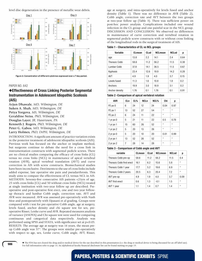
PAPERS, POSTERS & SCIENTIFIC EXHIBITS SPINE
986 u The FDA has not cleared the drug and/or medical device for the use described in this presentation (i.e. the drug or medical device is being discussed for an off label use). For full information refer to page 14. An alphabetical faculty financial disclosure list can be found starting on page 19.
level disc degeneration in the presence of metallic wear debris.
PAPER NO. 642
uEffectiveness of Cross Linking Posterior Segmental Instrumentation in Adolescent Idiopathic Scoliosis (AIS)Arjun Dhawale, MD, Wilmington, DE Suken A. Shah, MD, Wilmington, DE Petya Yorgova, MS, Wilmington, DE Geraldine Neiss, PhD, Wilmington, DE Douglas Layer, JR, Havertown, PA Kenneth J. Rogers, PhD, Wilmington, DE Peter G. Gabos, MD, Wilmington, DE Larry Holmes, PhD, DrPH, Wilmington, DEINTRODUCTION: A significant amount of practice variation exists in the posterior treatment of adolescent idiopathic scoliosis (AIS). Previous work has focused on the anchor or implant method, but surgeons continue to debate the need for a cross link in instrumentation constructs with segmental pedicle screws. There are no clinical studies comparing the efficacy of cross links (CL) versus no cross links (NCL) in maintenance of apical vertebral rotation (AVR), apical vertebral translation (AVT) and curve correction in AIS with screw constructs. Biomechanical studies have been inconclusive. Detriments to the use of cross links include added expense, late operative site pain and pseudarthrosis. This study aims to compare the effectiveness of CL versus NCL in AIS.METHODS: Seventy-five consecutive AIS patients <21yrs of age, 25 with cross links (CL) and 50 without cross links (NCL) treated at single institution with two-year follow up are described. Pre-operative and post-operative first erect, one and two year follow-up thoracic and lumbar Cobb angle, correction rate, AVT and AVR were measured. AVR was assessed pre-operatively with Nash Moe and postoperatively with Upasani et al grading. Groups were compared with t test for pre-operative Cobb angle, age at surgery, levels fused, anchor density and chi square test for sex, pre-operative Risser, Lenke curve and AVR. Repeated measures analysis of variance (ANOVA) and Chi square test were used for comparing continuous and categorical data respectively. Analysis was performed using SPSS and STATA, with significance set at p<0.05.RESULTS: The average age at surgery was 14 years, the mean pre-op Cobb angle was 57°. The groups were similar pre-operatively with respect to age, sex, Lenke curve, Cobb angle, AVT, Risser,
age at surgery, and intra-operatively for levels fused and anchor density (Table 1). There was no difference in AVR (Table 2), Cobb angle, correction rate and AVT between the two groups at two-year follow up (Table 3). There was sufficient power on post-hoc power analysis. Complications included one wound infection in the CL group and one painful scar in the NCL group.DISCUSSION AND CONCLUSION: We observed no differences in maintenance of curve correction and vertebral rotation in segmental pedicle screw constructs with or without cross linking of the longitudinal rods in the surgical treatment of AIS.
Table 1 - Characteristics of CL vs NCL groups
Variable CLmean CLsd NCLmean NCLsd p
age 13.8 2.2 14.1 2.4 0.64
Thoracic Cobb 58.6 11.2 56.2 11.5 0.38
Lumbar Cobb 37.8 14.1 36.5 11.5 0.67
Kyphosis 23.4 12.6 19.9 14.2 0.28
AVT 4.9 1.9 4.0 3.7 0.15
Levels fused 11.3 1.6 10.8 1.8 0.2
Anchors 19.9 3.0 18.9 3.1 0.2
Anchor density 1.76 0.1 1.76 0.1 0.91
Table 2 -Comparison of apical vertebral rotation
AVR CLn CL% NCLn NCL% Chi p
FE po 0 6 24 12 24 0.04 0.97
FE po 1 13 52 27 54 “ “
FE po 2 6 24 11 22 “ “
1 yr po 0 5 21 11 22 0.5 0.78
1 yr po 1 15 60 26 52 “ “
1 yr po 2 5 20 13 26 “ “
2 yr po 0 5 20 10 20 0.01 0.99
2 yr po 1 13 52 26 52 “ “
2 yr po 2 7 28 14 28 “ “
Table 3 - Comparison of Cobb angle and AVT
variable CLmean CLsd NCLmean NCLsd p
Thoracic Cobb pre-op 58.6 11.2 56.2 11.5 0.4
Thoracic Cobb first erect 16.1 6.2 15.6 5.8 “
Thoracic Cobb 1 year 17.7 5.2 19.6 6.9 “
Thoracic Cobb 2 years 20.5 6.3 20.4 7.2 “
AVT pre-op 4.9 1.9 4.0 3.7 0.09
AVT first erect 0.6 1.3 -0.1 1.5 “
AVT 1 year 1.1 1.4 0.7 1.4 “
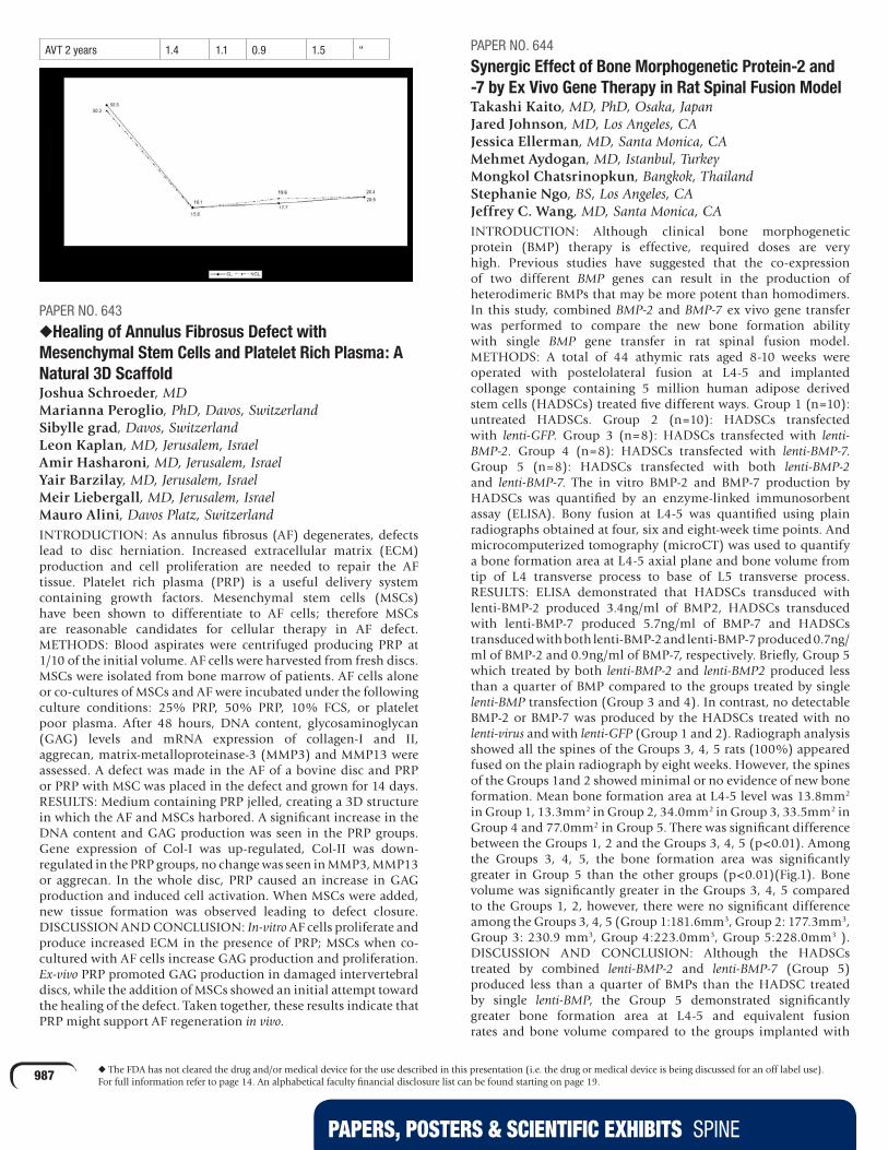
PAPERS, POSTERS & SCIENTIFIC EXHIBITS SPINE
987 u The FDA has not cleared the drug and/or medical device for the use described in this presentation (i.e. the drug or medical device is being discussed for an off label use). For full information refer to page 14. An alphabetical faculty financial disclosure list can be found starting on page 19.
AVT 2 years 1.4 1.1 0.9 1.5 “
PAPER NO. 643
uHealing of Annulus Fibrosus Defect with Mesenchymal Stem Cells and Platelet Rich Plasma: A Natural 3D ScaffoldJoshua Schroeder, MD Marianna Peroglio, PhD, Davos, Switzerland Sibylle grad, Davos, Switzerland Leon Kaplan, MD, Jerusalem, Israel Amir Hasharoni, MD, Jerusalem, Israel Yair Barzilay, MD, Jerusalem, Israel Meir Liebergall, MD, Jerusalem, Israel Mauro Alini, Davos Platz, SwitzerlandINTRODUCTION: As annulus fibrosus (AF) degenerates, defects lead to disc herniation. Increased extracellular matrix (ECM) production and cell proliferation are needed to repair the AF tissue. Platelet rich plasma (PRP) is a useful delivery system containing growth factors. Mesenchymal stem cells (MSCs) have been shown to differentiate to AF cells; therefore MSCs are reasonable candidates for cellular therapy in AF defect.METHODS: Blood aspirates were centrifuged producing PRP at 1/10 of the initial volume. AF cells were harvested from fresh discs. MSCs were isolated from bone marrow of patients. AF cells alone or co-cultures of MSCs and AF were incubated under the following culture conditions: 25% PRP, 50% PRP, 10% FCS, or platelet poor plasma. After 48 hours, DNA content, glycosaminoglycan (GAG) levels and mRNA expression of collagen-I and II, aggrecan, matrix-metalloproteinase-3 (MMP3) and MMP13 were assessed. A defect was made in the AF of a bovine disc and PRP or PRP with MSC was placed in the defect and grown for 14 days.RESULTS: Medium containing PRP jelled, creating a 3D structure in which the AF and MSCs harbored. A significant increase in the DNA content and GAG production was seen in the PRP groups. Gene expression of Col-I was up-regulated, Col-II was down-regulated in the PRP groups, no change was seen in MMP3, MMP13 or aggrecan. In the whole disc, PRP caused an increase in GAG production and induced cell activation. When MSCs were added, new tissue formation was observed leading to defect closure.DISCUSSION AND CONCLUSION: In-vitro AF cells proliferate and produce increased ECM in the presence of PRP; MSCs when co-cultured with AF cells increase GAG production and proliferation. Ex-vivo PRP promoted GAG production in damaged intervertebral discs, while the addition of MSCs showed an initial attempt toward the healing of the defect. Taken together, these results indicate that PRP might support AF regeneration in vivo.
PAPER NO. 644
Synergic Effect of Bone Morphogenetic Protein-2 and -7 by Ex Vivo Gene Therapy in Rat Spinal Fusion ModelTakashi Kaito, MD, PhD, Osaka, Japan Jared Johnson, MD, Los Angeles, CA Jessica Ellerman, MD, Santa Monica, CA Mehmet Aydogan, MD, Istanbul, Turkey Mongkol Chatsrinopkun, Bangkok, Thailand Stephanie Ngo, BS, Los Angeles, CA Jeffrey C. Wang, MD, Santa Monica, CAINTRODUCTION: Although clinical bone morphogenetic protein (BMP) therapy is effective, required doses are very high. Previous studies have suggested that the co-expression of two different BMP genes can result in the production of heterodimeric BMPs that may be more potent than homodimers. In this study, combined BMP-2 and BMP-7 ex vivo gene transfer was performed to compare the new bone formation ability with single BMP gene transfer in rat spinal fusion model.METHODS: A total of 44 athymic rats aged 8-10 weeks were operated with postelolateral fusion at L4-5 and implanted collagen sponge containing 5 million human adipose derived stem cells (HADSCs) treated five different ways. Group 1 (n=10): untreated HADSCs. Group 2 (n=10): HADSCs transfected with lenti-GFP. Group 3 (n=8): HADSCs transfected with lenti-BMP-2. Group 4 (n=8): HADSCs transfected with lenti-BMP-7. Group 5 (n=8): HADSCs transfected with both lenti-BMP-2and lenti-BMP-7. The in vitro BMP-2 and BMP-7 production by HADSCs was quantified by an enzyme-linked immunosorbent assay (ELISA). Bony fusion at L4-5 was quantified using plain radiographs obtained at four, six and eight-week time points. And microcomputerized tomography (microCT) was used to quantify a bone formation area at L4-5 axial plane and bone volume from tip of L4 transverse process to base of L5 transverse process.RESULTS: ELISA demonstrated that HADSCs transduced with lenti-BMP-2 produced 3.4ng/ml of BMP2, HADSCs transduced with lenti-BMP-7 produced 5.7ng/ml of BMP-7 and HADSCs transduced with both lenti-BMP-2 and lenti-BMP-7 produced 0.7ng/ml of BMP-2 and 0.9ng/ml of BMP-7, respectively. Briefly, Group 5 which treated by both lenti-BMP-2 and lenti-BMP2 produced less than a quarter of BMP compared to the groups treated by single lenti-BMP transfection (Group 3 and 4). In contrast, no detectable BMP-2 or BMP-7 was produced by the HADSCs treated with no lenti-virus and with lenti-GFP (Group 1 and 2). Radiograph analysis showed all the spines of the Groups 3, 4, 5 rats (100%) appeared fused on the plain radiograph by eight weeks. However, the spines of the Groups 1and 2 showed minimal or no evidence of new bone formation. Mean bone formation area at L4-5 level was 13.8mm2
in Group 1, 13.3mm2 in Group 2, 34.0mm2 in Group 3, 33.5mm2 in Group 4 and 77.0mm2 in Group 5. There was significant difference between the Groups 1, 2 and the Groups 3, 4, 5 (p<0.01). Among the Groups 3, 4, 5, the bone formation area was significantly greater in Group 5 than the other groups (p<0.01)(Fig.1). Bone volume was significantly greater in the Groups 3, 4, 5 compared to the Groups 1, 2, however, there were no significant difference among the Groups 3, 4, 5 (Group 1:181.6mm3, Group 2: 177.3mm3, Group 3: 230.9 mm3, Group 4:223.0mm3, Group 5:228.0mm3 ). DISCUSSION AND CONCLUSION: Although the HADSCs treated by combined lenti-BMP-2 and lenti-BMP-7 (Group 5) produced less than a quarter of BMPs than the HADSC treated by single lenti-BMP, the Group 5 demonstrated significantly greater bone formation area at L4-5 and equivalent fusion rates and bone volume compared to the groups implanted with

PAPERS, POSTERS & SCIENTIFIC EXHIBITS SPINE
988 u The FDA has not cleared the drug and/or medical device for the use described in this presentation (i.e. the drug or medical device is being discussed for an off label use). For full information refer to page 14. An alphabetical faculty financial disclosure list can be found starting on page 19.
HADSCs treated by single lenti-BMP gene transfer (Group 3 and 4). Combined BMP-2 and BMP-7 ex vivo gene transfer is significantly more effective in inducing new bone formation than individual BMP ex vivo gene transfer in rat spinal fusion model.
PAPER NO. 645
Intervertebral Disc Degeneration and Ectopic Bone Formation in Apolipoprotein E Knockout MiceDawei Zhang, PhD, Charlottesville, VA Li Jin, PhD, Charlottesville, VA Davis Reames, MD, Charlottesville, VA Francis H. Shen, MD, Charlottesville, VA Xudong Li, MD, Charlottesville, VAINTRODUCTION: Intervertebral disc degeneration is a multi-factorial disease. Recently accumulating body of evidence suggests an association between cardiovascular risk factors and disc degeneration. Apolipoprotein E (ApoE) has been clearly implicated in the development of atherosclerosis. The goal of the present study is to elucidate the role of ApoE in disc degeneration.METHODS: ApoE KO and wild-type (WT) mice were characterized by histological/immunological, biochemical, and real-time RT-PCR studies. Disc cells and bone marrow cells (MSCs) were isolated to compare the extracellular matrix and osteogenic differentiation capabilities.RESULTS: ApoE was highly expressed in the endplates of WT discs, and ectopic bone formed in the endplates of ApoE KO mice. The bone MSCs of the KO mice showed significant higher osteogenic differentiation than WT mice. Glycosaminoglycan content was decreased in ApoE KO annulus fibrosus (AF) and nucleus pulposus (NP) cells. Collagen levels were increased in AF and decreased in NP cells. Matrix metalloproteinases-3, 9, and 13 expression was increased which may at least partially explain the impaired matrix production. The expression of collagen I, II, aggrecan and biglycan increased in AF cells but decreased in NP cells. Apoptosis was increased in the ApoE KO NP cells when compared to WT mice.DISCUSSION AND CONCLUSION: ApoE, in addition to its importance to cardiovascular disease, may play a critical role in disc integrity and function and may represent a potential therapeutic target. The relationship between cardiovascular disease and disc degeneration warrants further investigation.
PAPER NO. 691
Vertebral Column Resection for Treatment of Adult Spinal Deformities: Outcomes and ComplicationsMostafa H. El Dafrawy, MD, Baltimore, MD Firas Chamas, MD, PhD, Homewood, AL Hamid Hassanzadeh, MD, Baltimore, MD Khaled M. Kebaish, MD, Baltimore, MDINTRODUCTION: Correction of severe rigid spinal deformities has been traditionally obtained via a combined anterior/posterior approach. Single incision, all posterior vertebral column resection (VCR) offers a potentially superior alternative. There are few published reports with the largest series describing VCR in the pediatric population.METHODS: Retrospective review of 50 consecutive adult patients (17 men and 33 women) with an average age of 50 years (21-81) underwent posterior VCR by a single surgeon between 2004 and 2010. The deformities were divided into three main groups: coronal (8), sagittal (31) and combined (coronal and sagittal, 11). The average follow up was 22.6 months (3-76).RESULTS: There were 27 lumbar and 26 thoracic VCRs (28 revisions & 22 primary). Forty-seven patients had a single level and three patients had two nonadjacent levels VCRs. The average number of levels fused was eight (2-16), average EBL of 3040cc (550-9000) and average operative time 415 min (220-660). All patients were ASA class 2 (28) and 3 (21). The average major Cobb angle for coronal deformities (scoliosis and kyphoscoliosis) was 61° (20° to 105°) pre-op and corrected to 17.6° (1°to 39°). The average preoperative thoracic kyphosis for thoracic deformity was 87.4° (38° to 137°) and corrected to 53.3° (34°to 72°). The average lumbar lordosis for lumbar deformity was -27.2° (+30° to -65°) and corrected to -40.3° (-11° to -87°). In the coronal group the average focal correction in the patients with scoliosis was 34° (9°to 75°), while the average focal correction in the sagittal plane for patients with kyphosis was 36° (6° to 76°). In the kyphoscoliosis group the combined average correction for both planes was 62°. The average preoperative sagittal imbalance was 83 mm (-61 to 351) pre-op and 30 mm (-48 to 187) at the last follow up. The average preoperative coronal imbalance was 27 mm pre-op and 18 mm at the last follow up. The ODI, VAS and the SF 12 scores were significantly improved at the last follow up (see table). There were 13 major and 15 minor complications. Major complications included six neurologic deficits manifested by distal lower extremity weakness in a single nerve root, five in lumbar (four in L5 VCR for treatment of high grade spondylolisthesis) and one in a thoracic VCR. Four completely recovered and two had partial recovery. There were two deep wound infections that resolved with irrigation and drainage. Two patients suffered respiratory failure requiring re-intubation. One patient developed pulmonary embolism and one patient had pneumonia and one myocardial infarction (NSTEMI). There were eight reoperations, two patients required decompression for postoperative weakness, three had extension of instrumentation for PJK while three patients had their instrumentation removed. Minor complications: there was a total of eight incidental durotomies (six in revision surgeries and two in primary cases). There was one superficial wound infection and two wound dehiscences treated with irrigation and drainage, two patients had pleural effusion requiring chest tube insertion.DISCUSSION AND CONCLUSION: All posterior VCR is a safe and effective method in treating severe adult spinal deformities. Key words: scoliosis, VCR, spine deformity, Kyphosis, Kyphoscoliosis

PAPERS, POSTERS & SCIENTIFIC EXHIBITS SPINE
989 u The FDA has not cleared the drug and/or medical device for the use described in this presentation (i.e. the drug or medical device is being discussed for an off label use). For full information refer to page 14. An alphabetical faculty financial disclosure list can be found starting on page 19.
HRQL functional outcome
Pre-operative Last follow up P values
ODI 76.3 (48-92) 28.7 (21.9-36.4) 0.001
SF12Physical health
32.1 (23.1-41.9) 54.9 (39.7-71.3) 0.012
SF12Mental health
38.4 (28.2-47.7) 56.8 (41.3-74.8) 0.032
SRSSelf image
2.29 (1.2-3.7) 4.29 (3.2-5.0) 0.012
SRSActivity
2.15 (1.1-2.6) 4.23 (2.8-5.0) 0.009
SRSPain
2.10 (1.4-2.9) 4.35 (3.0-5.0) 0.013
SRSMental
2.96 (2.0-3.7) 4.22 (2.6-5.0) 0.031
PAPER NO. 692
The Fate of the Adult Revision Spinal Deformity Patient: A Single Institution ExperienceMichael P. Kelly, MD, St Louis, MO Lawrence G. Lenke, MD, Saint Louis, MO Keith H. Bridwell, MD, Saint Louis, MO Linda A. KoesterINTRODUCTION: Reported revision rates for primary adult spinal deformity (SD) surgeries have ranged from 9% to 25%, but to our knowledge, the revision rate following revision SD surgery has not been reported. The reported improvements in health-related quality of life (HRQL) measures following revision SD surgery have also been quite modest. The aim of this study was to determine subsequent revision rates for all revision SD surgeries performed at a single center and to investigate the changes in measures of HRQL in these patients.METHODS: A total of 504 consecutive adult revision SD surgeries (1995-2008) were identified and the records were reviewed to determine the reason for and timing to any additional operation(s). SRS outcomes scores were recorded at the first visit, and at planned follow-up (F/U) visits.RESULTS: Ninety-six of 504 patients underwent further surgeries for a subsequent revision rate of 19%. Two-year F/U was available for 73 (77%) of these patients (mean F/U 6.0yrs, range 2.3-12.6, gender: F=60, M=13, mean age 52.7yrs, range 21-78). The most common causes of reoperation following revision surgery were pseudarthrosis (N=28, 38%), adjacent segment disease (N=23, 32%), infection (N=13, 18%), and implant prominence/pain (N=12, 16%). 15 (21%) patients underwent more than one revision procedure. SRS outcomes scores were available for 50 (68%) patients, at an average F/U of 4.9yrs (range 2-11.4). The mean improvements in the SRS outcomes measures were Pain: 0.74 (p<0.001), Self-Image: 0.8 (p<0.001), Function: 0.5 (p<0.001), Satisfaction: 1.2 (p<0.001) and Mental Health: 0.3 (p=0.012).DISCUSSION AND CONCLUSION: The rate of repeat revision following revision spinal deformity surgery was 19%, most commonly due to pseudarthrosis, adjacent segment disease, infection and implant prominence/pain. However, significant improvements in SRS outcome scores were still observed in those patients requiring additional revision procedures.
PAPER NO. 693
Establishment of Parameters for Congenital Stenosis of the Cervical Spine: A Study of 1,072 Cadaveric SpecimensNavrikat Bajwa, Cleveland, OH Nicholas U. Ahn, MD, Shaker Heights, OHINTRODUCTION: ongenital cervical stenosis occurs when the bony anatomy of the cervical canal is smaller than expected in the general population. This may predispose an individual to symptomatic neural compression. However, no studies have defined congenital cervical stenosis (CCS) based on the normal population. The diagnosis is currently made based on clinical impression from radiographic studies which is subjective at best. Exact measurements are needed which define this condition, as are simple parameters that will accurately predict if CCS is present. The purpose of this study is to provide a working definition for congenital cervical stenosis, based on objective measurements of cervical spine specimens, and to establish parameters that will accurately predict if CCS is present.METHODS: A total of 1,072 adult skeletal specimens from the Hamann Todd Collection in the Cleveland Museum of Natural History were selected. Baseline data including age, sex, and race of subjects were collected. Digital calipers were used to measure the following (C3-C7): sagittal canal diameter (sd), interpedicular distance (ipd), and pedicle length (pl). Canal area at each level was also calculated using a formula that was verified by computerized measurements. A standard distribution for each level was created, and values that were -2SD below mean were considered as being congenitally stenotic. Once defined, an analysis of deviance was performed to identify parameters that were predictive of CCS. Logistic regression analysis was used to determine odds ratios for CCS using these parameters.RESULTS: CCS was defined at each level as: C3=1.82cm2; C4=1.80cm2; C5=1.84cm2; C6=1.90cm2 ,C7=1.88 cm2. The sagittal diameter (sd) and interpedicular distance (ipd) were found to be predictive of CCS at each level with high sensitivity and specificity. Predictive values were sd<13mm and ipd<22.5mm, which yielded sensitivities and specificities of 88-100% at each level. Logistic regression demonstrated a significant association between these parameters and presence of CCS with OR>40 at each level. Interestingly, pedicle length (pl) was not a good predictor of CCS. CONCLUSIONS: Based on our study of a large population of adult skeletal specimens, we have defined congenital cervical stenosis at each level. Values of sd<13mm or ipd<22.5mm strongly predict the presence of CCS at all levels C3- C7.
PAPER NO. 694
Nationwide Trends in the Surgical Management of Lumbar Spinal Stenosis in all Age GroupsHyun W. Bae, MD, Los Angeles, CA Sean Rajaee, MS, Woodland Hills, CA Linda E A Kanim, MA, Los Angeles, CAINTRODUCTION: Over the last several decades, the number of surgeries performed for patients with lumbar spinal stenosis (LSS) has increased significantly. Several studies have reviewed indications for surgical management of these patients and controversy still exists as to what type of surgery should be performed. Select group of patients are indicated for de-compressive surgery alone and others are indicated for decompression with fusion with or without instrumentation. Since lumbar spinal stenosis is most prevalent in the older patient population who generally carry more

PAPERS, POSTERS & SCIENTIFIC EXHIBITS SPINE
990 u The FDA has not cleared the drug and/or medical device for the use described in this presentation (i.e. the drug or medical device is being discussed for an off label use). For full information refer to page 14. An alphabetical faculty financial disclosure list can be found starting on page 19.
co-morbidities, benefits and risks must be carefully weighed in the choice of surgical procedure. This current study aims to characterize the epidemiology of surgical treatment for lumbar spinal stenosis in the United States in all age groups from 2004 to 2008.METHODS: Data were obtained from the Healthcare Cost and Utilization Project Nationwide Inpatient Sample for the years 2004-2008. Discharges were identified using International Classification of Diseases, Ninth Revision, Clinical Modification diagnostic and procedure codes for patients with lumbar spinal stenosis. Population-based utilization rates were calculated from United States census data.RESULTS: Between 2004 and 2008, the annual number of inpatient discharges with a primary diagnosis of lumbar spinal stenosis increased from 91,630 to 101,936. The proportion of patients (pts/xxx) between the age of 45 and 65 years with lumbar spinal stenosis increased from 31.7% to 34.2% of all cases, while all other age groups decreased in proportion. In 2004, 53.1% of all patients with a primary inpatient diagnosis of LSS were treated with surgical decompression alone without spinal fusion, while in 2008, this decreased to 43.7% of cases. Between 2004 and 2008, the percentage of LSS discharges requiring a fusion increased from 24.5% of all cases in 2004 to 34.2% in 2008. Out of all LSS cases that required fusion in 2008, 32.8% involved the use of bone morphogenetic protein, 43% used an interbody spinal fusion device (cages/spacers), 0.5% used an interspinous process spacer, and 11.7% of cases involved fusion of more than four vertebral levels. Between 2004 and 2008, length of hospital stay for patients receiving a fusion decreased from 4.60 days to 3.95 days, in hospital mortality decreased from 0.3% to 0.1% and mean total hospital charges increased from $52,996 to $84,032.DISCUSSION AND CONCLUSION: Among all inpatient discharges for lumbar spinal stenosis, the proportion of cases requiring fusion increased over the time period of 2004 to 2008. Mean hospital charges for fusion cases increased by 56.8% from 2004 to 2008 while charges for decompression procedures increased by only 35.7%. The usage of bone morphogenetic protein and interbody spinal fusion devices (spacers/cages) also increased over the time period of this study. In addition to these findings, other patient variables and hospital characteristics changed significantly throughout the duration of this study.
PAPER NO. 695
Retrolisthesis and Lumbar Disc Herniation: A Postoperative Assessment of Patient FunctionKevin Kang, MD, Brooklyn, NY Michael Shen, MD, Broomfield, CO Wenyan Zhao, PhD, Hanover, NH Jonathan Lurie, MD, Lebanon, NH Afshin Razi, MD, New York, NYINTRODUCTION: Retrolisthesis in patients with L5-S1 disc herniation has not been shown to have significant relationship with worse baseline pain or function. Whether it can affect outcomes following discectomy has yet to be established. The purpose of this study was to determine the relation between retrolisthesis (alone or in combination with other degenerative conditions) and postoperative low back pain, physical function, and quality of life. This study was intended to be a follow up to a previous investigation that looked at preoperative assessment of patient function.METHODS: This cross-sectional study was performed on patients enrolled in the SPORT (Spine Patient Outcomes Research Trial) study who underwent L5-S1 discectomy and who had a complete MRI scan available for review (n=125). The SF-36 bodily pain scale, SF-36 physical function scale, Oswestry Disability Index (ODI), and Sciatica Bothersomeness Index (SBI) were measured. Longitudinal regression models were used to compare the time-weighted outcomes over four years.RESULTS: Patients with retrolisthesis did significantly worse in regard to bodily pain and physical function over four years. However, there was no significant difference in terms of ODI or SBI. Similarly, retrolisthesis was not a significant factor in operative time, blood loss, length of stay, complications, rate of additional spine surgeries, or recurrent disc herniations. Disc degeneration, modic changes, and posterior degenerative changes did not affect outcomes.DISCUSSION AND CONCLUSION: Although retrolisthesis in patients with L5-S1 disc herniation did not affect baseline pain or function, postoperative outcomes appeared to be somewhat worse. It is possible that the contribution of pain or dysfunction related to retrolisthesis became more evident after removal of the disc herniation.

PAPERS, POSTERS & SCIENTIFIC EXHIBITS SPINE
991 u The FDA has not cleared the drug and/or medical device for the use described in this presentation (i.e. the drug or medical device is being discussed for an off label use). For full information refer to page 14. An alphabetical faculty financial disclosure list can be found starting on page 19.
PAPER NO. 696
Scheuermann’s Kyphosis: Impact on Quality of Life in 86 PatientsBaron Lonner, MD, New York, NY Jamie S. Terran, BS, New York, NY Peter O. Newton, MD, San Diego, CA Randal R. Betz, MD, Philadelphia, PA Suken A. Shah, MD, Wilmington, DE Paul D. Sponseller, MD, Baltimore, MD Amer Samdani, MD Harry L. Shufflebarger, MD, Miami, FLINTRODUCTION: The clinical presentation and impact on health-related quality of life (HRQOL) of Scheuermann’s kyphosis (SK) has not been previously evaluated in a prospective manner. Previous studies have assessed the impact of adolescent idiopathic scoliosis (AIS) and kyphosis in AIS patients on HRQOL. The purpose of this study was to identify the clinical impact of Scheuermann’s kyphosis on HRQOL compared to that for AIS and normal controls (NC).METHODS: A total of 86 patients enrolled in a prospective study of patients with operative Scheuermann’s kyphosis were evaluated. Impact of kyphosis magnitude, apex location (thoracic, TL) were evaluated with SRS-22 and VAS outcome instruments. These patients were compared to a cohort of operative AIS patients from a prospective database as well as to normal controls. ANOVA and the Bonferroni post hoc comparison were utilized to compare the groups. Pearson correlation was utilized for correlation comparisons.RESULTS: Mean age for SK, AIS, and NC, were 14.96, 14.16 and 16.10 respectively (p<0.001). SK, AIS and NC were 39.5%, 75% and 74.19 % female respectively (p< 0.001). Preoperatively, SK patients have significantly lower scores in all domains of the SRS-22 compared to AIS patients. SK and AIS patients scored significantly lower than NC on the pain and image domains, as well as mean scores (p<0.05). There was no significant difference between SK and NC for the mental health domain. SK patients with TL apex scored significantly lower than those with T apex in the pain domain. There were no significant differences in SRS scores between patients with kyphosis < 80° and > 80°. When AIS and SK T5-12 K was pooled, there was a significant negative correlation to all domains of the SRS. SK patients mean VAS score was 3.36 ± 2.74. VAS score negatively correlates to pain, image mental health and total SRS score. There is no correlation of VAS to curve magnitude or apex location for AIS or SK.DISCUSSION AND CONCLUSION: The clinical impact on HRQOL of SK as compared to AIS and NC has been demonstrated for the first time. Kyphosis appears to impart a significantly negative impact on quality of life in the adolescent population.

PAPERS, POSTERS & SCIENTIFIC EXHIBITS SPINE
992 u The FDA has not cleared the drug and/or medical device for the use described in this presentation (i.e. the drug or medical device is being discussed for an off label use). For full information refer to page 14. An alphabetical faculty financial disclosure list can be found starting on page 19.
PAPER NO. 697
Partial Facetectomy for Lumbar Foraminal StenosisKevin Kang, MD, Brooklyn, NY Juan Carlos C. Rodriguez-Olaverri, MD, PhD, New York, NY Jean-Pierre C. Farcy, MD, New York, NYINTRODUCTION: Several techniques exist to address the pain and disability caused by nerve root impingement. Failure to adequately decompress the lumbar foramen has been cited as a significant cause of failed back surgery syndrome. However, aggressive treatment often causes spinal instability or may require fusion for satisfactory results. The purpose of this study was to describe a novel technique for decompression of the lumbar nerve root and to demonstrate its effectiveness in relief of radicular symptoms. This study was intended to be an evaluation of outcomes following the removal of the medial portion of the superior facet in patients with lumbar foraminal stenosis.METHODS: Work status and activity level were measured in patients who underwent partial facetectomy of the lumbar spine (n=47). Average follow-up was 3.9 years. Those who demonstrated neurogenic claudication without spinal instability or central canal stenosis and failed conservative management were eligible. Radiculopathy was defined as pain following a nerve root distribution which was exacerbated by standing or walking. On physical exam, patients exhibited muscle weakness without atrophy as well as decreased reflexes. Conservative measures and non-invasive rehabilitation exercises were used in all patients for an average of 12 weeks. Patients with previous surgical intervention, instability of the lumbar spine documented on flexion/extension films, and central canal stenosis were excluded.RESULTS: The average age of patients at time of surgery was 59 years (range 47-79). Twenty-seven of 47 (57%) report no back pain and no functional limitations. They are able to participate in recreational activities and work full time. Five of 47 (11%) required additional surgery for continued degenerative symptoms.DISCUSSION AND CONCLUSION: Partial facetectomy or removal of the medial portion of the superior facet is an effective means to decompress the lumbar nerve root foramen without causing spinal instability.
PAPER NO. 698
Nurick Grade based Surgical Intervention in Cervical Spondylotic Myelopathy Affects Improvement in SymptomsMatthias Pumberger, MD, Berlin, Germany Han Jo Kim, MD, Saint Louis, MO Arvind G. Von Keudell, MD, Chestnut Hill, MA Alexander P. Hughes, MD Andrew A. Sama, MD, New York, NY Frank P. Cammisa, Jr, MD, New York, NY Federico P. Girardi, MD, New York, NYINTRODUCTION: Cervical spondylotic myelopathy is a relatively common spinal disorder among elderly. If patients fail, conservative management surgical treatment is indicated. Although several surgical procedures are available the underlying difficulty of timing and prediction of outcome remains poorly defined. We hypothesize that clinical and functional improvement of myelopathic symptoms is dependent on pre-operative severity of disease.METHODS: We reviewed a consecutive series of all cervical myelopathy patients who underwent surgical intervention by three spine surgeons between 1/2000 and 10/2010 at our institution.

PAPERS, POSTERS & SCIENTIFIC EXHIBITS SPINE
993 u The FDA has not cleared the drug and/or medical device for the use described in this presentation (i.e. the drug or medical device is being discussed for an off label use). For full information refer to page 14. An alphabetical faculty financial disclosure list can be found starting on page 19.
Patient’s medical records, operative reports and imaging studies were studied. Patient demographics, persistence of myelopathic symptoms, type of surgery and operative surgical levels were collected. Patients were grouped according to their pre-operative Nurick classification. The improvement of a pre-operative Nurick Grade 0-2 was compared to the improvement of pre- operative 3 and 4. This distinction was instituted because those with Grade 3 and 4 disease cannot be employed due to their cervical myelopathy.RESULTS: A total of 258 patients (71 F; 188 M) were included in this study. The average age was 59.22±13.27 and average BMI was 28.3±5.1. Ten patients were lost during follow up time (seven incomplete data; three deceased). Pre-operative duration of myelopathic symptoms until surgical intervention was 14.93 months (1-101 months) and the average follow up was 15.3 (range 1-79 months). Of all patients, 213 patients underwent anterior cervical decompression and fusion, three patients decompression only and 42 patients (161 levels) underwent posterior cervical decompression and fusion. In total, 110 patients had Nurick grade 1, 90 grade 2, 48 grade 3 and 10 grade 4. Patients with grade 2 (p=0.003) and grade 3 (p=0.005) were most likely to have cure of all myelopathic symptoms. Largest improvement of at least one Nurick grade was observed in pre-operative grade 2 (60%), grade 3 (50%), grade 1 (15%) and grade 4 (10%). Parallel the greatest improvement with surgical intervention to employment capable status based on pre-operative Nurick Grade was observed in grade 2 (p<0.001) and grade 3 (p<0.001).DISCUSSION AND CONCLUSION: Our data suggests that patients with Grade 2 disease have the greatest likelihood of improvement with surgery. Those with Grade 3 disease have an intermediate prognosis for symptom resolution while those with Grade 4 disease have a poorer prognosis even with surgical intervention.
PAPER NO. 699
A Review of Medicolegal Cases for Cauda Equina Syndrome: What Factors Lead to an Adverse Outcome for the Provider?Eldra Daniels, Baltimore, MD Zachary Gordon, MD, Lyndhurst, OH Keisha French, Cleveland, OH Nicholas U. Ahn, MD, Shaker Heights, OHINTRODUCTION: Cauda equina syndrome (CES) is one of a few true surgical emergencies involving the lumbar spine. While treatment within a 48 hour period of time has been found to correlate with improved outcomes, recovery of bowel and bladder control unfortunately does not always occur and loss of this function can be distressing to the patient. An understanding of factors which affect the legal outcome can aid the clinician in determining risk management for medicolegal cases of CES.METHODS: The LexisNexis Academic legal search system was used to identify 104 cases of CES. Cases involving social security disability or claims against an employer were removed from the study. This left 14 cases in which lawsuits were filed against treating physicians for the management of CES. Outcomes data on trial verdicts were collected, as were associated penalties. Case data was also compiled on age, sex, initial site of presentation, initial diagnosis, whether or not a rectal exam was performed, time to consultation with a specialist, time to completion of advanced imaging study, time to surgery, and neurosurgical versus orthopaedic consultation. Univariate logistic regression analyses were performed to determine the association between adverse decisions and case data. Linear regression analyses were performed to determine the association between the penalties and case data as well.
RESULTS: Of the 14 cases reviewed, six were for the plaintiff. A significant association was found between an adverse decision and cases in which time to surgery was greater than 48 hours (p<0.05, OR=2.1). There was no association with the functional outcome, neurosurgical vs. orthopaedic consultation, initial site of presentation, or initial diagnosis. Interestingly, it did not appear that a rectal examination was performed in any of the cases. Only four of the 14 cases (28.5%) involved an initial presentation which included loss of bowel or bladder control.DISCUSSION AND CONCLUSION: Based a study of court cases involving CES, a positive association was found between time to surgery greater than 48 hours and an adverse decision. The actual degree of functional loss did not appear to affect the verdicts. This study emphasizes the importance of early diagnosis of cauda equina syndrome. As only 28.5% of the cases involved an initial presentation which included loss of bowel or bladder control, it also emphasizes the importance of cautioning all patients with lumbar complaints of the risk of CES.
PAPER NO. 700
CT-Based Evaluation of 8,661 Pedicles: Curve Characteristics Associated with Abnormal MorphologyAdam L. Wollowick, MD, New York, NY Terry D. Amaral, MD, Bronx, NY Beverly Thornhill, MD, Bronx, NY Joshua D. Grossman, BA Jonathan Horn, Bronx, NY Etan Sugarman, MD, Bronx, NY Preethi M. Kulkarni, Bronx, NY Vishal Sarwahi, MD, Bronx, NYINTRODUCTION: APs are associated with spinal deformity. It is thought that more APs are found with larger curves, greater rotation, and more complex pathology. The objective of this study is to study the incidence, subtypes, and distribution of APs in spinal deformity patients.METHODS: Low dose CT scans of 272 adolescent patients were reviewed. Pedicles were classified as Type A: >4mm cancellous channel, Type B: 2-4 mm channel, Type C: cortical channel, and Type D: cortical or cancellous channel < 2mm channel. Group I had 104 AIS patients, Group II had 69 patients with other spinal deformities, and Group III had 99 patients with non-spinal pathology. Curve and patient characteristics were analyzed.RESULTS: A total of 8,661 pedicles were studied. In Group I, the incidence of APs was 20.4%. In Group II, it was 19.7%. In Group III, it was 9.8% (p<0.001). No significant difference was seen between AIS and Non-AIS group (p=0.484). In the AIS group, the incidence of thoracic APs was 29.8%, comprising 97.4% of APs. Of those, 26.7% were type C or D. In the Non-AIS group, the incidence of APs in the thoracic spine was 25.5%, comprising 92.8% of APs. Of those, 16.1% were type C or D. In the AIS group, APs are 2.2X more likely to be located on the concave side of the curve (p<0.001). In the Non-AIS group, APs are 2.4X more likely to be located on the concave side of the curve (p<0.001). In the AIS group, 31.9% of pedicles within severe curves (Cobb >70°) are abnormal as compared to 21.8% in moderate curves (Cobb 40°-70°) (p<0.05). However, in the Non-AIS group, curve size did not affect incidence of APs (p>0.05). Degree of kyphosis did not impact pedicle morphology for both AIS and Non-AIS groups (p>0.05). Female patients in both AIS and Non AIS groups had significantly higher number of abnormal pedicles (p<0.0001).DISCUSSION AND CONCLUSION: APs are more commonly found in AIS and Non-AIS deformity patients than patients

PAPERS, POSTERS & SCIENTIFIC EXHIBITS SPINE
994 u The FDA has not cleared the drug and/or medical device for the use described in this presentation (i.e. the drug or medical device is being discussed for an off label use). For full information refer to page 14. An alphabetical faculty financial disclosure list can be found starting on page 19.
without spinal deformity. There is no difference between AIS and non-AIS in total APs. Significantly fewer Type C and D pedicles (highly abnormal) were seen in the Non-AIS group. Abnormal pedicles are common in patients with spinal deformity of all types. Knowing the location of APs can aid in pre-operative planning and help surgeons anticipate difficult screw placement.
PAPER NO. 701
Coronal Magnetic Resonance Imaging (MRI) Improves the Diagnosis of Foraminal StenosisTrichy S. Rajagopal, Dr, Nottigham, United Kingdom Robert W. Marshall, FRCS, Reading, United Kingdom Neta Raz, MD, Berkshire, United Kingdom Colin Archibald, Reading, United KingdomINTRODUCTION: Magnetic resonance imaging (MRI) has become established as the investigation of choice in degenerative disease of the lumbar spine. Conventional sagittal and axial images demonstrate central and lateral recess stenosis well, but do not detect foraminal stenosis with certainty. This is possibly because scanning is not done in the plane of the exiting nerves. Foraminal stenosis may thus be over-diagnosed. We found that the diagnosis of foraminal stenosis can be improved by additional coronal T2- weighted and short T1 inversion recovery (STIR) sequences. We studied the true incidence of foraminal stenosis using coronal MRI in addition to conventional sequences.METHODS: Two studies were done: Study 1 - a retrospective analysis of conventional sagittal and axial MRI in 100 patients with spinal stenosis treated by surgical decompression. We studied the radiologists’ reports and noted their reported incidence of foraminal stenosis and lateral recess stenosis. Study 2 - a prospective analysis of lumbar spine MRI in 100 consecutive patients with clinical features of spinal stenosis to assess the relative incidence of foraminal stenosis and lateral recess stenosis. Three spinal surgeons and a radiologist independently reviewed conventional sagittal and axial T1 and T2-weighted sequences to identify signs of nerve compression from L3 to S1 level. Each observer then studied additional coronal scans to see if any nerve compression could be identified. Any differences in diagnosis were recorded.RESULTS: Study 1 - the analysis of these 100 MRI reports in the study found that “foraminal stenosis” was reported by radiologists in 46% using conventional sequences. The radiologists based their diagnosis on sagittal and axial images only. Study 2 - the prospective study of 100 patients suspected of having stenosis included 48 men and 52 women. Their age ranged from 29 to 89 with a mean age of 67 years. MRI scans of 100 patients suspected of having stenosis showed: spinal stenosis - 40; degenerative spondylolisthesis - 14; posterolateral disc herniation - 14; normal - 13; far lateral disc herniation - 7; isthmic spondylolisthesis - 6; degenerative scoliosis - 6. Conventional sequences in patients with spinal stenosis showed lateral recess stenosis reliably, but also suggested foraminal stenosis in 43% (17 out of 40). However, coronal sequences clearly showed no foraminal nerve compression at all. In degenerative spondylolisthesis conventional scans suggested foraminal stenosis in 10 of 14 cases. Coronal imaging again showed no foraminal stenosis. In degenerative scoliosis conventional scans suggested foraminal stenosis in all six cases. Coronal imaging confirmed foraminal stenosis in five out of six cases. In far lateral disc herniation (six out of seven) and isthmic spondylolisthesis (six out of six), foraminal nerve compression was confirmed by both conventional and coronal imaging.DISCUSSION AND CONCLUSION: In cases of spinal stenosis and degenerative spondylolisthesis, fine slice coronal MRI sequences show that the exiting nerve roots are free, but
traversing nerve compression occurs in the lateral recesses beneath the facet joints. Although axial and sagittal sequences suggested foraminal stenosis, this impression seems mistaken because the imaging is not in the plane of the nerve roots.True foraminal stenosis with compression of the exiting nerves does occur in conditions like isthmic spondylolisthesis, far lateral disc herniation and degenerative scoliosis.The use of coronal imaging tilted into the plane of the exiting nerve roots can improve the accuracy of diagnosis of foraminal stenosis.
PAPER NO. 702
Are Thoracic Curves with a Low Apex (T11 or T11/12) Really Thoracic Curves?Milad Alam, San Diego, CA Peter O. Newton, MD, San Diego, CA Burt Yaszay, MD, San Diego, CA Tracey Bastrom, MA, San Diego, CAINTRODUCTION: Curves with an apex at T11 and T11/T12 are classified by the SRS and Lenke classifications as thoracic curves. In this study, we wished to determine whether this classification is accurate or if curves with an apex at T11 or T11/T12 represent a combination of both “thoracic” and “thoracolumbar” curves or constitute their own class of curves.METHODS: A review of data collected on 1,742 AIS cases from a multi-center study was performed. Based on an analysis of the most common curve apex levels (T9 for thoracic (TH) curves and L1 for thoracolumbar/lumbar (TL/L) curves), two comparison groups were created that represented “classic examples” of TH and TL/L curves (Figure). Sixty-six cases with an apex at T11-T11/T12 were identified and compared to 320 cases with an apex at T9 and 126 cases with an apex at L1. A distribution analysis of the curve lengths and a Chi-square test comparing curve direction of the TH, TH/L, and T11 - T11/T12 curves was performed (p<0.05). Additionally, the three groups were compared with respect to the major Cobb magnitude, trunk shift, and age at time of surgery. Finally, a radiographic review of the T11-T11/T12 group was completed.RESULTS: The curve direction of the three groups was significantly different (TL/L: 84% Left, TH: 3% Left, and T11-T11/T12:16% Left, p<.001). The mean number of vertebra in curves for the T11-T11/T12 group (7.1 ± 1.2) fell in between the value for the TL/L (5.7± 0.8) and TH (7.3 ± 1.0) groups. The T11-T11/T12 curve length distribution was a combination of a typical TH curve and a typical TL/L curve. The T11-T11/T12 curves have a greater trunk shift than both TL/L (p=0.002) and TH (p=0.011) curves. There was not a significant difference between the three groups in terms of major curve Cobb magnitude (p=0.09) or age at time of surgery (p=0.76). The radiographic review of the T11-T11/T12 curves revealed three curve patterns: 21 (32%) long single curves, 28 (42%) short single curves, which are more typical of a TL/L curves, and 17 (26%) double thoracic curves.DISCUSSION AND CONCLUSION: We suggest caution in lumping curves with an apex at either T11 or T11-12 disc together with other thoracic apices in studies which involve principally primary thoracic curves.

PAPERS, POSTERS & SCIENTIFIC EXHIBITS SPINE
995 u The FDA has not cleared the drug and/or medical device for the use described in this presentation (i.e. the drug or medical device is being discussed for an off label use). For full information refer to page 14. An alphabetical faculty financial disclosure list can be found starting on page 19.
PAPER NO. 703
Restoring Thoracic Kyphosis During Posterior Instrumentation for Adolescent Idiopathic ScoliosisShafagh Monazzam, MD, San Diego, CA Peter O. Newton, MD, San Diego, CA Tracey Bastrom, MA, San Diego, CA Burt Yaszay, MD, San Diego, CAINTRODUCTION: Correcting hypokyphosis in adolescent idiopathic scoliosis (AIS) patients is important in preventing junctional kyphosis and increasing pulmonary function. We wished to determine what factors were most predictive of postop correction of hypokyphosis when segmental posterior implants are used in treating thoracic AIS.METHODS: Prospectively collected cases from a multi-center study were analyzed. Lenke 1-4 AIS patients with preop kyphosis of 5-20 degrees, treated with posterior pedicle screws, with a surgeon who had at least 20 patients in the database were included. Patients were divided into two groups postoperatively based on first erect x-rays: those remaining hypokyphotic (HK, <20° or an increase <5°) and those restored to normal kyphosis (NK, >20° with >5° increase). Patients whose correction was less than 5 degrees were considered unchanged (hypokyphotic). Regression analysis was done on four preop factors thought to influence the postop kyphosis: pre-operative kyphosis, surgeon, rod material utilized (steel vs. Ti) and use or not of a posterior release (Ponte).RESULTS: Of the 280 patients included in the study, 227 reMEd hypokyphotic and 53 achieved normal kyphosis. There were no differences in preoperative kyphosis (13.4±5 HK group vs 14.5 ±4 NK group) and age (14.7 HK group vs 14.6 NK group). Of the factors evaluated, the surgeon was the most significant predictor of restoration of normal kyphosis. Comparison of surgeons showed the rate of normalizing kyphosis ranged from 6% to 42% (p=.004) and there was no difference in the degree of preoperative kyphosis between surgeons. Pre-operative kyphosis (p=.093), rod material (p=.56), and use or not of a posterior release (p=.622) were not significant predictors of restoration of normal kyphosis. DISCUSSION AND CONCLUSION: Restoration of thoracic kyphosis remains a challenge in posteriorly treated thoracic AIS cases. “Surgeon” was the only significant predictor of restoring normal kyphosis, emphasizing the importance of intra-operative techniques not presently measured in our study (e.g., extent of release, compression/distraction forces, rod contouring techniques).
PAPER NO. 704
Should Shoulder Balance Determine Proximal Fusion Levels in Patients with Lenke 5 Curves?Burt Yaszay, MD, San Diego, CA Tracey Bastrom, MA, San Diego, CA Peter O. Newton, MD, San Diego, CAINTRODUCTION: A high left shoulder is an indication to extend the fusion proximally in a right thoracic curve. Some apply these same rules to high right shoulders in patients with left thoracolumbar/lumbar curves. The purpose of this study was to indentify the frequency of an opposite high shoulder in Lenke 5 patients, and evaluate the factors that influence shoulder balance both pre-op and post-op.METHODS: A prospective multi-center AIS database was queried for patients with Lenke 5 curves and a minimum of two-year follow up. Pre-op and post-op shoulder height differences were recorded and categorized by the opposite shoulder (right shoulder high in a left thoracolumbar curve) as high (>1cm), level (within 1 cm), low (>1cm). Pre-op and post-op radiographic variables were compared based on the patients pre-op and post-op shoulder height.RESULTS: Of the 104 patients identified, 37% patients had level shoulders and 53% had a high opposite shoulder. The high and low were further classified as slight (1-2 cm), moderate (2-3 cm), significant (>3 cm) as per Kuklo et al. A high shoulder was associated with a greater mean thoracic Cobb (31o) than those with a level or low shoulder (24o and 26o, respectively; p=0.008). Post-op, 64% of patients had level shoulders (<1 cm) and 93% had a shoulder difference less than 2 cm. There were no significant factors predictive for a high post-op shoulder including whether the thoracic curve was included in the fusion (non-selective fusion). A slightly greater proportion of the pre-op high shoulders (36%) had a non-selective fusion than those with a level (27%) or low (9%) shoulder. Among the 29 patients with a pre-op moderate or significant high shoulder (>2 cm), only three continued to have a high shoulder greater than 2 cm which was not influenced by fusion of the thoracic spine.DISCUSSION AND CONCLUSION: Half of all Lenke 5 curves have a high opposite shoulder which is influenced by the size of the compensatory thoracic curve. Post-op, the majority of patients had level shoulders. Inclusion of the thoracic spine (non-selective fusion) did not appear to influence post-op shoulder balance even among those with a moderate or significant high opposite shoulder.

PAPERS, POSTERS & SCIENTIFIC EXHIBITS SPINE
996 u The FDA has not cleared the drug and/or medical device for the use described in this presentation (i.e. the drug or medical device is being discussed for an off label use). For full information refer to page 14. An alphabetical faculty financial disclosure list can be found starting on page 19.
PAPER NO. 705
urhBMP-2 and Modern Surgical Techniques Reduce the Pseudarthrosis Rate in Adult Spinal DeformityLukas P. Zebala, MD, Saint Louis, MO Jacob M. Buchowski, MD, Saint Louis, MO Keith H. Bridwell, MD, Saint Louis, MO Samuel Kang-Wook Cho, MD, Palisades Park, NJ Matthew M. Kang, MD Joshua Pahys, MD, Wynnewood, PA Woojin Cho, MD, New York, NYINTRODUCTION: Pseudarthrosis (PA) rates up to 30% have been reported in adult spinal deformity fusion to the sacrum. This study assessed outcomes of upper thoracic (T2-T5) to sacrum spinal fusion (UT SF) with BMP and modern surgical techniques in adult deformity surgery.METHODS: We analyzed a single-center prospective cohort of 48 patients (47 F) with primary UT SF from 2002-2008 at mean f/u of 2.7 years (2-5.1 yrs). Study inclusion criteria were minimum mean 5 mg BMP/ level and mean 1.7 fixation points/level. The study had a return rate of 84% (eight pts < 2-yr f/u, one pt died from cancer). Fusion was done with autograft/local bone (no iliac crest harvest), allograft and BMP. PA was diagnosed as implant failure. Forty patients had additional oblique x-rays or CT scan for fusion assessment. SRS scores, ODI and complications were recorded.RESULTS: The cohort averaged 61.7 years (43.1-80.9 yrs) with a BMI of 26.4 (18.7-46.1). SF averaged 15.2 posterior (mean 1.9 fixation points/level) and 1.5 anterior (71% of patients; 79% TLIF) levels. BMP averaged 12.1 mg/posterior and 9.7 mg/anterior level. Major coronal curve correction averaged 59%. Mean surgical time was 493 minutes (330-660 min) with a mean EBL of 1.7 liters (0.3-4.7 L). Mean hospital stay was 9.9 days (6-36 days). One patient (2.1%) developed a pseudarthrosis. This patient had a T2-sacrum PSF (5 mg BMP/posterior level) with L5-S1 TLIF (12 mg BMP) and presented with pain/broken rods at L3-L4 at 1.6 year f/u. Revision surgery confirmed L2-L5 PA treated with BMP/allograft. Eight patients had intraoperative complications (six minor, two major). Some 23% had a major acute perioperative and 10% had a long-term complication. There were no local or systemic complications due to BMP. Mean improvement in SRS self-image (1.6), satisfaction (1.5), pain (0.8), subscore (0.7), mental health (0.5) and ODI (-14.2) were significant.DISCUSSION AND CONCLUSION: BMP, aggressive local bone graft harvest and pedicle screw fixation may be a competitive alternative to PSF with ICBG. This technique resulted in a 2.1% pseudarthrosis rate in 48 adult deformity fusions. No complications were directly attributable to BMP use. HRQOL scores significantly improved and overall complication rate was consistent with established norms. A surgical technique of aggressive local bone graft harvesting combined with an average of 10 mg BMP/posterior level and pedicle screw fixation resulted in only one pseudarthrosis in upper thoracic to sacrum adult deformity fusions. This rate is much lower than prior published rates for these difficult adult deformity fusions. No local or systemic complications were attributed to BMP use and Health Related Quality of Life scores improved significantly for this patient cohort.
POSTERSPOSTER NO. P346
Staging Regimen in Adult Spinal Deformity Surgery Impacts Functional Outcome, Complications & Total Hospital DaysHamid Hassanzadeh, MD, Baltimore, MD Joseph P. Gjolaj, MD, Miami, FL Mostafa H. El Dafrawy, MD, Baltimore, MD Amit Jain, BS, Portland, OR David A. Cohen, MD, Baltimore, MD Khaled M. Kebaish, MD, Baltimore, MDINTRODUCTION: The effect of the staging regimen on the surgical outcome in patients undergoing combined anterior/posterior surgery for treatment of spinal deformity has not been studied. We hypothesize that in patients undergoing combined posterior and anterior procedures, delaying the second surgery (>21 days) is associated with less complications, shorter hospital stay and better outcome compared to performing both surgeries during the same hospitalization.METHODS: Retrospective review of a prospectively collected data in 63 consecutive patients (50 F, 13 M) >40 years undergoing combined anterior and posterior fusions for spinal deformities. We compared the clinical and radiographic parameters, complications and functional outcome. All patients completed a minimum two years follow up.RESULTS: Twenty-nine patients underwent posterior and anterior surgery during the same hospitalization (group I), average age 62 years (41-80). Thirty-four patients underwent their surgeries in two separate hospitalization, staged (>21 days)(group II) average age 60 ys (41-81). Group I and group II had similar preoperative ASA status 2.7 (2-3) vs. 2.7 (2-4) and number of previous surgeries 1.03 (0-2) vs. 1.08 (0-6). The average levels fused were 9(4-16) posterior & 1.6(1-3) anterior for group I and 9.5 (5-17) and 1.03 (1-2) for the group II. The average time between the two stages was seven days (0-15) for group I, and 38 days (22-61) for group II. Average operative time; two stages combined was similar in both groups; 624 min (430-820) for group I & 623 min (505-770) for group II. The average combined hospital days was lower for group II, 12 days (6-44) compared to group I, 14 days (7-70). The combined total estimated blood loss, 4L (1.8-10.1L) was lower for the group II than the group I with 4.5L (1.90-8.75L). Preoperative SRS-24 and ODI scores were similar in the two groups, but significantly better in group II compared to group I at six weeks and at final follow up, respectively (Table 1).DISCUSSION AND CONCLUSION: When combined anterior and posterior surgery is indicated, staging the two procedures in two different hospitalizations (>21 days) decreased total hospital days and complications with significantly improved outcome. Staging combined anterior and porterior surgery in adult spinal deformity should be considered whenever possible, as it may decrease cost, lessen complications and improve outcome.

PAPERS, POSTERS & SCIENTIFIC EXHIBITS SPINE
997 u The FDA has not cleared the drug and/or medical device for the use described in this presentation (i.e. the drug or medical device is being discussed for an off label use). For full information refer to page 14. An alphabetical faculty financial disclosure list can be found starting on page 19.
POSTER NO. P347
Metanalysis of Class I Results of Anterior Cervical Decompression and Fusion with Allograft and PlatingKenneth A. Pettine, MD, Loveland, CO Lukas Eiserman, BS, San Diego, CAINTRODUCTION: What are the clinical results of anterior cervical discectomy and fusion (ACDF) with plating? Most spine surgeons would answer a one-level ACDF has a 95% fusion rate and 95% excellent clinical results. This perception is based on class III or class IV data, retrospective reviews typically performed by a spine fellow or resident on a senior author’s surgical series.METHODS: Class I data from six FDA IDE studies involving ACDF allograft with plating were reviewed. The studies include: The Prestige (265 patients), ProDisc (103 patients), Bryan (221 patients), PCM (185 patients), Kineflex-C (133 patients), and Secure-C (144 patients) artificial discs versus intervertebral allograft with plating. Total number of patients included in this metanalysis was 1,051. FDA clinical success was very similar in all studies and defined as a 15 point or 20% improvement in NDI, no reoperation, and no neurologic deterioration.RESULTS: The average re-operation rate for a pseudoarthrosis, adjacent level degeneration, or index level revision at two-year follow up was 9.8% (table one illustrates individual study results). Clinical success rates at two-year follow up averaged 68% (table two illustrates individual study results).
Table One: Reoperation Rates at Two Year Follow Up
Prestige 19.9%
ProDisc-C 08.5%
Bryan 04.1%
PCM 06.6%
Kineflex-C 13.5%
Secure-C 06.25%
Table Two: Clinical Success at Two Year Follow Up
Prestige 72.6%
ProDisc-C 68.3%
Bryan 72.7%
PCM 60.9%
Kineflex-C 61.7%
Secure-C 71.7%
DISCUSSION AND CONCLUSION: Based on a metanalysis of class I data, the results of ACDF with allograft and plating are a 9.8% reoperation rate at two-year follow up due to pseudoarthrosis, adjacent level degeneration or revision of the index surgical site and a 68% clinical success. These results emphasize the importance in differentiating the validity of information gained from class I versus class III and IV data.
POSTER NO. P348
uOutcome of Operative Treatment for Spinal Deformity in Patients with Syringomyelia Compared to AIS PatientsDaniel J. Sucato, MD, Dallas, TX Anna McClung, RN, Dallas, TX Trey Allen, BS, Dallas, TX Benjamin D. Martin, MD, Washington, DCINTRODUCTION: Spinal deformity associated with a syringomyelia may be more challenging to treat than adolescent idiopathic scoliosis (AIS) due to kyphosis, curve stiffness and trying to determine fusion levels. The purpose of this study is to quantify the differences between patients with syringomyelia-associated scoliosis and AIS and to determine correct fusion levels to achieve a balanced spine in the coronal and sagittal planes.METHODS: An IRB-approved retrospective review of a consecutive series of patients who had scoliosis associated with syringomyelia (SS group) were compared to AIS patients (AIS group) matched by curve type and magnitude (1:2 ratio). Medical record and radiographs were reviewed preop, postop and at two years. Radiographic outcome was characterized as satisfactory at two years if the coronal balance ≤2 cm.RESULTS: There was no difference between the SS (N= 38) and AIS group (N= 82) for age (13.3 vs. 14.1 yrs), ethnicity and curve type, however, there were more males (39.5% vs. 17.1%, p=0.01) in the SS group. SS group had a higher incidence of being pre-menarchal (57.9% vs. 28.4%, p=0.02) and a Risser sign of 2 or less (60.5% vs. 39.5%, p=0.04). The coronal major curve preoperatively was similar (62.6º vs. 64.5º), but curves were stiffer (flexibility index: 39.7% vs. 54.2%, p=0.001) particularly in patients with a neurosurgical decompression of the syrinx (37%, vs. 45.1% (no decompression) and 54.4% (AIS), p=0.004), and thoracic curves were more often to the left (51.5% vs. 2.8%, p<.0001) in the SS group. Thoracic kyphosis was greater in the SS group preop (38.8º vs. 21.2º, p<.0001), and at two years (31.7º vs. 23.9º, p=0.01). Major coronal curve correction was similar postoperatively (56.1% vs. 60.2%) and final follow up (47.4% vs. 53.5%), without a difference in number of fusion levels (10.7 vs. 10.2). Preoperative coronal (1.6cm vs. 1.7cm) and sagittal balance (2.3cm vs. 2.6cm) and final follow up (1.3cm vs. 1.1cm) (3.0cm vs. 3.4cm) were similar. Selection of the LIV was no different between the SS and the AIS groups relative to the neutral (p=0.05), stable (p=0.08), distal end vertebra (p=0.2) or the last vertebra touched by the center sacral line (p=0.4). Intraoperative neuromonitoring had similar rates for baseline SSEP (97.4% vs. 93.9%, p=0.6), however patients with decompression of syrinx had decreased rates of obtaining baseline MEP (64% vs. 100% (No decompression) and 94 % (AIS), p=0.003). Rates of change in SSEP (5.3% vs. 0.0%, p=0.6) and MEP (2.6% vs. 0.0%, p=0.3) monitoring were similar. There were no neurologic deficits in this series. Overall rates of complications were similar between both groups (7.9% (n=3) vs. 10% (n=4), p=1.0).DISCUSSION AND CONCLUSION: Surgical treatment of scoliosis associated with a syrinx has a high likelihood of achieving a satisfactory result at two years postoperatively using similar fusion levels and selection of an LIV that is similar to AIS patients. The need to include more fusion levels for the syrinx group similar to a neuromuscular construct was not seen in this series; despite syrinx patients’ greater skeletal immaturity, having less flexible curves and greater kyphosis.

PAPERS, POSTERS & SCIENTIFIC EXHIBITS SPINE
998 u The FDA has not cleared the drug and/or medical device for the use described in this presentation (i.e. the drug or medical device is being discussed for an off label use). For full information refer to page 14. An alphabetical faculty financial disclosure list can be found starting on page 19.
POSTER NO. P349
The Ventral Lamina and Superior Facet Rule: The Key to Accurate Placement for Thoracic Pedicle ScrewsRonald A. Lehman, MD, Potomac, MD Daniel Kang, MD, Bethesda, MD Lawrence G. Lenke, MD, Saint Louis, MO Rachel E. Gaume, BS Haines Paik, MD, Fairfax, VA Adam Bevevino, MD, Washington, DCINTRODUCTION: With the increasing popularity of thoracic pedicle screws, the freehand technique has been espoused to be safe and effective. However, there is currently no objective, definable landmark to assist with freehand insertion of pedicle screws in the thoracic spine. With our own increasing surgical experience, we have noted a reproducible and unique anatomic structure known as the ventral lamina. We set out to define the morphologic relationship of the ventral lamina (VL) to the superior articular facet (SAF) and pedicle, and describe an optimal pedicle screw starting point in the thoracic spine, which we term the “Superior Facet Rule.” METHODS: A total of 115 thoracic spine vertebral levels (n=230 pedicles) were evaluated. After the vertebral body was removed at the junction of the pedicle, Kirschner wires were inserted retrograde along the four boundaries of the pedicle (medial, lateral, caudad and cephalad). Using digital calipers, we measured width of the SAF and pedicle at the isthmus, and from the borders of the SAF to the boundaries of the pedicle. We calculated the morphologic relationship of the ventral lamina and center of the pedicle (COP), to the SAF.RESULTS: A total of 229 pedicles were measured (one excluded due to SAF fracture). The VL was clearly identifiable in all specimens at all levels forming the roof of the spinal canal, and confluent with the medial pedicle wall (MPW). The mean distance from SAF midline to the MPW was 1.34±1.25 mm medial. The MPW was lateral to SAF midline in 34 (14.85%) pedicles, with a mean distance of only 0.52±0.51 mm lateral. The mean distance from SAF midline to COP was 2.22±1.49 mm lateral. The COP was medial to SAF midline in only nine (3.39%) pedicles. The mean distance from the SAF superior border to the COP was 13.15±2.47 mm.DISCUSSION AND CONCLUSION: The ventral lamina is a valid and anatomically reproducible structure, consistently located medial to the SAF midline (85%). We also found the COP consistently lateral to the SAF midline (97%). Based on these morphologic findings the optimal starting point for thoracic pedicle screws, termed the “Superior Facet Rule,” should be 2-3 mm lateral to the SAF midline, allowing screw placement in the center of the pedicle and avoiding penetration into the spinal canal.
POSTER NO. P350
Radiation Dose Imparted by Cone-beam Computed Tomography in Thoraco-Lumbar Spinal SurgeryJeffrey K. Lange, MD, Worcester, MA Andrew Karellas, PhD, Worcester, MA John T. Street, MD, Vancouver, BC, Canada Natalie M. Egge, MD, Worcester, MA Jason C. Eck, DO, Worcester, MA Anthony S. Lapinsky, MD, Holden, MA Patrick J. Connolly, MD, Worcester, MA Christian DiPaola, MD, Worcester, MAINTRODUCTION: The use of surgical navigation coupled with intraoperative cone-beam CT-guided technology in spinal surgery is primarily utilized to enhance safe insertion of spinal instrumentation. The consequent increased use of CT scans, however, may pose long-term health risks to patients as a result of radiation exposure. To our knowledge, a clinically targeted assessment of radiation exposure for typical posterior spinal procedures using intraoperative cone-beam CT technology does not exist. The purpose of this study is to evaluate the radiation dose to the patient imparted during typical posterior thoraco-lumbar instrumented spinal procedures using intraoperative cone-beam CT.METHODS: This is an observational study quantifying radiation exposure to simulated patients in a simulated operating room. Cone-beam CT scans were obtained using an O-arm in standard mode. Thermoluminescence radiation dosimeters were placed in a linear array on a foam-plastic thoraco-lumbar spine model centered above the radiation source for O-arm presets of lumbar scans for small or large patients. In-air dosimeter measurements were converted to skin surface dose measurements using published conversion factors. Dose-length product (DLP) was calculated from these values, and effective dose (ED) was estimated using published ED:DLP conversion factors.RESULTS: Radiation dose from a single CT scan using the O-arm “small patient” setting was 3.24 +/- 0.04 mSv (error range 2.37-4.12 mSv). Radiation dose from a single CT scan using the O-arm “large patient” setting was 8.09 +/- 0.23 mSv (error range 5.91-10.27 mSv). These doses tend to be lower than published typical effective doses for abdominal CT scans (10-20 mSv). For the “small patient” setting, two to three scans will impart an effective radiation dose approaching the dose imparted by a typical abdominal CT scan (Table 1). For the “large patient” setting, one scan will impart an effective radiation dose approaching the dose imparted by a typical abdominal CT scan (Table 1).

PAPERS, POSTERS & SCIENTIFIC EXHIBITS SPINE
999 u The FDA has not cleared the drug and/or medical device for the use described in this presentation (i.e. the drug or medical device is being discussed for an off label use). For full information refer to page 14. An alphabetical faculty financial disclosure list can be found starting on page 19.
DISCUSSION AND CONCLUSION: Single thoraco-lumbar cone-beam CT scans using an O-arm tend to deliver less radiation to patients than standard body CT scans. However, the radiation dose increases as patient size increases, and the radiation dose received by larger patients for single scans may approach the dose received during standard body CT scans. Quantitative information regarding the radiation imparted to patients by cone-beam CT is important for assessing the risks and benefits of this technology, especially when spinal surgical procedures will require multiple intraoperative scans.
Effective radiation dose (ED) imparted by multiple O-arm scans expressed in milli-Sieverts (mSv).
Average ED in mSv, Small Lumbar Preset
Average ED in mSv, Large Lumbar Preset
1 scan 3.24 +/- 0.04 (2.37-4.12) 8.09 +/- 0.23 (5.91-10.27)
2 scans 6.48 +/- 0.08 (4.74-8.24) 16.18 +/- 0.46 (11.82-20.54)
3 scans 9.72 +/- 0.12 (7.11-12.36) 24.27 +/- 0.69 (17.73-30.81)
POSTER NO. P351 ALTERNATE PAPER: SPINE I
Comparison of Simple Posterior Decompression with Laminoplasty for Patients with Cervical Compressive MyelopathyMamoru Kawakami, MD, Wakayama, Japan Shin-ichi Nakao, MD, Katsuragi-Cho, Japan Daisuke Fukui, MD, Wakayama, JapanINTRODUCTION: It has been reported that cervical laminoplasty with structural reconstruction of the posterior elements results in favorable outcomes for patients with cervical compressive myelopathy. However, it is still unknown if the reconstruction of the posterior elements itself is clinically needed in a laminoplasty. We have developed a simple posterior decompression surgery using mesh plate and cancellous screws at the lateral mass through splitting spinous processes approach. The purpose of this retrospective cohort study was to compare the clinical outcomes in patients with cervical myelopathy treated with two types of posterior decompression surgery.METHODS: We compared 89 consecutive patients with myelopathy undergoing posterior surgery into two groups: Group I, 44 patients received a laminoplasty with use of laminar spacers to secure expansive spinal canal and reattach the spinous process and extensor musculatures and Group II, 45 patients had a simple posterior decompression through splitting spinous processes approach. We evaluated the clinical outcomes utilizing the Japanese Orthopaedic Association Cervical Myelopathy Evaluation Questionnaire (JOACMEQ) and radiological changes of the cervical spine at two years postoperatively. Five subscales, which consisted of cervical spine function, upper extremity function, lower extremity function, bladder function and QOL, were measured as a point calculated with the JOACMEQ. These evaluations were based on a structured protocol established prior to patient enrollment. The reviewers were blinded as to treatment group.RESULTS: The demographics and preoperative subscales of JOACMEQ were similar between the groups. Mean operation time was no different, but mean blood loss in Group II (115 ml) was significantly less than that in Group I (209 ml) (P<0.05). Three patients were lost to follow up. Two patients in Group I, who had revision surgeries due to postoperative neurological deterioration, were excluded at the final examination. Postoperative changes of upper extremity function, lower extremity function and bladder function of JOACMEQ subscales were similar between the groups.
However, cervical spine function and QOL of JOACMEQ subscales were improved in Group II more than those in Group I (Cervical spine function: Group I, 12.9 points vs. Group II, 19.8 points, QOL: Group I, 15.8 points vs. Group II, 24.1 points, P<0.05). Improvement of axial pain (a recovery scale using Visual Analog Scale, mm) in Group II was superior to that in Group I (Group I: 11mm vs. Group II: 26mm, P age of 65 years), respectively.DISCUSSION AND CONCLUSION: This simple posterior decompression surgery resulted in a significant improvement in axial pain, cervical spine function and QOL of patients with cervical myelopathy compared to a laminoplasty. Postoperative sagittal alignment of the cervical spine did not show any significant change in both groups. Collectively, these results suggest that structural reconstruction of the posterior elements is not needed in cervical posterior decompression surgery.
POSTER NO. P352
uEvaluation of Spinal Fusion Capacity by a Biodegradable NanogelWellington K. Hsu, MD, Chicago, IL Marco Mendoza, MD, Chicago, IL Jacob Babu, MS, BS, Burr Ridge, IL Mahesh Polavarapu, Chicago, IL Stuart Kaltz, MS, Oak Park, IL Samuel I. Stupp, PhD, Chicago, IL Jeff McKee, PhD, Round Lake, IL Shawn F. Bairstow, PhD, Round Lake, IL Erin L. L. Hsu, PhD, Chicago, ILINTRODUCTION: The use of recombinant BMP-2 (rhBMP-2) in the cervical spine requires supra-physiologic doses, which has led to complications such as localized swelling and edema, vertebral body resorption, and airway difficulties. Although rhBMP-2 would benefit patients who are at high risk of nonunion, its current use has been warned against by the FDA. Improvements in carrier technology may allow surgeons to utilize rhBMP-2 in the cervical spine safely by decreasing the exogenous dose necessary to achieve successful fusion. This study investigates the capability of a biodegradable nanofiber matrix containing a BMP-2-binding epitope to elicit spine fusion. By sequestration of BMP-2 at the fusion bed, such a technology could decrease or eliminate the reported complications associated with the use of this growth factor.METHODS: Posterolateral lumbar intertransverse spinal fusions were performed on Sprague-Dawley rats assigned to groups treated with one of two scaffolds: absorbable collagen sponge (ACS) or BMP-2-binding nanogel (BMP-2PA). Each group was further divided into sub-groups, where scaffolds were preloaded with 0, 0.1, or 1.0µg rhBMP-2. Ten µg rhBMP-2/ACS served as a positive control. Fusion was assessed via AP radiographs and manual palpation. Spines were scored by three blinded observers using a previously-established scoring system: 0=no bridging bone; 1=unilateral bridging; 2=bilateral bridging; 3=bilateral bridging with abundant bone. Spines which scored an average of ≥1.0 were considered successfully fused. The ability to sequester rhBMP-2 in vitro was evaluated in both the ACS and the nanogel groups via ELISA.RESULTS: When preloaded with 0, 0.1, or 1.0µg rhBMP-2, the BMP-2PA elicited significantly higher fusion scores relative to ACS (Fig 1A). The BMP-2PA preloaded with 0, 0.1, and 1.0µg rhBMP-2 elicited fusion rates of approximately 42%, 33%, and 100% respectively, which were also significantly higher than that of ACS. Notably, BMP-2PA preloaded with 1.0 ug rhBMP-2 had equivalent fusion scores and rates to the positive control (10 ug rhBMP-2/ACS). An in vitro BMP-2 release assay demonstrated a slower release pattern for

PAPERS, POSTERS & SCIENTIFIC EXHIBITS SPINE
1000 u The FDA has not cleared the drug and/or medical device for the use described in this presentation (i.e. the drug or medical device is being discussed for an off label use). For full information refer to page 14. An alphabetical faculty financial disclosure list can be found starting on page 19.
BMP-2PA compared to an early burst-type pattern for ACS (Fig 1B).DISCUSSION AND CONCLUSION: Animals implanted with the BMP-2PA nanogel demonstrated superior spine fusion rates and scores relative to those treated with ACS, decreasing the required rhBMP-2 dose by 10-fold. Furthermore, the nanogel resulted in a spine fusion rate of 42% without any exogenous rhBMP-2 compared to 0% for ACS. In vitro studies suggest that the BMP2-binding epitope included in the nanogel sequesters and releases rhBMP-2 in a more optimal fashion than ACS. Consequently, this nanogel may maintain higher levels of endogenous BMP-2 at the fusion bed site. Further studies will evaluate the potential of this technology to lower the dose of growth factor required for successful spine fusion in humans.
POSTER NO. P353
uRecombinant Human Bone Morphogenetic Protein-2 in Posterolateral Spine Fusion: What is the Correct Dose?Martin Hoffmann, MD, Grand Rapids, MI Clifford B. Jones, MD, FACS, Grand Rapids, MI Debra Sietsema, PhD, Grand Rapids, MIINTRODUCTION: There are continued efforts to enhance the process of achieving spine fusion and to eliminate the need for autogenous iliac bone graft harvest. The use of osteobiologics to enhance fusion has become an important role in these procedures. Despite the fact of restricted approval, off-label use of BMPs has become a permanent trend. About 85% of the procedures using BMP were for off-label use. Especially rhBMP-2 in combination with bone void fillers without or in combination with iliac crest autograft for posterolateral intertransverse process fusion has been studied and became more popular over time because
of the need for a higher bonegraft amount for this procedure especially in patients with poor bone quality. The adjunctive use of rhBMP-2 has been found to result in larger and more consistent fusion masses. The purpose of this study was to define the amount of rhBMP-2 necessary to achieve reliable fusion rates in posterolateral spine fusion by avoiding greater complication rates.METHODS: During a seven-year period of time (2002-2009), all patients undergoing lumbar posterolateral fusion utilizing of rhBMP-2 were retrospectively evaluated within a large orthopaedic surgery private practice. Patient demographics, comorbidities, number of levels, type of surgery, and types of bone void filler were analyzed. Complications related to rhBMP were defined as reoperation secondary to symptomatic failed fusion, hyper reaction resulting in compressive fluid collections, hyper formation of bone resulting in neural compression, and infections.RESULTS: A total of 559 consecutive patients were evaluated with 232 (41.5%) males and 327 (58.5%) females. Average age was 63 years and BMI was 31.3 kg/m². Number of levels fused was: 1 (169, 30.2%), 2 (244, 43.6%), 3 (92, 16.5%), 4 (35, 6.3%), 5 (8, 1.4%), 6 (5, 0.9%), 7 (2, 0.4%), and 8 (4, 0.7%). Sixty-three of 559 patients (11.3%) had surgical complications. Seroma with acute neural compression 18 (3.2%), excess bone formation with development of neural compression requiring re-decompression 1 (0.2%), infection requiring debridement 18 (3.2%), and symptomatic nonunion requiring redo fusion and instrumentation 21 (3.8%). Nonunion was not related to smoking, number of levels fused, or steroid medication. There was an increased risk for nonunion in female patients with increasing BMI (F=6.197, p=0.002). There was no difference in nonunion risk for patients who received 6-11.9 mg rhBMP-2/level (2.4%) compared to patients with ≥12 mg rhBMP-2/level (2.3%). Patients undergoing fusion with 4-5.9 mg rhBMP-2/level had a significant higher risk of nonunion compared to those who received ≥ 6mg rhBMP-2/level (9.1% vs. 2.4%, p=0.012). No significant differences were found for infection or seroma formation in patients undergoing fusion with different levels of rhBMP-2.DISCUSSION AND CONCLUSION: Different doses and concentrations per level have been used in the past to achieve the highest possible fusion rate. Studies in nonhuman primates suggested 18 to 32 mg BMP-2 per side resulting in 36 to 64 mg BMP-2 per level to achieve solid fusion. In the following, multiple clinical studies have been performed using 40 mg per level fused. Fusion rates up to 100% were reported. There was also a growing concern of complications and adverse effects. A systematic literature review showed that the use of BMP in the lumbar spine can be associated with graft resorption, extradiscal, ectopic, and heterotopic bone formation, radiculopathies, epidural cyst formation, and seromas. According to our findings, we recommend a dose of 6-12 mg rhBMP-2 per level for successful instrumented lumbar fusion.
POSTER NO. P354
Image Changes of Multifidus Lumborum and Clinical Correlations in Patients with Lumbar StenosisYan-Yu Chen, MD, Taipei, Taiwan Jwo-Luen Pao, MD, Taipei, Taiwan Rong-Sen Yang, MD, Taipei, Taiwan Karl Wu, MD, Taipei, Taiwan Chih-Wei Chen, MD, Taipei City, Taiwan Che-Nan Huang, MD, Taipei City, Taiwan Yeong-Jang Chen, MD, Taipei, Taiwan I-Ting Liao, MD, Taipei City, TaiwanINTRODUCTION: Fatty infiltration (FI) of paraspinal muscles has been correlated to chronic back pain in severity. The
PAPERS, POSTERS & SCIENTIFIC EXHIBITS SPINE
1001 u The FDA has not cleared the drug and/or medical device for the use described in this presentation (i.e. the drug or medical device is being discussed for an off label use). For full information refer to page 14. An alphabetical faculty financial disclosure list can be found starting on page 19.
clinical role of paraspinal muscle volume in patients with spinal stenosis has yet been clarified. This study focused on the clinical correlations of image changes of multifidus lumborum to functional performance in patients with lumbar stenosis.METHODS: Seventy patients diagnosed as lumbar stenosis at L4-5 level without mechanical back pain or segmental instability were included. Age, sex, body mass index (BMI), number of level involved, and symptom duration were recorded and analyzed. Cross-section MRI images of L4-5 intervertebral level were evaluated. FI of multifidus lumborum was evaluated using T1-weighted MRI images. Relative cross-sectional area (RCSA) of multifidus lumborum was defined as the ratio of multifidus to ipsilateral psoas muscle at L4-5 level. Functional performance was evaluated with Japanese Orthopedic Association score (JOA score).RESULTS: Age more than 60 was correlated with more FI (p<0.01) while male gender was correlated with larger RCSA (p<0.01). FI and RCSA were neither correlated with BMI, number of level involved, nor symptom duration. More FI was correlated with poorer JOA score in motor disturbance and urinary bladder function (p<0.05). Smaller RCSA was correlated with poorer JOA score in motor disturbance, turn-over when lying, taking shower, leaning forward, and urinary bladder function (p<0.05).DISCUSSION AND CONCLUSION: FI in multifidus lumborum has been correlated with patients with chronic low back pain, but its correlation with functional performance has not been established. RCSA was first described in this study and was independent to different BMI and image modalities. The result of this study may provide useful information for explanation of different life quality and treatment outcome in patients with lumbar stenosis.
POSTER NO. P355
Clinical Results and Functional Outcome in Adult Patients Following Spinal Deformity Surgery: Primary vs. RevisionHamid Hassanzadeh, MD, Baltimore, MD Amit Jain, BS, Portland, OR Mostafa H. El Dafrawy, MD, Baltimore, MD A. J. Khanna, MD, Baltimore, MD Philip R. Neubauer, MD, White Hall, MD Addisu Mesfin, MD, St Louis, MO Richard L. Skolasky, Jr, Prof., Baltimore, MD Khaled M. Kebaish, MD, Baltimore, MDINTRODUCTION: Few reports examined the outcomes of surgery in adults with spinal deformities; even fewer studies evaluated the outcome of revision surgery. We hypothesize that although the perioperative complications in revision surgery may be higher than those of primary procedures, the long term outcome in the two groups is comparable.METHODS: We retrospectively reviewed 167 (128F, 39M) consecutive patients ≥40 years, undergoing surgeries for spinal deformity using a prospectively collected data. We compared the clinical and radiographic parameters, complications and outcome. All patients had minimum two years follow up.RESULTS: Fifty-eight patients average age 60 years (40-87) underwent primary surgery (group I) and 109 patients, age 59 (40-80) underwent revision (group II). Group II had on average two (1-14) prior surgeries. Number of levels fused in group I was seven (4-18) & in group II six (3-17). Most commonly fused levels in both groups were T11-S2, and most common upper instrumented vertebra (UIV) was T11. Preop, postop and final f/u radiographic data in the two groups are listed (Table). Average operative time was 540 min (232-690,(EBL) 2.35L (0.4-8.5) in group I and 495
min (270-660) EBL 2.75L (0.3-8.5) in group II. There were 10 major (17%) and 15 minor (26%) complications in group I, and 18 major (17%) and 37 minor (34%) in group II. The SRS-24 and ODI for both groups are listed in the Table. All participants had significant improvements in all SRS domains (Self Image, p<.001; Pain, p<.001; Activity, p<.001; and Mental, p<.001). There was significant reduction in disability as measured by ODI (p<.001). Preoperative, there was no significant difference in the SRS domain scores (Self Image, p=789; Pain, p=.481; Activity, p=.360; Mental, p=.955) or ODI (p=.989) between the two groups. Post-operative, there was significant difference in mental health (p= 0.04). At final follow up, there was significant difference in self-image (p= 0.02) with better outcome favoring revision patients.DISCUSSION AND CONCLUSION: Revision surgery for spinal deformity in adult patients, although technically challenging and considered higher risk by most surgeons, has comparable complications and favorable outcome compared to primary surgery in the properly selected individuals. Revision surgery for spinal deformity in adults should not be contraindicated in the properly selected patients.
POSTER NO. P356
A Five-year Review of Response to Abnormal Somatosensory Evoked Potential Monitoring in Complex Spinal SurgeryIvor Vanhegan, BSc(Hons), MBBS, MRCS, Dip SEM, London, United Kingdom Gemma Cannon, MSc, BS, Stanmore, United Kingdom Syed Kabir, London, United Kingdom Joseph Cowan, MB, ChB, Stanmore, United Kingdom Adrian Casey, FRCS, London, United KingdomINTRODUCTION: Evidence suggests that intra-operative spinal cord monitoring is sensitive and specific for detecting neurological injury. However, little is known about surgeons’ responses to trace changes and the resultant neurological outcome. The objective is to examine the role of intra-operative somatosensory evoked potential (SSEP) monitoring in the prevention of neurological injury, specifically sensitivity and sensitivity, and whether the abnormalities were reversible.METHODS: A total of 2,953 consecutive complex spine operations (male 36% female 64%, median age 25yrs) prospectively performed using spinal cord monitoring (SCM) at a single institution (2005-2009). All traces and neurophysiological events were prospectively recorded by the neurophysiology technician. All patients with a significant neurophysiology event were examined clinically by a neurologist, separate from the spinal surgery team. Significant trace abnormality was defined as a decrease in signal amplitude of 50% or a 10% increase in latency. Timing of trace abnormality, surgeon’s response and prospective neurological outcome were recorded. Sensitivity, specificity, positive / negative predictive value were calculated. A Chi-squared test was performed to assess the impact of intervention on neurological outcome (p < 0.05).

PAPERS, POSTERS & SCIENTIFIC EXHIBITS SPINE
1002 u The FDA has not cleared the drug and/or medical device for the use described in this presentation (i.e. the drug or medical device is being discussed for an off label use). For full information refer to page 14. An alphabetical faculty financial disclosure list can be found starting on page 19.
RESULTS: A total of 2,953 operations involving SCM were performed and 106 recorded a significant trace abnormality. This most often occurred during instrumentation and the most common reaction was adjustment of metalwork. SSEP monitoring had a sensitivity of 100%, specificity 97.3%, PPV 24%, NPV 100%. There were 79 false positives and no false negatives in this series. Chi-squared test was not significant (p=0.18) possibly suggesting intervention did not affect neurological outcome.DISCUSSION AND CONCLUSION: Triggering events are uncommon and the development of a persistent neurological deficit is rare with an incidence of 0.85% in this series of 2,953 operations. In the majority of cases detection of a monitoring abnormality prompts a corrective reaction by the surgeon. Of those with an abnormal trace, 76% were neurologically normal at follow up.
POSTER NO. P357
Correlation of Cervical Disc Disease with Fibronectin-Aggrecan Complex, Cytokines and Matrix MetalloproteinasesS. R. Golish, MD, PHD, Ridgefield, WA Matthew Smuck, MD, Redwood City, CA Ma Agnes Ith, Redwood City, CA Eugene Carragee, MD, Redwood City, CA Ivan Cheng, MD, Redwood City, CA Lewis Hanna, PhD, Jupiter, FL Gaetano J. Scuderi, MD, Redwood City, CAINTRODUCTION: Several classes of molecular biomarkers have been implicated in intervertebral disc disease, including inflammatory mediators, proteases, structural matrix proteins and their fragments. We investigated the presence of proinflammatory cytokines, matrix metalloproteinases, and a structural breakdown product termed fibronectin-aggrecan complex (FAC) in patients undergoing discectomy at a single level for cervical radiculopathy due to herniated disc and/or stenosis.METHODS: This study was a single center, prospective, consecutive case series of patients undergoing treatment for radiculopathy due to cervical disc herniation or stenosis. Inclusion criteria included patients with radiculopathic pain, numbness or weakness with MRI positive for disc herniation and/or stenosis correlating with their symptoms at a single level. Each patient elected for anterior cervical discectomy and fusion with instrumentation, and gave informed consent for study participation. Lavage was performed by excision of diseased disc after fluoroscopic localization of the correct level. The lavage fluid was assayed for FAC, MMP-1, MMP-3, MMP-9, IL-1beta, IL-1ra, IL-6, eotaxin, IFN-gamma, IP-10, MCP-1, MIP-1beta, PDGF-BB, RANTES, TNF-alpha, VEGF.RESULTS: There were 23 patients including eight females and 15 males with mean (±standard error of mean) age of 56.5 (±2.8) and with symptoms for 32.2 (±4.6) weeks. Weakness was present in six patients (26.1%) with sensory symptoms only in the remainder. Herniated nucleus pulposus predominated in 13 patients (56.5%) with stenosis predominating in 10 patients (43.5%). The surgical level was C5-6 for 11 patients (47.8%), C6-7 for eight patients (34.8%), and other levels for four patients (17.4%). Symptoms were left sided in 12 patients (52.2%), right sided in seven patients, and bilateral in four patients (17.4%). FAC was detectable in 16 of 23 patients (69.6%). Between patients with and without FAC, there were significant differences in MMP-1 (p=0.002), MCP-1 (p=0.003), MIP-1beta (p=0.001), and RANTES (p<0.001) by Mann-Whitney U test after Bonferroni multitest correction. The mean (±standard error of mean) in picograms/milliliter between
patients with and without FAC was as follows: MMP-1 was 611.0 (±191.8) vs. 84.2 (±52.3); MCP-1 was 68.7 (±22.3) vs. 12.7 (±4.1); MIP-1b was 5.32 (±1.33) vs. 0.053 (±0.053); and RANTES was 310.2 (±72.5) vs. 6.55 (±2.11). Figures 1 and 2 illustrate the differences for MIP-1beta. Both graphs are included due to the presence of outliers.DISCUSSION AND CONCLUSION: Fragments of structural matrix proteins, such as FAC, are significantly associated with proinflammatory cytokines and matrix metalloproteinases in cervical disc disease w/ radiculopathy. Complex interactions among inflammatory markers, proteases, and structural matrix proteins have been observed in the symptomatic lumbar disc disease and in painful intraarticular pathology of the knee. Further evaluation of FAC and associated biomarkers in painful disc disease in the cervical spine appears warranted.
POSTER NO. P358
Surgical Treatment of Cervical Spondylotic Amyotrophy Predicting Factor Related with Poor OutcomeRyoji Tauchi, MD, Nagoya, Japan Shiro Imagama, MD, Nagoya, Japan Zenya Ito, Nagoya, Japan Kei Ando, MD, Nagoya, Japan Kenichi Hirano, MD, Nagoya, Japan Akio Muramoto, Nagoya, Aichi, Japan Hiroki Matsui, Nagoya, Japan Naoki Ishiguro, MD, Nagoya, JapanINTRODUCTION: Cervical spondylosis causes upper extremity muscle atrophy without sensory disturbance, which is called

PAPERS, POSTERS & SCIENTIFIC EXHIBITS SPINE
1003 u The FDA has not cleared the drug and/or medical device for the use described in this presentation (i.e. the drug or medical device is being discussed for an off label use). For full information refer to page 14. An alphabetical faculty financial disclosure list can be found starting on page 19.
as cervical spondylotic amyotrophy (CSA). Since Keegan first reported autopsy case of this patient, there have been various reports regarding treatment strategies, diagnostic methods, conservative treatments, and surgical results for CSA, as well as the conditions of CSA. However, several questions associated with CSA still remain. In this study, we evaluate the predicting factors related with poor outcome after surgical treatment in CSA patients.METHODS: Of 27,807 registered patients who underwent spinal surgery between 1995 and December 2009, 57 underwent surgery for CSA, of whom 40 could be followed up for one year or longer, and they were evaluated. The subjects were 37 men and three women, with an average age of 59 years (39 to 78). The mean follow-up period was three years (1 year to 12 years and 9 months). To evaluate the surgical treatment effect, MMT (manual muscle test) was used, and improvements in the muscle strength of the most atrophic impaired muscle were classified in four grades. These were: “excellent,” full recovery; “good,” 1 grade of recovery by MMT; “fair,” no improvement by MMT; “poor,” worsening by MMT before surgery and at the time of the last follow up. The evaluation items included the duration of CSA, time needed for an improvement of at least 1 MMT grade postoperatively, whether the subjects had proximal- or distal-type CSA, ranges of spinal cord compression shown on cervical MRI, and surgical methods.RESULTS: The duration of CSA was 6.8 months on average, 28 patients had proximal-type CSA, and 12 patients had distal-type CSA. The surgical results were: excellent for 22 patients, good for eight, fair for nine, and poor for one. Intramedullary signal intensity changes were confirmed in 13 of 38 subjects, and spinal cord compressions were found at an average of 2.8 intervertebral levels. In terms of the surgical method, laminoplasty with or without foraminotomy was performed for 31 patients, posterior fusion for four patients, and anterior spinal fusion for five patients. The time needed for a postoperative improvement of at least 1MMT grade was approximately five months. In comparison between patients rated as excellent or good and those rated as fair, the patients who had fair or poor outcome after surgery revealed that the duration of CSA was longer (284 vs. 176 days, respectively), preoperative MMT grades showed a tendency to be lower (grade: 1.8 vs. 2.5, respectively), and the proportion of distal-type CSA was higher in the poor outcome group. In addition, seven patients of poor outcome group underwent laminoplasty without foraminotomy, and the remaining two underwent posterior fusion.DISCUSSION AND CONCLUSION: From these results, it was demonstrated that the duration of CSA was longer, and preoperative MMT grades showed a tendency to be lower in the patients with poor surgical outcome than in those with good outcome. Based on these results, early surgery is recommended for patients in whom a diagnosis of CSA has been made and conservative treatments have not been successful. On the subject about surgical methods, laminoplasty without foraminotomy was performed at a higher probability in those with poor results, and, hence, if the CSA patients present ventral nerve root impingement, appropriate foraminotomy should be performed in consideration of the decompression site based on imaging findings.
POSTER NO. P359
Hypertrophy of Ligamentum Flavum in Lumbar Spinal Stenosis is Associated with Increased bFGF ExpressionSittisak Honsawek, MD, PhD, Bangkok, Thailand Chookiet Chalermpanpipat, MD, Bangkok, Thailand Wicharn Yingsakmongkol, MD, Bangkok, ThailandINTRODUCTION: Lumbar spinal canal stenosis is the most common spinal disorder in elderly patients, causing low back and leg
pain, radiculopathy, and cauda equina syndrome. Canal narrowing partly results from hyperthrophy of ligamentum flavum (LF), which mechanically compresses nerve roots. Basic fibroblast growth factor (bFGF) is a potent regulator of many cellular functions including proliferation, differentiation, wound healing, and angiogenesis.METHODS: The purpose of this study was to investigate the pattern of bFGF expression in the ligamentum flavum (LF) of patients with lumbar spinal stenosis. We quantified and localized bFGF expression in LF tissues obtained during surgery from 19 patients with lumbar spinal stenosis. bFGF expression was determined with in situ using immunohistochemistry, reverse transcriptase-polymerase chain reaction (RT-PCR), and quantitative real-time PCR. The values of bFGF in the surgically obtained LF specimens were analyzed by enzyme-linked immunosorbent assay.RESULTS: The bFGF expression was significantly higher in hypertrophic LF of spinal stenosis than that in nonpathologic LF of controls. bFGF was detected in the cytoplasm of LF fibroblasts. The mean concentration of bFGF in the hypertrophic LF was remarkably greater in the pathologic LF of spinal stenosis when compared to the nonpathologic LF of controls (P=0.003). In RT-PCR, the mean optical density of bFGF was substantially higher in the hypertrophic LF than controls (P=0.006). There was greater bFGF expression in lumbar spinal stenosis patients as quantified by real-time PCR (P=0.001). Moreover, there was a positive correlation between the tissue bFGF expression of the pathologic LF and patient age in spinal stenosis patients (r = 0.63, P < 0.001). DISCUSSION AND CONCLUSION: This data demonstrated that increased bFGF expression was associated with the degenerative changes of hypertrophic LF, suggesting that bFGF could play a potential role in pathogenesis of hypertrophic ligamentum flavum in lumbar spinal stenosis patients.
POSTER NO. P360
Diagnostic Classification for Lumbar Spine Registry DevelopmentSteven D. Glassman, MD, Louisville, KY Leah Y. Carreon, MD, Louisville, KY Paul A. Anderson, MD, Madison, WI Daniel Resnick, MD, MS, Madison, WIINTRODUCTION: Low back pain (LBP) is a symptom not a diagnosis. The failure to differentiate underlying diagnoses in patients complaining of LBP is one of the primary reasons that studies examining treatments for LBP have yielded inconsistent results. In order to design a lumbar spine registry such that the accumulated data provides applicable guidance for clinical treatment, the incorporation of a functional diagnostic matrix is critical. One of the primary difficulties in evaluating the effectiveness of lumbar fusion is that, outside of spondylolisthesis, specific diagnostic indications for surgery are poorly defined. This lack of diagnostic specificity markedly limits the ability to accurately determine either relative benefit of surgery versus medical management or the optimal surgical procedure for a given clinical scenario. The purpose of this study is to propose a clinically relevant diagnostic classification scheme, simple enough for use in clinical practice but granular enough to differentiate characteristics which impact clinical outcome. METHODS: Thirty case histories were compiled. Each case consisted of a brief clinical history with PE findings as well as pertinent radiographic images, including CT scan and MRI when available. Thirty-six physicians were asked to provide a three-digit diagnostic code, specifying Symptoms, Structural Pathology and Compressive Pathology for each case (Figure 1). The cases were then randomly re-arranged and sent back to
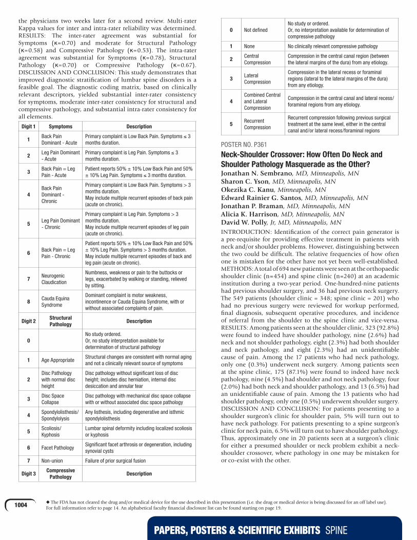
PAPERS, POSTERS & SCIENTIFIC EXHIBITS SPINE
1004 u The FDA has not cleared the drug and/or medical device for the use described in this presentation (i.e. the drug or medical device is being discussed for an off label use). For full information refer to page 14. An alphabetical faculty financial disclosure list can be found starting on page 19.
the physicians two weeks later for a second review. Multi-rater Kappa values for inter and intra-rater reliability was determined.RESULTS: The inter-rater agreement was substantial for Symptoms (κ=0.70) and moderate for Structural Pathology (κ=0.58) and Compressive Pathology (κ=0.53). The intra-rater agreement was substantial for Symptoms (κ=0.78), Structural Pathology (κ=0.70) or Compressive Pathology (κ=0.67).DISCUSSION AND CONCLUSION: This study demonstrates that improved diagnostic stratification of lumbar spine disorders is a feasible goal. The diagnostic coding matrix, based on clinically relevant descriptors, yielded substantial inter-rater consistency for symptoms, moderate inter-rater consistency for structural and compressive pathology, and substantial intra-rater consistency for all elements.
Digit 1 Symptoms Description
1 Back Pain Dominant - Acute
Primary complaint is Low Back Pain. Symptoms ≤ 3 months duration.
2 Leg Pain Dominant - Acute
Primary complaint is Leg Pain. Symptoms ≤ 3 months duration.
3 Back Pain = Leg Pain - Acute
Patient reports 50% ± 10% Low Back Pain and 50% ± 10% Leg Pain. Symptoms ≤ 3 months duration.
4Back Pain Dominant - Chronic
Primary complaint is Low Back Pain. Symptoms > 3 months duration. May include multiple recurrent episodes of back pain (acute on chronic).
5 Leg Pain Dominant - Chronic
Primary complaint is Leg Pain. Symptoms > 3 months duration. May include multiple recurrent episodes of leg pain (acute on chronic).
6 Back Pain = Leg Pain - Chronic
Patient reports 50% ± 10% Low Back Pain and 50% ± 10% Leg Pain. Symptoms > 3 months duration. May include multiple recurrent episodes of back and leg pain (acute on chronic).
7 Neurogenic Claudication
Numbness, weakness or pain to the buttocks or legs, exacerbated by walking or standing, relieved by sitting.
8 Cauda Equina Syndrome
Dominant complaint is motor weakness, incontinence or Cauda Equina Syndrome, with or without associated complaints of pain.
Digit 2Structural Pathology
Description
0No study ordered. Or, no study interpretation available for determination of structural pathology
1 Age AppropriateStructural changes are consistent with normal aging and not a clinically relevant source of symptoms
2Disc Pathology with normal disc height
Disc pathology without significant loss of disc height; includes disc herniation, internal disc desiccation and annular tear
3 Disc Space Collapse
Disc pathology with mechanical disc space collapse with or without associated disc space pathology
4 Spondylolisthesis/Spondylolysis
Any listhesis, including degenerative and isthmic spondylolisthesis
5 Scoliosis/Kyphosis
Lumbar spinal deformity including localized scoliosis or kyphosis
6 Facet PathologySignificant facet arthrosis or degeneration, including synovial cysts
7 Non-union Failure of prior surgical fusion
Digit 3Compressive
PathologyDescription
0 Not definedNo study or ordered. Or, no interpretation available for determination of compressive pathology
1 None No clinically relevant compressive pathology
2 Central Compression
Compression in the central canal region (between the lateral margins of the dura) from any etiology.
3 Lateral Compression
Compression in the lateral recess or foraminal regions (lateral to the lateral margins of the dura) from any etiology.
4Combined Central and Lateral Compression
Compression in the central canal and lateral recess/foraminal regions from any etiology.
5 Recurrent Compression
Recurrent compression following previous surgical treatment at the same level, either in the central canal and/or lateral recess/foraminal regions
POSTER NO. P361
Neck-Shoulder Crossover: How Often Do Neck and Shoulder Pathology Masquerade as the Other?Jonathan N. Sembrano, MD, Minneapolis, MN Sharon C. Yson, MD, Minneapolis, MN Okezika C. Kanu, Minneapolis, MN Edward Rainier G. Santos, MD, Minneapolis, MN Jonathan P. Braman, MD, Minneapolis, MN Alicia K. Harrison, MD, Minneapolis, MN David W. Polly, Jr, MD, Minneapolis, MNINTRODUCTION: Identification of the correct pain generator is a pre-requisite for providing effective treatment in patients with neck and/or shoulder problems. However, distinguishing between the two could be difficult. The relative frequencies of how often one is mistaken for the other have not yet been well-established.METHODS: A total of 694 new patients were seen at the orthopaedic shoulder clinic (n=454) and spine clinic (n=240) at an academic institution during a two-year period. One-hundred-nine patients had previous shoulder surgery, and 36 had previous neck surgery. The 549 patients (shoulder clinic = 348; spine clinic = 201) who had no previous surgery were reviewed for workup performed, final diagnosis, subsequent operative procedures, and incidence of referral from the shoulder to the spine clinic and vice-versa.RESULTS: Among patients seen at the shoulder clinic, 323 (92.8%) were found to indeed have shoulder pathology, nine (2.6%) had neck and not shoulder pathology, eight (2.3%) had both shoulder and neck pathology, and eight (2.3%) had an unidentifiable cause of pain. Among the 17 patients who had neck pathology, only one (0.3%) underwent neck surgery. Among patients seen at the spine clinic, 175 (87.1%) were found to indeed have neck pathology, nine (4.5%) had shoulder and not neck pathology, four (2.0%) had both neck and shoulder pathology, and 13 (6.5%) had an unidentifiable cause of pain. Among the 13 patients who had shoulder pathology, only one (0.5%) underwent shoulder surgery.DISCUSSION AND CONCLUSION: For patients presenting to a shoulder surgeon’s clinic for shoulder pain, 5% will turn out to have neck pathology. For patients presenting to a spine surgeon’s clinic for neck pain, 6.5% will turn out to have shoulder pathology. Thus, approximately one in 20 patients seen at a surgeon’s clinic for either a presumed shoulder or neck problem exhibit a neck-shoulder crossover, where pathology in one may be mistaken for or co-exist with the other.

PAPERS, POSTERS & SCIENTIFIC EXHIBITS SPINE
1005 u The FDA has not cleared the drug and/or medical device for the use described in this presentation (i.e. the drug or medical device is being discussed for an off label use). For full information refer to page 14. An alphabetical faculty financial disclosure list can be found starting on page 19.
POSTER NO. P362
Anterior Release Generates More Thoracic Rotation Than Ponte Osteotomy: A Biomechanical Human Cadaver Spine StudyAdam L. Wollowick, MD, New York, NY Erin Farrelly, MD, New York, NY Kate Meyers, MS, New York, NY Terry D. Amaral, MD, Bronx, NY Seth Grossman, MD, Bronx, NY Timothy M. Wright, PhD, New York, NY Vishal Sarwahi, MD, Bronx, NYINTRODUCTION: Historically, large and/or stiff spinal deformities were treated with anterior release to facilitate correction. However, anterior release increases risks and requires a two-part procedure. Recently, large or rigid deformities have been treated with a single posterior procedure using pedicle screws and osteotomies. No study in the literature has evaluated the effect of anterior release or posterior osteotomy on thoracic rotation.METHODS: Fourteen fresh frozen human thoracic spines were randomly assigned to anterior or posterior groups. Specimens were disarticulated at T4-T5 and T8-T9 to test upper, middle, and lower thoracic segments. Sections were potted, and reflective markers were placed on the vertebrae of interest (T2-T3, T6-T7, T10-T11). Specimens were mounted on a servo-hydraulic load frame. Specimens were cyclically loaded to ±5Nm axial rotation for 10 cycles with data from the tenth cycle analyzed. Specimens were tested intact then retested after sequential sectioning or removal of various structures. Anterior structures removed were: ALL, annulus fibrosis, nucleus pulposis, and PLL. Posterior structures removed were: intraspinous ligament, inferior facets, superior facets, spinous process, lamina, and ligamentum flavum. Motion was recorded using a 3D motion capture camera, and the relative motion of one vertebra to the other in axial rotation was calculated.RESULTS: Posterior sectioning produced a 27-82% increase in rotation from the intact specimens, while anterior release generated a 201-534% increase. Removal of the annulus, nucleus, and PLL led to a significant increase in rotation compared to intact specimens. Ponte osteotomy increased rotation 1.8-4.2°, while anterior release increased rotation 6.6-13.4°.DISCUSSION AND CONCLUSION: Anterior release generated significantly more thoracic rotation than Ponte osteotomy in biomechanical testing of human cadaver spines. An anterior or posterior structure that contributed most to rotational stability was not identified. Significance: Although many surgeons favor a single posterior approach to correct severe spinal deformity, anterior release may be needed to maximize correction. With increased emphasis being placed on spinal derotation, the use of anterior release should be reconsidered.
POSTER NO. P363
Sagittal Plane Spinal Deformity: Correction Thresholds and Health Related Quality of Life at Two Years Follow UpFrank J. Schwab, MD, New York, NY Virginie Lafage, PhD, New York, NY Benjamin Blondel, MD, New York, NY Justin S. Smith, MD, Charlottesville, VA Edward Chay, New York, NY Keith H. Bridwell, MD, Saint Louis, MO Steven D. Glassman, MD, Louisville, KY Christopher I. Shaffrey, MD, Charlottesville, VAINTRODUCTION: Recent studies have defined sagittal plane mal-alignment as the main driver of disability in adult spinal deformity (ASD). While normative values for sagittal plane parameters have been defined, this study aims to evaluate the amount of sagittal correction needed to achieve a minimal clinically important difference (MCID) in health related quality of life (HRQOL) scores for ASD patients.METHODS: This was a multicenter, retrospective analysis of consecutively enrolled ASD patients in a prospective database. Using demographic, radiographic, and HRQOL pre-operative and two-year post-surgery data, ASD patients were divided into two groups: pre-operative lumbar lordosis (LL) 80mm. Further subdivision was based on amount of correction: 15°, 15°-30°, and >30° for LL, and 120mm for SVA. HRQOL measures were Oswestry Disability Index (ODI), Scoliosis Research Society Questionnaire (SRS-22r) and Short Form 12 (SF-12). Statistical analysis was performed using paired t-test and one-way ANOVA.RESULTS: There were 79 and 76 patients in LL and SVA, respectively. Each group showed HRQOL improvements between pre-operative and two-year evaluations (p<0.05). In LL (mean pre-op LL=13°), an increase of <15° improved HRQOL (SRS-22r: 0.763 points; ODI: -15 points; p<0.05). Increasing LL by 15°-30° increased the likelihood of MCID for SRS activity (92%) and appearance (91%) (p30° increase in LL decreased the likelihood of MCID for SRS activity (61%) and appearance (74%) (p<0.05). In SVA (mean pre-op SVA=14cm), correcting by <60mm resulted in HRQOL improvements (SRS-22r: 0.757 points; ODI: -12 points, p120mm increased the likelihood of MCID in SRS appearance, ODI and SF-12 PCS (p<0.05).DISCUSSION AND CONCLUSION: The best HRQOL outcomes for ASD patients with severe sagittal plane deformity were obtained with corrections of 15°-30° for LL and >120mm for SVA. These results identify surgical correction thresholds for reaching HRQOL improvements for severe ASD patients.
POSTER NO. P364
Myelomalacia on MRI is Poorly Correlated with Severity of Disease in Cervical Spondylitic MyelopathyHan Jo Kim, MD, Saint Louis, MO Matthias Pumberger, MD, Berlin, Germany Joseph Nguyen, MPH, New York, NY Dara Froemel, MD, New York, NY Alexander P. Hughes, MD Andrew A. Sama, MD, New York, NY Frank P. Cammisa, Jr, MD, New York, NY Federico P. Girardi, MD, New York, NYINTRODUCTION: Myelomalacia is an MRI finding which suggests spinal cord damage. However, it is not uniformly found in patients with cervical spondylitic myelopathy (CSM)
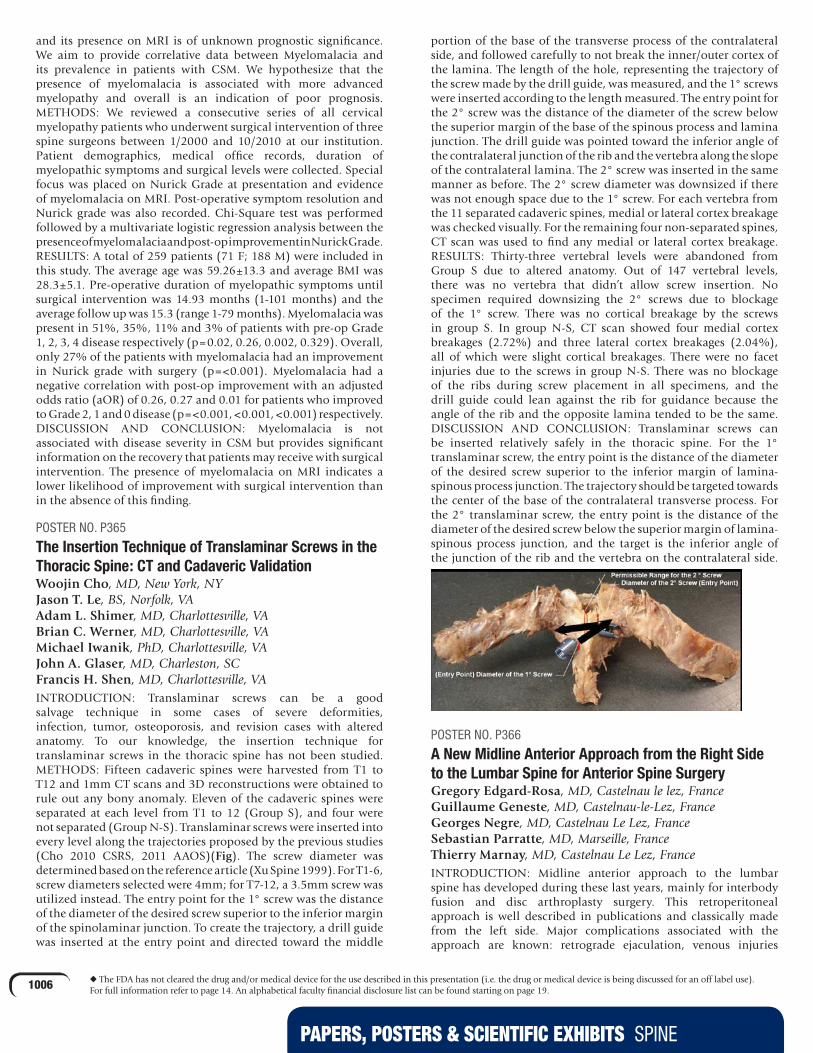
PAPERS, POSTERS & SCIENTIFIC EXHIBITS SPINE
1006 u The FDA has not cleared the drug and/or medical device for the use described in this presentation (i.e. the drug or medical device is being discussed for an off label use). For full information refer to page 14. An alphabetical faculty financial disclosure list can be found starting on page 19.
and its presence on MRI is of unknown prognostic significance. We aim to provide correlative data between Myelomalacia and its prevalence in patients with CSM. We hypothesize that the presence of myelomalacia is associated with more advanced myelopathy and overall is an indication of poor prognosis.METHODS: We reviewed a consecutive series of all cervical myelopathy patients who underwent surgical intervention of three spine surgeons between 1/2000 and 10/2010 at our institution. Patient demographics, medical office records, duration of myelopathic symptoms and surgical levels were collected. Special focus was placed on Nurick Grade at presentation and evidence of myelomalacia on MRI. Post-operative symptom resolution and Nurick grade was also recorded. Chi-Square test was performed followed by a multivariate logistic regression analysis between the presence of myelomalacia and post-op improvement in Nurick Grade.RESULTS: A total of 259 patients (71 F; 188 M) were included in this study. The average age was 59.26±13.3 and average BMI was 28.3±5.1. Pre-operative duration of myelopathic symptoms until surgical intervention was 14.93 months (1-101 months) and the average follow up was 15.3 (range 1-79 months). Myelomalacia was present in 51%, 35%, 11% and 3% of patients with pre-op Grade 1, 2, 3, 4 disease respectively (p=0.02, 0.26, 0.002, 0.329). Overall, only 27% of the patients with myelomalacia had an improvement in Nurick grade with surgery (p=<0.001). Myelomalacia had a negative correlation with post-op improvement with an adjusted odds ratio (aOR) of 0.26, 0.27 and 0.01 for patients who improved to Grade 2, 1 and 0 disease (p=<0.001, <0.001, <0.001) respectively.DISCUSSION AND CONCLUSION: Myelomalacia is not associated with disease severity in CSM but provides significant information on the recovery that patients may receive with surgical intervention. The presence of myelomalacia on MRI indicates a lower likelihood of improvement with surgical intervention than in the absence of this finding.
POSTER NO. P365
The Insertion Technique of Translaminar Screws in the Thoracic Spine: CT and Cadaveric ValidationWoojin Cho, MD, New York, NY Jason T. Le, BS, Norfolk, VA Adam L. Shimer, MD, Charlottesville, VA Brian C. Werner, MD, Charlottesville, VA Michael Iwanik, PhD, Charlottesville, VA John A. Glaser, MD, Charleston, SC Francis H. Shen, MD, Charlottesville, VAINTRODUCTION: Translaminar screws can be a good salvage technique in some cases of severe deformities, infection, tumor, osteoporosis, and revision cases with altered anatomy. To our knowledge, the insertion technique for translaminar screws in the thoracic spine has not been studied.METHODS: Fifteen cadaveric spines were harvested from T1 to T12 and 1mm CT scans and 3D reconstructions were obtained to rule out any bony anomaly. Eleven of the cadaveric spines were separated at each level from T1 to 12 (Group S), and four were not separated (Group N-S). Translaminar screws were inserted into every level along the trajectories proposed by the previous studies (Cho 2010 CSRS, 2011 AAOS)(Fig). The screw diameter was determined based on the reference article (Xu Spine 1999). For T1-6, screw diameters selected were 4mm; for T7-12, a 3.5mm screw was utilized instead. The entry point for the 1° screw was the distance of the diameter of the desired screw superior to the inferior margin of the spinolaminar junction. To create the trajectory, a drill guide was inserted at the entry point and directed toward the middle
portion of the base of the transverse process of the contralateral side, and followed carefully to not break the inner/outer cortex of the lamina. The length of the hole, representing the trajectory of the screw made by the drill guide, was measured, and the 1° screws were inserted according to the length measured. The entry point for the 2° screw was the distance of the diameter of the screw below the superior margin of the base of the spinous process and lamina junction. The drill guide was pointed toward the inferior angle of the contralateral junction of the rib and the vertebra along the slope of the contralateral lamina. The 2° screw was inserted in the same manner as before. The 2° screw diameter was downsized if there was not enough space due to the 1° screw. For each vertebra from the 11 separated cadaveric spines, medial or lateral cortex breakage was checked visually. For the remaining four non-separated spines, CT scan was used to find any medial or lateral cortex breakage.RESULTS: Thirty-three vertebral levels were abandoned from Group S due to altered anatomy. Out of 147 vertebral levels,there was no vertebra that didn’t allow screw insertion. No specimen required downsizing the 2° screws due to blockage of the 1° screw. There was no cortical breakage by the screws in group S. In group N-S, CT scan showed four medial cortex breakages (2.72%) and three lateral cortex breakages (2.04%), all of which were slight cortical breakages. There were no facet injuries due to the screws in group N-S. There was no blockage of the ribs during screw placement in all specimens, and the drill guide could lean against the rib for guidance because the angle of the rib and the opposite lamina tended to be the same.DISCUSSION AND CONCLUSION: Translaminar screws can be inserted relatively safely in the thoracic spine. For the 1° translaminar screw, the entry point is the distance of the diameter of the desired screw superior to the inferior margin of lamina-spinous process junction. The trajectory should be targeted towards the center of the base of the contralateral transverse process. For the 2° translaminar screw, the entry point is the distance of the diameter of the desired screw below the superior margin of lamina-spinous process junction, and the target is the inferior angle of the junction of the rib and the vertebra on the contralateral side.
POSTER NO. P366
A New Midline Anterior Approach from the Right Side to the Lumbar Spine for Anterior Spine SurgeryGregory Edgard-Rosa, MD, Castelnau le lez, France Guillaume Geneste, MD, Castelnau-le-Lez, France Georges Negre, MD, Castelnau Le Lez, France Sebastian Parratte, MD, Marseille, France Thierry Marnay, MD, Castelnau Le Lez, FranceINTRODUCTION: Midline anterior approach to the lumbar spine has developed during these last years, mainly for interbody fusion and disc arthroplasty surgery. This retroperitoneal approach is well described in publications and classically made from the left side. Major complications associated with the approach are known: retrograde ejaculation, venous injuries

PAPERS, POSTERS & SCIENTIFIC EXHIBITS SPINE
1007 u The FDA has not cleared the drug and/or medical device for the use described in this presentation (i.e. the drug or medical device is being discussed for an off label use). For full information refer to page 14. An alphabetical faculty financial disclosure list can be found starting on page 19.
and arterial thrombosis.The aim of this prospective study was to describe a midline anterior approach to the lumbar spine from the right side, below the aortic bifurcation to L5S1 and by mobilizing the vena cava from right to left between L2 and L5 and to evaluate the feasibility and complication rate.METHODS: A total of 469 patients were included in a prospective study between August 2003 and November 2010, either for interbody fusion by anterior approach or for total disc replacement, on one or several levels between L2-L3 and L5-S1.RESULTS: Of the 154 patients who had a mobilization of the vena cava, no injury occurred. Only four major venous injuries occurred. There was no arterial complication and the oxygen saturation signal was interrupted in only one case. No case of retrograde ejaculation was found.DISCUSSION AND CONCLUSION: The midline anterior retroperitoneal approach from the right side is a safe alternative compared to the classical approach from the left side. The low rate of venous injury is explained by the sidewall thickness of the vena cava compared to the left iliac vein sidewall. Contrary to what happens by left sided approach, the vascular retraction required for access to L4-L5 and above does not lead to arterial occlusion and therefore diminishes the risk in atheromatous patients. The absence of retrograde ejaculation confirms previous studies made on the left anastomosis of the superior hypogastric plexus suggesting that its approach and mobilization by the left side is delicate.
POSTER NO. P367
Analysis of Variance in Vertebral Bone Mineral DensityShaun P. Patel, BS, Ann Arbor, MI Sven Holcombe, BS, Ann Arbor, MI Stewart C. Wang, Ann Arbor, MI James A. Goulet, MD, Ann Arbor, MIINTRODUCTION: Worldwide, vertebral fractures are estimated to occur once every 22 seconds. They are a source of significant morbidity and healthcare expenditure, which will continue to increase as our population ages. Along with increased age, low bone mineral density is known to be a significant risk factor contributing to vertebral fractures. However, few studies have investigated whether there is variance in bone mineral density across vertebral levels and with respect to sex and age.METHODS: We identified 2,622 trauma patients between 2002 and 2010 who underwent a CT scan encompassing any part of their thoracic or lumbar spine. All CT scans were processed using semi-automatic algorithms through MATLAB R2011a. The mean Hounsfield Unit (quantitative scale for describing radiodensity) within each vertebral body was ascertained, which served as our measure of bone mineral density. This was performed at each vertebral level present on a given CT scan. Mean bone mineral density values were subsequently analyzed with respect to vertebral level, sex, and age.RESULTS: Analysis of CT scans yielded 26,346 unique vertebrae (17,067 male, 9,279 female) for which bone mineral density measurements were obtained. Across both sexes, there was a downward trend in bone mineral density with age (Figures 1 and 2). Across both sexes and nearly all age ranges, the lowest mean bone mineral density among thoracic vertebrae was at T8 and among lumbar vertebrae it was at L3. These were significantly lower when compared to the mean bone mineral density of the cumulative vertebrae for any corresponding age range (p < 0.05).DISCUSSION AND CONCLUSION: With this work, we present a large scale analysis of vertebral bone mineral density values across a diverse population. Our findings not only reaffirm declining bone
mineral densities with age, but also suggest specific vertebral levels at higher risk for fractures. These data may help further identify patients at risk for fractures, and, as a result, may help inform clinical decision making and targeted therapeutic interventions.
POSTER NO. P368
uTransforaminal Lumbar Interbody Fusion: Outcomes Among 155 Consecutive Patients from a High-Demand PopulationDimitri M. Thomas, MD, El Paso, TX Julia O. Bader, PhD, El Paso, TX Andrew Schoenfeld, MD, Canutillo, TXINTRODUCTION: Transforaminal interbody fusion (TLIF) is a safe and effective technique for achieving lumbar fusion with anterior column support. Few studies, however, have addressed outcomes following TLIF, especially in physically active and high demand populations, such as the U.S. military. This study sought to evaluate the efficacy of TLIF among active duty service members using retention within the military as an endpoint. The study also aimed to identify factors predictive of successful outcome following TLIF.METHODS: A consecutive cohort study was performed for all patients undergoing TLIF within our department from 2005-2008. In total, 155 active duty service members receiving TLIF were followed for a minimum of two years. The electronic medical record was abstracted for demographic information including age, gender, rank, branch of service, and smoking status. Pre-operative diagnosis, presence of fusion, number of levels at which TLIF was performed, complications, and revision surgeries were also noted. The ability of the patient to remain on active duty service, and whether they received a medical discharge for their spinal condition were determined as the dependent variable. Univariate
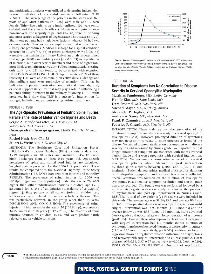
PAPERS, POSTERS & SCIENTIFIC EXHIBITS SPINE
1008 u The FDA has not cleared the drug and/or medical device for the use described in this presentation (i.e. the drug or medical device is being discussed for an off label use). For full information refer to page 14. An alphabetical faculty financial disclosure list can be found starting on page 19.
and multivariate analyses were utilized to determine independent factors predictive of successful outcome following TLIF.RESULTS: The average age of the patients in the study was 36.3 years of age. Most patients (n= 136) were male and 19 were female. Thirty-five patients were junior enlisted, 106 were senior enlisted and there were 14 officers. Ninety-seven patients were non-smokers. The majority of patients (n=148) were in the Army and most carried a diagnosis of degenerative disc disease (n=129). Eighty-one patients had single level fusions, whereas 74 had two or more levels. There were six complications that required seven subsequent procedures. Medical discharge for a spinal condition occurred in 30.3% (47/155) of patients, whereas 69.7% (108/155) were able to remain in the military. Univariate analysis determined that age (p < 0.001) and military rank (p < 0.0003) were predictive of retention, with older service members and those of higher rank more likely to remain on active duty. Following multivariate testing, only rank (p < .02) was found to be correlated with retention.DISCUSSION AND CONCLUSION: Approximately 70% of those receiving TLIF were able to remain on active duty. Older age and high military rank were predictive of retention. This may be indicative of patient motivation, occupational demands, and/or social support structures that may play a role in influencing a patient’s ability to remain in the military following TLIF. Results presented here show that TLIF is a successful intervention for younger, high-demand patients serving within the military.
POSTER NO. P369
The Age-Specific Prevalence of Pediatric Spine Injuries Parallels the Rate of Motor Vehicle Injuries and DeathSergio A. Mendoza-Lattes, MD, Iowa City, IA Yubo Gao, PhD, Iowa City, IA Gnanapradeep Gnanapragasam, MBBS, West Des Moines, Iowa Rachel Nash, Iowa City, IA Stuart L. Weinstein, MD, Iowa City, IAMETHODS: The Healthcare Cost and Utilization Project (HCUP) Kid’s Inpatient Database (KID) consists of data from 3,739 hospitals in 38 states and includes 3,452,325 non-birth discharges from children 0-19 years old. Age-specific prevalence of spine and spinal cord injuries are calculated. Weighed data is presented for the 2006 U.S. population. These results are compared with the National Highway Traffic Safety Administration (U.S. DOT) 2006 report on injuries and mortality.RESULTS: The prevalence of spinal injuries for 2006 was 106.8pmp (per million population) under the age of 20; 66% higher than other industrialized nations. Children age 15-19 accounted for 81.2% of all injuries (prevalence of 240.2pmp) (figure 1). Sixty percent of all spine injuries in children and adolescents were related to motor vehicle collisions. This was particularly relevant, in the group older than 15 years.DISCUSSION AND CONCLUSION: The prevalence of spinal injuries in children under the age of 20 (106.8pmp), represents a 34.7% increase in 10 years (1997 - 2006). The majority of spine injuries occurred in children 15-19, and were predominantly related to motor vehicle collisions.
POSTER NO. P370
Duration of Symptoms has No Correlation to Disease Severity in Cervical Spondylitic MyelopathyMatthias Pumberger, MD, Berlin, Germany Han Jo Kim, MD, Saint Louis, MO Dara Froemel, MD, New York, NY Michael Mayer, MD, Salzburg, Austria Alexander P. Hughes, MD Andrew A. Sama, MD, New York, NY Frank P. Cammisa, Jr, MD, New York, NY Federico P. Girardi, MD, New York, NYINTRODUCTION: There is debate over the association of the duration of symptoms and disease severity in cervical spondylitic myelopathy (CSM). However, prolonged duration of symptoms may not necessarily correlate to more progressive forms of the disease. We aimed to associate duration of symptoms with disease severity in CSM measured by Nurick grade. We hypothesize that longer duration of symptoms are associated with more advanced Nurick Grade and a worse prognosis with surgical intervention.METHODS: We reviewed a consecutive series of all cervical myelopathy patients who underwent surgical intervention by three spine surgeons between 1/2000 and 10/2010 at our institution. Patient demographics, medical office records, duration of myelopathic symptoms and surgical levels were collected. Special attention was focused on the duration of myelopathy symptoms. Post-operative symptom resolution and Nurick grade was also recorded. Chi-Square test was performed followed by a multivariate logistic regression analysis between the presence of myelomalacia and post-op improvement in Nurick Grade.RESULTS: A total of 259 patients (71 F; 188 M) were included in this study. The average age was 59.26±13.3 and average BMI was 28.3±5.1. Pre-operative duration of myelopathic symptoms until surgical intervention was 14.93 months (1-101 months) and the average follow up was 15.3 (range 1-79 months). More advanced Nurick grades did not correlate with longer duration of symptoms (p=0.813). However, those who improved at least one Nurick grade with surgical intervention had 6.1 months shorter duration of symptoms than those who stayed the same or worsened with surgery (11.2 vs. 17.3 months respectively, p = 0.003). Multivariate logistic regression showed a negative correlation with duration of symptoms and recovery with surgical intervention to at least grade 2, 1 or 0 disease (aOR 0.96, 0.97, 0.97 respectively, p=0.002, 0.004, 0.029).DISCUSSION AND CONCLUSION: Duration of myelopathic

PAPERS, POSTERS & SCIENTIFIC EXHIBITS SPINE
1009 u The FDA has not cleared the drug and/or medical device for the use described in this presentation (i.e. the drug or medical device is being discussed for an off label use). For full information refer to page 14. An alphabetical faculty financial disclosure list can be found starting on page 19.
symptoms do not correlate with severity of disease which question the natural history of CSM. However, those patients who did not improve with surgery had a longer duration of symptoms by approximately six months.
POSTER NO. P371
Preoperative Vitamin D Status of Adults Undergoing Spinal Fusion SurgeryGeoffrey Stoker, BS, Saint Louis, MO Jacob M. Buchowski, MD, Saint Louis, MO Keith H. Bridwell, MD, Saint Louis, MO Lawrence G. Lenke, MD, Saint Louis, MO K. D. Riew, MD, Saint Louis, MO Lukas P. Zebala, MD, Saint Louis, MOINTRODUCTION: Vitamin D plays a critical role in establishing optimal bone health, which, in turn, is vital to the success of spinal arthrodesis. Furthermore, deficiency-induced osteomalacia and osteoporosis predispose to postoperative screw pullout and instrumentation failure. The purpose of this study was to characterize the prevalence of preoperative vitamin D abnormality in adults undergoing spinal fusion and to determine whether previously identified risk factors for suboptimal vitamin D can be applied to our population.METHODS: Serum 25-hydroxyvitamin D (25OHD) levels were prospectively measured in adults (at least 18 years old) undergoing spinal fusion at a single institution. Between 1/2010 and 3/2011, 313 consecutive patients were identified for inclusion in this cross-sectional investigation. We generated a composite disability instrument by pooling Neck and Oswestry Disability Index scores of cervical and thoracolumbar patients, respectively. Potential risk factors for vitamin D deficiency were analyzed using Fisher’s exact, chi-squared, and Mann-Whitney U tests as well as multivariate logistic regression.RESULTS: The mean baseline 25OHD level was 29 ± 14 ng/mL. The prevalence of vitamin D inadequacy (<30 ng/mL) was 57%, and that of deficiency (<20 ng/mL) was 27%. There were 176 (56%) females. The overall mean age and BMI were 55 ± 13 years and 29 ± 5.8 kg/m^2, respectively. While 260 patients were diagnosed with degenerative disease, 99 had spinal deformity, and there were 73 revision cases. The cervical spine was included in 48% of fusion constructs; thoracic spine, 39%; lumbar spine, 51%. On average, 4.8 ± 4.7 motion segments were included. Deficient patients were younger (P = 0.009) and more likely to be <50 years old (P < 0.050). There was no gender difference. Bone mineral density was not lower in the setting of deficiency (P = 0.734). Deficient patients had significantly higher body mass index (BMI; P = 0.001) and disability scores (P = 0.004); likewise, they were more liable to be obese than of normal BMI (P = 0.001) and rate their disability greater than or equal to 60 than <60 (P = 0.005). The subgroup of patients with prior vitamin D and/or multivitamin supplementation was significantly older (P < 0.001) and more likely to be at least 50 years of age than those without prior repletion (P = 0.001). Increasing BMI (OR = 0.92, 95% CI = 0.87 - 0.97, P = 0.003), increasing disability scores (OR = 0.97, 95% CI = 0.95 - 0.99, P = 0.028), and a lack of prior vitamin D or multivitamin supplementation (OR = 0.14, 95% CI = 0.05 - 0.39, P < 0.001) reMEd significant predictors upon multivariate analysis. In other words, each one-unit increase in disability index equated to a 3% decrease in the likelihood of having 25OHD of at least 20 ng/mL. In contrast, a lack of prior supplementation conferred an 86% decrease in those odds.DISCUSSION AND CONCLUSION: Our investigation revealed an alarmingly high rate of vitamin D abnormality in the
analyzed population. Since augmenting serum 25OHD is straightforward and inexpensive, and hypovitaminosis-induced spinal osteoporosis and osteomalacia may predispose to poor surgical outcome, we advocate repletion for spine patients with documented deficiency. Although advanced age is a well-established risk factor for deficiency, young adults undergoing spinal fusion should not be overlooked with regard to preoperative vitamin D screening; younger patients are less likely to have been previously supplemented. Moreover, as validated indices of spine-related disability are predictive of vitamin D deficiency, these may be clinically useful tools in identifying at-risk patients in the absence of other well-established, physiologic risk factors.
POSTER NO. P372
uThe Reliability of X-Ray Based Evaluation of Pedicle Screw Misplacement in Adolescent Spinal DeformityPaul Haynes, MD, Ocean, NJ Beverly Thornhill, MD, Bronx, NY Gordon Sims, BA, Bronx, NY Jonathan Horn, Bronx, NY Adam L. Wollowick, MD, New York, NY Terry D. Amaral, MD, Bronx, NY Preethi M. Kulkarni, Bronx, NY Vishal Sarwahi, MD, Bronx, NYINTRODUCTION: Post-operative x-rays are routinely used to detect misplaced pedicle screws. Kim, et al. have defined radiographic criteria for evaluation of screw placement in spinal deformity. This study evaluates pedicle screw misplacement on x-ray using these criteria as well as anterior placement on lateral x-ray, and compared them to screw misplacements seen on post-operative CT scan.METHODS: Post-op x-rays and low dose CT scans of 104 adolescent spinal deformity patients who underwent PSF were reviewed. A blinded review of screw placement on x-ray was carried out using Kim et al.’s criteria: 1)violation of the harmonious change; 2)no crossing of medial pedicle wall by screw tip; 3)violation of imaginary midline of the vertebral body. On lateral x-ray, a screw was considered misplaced if: 1) an anterior breach was seen or 2) the length of the screw inside the vertebral body was ≥ 80% of the width of the vertebral body. Kappa analysis was used for overall agreement as well as agreement within specific regions of the curve.RESULTS: A total of 2,087 screws were evaluated on x-ray and CT. CT classified 1,820 screws as acceptable, 143 lateral, 30 medial, and 94 anterior. X-ray had 908 acceptable, 304 lateral, 241 medial, and 634 anterior. X-ray correlated with CT Scan in 50% of acceptable screws, 213% of laterally placed screws, 803% of medially placed screws, and in 674% of anteriorly placed screws. X-ray overestimated the number of misplaced screws and had poor reliability for detecting properly placed screws. Overall agreement was 0.08, indicating poor correlation. 53 screws were identified by CT scan as concerning - either breeching the canal or lying adjacent to the aorta. Of these, only 35% were correctly classified on x-ray. Further review of these screws on x-ray was unable to identify any relationship to structures of concern.DISCUSSION AND CONCLUSION: X-ray evaluation of screw placement showed poor correlation with CT data. X-rays were found to be inadequate to evaluate screw misplacement or relationship to structures of concern. Routine postoperative x-rays have significantly high false positive rates for screw misplacement. The practice of evaluating accuracy on x-ray needs to be examined. Low dose CT scan or intra-op image guidance should be considered.

PAPERS, POSTERS & SCIENTIFIC EXHIBITS SPINE
1010 u The FDA has not cleared the drug and/or medical device for the use described in this presentation (i.e. the drug or medical device is being discussed for an off label use). For full information refer to page 14. An alphabetical faculty financial disclosure list can be found starting on page 19.
POSTER NO. P373 ALTERNATE PAPER: SPINE VI
Patient Impressions of Reimbursement for Orthopedic Spine SurgeonsM. M. Gomberawalla, MD, Ann Arbor, MI Jeffrey S Fischgrund, MD, Southfield, MI Gregory Graziano, MD, Ann Arbor, MI Rakesh Patel, MD, Ann Arbor, MIINTRODUCTION: The purpose of this study was to understand patients’ impressions of reimbursement to orthopedic spine surgeons.METHODS: A survey was mailed to patients who underwent a spine procedure within the past three to 15 months at an academic and private institution. Surveys were returned in an anonymous fashion.RESULTS: A total of 103 surveys were completed. Some 76% of patients underwent a larger procedure (cervical fusion, lumbar fusion, or laminectomy with fusion), while others underwent a smaller procedure (kyphoplasty, discectomy, or laminectomy without fusion). Of those who underwent smaller procedures, 69% of patients felt the per-procedure reimbursement was between $1,000 and 5,000. Of those who underwent larger fusion-based procedures, 84% of patients felt the reimbursement was greater than $5,000, of which 28% believed the reimbursement was greater than $15,000 for their procedure (Figure 1). Most patients (73%) believed procedural reimbursement should primarily be based on technical difficulty. Only a minority of patients felt that in-hospital rounding and outpatient follow up in the first 90 days were included in the procedural reimbursement (38% and 26%, respectively). A total of 41% of patients believed spine surgeons’ annual salaries are between $250,000 and $500,000, and 22% believed it was between $500,000 and $750,000. Most patients (73%) felt that spine surgeons are appropriately compensated. Thirty-eight percent of patients believed reimbursement should be addressed as a part of healthcare reform.DISCUSSION AND CONCLUSION: A majority of patients believe spine surgeons are reimbursed a higher amount per procedure than factually true. Despite this, most patients feel that spine surgeons are appropriately compensated. Additionally, most patients are not aware that early post-operative inpatient rounding and outpatient clinical care are included within the global billing period.
POSTER NO. P374
Cervical Sagittal Contour Decompensation Before and After Surgery in Adolescent Idiopathic ScoliosisEdward P. Curry, MD, Philadelphia, PA Brian Winters, MD, Mount Laurel, NJ Petya Yorgova, MS, Wilmington, DE Geraldine Neiss, PhD, Wilmington, DE Alan S. Hilibrand, MD, Philadelphia, PA Suken A. Shah, MD, Wilmington, DEINTRODUCTION: The cervical sagittal contour of patients with adolescent idiopathic scoliosis (AIS) may be clinically important, as neck pain is a major complaint in long term follow up studies. Cervical spine alignment and sagittal vertical axis are affected by thoracic kyphosis. Thoracic hypokyphosis is a common feature of AIS and can be aggravated by contemporary instrumentation techniques with segmental pedicle screw systems. This study sought to examine the relationship of thoracic hypokyphosis and cervical spine sagittal contour before and after posterior spinal fusion for AIS.METHODS: A single-surgeon’s prospective, consecutive series of patients (2005-2008) treated with segmental pedicle screw instrumentation for thoracic AIS (Lenke 1,2) with minimum two year follow up yielded 65 patients with adequate cervical spine views for analysis. Cervical sagittal contour was measured by one of two observers from the inferior endplate of C2 to the inferior endplate of C6, as described by Hilibrand, et al (1995) preoperatively, at the first erect film and at two year follow up. Arm position was standardized with hands resting on poles and arms flexed forward 20-30°. A comprehensive collection of global and local sagittal parameters were analyzed.RESULTS: “Flattening” of the cervical sagittal contour was observed in patients with AIS before and after surgery. Preoperatively, the sagittal contour from C2-C6 was 7.0 ± 2° of kyphosis and 4.6 ± 2° of kyphosis two years after surgery (p<0.01). Thoracic sagittal contour preoperatively was 28 ± 6° of kyphosis (T2-T12) and was preserved after surgery (29.6 ± 3°). A strong correlation existed between thoracic hypokyphosis and the development of cervical kyphosis - and this was significant preoperatively (p=0.003) and two years postoperatively (p=0.004). A relatively small correction of the thoracic kyphosis (4.8%) translated into a substantial and spontaneous correction in the cervical lordosis of -39%.DISCUSSION AND CONCLUSION: In AIS, thoracic hypokyphosis is associated with a risk of cervical decompensation into kyphosis. Efforts to restore even some thoracic kyphosis postoperatively during scoliosis surgery may have profound effects in establishment of cervical lordosis.
POSTER NO. P375
Ultrasonographic Quantification of Spinal Cord and Dural Pulsations during Cervical LaminoplastyAtsushi Kimura, MD, Shimotsuke, Japan Atsushi Seichi, MD, Shimotsuke, Japan Hirokazu Inoue, MD, Shimotsuke, Tochigi, Japan Teruaki Endo, Shimotsuke City, Japan Yuichi Hoshino, MD, Tochigi, JapanINTRODUCTION: Pulsatile movements of the spinal cord and dura mater have been interpreted as a sign that the cord is free within the subarachnoid space, with no extrinsic compression. However, the association between restoration of pulsation and adequate decompression of the spinal cord has not been established. The purpose of this study was to investigate the relationship between the extent of spinal cord decompression and spinal cord and dural

PAPERS, POSTERS & SCIENTIFIC EXHIBITS SPINE
1011 u The FDA has not cleared the drug and/or medical device for the use described in this presentation (i.e. the drug or medical device is being discussed for an off label use). For full information refer to page 14. An alphabetical faculty financial disclosure list can be found starting on page 19.
pulsations by quantitative analysis of intraoperative ultrasonopraphy.METHODS: The study enrolled 85 consecutive patients with cervical myelopathy (55 male, 30 female; mean age 64 ±13 years). Spinal cord decompression status was classified as: Type 1, noncontact, retaining the presence of the subarachnoid space on the ventral side of the cord; Type 2, contact and apart, with the cord showing contact with and separation from the anterior element of the cervical spine; and Type 3, contact, with the cord in continuous contact with the anterior element. Spinal cord and dura mater dynamics were analyzed quantitatively using automatic video-tracking software.RESULTS: The amplitude of spinal cord pulsations ranged from 0.01-0.84 mm (mean 0.30 ± 0.16 mm) and that of dural pulsations ranged from 0.01-0.38 (mean 0.14 ± 0.08). The average spinal cord pulsation amplitude in type 2 patients was significantly larger than in other types. However, the average dural pulsation amplitudes were similar in all groups. There was a significant correlation between spinal cord and dural pulsation amplitudes in type 1, but not in type 2 or type 3 patients. Type 3 patients showed a particularly poor correlation between spinal cord and dural pulsations.DISCUSSION AND CONCLUSION: The results of this study revealed that restoration of dural pulsation is not a reliable indicator of sufficient spinal cord decompression following a decompression procedure.
POSTER NO. P376
Protective Changes in Cervical Spine Geometry Occurring with Age: A Cadaveric Study of 1,072 Human SkeletonsNavriakt Bajwa, Cleveland, OH Nicholas U. Ahn, MD, Shaker Heights, OHINTRODUCTION: Prior studies have shown changes in lumbar vertebral parameters such as vertebral body diameter, sagittal diameter, interpedicular distance and pedicle length with age. Few studies have been done to evaluate changes in cervical spine morphology with age, and these have primarily been in the pediatric population. However no studies have been done which study the changes in cervical anatomy with age in adulthood. This study was performed to determine changes in cervical morphology which occur with age in adults, and whether these changes have an impact on development of cervical stenosis with advancing age.METHODS: A total of 1,072 cadaveric lumbar specimens from Hamann-Todd Osteological Collection in Cleveland, OH were examined by a single examiner. Baseline data including age, sex, and race of subjects were collected. The gross specimens were measured subjectively with digital caliper. The following measurements were made; body diameter, measured as the anteroposterior distance of each vertebral body, the interpedicular distance, the sagittal diameter and pedicle length at each of the cervical levels (C3-C7). Linear regression analyses were then used to determine the relationship between these parameters while correcting for confounding factors of age, sex and race.RESULTS: In all the specimens, there was a significant positive association between changes in body diameter with age from C3 to C7 (p<0.01). At all levels there was also a significant association between sagittal diameter and age (p<0.01). Pedicle length was associated with age only at levels C4, C5 and C7 (p<0.03). Interpedicular distance was not significantly associated age at any level (p>0.1).DISCUSSION AND CONCLUSION: This study suggests that changes occur in body diameter, sagittal diameter and pedicle length with age in adulthood. The increases in cervical sagittal diameter, in particular, are interesting as this parameter impacts the presence of cervical stenosis. The forces across the cervical spine may result in degenerative changes which lead to cord

PAPERS, POSTERS & SCIENTIFIC EXHIBITS SPINE
1012 u The FDA has not cleared the drug and/or medical device for the use described in this presentation (i.e. the drug or medical device is being discussed for an off label use). For full information refer to page 14. An alphabetical faculty financial disclosure list can be found starting on page 19.
compression, but these same forces also appear to cause alterations in bony anatomy with age, with an increase in sagittal diameter and which in turn may have a protective effect in preventing cervical stenosis. This may explain why cervical stenosis leading to cord compression and myelopathy is uncommon despite tremendous forces on cervical spine that occur over the course of time.
POSTER NO. P377
The Prevalence of Abnormal Preoperative Neurologic Exam in Scheuermann’s KyphosisWoojin Cho, MD, New York, NY Lawrence G. Lenke, MD, Saint Louis, MO Keith H. Bridwell, MD, Saint Louis, MO Guangxun Hu, Shenzhen City, China Jacob M. Buchowski, MD, Saint Louis, MO Joshua Pahys, MD, Wynnewood, PA Samuel Kang-Wook Cho, MD, Palisades park, NJ Matthew M. Kang, MD Lukas P. Zebala, MD, Saint Louis, MO Linda A. KoesterINTRODUCTION: There have been sporadic reports about abnormal neurologic findings in Scheuermann’s kyphosis patients. The purpose of this study was to report the prevalence of abnormal neurologic findings detected by physical exam in Scheuermann’s kyphosis, and to correlate it to x-rays, MRI findings, and results of operative treatment.METHODS: Among 82 Scheuermann’s kyphosis patients who underwent corrective surgery, 69 primary cases were selected. Patient charts were reviewed retrospectively in terms of pre and postop neurological exams. Sensory or motor change was defined as an abnormal neurologic exam. Their duration, associated problems, and various parameters on preop x-rays and MRI exams were also measured to search for any atypical findings associated with an abnormal neurologic exam.RESULTS: There were six cases (9%)(Group AbN) with an abnormal neurologic exam ranging from severe myelopathy to a subtle change (eg. sensory paresthesias on trunk). Five patients recovered to a normal neurologic exam after corrective surgery. The remaining one severe myelopathic patient also showed marked improvement and was ambulatory unassisted by two-year follow up. In patients with a normal neurologic exam (Group N, n=63), only one patient had neurologic sequelae due to anterior spinal artery syndrome after combined A/P correction. No preop x-ray parameters were significantly different between groups. Average age was 21.3 (AbN) and 18.6 (N) yrs (P=0.55). Average preop T5-12 kyphosis was 69.0º(AbN) and 72.5º(N) (P=0.61). Forty-two MRIs were obtained, all showed typical findings of Scheuermann’s kyphosis. In AbN, five patients had MRI (one had a Ct/myelo) and in 37 in N.DISCUSSION AND CONCLUSION: The prevalence of abnormal neurology in Scheuermann’s kyphosis was 9%, emphasizing the importance of detailed preop neurologic exam. If congenital stenosis or herniated thorathic disc is combined, myelopathy can occur. No x-ray findings correlated with the abnormal preop neurologic exam. A normal MRI can exist in the face of an abnormal neurologic exam, and conversely, a normal neurologic exam can be seen with an abnormal MRI as well. Surgery was successful in alleviating abnormal neurologic issues. Deformity surgeons who correct Scheuermann’s kyphosis should rule out neurologic issues preoperatively.
POSTER NO. P378
Increased Incidence of C2 Fractures in the Elderly PopulationYohan Robinson, MD, Uppsala, Sweden Bengt Sanden, MBBS, Uppsala, Sweden Claes Olerud, MD, Uppsala, SwedenINTRODUCTION: Fractures of the second cervical vertebra - axis - are a common injury in the elderly population and treatment is often complicated due to patient morbidity. The number of geriatric patients increased during the last decades due to an increase in mean population age. Until now little epidemiological data is available allowing investigating whether the number of C2 fractures increased during the last decade.METHODS: Data for all patients with C2 fractures admitted to hospital between 1997 and 2009 were abstracted from the Swedish National Hospital Discharge Register (SNHDR). The data in the register are collected prospectively, recording all inpatient admissions throughout Sweden. The SNHDR uses the codes for diagnoses at discharge and surgical procedures according to the Swedish version of the International Classification of Diseases (ICD).RESULTS: A total number of 4,444 patients (2,072 women, 2,372 men) with C2 fractures were treated as inpatients in Sweden during the years from 1997 to 2009. Of these 1,267 were operated. The annual incidence of C2 fractures showed a linear increase during the years (r=0.92). This was mainly due to an increase in the geriatric subgroup, while the younger age groups reMEd unchanged during the observation period. Interestingly the percentage of operated patients rather decreased until 2009 (r=-0.65).DISCUSSION AND CONCLUSION: While the elderly population grows dramatically, the number of hospital admissions due to elderly-specific C2 fractures increased during the last decade. Possible explanations are greater awareness of fractures, improved diagnostics, and a higher activity level of the patients. Due to the continuous increase of odontoid fractures treatment modalities have to be optimised to reduce fatalities. Obviously there is a trend to more conservative treatment in Sweden. Randomized controlled trials allowing evidence-based recommendations are needed to establish a treatment rationale for C2 fractures.

PAPERS, POSTERS & SCIENTIFIC EXHIBITS SPINE
1013 u The FDA has not cleared the drug and/or medical device for the use described in this presentation (i.e. the drug or medical device is being discussed for an off label use). For full information refer to page 14. An alphabetical faculty financial disclosure list can be found starting on page 19.
POSTER NO. P379
Clinical Outcomes after Lumbar Fusion Complicated by Deep Wound Infection: A Case-Control StudyJulio Petilon, MD, Portsmouth, VA Steven D. Glassman, MD, Louisville, KY John R. Dimar, II, MD, Louisville, KY Leah Y. Carreon, MD, Louisville, KYINTRODUCTION: Postoperative infection following instrumented spinal fusion is a major complication, often resulting in substantial short term morbidity. However, there is little literature reviewing how these patients do in the longer term, after their infection has been managed. This study evaluated the two-year clinical outcomes of patients who had instrumented spinal fusions complicated by deep wound infections and compared them to a propensity-matched control group.METHODS: Surgical and clinical databases from 2001 to 2008 were reviewed for eligible subjects. Inclusion criteria consisted of patients who underwent instrumented lumbar spinal fusion with complete pre-operative and two-year postoperative outcome measures and had acute (≤3 months) postoperative deep wound infections necessitating irrigation and debridement. Thirty subjects met criteria for analysis. Outcome measures included the Oswestry Disability Index (ODI), SF-36 Physical (PCS) and Mental (MCS) composite summaries and Numeric rating scales (0-10) for back and leg pain. A non-infected control group was identified using propensity-matching techniques based on demographics, baseline clinical outcome measures and surgical characteristics. Two year postoperative outcome measures of both groups were compared. The proportion of patients in each group achieving the minimum clinically important difference (MCID) for the outcome measures was also assessed. Independent t-tests were used to compare continuous variables and Fisher’s exact test was used to compare categorical variables between the two groups.RESULTS: Consistent with the propensity-matching technique, there were no significant demographic or surgical differences between the two groups at baseline (Table 1). ODI, PCS, back and leg pain scores were statistically significantly better at two years post-operative compared to baseline in both groups. However, at two years post-operative, the infection group had statistically significantly worse back pain scores compared to the control group (6.45 vs. 4.7, p=0.020). Also, a greater proportion of patients in the control group (18, 60%) achieved MCID for ODI compared to the infection group (8, 27%, p=0.018).DISCUSSION AND CONCLUSION: This study demonstrates that patients with acute postoperative deep wound infections following instrumented lumbar spinal fusion have improved outcome measures after surgery but have greater back pain and a decreased probability of achieving MCID for ODI than patients without infection two years following surgery.
NO INFECTION INFECTION p-value
N 30 30
Sex (male) 12 8 0.402
Surgery
ALIF 2 2 1.000
TLIF 8 8
PSF 17 17
ASF/PSF 3 3
Smoker 6 8 0.761
Workers’’ comp 2 1
Diagnosis
Spondylolisthesis 8 8 1.000
Instability 1 1
Stenosis 2 2
Scoliosis 2 2
Disc pathology 5 5
Non-union 3 3
Post-decompression 5 5
Adjacent level 4 4
Age 44.77 44.57 0.864
Length of Stay 5.37 5.57 0.731
Operative Time 288.17 259.27 0.298
Estimated Blood Loss 857.59 750.86 0.617
ASA Grade 2.63 2.83 0.136
No. of Levels 1.93 1.83 0.705
BMI 31.62 34.22 0.200
Baseline
Back Pain 7.31 7.87 0.413
Leg Pain 7.52 7.67 0.821
ODI 56.82 55.30 0.721
PCS 26.91 26.49 0.832
MCS 35.06 37.40 0.548
Two-Year Post-operative
Back Pain 4.70 6.45 0.020
Leg Pain 5.07 5.73 0.392
ODI 39.73 48.20 0.198
PCS 34.25 30.04 0.144
MCS 37.98 37.65 0.931
Proportion achieving MCID
Back Pain 17 13 0.439
Leg Pain 23 22 1.000
ODI 18 8 0.018
PCS 15 15 1.000
POSTER NO. P380
Sagittal Classification for Ossification of the Ligamentum Flavum of the Thoracic SpineKei Ando, MD, Nagoya, Japan Shiro Imagama, MD, Nagoya, Japan Zenya Ito, PhD, Nagoya, Japan Yukihiro Matsuyama, MD, Hamamatsu, Japan Naoki Ishiguro, MD, Nagoya, JapanINTRODUCTION: Thoracic myelopathy due to an ossified ligamentum flavum (OLF) generally requires surgical intervention due to its progressive nature and its poor response to conservative therapy. The purpose of this study was to provide the first evidence for the influence of ossified anterior longitudinal ligament (OALL) on the clinical features and surgical outcomes in thoracic ossified ligamentum flavum (OLF).METHODS: Sixty-three patients who underwent surgery for a one level thoracic OLF were identified, and preoperative symptoms, severity of symptoms and myelopathy, disease duration, MR imaging and CT findings, surgical procedure, intraoperative findings, complications, and postoperative recovery were investigated in these patients. Findings on sagittal CT images that was adjacent to or at the same vertebral level were classified into four types: No discernible type (Type N); One sided type (Type O); Discontinuous (Type D); and continuous type (Type C).RESULTS: The duration of symptoms was especially long in the Type D and C OALLs. Patients with Type D had a significantly poorer percentage of recovery, the same as their preoperative JOA scores.

PAPERS, POSTERS & SCIENTIFIC EXHIBITS SPINE
1014 u The FDA has not cleared the drug and/or medical device for the use described in this presentation (i.e. the drug or medical device is being discussed for an off label use). For full information refer to page 14. An alphabetical faculty financial disclosure list can be found starting on page 19.
DISCUSSION AND CONCLUSION: The authors’ results showed that a Type D OALL had strong relationships with preoperative severity of symptoms and surgical outcomes. These findings may allow surgeons to determine the severity of preoperative symptoms and the probable surgical outcomes from the OALL classifications. Moreover, the surgery with instrumentation for Type D OALL may have better surgical outcome.
POSTER NO. P381
uAre Pedicle Screw Constructs Really More Expensive than Hybrid Constructs?Le-qun Shan, MD, PhD, Xi’An, China David L. Skaggs, MD, Los Angeles, CA Christopher Lee, East Greenwich, RI Karen Myung, MD, Los Angeles, CAINTRODUCTION: The use of pedicle screws in the treatment of scoliosis has been increasingly adopted, with most studies showing greater correction of spinal deformity than hybrid constructs. However, the increased implant expense of pedicle screws compared to hybrid constructs are concerning. The hypothesis of this study is that if revision surgery secondary to implant failure is taken into account, the total expense of pedicle screw versus hybrid constructs in the treatment of adolescent idiopathic scoliosis (AIS) is similar.METHODS: A retrospective review of 872 consecutive cases of AIS patients that underwent posterior fusion (PSF) with hybrid instrumentation or pedicle screw instrumentation at a single institution from December 1995 to 2009 was performed. Inclusion criteria consisted of diagnosis of AIS, posterior spinal fusion with segmental instrumentation, and a minimum of two-year follow up. Patient demographics, radiographic parameters, operative data, and comprehensive charges for implant related revision operations were recorded. Revision surgery for infection was not included in the financial analysis.RESULTS: A total of 167 patients with an average age of 14 years and 11 levels fused (6-15) met inclusion criteria. Mean major curve correction was 72% in the pedicle screw group and 54% in the hybrid group (p=0.00). There was no significant difference in average preoperative major Cobb angle (p=0.09), operative time (p=0.48), anesthesia time (p=0.82) and average days in ICU (p=0.42) between the two groups. Among the 44 patients treated with all pedicle screws there were no implant related surgical revisions. Of the 123 patients in the hybrid group, there were 29 revision surgeries in 21 patients for implant failure (revision rate of 17%). We calculated the present day charges in our institution for an 11 level fusion using titanium implants to be $42,450 for the average pedicle screw construct and $26,064 for the average hybrid construct. The mean charges of revision surgery totaled $1,639,910, or $13,333 on average for each patient in the hybrid group including implant charges, professional fees, and hospital charges. Though the charge of initial surgery was roughly $16,400 less in the hybrid construct group, when revision charges for implant failure were added in, average estimated charges for the pedicle screw group at final follow up was 7.2% higher than the hybrid group ($42,450 vs. $39,397).DISCUSSION AND CONCLUSION: Patients with AIS undergoing PSF with all pedicle screw constructs had a markedly lower revision rate (0% vs. 17%) when compared to hybrid constructs, but total charges were 7.2% higher. In order to judge if new technology is worth increased initial expense, a financial analysis of complications over time should be assessed. In our opinion, the markedly decreased revision rate with all pedicle screw constructs compared to hybrid constructs are worth a 7.2% increase in spinal implant charges.
POSTER NO. P382
uTwo Year Bone Morphogenetic Protein (BMP) Clinical Effects after Lumbar Fusion in Degenerative Disc Disease (DDD)Evalina L Burger, MD, Aurora, CO Vikas V. Patel, MD, Denver, CO Andriy Noshchenko, MD, PhD, Aurora, COINTRODUCTION: Several prospective randomized controlled clinical trials (RCT) have reported that bone morphogenetic protein (BMP) significantly improves lumbar fusion clinical outcomes and fusion rates at 12 months postoperative, particularly in patients with spondylolisthesis. However, other relatively small studies have reported higher complication rates after BMP use. The purpose of the present meta-analysis study was to evaluate the effects of BMP on long-term lumbar fusion clinical outcomes in patients with degenerative disc disease (DDD).METHODS: The Cochrane database of randomized controlled trails (RCT), Cochrane Database of Systematic Reviews, PubMed, Medline, Embase, ClinicalTrial.gov, and published reviews were used to search for published studies that met our inclusion/exclusion criteria. Inclusion criteria were: 1) age >18 years, 2) lumbar DDD with or without: stenosis, 3) grade I-II degenerative spondylolisthesis, disk herniation, and other disk pathology, 4) follow up ≥ 24 month for clinical outcomes and fusion rate, 5) use of Oswestry disability index (ODI), back and/or leg pain Visual analog scale (VAS), and/or SF-36 questionnaire, 6) not less than two year postoperative complication rate, 7) employment rate before and two years after surgery, 8) published between 2000 and 2010, 9) English language, 10) level of evidence ≥5 by van Tulder’s scale for RCT, and ≥7 by Cowley’s scale for cohort studies and case series. Exclusion criteria: scoliosis, trauma, isthmic spondylolisthesis, tumors, radiculopathy. A meta-analysis was performed to assess pooled treatment effect size, heterogeneity, and the risk of publication bias. The data were grouped by whether or not BMP was used. Data pooling was performed by randomized effect model.RESULTS: A total of 18 studies from seven countries met the inclusion/exclusion criteria: 14 RCT, three cohort studies, and one retrospective case series. Pooled data showed significant improvements in disability, pain, and physical health status after lumbar fusion regardless of whether or not BMP was used, (Table). Fusion rates were relatively high and approximately similar for both BMP and non-BMP groups. Employment rate did not improve significantly after surgery in either group. BMP was not associated with an increased complication rate. The risk of publication bias was not significant (p<0.001).DISCUSSION AND CONCLUSION: Fusion outcomes at 24-48 months postoperative for lumbar DDD are not significantly improved by the use of BMP. This result suggests that the potential benefits of BMP are limited to the early stages of fusion.

PAPERS, POSTERS & SCIENTIFIC EXHIBITS SPINE
1015 u The FDA has not cleared the drug and/or medical device for the use described in this presentation (i.e. the drug or medical device is being discussed for an off label use). For full information refer to page 14. An alphabetical faculty financial disclosure list can be found starting on page 19.
POSTER NO. P383
Loss of Lordosis Following Cervical LaminoplastySamuel M. Davis, MD, Atlanta, GA John J. Rhee, MD, Atlanta, GAINTRODUCTION: Although laminoplasty is not commonly associated with the severe postoperative kyphotic deformities that may occur with multilevel laminectomy alone, changes in sagittal alignment, including loss of lordosis, can occur. However, this process has not been extensively studied in the literature. The purpose of this study was to evaluate the loss of lordosis that occurs after open door laminoplasty, its pattern, extent, and timecourse.METHODS: Thirty-one patients with satisfactory neutral lateral radiographs at preop, six weeks, three months, six months, and one year were selected from a cohort of patients who underwent open door cervical laminoplasty between 2007-2009 for cervical spondylotic myelopathy. At surgery, C2 muscle attachments were preserved and not detached whenever possible or reattached if necessary during closure. All patients were encouraged to begin active range of motion and extensor muscle strengthening exercises as soon as tolerable postop and to continue them throughout the postoperative course. Sagital cobb angles of the instrumented cervical levels were determined. Forward pitch (FP) was measured as the distance from a vertical plumb line drawn from the center of the cortical ring of C2 to the anteroinferior corner of the lowest instrumented vertebrae (Fig 1). The change from preoperative was calculated for all patients at each time interval. Per convention, negative numbers were used to denote lordosis, positive numbers to denote kyphosis.RESULTS: On average, patients lost 6o of lordosis from preop (-9.6o) to one year postop (-3.6 o). The greatest loss of lordosis occurred between preop to six weeks and averaged 11 oof loss. Between three to six months, the loss of lordosis gradually improved compared to six weeks, but the overall lordosis never returned to preoperative levels. There was no significant difference in the amount of lordosis lost among those with a preop Cobb > -10 o versus those with < -10 o
at one year (-6.5 o vs. -5.7 o , p=0.65). There was no difference in the preoperative Cobb angle between those with lower versus higher forward pitch (p=0.52). However, those with less forward pitch (FP <1 cm; n=14) lost 4.5o of lordosis at one year, whereas those with greater forward pitch (FP > 1cm; n=17) lost 7.3 o (p=0.072)
Preop Cobb
6 wks (change)
3 mo (change)
6 mo (change)
1 yr (change)
All (n=31) -9.6 o+1.8 o (11.4 o)
-1.1 o (8.5o)
-4.4 o (5.2 o)
-3.6 o (6.0 o)
Preop cobb > -10 o (n=13)
-19.3 o -8.6 o (10.7 o)
-12.5 o (6.8 o)
-11.9 o (7.4 o)
-12.8 o (6.5 o)
Preop cobb < -10 o (n=18)
-2.6 o+9.1 o (11.7 o)
+10.4 o (13 o)
+1.1 o (3.7 o)
+3.0 o (5.6 o)
FP < 1cm (n=14)
-9.5 o-1.2 o (8.3 o)
-5.4 o (4.1 o)
-4.8o (4.7 o)
-5.0 o (4.5 o)
FP > 1cm (n=17)
-9.7 o+4.6 o (14.3 o)
+1.3o (11 o)
-4.0o (5.7 o)
-2.5 o (7.2 o)
DISCUSSION AND CONCLUSION: Patients lose on average 6 o of lordosis at one year after open door cervical laminoplasty, with the greatest loss occurring at six weeks postop and then partially recovering over time. The changes at six weeks may reflect pain and splinting, whereas those at one year may reflect chronic muscle denervation or atrophy. There was no significant difference in loss of lordosis at one year between those who had more or less than -10o of preoperative lordosis. However, those with greater forward pitch of more than 1 cm demonstrated greater ultimate loss of
lordosis. These data provide potentially important considerations in preoperative preparation and patient counseling of patients requiring surgery for multilevel myelopathy.
POSTER NO. P384 ALTERNATE PAPER: SPINE III
The Relation Between Pulmonary Function and Selection of Upper Instrumented Vertebra in AISSatoru Demura, MD, Kanazawa, Japan Peter O. Newton, MD, San Diego, CA Burt Yaszay, MD, San Diego, CA Tracey Bastrom, MA, San Diego, CA John Schlechter, DO, Irvine, CAINTRODUCTION: There seems to be increasing concern that a more proximal extent of posterior thoracic spinal instrumentation and fusion (PSF) reduces postoperative pulmonary function. However, there are few reports to analyze the relation between the selection of upper instrumented vertebra (UIV) and pulmonary function in adolescent idiopathic scoliosis (AIS). The objective of this study was to evaluate pulmonary function to determine whether a more proximal UIV negatively impacts pulmonary function in surgically treated AIS patients.METHODS: Pulmonary function testing (PFT) and radiographic examination of 154 patients with major thoracic AIS (Lenke type 1 to 4) undergoing PSF without thoracoplasty were completed prospectively.Patients were divided into groups based on UIV (T1 to T3 vs. T4 to T5) and Lenke curve type (2 & 4 vs. 1 & 3) and analyzed respectively. Demographic, radiographic measurements, and PFT data from preoperative and two year time points were analyzed.RESULTS: Patients with a structural upper thoracic curve (Lenke 2 and 4) had significantly lower preoperative PFT values than those without a structural upper thoracic curve (Lenke 1 and 3).Lenke 2 and 4 patients were also more likely to be fused proximally (82% T1-T3) than those in the Lenke 1 and 3 group (42% T1-T3, p<0.05). Pre-operatively, those with UIV from T1 to T3 had lower PFT values than those with UIV from T4-T5; however, only %TLC was statistically significant (p<0.05). Both UIV groups showed significant increases in all absolute values (FVC, FEV1, TLC) at two-year follow up (p<0.05), and the percent predicted values (%FVC, %FEV1, %TLC) reMEd stable. DISCUSSION AND CONCLUSION: Although patients with UIV: T1 - T3 showed slightly lower PFT values compared with UIV: T4 - T5, the presence of a double thoracic curve was the primary cause of PFT reduction in these patients. Including the upper thoracic spine in the fusion had no significant effect on pulmonary function two years after surgical correction of AIS. In the AIS population, there does not appear to be a pulmonary function consequence to fusing into the upper thoracic spine.

PAPERS, POSTERS & SCIENTIFIC EXHIBITS SPINE
1016 u The FDA has not cleared the drug and/or medical device for the use described in this presentation (i.e. the drug or medical device is being discussed for an off label use). For full information refer to page 14. An alphabetical faculty financial disclosure list can be found starting on page 19.
POSTER NO. P385
Anterior vs. Posterior Surgery for Lenke 5 Adolescent Idiopathic Scoliosis: Clinical and Radiographic OutcomesDarren R. Lebl, MD, New York, NY Oheneba Boachie-Adjei, MD, New York, NY Behrooz A. Akbarnia, MD, La Jolla, CA Jaspaul S. Gogia, MD, Fair Oaks, CA J. I. Krajbich, MD, Portland, OR Raymund Woo, MD, Orlando, FL Mark D. Rahm, MD, Temple, TX Akilah B. King, BA, New York, NY Matthew E. Cunningham, MD, PhD, New York, NYINTRODUCTION: Lenke 5 adolescent idiopathic scoliosis (AIS) that fails conservative management has traditionally been treated by anterior spinal fusion (ASF). Recent advances in segmental instrumentation have made posterior spinal fusion (PSF) an attractive alternative utilizing the more common posterior approach. Our multicenter database review showed similar fusion rates and curve correction for ASF and PSF at a minimum two-year follow up. PSF was associated with shorter operative times and improved lumbar lordosis. The traditional approach for Lenke 5 AIS has been ASF. Advances in segmental instrumentation have made PSF an alternative. The optimal approach has yet to be determined.METHODS: A multicenter database was searched for Lenke 5 AIS patients undergoing ASF or PSF with a minimum two-year follow up. Clinical charts, operative records and radiographs were reviewed.RESULTS: Lenke 5 AIS patients (n=34: 31F, 3M) at a mean follow up of 44(+/-)4 months underwent ASF (n=25) with both large diameter single rods (n=11), small diameter dual rods (n=14), or PSF (n=9). Patients undergoing ASF were younger (ASF 14.3(+/-).4yrs vs. PSF 15.8(+/-).3yrs) (p<.05) and had fewer motion segments fused (ASF 3.9(+/-)0.1 levels vs. PSF 4.8(+/-)0.3 levels) (p=.001). Hospital LOS was lower in the PSF than the ASF group (ASF 7.1(+/-)0.3 days vs. PSF 6.0(+/-)0.6 days) but was not significant (p=0.09). Operative times trended toward being longer in the ASF (239(+/-)10 min) group compared to the PSF group (199(+/-)24 min)(p=.07). There was no significant difference in EBL (p=.37). Coronal curve correction was: thoracolumbar (ASF 75(+/-)2% vs. PSF 67(+/-)7%; p=.23) and thoracic (31(+/-)5% ASF vs. 58(+/-)13%; PSF) (p=.08). There was no significant difference in increase in lumbar lordosis between groups (PSF 14(+/-)11 deg vs. ASF 1(+/-)2 deg) (p=.09). There was no difference in preop (p=.73), postop (p=.13), and change in kyphosis (p=.78), preop (p=.46), postop (p=.50), and change in SVA (p=.69). There was no difference in LIV Tilt correction (p=.64), CSVL correction (p=.14), and PJK increase (p=.77). SRS22 scores were similar between groups and there were no nonunions at most recent follow up. One major complication occurred in the PSF that required revision surgery.DISCUSSION AND CONCLUSION: Both ASF and PSF remain options for Lenke 5 AIS. Fusion rates, blood loss, and curve correction were similar between groups. PSF patients trended towards shorter operative times and hospital stays. Patients selected for ASF were younger and had fewer motion segments fused than PSF patients. ASF involves an extensive surgical approach; however it allows fusion of fewer segments with similar curve correction. The more familiar posterior approach for Lenke 5 AIS curves may be a better option for shorter operative times and hospital lengths of stay.
POSTER NO. P386
Cervical Spine Disease in Surgeons Performing Arthroscopy or LaparoscopyElizabeth P. Norheim, MD, Torrance, CA Miwa Takayanagi, MS, Pasadena, CA MaryHelen Black, MS, PhD, Pasadena, CA Marc Safran, MD, Redwood City, CA Ronald A. Navarro, MD, Rolling Hills, CAINTRODUCTION: There appears to be anecdotal evidence of increased neck problems among arthroscopists. Surgeons who perform arthroscopy or laparoscopy typically carry out these surgical techniques with the cervical spine extended. This posture places strain on the cervical spine which may lead to chronic neck pain and cervical radiculopathy/myelopathy.METHODS: A questionnaire assessing neck pain (NP), spine disease (SD), and radiculopathy/myelopathy (R/M) was administered to surgeons performing endoscopy at Kaiser Permanente Southern CA. The sample was comprised of 722 surgeons from the disciplines of orthopaedic surgery (n=163), general surgery (n=173), obstetrics and gynecology (n=301), and urology (n=85). We examined whether surgeons who routinely perform arthroscopy and laparoscopy are at increased risk of neck pain and cervical radiculopathy/myelopathy compared to their colleagues without significant endoscopic experience. Among surgeons with endoscopic experience, we explored demographic and clinical risk factor associations with NP, SD and R/M.RESULTS: A total of 392 (54%) surgeons completed the questionnaire. The majority of participants were over 40 years of age (74%); 260 (66%) male and 132 (34%) female. A total of 361 (92%) had experience in endoscopy, of which 211 (58%) reported having ever had NP, 75 (21%) had SD, and 25 (7%) had R/M. The prevalence of current neck pain was 26.7% in this surgeon population, which was significantly higher than the 4.4% reported in recent a U.S.-based general population study (p<0.0001) [Strine 2007]. Female sex [Odds Ratio (OR) 1.62; 95% CI: 1.02, 2.59] and elevated job stress level [OR 1.39; 95% CI: 1.10, 1.76] were significantly associated with NP, while moderate [OR 0.87; 95% CI: 0.77, 0.98] and vigorous [0.89; 95% CI: 0.79,1.00] physical activity had a protective effect. Increasing age [OR 1.40; 95% CI: 1.09, 1.81] and elevated job stress level [OR 1.38; 95% CI: 1.04, 1.84] were associated with SD. Increasing age was also associated with R/M [OR 1.59; 95% CI: 1.06, 2.39]. After adjusting for demographic and clinical covariates, there was a marginally significant association between number of years of endoscopic experience and SD (p=0.07***). In adjusted models, surgeons who used a digital operating room for > 4 years were less likely to have R/M compared to those who used < 4 years (p=0.06**). No significant associations were observed between increasing endoscopy experience and NP after adjusting for covariates.DISCUSSION AND CONCLUSION: The point prevalence of neck pain in our surgeon population was significantly higher than a U.S. based general population study. Surgeons with greater endoscopy experience were more likely to have spine disease, while those with > 4 years of using a boom monitor were less likely to have radiculopathy/myelopathy. Increasing age, female sex, and elevated stress level significantly increased risk for these outcomes, whereas physical activity had protective effect.

PAPERS, POSTERS & SCIENTIFIC EXHIBITS SPINE
1017 u The FDA has not cleared the drug and/or medical device for the use described in this presentation (i.e. the drug or medical device is being discussed for an off label use). For full information refer to page 14. An alphabetical faculty financial disclosure list can be found starting on page 19.
Table 2: Adjusted Odds Ratio for factors that are related to each outcome in Table 1
Among endo surgeons (n=361): Risk Factors
Neck Pain vs. No Pain (211 vs. 135) Mean (SD) or Number (%) of Pain
Neck Pain vs. No Pain (211 vs. 135) Odds Ratio (95% CI)
Spine Disease vs. No SD (75 vs. 286) Mean (SD) or Number (%) of SD
Spine Disease vs. No SD (75 vs. 286) Odds Ratio (95% CI)
Radiculopathy/Myelopathy vs. No RP/MP (25 vs. 314) Mean (SD) or Number (%) of R/M
Radiculopathy/Myelopathy vs. No RP/MP (25 vs. 314) Odds Ratio (95% CI)
Duration, years (continuous)
17.1 (9.36)1.01 (0.99, 1.04)
20.5 (8.82)1.03 (1.00, 1.07)***
21.0 (8.81)1.01 (0.96, 1.07)
% of surgeries with Endoscopy, 10% increment (continuous)
4.1 (2.89)1.01 (0.92, 1.10)
4.0 (2.88)0.98 (0.90, 1.08)
4.8 (2.44)1.09 (0.93, 1.27)
Frequency/week (continuous)
3.2 (3.28)1.01 (0.93, 1.10)
3.1 (3.49)0.97 (0.89, 1.06)
2.9 (3.32)0.99 (0.85, 1.15)
Digital OR user (Boom monitor): No (reference)
85 (63%) 1 20 (26.3%) 1 9 (12%) 1
Digital OR user (Boom monitor): Yes
107 (60.1%)0.98 (0.57, 1.67)
45 (24.9%)1.30 (0.74, 2.27)
14 (8%)0.88 (0.37, 2.10)
Years of digital OR use: 3 or less years (reference)
48 (64%) 1 20 (26.3%) 1 9 (12%) 1
Years of digital OR use: 4 or more years
58 (57.4%)0.70 (0.34, 1.44)
25 (24.5%)0.79 (0.39, 1.62)
5 (5.1%)0.32 (0.10, 1.07)**
Table 1: Univariate analysis of risk factors associated with outcomes
Among endo surgeons (n=361): Risk Factors
Pain vs. No Pain Mean (sd) or Frequency (%)
Pain vs. No PainOdds Ratio (95% CI)
Spine Disease vs. No SDMean (sd) or Frequency (%)
Spine Disease vs. No SDOdds Ratio (95% CI)
Radiculopathy/Myelopathy vs. No RP/MPMean (sd) or Frequency (%)
Radiculopathy/Myelopathy vs. No RP/MPOdds Ratio (95% CI)
Age, less than 30, 10 year increment, greater than 70
Mode=41-50
0.97 (0.77, 1.21)
Mode=41-50
1.40 (1.09, 1.81)*
Mode=41-501.59 (1.06, 2.39)*
Gender: Male
129 (57.1%) 1 46 (19.4%) 1 12 (5.5%) 1
Gender: Female
82 (68.3%)1.62 (1.02, 2.59)*
29 (23.4%)1.27 (0.75, 2.14)
13 (10.9%)2.13 (0.94, 4.82)***
Stress On Job: Scale 1 to 5 (trend)
3.5 (0.97)1.39 (1.10, 1.76)*
3.6 (1.00)1.38 (1.04, 1.84)*
3.6 (0.96)1.31 (0.84, 2.04)
Physical Activity: Days/wk with Mild activity (1 to 7)
3.6 (2.47)0.99 (0.88, 1.10)
3.7 (2.35)1.00 (0.88, 1.14)
2.9 (2.11)0.87 (0.70, 1.08)
Physical Activity: Days with Moderate activity (1 to 7)
2.6 (1.87)0.87 (0.77, 0.98)*
3.2 (1.89)1.12 (0.97, 1.30)
3.1 (2.02)1.07 (0.86, 1.33)
Physical Activity: Days with Strenuous activity (1 to 7)
2.2 (2.01)0.89 (0.79, 1.00)
2.7 (2.13)1.08 (0.94, 1.24)
1.8 (1.94)0.85 (0.65, 1.11)
Smoking: Never (reference)
112 (39.9%) 1 61 (21.6%) 1 264 (93.6%) 1
Smoking: Ever/Current
16 (44.4%)0.83 (0.41, 1.67)
7 (18.9%)0.85 (0.35, 2.02)
33 (89.2%)1.78 (0.57, 5.57)
POSTER NO. P387
Anterior L5-S1 Fusion Does Not Improve Sagittal Alignment Following Pedicle Subtraction OsteotomyMunish C. Gupta, MD, Sacramento, CA Eric O. Klineberg, MD, Sacramento, CA Virginie Lafage, PhD, New York, NY Robert S. Bess, MD, Castle Rock, CO Frank J. Schwab, MD, New York, NY Oheneba Boachie-Adjei, MD, New York, NY Khaled M. Kebaish, MD, Baltimore, MD Kirkham B. Wood, MD, Boston, MA Behrooz A. Akbarnia, MD, La Jolla, CAINTRODUCTION: Lumbar pedicle subtraction osteotomy (LPSO) improves lumbar lordosis (LL), sagittal vertical axis (SVA), and spino-pelvic alignment (SPA). Reports have indicated that interbody fusion at the L5-S1 improves arthrodesis rates at the lumbosacral junction; however, the contribution of L5-S1 interbody procedure toward sagittal alignment correction when performing LPSO is unknown.METHODS: Multi-center, retrospective, radiographic analysis of adult spinal deformity (ASD) patients undergoing LPSO with fusion to the sacro-pelvis for sagittal spinal malalignment (SSM) using a prospective collected database. Inclusion criteria: age >18 years, pre and postoperative spine radiographs permitting spino-pelvic parameter measurement. Exclusion criteria: post-traumatic, infectious, neuromuscular or tumor associated spinal deformities. Patients evaluated according to type of interbody fusion performed at L5-S1 (anterior approach= ALIF, posterior approach=T/PLIF, no interbody= NONE). ALIF patients divided into timing of the ALIF procedure: prior to (ALIFpre) or after (ALIFpost) the LPSO procedure. Radiographic analysis included coronal and sagittal spino-pelvic parameters and degree of focal PSO correction.RESULTS: A total of 105 patients were treated with LPSO with fusion to the sacro-pelvis, of which 77 patients met inclusion criteria. Interbody procedures included: NONE, n=32; T/PLIF, n=15; ALIFpre, n=19; ALIFpost, n=11. Mean preoperative radiographic parameters, correction of and postoperative values for SVA, L5-S1 angle, lumbar lordosis and PSO angle were similar for all treatment groups (ANOVA<0.05). T/PILF had greater postoperative pelvic tilt (PT) than ALIFpost (29.4° vs. 17.1° and p=0.014), however PT correction was similar for all groups.DISCUSSION AND CONCLUSION: Anterior interbody graft at L5-S1 has been reported to enhance fusion rates at the lumbosacral junction despite higher reported complication rates. There was, however, no added benefit of ALIF vs. TLIF with respect to sagittal SPA correction when performing LPSO and fusion to the sacro-pelvis. Further research is needed to evaluate long-term outcomes to determine the ideal interbody approach at L5-S1 when performing LPSO.

PAPERS, POSTERS & SCIENTIFIC EXHIBITS SPINE
1018 u The FDA has not cleared the drug and/or medical device for the use described in this presentation (i.e. the drug or medical device is being discussed for an off label use). For full information refer to page 14. An alphabetical faculty financial disclosure list can be found starting on page 19.
POSTER NO. P388
Lenke 1 AIS Curves: Where to Stop and How does it Affect Shoulder Balance? Selective vs Non-Selective Thoracic FusionsJaspaul S. Gogia, MD, Fair Oaks, CA Darren R. Lebl, MD, New York, NY Akilah B. King, BA, New York, NY Matthew E. Cunningham, MD, PhD, New York, NY John S. Blanco, MD, Pelham, NY Roger F. Widmann, MD, New York, NY Oheneba Boachie-Adjei, MD, New York, NYINTRODUCTION: A retrospective radiographic evaluation of Lenke 1 adolescent idiopathic scoliosis (AIS) patients treated with fusion and pedicle screw fixation demonstrated no differences in proximal thoracic (PT) curve correction or shoulder symmetry when the fusion did not include the flexible non-structural proximal thoracic curve. Instrumentation of proximal thoracic curves in the treatment of Lenke 1 AIS patients may not be indicated. Shoulder symmetry is an important factor to consider in surgery for AIS. Many surgeons currently fuse flexible PT curves in an effort to obtain improve shoulder balance. This study investigated the need to include or exclude the PT curve in Lenke 1 patients with the hypothesis that no difference would be present with respect to shoulder symmetry.METHODS: A retrospective review of 148 consecutive AIS patients treated with fusion was performed. Twenty-two patients met inclusion criteria of Lenke 1 curve type, posterior pedicle screw fixation, and two-year follow up (f/u). The 22 patients were divided into two groups: Group 1 (n=9) included those who were fused to the upper end vertebra or one level higher. Group 2 (n=13) included those fused two or three levels proximal to the upper end vertebra. Radiographic measurements including PT, main thoracic (MT), T1 tilt, clavicle angle (CA) and shoulder height difference (SHD) were performed for each patient at preop, six week and final f/u.RESULTS: The average age in both groups was 14 years (10-17yrs). The avg f/u in Group 1 was 28 mos (24-36mos) and 30 mos (24-47mos) in Group 2. There was no difference between the two groups in preop angles for MT (p=0.77), T1 tilt (p=0.25), CA (p=0.54), and SHD (p=0.22). Preop PT curves averaged 19.6 deg (12-28deg) in Group 1 and 28.3 deg (14-41deg) in Group 2 (p=0.02). Preop PT curve flexibility averaged 35% (range 21-61) in Group 1 and 50% (range 14-73) in Group 2 (p=0.04). Postop, no significant differences were seen in PT (p=0.21) or MT (p=0.06) curve correction or changes in T1 tilt (p=0.44) or SHD (p=0.19). Changes in CA averaged 4.1 deg in Group 1 and 2.1 deg in Group 2 (p=0.02).DISCUSSION AND CONCLUSION: While Group 2 had larger preop PT curves, these curves were also much more flexible. No significant differences were seen in either group with respect to
postop measures of T1 tilt or SHD. Increased changes in CA were seen in Group 1. This study indicates that there is no benefit to incorporating the PT curve in the surgical Lenke 1 AIS patients. This can potentially avoid the increased implant cost, neurologic risk, and operative time required to extend instrumentation proximally.
POSTER NO. P389
Anatomy of Lamina in the Thoracolumbar Spine with the Special Reference to Translaminar ScrewsWoojin Cho, MD, New York, NY Jason T. Le, BS, Norfolk, VA Adam L. Shimer, MD, Charlottesville, VA Brian C. Werner, MD, Charlottesville, VA Michael Iwanik, PhD, Charlottesville, VA John A. Glaser, MD, Charleston, SC Francis H. Shen, MD, Charlottesville, VAINTRODUCTION: The original development of translaminar screws was for fixation of C2. Over the years, the use of translaminar screws has grown to almost all parts of the spine, including the subaxial cervical, thoracic, and lumbar spine. In terms of examining the specific anatomy important for thoracic and lumbar spine insertion of translaminar screws, to our understanding, there has not been a study to do so.METHODS: T1 to L5 were harvested from 17 cadaveric spines and 3D reconstructions were acquired using 1-mm CT scans. One manufacturer’s platform was used to simulate translaminar screw insertion points and trajectories bilaterally from T1 to L5. These translaminar screw simulations were created with the goal of obtaining optimal bony purchase with the first screw (1° screw) and the best allowable diameter with the second screw (2° screw) taking into account the trajectory of the 1° screw. For the 2° screw, it was important to prevent cortical breakage even if the theoretical best diameter for purchase was not obtained. Figure shows the screws’ simulated trajectories. A previous study (Xu et al. Spine 1999) was used as a reference for the diameter of the simulated translaminar screws to be utilized. For T1-6, L1-2, the screw diameters selected were 4mm; however, for T7-12, L3-5, a 3.5mm diameter screw was utilized instead. The length ofthe midlaminar line was measured, and the diameter of the 1° screw was subtracted from it to determine if the same screw size could be used as the 2° screw. From the CT 3D reconstructions, anatomical measurements were made based on the simulated 1° and 2° screw trajectories. These measurements included the screw length, vertical angle, horizontal angle, thickness of the narrowest section of lamina, and the cancellous and cortical thickness of that thinnest lamina section. From the individually sectioned cadaveric thoracolumbar spine segments (11 out of 17), caliper measurements were obtained that mirrored those from CT, nevertheless, the 3D characteristics of the specimens limited the ability to acquire the measurements of the exact same portions that were done on CT.RESULTS: There were no specimens that did not allow the use of the same size 2° screw as was used for the 1° screw. The lengths and the angles of the 1° and the 2° screws are illustrated in Table.DISCUSSION AND CONCLUSION: This study represents a description of the lamina anatomy in the thoracolumbar spine in terms of translaminar screws. This special reference to translaminar screws in the thoracolumbar spine takes into account the simulated trajectories of 1° and 2° screws using cadaveric and CT analysis. Future studies into the indications for clinical use and effectiveness of inserting translaminar screws into actual thoracolumbar spines would be valuable.

PAPERS, POSTERS & SCIENTIFIC EXHIBITS SPINE
1019 u The FDA has not cleared the drug and/or medical device for the use described in this presentation (i.e. the drug or medical device is being discussed for an off label use). For full information refer to page 14. An alphabetical faculty financial disclosure list can be found starting on page 19.
POSTER NO. P390
A Retrospective Review of Cases of Osteomyelitis of the Cervical Spine Requiring Surgical InterventionDrew Lansdown, MD, San Francisco, CA Robert McClellan, MD, San Francisco, CA Christopher Ames, MD, San Francisco, CA Dean Chou, MD, San Francisco, CA Vedat Deviren, MD, San Francisco, CA Murat Pekmezci, MD, San Francisco, CAINTRODUCTION: Osteomyelitis of the spine is a condition that must be diagnosed quickly and treated appropriately, as the potential consequences can be devastating. The cervical spine is the least likely region in the spine to have an infection. This disease has not been well documented in the literature, and it is important for clinicians to understand the salient features so these patients can receive treatment in a timely fashion. The purpose of this study is to evaluate patients with osteomyelitis of the cervical spine that had surgical treatment in two institutions.METHODS: Following an Institutional Review Board approval, a retrospective database review was performed in two institutions from January 2000 to November 2010. The inclusion criteria were age>18, surgical treatment for primary osteomyelitis of the cervical spine. A chart review was performed to document demographic characteristics; medical comorbidities, initial presentation, infectious organism, surgical procedure, post-operative antibiotics, perioperative complications and neurological function on follow up were recorded. A radiographic review was performed to assess initial presentation, levels involved and for any signs of loosening or failure on follow up.RESULTS: A total of 21 cases (17 male, four female) of osteomyelitis of the cervical spine that were treated surgically were identified. The average age of affected patients was 48 years (range 20-81). The most frequent presenting symptom was a neurological deficit (12/21). Pain was also frequently documented on presentation (9/21), and bacteremia (4/21), chronic infection (3/21) or structural changes (2/21) were other symptoms on presentation. The infection was attributed to IV drug abuse in nine patients. Four patients had known HIV. On average, there was involvement of 2.4 vertebral bodies (range 1-4), and the most commonly affected vertebral segment was C6, seen in 19/21 cases. The majority of patients underwent anterior and posterior procedures (15/21) with instrumentation. Surgical complications included an epidural hematoma in two patients and a wound infection in one patient. Four patients developed pneumonia post-operatively. Staphylococcus aureus was the most frequently implicated primary organism, seen in 12 cases, with six cases of MRSA, six cases of MSSA. Two patients had a polymicrobial infection, and both had a concomitant fungal infection. All patients received long-term IV antibiotics following surgical intervention. Clinical follow up was available for an average of 17.0 months (1-38 months). Of 11 patients with preoperative neurologic deficit, nine had persistent neurologic dysfunction at the final follow up. Two patients died during the follow-up period from unrelated causes.DISCUSSION AND CONCLUSION: Osteomyelitis of the cervical spine is a rare condition that often presents with a neurological deficit, and despite surgical intervention majority of the patients are left with residual neurologic dysfunction. Therefore, timely surgical intervention is vital in management of these patients. This study also showed that despite significant comorbidities, aggressive debridement with circumferential instrumented fusion provided a high rate of union without an increased morbidity secondary to using implants in an infected surgical field.

PAPERS, POSTERS & SCIENTIFIC EXHIBITS SPINE
1020 u The FDA has not cleared the drug and/or medical device for the use described in this presentation (i.e. the drug or medical device is being discussed for an off label use). For full information refer to page 14. An alphabetical faculty financial disclosure list can be found starting on page 19.
POSTER NO. P391
Can Monitoring Spinal Cord Blood Flow Identify Pre-injury State During Surgery?Vishal Sarwahi, MD, Bronx, NY Adam L. Wollowick, MD, New York, NY Seth Grossman, MD, Bronx, NY Terry D. Amaral, MD, Bronx, NY Farzin Kabaei, MD, Los Angeles, CA Etan Sugarman, MD, Bronx, NY Chistian E. Keller, MD, Bronx, NY Alan Legatt, MD, PhD, Bronx, NYINTRODUCTION: Spinal cord injuries during spinal surgery most commonly occur during cord manipulation or from iatrogenic compression due to misplaced hardware. Ischemia and reperfusion of the spinal cord play a significant role in both pathogenesis and functional outcome. Measuring spinal cord blood flow in real time may detect impending spinal cord injury. The objective of this study was to determine the effect of compressive spinal cord injury on spinal cord blood flow and to correlate circulatory disturbances with trans-cranial motor evoked potential signals.METHODS: Seven farm-raised pigs were studied. An inflatable, balloon catheter with pressure monitor was used in the mid-thoracic spine to apply gradual compression to the spinal cord. Dual channel LASER doppler leads were placed posteriorly and laterally on the dura, immediately caudal to the level of compression. The balloon was inflated in 0.55 cc increments at five-minute intervals until MEPs decreased to less than 90%. Continuous TcMEP, SCBF, and ABP monitoring was carried out. Thirty minutes after the MEP changes were seen, a wake-up test was performed, the animal was sacrificed, and a spinal cord biopsy was obtained.RESULTS: Two animals died during intubation. Between 6 to 9 psi, a 30% increase in the spinal cord blood flow was seen on the posterior leads while MEP reMEd at baseline. Significant decrease in MEPs occurred around 11 psi and corresponded to a 50% decrease in spinal cord blood flow on both posterior and lateral leads. MEPs did not return and the wake-up test was unsuccessful in all five pigs. Spinal cord histopathology showed eosinophillic infiltrates and microhemorrhages consistent with acute ischemia.DISCUSSION AND CONCLUSION: Real time SCBF measurement corresponds well with changes in MEPs. In the presence of cord compression, the LASER Doppler can detect spinal cord injury earlier than MEPs. This pre-injury, hyperemic state can provide the operating surgeon with an opportunity to intervene before complete spinal cord injury occurs. Significance: Real time SCBF monitoring with LASER doppler allows for detection of a pre-injury state. Awareness of this time period has the potential to significantly impact the safety of complex spine surgery.
POSTER NO. P392
The Anchor Type in Spinal Deformity Surgery Significantly Impact the Incidence of Proximal Junctional KyphosisHamid Hassanzadeh, MD, Baltimore, MD Amit Jain, BS, Portland, OR Mostafa H. El Dafrawy, MD, Baltimore, MD Richard L. Skolansky, Jr, Prof., Baltimore, MD Khaled M. Kebaish, MD, Baltimore, MDINTRODUCTION: Proximal junctional kyphosis (PJK) develops as a result of stresses on the proximal mobile segment by the stiff instrumented vertebrae; ligamentous disruption and fractures are
among the factors implicated. We hypothesize that using transverse process hooks (TPH) as an anchor at the most cephalad vertebra lowers the incidence of PJK compared to pedicle screws (PS).METHODS: We prospectively collected and retrospectively reviewed clinical and radiographic data and functional outcome of 52 consecutive patients who underwent long posterior spinal fusion using TPH or PS at the UIV between 2004 -09. We compared the results of TPH vs. PS at the UIV. PJK was defined as sagittal Cobb angle ≥10° between the lower endplate of UIV and the upper endplate of the two superior vertebrae and ≥10° increase from prior measurement.RESULTS: Forty-seven patients completed two years follow up, 20 had TPH at the UIV (group I) average age 45 ys (22-78) (17F, 3M). Twenty-seven (22F, 5M) had PS at the UIV (group II) age 57 years (20-78) . There were 13 levels fused in group I and group II (9-17) and (9-18) respectively. Pre- and post-op radiographic characteristics were similar in both groups (Table 1). Comparing post-op to final follow up, 0/20 (0%) of patients in TPH group compared to 8/27 (29.6%) of PS group developed PJK (p=0.023). Comparing pre-op and final follow up, 2/22 (9.1%) in TPH group and 13/27 (48.1%) in PS group developed PJK (p=0.008). Of the eight patients in PS group who developed PJK, three required additional surgery. Complication rate was higher in group II than group I; five major and 13 minor vs. nine major and 13 minor respectively. The SRS-24 and ODI at final follow up were significantly better in the TP hook group compared to those in the pedicle screw group (Table 2).DISCUSSION AND CONCLUSION: The use of TP hooks at UIV in long spinal fusion appears to reduce the risk of PJK. Several reasons may contribute to that; the surgical approach for TPH placement is less extensive, preserving the soft tissues and joint capsule, TPHs provide a less rigid proximal segment, exerting less mechanical stress on the level above. TPH do not weaken the anterior vertebral body potentially preventing compression fracture at UIV. TP hooks should be strongly considered in the choice of the posterior anchors at the UIV in long spinal fusions to the upper thoracic spine in adult deformity surgery.
POSTER NO. P393
Protective Changes in Lumbar Anatomy Occur with Age in Adulthood: An Anatomic Study of 1,072 Cadaveric SpecimensNavrikat Bajwa, Cleveland, OH Ernest Young, MS, Cleveland Heights, OH Nicholas U. Ahn, MD, Shaker Heights, OHINTRODUCTION: Prior studies have demonstrated that changes in lumbar vertebral parameters such as vertebral body diameter (VBD), sagittal diameter and interpedicular distance (IPD) occur with age. However, most of these prior anatomic studies have studied the alterations which occur with growth in the pediatric population. However no studies have studied the changes in lumbar morphology which occur in adult specimens with age. This study was performed to determine changes in lumbar morphology

PAPERS, POSTERS & SCIENTIFIC EXHIBITS SPINE
1021 u The FDA has not cleared the drug and/or medical device for the use described in this presentation (i.e. the drug or medical device is being discussed for an off label use). For full information refer to page 14. An alphabetical faculty financial disclosure list can be found starting on page 19.
which occur with age, and to determine if these changes may have an impact on development of lumbar stenosis with advancing age.METHODS: A total of 1,072 cadaveric specimens from Hamann-Todd Osteological Collection in Cleveland, OH were examined by a single examiner. Baseline data including age, sex, and race of subjects were collected. The lumbar vertebrae (L1-L5) were measured subjectively with digital caliper. The vertebral body diameter (VBD) was measured as the anteroposterior distance of each vertebral body, while the interpedicular distance (IPD) was measured from the superior aspect as the minimum distance between the medial aspects of the pedicles on either side. Linear regression analyses were then used to determine the relationship between vertebral body diameter (VBD) and age, and interpedicular distance (IPD) and age, correcting for confounding factors of sex and race.RESULTS: A significant positive association was found between VBD and age at all lumbar levels L1-L5 (p<0.02). A significant positive association was found between IPD and age at lumbar levels L1, L2, and L3 (p<0.02). The average body diameter for specimens over 50 years of age was 34.2 mm as compared to 32.5 mm for specimens under the age of 50, while the average interpedicular distance for specimens over 50 years of age was 34.2 mm, compared to 32.5 mm for specimens under the age of 50.DISCUSSION AND CONCLUSION: This study demonstrates that VBD and IPD increase with age. The axial stresses which occur over the course of years may cause the vertebral bodies to expand with age. However, while VBD increases with age at all lumbar levels, the IPD only increases with age in the upper lumbar spine. The lumbar spine is more lordotic at L4 and L5, and as a result the axial stress on the lower lumbar levels is more anteriorly located. This would place the axis of the stress away from the posterior half of the vertebral bodies, which is where the pedicles are located, and may explain why the IPD does not increase with age at these levels. Alternately, the L1, L2, and L3 vertebrae are not lordotic in position, and the axial forces exerted across these levels may be more evenly distributed, causing the IPD at the upper lumbar levels to expand with age. This expansion of the IPD in the upper lumbar spine may protect the aging individual from stenosis at these levels. The fact that the canal widens in the upper lumbar spine with age, while it does not in the lower lumbar spine may explain, in part, why lumbar stenosis occurs so frequently at the lower lumbar levels while it is relatively uncommon in the upper lumbar spine.
POSTER NO. P394
Spinal Deformity Correction in Marfan Syndrome versus Adolescent Idiopathic ScoliosisJoseph P. Gjolaj, MD, Miami, FL Paul D. Sponseller, MD, Baltimore, MD Suken A. Shah, MD, Wilmington, DE Peter O. Newton, MD, San Diego, CA Jack M. Flynn, MD, Philadelphia, PA Michelle MAK, NMD, Tucson, AZ Philip R. Neubauer, MD, White Hall, MD Tracey Bastrom, MA, San Diego, CAINTRODUCTION: Patients with Marfan syndrome commonly require spinal deformity surgery, but practice guidelines and results are not as established as those for patients with adolescent idiopathic scoliosis (AIS).METHODS: We matched 34 adolescents with Marfan syndrome 1:2 with AIS patients for age, gender, and degree of major deformity. Mean age was 14 ± 2 years. Mean curves were 51° thoracic and 46° lumbar for both groups. Mean follow up was 5.3 and 3. 6 years, respectively.RESULTS: The Marfan group had significantly more thoracolumbar
kyphosis correction (9.5° vs. 0.1°, P = 0.05), significantly more levels fused (12 ± 2 vs. 9 ± 3, P = 0.01), significantly more fusions to the pelvis (6 vs. 0, P = 0.01), and significantly more correction of preoperative sagittal imbalance (2.4 vs. -0.6 cm, P = 0.035) than did the AIS group. The Marfan group also had more intraoperative cerebrospinal fluid leaks (3 vs. 0, P = 0.01), more instrumentation complications (3 vs. 1, P = 0.007), more reoperations for indications such as fixation failure, spine fracture, curve decompensation, add-on deformity, distal degeneration and pseudoarthrosis (8 vs. 0, P = 0.01) and lower SRS-22 total (3.9 vs. 4.5, P = 0.01) and partial (P < 0.015) subscores. There were no significant differences between the groups in progression of unfused proximal thoracic curves, blood loss, neurologic deficit, hospital stay, percent correction, or infection rate.DISCUSSION AND CONCLUSION: Marfan patients differ in several ways from those with AIS: they require more levels of surgical correction, more distal fusion, greater correction of sagittal balance, more reoperations, and have more intraoperative CSF leaks and instrumentation-related complications. Knowledge of these differences is important for successful surgery planning.
POSTER NO. P395 ALTERNATE PAPER: SPINE II
Preoperative Methicillin-resistant Staphylococcus aureus (MRSA) Screening and Treatment in Elective Spine CasesAntonia Chen, MD, Pittsburgh, PA Srinivas Chivukula, BS, Pittsburgh, PA Lloydine Jacobs, MD, Pittsburgh, PA Matthew Tetreault, BA, Chicago, IL Joon Y. Lee, MD, Pittsburgh, PAINTRODUCTION: The prevalence of methicillin-resistant Staphylococcus aureus (MRSA) is increasing. Screening for MRSA and treating MRSA colonized patients with intranasal mupirocin and topical chlorhexidine has been shown to be effective for reducing surgical site infections (SSIs) in arthroplasty patients. However, there have been no studies conducted characterizing the rates of MRSA colonization among spine surgery patients. The purposes of this study was to determine the incidence of MRSA colonization in an elective spine population and to evaluate if MRSA screening and treatment reduced SSIs.METHODS: A retrospective analysis of 1,160 consecutive elective spine patients was conducted in 2010. Thirty-three were lost to follow up, and 578 were screened for MRSA and 549 were not. For those patients who underwent screening, a pre-operative treatment protocol with muprocin and cholorohexidine was performed if they were colonized with MRSA. A SSI was defined as an infection that was irrigated and debrided in the operating room. Data was collected for patient demographics (age, gender, BMI), surgical data (location of surgery, primary versus revision surgery, instrumentation, harvesting of autologous iliac crest bone graft - ICBG), and clinical data (follow up, SSI, cultures). Statistical analysis of continuous variables was performed by Mann-Whitney U tests and nominal variables were analyzed by chi-squared tests. Statistical significance was defined as a p-value <0.05.RESULTS: In the study population, there were 570 males (50.6%) and 557 females (49.4%). The average age was 54.2 years±17.2 and average BMI was 29.8 kg/m2±6.3. Surgery was performed in the cervical (398, 35.3%), thoracic (26, 2.3%) and lumbar (703, 62.4%) regions. There were 840 (74.5%) primary and 287 (25.5%) revision surgeries. Instrumentation was used in 849 (75.3%) cases and ICBG was taken in 773 (68.6%). The average follow up was 171.7

PAPERS, POSTERS & SCIENTIFIC EXHIBITS SPINE
1022 u The FDA has not cleared the drug and/or medical device for the use described in this presentation (i.e. the drug or medical device is being discussed for an off label use). For full information refer to page 14. An alphabetical faculty financial disclosure list can be found starting on page 19.
days (3-481). Patients who were screened for MRSA had similar demographics compared to the group not screened for MRSA (see Table 1). Of the patients screened for MRSA, 16/578 (2.8%) tested MRSA positive. There were 36 SSIs for the entire study population (3.1%), with 25 (2.2%) from the surgical site, nine (0.8%) from the ICBG, and two (0.2%) from both. The rates of SSIs were similar for patients who were screened then pre-treated for MRSA (18/578, 3.1%) compared to those who were not (18/549, 3.3%) (p=0.874).DISCUSSION AND CONCLUSION: The colonization rate for MRSA in an elective spine population has not yet been determined. The results of our study indicate that 2.8% of elective spine patients in our population were colonized with MRSA. This rate is comparable to orthopaedic arthroplasty literature. Although MRSA decolonization has been shown to reduce SSIs, our study did not demonstrate this finding. Although patients were provided prescriptions for mupirocin and chlorhexidine, there was no method of measuring compliance with treatment. Further studies must be conducted to evaluate the screening of both MRSA and methicillin-sensitive Staphylococcus aureus (MSSA) to determine if treatment can reduce the rate of SSIs.
POSTER NO. P396
Pelvic Tilt, Pelvic Incidence/Lumbar Lordosis Mismatch and Sagittal Alignment Predict Spinal DisabilityFrank J. Schwab, MD, New York, NY Robert S. Bess, MD, Castle Rock, CO Benjamin Blondel, MD, New York, NY Christopher I. Shaffrey, MD, Charlottesville, VA Justin S. Smith, MD, Charlottesville, VA Richard A. Hostin, MD, Plano, TX Douglas C. Burton, MD, Kansas City, KS Robert A. Hart, MD, Portland, OR Virginie Lafage, PhD, New York, NYINTRODUCTION: Sagittal spinal malalignment (SSM) is commonly defined by increased sagittal vertical axis (SVA), however, SVA alone may underestimate the severity of SSM. Spino-pelvic parameters provide a more complete assessment of SSM. Little data has correlated spino-pelvic parameters with disability. The purpose of this study is to evaluate correlations between sagittal spino-pelvic parameters and health related quality of life (HRQOL) scores.METHODS: Demographic, radiographic, and HRQOL data were obtained from patients consecutively enrolled into a multi-center, prospective study evaluating operative (OP) vs. nonoperative (NON)
treatment for adult spinal deformity (ASD). Inclusion criteria: age >18 years and radiographic diagnosis of ASD (scoliosis >20°, or SVA > 5cm, or pelvic tilt > 25°, or thoracic kyphosis > 60°). Radiographic evaluation: frontal and lateral spino-pelvic measurements. HRQOL questionnaires: Oswestry Disability Index (ODI), Scoliosis Research Society Questionnaire (SRS-22r). Radiographic parameters were correlated with HRQOL values. Radiographic parameters demonstrating highest correlation with HRQOL values were evaluated to determine a disability threshold of ODI≥40.RESULTS: Between 10/2008 to 12/2010, 492 consecutive ASD patients (mean age 51.9 years, SD 16.8) were enrolled. Patients treated OP (n=178) were older (55 vs. 50.1 years, p<0.05), had greater SVA (5.5 vs. 1.7cm, p<0.05), greater pelvic tilt (PT; 22° vs. 11°, p<0.05) and greater pelvic incidence/ lumbar lordosis mismatch (PI-LL; 12.2 vs. 4.3; p<0.05) than NON (n=314). OP demonstrated greater disability on all HRQOL measures compared to NON (ODI =41.4 vs. 23.9, p<0.05; SRS total=2.9 vs. 3.5, p<0.05). Pearson analysis demonstrated PT, SVA, and PI-LL correlated most strongly with disability for both OP and NON patients (p<0.001). Linear regression models demonstrated threshold radiographic spino-pelvic parameters for ODI≥40 included: PT≥22° (r=0.38), SVA ≥46 mm (r=0.47), PI-LL≥11 (r=0.45).DISCUSSION AND CONCLUSION: SSM is a disabling condition. Prospective analysis of consecutively enrolled ASD patients demonstrated PT and PI-LL combined with SVA predict patient disability and provide a guide for patient assessment. Threshold values for severe disability included: PT≥22°, SVA ≥46mm, and PI-LL≥11°.
POSTER NO. P397
Asymmetric Pedicle Subtraction Osteotomy: A Useful Tool for Severe Scoliotic DeformitiesMohammad M. El-Sharkawi, MD, Assiut, Egypt Wael Koptan, MD, Cairo, Egypt Yasser H. El Miligui, MD, FRCS, Cairo, EgyptINTRODUCTION: Different spinal osteotomies have been described to improve the correction power and to eliminate the need for anterior release, application of traction as well as staged surgeries in severe spinal deformities. Pedicle subtraction osteotomy (PSO) has been extensively applied for correcting kyphosis of various etiologies. However, the use of asymmetric PSO for correcting coronal plane deformities has been inadequately reported in the literature. The aim of this work is therefore to study the outcome and safety of using asymmetric PSO in treating severe scoliotic deformity.METHODS: Twenty-two patients (14 females and eight males, age range 15-27 years) with severe rigid scoliosis that does not correct on bending to less than 50° were treated by asymmetric PSO and were prospectively followed for a minimum of two years. This group was compared to a historical group of 25 patients treated earlier by the same surgeons by staged anterior release and posterior fixation and fusion two weeks later. Preoperative Cobb angle ranged between 75°-145° in the asymmetric PSO and between 70°-150° in the staged group. Both groups were stabilized posteriorly with pedicle screws only.RESULTS: The total operative time and the duration of hospital stay were significantly shorter in the asymmetric PSO group. The amount of blood loss was also significantly less in the asymmetric

PAPERS, POSTERS & SCIENTIFIC EXHIBITS SPINE
1023 u The FDA has not cleared the drug and/or medical device for the use described in this presentation (i.e. the drug or medical device is being discussed for an off label use). For full information refer to page 14. An alphabetical faculty financial disclosure list can be found starting on page 19.
PSO group. The average preoperative Cobb angle improved from 110° to 38° postoperatively in the asymmetric PSO group (65%), and from 102° to 50° in the staged group (50%). The difference between the two groups was statistically significant in favor of the PSO group. Complications were minimal in both groups.DISCUSSION AND CONCLUSION: Asymmetric PSO appears to be a very effective tool to correct severe coronal plane deformities. It also minimizes blood loss, operative time and the duration of hospitalization when compared to two-stage procedures.
POSTER NO. P398
Effect of Intravenous Steroid on Soft Tissue Swelling after Anterior Cervical Discectomy and FusionJung S. Lee, MD, Pusan, Republic of Korea Kuen-Tak Suh, MD, Yangsan, Republic of Korea Jeung Il Kim, MD, Pusan, Republic of Korea Il Soo Eun, Pusan, Republic of Korea Jong Min Lim, MD, Busan, Republic of KoreaINTRODUCTION: There were few clinical trials for reducing prevertebral soft tissue swelling after anterior cervical discectomy and fusion (ACDF). Therefore, we compared the effect of postoperative administration of corticosteroids (dexamethasone) with that of a placebo in ACDF for reducing prevertebral soft tissue swelling.METHODS: Sixty-two consecutive patients with degenerative disc disease of the cervical spine, who were treated by one level ACDF, were examined prospectively. Subjects were assigned randomly to three treatment groups. Group 1 received total of 40 mg dexamethasone, group 2 received total of 80 mg dexamethasone, and group 3 received placebo (normal saline); the first dose was given immediately postoperatively, with subsequent doses given 24 hours and 48 hours after the operation. Plain cervical spine lateral radiographs in the supine position were taken preoperatively, immediately postoperatively, and then daily for five days after operation. The area of the prevertebral soft tissue was measured from lower border of C1 to upper end plate of C7 on the cervical spine lateral radiographs using a PACS digital measuring instrument. The patients were also examined to evaluate dyspnea and dysphagia using 10-point visual analog scale (VAS). The area of prevertebral soft tissue, dysphagia VAS and dyspnea VAS were peak on the third day, first day and second day in all three groups, respectively.RESULTS: There were 20 patients who received 40 mg dexamethasone (group 1) immediately postoperatively, 24 hours and 48 hours after operation, 20 patients who received 80 mg and 22 received placebo (group 3). The three groups did not differ statistically with regard to age, gender, weight, height, smoking status, length of surgery and fusion level. No patient needed reintubation due to acute airway obstruction caused by soft tissue swelling. The area of prevertebral soft tissue, dysphagia VAS and dyspnea VAS were peak on the third day, first day and second day in all three groups, respectively. The three groups did not differ statistically with regard to the area of prevertebral soft tissue and dysphagia VAS immediately postoperatively and daily for five days after operation. However, group 1 and 2 showed significant reductions in dyspnea VAS compared to group 3 on immediate postoperative day, first and second day. There wasno significant difference of dyspnea VAS on the third, fourth and fifth days among the three groups. Group 1 and 2 showed no significant difference of dyspnea VAS on each postoperative day. There was no correlation between gender, operative time, fusion level, or smoking status and degree of soft tissue swelling immediately postoperatively and daily for five days after operation in all three groups. There was no correlation among soft tissue swelling, dysphagia VAS and dyspnea VAS on each postoperative day in all three groups.
The mean Pearson’s correlation coefficients for intraobserver and interobserver reliabilities were 0.912 and 0.899, respectively.DISCUSSION AND CONCLUSION: Our data suggests that steroids are not effective in reducing postoperative prevertebral soft tissue swelling but steroid could reduce symptom of dyspnea during initial postoperative two days. A higher dosage is not more effective in reducing postoperative edema.
POSTER NO. P399
Can the Potential for Recovery of Upper Extremity Function be Predictable Following Cervical Spinal Cord Injury?Tetsuo Hayashi, MD, Fukuoka, Japan Osamu Kawano, MD Takeshi Maeda, Iizuka, Japan Yuichiro Morishita, MD, PhD, Iizuka, Japan Tsuneaki Takao, MD, Iizuka, Japan Hiroaki Sakai, MD Keiichiro Shiba, MD, Iizuka, JapanINTRODUCTION: Spinal cord injuries without radiographic abnormality (SCIWORA) have been dramatically increased with aging society. We have been treating all of these injuries conservatively even if spinal cord compression may exist. It is well known that the patient with incomplete tetraplegia due to spinal cord injury may have the potential for neural recovery, however, the prognostic factors of upper extremity functional recovery still remain unclear. The purpose of this study is to detect the prognostic factors of upper extremity functional recovery following cervical cord injury. METHODS: A total of 526 patients with acute spinal injuries were treated in our facility from 2005. Of these, 60 patients were selected retrospectively based on following criteria; 1) admission within three days following trauma, 2) more than six months for observation, 3) evidence of C3-4 cord injury with T2 weighted MR images. The following subjects were excluded from the study; patients who were with dementia, severe complications in general, or demonstrated cervical myelopathy before trauma. The American Spinal Injury Association (ASIA) impairment scale, ASIA motor score for upper (ranged 0-50) and lower extremities (ranged 0-50), and modified Zancolli classification were documented. The functional evaluation of upper extremity was made based on the ability to have meal by oneself: The upper extremity function scale: (I) unable to bring hand to mouth (full-support), (II) unable to use spoon without brace, (III) able to use reformed spoon, (IV) able to use normal spoon, (V) normal. 60 patients were evaluated at the time of admission, three days, two

PAPERS, POSTERS & SCIENTIFIC EXHIBITS SPINE
1024 u The FDA has not cleared the drug and/or medical device for the use described in this presentation (i.e. the drug or medical device is being discussed for an off label use). For full information refer to page 14. An alphabetical faculty financial disclosure list can be found starting on page 19.
weeks, one month, three months, six months following trauma.RESULTS: Sixty patients included 54 men and six women, and the average age was 63.2 years when they injured. Complete paralysis (grade A in the ASIA impairment scale) at six months following trauma was observed only in three out of 60 cases. One out of five cases (20%) of C4 in modified Zancolli classification at three days after trauma, seven out of 29 cases (29%) of C5, none out of two cases (0%) of C6, six out of seven cases (86%) of C7, 18 out of 19 cases (95%) of C8, and all three cases (100%) of T1 demonstrated greater than scale III, which was usable finger function, at six months following trauma (Table). When compared the subjects with useless function of upper extremity (the upper extremity function scale I and II) and useful function (the upper extremity function scale III, IV, and V) at six months following trauma, significant differences were observed in ASIA motor score, both in upper (17.7 and 39.8, respectively) and lower (15.6 and 41.2, respectively) extremities, at three days following trauma (p<0.05, p<0.05).DISCUSSION AND CONCLUSION: Our results suggested that reMEd motor neurological level evaluated with modified Zancolli classification and ASIA motor score, both in upper and lower extremities, might predict the future improvement of upper extremity function.
POSTER NO. P400 ALTERNATE PAPER: SPINE V
Hypertrophy of Ligamentum Flavum in Lumbar Spinal Stenosis is Associated with Increased bFGF ExpressionSittisak Honsawek, MD, PhD, Bangkok, Thailand Chookiet Chalermpanpipat, MD, Bangkok, Thailand Wicharn Yingsakmongkol, MD, Bangkok, ThailandINTRODUCTION: Lumbar spinal canal stenosis is the most common spinal disorder in elderly patients, causing low back and leg pain, radiculopathy, and cauda equina syndrome. Canal narrowing partly results from hyperthrophy of ligamentum flavum (LF), which mechanically compresses nerve roots. Basic fibroblast growth factor (bFGF) is a potent regulator of many cellular functions including proliferation, differentiation, wound healing, and angiogenesis.METHODS: The purpose of this study was to investigate the pattern of bFGF expression in the ligamentum flavum of patients with lumbar spinal stenosis. We quantified and localized bFGF expression in LF tissues obtained during surgery from 19 patients with lumbar spinal stenosis. bFGF expression was determined with in situ using immunohistochemistry, reverse transcriptase-polymerase chain reaction (RT-PCR), and quantitative real-time PCR. The values of bFGF in the surgically obtained LF specimens were analyzed by enzyme-linked immunosorbent assay.RESULTS: The bFGF expression was significantly higher in hypertrophic LF of spinal stenosis than that in nonpathologic LF of controls. bFGF was detected in the cytoplasm of LF fibroblasts. The mean concentration of bFGF in the hypertrophic LF was
remarkably greater in the pathologic LF of spinal stenosis when compared to the nonpathologic LF of controls (P=0.003). In RT-PCR, the mean optical density of bFGF was substantially higher in the hypertrophic LF than controls (P=0.006). There was greater bFGF expression in lumbar spinal stenosis patients as quantified by real-time PCR (P=0.001). Moreover, there was a positive correlation between the tissue bFGF expression of the pathologic LF and patient age in spinal stenosis patients (r = 0.63, P < 0.001). DISCUSSION AND CONCLUSION: This data demonstrated that increased bFGF expression was associated with the degenerative changes of hypertrophic LF, suggesting that bFGF could play a potential role in pathogenesis of hypertrophic ligamentum flavum in lumbar spinal stenosis patients.
POSTER NO. P401 ALTERNATE PAPER: SPINE IV
Radiological Risk Factors of Curve Progression in Degenerative Lumbar ScoliosisHideki Murakami, Morioka, Japan Ken Yamazaki, MD,PhD, Morioka, Japan Endo Hirooki, Morioka City, Japan Daisuke Yamabe, Morioka City, JapanINTRODUCTION: Surgery could be selected for lumbar degenerative scoliosis cases with disabling symptoms and progressive deformity. However, there is lack of consensus for surgical treatment because natural history of lumbar curve progression is still unknown. It is important to find objective risks of curve progression in especially patients with low magnitude degenerative lumbar curve are more common in our clinical practice. Therefore we analyzed radiological findings in a patient with de novo low magnitude but measurable degenerative coronal curvature of the lumbar spine and documented potential associated risk factors. METHODS: A total 53 de novo degenerative scoliosis patients (age ≥50 years) were followed non-operatively for a minimum of 24 months and measured curve magnitude at presentation and latest follow up. They were classified patients with curve progression of less than 5° and 5° as non-progression group (31 patients: 9 males and 22 females) and those of more than 5° as progression group (22 patients: 6 males and 16 females). Tilt of vertebral body, wedging angle of disc space, wedging angle of vertebral body, lateral listhesis, vertebral rotation, and lateral translation of apical vertebra were measured at presentation and latest follow up and compared between progression group and non-progression group. Furthermore, correlations between curve progression and changes of each parameter were analyzed using Spearman’s correlation coefficient by rank test. RESULTS: Tilt of L4 vertebral body was 9.50±5.98° in progression group and 6.39±4.78° in non-progression group. L2 vertebral rotation was 0.95±0.80° in progression group and 0.52±0.51° in non-progression group. Lateral translation of apical vertebra was 17.90±8.0 mm in progression group and 13.43±7.47 mm in non-progression group. There were significant differences between two groups, respectively. There existed a correlation between curve progression and change of lateral listhesis at L3-4 with 0.57 (P=0.007) of correlation coefficient. There was no correlation between curve progression and other parameters. DISCUSSION AND CONCLUSION: We should attend to curve progression in degenerative lumbar scoliosis with large tilt of L4, L2 vertebral rotation and lateral translation of apical vertebra. Patients with degenerative lumbar scoliosis might be at increased risk for curve progression if they have progressive lateral listhesis at L3-4.
PAPERS, POSTERS & SCIENTIFIC EXHIBITS SPINE
1025 u The FDA has not cleared the drug and/or medical device for the use described in this presentation (i.e. the drug or medical device is being discussed for an off label use). For full information refer to page 14. An alphabetical faculty financial disclosure list can be found starting on page 19.
POSTER NO. P402
Instrumented Lumbar Surgery in Patients with Liver Cirrhosis - A Gender, Age, Diagnosis Matched Cohort AnalysisJen-Chung Liao, MD, Kweishian, Taoyuan, TaiwanINTRODUCTION: Liver cirrhosis means an irreversible, diffused fibrosis, and nodule formation after hepatic necrosis. Malnutrition, impaired immunity, coagulopathy, and encephalopathy often develop in patients with liver cirrhosis; which means liver cirrhotic patients carry high surgical risks. However, there is no information in English literature about the results of liver cirrhotic patients who underwent instrumented lumbar surgery. The purposes of the present study were to review the results of instrumented lumbar surgery in patients with liver cirrhosis and determine the surgical risk factor in these patients. METHODS: After obtaining IRB approval, we retrospectively reviewed patients with liver cirrhosis who underwent spine surgery between 1997 and 2009. Twenty-nine patients with liver cirrhosis who had undergone instrumented lumbar surgeries for degenerative lumbar disease were studied. The hepatic function reserve of the patients was recorded according to the Child-Turcotte-Pugh scoring system. The medical records and laboratory data were collected. We documented every complication during admission. Any event causing reoperation, requirement of intensive cage, prolonged hospital stay (over 14 days), or admission after discharge within 30 days of surgery was also defined as perioperative complication. The clinical outcomes were assessed by a 5-grade patient-centered general outcome assessment questionnaire. All data were compared with those for matched patients without liver cirrhosis.RESULTS: Liver cirrhotic patients had significantly lower preoperative hemoglobin, white blood cell count, platelet, albumin level; higher prothrombin time and bilirubin level. Instrumented lumbar surgery was associated with significantly more blood loss, a longer hospital stay, more complication in patients with liver cirrhosis as compared with control patients (Table I). Although the final satisfactory rate was higher in the control group, it was not statistically different (85% vs. 65%, p= 0.240). In the cirrhotic group, 22 patients (76%) were Child class A and seven patients (24%) belonged to Child class B; 12 patients developed one or more complications (Table II). Patients with Child class B carried a significantly higher incidence of complications than those with Child class A (p=0.006). Inside patients with Child class A, those with 6-point also had a significantly higher incidence of complications than those with 5-point (p<0.001) (Table III). Female (p=0.035), a low level of albumin (p=0.002), presence of ascites (p=0.029), and an increased blood loss (p=0.044) were associated with a higher risk of complications.DISCUSSION AND CONCLUSION: The rate of complications after instrumented lumbar surgery was significantly higher in cirrhotic patients than in control patients (P= 0.007), especially in those with a 6 or higher Child-Turcotte-Pugh point. Perioperative complications might result in unsatisfactory clinical outcomes. The surgeon should counsel these patients on the possibility of development of early complications. Several factors were associated with surgical complications; spine surgeons should correct these factors before or when they perform these elective instrumented lumbar surgeries.
POSTER NO. P403
Revision Surgery after Instrumentation and Fusion in Adolescent Idiopathic ScoliosisKelley E. Banagan, MD, Baltimore, MD Peter F. Sturm, MD, Cincinnati, OH Jessica Day, Chicago, IL Anne Riordan, BA, Chicago, IL Melanie Bland, BS, Chicago, IL Kim W. Hammerberg, MD, Chicago, ILINTRODUCTION: Spine fusion is expected to be the definitive treatment for adolescent idiopathic scoliosis; however recent studies have shown that reoperation rates for revision surgery may be as high as 12%. The purpose of this study was to examine the rates and long term outcomes of patients who have undergone revision surgery for complications after a primary instrumentation and fusion for adolescent idiopathic scoliosis at single institution over a 20-year period.METHODS: After study approval from the Institutional Review Board, the billing code databases at our institution were searched to identify those patients with the diagnosis of adolescent idiopathic scoliosis from 1986 to 2006. A retrospective chart review was then performed on the patients identified by the databases to identify those patients that had a diagnosis of adolescent idiopathic scoliosis and underwent a primary fusion procedure at the Shriners Hospital for Children in Chicago. Patients were followed for a minimum of five years, and return visits to the operating room for a secondary, unplanned surgery were recorded.RESULTS: There were 1,136 patients identified by the database query. Of those, 327 patients met the inclusion criteria; they carried a diagnosis of adolescent idiopathic scoliosis, and underwent a fusion procedure at the Shriners Hospital for Children between 1986 and 2006. There were 282 female, and 45 male patients. Average Cobb angle at time of surgery was 59.9, and average age at initial surgery was 14.6 years. There were 223 posterior only procedures, 60 anterior only surgeries and 44 combined procedures. There were a total of 16 revision procedures in 13 patients (3.97%); nine of the patients were female and four were male. There were four return visits for irrigation and debridement secondary to infection. Two of the procedures were in the same patient. There were five revision procedures for progression of the curve, or junctional deformity, three procedures for symptomatic hardware, two procedures for scar revision, and two revision surgeries for pseudoarthroses.DISCUSSION AND CONCLUSION: Instrumentation and fusion for the treatment of adolescent idiopathic scoliosis is not always the definitive surgical intervention. However, based on our findings at a single institution over a 20-year time period, we feel that the operation is successful, with an acceptably low revision rate of 3.97%.
POSTER NO. P404
Proteonomic Analysis of CSF Biomarkers in Cervical MyelopathyLouis G. Jenis, MD, Newton, MA Robert J. Banco, MD, Newton Corner, MAINTRODUCTION: Cervical spondylotic myelopathy (CSM) is a common condition leading to dysfunction of the upper and lower extremities. Natural history studies suggest progression of symptoms and the mainstay of treatment for neurological deterioration remains surgical intervention. Prognostic factors have been long sought for including examination findings, characteristics on MRI including myelomalacia, and duration of

PAPERS, POSTERS & SCIENTIFIC EXHIBITS SPINE
1026 u The FDA has not cleared the drug and/or medical device for the use described in this presentation (i.e. the drug or medical device is being discussed for an off label use). For full information refer to page 14. An alphabetical faculty financial disclosure list can be found starting on page 19.
symptoms among others. While numerous studies have evaluated altered cerebrospinal fluid (CSF) protein levels in CNS pathologies, including acute spinal cord injury (SCI) secondary to trauma or ischemia during surgery, multiple sclerosis and amyotrophic lateral sclerosis, there is very limited research specifically addressing the role of proteonomic biomarkers in CSF as a means of establishing prognosis in CSM. The purpose of this study is to prospectively evaluate patients with CSM and correlate protein biomarkers within the CSF compared to a normal control population.METHODS: Twenty patients were prospectively enrolled into the study. Institutional Review Board approval was obtained. Five patients (control) scheduled for a total knee replacement and undergoing a spinal anesthetic with lumbar puncture were enrolled. Each of these patients provided a screening evaluation to rule out the presence of neck pain or potential neurological symptoms. Fifteen patients with CSM were identified and also entered into the study. Each of these patients underwent a lumbar puncture at the time of their surgery. 1-2ml of CSF was obtained from both the control and CSM patients. Twenty CSF candidate protein biomarkers were analyzed and total protein concentration calculated. No patient sustained complications related to the lumbar puncture. Demographic data was collected from the CSM patients.RESULTS: CSM patients - average age was 56.5+/-8.7 years (41 - 68) and duration of their symptoms were 9.9+/-12.9 months (1 - 48). Average levels of cord compression were 2.0+/-1.1 (1-4) and modified Japanese Orthopedic Association preoperative score was 13+/-2.6 (6-16). When comparing control versus patients with CSM, four proteonomic analyses revealed differences. Human Brain-Derived Neurotropic Factor (BDNF) and Pigment Epithelium Derived Factor (PEDF) were significantly decreased in myelopathy patients whereas Apolipoprotein A-1 (ApoA1) and Vascular Endothelial Growth Factor (VEGF) were significantly elevated. Within the CSM patient group, no significant difference was noted in CSF protein concentration when comparing age or the presence of intrinsic cord changes. However, symptom duration of greater than six months resulted in significant elevation of Platelet Derived Growth Factor (PDGF) and Transforming Growth Factor beta (TGFβ).DISCUSSION AND CONCLUSION: The results from this pilot study confirm significant differences in protein biomarkers within the CSF in patients with and without CSM. Protein levels may reflect structural or functional degenerative change, repair or regeneration within the central nervous system. The differences noted may allow for clinical prognostication in terms of surgical timing and eventual outcome and, more importantly, may provide for a better understanding of the pathological process of CSM. While small numbers of patients were enrolled in this pilot study, the results suggest that further research within this area may be beneficial.
POSTER NO. P405
uHydrogel Safety and Effectiveness in Reducing Risk of Posterior Spinal Revision Surgery Neural Tissue DamagePaul Jeffords, MD, Atlanta, GA Guilhem Denoziere, Kennesaw, GAINTRODUCTION: The development of scar tissue and adhesions following posterior spinal surgery presents a significant problem when revision surgery is required. Adhesion involvement of overlying structures can present a difficult surgical environment and significantly increase the risk of major neural tissue injury.METHODS: Bilateral laminectomies and discectomies were completed at the L2/3 and L4/5 lumbar intervertebral discs in eight sheep using a standard posterior approach. One surgical site was
randomly assigned to the treatment group and the laminectomy defect was overlaid with the permanent hydrogel film, which was secured by one suture to an adjacent spinous process. The remaining surgical site was assigned to the control group, for which the laminectomy defect was left uncovered. During the in-life phase, the animals were subjected to clinical pathology evaluations as well as multiple neurological assessments to monitor any deficit. Three sheep were evaluated at 30 and 93 days and two at 184 days to assess the presence and quality of a plane of dissection by performing a revision surgery at the same levels. Tissues surrounding the surgical sites as well as main organs were histologically processed to assess biological response to the material.RESULTS: All animals reMEd healthy and had blood hematology and chemistry results within normal limit throughout the study and survived their respective time point. While minor neurological deficits were reported immediately after surgery, those observations resolved within few days post-implant and there was no subsequent abnormal finding until termination. At the time of the revision surgery, normal cytology was reported at all sites (impression smears), the structural properties and appearance of the device reMEd intact (flexible, hydrophilic, no visible decomposition) even though some damages were induced while removing prominent bone growth in one animal of the 180-day time point. The plane of dissection assessments demonstrated that the PVA hydrogel film provided significantly reduced levels of adhesions (scores of 1-2) compared to the control group (scores of 2-4) using scoring system in table below and allowed easy localization of the prior surgical site by the surgeons conducting the revision surgery. The material was well tolerated by the animals, with minimal histological signs of inflammation or rejection locally and in main organs.DISCUSSION AND CONCLUSION: The PVA hydogel film was proven to be safe and provided a clear plane of dissection during revision surgery, allowing for facilitated access to the initial laminectomy defect while reducing the risks of neural tissue damage that may occur with high level of tissue adhesions at the surgical site, as seen in the control group, where the device was not used.
Score scheme for the presence and quality of a plane of dissection
Score Description
1 Separates with no adhesion – applicable tissues can be safely separated from the study site without the use of surgical tools
2Easily detachable – applicable tissues can be safely separated from the study site with minimal use of blunt surgical tools to overcome light adhesion
3 Dissection required – applicable tissues can be safely separated from the study site while using blunt surgical tools to overcome moderate adhesion
4Sharp dissection required – applicable tissues cannot be separated from the study site without risk of damage as the use of sharp surgical tools is required to overcome tenacious adhesion

PAPERS, POSTERS & SCIENTIFIC EXHIBITS SPINE
1027 u The FDA has not cleared the drug and/or medical device for the use described in this presentation (i.e. the drug or medical device is being discussed for an off label use). For full information refer to page 14. An alphabetical faculty financial disclosure list can be found starting on page 19.
SCIENTIFIC EXHIBITSSCIENTIFIC EXHIBIT NO. SE66
The Performance of a Lumbar Total Disc Replacement In Vivo: A Retrieval Analysis of Wear Properties and FixationDarren R. Lebl, MD, New York, NY Frank P. Cammisa, Jr, MD, New York, NY Federico P. Girardi, MD, New York, NY Timothy M. Wright, PhD, New York, NY Celeste Abjornson, PhD, New York, NYIntroduction: To understand the performance of lumbar total disc replacement(LTDR) devices in vivo, we performed a prospective analysis of retrieved devices. We hypothesized that resection of the anterior longitudinal ligament necessary for implantation results in evidence of posterior component impingement.Methods: Twenty ProDisc-L devices from 19 patients (age 44.7±2.9 years) were studied after a mean implantation time of 1.0±.31yrs (range 2days-5.8 years). Polyethylene(PE) and metallic(CoCrMo) components were examined using light stereo-microscopy(6X-31X) for wear, surface damage, and ongrowth. Areas of interest were evaluated using scanning electron microscopy(SEM) and 3rd body debris using energy dispersive x-ray analysis (EDAX).Results: The operative level was L4-L5 in 30%(n=6), L5-S1 in 60%(n=12), and unknown in 10%(n=2). Indications for explantation were pain 45%(9/20), prosthesis subluxation/migration 20%(4/20), endplate collapse/prosthesis subsidence 15%(3/20), PE dislodgement 15%(3/20), and unknown 10%(2/20). Areas of ongrowth were present on the Ti plasma sprayed coating on both components in 75%(15/20), one component in 5%(1/20), and neither component in 20%(4/20). Burnishing consistent with component impingement was present most commonly in the posterior quadrant in 60%(12/20) (p<.001). Anterior (40%), and lateral (15%) patterns were also seen. Circumferential burnishing patterns were not observed. Backside wear was observed in 75%(12/16) of the implants that were disassembled and 3rd body wear was observed in 30%(6/20).Discussion and Conclusion: Burnishing consistent with posterior impingement was seen in the majority of retrieved LTDR devices. The high loads in the lumbar spine predispose to backside wear at early time points. Long-term follow-up studies will determine the clinical sequelae of backside wear and posterior impingement.
SCIENTIFIC EXHIBIT NO. SE67
Cervical Artificial Disc Wear: The Influence of Surgical PlacementSanghita Bhattacharya, PhD, Cleveland, OH Vijay Goel, PhD, Toledo, OH A. S. Greenwald, DPhil Oxon, Cleveland Heights, OHIntroduction: Relative motion at interacting implant surfaces generates wear debris over time leading to periprosthetic osteolysis and device failure. Factors related to implant design, patient habitus and surgical approach will impact the generation of wear debris and influence the clinical longevity of artificial spinal disc devices. Further, surgeons play an important role in selecting the appropriate implant size as well as its placement within the disc space to optimize soft tissue balance and alignment. Current wear testing standards for artificial discs do not account for the influence of anatomic structures or variations in disc placement. This exhibit describes the influence of neutral, anterior and posterior cervical
disc positioning on surface and subsurface stresses as well as polymeric wear volumes using a finite element model and how they corroborate with clinical wear patterns observed in retrieved devices.Methods: Finite element (FE) model of an artificial ball-on-socket cervical disc (metal-on-polymer similar to Prodisc-C) was created in AbaqusTM software (Dassault Systems, Providence RI). The disc was then placed in an experimentally validated ligamentous C5-C6 FE model simulating appropriate surgery and subjected to boundary/loading conditions of flexion/extension= ±7.5°, lateral bending = ±6° and rotation= ±4° via time-dependent amplitudes within a single loading step as per ISO18192 . This model was subjected to a follower load to emulate the effect of muscles. An adaptive meshing technique was utilized to compute the wear depth on the surface of the polymeric core. These models were further modified by moving the device by 0.5 mm in anterior and posterior directions from the neutral position. The positional wear data were compared.Results: The wear patterns for the implanted cases were lopsided irrespective of their position. Lift-off/separation was also observed at the device interface for all cases during extension and bending. An increase in surface and subsurface stresses was observed for both the anterior and posterior test cases in comparison to the neutral position. The maximum linear wear was computed for the posterior test case while the minimum wear was observed for the neutral case (Fig 1.a). The posterior case also reported the maximum cumulative volumetric wear of 1.14 mm3 compared to the neutral value of 0.6 mm3 (Fig 1.b).This study demonstrates that wear is not only dependent on stress but also on device placement. The maximum wear, both linear and volumetric, resulting from posterior positioning of cervical disc replacements is attributed to an increase in the sliding distance between the interacting implant surfaces. This finding strongly suggests that accurate device placement is requisite for achieving optimal clinical outcome. Comparison with retrievals supports this finding. Posterior placement of cervical disc replacements should be avoided.

PAPERS, POSTERS & SCIENTIFIC EXHIBITS SPINE
1028 u The FDA has not cleared the drug and/or medical device for the use described in this presentation (i.e. the drug or medical device is being discussed for an off label use). For full information refer to page 14. An alphabetical faculty financial disclosure list can be found starting on page 19.
SCIENTIFIC EXHIBIT NO. SE68
Algorithm for Decision Making Process of Spine Metastatic DiseasesAlessandro Gasbarrini, MD, Bologna, Italy Simone Colangeli, MD, Bologna, Italy Riccardo Ghermandi, MD, Bologna, Italy Cesare Faldini, MD, Bologna, Italy Sandro Giannini, MD, Bologna, Italy Stefano Boriani, MD, Bologna, ItalyIntroduction: The target of treatment of spine metastatic disease should be to improve quality of life, but there is no consensus on the treatment selection. Since 2004 an algorithm was proposed. The decision making process includes: anesthesiological evaluation, sensitivity to medical and radiation oncology treatments, neurological conditions, pathological fractures, systemic spread of the disease and possible treatment. The treatment options are: decompression and fixation, debulking, en bloc excision, non surgical treatment. Methods: 43 consecutive patients (20 male, 23 female), average age 56,2 (min 32-max 81) affected by spine metastasis from carcinoma were treated and retrospectively evaluated. To evaluate the results the concept of “target achievement” was introduced; the target is achieved when the following findings are present: survival rate not inferior to the average, calculated on epidemiological data for each histotypes; improvement or maintenance of neurological condition; achievement of local control, no local recurrences and no increase of tumor size. Results: The algorithm was followed in 26 cases and the target was achieved in 20 cases (77%). The algorithm was not followed in 17 cases: 14 cases were defined as overtreatment (target achievement: 36%), 3 cases as undertreatment (target achievement: 67%). Discussion and Conclusion:: The algorithm is a series of logical sequences, patient centered and based on multidisciplinary approach. The results demonstrate that the majority of the patients have a good residual quality of life. Worse results are obtained when a more aggressive treatment is selected.
SCIENTIFIC EXHIBIT NO. SE69
uImpingement Characterization of Total Disc ReplacementsRyan Siskey, MS, Philadelphia, PA Genevieve A. Hill, Silver Spring, MD Jonathan Peck, ME, Silver Spring, MD Steven A. Rundell, PhD, Farmingtn Hls, MI Steven M. Kurtz, PhD, Philadelphia, PAIntroduction: Total disc arthroplasty is an alternative treatment to spinal fusion in the treatment of neck or low back pain, as it is thought to reduce the risk of disease progression in the adjacent spinal levels. To date, five Total Disc Replacements (TDRs) have been PMA-approved and approximately fifty TDR designs are in development. In light of the ongoing development of new designs, validated in vitro test methods for TDRs are crucially needed. Ongoing retrieval analyses of approved TDRs of varying device designs show evidence of impingement damage in regions other than the primary, intended bearing surface. These finding have lead the FDA to begin requesting impingement testing as part of the preclinical testing package for total disc devices. Through its critical path initiative, the FDA has funded research to investigate and develop an internal standard for impingement characterization of total disc devices. The objective of this effort is to develop and validate an impingement test methodology that mimics the clinical impingement scenario.
Methods: Impingement of TDRs occurs when the device reaches the limits of its functional range of motion causing peripheral regions of the device to contact. Depending on the design, impingement regions can be bearing surfaces or non-bearing surfaces and can be associated with increased wear and mechanical damage. Impingement, Mode IV wear, is not accounted for in the current pre-clinical test methods, rather they only focus on intended bearing wear, Mode I wear. Therefore, results from bench testing do not accurately mimic some of the more worst-case wear and damage observed in vivo. Using wear and damage maps collected from 55 mobile bearing Charité retrievals, in combination with finite element analyses of a subset of these clinical cases, the in vivo boundary conditions associated with TDR impingement were developed. Therefore, a set of fixtures, intended to mimic facet contact during extension were developed. A mode I and mode IV wear tests were completed to establish the baseline wear performance and impinged wear performance of a mobile bearing lumbar disc design. The resulting wear morphology under Mode I and Mode IV conditions for the in vitro tested samples were compared to in vivo retrieved devices to assess the validity of the impingement test protocol.Results: All samples demonstrated multidirectional scratching on both the inferior and superior domes under standard Mode I conditions. For all samples, under impingements conditions, the wear shifted to posterior rim of the cores. The wear morphology was consistent between both the aged and non-aged samples and consistent with the wear observed on the retrieved devices. Using a protocol, which mimics in vivo facet contact, posterior impingement can be replicated on the bench. In this scientific exhibit we will present the methods used to characterized the retrieved and in vitro devices. We will also present the finds from the validation study and explain how the protocol can be used to evaluate other device designs and bearing combinations.Discussion: The end goal of the FDA critical path project is to developing an international standard, through ASTM, for impingement characterization of total disc devices. This scientific exhibit will demonstrate the design, implementation and validation of an impingement procedure. It will then extrapolate its application to other designs and material bearing combinations. All of this will be presented in the context of retrieval analysis as a basis for procedure verification and validation and the use of retrievals as a feedback mechanism to the device design process.