spinal disorders - readingsample
TRANSCRIPT
Spinal Disorders
Fundamentals of Diagnosis and Treatment
Bearbeitet vonNorbert Boos, Max Aebi
1. Auflage 2008. Buch. lxvi, 1165 S. HardcoverISBN 978 3 540 40511 5
Format (B x L): 20,3 x 27,6 cm
Weitere Fachgebiete > Medizin > Sonstige Medizinische Fachgebiete > Orthopädie,konservativ
schnell und portofrei erhältlich bei
Die Online-Fachbuchhandlung beck-shop.de ist spezialisiert auf Fachbücher, insbesondere Recht, Steuern und Wirtschaft.Im Sortiment finden Sie alle Medien (Bücher, Zeitschriften, CDs, eBooks, etc.) aller Verlage. Ergänzt wird das Programmdurch Services wie Neuerscheinungsdienst oder Zusammenstellungen von Büchern zu Sonderpreisen. Der Shop führt mehr
als 8 Millionen Produkte.

31Thoracolumbar Spinal Injuries
Michael Heinzelmann, Guido A. Wanner
Core Messages
✔ Spinal fractures are frequently located at thethoracolumbar junction for biomechanical rea-sons
✔ The AO classification has gained widespreadacceptance in Europe for the grading of thora-columbar fractures: Type A: vertebral compres-sion fractures; Type B: anterior and posteriorcolumn injuries with distraction; Type C: ante-rior and posterior element injury with rotation
✔ The initial focus of the physical examination ofa patient with a spinal injury is on the vital andneurological functions, because effective resus-citation is critical to the management of poly-traumatized patients and patients with spinalcord injury
✔ The imaging modalities of choice are standardradiographs and CT scans. A CT scan shouldroutinely be made to visualize bony injury. MRIis helpful to diagnose discoligamentous injuriesand to identify a possible cord lesion
✔ Primary goals of treatment are prevention andlimitation of neurological injury as well as res-toration of spinal stability, regardless ofwhether operative or non-operative therapy ischosen
✔ Secondary goals consist of correction of defor-mities, minimizing the loss of motion, and facili-tating rapid rehabilitation
✔ Early stabilization and fusion is generallyaccepted for patients with unstable fracturesand neurological deficits
✔ The optimal treatment for patients with lessinstability, moderate deformity and absence ofneurological compromise is not based onscientific evidence and remains a matter ofdebate.
✔ Good clinical outcome can be achieved withnon-operative as well as operative treatment
Epidemiology
Fractures most frequently
affect the thoracolumbar
junction
Systematic epidemiologic data on traumatic thoracolumbar fractures are rare anddiffer depending on the area studied and on the treating center. The studies avail-able from western countries reveal typical and comparable data on incidence, local-ization, and mechanisms of injury. Thoracolumbar fractures are more frequent inmen (2/3) than in women (1/3) and peak between the ages of 20 and 40 years [30, 47,65, 81, 94]. Approximately, 160000 patients/year sustain an injury of the spinal col-umn in the United States. The majority of these injuries comprise cervical and lum-bar (L3–L5) spine fractures. However, between 15% and 20% of traumatic fracturesoccur at the thoracolumbar junction (T11–L2), whereas 9–16% occur in the tho-racic spine (T1–T10) [36, 46]. Hu and coworkers [56] studied the total population ofa Canadian province over a period of 3 years. The incidence of spine injuries was 64/100000 inhabitants per year, predominantly younger men and older women. A totalof 2063 patients were registered and 944 patients were treated in hospital: 182patients (20%) with a cervical spine injury, 286 patients (30%) with a thoracic spineinjury and 403 patients (50%) with an injury of the lumbosacral spine. Traumaticcross-section spinal cord injury occurred in 40 out of 1 million inhabitants. About
Fractures Section 883

a b c d
e f g h
i j k l
Case Introduction
This 23-year-old female sustained a motor vehicle accident as an unrestrained passenger. Clinically, she presented withan incomplete paraplegia (ASIA C) and an incomplete conus-cauda syndrome. The initial CT (a–d) scan demonstrates anunstable complete burst fracture of L1 (Type A3.3). The 3D reconstruction (a, b) gives a good overview of the degree ofcomminution and the deformity; the posterior fragment is best visualized in the lateral 2D reconstruction (c) and theaxial view (d). In an emergency procedure, the myelon was decompressed by laminectomy and the fracture was reducedand stabilized with an internal fixator (e–h). Interestingly, the prone position alone (e) reduced the fracture to a certaindegree when compared to the CT scan taken with the patient in a supine position. With the internal fixator (RecoFix), theanatomical height and physiological alignment was restored (f ) and the posterior fragment was partially reduced (g, h).This indirect reduction of bony fragments, called ligamentotaxis, is possible if the posterior ligaments and the attach-ment to the anulus fibrosus are intact. We performed a complete clearance of the spinal canal by an anterior approach5 days later (i–l). In this minimally invasive technique, the spine is approached by a small thoracotomy from the left, theruptured disc and bony fragments are removed, and an expandable cage is inserted. One of the first steps in this tech-nique is the positioning of a K-wire in the upper disc space of the fractured vertebra (i). In this figure, the four retractorsof the Synframe and the endoscopic light source are seen. The final result after 9 months (j–l) demonstrates the cage(Synex), the physiological alignment without signs of implant failure or kyphosis, a good clearance of the spinal canalfrom anterior and the laminectomy from posterior (k), and a bony healing of the local bone transplant of the lateral sideof the cage (l). Fortunately, the patient completely recovered from her neurological deficit (ASIA E).
50–60% of thoracolumbar fractures affect the transition T11–L2, 25–40% thethoracic spine and 10–14% the lower lumbar spine and sacrum [80, 86].
In a study by Magerl and Engelhardt [81] on 1446 thoracolumbar fractures,most injuries concerned the first lumbar vertebra, i.e., 28% (n =402), followed byT12 (17%, n =246) and L2 (14%, n =208). The epidemiologic multicenter studyon fractures of the thoracolumbar transition (T10–L2) by the German TraumaSociety studied 682 patients and revealed 50% (n =336) L1 fractures, 25%
884 Section Fractures

(n=170) T12 fractures, and 21% (n=141) L2 fractures [65]. Our own series at theUniversity Hospital in Zürich demonstrated a very similar distribution for oper-ated spine fractures (1992–2004, n= 1744): 20% cervical spine (n =350), 8% tho-racic spine T1–T10 (n=142), 62% thoracolumbar spine T11–L2 (n =1075), and10% lumbosacral spine L3-sacrum (n =176). The susceptibility of the thoraco-lumbar transition is attributed mainly to the following anatomical reasons:
) The transition from a relatively rigid thoracic kyphosis to a more mobilelumbar lordosis occurs at T11–12.) The lowest thoracic ribs (T11 and T12) provide less stability at the thoraco-
lumbar junction region compared to the rostral thoracic region, becausethey do not connect to the sternum and are free floating.) The facet joints of the thoracic region are oriented in the coronal (frontal)
plane, limiting flexion and extension while providing substantial resistanceto anteroposterior translation [36]. In the lumbosacral region, the facetjoints are oriented in a more sagittal alignment, which increases the degreeof potential flexion and extension at the expense of limiting lateral bendingand rotation.
Spinal cord injury occurs in
about 10 – 30 % of traumatic
fractures
Spinal cord injury occurs in about 10–30% of traumatic spinal fractures [37, 56].In thoracolumbar spine fractures (T1–L5), Magerl et al. [81] and Gertzbein [47]reported 22% and 35.8% neurological deficiencies, respectively. The epidemio-logic multicenter study on fractures of the thoracolumbar transition (T10–L2) bythe German Society of Traumatology [65] revealed neurological deficiencies in22–51%, depending on the fracture type (22% in Type A fractures, 28% in TypeB fractures, and 51% in Type C fractures, according to the AO classification).Complete paraplegia was found in 5% of the patients with fractures of the thora-columbar transition.
Pathomechanisms
At the time of injury, several forces may act together to produce structural dam-age to the spine. However, most frequently, one or two major forces, defining themajor injury vector, account for most of the bony and ligamentous damage. Themost relevant forces are:
) axial compression) flexion/distraction) hyperextension) rotation) shear
Axial Compression
Axial load may result
in a burst fracture
While axial loading of the body results in anterior flexion forces in the kyphotic tho-racic spine, mainly compressive forces occur in the straight thoracolumbar region[64]. Axial loading of a vertebra produces endplate failure followed by vertebralbody compression [98]. Depending on the energy, the axial load may result inincomplete or complete burst fractures, i.e., vertical fractures with centripetal dis-placement of the fragments [12, 33]. The posterior elements are usually intact; how-ever, with severe compression, significant disruption of these elements may occur.The combination of an axially directed central compressive force with an eccentriccompressive force anterior to the axis of rotation (center of nucleus pulposus) typi-cally leads to wedge compression fractures. Herein, the vertebral body fails in(wedge) compression, while the posterior ligamentous and osseous elements may
Thoracolumbar Spinal Injuries Chapter 31 885
remain intact or fail in tension, depending on the energy level of the injury. In thelatter case, the injury is classified as flexion-distraction injury. Violent trauma is themost common cause of compression fractures in young and middle-aged adults.The most frequent causes are motor vehicle accidents and falls from a height, fol-lowed by sports and recreational activity injuries. In the elderly population, osteo-porotic compression fractures following low-energy trauma are most common.
Flexion/Distraction
Flexion forces cause eccentric compression of the vertebral bodies and discs andcause tension to the posterior elements. If the anterior wedging exceeds 40–50%,rupture of the posterior ligaments and facet joint capsules must be assumed[117]. In flexion/distraction injuries, the axis of flexion is moved anteriorly(towards the anterior abdominal wall), and the entire vertebral column is sub-jected to large tensile forces. These forces can produce:
) pure osseous lesion) mixed osteoligamentous lesion) pure soft tissue (ligamentous or disc) lesion
In flexion/distraction
injuries, the posterior
ligamentous and osseous
elements fail in tension
Distraction leads to a horizontal disrupture of the anterior and/or posterior ele-ments. A distraction fracture that extends through the bone was first describedby Chance [22]. This lesion involves a horizontal fracture, which begins in thespinous process, progresses through the lamina, transverse processes, and pedi-cles, and extends into the vertebral body. Depending on the axis of flexion thevertebral body and disc may rupture or may be compressed anteriorly asdescribed above. Although any accident providing significant forward flexioncombined with distraction can produce this type of injury, the typical cause is amotor vehicle accident with the victim wearing a lap seat belt. These injuries areassociated with a high rate of hollow visceral organ lesions, typically of the smallbowel, colon or stomach, but also pancreatic injuries have been reported [3, 13].
Hyperextension
Hyperextension may result
in anterior discoligamentous
disruption and posterior
compression fractures
of facets, laminae,
or spinous processes
Extension forces occur when the upper part of the trunk is thrust posteriorly. Thisproduces an injury pattern that is the reverse of that seen with flexion. Tension isapplied anteriorly to the strong anterior longitudinal ligaments and anterior por-tion of the anulus fibrosus, whereas compression forces are transmitted to theposterior elements. This mechanism results in a rupture from anterior to poste-rior and may result in facet, lamina, and spinous process fractures [43]. Denis andBurks reported on a hyperextension injury pattern that they termed lumberjackfracture-dislocation [32]. The mechanism of this injury is a falling mass, oftentimber, striking the midportion of the patient’s back. The injury involves com-plete disruption of the anterior ligaments and is an extremely unstable injury pat-tern. These injuries are the result of a reversed trauma mechanism. The interver-tebral disc ruptures from anterior to posterior. The lesion may proceed into theposterior column and is then unstable against extension and shearing forces.
Rotational Injuries
Rotational injuries combine
compressive forces and flex-
ion/distraction mechanisms
and are highly unstable
Both compressive forces and flexion-distraction mechanisms may be combinedwith rotational forces and lead to rotational fracture dislocations. As rotationalforces increase, ligaments and facet capsules fail and lead to subsequent disrup-tion of both the anterior and posterior elements. A highly unstable injury patternwill develop, i.e., the posterior ligaments and joint capsule will rupture and the
886 Section Fractures
anterior disc and vertebral body will disrupt obliquely or will be compressed.Rotational forces may further be combined with shearing forces and lead to mostunstable fractures (slice fractures, Holdsworth) [54]. These patients have oftenbeen thrown against an obstacle or hit by a heavy device. Thus, the patients oftenhave widespread dermabrasions and contusions on the back.
Shear
Shear forces produce severe
ligamentous disruption and
are often associated with
spinal cord injury
Shear forces produce severe ligamentous disruption and may result in anterior, pos-terior or lateral vertebral displacement [98]. The most frequent type is traumaticanterior spondylolisthesis that usually results in a complete spinal cord injury.
Classification
Vertebral spine injuries are very heterogeneous in nature. Most important for theunderstanding and treatment of these injuries is the evaluation of spinal stabilityor instability, respectively. However, the conclusive evaluation of this question isdifficult because the term “instability” is not yet clearly defined in the context ofspinal disorders.
Several classifications of spinal injuries have been introduced based primarilyon fracture morphology and different stability concepts. White and Panjabi [118]defined clinical instability of the spine as shown in Table 1:
Table 1. Definition of spinal instability
) Loss of the ability of the spine under physiologic loads to maintain relationshipsbetween vertebrae in such a way that there is neither damage nor subsequent irrita-tion to the spinal cord or nerve root and, in addition, there is no development ofincapacitating deformity or pain from structural changes
Physiologic loads are defined as loads during normal activity, incapacitatingdeformity as gross deformity unacceptable to the patient, and incapacitatingpain as discomfort uncontrolled by non-narcotic analgesics.
Presently, there is no generally used classification for thoracolumbar injuries.However, the most important classification of spinal injuries aims to differenti-ate between:
) stable fractures) unstable fractures
This concept was first introduced by Nicoll in 1949 [89] and is still the mostwidely accepted differentiation. However, this classification is insufficient to givedetailed treatment recommendations.
Holdsworth [54] was the first to stress the mechanism of injury to classify spi-nal injuries and described five different injury types. Kelly and Whitesides [61,119] reorganized the mechanistic classification and defined the two column con-cept, which became the basis of the AO classification (see below). Louis furthermodified this structural classification scheme and suggested the posterior facetjoint complex of each side to become a separate column [79]. The ventral columnconsists of the vertebral body; the two dorsal columns involve the facet articula-tions of both sides. Roy-Camille was concerned about the relationship of theinjury to vertebra, especially the neural ring, and the spinal cord. He describedthe “segment moyen,” referring to the neural ring, and related injury of the seg-ment moyen to instability [99]. This aspect led to the term of the so-called “mid-dle column,” which is not a distinct anatomic column.
Thoracolumbar Spinal Injuries Chapter 31 887
Denis Classification
The middle column became a central part of the classification of spinal injuriesaccording to Denis [30], which is in widespread use in the United States. Accord-ingly, the vertebral column is divided into three columns [30]:
) anterior column) middle column) posterior column
The anterior column consists of the ventral longitudinal ligament (VLL), theanterior anulus fibrosus, and the anterior half of the vertebral bodies. The middlecolumn consists of the posterior longitudinal ligament (PLL), the dorsal anulusfibrosus, and the dorsal half of the vertebral bodies. Finally, the posterior columnconsists of the bony neural arch, posterior spinous ligaments and ligamentumflavum, as well as the facet joints.
Denis considered the middle column to be the key structure. A relevant injuryto the middle column was therefore the essential criterion for instability. Accord-ing to the Denis classification, rupture of the posterior ligamentous complex onlycreates instability if there is concomitant disruption of at least the PLL and dorsalanulus. However, the middle column is not clearly defined either anatomically orbiomechanically, i.e., the middle column bony part resists compression forces,
The Denis classification
does not allow for a detailed
fracture classification
and the ligamentous part resists distraction forces. Although the three columnconcept by Denis raised several concerns, his classification is still frequentlyused, because it is simple and includes all the injury patterns most commonlyseen. Denis distinguished minor and major injuries: minor injuries includedfractures of the articular, transverse, and spinous processes as well as the parsinterarticularis. Major spinal injuries were divided into compression fractures,burst fractures, flexion-distraction (seat-belt) injuries, and fracture dislocations.
AO Classification
The AO/ASIF (Arbeitsgemeinschaft für Osteosynthesefragen/Association for theStudy of Internal Fixation) classification introduced by Magerl et al. in 1994 [80]is increasingly being accepted as the gold standard for documentation and treat-ment of injuries of the vertebral spine.
The AO classification is based on the “two column theory” described byHoldsworth [54, 55] and Kelly and Whitesides [61, 119]. The AO classificationconsiders the spine to comprise two functionally separate supportive columns.The anterior column consists of the vertebral body and the intervertebral discsand is loaded in compression. The posterior column consists of the pedicles, thelaminae, the facet joints, and the posterior ligamentous complex, and is loaded intension. According to the common AO classification system, injuries are catego-rized with increasing severity into types (Fig. 1):
) Type A: compression injuries) Type B: distraction injuries) Type C: rotational injuries
Type A injuries are the result of compression by axial loading (e.g., compressionand burst fractures). Type B injuries are flexion-distraction or hyperextensioninjuries and involve the anterior and posterior column. Disruption may occur inthe posterior or anterior structures. Type C fractures are the result of a compres-sion or flexion/distraction force in combination with a rotational force in thehorizontal plane (e.g., fracture dislocations with a rotatory component). Eachtype is classified into three major groups (1 – 3) of increasing severity (Fig. 2) andcan further be divided into subgroups and specifications (Table 2).
888 Section Fractures
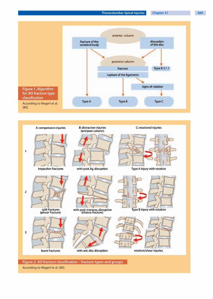
Figure 1. Algorithmfor AO fracture typeclassification
According to Magerl et al.[80].
Figure 2. AO fracture classification – fracture types and groups
According to Magerl et al. [80].
Thoracolumbar Spinal Injuries Chapter 31 889

Table 2. AO fracture classification
Type A: vertebral bodycompression
Type B: anterior and posteriorelement injury with distraction
Type C: anterior and posterior element injurywith rotation
A1. Impaction fracturesA1.1. Endplate impactionA1.2. Wedge impaction fracturesA1.2.1. Superior wedge impaction
fractureA1.2.2. Lateral wedge impaction
fractureA1.2.3. Inferior wedge impaction
fracture
B1. Posterior disruption pre-dominantly ligamentous(flexion-distraction injury)
B1.1. With transverse disruptionof the disc
B1.1.1. Flexion-subluxationB1.1.2. Anterior dislocationB1.1.3. Flexion-subluxation/anterior
dislocation with fracture ofthe articular processes
B1.2. With Type A fracture of thevertebral body
B1.2.1. Flexion-subluxation +Type A fracture
B1.2.2. Anterior dislocation +Type A fracture
B1.2.3. Flexion-subluxation/anteriordislocation with fracture ofthe articular processes +Type A fracture
C1. Type A injuries with rotation (compres-sion injuries with rotation)
C1.1. Rotational wedge fractureC1.2. Rotational split fracturesC1.2.1. Rotational sagittal split fractureC1.2.2. Rotational coronal split fractureC1.2.3. Rotational pincer fractureC1.2.4. Vertebral body separationC1.3. Rotational burst fracturesC1.3.1. Incomplete rotational burst fracturesC1.3.2. Rotational burst-split fractureC1.3.3. Complete rotational burst fracture
A2. Split fracturesA2.1. Sagittal split fractureA2.2. Coronal split fractureA2.3. Pincer fracture
B2. Posterior disruption pre-dominantly osseous (flexion-distraction injury)
B2.1. Transverse bicolumn frac-ture
B2.2. With transverse disruptionof the disc
B2.2.1. Disruption through thepedicle and disc
B2.2.2. Disruption through the parsinterarticularis and disc(flexion-spondylolysis)
B2.3. With Type A fracture of thevertebral body
B2.3.1. Fracture through the pedicle+ Type A fracture
B2.3.2. Fracture through the parsinterarticularis (flexion-spon-dylolysis) + Type A fracture
C2. Type B injuries with rotationC2.1. B1 injuries with rotation (flexion-
distraction injuries with rotation)C2.1.1. Rotational flexion subluxationC2.1.2. Rotational flexion subluxation with
unilateral articular process fractureC2.1.3. Unilateral dislocationC2.1.4. Rotational anterior dislocation without/
with fracture of articular processesC2.1.5. Rotational flexion subluxation without/
with unilateral articular process + Type Afracture
C2.1.6. Unilateral dislocation + Type A fractureC2.1.7. Rotational anterior dislocation without/
with fracture of articular processes +Type A fracture
C2.2. B2 injuries with rotation (flexiondistraction injuries with rotation)
C2.2.1. Rotational transverse bicolumn fractureC2.2.2. Unilateral flexion spondylolysis with
disruption of the discC2.2.3. Unilateral flexion spondylolysis +
Type A fractureC2.3. B3 injuries with rotation (hyperexten-
sion-shear injuries with rotation)C2.3.1. Rotational hyperextension-subluxation
without/with fracture of posterior ver-tebral elements
C2.3.2. Unilateral hyperextension-spondylolysisC2.3.3. Posterior dislocation with rotation
A3. Burst fracturesA3.1. Incomplete burst fractureA3.1.1. Superior incomplete burst
fractureA3.1.2. Lateral incomplete burst
fractureA3.1.3. Inferior incomplete burst
fractureA3.2. Burst-split fractureA3.2.1. Superior burst-split fractureA3.2.2. Lateral burst-split fractureA3.2.3. Inferior burst-split fractureA3.3. Complete burst fractureA3.3.1. Pincer burst fractureA3.3.2. Complete flexion burst fractureA3.3.3. Complete axial burst fracture
B3. Anterior disruption throughthe disc (hyperextension-shear injury)
B3.1. Hyperextension-subluxa-tions
B3.1.1. Without injury of the poste-rior column
B3.1.2. With injury of the posteriorcolumn
B3.2. Hyperextension-spondylo-lysis
B3.3. Posterior dislocation
C3. Rotational-shear injuriesC3.1. Slice fractureC3.2. Oblique fracture
Types, groups, subgroups and specifications allow for a morphology based classification of thoracolumbar fractures accordingto Magerl et al. [80]
890 Section Fractures
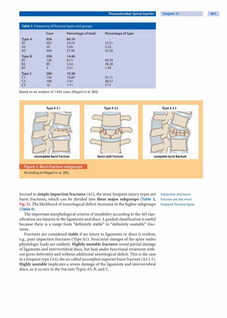
Table 3. Frequency of fracture types and groups
Case Percentage of total Percentage of type
Type A 956 66.16A1 502 34.74 52.51A2 50 3.46 5.23A3 404 27.96 42.26
Type B 209 14.46B1 126 8.72 60.29B2 80 5.54 38.28B3 3 0.21 1.44
Type C 280 19.38C1 156 10.80 55.71C2 108 7.47 38.57C3 16 1.11 5.71
Based on an analysis of 1 445 cases (Magerl et al. [80])
a b c
Figure 3. Burst fracture subgroups
According to Magerl et al. [80].
Impaction and burst
fracture are the most
frequent fracture types
Second to simple impaction fractures (A1), the most frequent injury types areburst fractures, which can be divided into three major subgroups (Table 3,Fig. 3). The likelihood of neurological deficit increases in the higher subgroups(Table 4).
The important morphological criteria of instability according to the AO clas-sification are injuries to the ligaments and discs. A graded classification is usefulbecause there is a range from “definitely stable” to “definitely unstable” frac-tures.
Fractures are considered stable if no injury to ligaments or discs is evident,e.g., pure impaction fractures (Type A1). Structural changes of the spine underphysiologic loads are unlikely. Slightly unstable fractures reveal partial damageof ligaments and intervertebral discs, but heal under functional treatment with-out gross deformity and without additional neurological deficit. This is the casein a frequent type (A3), the so-called incomplete superior burst fracture (A3.1.1).Highly unstable implicates a severe damage of the ligaments and intervertebraldiscs, as it occurs in the fracture Types A3, B, and C.
Thoracolumbar Spinal Injuries Chapter 31 891

Table 4. Frequency of neurological deficits
Types and groups Number of injuries Neurological deficit (%)
Type A 890 14A1 501 2A2 45 4A3 344 32
Type B 145 32B1 61 30B2 82 33B3 2 50
Type C 177 55C1 99 53C2 62 60C3 16 50
Total 1 212 22
Based on an analysis of 1 212 cases (Magerl et al. [80])
Clinical Presentation
The clinical assessment of patients with a putative trauma to the spine has threemajor objectives, i.e., to identify:
) the spinal injury) neurological deficits) concomitant non-spinal injuries
Spinal Injuries
About 30 % of
polytraumatized patients
have a spinal injury
It is obvious that the management and the priorities differ between a life-threat-ening polytrauma that includes a spinal injury and a monotrauma of the spine. Inthe case of a polytrauma, about one-fourth to one-third of patients have a spinalinjury [120]. In our institution, we found spinal injuries in 22% of polytrauma-tized patients. In a series of 147 consecutive patients with multiple trauma, Dai etal. [24] found a delayed diagnosis of thoracolumbar fractures in 19%, confirmingan earlier study by Anderson et al. [5], in which 23% of patients with major tho-racolumbar fractures were diagnosed after the patient had left the emergencydepartment. A delay in the diagnosis of thoracolumbar fractures is frequentlyassociated with an unstable patient condition that necessitates higher-priorityprocedures than thoracolumbar spine radiographs in the emergency depart-ment. However, with the routine use of multi-slice computed tomography (CT) in
Polytraumatized patients
should be screened
for spinal fracture by CT
polytraumatized patients, the diagnostic work-up is usually adequate [57, 106]and delayed diagnosis of spine fractures should become rare. Multiple burst frac-tures occur in approximately 10–34% [10, 11, 53].
Neurological Deficit
Sacral sparing indicates
an incomplete lesion
with a better prognosis
An accurate and well-documented neurological examination is of great impor-tance. With an inaccurate or incomplete examination and a subsequent variation ofthe patient’s neurological deficit, it will be unclear if the situation has changed or ifthe initial assessment was simply inappropriate. In the case of a progressive neuro-logical deficit, this may hinder urgent further management, i.e., the need for a sur-gical intervention with spinal decompression. Neurological assessment is usuallydone according to the guidelines of the American Spinal Injury Association (seeChapter 11 ). Importantly, the examination has to include the “search for a sacralsparing” which will determine the completeness of the deficit and the prognosis.
892 Section Fractures
Concomitant Non-spinal Injuries
About one-third of all spine injuries have concomitant injuries [65, 100, 120]. Ina review of 508 consecutive hospital admissions of patients with spinal injuries,Saboe et al. [100] identified the presence of associated injuries in 240 (47%) indi-viduals. Most frequently found concomitant injuries were:
) head injuries (26%)) chest injuries (24%)) long bone injuries (23%)
About one-third of all spinal
injuries have concomitant
injuries
One associated injury was found in 22%, two injuries in 15%, and 10% of thepatients had three or more associated injuries. Most spine fractures involved thelower cervical spine (29%) or the thoracolumbar junction (21%). Eighty-twopercent of thoracic fractures and 72% of lumbar fractures had associated injuriescompared to 28% of lower cervical spine fractures [100]. There is an association
Flexion injuries are
frequently associated
with abdominal injuries
between flexion injuries of the lumbar spine (Chance type) and abdominal inju-ries in seat belt injuries. Anderson et al. [2] reviewed 20 cases of Chance-typethoracolumbar flexion-distraction fractures and found that 13 patients (65%)had associated life-threatening intra-abdominal trauma. Twelve of these patientshad bowel wall injury. Conversely, specific injury mechanisms and fracture pat-terns should lead to a targeted search for concomitant spinal injuries. It is wellestablished that calcaneus or tibia plateau fractures following a fall from a greatheight are associated with spinal burst fractures. Also, sternal injuries may beassociated with spinal fractures. Injury to the sternum, when due to indirect vio-lence, is almost always associated with a severe spinal column injury [48].
History
The history of a patient who sustained a thoracolumbar spinal injury is usuallyobvious. The cardinal symptoms are:
) pain) loss of function (inability to move)) sensorimotor deficit) bowel and bladder dysfunction
History should include
the trauma type
and injury mechanism
The history should include a detailed assessment of the injury, i.e.:
) type of trauma (high vs. low energy)) mechanism of injury (compression, flexion/distraction, hyperextension,
rotation, shear injury)
Fractures of the thoracolumbar spine usually result from high-energy traumasuch as traffic accidents and falls from a great height. Recreational activities fre-quently associated with spinal injuries are skiing, snowboarding, paragliding orhorseriding. A spinal fracture should be suspected in any patient who has had ahigh-energy trauma. Consequently, patients should be treated as if they have aspinal injury unless proven otherwise [97]. On the contrary, vertebral compres-sion fractures can also occur in less severe accidents or more or less spontane-ously in elderly patients with osteoporotic bones (see Chapter 32 ) [63].
In patients with neurological deficits, the history must be detailed regarding:
) time of onset) course (unchanged, progressive, or improving)
The time course of the
neurological deficit matters
As outlined in Chapter 30 , polytraumatized and unconscious (head-injured)patients are difficult to assess. Polytraumatized patients carry a high risk (up to
Thoracolumbar Spinal Injuries Chapter 31 893

30%) of having suffered a spinal fracture and must be scrutinized for such aninjury. Assessing the history is not possible in unconscious patients and the diag-nosis must therefore be based on thorough imaging studies.
Physical Findings
Similarly to the assessment of the patient with a cervical spine injury (see Chap-ter 30 ), the initial focus of the physical examination is on the assessment of:
) vital functions) neurological deficits
Assess vital functions
and neurological deficits
The goal is to immediately secure vital functions, which can be compromised inpolytraumatized patients and patients with a spinal cord injury. Often hypoten-sion and hypovolemia is encountered both in polytraumatized and spinal cordinjured patients. Importantly, secondary deterioration of spinal cord functionthat results from hypotension and inadequate tissue oxygenization has to beavoided by timely and appropriate treatment.
Neurological deficits due
to thoracolumbar fractures
vary considerably
A thorough neurological examination is indispensable (see Chapter 11 ). Thespinal cord usually terminates at the level of L1 in adults, although it may extendto L2 in some patients. Therefore, fractures at the thoracolumbar junction mayresult in a variety of neurological injury types and symptoms, i.e., damage to:
) distal spinal cord with complete/incomplete paraplegia) conus medullaris with malfunction of the vegetative system) cauda equina) thoracolumbar nerve roots
Consider a spinal shock in
patients with neurological
deficits
In the case of a neurological deficit, the differentiation between a complete andincomplete paraplegia is of great importance for the prognosis, because approxi-mately 60% of patients with an incomplete lesion have the potential to make afunctionally relevant improvement. In thoracolumbar fractures, the clinical pic-ture of a complete neurogenic shock will not develop, because only the caudalparts of the sympathetic system are possibly damaged. However, a spinal shockmay be present (see Chapter 30 ). It is mandatory to exclude a spinal shockbecause spinal shock can disguise remaining neural function and has an impacton the treatment decision and timing.
Thoracolumbar factures may damage the parasympathic centers located inthe conus medullaris. This injury will lead to bladder dysfunction, bowel dys-function as well as sexual dysfunction. In the case of damage to the cauda equinaor in a combination with damage to the conus medullaris, a more diffuse distri-bution of lower extremity paresthesia, weakness and loss of reflexes is found.Radiculopathy can be identified by a segmental pattern of sensory alterationsthat do not have to be combined with motor dysfunction. As outlined in the pre-vious chapter, the neurological function must be precisely documented. TheASIA protocol [84] has become an assessment standard for this objective (seeChapter 11 ).
The inspection and palpation of the spine should include the search for:
) skin bruises, lacerations, ecchymoses) open wounds) swellings) hematoma) spinal (mal)alignment) gaps
894 Section Fractures
Diagnostic Work-up
Imaging Studies
The radiographic examination is an extension of the physical examination thatconfirms clinical suspicions and documents the presence and the extent of manyinjuries. Similarly to the “clearance of the cervical spine” [97], the clinical assess-ment is of great importance to evaluate the necessity of imaging studies. In thealert patient who has no distracting injuries, and is not affected by sedativedrugs, alcohol, or neurological deficit, the requirement for imaging is guided byclinical symptoms. The absence of back pain and tenderness has been shown toexclude a thoracolumbar injury [101].
Modern imaging studies such as computed tomography (CT) and magneticresonance imaging (MRI) have substantially improved the diagnosis of osseousand discoligamentous injuries after spinal trauma. Thus, changes such asimprovement in scan availability, image quality, acquisition time, and imagereformatting have changed commonly used algorithms [6]. However, plain filmsare still helpful, because they allow a quick overview of the bony deformity. Also,standard radiographs are important for analyzing long-term results and defor-mities at follow-up.
Static imaging studies
may disguise the real extent
of displacement at the time
of impact
It is important to remember that any static imaging study is a “snapshot intime” that is taken after the major impact has hit the spine. Thus, even CT scansor MRI do not reveal the actual degree of spinal displacement that may have hap-pened during the injury. Also, routine plain X-rays, CT and MRI studies are takenwith the patient in a prone position, i.e., in a position that lacks physiologicalload, and may therefore lead to a misjudgement of the severity and instability ofthe spine injury.
Standard Radiographs
Supine radiographs
underestimate the kyphotic
deformity
In most institutions, anterior-posterior and lateral radiographs of the entirespine are standard imaging studies after a spinal trauma. If there is a clinical sus-picion of a spinal injury, plain radiographs (anterior-posterior and lateral view)should be obtained. Radiographs taken with the patient in the prone positionunderestimate the extent of kyphotic deformity. Films taken with the patient inthe standing position can demonstrate a possible loss of integrity of the posteriortension band under axial loading and should be done in equivocal cases.
Emergency radiographs
often do not suffice because
of their poor quality
Krueger and coworkers [74] studied 28 patients with fractures of the lumbartransverse process and found that three patients (11%) had a lumbar spine frac-ture that was identified by CT but was overlooked on plain radiographs. They con-cluded that patients with acute trauma and fractures of the transverse processshould be examined with CT, because CT scanning decreases the risk of missingpotentially serious injuries. In a prospective series, Hauser et al. [52] comparedplain films and initial CT of the chest, abdomen, and pelvis with thin cut CT scans.The authors found that all unstable fractures were diagnosed with plain radio-
CT has replaced radiographs
for the assessment
of seriously injured patients
graphs. However, the initial CT detected acute fractures that were missed with theconventional X-rays and correctly classified old fractures that plain films read as“possibly” acute. The total misclassification rate for plain films was 12.6% com-pared to 1.4% for the initial CT. In an emergency situation radiographs are oftenof poor quality and CT is prompted if a fracture cannot be ruled out with certainty.
Measurements should be made at the level of injury and be compared with thevertebrae at the more cranial and caudal levels. Any posterior cortical disruptionseen in the lateral view or any interpedicular widening seen in the anteroposte-rior view suggests a burst fracture that should be further analyzed by CT scan.
Thoracolumbar Spinal Injuries Chapter 31 895
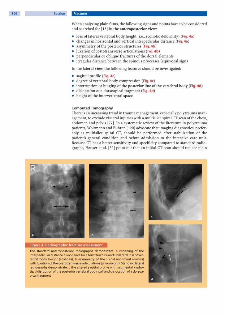
When analyzing plain films, the following signs and points have to be consideredand searched for [13] in the anteroposterior view:
) loss of lateral vertebral body height (i.e., scoliotic deformity) (Fig. 4a)) changes in horizontal and vertical interpedicular distance (Fig. 4a)) asymmetry of the posterior structures (Fig. 4b)) luxation of costotransverse articulations (Fig. 4b)) perpendicular or oblique fractures of the dorsal elements) irregular distance between the spinous processes (equivocal sign)
In the lateral view, the following features should be investigated:
) sagittal profile (Fig. 4c)) degree of vertebral body compression (Fig. 4c)) interruption or bulging of the posterior line of the vertebral body (Fig. 4d)) dislocation of a dorsoapical fragment (Fig. 4d)) height of the intervertebral space
Computed TomographyThere is an increasing trend in trauma management, especially polytrauma man-agement, to exclude visceral injuries with a multislice spiral CT scan of the chest,abdomen and pelvis [77]. In a systematic review of the literature in polytraumapatients, Woltmann and Bühren [120] advocate that imaging diagnostics, prefer-ably as multislice spiral CT, should be performed after stabilization of thepatient’s general condition and before admission to the intensive care unit.Because CT has a better sensitivity and specificity compared to standard radio-graphs, Hauser et al. [52] point out that an initial CT scan should replace plain
a b
c
d
Figure 4. Radiographic fracture assessment
The standard anteroposterior radiographs demonstrate: a widening of theinterpedicular distance as evidence for a burst fracture and unilateral loss of ver-tebral body height (scoliosis); b asymmetry of the spinal alignment (arrows)with luxation of the costotransverse articulations (arrowheads). Standard lateralradiographs demonstrate: c the altered sagittal profile with segmental kypho-sis; d disruption of the posterior vertebral body wall and dislocation of a dorsoa-pical fragment
896 Section Fractures

a b c
d
Figure 5. CT fracture assessment
The axial CT scan reveals: a significant spinal canal compromise bya retropulsed bony fragment. Note the double contour of the ver-tebral body indicating a “burst” component. b Sagittal 2D imagereformation demonstrating fracture subluxation. Note the bonyfragment behind the vertebral body which may cause neuralcompression when the fracture is reduced. c Severe luxation frac-ture of the spine. d The 3D CT reformation nicely demonstratesthe rotation component indicating a Type C lesion
radiographs in high-risk trauma patients who require screening. In their pro-spective series of 222 patients with 63 thoracic and lumbar injuries, the results ofconventional X-ray compared to initial CT scan were as follows: sensitivity 58%vs. 97%, specificity 93% vs. 99%, positive predictive value 64% vs. 97%, negativepredictive value 92% vs. 99%, respectively.
CT is the imaging study
of choice to demonstrate
bony injuries
The axial view allows an accurate assessment of the comminution of the frac-ture and dislocation of fragments into the spinal canal (Fig. 5a). Sagittal andcoronal 2D or 3D reconstructions are helpful for determining the fracture pat-tern (Fig. 5b–d). The canal at the injured segment should be measured in theanteroposterior and transverse planes and compared with the cephalad and cau-dal segments.
Magnetic Resonance Imaging
MRI is helpful in ruling out
discoligamentous lesions
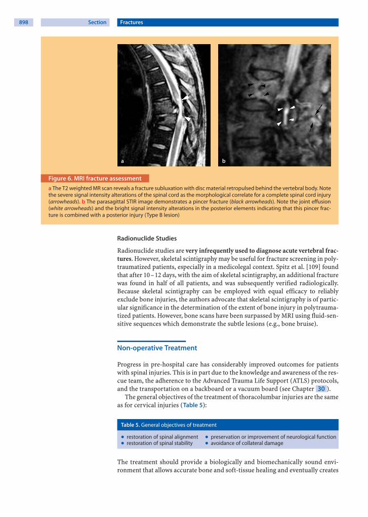
In the presence of neurological deficits, MRI is recommended to identify a possi-ble cord lesion or a cord compression that may be due to disc or fracture frag-ments or to an epidural hematoma (Fig. 6a). In the absence of neurological defi-cits, MRI of the thoracolumbar area is usually not necessary in the acute phase.However, MRI can be helpful in determining the integrity of the posterior liga-mentous structures and thereby differentiate between a Type A and an unstableType B lesion. For this purpose a fluid sensitive sequence (e.g., STIR) is fre-quently used to determine edema (Fig. 6b).
Thoracolumbar Spinal Injuries Chapter 31 897
a b
Figure 6. MRI fracture assessment
a The T2 weighted MR scan reveals a fracture subluxation with disc material retropulsed behind the vertebral body. Notethe severe signal intensity alterations of the spinal cord as the morphological correlate for a complete spinal cord injury(arrowheads). b The parasagittal STIR image demonstrates a pincer fracture (black arrowheads). Note the joint effusion(white arrowheads) and the bright signal intensity alterations in the posterior elements indicating that this pincer frac-ture is combined with a posterior injury (Type B lesion)
Radionuclide Studies
Radionuclide studies are very infrequently used to diagnose acute vertebral frac-tures. However, skeletal scintigraphy may be useful for fracture screening in poly-traumatized patients, especially in a medicolegal context. Spitz et al. [109] foundthat after 10– 12 days, with the aim of skeletal scintigraphy, an additional fracturewas found in half of all patients, and was subsequently verified radiologically.Because skeletal scintigraphy can be employed with equal efficacy to reliablyexclude bone injuries, the authors advocate that skeletal scintigraphy is of partic-ular significance in the determination of the extent of bone injury in polytrauma-tized patients. However, bone scans have been surpassed by MRI using fluid-sen-sitive sequences which demonstrate the subtle lesions (e.g., bone bruise).
Non-operative Treatment
Progress in pre-hospital care has considerably improved outcomes for patientswith spinal injuries. This is in part due to the knowledge and awareness of the res-cue team, the adherence to the Advanced Trauma Life Support (ATLS) protocols,and the transportation on a backboard or a vacuum board (see Chapter 30 ).
The general objectives of the treatment of thoracolumbar injuries are the sameas for cervical injuries (Table 5):
Table 5. General objectives of treatment
) restoration of spinal alignment ) preservation or improvement of neurological function) restoration of spinal stability ) avoidance of collateral damage
The treatment should provide a biologically and biomechanically sound envi-ronment that allows accurate bone and soft-tissue healing and eventually creates
898 Section Fractures
The main advantage
of non-operative treatment
is the avoidance of surgery-
related complications
a stable and pain-free spinal column. These goals should be accomplished with aminimal risk of morbidity. Hence, the main advantage of non-operative treatmentof thoracolumbar fracture is avoidance of surgery-related complications such as:
) infection) iatrogenic neurological injury) failure of instrumentation) anesthesia-related complications
The relationship between post-traumatic kyphotic deformity and chronic backpain is not well established in the literature. Most clinicians believe that kyphoticdeformity of the thoracolumbar area is synonymous with a poor clinical out-come. Although few studies provide some evidence that moderate kyphosis isassociated with either pain or disability [47], several studies suggest that there isno direct relationship between kyphosis and back pain or functional impairment[20, 73, 87, 89, 116].
Steroid Treatment of Spinal Cord Injury
High-dose steroid treatment
is highly controversial
The controversy over steroid treatment of thoracolumbar spinal cord injury isdiscussed in the previous chapter (see Chapter 30 ). The overall consensus isthat high-dose steroid treatment is regarded as an option for spinal monotraumain young patients but not as a guideline for standard of care.
Non-operative Treatment Modalities
As more and more data are collected, information emerges that supports bothsurgical and non-operative treatment. Non-operative treatment is still a viableand effective treatment for the vast majority of thoracolumbar fractures (Table 6)and should be part of the armamentarium available to all clinicians that treatthese patients [92].
Table 6. Favorable indications for non-operative treatment
) pure osseous lesions ) absence of malalignment) absence of neurological deficits ) absence of gross bony destruction) only mild to moderate pain on mobilization ) absence of osteopenia/osteoporosis
There are three different methods of non-operative treatment:
) repositioning and cast stabilization) functional treatment and bracing without repositioning) functional treatment without bracing
However, functional treatment without bracing is not applicable to all fracturetypes, while basically all fractures can be treated with repositioning and formalcasting (Böhler technique).
Repositioning and Cast Stabilization
Böhler [18] was one of the first to advocate a conservative treatment with reposi-tioning and retention in a cast. The correct technique of repositioning andimmobilization in a plaster of Paris cast is quite sophisticated and needs to beperformed perfectly to obtain good results [13, 58]. The fracture is reduced usinga fracture table with the abdomen hanging freely. The hyperextension results ina fracture reduction by ligamentotaxis (Case Study 1). As a general rule, Böhler
Thoracolumbar Spinal Injuries Chapter 31 899
a b
c
d e f
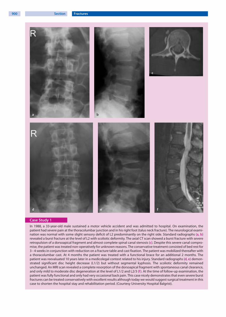
Case Study 1
In 1988, a 33-year-old male sustained a motor vehicle accident and was admitted to hospital. On examination, thepatient had severe pain at the thoracolumbar junction and in his right foot (talus neck fracture). The neurological exami-nation was normal with some slight sensory deficit of L2 predominantly on the right side. Standard radiographs (a, b)revealed a burst fracture at the level of L2 with scoliotic deformity. The axial CT scan showed a burst fracture with severeretropulsion of a dorsoapical fragment and almost complete spinal canal stenosis (c). Despite this severe canal compro-mise, the patient was treated non-operatively for unknown reasons. The conservative treatment consisted of bed rest for3 – 4 weeks in conjunction with reduction on a fracture table and cast fixation. The patient was mobilized thereafter witha thoracolumbar cast. At 4 months the patient was treated with a functional brace for an additional 2 months. Thepatient was reevaluated 10 years later in a medicolegal context related to his injury. Standard radiographs (d, e) demon-strated significant disc height decrease (L1/2) but without segmental kyphosis. The scoliotic deformity remainedunchanged. An MRI scan revealed a complete resorption of the dorsoapical fragment with spontaneous canal clearance,and only mild to moderate disc degeneration at the level of L1/2 and L2/3 (f ). At the time of follow-up examination, thepatient was fully functional and only had very occasional back pain. This case nicely demonstrates that even severe burstfractures can be treated conservatively with excellent results although today we would suggest surgical treatment in thiscase to shorten the hospital stay and rehabilitation period. (Courtesy University Hospital Balgrist).
900 Section Fractures

used the kyphosis angle in degrees to calculate the numbers of weeks of immobi-lization (minimum 12 weeks, maximum 5 months). Patients were allowed toambulate almost immediately and were discharged home after a couple of days.Regular clinical and radiological exams were performed, initially every 2 weeks,then every 4 weeks, and the cast had to be changed if it became loose. Impor-tantly, an intense and skillful physical therapy was, and still is, paramount toachieving good or satisfactory results.
Böhler’s fracture treatment
today is still a viable treat-
ment option
The disadvantage of the Böhler technique is that it is very uncomfortable andpainful for the patient and often requires sedation and strong analgesics. TheBöhler technique is also prone to plaster cast related pressure sores. In patientswith an indication for conservative treatment, we prefer to apply the cast in thestanding position in hyperextension. This is possible in the vast majority ofpatients after a few days post-trauma and after orthostatic training on a verticallytilted board (Fig. 7).
a
b c d
Figure 7. Non-operative treatment
a The patient with an orthostatic problem after a fracture is firstplaced on a motorized table which can be tilted vertically.b When the patient is able to stand upright for 15 – 20 min, he ispositioned between two vertical bars and moderately extendshis spine while the cast is applied. c, d The thoracolumbar castbuttresses onto the iliac crest and reaches up to the sternum
Thoracolumbar Spinal Injuries Chapter 31 901

Functional Bracing
Reduced kyphotic fractures
are prone to return
to the initial deformity,
placing a questionmark
over reduction
Magnus [82] advocated early functional treatment without repositioning. Accord-ing to this concept, a thoracolumbar fracture is bound to return to the initialdeformity and repositioning is therefore not necessary. The functional treatmentconcept was initiated with a phase of prone position on a stable bed and, if neces-sary, with lordotic support. The time of immobilization in bed depended on thefracture type. The next phases of treatment consisted of physical therapy toenhance muscle strength, mobilization in a waterbath, mobilization with a threepoint orthesis to prevent flexion and to assure an upright position of the patient,and a discharge home after approximately 3 weeks. Outpatient treatment was con-tinued for another 3–4 months and physical therapy to enhance spine mobilitywas initiated after radiologic consolidation of the fracture, i.e., after 3–4 months.
Functional Treatment
Functional treatment is indi-
cated only in unequivocal
stable fractures
In contrast to Böhler’s repositioning and stabilization [18] or Magnus’ functionalbracing [82], functional treatment does not include any bracing device. Espe-cially patients with stable fractures will benefit from this treatment (Table 7).Some braces are rather cumbersome and will hinder the patient in many activi-ties of daily life. In fact, braces can be considered an “aide-memoire” and remindthe patient not to perform painful movements. With the functional treatment,patients are advised to mobilize freely according to their capabilities and accord-ing to the resulting pain. Importantly, qualified physical therapy and adequatepain medication are necessary to obtain optimal results.
Table 7. Outcome of conservative and operative treatment
Authors Cases Studydesign
Fracturetype(numbers)
Type oftreatment
Neuro-logicaldeficit
Follow-up(months)
Outcome Conclusions
Wein-steinet al.(1988)[116]
42 retro-spec-tive
burstfractures(T10–L5)
non-operative:treatmentranged fromimmediateambulation ina body cast orbrace to3 months bedrest
22 % 240 neurological deteriora-tion: noneable to return to work:88 %kyphotic angle 26.4° in
flexion and 16.8° inextension
average back pain score3.5 (0 – 10)
non-operative treat-ment of thoracolumbarburst fractures withoutneurological deficit canlead to acceptablelong-term results
Mum-fordtet al.(1993)[87]
41 retro-spec-tive
single levelthoracolum-bar burstfracturesT11–L5:type I: 5 %type II: 78 %type III: 5 %type V: 12 %(Denis classi-fication)
non-operative:bedrest mean:
31.3 (range,7 – 68 days)
bracing mean11.9 (range,2 – 24 weeks)
none 24 functional results:excellent 49 %good 17 %fair 22 %poor 12 %one patient developedneurological deteriora-tion that required sur-gery
for patients with burstfractures withoutneurological deficit:
non-operative manage-ment yields accept-able results
bony deformity progres-ses marginally relativeto the rate of canalarea remodeling
radiographic severity ofinjury or residualdeformity does notcorrelate with long-term symptoms
Chowet al.(1996)[23]
24 retro-spec-tive
unstableburstfractures(T11–L2)
non-operative:casting or brac-ing and earlyambulation
None 34 no correlation betweenpost-traumatic kypho-sis and outcome
little/no pain 79 %return to work 75 %no restrictions at work75 %
hyperextension castingor bracing is a safe andeffective method fortreatment of thoraco-lumbar burst fractures
902 Section Fractures

Table 7. (Cont.)
Authors Cases Studydesign
Fracturetype(numbers)
Type oftreatment
Neuro-logicaldeficit
Follow-up(months)
Outcome Conclusions
Kanedaet al.(1997)[60]
150 retro-spec-tive
FrankelgradesA (24 %)B (58 %)C (6 %)D (7 %)E (4 %)
operative:single stageanterior spinaldecompres-sion, strut graf-ting, and ante-rior instrumen-tation
100 % 96(60 – 156)
neurological functionimproved at least onegrade in 95 % ofpatients. 72 % ofpatients with bladderdysfunction recoveredcompletely. 96 %returned to work, 86 %to their previous jobwithout restrictions
anterior decompressionand stabilization inpatients with burst frac-tures and neurologicaldeficit yielded goodfunctional results
Knopet al.(2001)[67]
372 pro-spec-tive,multi-center
thoracolum-bar fractures(T12–L2)type:A (69 %)B (17 %)C (14 %)
operative:Posterior (59 %)combined
anterior-pos-terior (35 %)
anterior (6 %)stabilization
20 % 27(4 – 61)
for detailed descriptionsee text
all treatment methodsresulted in compara-ble clinical and func-tional outcome
one-third of all patientshad severe and persist-ing functional disabili-ties
Khooet al.(2002)[62]
371 retro-spec-tive
N/A 35 % stand-alone ante-rior thora-coscopic sta-bilization
65 % additionalposterior pedi-cle screwinstrumenta-tion
15 % 24(4 – 72)
low rate of severe com-plications (1.3 %); onecase each of aorticinjury, splenic contu-sion, neurologicaldeterioration, CSF fluidleak, and severewound infection
42 % less narcotics forpostoperative paintreatment comparedto a group of 30patients treated withopen thoracotomy
anterior thoracoscopic-assisted reconstructionof thoracolumbar frac-tures can be safelyaccomplished, reducingpain and morbidityassociated with openapproaches
DefinoandScar-paro(2005)[29]
18 retro-spec-tive
type B and Cfractures(AO classifi-cation), T10–L4
operative:posteriormonosegmen-tal fixation andarthrodesis
38.9 % 78(24 – 144)
low residual pain ratesand high level patientsatisfaction with finalresult. 95.5 % returnedto work and presentedwith a low disabilityindex (Oswestry Disabil-ity Index = 10.33 %)
posterior monoseg-mental fixation is anadequate and satisfac-tory procedure in spe-cific types of thoraco-lumbar spine fractures
Woodet al.(2005)[122]
38 pro-spec-tive,ran-domi-zed
isolatedburst frac-tures (T10–L2)
operative:18 posteriorfusion20 anterior sta-bilization
none 43(24 – 108)
17 minor complicationsin patients treatedposteriorly, includingimplant removal, 3minor complicationswith anterior stabiliza-tion
similar functional out-comes
anterior fusion andinstrumentation mayexhibit fewer complica-tions and fewer addi-tional surgeries
Operative Treatment
General Principles
There is a general trend towards operative treatment of unstable fractures [31,47], mostly because surgical stabilizing allows for:
) early mobilization of the patient) diminished pain) facilitated nursing care (polytraumatized patients)) earlier return to work) avoidance of late neurological complications
Thoracolumbar Spinal Injuries Chapter 31 903
Despite theoretical
advantages, the superiority
of surgical fracture
treatment is not supported
by scientific evidence
However, evidence suggests that there is no difference as regards neurologicalrecovery (Frankel score) and no substantial difference in functional long-termoutcome between the operative and non-operative treatment [114]. This isclearly valid for compression fractures that are relatively stable, i.e., A1 and A2fractures, according to the AO classification. Quite frequently, however, studiespresented in the literature analyze a mixed cohort of fracture types without fur-ther differentiation, which leaves their results somewhat inconclusive.
In burst fractures, there is often some degree of canal compromise with apotential risk of neurological injury. Hence, progressive neurological deteriora-tion in the presence of substantial canal compromise is an indication for surgicaldecompression and stabilization. Importantly, neurological status, spinal stabil-ity, degree of deformity of the injured segment, degree of canal compromise, andassociated injuries are the most relevant factors that need to be considered when
Progressive neurological
deficit is an absolute
indication for surgery
deciding on operative or non-operative treatment for patients with a thoraco-lumbar spine fracture. Most surgeons agree on absolute indications for surgerywhile relative indications are debatable (Table 8):
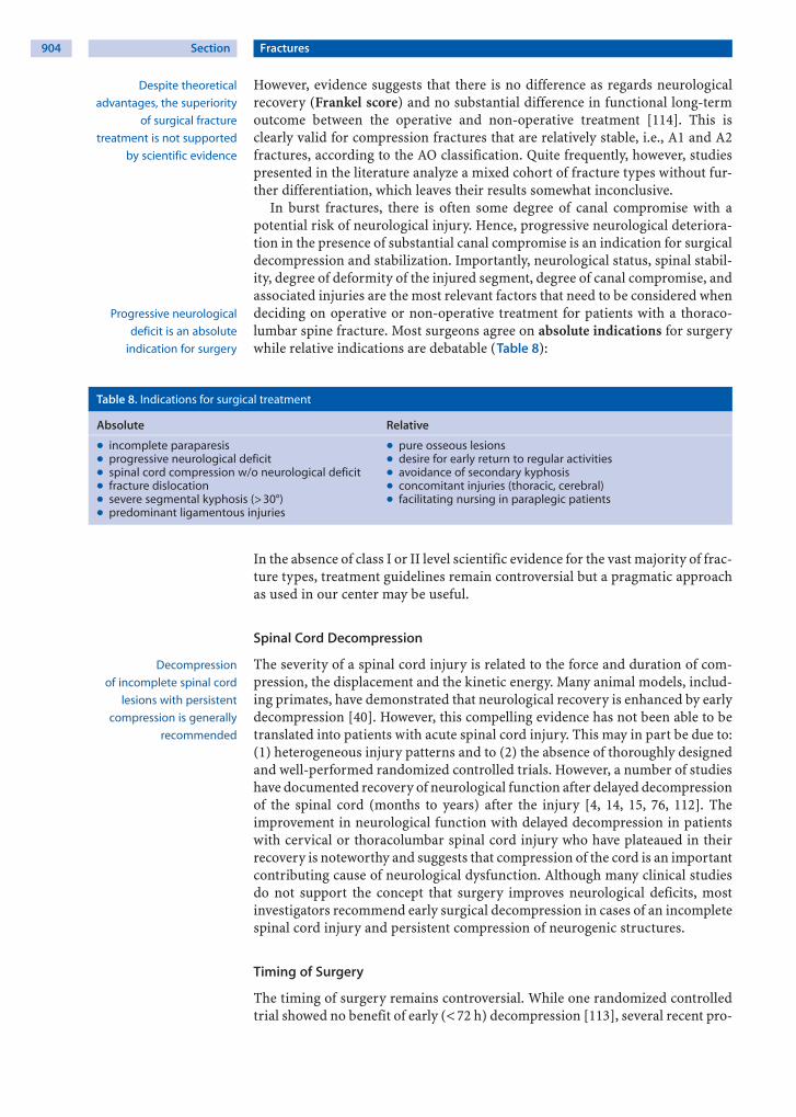
Table 8. Indications for surgical treatment
Absolute Relative
) incomplete paraparesis ) pure osseous lesions) progressive neurological deficit ) desire for early return to regular activities) spinal cord compression w/o neurological deficit ) avoidance of secondary kyphosis) fracture dislocation ) concomitant injuries (thoracic, cerebral)) severe segmental kyphosis (> 30°) ) facilitating nursing in paraplegic patients) predominant ligamentous injuries
In the absence of class I or II level scientific evidence for the vast majority of frac-ture types, treatment guidelines remain controversial but a pragmatic approachas used in our center may be useful.
Spinal Cord Decompression
Decompression
of incomplete spinal cord
lesions with persistent
compression is generally
recommended
The severity of a spinal cord injury is related to the force and duration of com-pression, the displacement and the kinetic energy. Many animal models, includ-ing primates, have demonstrated that neurological recovery is enhanced by earlydecompression [40]. However, this compelling evidence has not been able to betranslated into patients with acute spinal cord injury. This may in part be due to:(1) heterogeneous injury patterns and to (2) the absence of thoroughly designedand well-performed randomized controlled trials. However, a number of studieshave documented recovery of neurological function after delayed decompressionof the spinal cord (months to years) after the injury [4, 14, 15, 76, 112]. Theimprovement in neurological function with delayed decompression in patientswith cervical or thoracolumbar spinal cord injury who have plateaued in theirrecovery is noteworthy and suggests that compression of the cord is an importantcontributing cause of neurological dysfunction. Although many clinical studiesdo not support the concept that surgery improves neurological deficits, mostinvestigators recommend early surgical decompression in cases of an incompletespinal cord injury and persistent compression of neurogenic structures.
Timing of Surgery
The timing of surgery remains controversial. While one randomized controlledtrial showed no benefit of early (< 72 h) decompression [113], several recent pro-
904 Section Fractures
spective series suggest that early decompression (<12 h) can be performed safelyand may improve neurological outcomes [40].
Early rather than late
decompression
is recommended
La Rosa et al. [75] published a meta-analysis on the issue of early decompres-sion in acute spinal cord injury. They reviewed 1687 patients in studies publishedup to 2000. Patients were divided into three treatment groups: early decompres-sion (<24 h), delayed decompression (>24 h), and conservative treatment. Sta-tistically, early decompression resulted in better outcomes compared to bothdelayed decompression and conservative management. Because the analysis ofhomogeneity demonstrated that only data regarding patients with incompletespinal cord injury who underwent early decompression were reliable, the authorsconcluded that early decompression can only be considered a practice option.Currently, there are no standards regarding the role and timing of decompressionin acute spinal cord injury. Also, the presence and duration of a therapeutic win-dow, during which surgical decompression could attenuate the secondary mech-anisms of spinal cord injury, remains unclear. In a recent article, Fehlings et al.[40] provide evidence-based recommendations regarding spinal cord decom-pression in patients with acute spinal cord injury. Animal studies consistentlyshow that neurological recovery is enhanced by early decompression. One ran-domized controlled trial showed no benefit to early (<72 h) decompression. Sev-eral recent prospective series suggest that early decompression (< 12 h) can beperformed safely and may improve neurological outcomes. Currently, there areno standards regarding the role and timing of decompression in acute spinal cord
Early decompression
of progressive neurological
deficits is indicated
injury. On the other hand, no significant adverse effects of early decompressionhave been documented. In the absence of clear guidelines from the literature,early decompression of compressed neurological structures appears to be bestpractice.
Surgical Techniques
If surgical treatment is chosen, further debate arises over the appropriate type ofapproach. Similarly to the treatment decision of conservative vs. operative, scien-tific evidence is lacking for the superiority of one surgical technique over theother. Particularly for the frequent superior burst fracture (Fig. 3), a large varietyof surgical techniques are available. Finally, it depends on the surgical expertiseof the surgeon and their preference which technique is chosen. It is difficult tobase treatment recommendations on treatment outcome in the literature(Table 7).
Posterior Approach
Posterior Monosegmental Reduction and Stabilization
Posterior monosegmental
reduction and stabilization
is feasible in selected Type A
and B fractures
The group of Gotzen et al. [49, 59] was the first to publish their results aftermonosegmental reduction and stabilization (Case Study 2). In their initial report[49], 14 patients with unstable compression fractures Grade II were treated byposterior one-level internal fixation (9 patients had stabilization with plates andcerclage wire, 5 with internal fixator). The results were compared to a series of 11patients with equivalent fractures treated non-operatively. The authors concludethat posterior single level stabilization and fusion is a recommendable surgicalprocedure. In their second publication, Junge et al. [59] describe the technique,which always included a posterior allogenic bone grafting and to some extentalso transpedicular bone grafting. The 2-year follow-up of 39 patients demon-strated that 17 patients (43%) were completely free of pain and 17 patients wereonly sensitive to weather changes or had minor pain during great physical stress.
Thoracolumbar Spinal Injuries Chapter 31 905
a b c
d e f
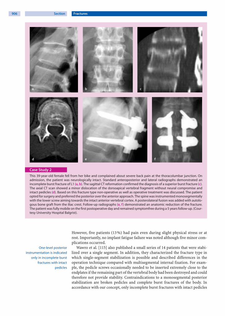
Case Study 2
This 39-year-old female fell from her bike and complained about severe back pain at the thoracolumbar junction. Onadmission, the patient was neurologically intact. Standard anteroposterior and lateral radiographs demonstrated anincomplete burst fracture of L1 (a, b). The sagittal CT reformation confirmed the diagnosis of a superior burst fracture (c).The axial CT scan showed a minor dislocation of the dorsoapical vertebral fragment without neural compromise andintact pedicles (d). Based on this fracture type non-operative as well as operative treatment was discussed. The patientopted for surgery and preferred the posterior over the anterior approach. The spine was instrumented monosegmentallywith the lower screw aiming towards the intact anterior vertebral cortex. A posterolateral fusion was added with autolo-gous bone graft from the iliac crest. Follow-up radiographs (e, f ) demonstrated an anatomic reduction of the fracture.The patient was fully mobile on the first postoperative day and remained symptomfree during a 5 years follow-up. (Cour-tesy University Hospital Balgrist).
However, five patients (13%) had pain even during slight physical stress or atrest. Importantly, no implant fatigue failure was noted although five minor com-plications occurred.
One-level posterior
instrumentation is indicated
only in incomplete burst
fractures with intact
pedicles
Wawro et al. [115] also published a small series of 14 patients that were stabi-lized over a single segment. In addition, they characterized the fracture type inwhich single-segment stabilization is possible and described differences in theoperation technique compared with multisegmental internal fixation. For exam-ple, the pedicle screws occasionally needed to be inserted extremely close to theendplates if the remaining part of the vertebral body had been destroyed and couldtherefore not provide stability. Contraindications to a monosegmental posteriorstabilization are broken pedicles and complete burst fractures of the body. Inaccordance with our concept, only incomplete burst fractures with intact pedicles
906 Section Fractures

and inferior endplate (i.e., Type A1 and A3.1) should be considered for posteriormonosegmental reduction and stabilization. Probably the pathophysiologicallymost sound indication for a monosegmental dorsal stabilization is a Type B frac-ture with only ligamentous posterior injury combined with a Type A1 or A3.1fracture of the vertebral body with intact endplates and intact pedicles, becausethe dorsal stabilization restores the tension band function of the ruptured liga-ments.
In a similar small series of 18 patients undergoing posterior monosegmentalstabilization, Defino et al. [29] report a clinical and radiological follow-up after2–12 years (mean 6.6±3 years) to demonstrate that posterior monosegmentalfixation is an adequate and satisfactory procedure in specific types of thoraco-lumbar spine fractures. Clinical evaluation revealed low residual pain rates and ahigh level of patient satisfaction with the final result. Functional evaluationshowed that 95.5% of the patients returned to work on a full-time basis and pre-sented with a low disability index (Oswestry Disability Index =10.33%). Radio-graphic evaluation demonstrated increased kyphosis in the fixed vertebral seg-ment during the late postoperative period, accompanied by a reduced height ofthe intervertebral disc. There was no implant failure, and no signs of pseudoar-throsis were observed in any patient.
Posterior Bisegmental Reduction and Stabilization
Posterior two-level reduction
and fracture stabilization
remains the gold standard
for the vast majority
of thoracolumbar fractures
The bisegmental, two-level posterior approach (short segmental stabilization) isthe “working horse” of the posterior techniques that allows a secure fixation ofthe pedicle screws in the intact vertebra one level above and below the fracture(Fig. 8). With this construct, a good reduction and stable fixation is reliablyachieved.
Fredrickson et al. [45] studied the mechanisms of ligamentotaxis to reduce theintracanal fragment of a burst fracture. Examination of anatomic data providedby microtome section indicated that the fibers that actually reduce the intracanalfragment originate in the anulus of the superior vertebra in the midportion of theendplate and insert into the lateral margins of the intracanal fragment. Investiga-tions using MRI confirmed that these obliquely directed fibers account for theindirect reduction of the fragment. Further studies demonstrate that the poste-rior longitudinal ligament provided only a minor contribution in the reductionof the fracture in comparison to the attachments of the posterior portion of theanulus fibrosus.
Harrington et al. [51] studied the biomechanics of indirect reduction of boneretropulsed into the spinal canal in vertebral fracture and made several clinicallyrelevant observations. It was not possible to produce an anteriorly directed forcein the posterior longitudinal ligament at less than 35% canal occlusion, partlybecause the posterior longitudinal ligament stands away from the midbody of thevertebra. Regardless of the relative sagittal plane angulation of the vertebrae, dis-traction was the governing factor in generating force in the posterior longitudi-nal ligament. Because positioning the vertebrae in lordosis before applying dis-traction significantly slackens the posterior longitudinal ligament, it is suggestedthat distraction be applied before angular positioning of the vertebrae is per-formed. However, this procedure risks overdistraction with deleterious resultsfor the spinal cord.
A comminuted anterior
column demands anterior
load sharing support
Depending on the comminution of the fractured vertebral body, additionalanterior load sharing support is needed. McLain et al. [85] reported early failureof short-segment pedicle instrumentation for thoracolumbar fractures. Out of 19patients with unstable thoracolumbar fractures, 10 patients had early failure offixation: progressive kyphosis, osseous collapse, vertebral translation, screw
Thoracolumbar Spinal Injuries Chapter 31 907

a b
c d
Figure 8. Surgical technique of two-level fracture reduction and stabilization
The technique demonstrates the use of the Fracture Module of Universal Spine System (Synthes) but the general princi-ples similarly apply to other fracture systems. a Schanz screws are inserted in the pedicles of the vertebral bodies superiorand inferior to the fracture. b Screw clamps connected with the rods are mounted and fixed (arrow). c The fracture can bereduced by lordosing both screwdrivers. However, it is often better to first tighten the two lower screws and reduce thefracture simultaneously by lordosing the cranial screw bilaterally with the help of the screwdriver. d If this reductionmaneuver does not suffice to restore vertebral height, a temporary C-clamp can be mounted and the fracture distractedafter loosening the upper screws. Care must be taken not to overdistract the fracture because of the inherent neurologi-cal risks. Finally, the Schanz screws are cut with a special screwcutter (not shown). Dependent on canal clearance andanterior vertebral column restoration, an additional anterior approach can be added (preferably in a second stage)
breakage or loosening. These results indicate the need for an adequate anteriorcolumn support and an optimal anterior-posterior column load sharing environ-ment.
Transpedicular cancellous
bone grafting is insufficient
to stabilize the anterior
column
If no anterior stabilization is planned, a posterolateral fusion [78, 88] is man-datory. In addition, transpedicular bone grafting in the disrupted disc space hasbeen a treatment option [26, 78, 90]. However, transpedicular bone graftingcould not prevent kyphosis after dorsal removal on implants [1, 68, 108]. Knopet al. [68] studied 56 patients after implant removal and concluded that, because
908 Section Fractures
of the disappointing results, they cannot recommend the additional transpedicu-lar cancellous bone grafting as an interbody fusion technique after posterior sta-bilization in cases of complete or incomplete burst injury to the vertebral body.Similarly, Alanay et al. [1] concluded that short-segment transpedicular instru-mentation of thoracolumbar burst fractures is associated with a high rate of fail-ure that cannot be decreased by additional transpedicular intracorporeal graf-ting.
Posterior Reduction and Multisegmental Stabilization
Fracture dislocations usually
require multilevel spinal
stabilization
Multilevel stabilization is indicated for the very unstable thoracolumbar luxationfractures (Type C lesions) which usually cannot be accurately reduced and stabi-lized with a short two-level construct. Usually, fixation of two to three segmentsabove and below the injury is recommended for a stable fixation. Unstable frac-tures of the thoracic spine that need to be stabilized are often combined with asignificant thorax trauma or a polytrauma. In these patients, an early posteriorstabilization with additional bone grafting allows for (1) a stable fixation of thespine with restoration of the dorsal tension band function, (2) the possibility ofearly and orthosis-free mobilization in the intensive care unit or later in a centerof rehabilitation, and finally (3) bony fusion.
Anterior Approach
Rationale for the anterior
approach is that the spine
should be treated where
the injury has occurred
From the biomechanical point of view, it is obvious that the damaged spine has tobe treated according to the injury mechanism and the site of injury. In a flexioninjury (e.g., Chance fracture) with fracture of the pedicles and the vertebral body,stabilization can be performed by a dorsal approach and restores the tensionband function until bony healing has occurred. Similarly, the biomechanics ofthe anterior column has to be considered in the case of a burst fracture. About80% of the axial load of an intact spine is supported by the anterior column.When the anterior column is substantially injured, the anterior support is dra-matically reduced to about 10%, leaving 90% of the load to be resisted by theimplant and the posterior elements. These general biomechanical considerationssupport the use of an anterior load sharing support (e.g., by a tricortical bonegraft or a cage).
The primary indications for the anterior approach are:
) insufficient spinal decompression) insufficient anterior column restoration
Spinal canal compromise in patients presenting with neurological deficits whichcannot adequately be resolved by a dorsal approach alone requires anteriordecompression. An additional indication is a vertebral body fracture with sub-stantial comminution and dislocation which cannot be adequately restored by aposterior approach alone [50].
Type A fractures can be
treated by an anterior
approach alone
However, Type A fractures can be treated by an anterior approach alone.Kaneda et al. [60] reported a study on 150 consecutive patients who had a burstfracture of the thoracolumbar spine and associated neurological deficits. Thepatients were managed with a single-stage anterior spinal decompression, strut-grafting, and anterior spinal instrumentation. At a mean of 8 years (range5–12 years) after the operation, radiographs showed successful fusion of theinjured spinal segment in 140 patients (93%). Ten patients had a pseudarthrosis,and all were managed successfully with posterior spinal instrumentation and aposterolateral arthrodesis. Despite breakage of the Kaneda device in ninepatients, removal of the implant was not necessary in any patient. None of the
Thoracolumbar Spinal Injuries Chapter 31 909
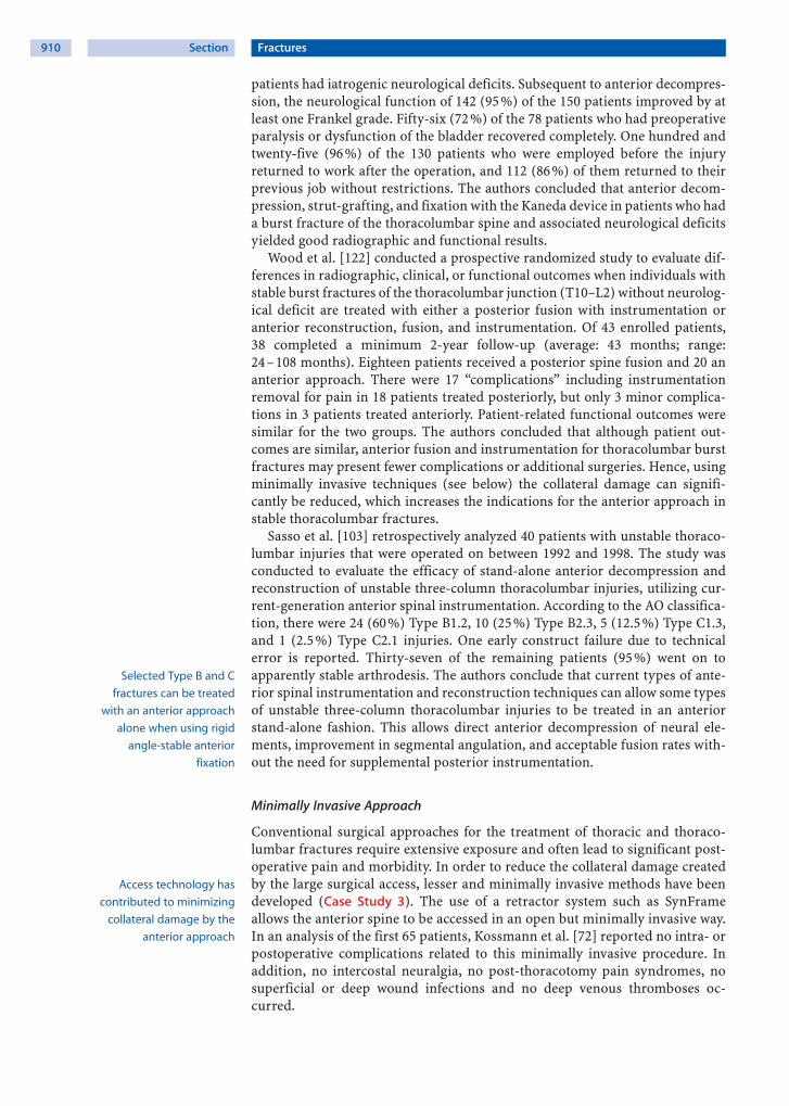
patients had iatrogenic neurological deficits. Subsequent to anterior decompres-sion, the neurological function of 142 (95%) of the 150 patients improved by atleast one Frankel grade. Fifty-six (72%) of the 78 patients who had preoperativeparalysis or dysfunction of the bladder recovered completely. One hundred andtwenty-five (96%) of the 130 patients who were employed before the injuryreturned to work after the operation, and 112 (86%) of them returned to theirprevious job without restrictions. The authors concluded that anterior decom-pression, strut-grafting, and fixation with the Kaneda device in patients who hada burst fracture of the thoracolumbar spine and associated neurological deficitsyielded good radiographic and functional results.
Wood et al. [122] conducted a prospective randomized study to evaluate dif-ferences in radiographic, clinical, or functional outcomes when individuals withstable burst fractures of the thoracolumbar junction (T10–L2) without neurolog-ical deficit are treated with either a posterior fusion with instrumentation oranterior reconstruction, fusion, and instrumentation. Of 43 enrolled patients,38 completed a minimum 2-year follow-up (average: 43 months; range:24–108 months). Eighteen patients received a posterior spine fusion and 20 ananterior approach. There were 17 “complications” including instrumentationremoval for pain in 18 patients treated posteriorly, but only 3 minor complica-tions in 3 patients treated anteriorly. Patient-related functional outcomes weresimilar for the two groups. The authors concluded that although patient out-comes are similar, anterior fusion and instrumentation for thoracolumbar burstfractures may present fewer complications or additional surgeries. Hence, usingminimally invasive techniques (see below) the collateral damage can signifi-cantly be reduced, which increases the indications for the anterior approach instable thoracolumbar fractures.
Sasso et al. [103] retrospectively analyzed 40 patients with unstable thoraco-lumbar injuries that were operated on between 1992 and 1998. The study wasconducted to evaluate the efficacy of stand-alone anterior decompression andreconstruction of unstable three-column thoracolumbar injuries, utilizing cur-rent-generation anterior spinal instrumentation. According to the AO classifica-tion, there were 24 (60%) Type B1.2, 10 (25%) Type B2.3, 5 (12.5%) Type C1.3,and 1 (2.5%) Type C2.1 injuries. One early construct failure due to technicalerror is reported. Thirty-seven of the remaining patients (95%) went on to
Selected Type B and C
fractures can be treated
with an anterior approach
alone when using rigid
angle-stable anterior
fixation
apparently stable arthrodesis. The authors conclude that current types of ante-rior spinal instrumentation and reconstruction techniques can allow some typesof unstable three-column thoracolumbar injuries to be treated in an anteriorstand-alone fashion. This allows direct anterior decompression of neural ele-ments, improvement in segmental angulation, and acceptable fusion rates with-out the need for supplemental posterior instrumentation.
Minimally Invasive Approach
Conventional surgical approaches for the treatment of thoracic and thoraco-lumbar fractures require extensive exposure and often lead to significant post-operative pain and morbidity. In order to reduce the collateral damage created
Access technology has
contributed to minimizing
collateral damage by the
anterior approach
by the large surgical access, lesser and minimally invasive methods have beendeveloped (Case Study 3). The use of a retractor system such as SynFrameallows the anterior spine to be accessed in an open but minimally invasive way.In an analysis of the first 65 patients, Kossmann et al. [72] reported no intra- orpostoperative complications related to this minimally invasive procedure. Inaddition, no intercostal neuralgia, no post-thoracotomy pain syndromes, nosuperficial or deep wound infections and no deep venous thromboses oc-curred.
910 Section Fractures
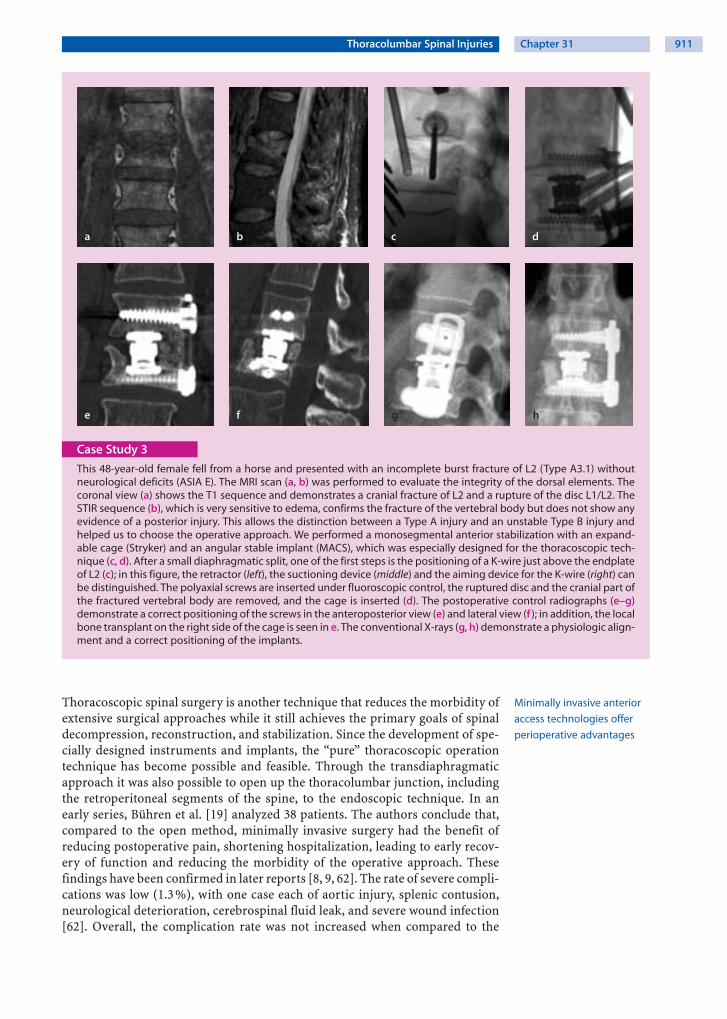
a b c d
e f g h
Case Study 3
This 48-year-old female fell from a horse and presented with an incomplete burst fracture of L2 (Type A3.1) withoutneurological deficits (ASIA E). The MRI scan (a, b) was performed to evaluate the integrity of the dorsal elements. Thecoronal view (a) shows the T1 sequence and demonstrates a cranial fracture of L2 and a rupture of the disc L1/L2. TheSTIR sequence (b), which is very sensitive to edema, confirms the fracture of the vertebral body but does not show anyevidence of a posterior injury. This allows the distinction between a Type A injury and an unstable Type B injury andhelped us to choose the operative approach. We performed a monosegmental anterior stabilization with an expand-able cage (Stryker) and an angular stable implant (MACS), which was especially designed for the thoracoscopic tech-nique (c, d). After a small diaphragmatic split, one of the first steps is the positioning of a K-wire just above the endplateof L2 (c); in this figure, the retractor (left), the suctioning device (middle) and the aiming device for the K-wire (right) canbe distinguished. The polyaxial screws are inserted under fluoroscopic control, the ruptured disc and the cranial part ofthe fractured vertebral body are removed, and the cage is inserted (d). The postoperative control radiographs (e–g)demonstrate a correct positioning of the screws in the anteroposterior view (e) and lateral view (f ); in addition, the localbone transplant on the right side of the cage is seen in e. The conventional X-rays (g, h) demonstrate a physiologic align-ment and a correct positioning of the implants.
Minimally invasive anterior
access technologies offer
perioperative advantages
Thoracoscopic spinal surgery is another technique that reduces the morbidity ofextensive surgical approaches while it still achieves the primary goals of spinaldecompression, reconstruction, and stabilization. Since the development of spe-cially designed instruments and implants, the “pure” thoracoscopic operationtechnique has become possible and feasible. Through the transdiaphragmaticapproach it was also possible to open up the thoracolumbar junction, includingthe retroperitoneal segments of the spine, to the endoscopic technique. In anearly series, Bühren et al. [19] analyzed 38 patients. The authors conclude that,compared to the open method, minimally invasive surgery had the benefit ofreducing postoperative pain, shortening hospitalization, leading to early recov-ery of function and reducing the morbidity of the operative approach. Thesefindings have been confirmed in later reports [8, 9, 62]. The rate of severe compli-cations was low (1.3%), with one case each of aortic injury, splenic contusion,neurological deterioration, cerebrospinal fluid leak, and severe wound infection[62]. Overall, the complication rate was not increased when compared to the
Thoracolumbar Spinal Injuries Chapter 31 911

open technique; however, there were clear advantages in terms of the reducedaccess morbidity.
Importantly, the endoscopic technique is also effective for anterior spinalcanal decompression. Beisse et al. [8] published a series of 30 patients with thora-columbar canal compromise that underwent endoscopic anterior spinal canaldecompression and report that 25% of patients with complete paraplegia and65% of those with incomplete neurological deficit improved neurologically.
The following factors have gradually opened up the entire spectrum of ante-rior spine surgery to endoscopic techniques [9]:
) a standardized operating technique) instruments and implants specially developed for the endoscopic procedure,
i.e.:) angle-stable plate and screw implants and) endoscopically implantable vertebral body replacements
Combined Anterior-Posterior Approach
Studies on posterior stabilization of thoracolumbar fractures demonstrated thatfractures with comminution of the anterior column often lead to early failure[85]. Therefore, in addition to the posterior two-level repositioning and stabili-zation, several techniques were introduced to stabilize the anterior column: iliacanterior crest [41], possibly in an inlay technique [71] or with vertebral bodyreplacements in different materials, shapes, sizes, and configurations (i.e., non-expandable vs. expandable cages). In our institution, we prefer to adhere to atwo-staged procedure that includes (Case Introduction):
) Stage 1: posterior fracture reduction and usually a two-level stabilization (w/o decompression depending on neural compromise)) Stage 2: delayed anterior surgery depending on the patients’ condition
Many peers recommend
a combined posterior/
anterior approach
for unstable fractures
It is evident that, although posterior reduction and stabilization provides effec-tive restoration of the sagittal alignment, the reduction capability of the intraca-nal bone fragments is distinctly limited [50, 107, 123]. The anterior reconstruc-tion method permits effective decompression of the spinal canal and offers supe-rior mechanical stability compared with the indirect decompression and stabili-zation of posterior instrumentation.
Treatment Guidelines
Most treatment recommen-
dations are not based
on scientific evidence
The conflicting results and the diversity of studies presented in this chapter indi-cate that there is no gold standard for the vast majority of fractures and treatmentdecisions are almost always lacking scientific evidence. Treatment options areoften based on the experience and the tradition of the institute and the treatingphysicians. Importantly, the patient and the treating team must be aware of theattainable results, the time course of the treatment, the pitfalls, and the complica-tion of the respective method, be it conservative or operative. Under these limita-
Critically evaluate anecdotal
treatment recommen-
dations before adaptation
tions, we have summarized some general guidelines (Fig. 9). However, we want toemphasize that these general recommendations may not apply to the individualcase and confounding variables have to be considered, e.g., general condition,injury pattern, polytrauma, age, associated diseases, etc.
Type A1 fractures are usually treated conservatively. However, if kyphosisbecomes relevant (more than 20°–25°) an operative correction of the kyphosishas to be considered. In this case, we advocate an early correction, i.e., when thefracture is not consolidated and still can be reduced to avoid more complex anddifficult correction surgery in a later stage. Also Type A2 fractures can be treated
912 Section Fractures

Figure 9. General treatment guidelines1 Corpectomy, interbody fusion with strut graft/cage, anterior instrumentation2 Two-level instrumentation, reduction, posterolateral fusion (optional with one-level fusion and posterior implant
removal after 10 – 12 months to liberate the uninjured segment)3 One-level stabilization and fusion possible in cases of monosegmental lesions (incomplete burst fractures, anterior disc
disruption)4 Additional anterior approach (corpectomy w/o decompression, interbody fusion with strut graft/cage) is indicated in
cases of persistent neural compression (incomplete canal clearance) or comminuted anterior column or to enhancefusion in discoligamentous injuries
5 One-level stabilization and fusion possible in cases of discoligamentous injuries or concomitant incomplete burst frac-tures
6 Multilevel stabilization often required (two or three levels above/below the injury)
Pincer fractures are prone
to non-union and are better
treated surgically
conservatively with the exception of A2.3 type fractures, the so-called “pincer”type. In this fracture type, both discs are usually ruptured and pushed into thefractured vertebral body. This injury pattern often leads to non-union andresults in painful instability. From a pathophysiological and biomechanical view,an anterior approach makes most sense in these A2.3 fractures, because thepathology is treated where the pathology is located. Probably the most contro-versy exists in A3 type fractures particularly the incomplete burst fracture
Type A3.1 fractures are the
most controversial ones
regarding treatment
recommendations
(A3.1). In this fracture type, the accepted treatments range from bracing to com-bined anterior/posterior approach all with acceptable results (Case Studies 2, 3).The treatment options depend on the comminution of the vertebral body, thedegree of kyphosis, and the presence or absence of neurology. If one decides tostabilize A3 fractures, the goal of neural decompression, sagittal alignment, andanterior support will dictate the operative approach. In an emergency situation,a primarily posterior approach will allow to reduce and stabilize the fracture withan internal fixator with or without laminectomy to decompress neural structure(Case Introduction). At a later stage, the surgeon can decide if an additional ante-rior approach is needed, based on the persistence of neurological compressionand the comminution of the anterior column. A CT scan after the postoperative
Thoracolumbar Spinal Injuries Chapter 31 913

approach is often helpful for decision making. Alternatively, an anteriorapproach only with corpectomy, interbody fusion with strut graft/cage, andanterior instrumentation will provide an appropriate stabilization (see CaseStudy 3).
The paradigm of a primarily posterior approach with or without an additionalanterior operation is also true for Type B and Type C fractures. One exception isthe purely osseous “Chance” fracture, because fractured bones heal better andfaster than ligamentous injuries. In this case, a thoracolumbar cast fixation thatprevents flexion/distraction movements of the injured segment is applied for6–8 weeks. Alternatively, one might also prefer to treat Chance fractures with anoperative stabilization and restore the ruptured tension band with a posteriorbisegmental stabilization without posterolateral fusion. Removal of the hardwareis then usually performed after 4 months. In B-type fractures, posterior stabiliza-tion is usually performed with a two-level instrumentation, reduction, and pos-terolateral fusion or optionally with a one-level fusion and posterior implant
The indication for an addi-
tional anterior approach
depends on neurological
compromise and anterior
column comminution
removal after 10–12 months to liberate the uninjured segment. Alternatively,two-level stabilization and fusion is possible in Type B cases with discoligamen-tous injuries or concomitant complete burst fractures. The decision whether anadditional anterior support is necessary or not depends on the persistence ofneural compression (incomplete canal clearance) or the comminution of theanterior column or the need to enhance arthrodeses by adding an interbody
Type C injuries are very
unstable and commonly
require multisegmental
fixation
fusion. In Type C injuries, multilevel stabilization is often required (two or threelevels above/below the injury) for reduction and stabilization. Additional ante-rior surgery again depends on canal clearance and anterior column reconstruc-tion.
Outcome of Operative Versus Non-operative Treatment
Despite many theoretical advantages of operative spinal fracture treatment, thereis a lack of scientific evidence which supports the benefits of surgery (Table 9).Many studies were not able to prove a substantial difference in functional out-come between the operative and non-operative treatment, regardless of the neu-rological injury [16, 17, 20, 73, 87, 92, 105, 116, 121]. Chow et al. [23] retrospec-tively reviewed 24 neurologically healthy patients (mean follow-up of 34 months)with unstable thoracolumbar burst fractures (T11–L2) managed with either cast-ing or bracing and early ambulation. Clinical follow-up examination was per-formed by the use of a questionnaire in which the patients were asked to rate theirpain, ability to work, ability to perform in recreational activities, and their over-all satisfaction with treatment. Kyphotic deformity could be corrected withhyperextension casting but tended to recur during the course of mobilization andhealing, as hypothesized by Magnus [82] and confirmed by other studies [96,111]. No correlation was found between kyphosis and clinical outcome. At finalfollow-up evaluation, 79% had little or no pain; 75% had returned to work; 75%stated that they had few or no restrictions in their ability to work; and 67% statedthat they had few or no restrictions in their ability to participate in recreational
Favorable outcome has
been reported with conser-
vative as well as operative
treatment when applying
the correct technique
activities. Only one patient (4%) reported being dissatisfied with the initial non-operative treatment of his spine fracture. The authors conclude that non-opera-tive management of thoracolumbar burst fractures with hyperextension castingor bracing is a safe and effective method of treatment in selected patients.
In the series of Daniaux et al. [27], 85% of patients with a thoracolumbar frac-ture were treated conservatively. In 40%, a functional treatment was possible;these were patients with stable impaction and split fractures as well as burst frac-tures that were considered to be stable and that had a kyphotic deformity of lessthan 10° for T12–L2 and 15° for T11, respectively. In 45%, a repositioning and
914 Section Fractures
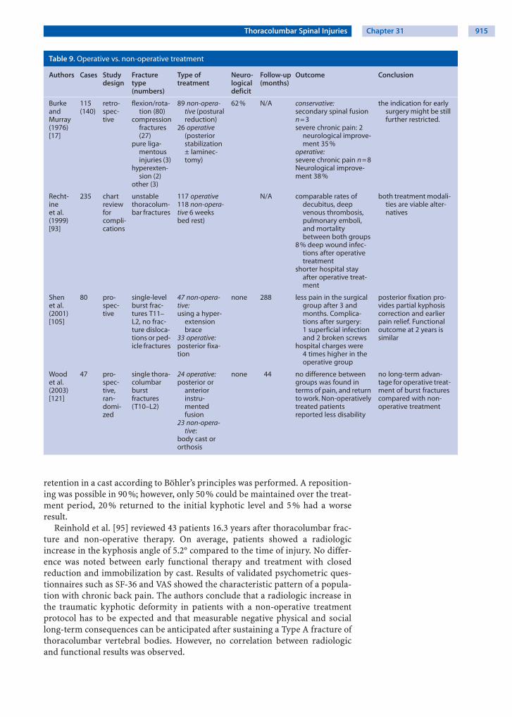
Table 9. Operative vs. non-operative treatment
Authors Cases Studydesign
Fracturetype(numbers)
Type oftreatment
Neuro-logicaldeficit
Follow-up(months)
Outcome Conclusion
BurkeandMurray(1976)[17]
115(140)
retro-spec-tive
flexion/rota-tion (80)
compressionfractures(27)
pure liga-mentousinjuries (3)
hyperexten-sion (2)
other (3)
89 non-opera-tive (posturalreduction)
26 operative(posteriorstabilization± laminec-tomy)
62 % N/A conservative:secondary spinal fusionn = 3severe chronic pain: 2
neurological improve-ment 35 %
operative:severe chronic pain n = 8Neurological improve-ment 38 %
the indication for earlysurgery might be stillfurther restricted.
Recht-ineet al.(1999)[93]
235 chartreviewforcompli-cations
unstablethoracolum-bar fractures
117 operative118 non-opera-tive 6 weeksbed rest)
N/A comparable rates ofdecubitus, deepvenous thrombosis,pulmonary emboli,and mortalitybetween both groups
8 % deep wound infec-tions after operativetreatment
shorter hospital stayafter operative treat-ment
both treatment modali-ties are viable alter-natives
Shenet al.(2001)[105]
80 pro-spec-tive
single-levelburst frac-tures T11–L2, no frac-ture disloca-tions or ped-icle fractures
47 non-opera-tive:using a hyper-
extensionbrace
33 operative:posterior fixa-tion
none 288 less pain in the surgicalgroup after 3 andmonths. Complica-tions after surgery:1 superficial infectionand 2 broken screws
hospital charges were4 times higher in theoperative group
posterior fixation pro-vides partial kyphosiscorrection and earlierpain relief. Functionaloutcome at 2 years issimilar
Woodet al.(2003)[121]
47 pro-spec-tive,ran-domi-zed
single thora-columbarburstfractures(T10–L2)
24 operative:posterior or
anteriorinstru-mentedfusion
23 non-opera-tive:
body cast ororthosis
none 44 no difference betweengroups was found interms of pain, and returnto work. Non-operativelytreated patientsreported less disability
no long-term advan-tage for operative treat-ment of burst fracturescompared with non-operative treatment
retention in a cast according to Böhler’s principles was performed. A reposition-ing was possible in 90%; however, only 50% could be maintained over the treat-ment period, 20% returned to the initial kyphotic level and 5% had a worseresult.
Reinhold et al. [95] reviewed 43 patients 16.3 years after thoracolumbar frac-ture and non-operative therapy. On average, patients showed a radiologicincrease in the kyphosis angle of 5.2° compared to the time of injury. No differ-ence was noted between early functional therapy and treatment with closedreduction and immobilization by cast. Results of validated psychometric ques-tionnaires such as SF-36 and VAS showed the characteristic pattern of a popula-tion with chronic back pain. The authors conclude that a radiologic increase inthe traumatic kyphotic deformity in patients with a non-operative treatmentprotocol has to be expected and that measurable negative physical and sociallong-term consequences can be anticipated after sustaining a Type A fracture ofthoracolumbar vertebral bodies. However, no correlation between radiologicand functional results was observed.
Thoracolumbar Spinal Injuries Chapter 31 915

In an earlier report, Weinstein et al. [116] also addressed the long-term results of42 patients with non-operative treatment for fractures of the thoracolumbarspine. Average time from injury to follow-up was 20.2 years. At follow-up, theaverage back pain score was 3.5, with 0 being no pain at all and 10 being verysevere pain. No patient required narcotic medication for pain control. Eighty-eight percent of patients were able to work at their usual level of activity. Follow-up radiographs revealed an average kyphosis angle of 26.4° in flexion and 16.8° inextension. The degree of kyphosis did not correlate with pain or function at fol-low-up.
Burke et al. [17] reported in his retrospective study that 3 of 89 patients withconservative therapy required a secondary spinal fusion for suspected instabilityafter a period of conservative treatment. Frankel [44] found that 2 of 394 conser-vatively treated patients required surgery because of instability.
Braakman et al. [16] prospectively studied 70 consecutive patients with inju-ries of the thoracic and lumbar spine with a neurological deficit. The authorscould not establish a difference in neurological recovery between those patientswho were managed conservatively and those in whom a surgical decompressionand stabilization procedure was performed. Surgical stabilizing procedures,however, resulted in immediate stabilization of the spine, diminished pain, facili-tated nursing care and allowed more rapid mobilization and earlier active reha-bilitation.
Shen at al. [104] studied 38 patients after functional treatment with a follow-up of 4.1 years. Four patients had moderate pain, 2 had moderate to severe pain,and 29 (76%) were able to work at the same level. The authors conclude thatactivity restriction and bracing may be important for pain control but probablydo not change the long-term result. The same authors [105] also conducted a pro-spective trial with 80 patients to compare the results of non-operative treatment(n=47) versus short-segment posterior fixation using pedicle screws; follow-upwas 2 years. They found that posterior fixation provides partial kyphosis correc-tion and earlier pain relief, but the functional outcome at 2 years is similar.
Wood et al. [121] published a prospective, randomized study comparing oper-ative (posterior or anterior arthrodesis and instrumentation) and non-operativetreatment (application of a body cast or orthosis) of stable thoracolumbar burstfractures in 47 patients without neurological deficit. After treatment, patientsindicated the degree of pain with use of the visual analog scale and they com-pleted the Roland and Morris Disability Questionnaire, the Oswestry Back-PainQuestionnaire, and the Short Form-36 (SF-36) Health Survey. No significant dif-ference was found between the two groups with respect to return to work. Thepreinjury scores were similar for both groups; however, at the time of the finalfollow-up (on average after 44 months), those who were treated non-operativelyreported less disability. The authors conclude that operative treatment of patientswith a stable thoracolumbar burst fracture and normal findings on the neurolog-ical examination provided no major long-term advantage compared with non-operative treatment.
The superiority of surgical
fracture treatment
is not well supported
in the literature
Rechtine et al. [93] reviewed the medical charts of 235 patients with thoraco-lumbar fractures to evaluate a difference in the occurrence of complications afterconservative (118 patients) or operative treatment (117 patients). There was nosignificant difference in the occurrence of decubitus, deep venous thromboses,pulmonary emboli, or mortality between the two groups. Deep wound infectionsoccurred in 8% of the operative cases. However, the length of stay was 24 dayslonger in the non-operative group. The authors conclude that the selection oftreatment method remains a matter of controversy.
916 Section Fractures
Complications
The reported complication
rate in the literature
varies largely
A surgery-related complication is a relevant shortcoming of any operative proce-dure with potentially devastating consequences, especially in spine surgery (seeChapter 39 ). The reported complication rate in the literature is largely variableand critically dependent on the pathology and type of surgery [7, 8, 19, 25, 34, 35,38, 39, 42, 62, 68, 70, 83, 102, 110, 115].
One of the largest series which considered complications in the surgical treat-ment of spinal fractures is the multicenter study of the Spine Study Group of theGerman Trauma Association (DGU). Knop et al. [69] reviewed sources of errorand specific complications [67, 65, 66]. A total of 682 patients were operated onfor acute traumatic injuries of the thoracolumbar spine. In 101 cases (15%) atleast one complication occurred intra- or postoperatively. In 41 patients (6%) arevision was performed, and in 60 patients (9%) complications without opera-tive revision were observed. Typical errors and possible complications duringoperations were related to different steps of the operation:
) positioning and closed reduction of fractures) approach) decompression of the spinal canal) instrumentation and stabilization) intervertebral fusion
Postoperative neurological
complications are rare
In addition, there are general surgical complications, which are not specific tospinal operations.
) Complications specific to the procedure that were revised included (n=40):deep infection 15 (2.2%), hematoma/wound healing disorder 12 (1.8%),instability or segmental malalignment 5 (0.7%), misplacement of screw/implant 4 (0.6%), persisting liquor fistula 2 (0.3%), sewn-in drain 1 (0.1%),arterial embolism of femoral artery 1 (0.1%).) Complications specific to the procedure that were (n= 29) not revised
included: intraoperative bleeding 10 (1.5%), iatrogenic pedicle fracture 5(0.7%), misplacement of screw/implant 3 (0.4%), instability or consecutivemalalignment 2 (0.3%), infection/healing disorder iliac crest 2 (0.3%), notspecified 2 (0.3%), iatrogenic rip fracture, approach related 1 (0.1%), iatro-genic lesion of pleura/peritoneum 1 (0.1%), narrowing of spinal canal withbone graft 1 (0.1%), fracture of iliac crest after graft harvesting 1 (0.1%),persisting liquor fistula 1 (0.1%).) Neurological complications (n= 13), revised and non-revised included:
peripheral lesion of nerve roots (0.7%), remittent neurologic deficit 4(0.6%), neurologic deterioration (Frankel/ASIA E to D) 2 (0.3%), neurologicdeterioration (Frankel/ASIA D to A) 1 (0.1%), paresthesia without neurolog-ical deficit 1 (0.1%).
Thoracolumbar Spinal Injuries Chapter 31 917

Recapitulation
Epidemiology. About 60 % of thoracic and lumbarspine fractures are located at the transition T11–L2,30 % in the thoracic spine and 10 % in the lowerlumbar spine. Spinal cord injury occurs in about10 – 30 % of traumatic spinal fractures.
Pathogenesis. The most relevant forces that pro-duce structural damage to the spine are axial com-pression, flexion/distraction, hyperextension, rota-tion, and shear. Axial load may result in a burst frac-ture; the posterior elements are usually intact. Inflexion/distraction injuries, the posterior ligamen-tous and osseous elements fail in tension; a wedgecompression fracture of the vertebral body is oftenassociated. Hyperextension may result in ruptureof the anterior ligament and the disc as well as incompression injuries of the posterior elements, i.e.,fracture of the facets, the laminae, or the spinousprocesses. Rotational injuries combine compres-sive forces and flexion/distraction mechanisms andare highly unstable injuries. Shear forces producesevere ligamentous disruption and usually result incomplete spinal cord injury.
Clinical presentation. In the case of a polytrauma,about 30 % of the patients have a spinal injury. Theneurological examination has to include the“search for a sacral sparing” which determines thecompleteness of the deficit and the prognosis.About one-third of all spinal injuries have concomi-tant injuries; the most frequent are: head injuries,chest injuries and long bone injuries. The historyshould include the type of trauma (high vs. low en-ergy injuries) and the time course of a possible neu-rological deficit. The initial focus of the physical ex-amination is on the assessment of vital functionsand neurological deficits. Because the spinal cordusually terminates at the level of L1, injuries to thethoracolumbar junction may result in various neu-rological symptoms: e.g., complete/incompleteparaplegia (distal spinal cord), malfunction of thevegetative system (conus medullaris), or caudaequina syndrome.
Diagnostic work-up. Static imaging studies are“snapshots in time” and do not reveal the real de-gree of spinal canal compromise that may havehappened during the injury. A posterior cortical dis-ruption seen in the lateral view or an interpedicularwidening seen in the anteroposterior view sug-gests a burst fracture that should be further ana-
lyzed by CT scan. CT is the imaging study of choiceto demonstrate bony destruction. MRI is recom-mended to identify a possible cord lesion or a cordcompression in patients with neurological deficits.MRI can be helpful in determining the integrity of theposterior ligamentous structures and thereby in dif-ferentiating between a Type A and a Type B lesion.
Non-operative treatment. Management of thora-columbar and sacral spinal fractures remains a con-troversial area in modern spinal surgery. The litera-ture demonstrates a wide range of conflicting re-sults and recommendations. Unfortunately, thevast majority of clinical studies can be criticized be-cause of their retrospective design, heteroge-neous patient populations and treatment strate-gies, limited follow-up, and poorly defined out-come measures.The main advantage of non-operative treatmentof thoracolumbar fracture is the avoidance of sur-gery-related complications. According to Böhler,the time of immobilization in a cast is usually3 –5 months depending on the fracture type. Im-portantly, skillful physical therapy is paramount toachieve good results. Because thoracolumbar frac-tures are bound to return to the initial deformity,functional bracing without repositioning is an alter-native to Böhler’s concept of repositioning and sta-bilization with a cast if the initial deformity is ac-ceptable. Many studies were not able to prove asubstantial difference in functional outcome be-tween the operative and non-operative treatment,regardless of the neurological injury.
Operative treatment. There is a general trend to-wards operative treatment of unstable fracturesmostly because surgical stabilizing procedures re-sult in early mobilization, diminished pain, facilitat-ed nursing care, earlier return to work, and avoid-ance of late neurological complications. In experi-mental animal models, persistent compression ofthe spinal cord is potentially reversible from a sec-ondary injury by early decompression. Most investi-gators recommend a surgical decompression inthe setting of major neurological deficit, progres-sive neurological loss, and substantial compromiseof the spinal canal. Currently, there are no goldstandards regarding the role and timing of de-compression in acute spinal cord injury. Posteriorbisegmental reduction and stabilization is the“working horse” of the posterior approach tech-nique that allows for fracture reduction and stable
918 Section Fractures

fixation. Depending on the persistence of spinalcanal compromise or comminution of the fracturedvertebral body, an additional anterior approach isneeded. Transpedicular cancellous bone graftingfor interbody fusion after posterior stabilization isnot recommended in complete or incomplete burstfractures. Only incomplete Type A burst fractureswith intact pedicles and a lower endplate should beconsidered for posterior monosegmental reduc-tion and stabilization. Compared to the openmethod, minimally invasive surgery reduces post-operative pain, shortens hospitalization, leads toearly recovery of function and reduces morbidity of
the operative approach. A combined posterior andanterior approach is used to reduce and stabilizeseverely comminuted vertebral body fractures andto decompress the spinal canal. In Type C lesionsoften multisegmental instrumentation is neededto reliably stabilize the spine.
Complications. The reported complication rate inthe literature varies largely and ranges from 3.6 % to10 %. Postoperative neurological complicationsrange from 0.1 % to 0.7 %. Only honest and accurateassessment of complications will lead to scientificand clinical progress.
Key Articles
Böhler L (1951) Die Technik der Knochenbruchbehandlung. Maudrich, ViennaLorenz Böhler was one of the first to advocate a conservative treatment with fracturereduction and retention in a cast.
Roaf R (1960) A study of the mechanics of spinal injuries. J Bone Joint Surg Br42B:810 – 23
In this article Roaf studies the biomechanics of spinal injuries and describes the results ofstudies of spinal units when subjected to forces of different magnitude and direction, i.e.,compression, flexion, extension, lateral flexion, rotation, and horizontal shear.
Denis F (1983) The three column spine and its significance in the classification of acutethoraco-lumbar spinal injuries. Spine 8:817 – 31
This article is a presentation of the concept of the three-column spine. The conceptevolved from a retrospective review of 412 thoracolumbar spine injuries and observa-tions on spinal instability. The posterior column consists of what Holdsworth describedas the posterior ligamentous complex. The middle column includes the posterior longitu-dinal ligament, posterior anulus fibrosus, and posterior wall of the vertebral body. Theanterior column consists of the anterior vertebral body, anterior anulus fibrosus, andanterior longitudinal ligament.
Dick W (1987) The “fixateur interne” as a versatile implant for spine surgery. Spine12:882 –900
This article introduced a new angle-stable fixation device which first allowed a short seg-mental reduction and fixation of fractures.
Magerl F, Aebi M, Gertzbein SD, Harms J, Nazarian S (1994) A comprehensive classifica-tion of thoracic and lumbar injuries. Eur Spine J 3:184 – 201
This article describes a classification of thoracic and lumbar injuries. As a result of morethan a decade of consideration of the subject matter and a review of 1445 consecutive tho-racolumbar injuries, a comprehensive classification of thoracic and lumbar injuries isproposed. The classification is primarily based on pathomorphological criteria. Threemechanisms classify the injury pattern according to the AO classification: axial compres-sion (Type A), flexion distraction (Type B) and rotational/shear injuries (Type C).
Kaneda K, Taneichi H, Abumi K, Hashimoto T, Satoh S, Fujiya M (1997) Anterior decom-pression and stabilization with the Kaneda device for thoracolumbar burst fracturesassociated with neurological deficits. J Bone Joint Surg Am 79:69 – 83
One hundred and fifty consecutive patients who had a burst fracture of the thoracolum-bar spine and associated neurological deficits were managed with a single-stage anteriorspinal decompression, strut-grafting, and Kaneda spinal instrumentation. The authorsconclude that anterior decompression, strut-grafting, and fixation with the Kaneda
Thoracolumbar Spinal Injuries Chapter 31 919
device in patients who had a burst fracture of the thoracolumbar spine and associatedneurological deficits yielded good radiographic and functional results. This article estab-lished the single stage anterior approach for this fracture type.
Knop C, Blauth M, Bühren V, Hax PM, Kinzl L, Mutschler W, Pommer A, Ulrich C, Wag-ner S, Weckbach A, Wentzensen A, Wörsdörfer O (1999) Surgical treatment of injuries ofthe thoracolumbar transition. 1: Epidemiology. Unfallchirurg 102:924 – 35
Knop C, Blauth M, Bühren V, Hax PM, Kinzl L, Mutschler W, Pommer A, Ulrich C, Wag-ner S, Weckbach A, Wentzensen A, Wörsdörfer O (2000) Surgical treatment of injuries ofthe thoracolumbar transition. 2: Operation and roentgenologic findings. Unfallchirurg103:1032 – 47
Knop C, Blauth M, Bühren V, Arand M, Egbers HJ, Hax PM, Nothwang J, Oestern HJ,Pizanis A, Roth R, Weckbach A, Wentzensen A (2001) Surgical treatment of injuries ofthe thoracolumbar transition – 3: Follow-up examination. Results of a prospective mul-ti-center study by the “Spinal” Study Group of the German Society of Trauma Surgery.Unfallchirurg 104:583 – 600
These three reports summarize the experience based on 682 patients included in a pro-spective multicenter study by the “Spinal” Study Group of the German Society of TraumaSurgery. All treatment methods under study were appropriate for achieving comparableclinical and functional outcome. The internal fixator was found superior in restoration ofthe spinal alignment. Best radiological outcomes were achieved by combined stabiliza-tion. Merely by direct reconstruction of the anterior column the postoperative re-kypho-sing is prevented and a gain in segmental angle is achieved. However, this benefit was notreflected in the clinical outcome.
Fehlings MG, Perrin RG (2005) The role and timing of early decompression for cervicalspinal cord injury: Update with a review of recent clinical evidence. Injury S-B13–S-B26
Evidence-based recommendations regarding spinal cord decompression in patients withacute spinal cord injury.
Beisse R (2006) Endoscopic surgery on the thoracolumbar junction of the spine. EurSpine J 15:687 – 704
This article summarizes the technique and results based on a large patient group from aGerman trauma center: A now standardized operating technique, instruments andimplants specially developed for the endoscopic procedure, from angle stable plate andscrew implants to endoscopically implantable vertebral body replacements, have gradu-ally opened up the entire spectrum of anterior spine surgery to endoscopic techniques.
References
1. Alanay A, Acaroglu E, Yazici M, Oznur A, Surat A (2001) Short-segment pedicle instrumenta-tion of thoracolumbar burst fractures: does transpedicular intracorporeal grafting preventearly failure? Spine 26:213– 7
2. Anderson PA, Henley MB, Rivara FP, Maier RV (1991) Flexion distraction and chance injuriesto the thoracolumbar spine. J Orthop Trauma 5:153–60
3. Anderson PA, Rivara FP, Maier RV, Drake C (1991) The epidemiology of seatbelt-associatedinjuries. J Trauma 31:60–7
4. Anderson PA, Bohlman HH (1992) Anterior decompression and arthrodesis of the cervicalspine: long-term motor improvement. Part II – Improvement in complete traumatic quadri-plegia. J Bone Joint Surg Am 74:683– 92
5. Anderson S, Biros MH, Reardon RF (1996) Delayed diagnosis of thoracolumbar fractures inmultiple-trauma patients. Acad Emerg Med 3:832–9
6. Bagley LJ (2006) Imaging of spinal trauma. Radiol Clin North Am 44:1–12, vii7. Been HD, Bouma GJ (1999) Comparison of two types of surgery for thoraco-lumbar burst
fractures: combined anterior and posterior stabilisation vs. posterior instrumentation only.Acta Neurochir (Wien) 141:349– 57
8. Beisse R, Muckley T, Schmidt MH, Hauschild M, Buhren V (2005) Surgical technique andresults of endoscopic anterior spinal canal decompression. J Neurosurg Spine 2:128–36
9. Beisse R (2006) Endoscopic surgery on the thoracolumbar junction of the spine. Eur Spine J15:687–704
920 Section Fractures
10. Bensch FV, Kiuru MJ, Koivikko MP, Koskinen SK (2004) Spine fractures in falling accidents:analysis of multidetector CT findings. Eur Radiol 14:618– 24
11. Bensch FV, Koivikko MP, Kiuru MJ, Koskinen SK (2006) The incidence and distribution ofburst fractures. Emerg Radiol 12:124– 9
12. Benson DR (1988) Unstable thoracolumbar fractures, with emphasis on the burst fracture.Clin Orthop Relat Res 14– 29
13. Blauth M, Knop C, Bastian L (1998) Wirbelsäule. In: Tscherne H, Blauth M (eds) TscherneUnfallchirurgie, vol 3. Springer, Berlin Heidelberg New York, pp 241– 381
14. Bohlman HH, Anderson PA (1992) Anterior decompression and arthrodesis of the cervicalspine: long-term motor improvement. Part I – Improvement in incomplete traumatic quad-riparesis. J Bone Joint Surg Am 74:671– 82
15. Bohlman HH, Kirkpatrick JS, Delamarter RB, Leventhal M (1994) Anterior decompressionfor late pain and paralysis after fractures of the thoracolumbar spine. Clin Orthop 24–9
16. Braakman R, Fontijne WP, Zeegers R, Steenbeek JR, Tanghe HL (1991) Neurological deficitin injuries of the thoracic and lumbar spine. A consecutive series of 70 patients. Acta Neuro-chir (Wien) 111:11–7
17. Burke DC, Murray DD (1976) The management of thoracic and thoraco-lumbar injuries ofthe spine with neurological involvement. J Bone Joint Surg Br 58:72–8
18. Böhler L (1951) Die Technik der Knochenbruchbehandlung. Maudrich Verlag, Vienna19. Bühren V, Beisse R, Potulski M (1997) [Minimally invasive ventral spondylodesis in injuries
to the thoracic and lumbar spine]. Chirurg 68:1076–8420. Cantor JB, Lebwohl NH, Garvey T, Eismont FJ (1993) Nonoperative management of stable
thoracolumbar burst fractures with early ambulation and bracing. Spine 18:971–621. Carl AL, Tromanhauser SG, Roger DJ (1992) Pedicle screw instrumentation for thoracolum-
bar burst fractures and fracture-dislocations. Spine 17:S317–2422. Chance G (1948) Note on a type of flexion fracture of the spine. Br J Radiol 21:452–323. Chow GH, Nelson BJ, Gebhard JS, Brugman JL, Brown CW, Donaldson DH (1996) Func-
tional outcome of thoracolumbar burst fractures managed with hyperextension casting orbracing and early mobilization. Spine 21:2170– 5
24. Dai L-Y, Yao W-F, Cui Y-M, Zhou Q (2004) Thoracolumbar fractures in patients with multi-ple injuries: diagnosis and treatment – a review of 147 cases. J Trauma 56:348– 55
25. Daniaux H (1986) Transpedicular repositioning and spongioplasty in fractures of the verte-bral bodies of the lower thoracic and lumbar spine. Unfallchirurg 89:197– 213
26. Daniaux H, Seykora P, Genelin A, Lang T, Kathrein A (1991) Application of posterior platingand modifications in thoracolumbar spine injuries. Indication, techniques, and results.Spine 16:S125– 33
27. Daniaux H, Wagner M, Kathrein A, Lang T (1999) [Fractures of the thoraco-lumbar junc-tion. Conservative management]. Orthopade 28:682–91
28. Defino HL, Rodriguez-Fuentes AE (1998) Treatment of fractures of the thoracolumbar spineby combined anteroposterior fixation using the Harms method. Eur Spine J 7:187–94
29. Defino HL, Scarparo P (2005) Fractures of thoracolumbar spine: monosegmental fixation.Injury 36 Suppl 2:B90– 7
30. Denis F (1983) The three column spine and its significance in the classification of acute tho-racolumbar spinal injuries. Spine 8:817– 31
31. Denis F, Armstrong GW, Searls K, Matta L (1984) Acute thoracolumbar burst fractures in theabsence of neurologic deficit. A comparison between operative and nonoperative treat-ment. Clin Orthop Relat Res 142–9
32. Denis F, Burkus JK (1992) Shear fracture-dislocations of the thoracic and lumbar spine asso-ciated with forceful hyperextension (lumberjack paraplegia). Spine 17:156–61
33. DeWald RL (1984) Burst fractures of the thoracic and lumbar spine. Clin Orthop Relat Res150– 61
34. Dick W (1987) The “fixateur interne” as a versatile implant for spine surgery. Spine 12:882– 900
35. Dickson JH, Harrington PR, Erwin WD (1978) Results of reduction and stabilization of theseverely fractured thoracic and lumbar spine. J Bone Joint Surg Am 60:799– 805
36. el-Khoury GY, Whitten CG (1993) Trauma to the upper thoracic spine: anatomy, biome-chanics, and unique imaging features. AJR Am J Roentgenol 160:95–102
37. Evans L (1988) Risk of fatality from physical trauma versus sex and age. J Trauma 28:368–7838. Eysel P, Meinig G, Sanner F (1991) Comparative study of various dorsal stabilization proce-
dures in recent fractures of the thoracic spine. Unfallchirurgie 17:264–7339. Faciszewski T, Winter RB, Lonstein JE, Denis F, Johnson L (1995) The surgical and medical
perioperative complications of anterior spinal fusion surgery in the thoracic and lumbarspine in adults. A review of 1223 procedures. Spine 20:1592–9
40. Fehlings MG, Perrin RG (2005) The role and timing of early decompression for cervical spi-nal cord injury: Update with a review of recent clinical evidence. Injury S-B13–S-B26
41. Feil J, Wörsdörfer O (1992) [Ventral stabilization in the area of the thoracic and lumbarspine]. Chirurg 63:856– 65
Thoracolumbar Spinal Injuries Chapter 31 921
42. Feil J, Wörsdörfer O (1992) Complications in surgical management of spinal injuries. Lan-genbecks Arch Chir Suppl Kongressbd 304–10
43. Floman Y, Fast A, Pollack D, Yosipovitch Z, Robin GC (1986) The simultaneous applicationof an interspinous compressive wire and Harrington distraction rods in the treatment offracture-dislocation of the thoracic and lumbar spine. Clin Orthop Relat Res 207–15
44. Frankel HL, Hancock DO, Hyslop G, Melzak J, Michaelis LS, Ungar GH, Vernon JD, Walsh JJ(1969) The value of postural reduction in the initial management of closed injuries of thespine with paraplegia and tetraplegia. I. Paraplegia 7:179– 92
45. Fredrickson BE, Edwards WT, Rauschning W, Bayley JC, Yuan HA (1992) Vertebral burstfractures: an experimental, morphologic, and radiographic study. Spine 17:1012– 21
46. Gertzbein SD (1992) Fractures of the thoracic and lumbar spine. Williams & Wilkins, Balti-more
47. Gertzbein SD (1992) Scoliosis Research Society. Multicenter spine fracture study. Spine17:528–40
48. Gopalakrishnan KC, el Masri WS (1986) Fractures of the sternum associated with spinalinjury. J Bone Joint Surg Br 68:178– 81
49. Gotzen L, Puplat D, Junge A (1992) Indications, technique and results of monosegmentaldorsal spondylodesis in wedge compression fractures (grade II) of the thoracolumbar spine.Unfallchirurg 95:445–54
50. Haas N, Blauth M, Tscherne H (1991) Anterior plating in thoracolumbar spine injuries. Indi-cation, technique, and results. Spine 16:S100– 11
51. Harrington RM, Budorick T, Hoyt J, Anderson PA, Tencer AF (1993) Biomechanics of indi-rect reduction of bone retropulsed into the spinal canal in vertebral fracture. Spine18:692–9
52. Hauser CJ, Visvikis G, Hinrichs C, Eber CD, Cho K, Lavery RF, Livingston DH (2003) Pro-spective validation of computed tomographic screening of the thoracolumbar spine intrauma. J Trauma 55:228–34; discussion 34–5
53. Henderson RL, Reid DC, Saboe LA (1991) Multiple noncontiguous spine fractures. Spine16:128–31
54. Holdsworth F (1963) Fractures, dislocations, and fracture-dislocations of the spine. J BoneJoint Surg Am 45:6– 20
55. Holdsworth F (1970) Fractures, dislocations, and fracture-dislocations of the spine. J BoneJoint Surg Am 52:1534–51
56. Hu R, Mustard CA, Burns C (1996) Epidemiology of incident spinal fracture in a completepopulation. Spine 21:492– 9
57. Inaba K, Munera F, McKenney M, Schulman C, de Moya M, Rivas L, Pearce A, Cohn S (2006)Visceral torso computed tomography for clearance of the thoracolumbar spine in trauma: areview of the literature. J Trauma 60:915–20
58. Jahna H, Wittich H (1985) Konservative Methoden in der Frakturbehandlung. Urban &Fischer, Vienna, pp 121–38
59. Junge A, Gotzen L, von Garrel T, Ziring E, Giannadakis K (1997) [Monosegmental internalfixator instrumentation and fusion in treatment of fractures of the thoracolumbar spine.Indications, technique and results]. Unfallchirurg 100:880–7
60. Kaneda K, Taneichi H, Abumi K, Hashimoto T, Satoh S, Fujiya M (1997) Anterior decom-pression and stabilization with the Kaneda device for thoracolumbar burst fractures associ-ated with neurological deficits. J Bone Joint Surg Am 79:69– 83
61. Kelly RP, Whitesides TE (1968) Treatment of lumbodorsal fracture-dislocations. Ann Surg167:705– 17
62. Khoo LT, Beisse R, Potulski M (2002) Thoracoscopic-assisted treatment of thoracic andlumbar fractures: a series of 371 consecutive cases. Neurosurgery 51:104–17
63. Kim DH, Silber JS, Albert TJ (2003) Osteoporotic vertebral compression fractures. InstrCourse Lect 52:541–50
64. King AG (1987) Burst compression fractures of the thoracolumbar spine. Pathologic anat-omy and surgical management. Orthopedics 10:1711–9
65. Knop C, Blauth M, Bühren V, Hax PM, Kinzl L, Mutschler W, Pommer A, Ulrich C, WagnerS, Weckbach A, Wentzensen A, Wörsdörfer O (1999) Surgical treatment of injuries of thethoracolumbar transition. 1: Epidemiology. Unfallchirurg 102:924– 35
66. Knop C, Blauth M, Bühren V, Hax PM, Kinzl L, Mutschler W, Pommer A, Ulrich C, Wagner S,Weckbach A, Wentzensen A, Wörsdörfer O (2000) Surgical treatment of injuries of the thora-columbar transition. 2: Operation and roentgenologic findings. Unfallchirurg 103:1032 –47
67. Knop C, Blauth M, Bühren V, Arand M, Egbers HJ, Hax PM, Nothwang J, Oestern HJ, PizanisA, Roth R, Weckbach A, Wentzensen A (2001) Surgical treatment of injuries of the thoraco-lumbar transition – 3: Follow-up examination. Results of a prospective multi-center studyby the “Spinal” Study Group of the German Society of Trauma Surgery. Unfallchirurg104:583– 600
68. Knop C, Fabian HF, Bastian L, Blauth M (2001) Late results of thoracolumbar fractures afterposterior instrumentation and transpedicular bone grafting. Spine 26:88– 99
922 Section Fractures

69. Knop C, Bastian L, Lange U, Oeser M, Zdichavsky M, Blauth M (2002) Complications in sur-gical treatment of thoracolumbar injuries. Eur Spine J 11:214– 26
70. Knop C, Fabian HF, Bastian L, Rosenthal H, Lange U, Zdichavsky M, Blauth M (2002) Fate ofthe transpedicular intervertebral bone graft after posterior stabilisation of thoracolumbarfractures. Eur Spine J 11:251–7
71. Kossmann T, Ertel W, Platz A, Trentz O (1999) [Combined surgery for fractures of the tho-raco-lumbar junction using the inlay-span method]. Orthopade 28:432– 40
72. Kossmann T, Jacobi D, Trentz O (2001) The use of a retractor system (SynFrame) for open,minimal invasive reconstruction of the anterior column of the thoracic and lumbar spine.Eur Spine J 10:396– 402
73. Kraemer WJ, Schemitsch EH, Lever J, McBroom RJ, McKee MD, Waddell JP (1996) Func-tional outcome of thoracolumbar burst fractures without neurological deficit. J OrthopTrauma 10:541– 4
74. Krueger MA, Green DA, Hoyt D, Garfin SR (1996) Overlooked spine injuries associated withlumbar transverse process fractures. Clin Orthop 191– 5
75. La Rosa G, Conti A, Cardali S, Cacciola F, Tomasello F (2004) Does early decompressionimprove neurological outcome of spinal cord injured patients? Appraisal of the literatureusing a meta-analytical approach. Spinal Cord 42:503– 12
76. Larson SJ, Holst RA, Hemmy DC, Sances A (1976) Lateral extracavitary approach to trau-matic lesions of the thoracic and lumbar spine. J Neurosurg 45:628–37
77. Leidner B, Adiels M, Aspelin P, Gullstrand P, Wallen S (1998) Standardized CT examinationof the multitraumatized patient. Eur Radiol 8:1630– 8
78. Lindsey RW, Dick W (1991) The fixateur interne in the reduction and stabilization of thora-columbar spine fractures in patients with neurologic deficit. Spine 16:S140–5
79. Louis R (1977) Unstable fractures of the spine. III. Instability. A. Theories concerning insta-bility. Rev Chir Orthop Reparatrice Appar Mot 63:423–5
80. Magerl F, Aebi M, Gertzbein SD, Harms J, Nazarian S (1994) A comprehensive classificationof thoracic and lumbar injuries. Eur Spine J 3:184–201
81. Magerl F, Engelhardt P (1994) Brust- und Lendenwirbelsäule – Verlaufsformen. In: Witt AN,Rettig H, Schlegel KF (eds) Orthopädie in Praxis und Klinik, Spezielle Orthopädie (Wirbel-säule – Thorax – Becken). Thieme, Stuttgart New York, pp 3.82– 3.132
82. Magnus G (1930) Die Begutachtung und Behandlung des Wirbelbruchs. Arch OrthopUnfallchir 29:277
83. Mayer H, Schaaf D, Kudernatsch M (1992) Use of internal fixator in injuries of the thoracicand lumbar spine. Chirurg 63:944– 9
84. Maynard FM, Jr, Bracken MB, Creasey G, Ditunno JF, Jr, Donovan WH, Ducker TB, GarberSL, Marino RJ, Stover SL, Tator CH, Waters RL, Wilberger JE, Young W (1997) InternationalStandards for Neurological and Functional Classification of Spinal Cord Injury. AmericanSpinal Injury Association. Spinal Cord 35:266– 74
85. McLain RF, Sparling E, Benson DR (1993) Early failure of short-segment pedicle instrumen-tation for thoracolumbar fractures. A preliminary report. J Bone Joint Surg Am 75:162– 7
86. Meyer PR, Heim S (1989) Fractures of the thoracic spine T1–T10. In: Meyer PR (ed) Surgeryof spine trauma. Churchill Livingstone, Edinburgh, pp 525– 72
87. Mumford J, Weinstein JN, Spratt KF, Goel VK (1993) Thoracolumbar burst fractures. Theclinical efficacy and outcome of nonoperative management. Spine 18:955– 70
88. Müller U, Berlemann U, Sledge J, Schwarzenbach O (1999) Treatment of thoracolumbarburst fractures without neurologic deficit by indirect reduction and posterior instrumenta-tion: bisegmental stabilization with monosegmental fusion. Eur Spine J 8:284–9
89. Nicoll EA (1949) Fractures of the dorso-lumbar spine. J Bone Joint Surg Br 31:376–9490. Olerud S, Karlstrom G, Sjostrom L (1988) Transpedicular fixation of thoracolumbar verte-
bral fractures. Clin Orthop Relat Res 227:44–5191. Place HM, Donaldson DH, Brown CW, Stringer EA (1994) Stabilization of thoracic spine
fractures resulting in complete paraplegia. A long-term retrospective analysis. Spine19:1726– 30
92. Rechtine GR (1999) Nonsurgical treatment of thoracic and lumbar fractures. Instr CourseLect 48:413– 6
93. Rechtine GR, 2nd, Cahill D, Chrin AM (1999) Treatment of thoracolumbar trauma: compar-ison of complications of operative versus nonoperative treatment. J Spinal Disord 12:406– 9
94. Reid DC, Hu R, Davis LA, Saboe LA (1988) The nonoperative treatment of burst fractures ofthe thoracolumbar junction. J Trauma 28:1188–94
95. Reinhold M, Knop C, Lange U, Bastian L, Blauth M (2003) Non-operative treatment of tho-racolumbar spinal fractures. Long-term clinical results over 16 years. Unfallchirurg 106:566– 76
96. Resch H, Rabl M, Klampfer H, Ritter E, Povacz P (2000) Surgical vs. conservative treatmentof fractures of the thoracolumbar transition. Unfallchirurg 103:281–8
97. Richards PJ (2005) Cervical spine clearance: a review. Injury 36:248– 6998. Roaf R (1960) A study of the mechanics of spinal injuries. J Bone Joint Surg Br 42B:810– 23
Thoracolumbar Spinal Injuries Chapter 31 923
99. Roy-Camille R, Saillant G (1984) Spinal injuries without neurologic complications. IntOrthop 8:155–62
100. Saboe LA, Reid DC, Davis LA, Warren SA, Grace MG (1991) Spine trauma and associatedinjuries. J Trauma 31:43– 8
101. Samuels LE, Kerstein MD (1993) ’Routine’ radiologic evaluation of the thoracolumbarspine in blunt trauma patients: a reappraisal. J Trauma 34:85– 9
102. Sasso RC, Cotler HB (1993) Posterior instrumentation and fusion for unstable fracturesand fracture-dislocations of the thoracic and lumbar spine. A comparative study of threefixation devices in 70 patients. Spine 18:450–60
103. Sasso RC, Best NM, Reilly TM, McGuire RA (2005) Anterior-only stabilization of three-col-umn thoracolumbar injuries. J Spinal Disord Tech 18 Suppl:S7–14
104. Shen WJ, Shen YS (1999) Nonsurgical treatment of three-column thoracolumbar junctionburst fractures without neurologic deficit. Spine 24:412– 5
105. Shen WJ, Liu TJ, Shen YS (2001) Nonoperative treatment versus posterior fixation for tho-racolumbar junction burst fractures without neurologic deficit. Spine 26:1038– 45
106. Sheridan R, Peralta R, Rhea J, Ptak T, Novelline R (2003) Reformatted visceral protocolhelical computed tomographic scanning allows conventional radiographs of the thoracicand lumbar spine to be eliminated in the evaluation of blunt trauma patients. J Trauma55:665– 9
107. Shono Y, McAfee PC, Cunningham BW (1994) Experimental study of thoracolumbar burstfractures. A radiographic and biomechanical analysis of anterior and posterior instru-mentation systems. Spine 19:1711– 22
108. Speth MJ, Oner FC, Kadic MA, de Klerk LW, Verbout AJ (1995) Recurrent kyphosis afterposterior stabilization of thoracolumbar fractures. 24 cases treated with a Dick internalfixator followed for 1.5–4 years. Acta Orthop Scand 66:406–10
109. Spitz J, Becker C, Tittel K, Weigand H (1992) [Clinical relevance of whole body skeletalscintigraphy in multiple injury and polytrauma patients]. Unfallchirurgie 18:133–47
110. Spivak JM, Neuwirth MG, Giordano CP, Bloom N (1994) The perioperative course of com-bined anterior and posterior spinal fusion. Spine 19:520– 5
111. Steindl A, Schuh G (1992) Late results after lumbar vertebrae fracture with Lorenz Böhlerconservative treatment. Unfallchirurg 95:439–44
112. Transfeldt EE, White D, Bradford DS, Roche B (1990) Delayed anterior decompression inpatients with spinal cord and cauda equina injuries of the thoracolumbar spine. Spine15:953– 7
113. Vaccaro AR, Daugherty RJ, Sheehan TP, Dante SJ, Cotler JM, Balderston RA, Herbison GJ,Northrup BE (1997) Neurologic outcome of early versus late surgery for cervical spinalcord injury. Spine 22:2609– 13
114. Vaccaro AR, Kim DH, Brodke DS, Harris M, Chapman JR, Schildhauer T, Routt ML, SassoRC (2004) Diagnosis and management of thoracolumbar spine fractures. Instr Course Lect53:359– 73
115. Wawro W, Konrad L, Aebi M (1994) Single segment internal fixator device in treatment ofthoracolumbar vertebral fractures. Unfallchirurg 97:114– 20
116. Weinstein JN, Collalto P, Lehmann TR (1988) Thoracolumbar “burst” fractures treatedconservatively: a long-term follow-up. Spine 13:33–8
117. Weitzman G (1971) Treatment of stable thoracolumbar spine compression fractures byearly ambulation. Clin Orthop 76:116– 22
118. White AA, 3rd, Panjabi MM (1978) The basic kinematics of the human spine. A review ofpast and current knowledge. Spine 3:12–20
119. Whitesides TE (1977) Traumatic kyphosis of the thoracolumbar spine. Clin Orthop 78–92120. Woltmann A, Bühren V (2004) Emergency room management of the multiply injured
patient with spine injuries. A systematic review of the literature. Unfallchirurg 107:911–9121. Wood K, Butterman G, Mehbod A, Garvey T, Jhanjee R, Sechriest V (2003) Operative com-
pared with nonoperative treatment of a thoracolumbar burst fracture without neurologi-cal deficit. A prospective, randomized study. J Bone Joint Surg Am 85-A:773–81
122. Wood KB, Bohn D, Mehbod A (2005) Anterior versus posterior treatment of stable thora-columbar burst fractures without neurologic deficit: a prospective, randomized study. JSpinal Disord Tech 18 Suppl:S15– 23
123. Young B, Brooks WH, Tibbs PA (1981) Anterior decompression and fusion for thoracolum-bar fractures with neurological deficits. Acta Neurochir (Wien) 57:287–98
924 Section Fractures