spigelian hernia a review of the literature and report of three cases · several anatomi factorc...
TRANSCRIPT
C L E V E L A N D C L I N I C Q U A R T E R L Y Copyright © 1971 by The Cleveland Clinic Foundation
Volume 38, January 1971 Printed in U.S.A.
Spigelian hernia
A review of the literature and report of three cases
NORMAN R . HERTZER, M . D . *
JOSEPH E . MONTIE, M . D . * Department of General Surgery
1ATERAL ventral abdominal wall (spigelian) hernia protruding through é the spigelian fascia should be considered in the differential diagnosis for
the patient with otherwise unexplained abdominal wall pain. The symptoms associated with spigelian hernia are protean, unless incarceration has oc-curred. Vague abdominal discomfort and burning sensations, aggravated by heavy lifting, coughing, and exertion, are common and easily diagnosed as neuromuscular strain. The musculoaponeurotic defect is usually small and may be overlooked unless suspected and sought with appropriate maneuvers.
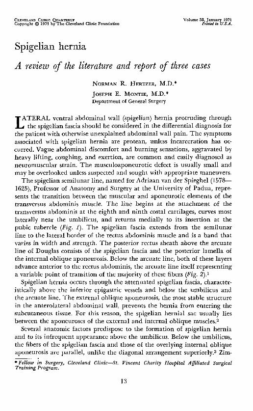
The spigelian semilunar line, named for Adriaan van der Spieghel (1578— 1625), Professor of Anatomy and Surgery at the University of Padua, repre-sents the transition between the muscular and aponeurotic elements of the transversus abdominis muscle. The line begins at the attachment of the transversus abdominis at the eighth and ninth costal cartilages, curves most laterally near the umbilicus, and returns medially to its insertion at the pubic tubercle (Fig. 1). The spigelian fascia extends from the semilunar line to the lateral border of the rectus abdominis muscle and is a band that varies in width and strength. The posterior rectus sheath above the arcuate line of Douglas consists of the spigelian fascia and the posterior lamella of the internal oblique aponeurosis. Below the arcuate line, both of these layers advance anterior to the rectus abdominis, the arcuate line itself representing a variable point of transition of the majority of these fibers (Fig. 2).1
Spigelian hernia occurs through the attenuated spigelian fascia, character-istically above the inferior epigastric vessels and below the umbilicus and the arcuate line. The external oblique aponeurosis, the most stable structure in the anterolateral abdominal wall, prevents the hernia from entering the subcutaneous tissue. For this reason, the spigelian hernial sac usually lies between the aponeuroses of the external and internal oblique muscles.2
Several anatomic factors predispose to the formation of spigelian hernia and to its infrequent appearance above the umbilicus. Below the umbilicus, the fibers of the spigelian fascia and those of the overlying internal oblique aponeurosis are parallel, unlike the diagonal arrangement superiorly.3 Zim-
* Fellow in Surgery, Cleveland Clinic—St. Vincent Charity Hospital Affiliated Surgical Training Program.
13

14 Hertzer and Montie
Fig. 1. Sketch shows relationship of the spigelian fascia and semilunar line to the muscles of the anterior abdominal wall. 1, The lateral border of the rectus abdominis muscle. 2, The spigelian fascia extending between the transversus abdominis and rectus abdominis muscles. 3, The semilunar line.
merman and associates4 demonstrated the irregular transitions of the mus-cular and fibrous elements of the transversus abdominis and internal oblique aponeuroses. Both layers contain interdigitating bands of attentuatcd muscle within the lateral aponeurotic margins. Associated fascial defects often allow protrusion of preperitoneal fat. Among 100 cadaver halves that were dis-sected, 45 specimens showed elongated defects in one layer or in both layers. Ten specimens contained defects in both layers, and in six instances4 the defects were superimposed. The presence of demonstrable musculoaponeu-rotic defects provides a more satisfactory explanation for spigelian hernia than does the former inconsistent theory that this hernia occurs through neurovascular perforations.5
Other conditions frequently associated with spigelian hernia are obesity, chronic cough, ascites, pregnancy, heavy exertion, and muscular atrophy of the aged.

Spigelian hernia 15
A B O V E T H E S E M I C I R C U L A R L I N E
T ra n s v e r s u s
a b d o m i n i s r
r T r a n s v e r s a l i s f a s c i a
I n t e r n a l
o b l i q u e m u
E x t e r n a l
o b l i q u e mu
B E L O W T H E S E M I C I R C U L A R L I N E
I n t e r n a l
o b l i q u e m u s e
E x t e r n a l
o b l i q u e m u s e
T r a n s v e r s u s
a b d o m i n i s m u s e i The s p i g e l i a n l i n e
T r a n s v e r s a l i s f a s c i a
Fig. 2. Sketch shows construction of the rectus sheath above and below the semicircular line. T h e transition of the spigelian fascia and the posterior lamella of the internal oblique aponeurosis from posterior to anterior rectus sheath is gradual rather than abrupt.
R e p o r t of cases
Case 1. A 74-year-old woman was admitted to St. Vincent Charity Hospital on April 19, 1970, for elective repair of a right spigelian hernia. For several weeks she had lower ab-dominal discomfort and burning sensations. An abdominal examination made while she was standing upright disclosed a defect in the abdominal wall. Her surgical history included cholecystectomy, abdominal hysterectomy, and repair of a left spigelian hernia.
The results of preoperative laboratory studies were normal. Roentgenograms of the colon and upper gastrointestinal tract showed the presence of a hiatal hernia but otherwise were normal. At operation, a 6-cm defect within the transversus abdominis and internal oblique aponeurotic layers was found, medial to the anterosuperior iliac spine and extending parallel with, but above, the conjoined tendon. A second defect, 1 cm in diameter, was beneath the external oblique aponeurosis at the level of the umbilicus. Both hernial sacs were excised and the defects closed in layers with interrupted silk sutures. The postoperative convalescence was uncomplicated.
Comment. This case illustrates the importance of an upright abdominal examination of the patient who has unexplained abdominal pain. The discovery at operation of a smaller, more medial hernial defect was unexpected.
Case 2. A 62-year-old woman was admitted to the Cleveland Clinic Hospital on Novem-ber 18, 1969, for repair of a hernia in the left lateral abdominal wall, which had caused episodic abdominal tenderness for one month. During that time the hernia had enlarged and was most prominent and tender after prolonged periods of standing upright. Physical examination showed an abdominal wall defect, 2 cm in diameter, located below the um-bilicus about 8 cm medial to the anterosuperior iliac spine. The fascial margins were firm and the hernial contents easily reducible. The results of the physical examination were otherwise normal, except for the presence of a benign endocervical polyp.
The preoperative laboratory evaluation and chest roentgenogram were normal. Roent-

16 Hertzer and M o n t i e
gcnograms of the upper gastrointestinal tract and colon were normal except for evidence of a small hiatal hernia and sigmoid diverticula. An intravenous pyelogram was normal.
At operation, an oblique lateral abdominal incision was made over the palpable fascial defect and continued through the external oblique aponeurosis. Superimposed 1-cm defects in both the spigelian fascia and the internal oblique aponeurosis allowed protrusion of preperitoneal fat and a small peritoneal sac. Redundant peritoneum was invaginated into the abdominal cavity, and the defects were closed in layers with interrupted silk sutures. After an uncomplicated convalescence the patient was free of symptoms.
Case 3. A 73-year-old, obese woman was admitted on an emergency basis to St. Vincent Charity Hospital on April 20, 1970, because of severe, cramping lower abdominal pain and vomiting of eight hours' duration. Several similar, but less severe attacks of abdominal pain had occurred within the 18 months previous to her admission to the hospital, but each episode was resolved with no treatment except bed rest. Several series of gastrointestinal roentgenograms had shown no evidence of the source of abdominal pain. The patient's surgical history included cholecystectomy, abdominal hysterectomy, and umbilical herni-orrhaphy.
The results of the physical examination were normal except for the presence in the left lower abdominal quadrant of a fluctuant, tender mass that was 10 cm long. The patient's temperature was 98.6 F, the pulse rate 88, and the blood pressure 110/80 mm Hg. The blood hemoglobin content was 14 g per 100 ml, the hematocrit 40, and the leukocyte count 19,100 per cubic millimeter. The serum electrolyte, blood urea nitrogen, and serum creati-nine values were normal. A chest roentgenogram was normal. Roentgenograms of the ab-domen showed a distended loop of small intestine in the left lower part of the abdomen (Fig- -?). After preoperative preparation with nasogastric suction, and intravenous adminis-
Fig. 3. Case 3. Plain abdominal roentgenogram shows the prominent loop of small bowel, which at operation was found to be incarcerated within a spigelian hernial sac.

Spigelian hernia 17
tration of fluids and antibiotics, laparotomy was performed because of the provisional diag-nosis of closed-loop small-bowel obstruction.
A left paramedian incision was used. A loop of jejunum protruded through an abdominal wall defect, 2 cm by 3 cm, located below and lateral to the junction of the arcuate line and the lateral margin of the rectus abdominis. The loop of bowel incarcerated within the in-terstitial layers of the abdominal wall showed no evidence of ischemia when returned to the abdominal cavity. T h e peritoneal sac was reduced and excised, and the firm margins of the aponeurotic defect were approximated with silk sutures. T h e patient had an un-eventful recovery.
Comment. Repeated physical examinations and roentgenographic studies had shown no evidence of the lateral ventral abdominal wall defect in this patient. Each examination had been done when she had been free of pain and without incarceration. It is likely that no defect was suspected before her admission to the hospital as a surgical emergency.
Discussion
Although reports of 200 cases of spigelian hernia proved at operation have been published, the true incidence is probably much greater, since the ma-jority of cases have been added in only the last 25 years.2' 6 The disease oc-curs generally in the fifth to seventh decades of life, and with equal frequency in men and women. Because the fascial defect is usually small, and has firm, thickened margins, one third of the hernias already described have been incarcerated.2 The defect is usually unilateral. To our knowledge, the pa-tient in our case 1 represents only the second case of bilateral spigelian hernia reported to this time.
The symptoms of a mildly symptomatic spigelian hernia may resemble those of neuromuscular strain. When operation was required because of incarceration, mistaken preoperative diagnoses have included appendicitis, periappendicular abscess, cholecystitis, abdominal tumor, and tumor of the anterior abdominal wall.2
Gastrointestinal roentgenograms may demonstrate chronically herniated bowel, but the most reliable means of early diagnosis is the physical examina-tion. Unless incarceration is present, the hernial contents usually return to the abdominal cavity when the patient is supine, and examination of a recum-bent patient will be unrewarding unless the fascial defect is prominent. When the patient is instructed to stand during the abdominal examination and turn in such a way that tension is applied to each of the anterior ab-dominal muscles, the hernial defect becomes palpable.6 When incarceration is present, an interstitial mass should be palpable within the abdominal wall, and when compressed may be associated with audible bowel sounds.
Elective repair of spigelian hernia is advisable because of the high in-cidence of incarceration and subsequent hernial strangulation. A transverse incision is made directly over the fascial defect or palpable mass. The hernial sac is isolated and excised after reduction of its contents, and a closure, in layers, of peritoneum and the fascial defect is performed. Imbrication of the internal oblique aponeurosis over the spigelian fascia may be required. When an incarcerated spigelian hernia is discovered at laparotomy, the hernial sac may be invaginated and excised and the defect closed from within

18 Hertzer and Montie
the peritoneal cavity. The latter procedure was used in the patient of case 2 of our report, with good results.
Spigelian hernia also occurs in the inguinal region, and may be mistaken for direct inguinal hernia. Unlike direct inguinal hernia, the spigelian hernia lies above the conjoined tendon. Both types of inguinal hernia are similarly repaired, by approximation of transversalis fascia and transversus abdominis aponeurosis to Poupart's ligament.3
Summary
Reports of three cases of lateral ventral (spigelian) hernia, one of which was bilateral, are presented and discussed. The structure of the anterolateral abdominal wall and anatomic factors contributing to the occurrence of spigelian hernia are described. Emphasis is placed upon the suspicion of spigelian hernia as a possible source both of protean symptoms and of sud-den visceral incarceration. Elective surgical repair of spigelian hernia is suggested, because of the relative frequency of incarceration and strangula-tion.
Acknowledgments
The authors are grateful to Anthony F. Spech, M.D., and Richard C. Robrock, M.D., St. Vincent Charity Hospital; and to Robert E. Hermann, M.D., Cleveland Clinic, for permission to report the data on their patients.
References 1. McVay, C. B., and Anson, B. J. : Composition of the rectus sheath. Anat. Rec. 77: 213-
225, 1940.
2. Bertelsen, S.: The surgical treatment of spigelian hernia. Surg. Gynec. Obstet. 122: 567-572, 1966.
3. Mersheimer, W. L.; Winfield, J . M., and Ruggiero, W. F.: Spontaneous lateral ventral hernia; so-called spigelian hernia. A.M.A. Arch. Surg. 63: 39-47, 1951.
4. Zimmerman, L. M., and others: Ventral hernia due to normal banding of the abdominal muscles. Surg. Gynec. Obstet. 78: 535-540, 1944.
5. Ignatius, J . A.: Spigelian hernia. Amer. J . Surg. 90: 388-391, 1955.
6. Harless, M. S., and Hirsch, J . E.: Spigelian or spontaneous lateral ventral hernia. Amer. J . Surg. 100: 515-521, 1960.