spectrum of mandibular fractures in motor vehicle ...jmscr.igmpublication.org/v4-i8/54 jmscr.pdfdr...
TRANSCRIPT

Dr Srinivas MR et al JMSCR Volume 04 Issue 08 August Page 12003
JMSCR Vol||04||Issue||08||Page 12003-12014||August 2016
Spectrum of Mandibular Fractures in Motor Vehicle Accidents:
MDCT Evaluation (Original Article)
Authors
Dr Srinivas MR1, Dr Vijay Kumar KR
2, Dr Vedaraju KS
3*
1Associate Professor, Dept of Radiodiagnosis and Imaging, Victoria Hospital Bangalore Medical College
and Research Institute, Fort, Bangalore, India- 560002
Email: [email protected], Ph: +919844962444, +919341724277 2Associate Professor, Dept of Radiodiagnosis and Imaging, Victoria Hospital Bangalore Medical College
and Research Institute, Fort, Bangalore, India-560002
Office: 91-80-26700120 Mob: +91-9632628899
Email: [email protected]
Email: [email protected] 3Head of the Department, Dept of Radiodiagnosis and Imaging, Victoria Hospital Bangalore Medical
College and Research Institute, Fort, Bangalore, India-560002
Office: 91-80-26700304, Mob: +91-99611129456
Email: [email protected]
Corresponding Author
Dr M R Srinivas
Associate Professor, Dept of Radiodiagnosis and Imaging, Victoria Hospital Bangalore Medical College and
Research Institute, Fort, Bangalore, India-560002
Email: [email protected], Ph: +919844962444, +919341724277
Abstract
Background: Mandibular fractures consist of significant number of facial fractures, especially in cases of
Motor vehicle accidents. MDCT plays a major role in determining the presence/ absence of fracture, gives
accurate information of displacement and associated soft tissue injury and thin section and multiplanar
reconstructions help in better evaluation of the fracture. This study describes the age and gender predilection
and site of fracture with some clinical and treatment relevance.
Materials and Methods: A retrospective and prospective study was conducted in the department of radio-
diagnosis, Bangalore Medical college and research institute for over a period of 1 year, from November 2014-
October 2015 which included 98 cases of mandibular fracture by using multidetector computed tomography
(MDCT). Cases were divided and assessed based on age, gender predominance, unifocal/ multiplicity, site of
fracture and associated facial fractures.
Results: Of the 98 cases, male constituted 88% and remaining 12% were females. Most common age group
was between 21-30 years. Most cases had multifocal fractures and the most common site of injury was
condylar process (32%).
Conclusion: Road traffic accidents are very common in metropolitan cities like Bangalore and facial injuries
www.jmscr.igmpublication.org
Impact Factor 5.244
Index Copernicus Value: 83.27
ISSN (e)-2347-176x ISSN (p) 2455-0450
DOI: http://dx.doi.org/10.18535/jmscr/v4i8.54

Dr Srinivas MR et al JMSCR Volume 04 Issue 08 August Page 12004
JMSCR Vol||04||Issue||08||Page 12003-12014||August 2016
with mandibular fractures constitute a significant number. Young and middle aged males are more prone for
injury because of increased habit of risk taking and more exposure to risk. MDCT plays a major role in
evaluation of patients with facial and mandibular trauma. It not only gives information about site and
displacement of the fracture; but also helps in detection of adjacent soft tissue injury and airway.
Keywords: Multidetector computed tomography, fractures, mandible, zygomatic fractures.
Introduction
Mandibular fractures consist of significant number of facial fractures, especially in cases of Motor vehicle
accidents. Prompt recognition and stabilization of such fractures are important as they can be life threatening
by blocking the airway and have significant long term complications like mal-occlusion, mal- union and
osteomyelitis. They can be unifocal or multiple depending on the force and direction of injury and position
of the individual. In metropolitan like Bangalore, motor vehicle accidents are very frequent owing to
increasing number of motorcycles on road, increasing need to travel distance to work, especially during dark
hours of the day and drink and drive by the drivers, more so of heavy motor vehicles like lorries and tempos.
Classification based on location (Figure 1 and 2) is the most useful classification, because both the signs and
symptoms, and treatment are dependent upon the location of the fracture [1]
.
Condylar fractures are classified by location with respect to capsule of ligaments that hold the
temporomandibular joint (intracapsular or extracapsular), based on presence of absence of dislocation
(whether or not the condylar head has come out of the glenoid fossa as the lateral pterygoid tend to pull the
condyle antero-medially) and neck of the condyle fractures. Because the coronoid process lies deep to many
structures, like the zygomatic complex (ZMC), it is rare to be broken in isolation. It usually occurs with
mandibular fractures in other sites or with fracture of zygomatic arch or complex. Isolated fractures of the
coronoid process must be viewed with suspicion and fracture of the ZMC should be ruled out [2]
.
Ramus fractures are said to be present if it involves a region inferiorly bounded by an oblique line starting
from the lower third molar region to the postero-inferior insertion of the masseter muscle, and which could
not be classified as either condylar or coronoid fractures. The angle of the mandible refers to the angle
created by the arrangement of the body and the ramus of the mandible. Angle fractures are those that involve
a triangular region bounded anteriorly by the anterior border of masseter muscle and an oblique line
extending from the lower third molar to the postero-inferior attachment of the masseter muscle. Fracture of
the mandibular body is defined as that involving a region bounded anteriorly by the para-symphysis (vertical
line just distal to the canine tooth) and posteriorly by the anterior border of masseter muscle. Para-
symphyseal fractures are defined as mandibular fractures that involve an area bounded on both sides by
vertical lines distal to the canine tooth. Symphyseal fractures are linear fractures that run in the midline of
the mandible (symphysis menti). Alveolar fracture involves the alveolar process of the mandible [1]
.
Dentition of mandible – Each Hemi mandible in adults is composed of two incisor teeth (Central and lateral
incisor), one canine, two pre molars and three molars. Thus eight teeth in each hemi mandible and 16 teeth
in mandible or lower jaw (Figure 3)
MDCT plays a major role in determining the presence/ absence of fracture, gives accurate information of
displacement and associated soft tissue injury and thin section and multiplanar reconstructions help in better
evaluation of the fracture. This study describes the age and gender predilection and site of fracture with
some clinical and treatment relevance.
Methods and Materials
This study is conducted in Department of radio-diagnosis, Bangalore medical college and research institute,
Bangalore which is a referral as well as a teaching institute. Multi slice CT GE somatom scanner was used to
evaluate every patient who was referred to the department with motor vehicle accident. Scans were acquired

Dr Srinivas MR et al JMSCR Volume 04 Issue 08 August Page 12005
JMSCR Vol||04||Issue||08||Page 12003-12014||August 2016
in bone soft tissue algorithm and thinner reconstructions, multiplanar imaging and volume rendering
reconstruction were done in workstation. Each case was evaluated for the presence of mandibular fracture,
site and multiplicity. Cases were collected retrospectively as well as prospectively over a period of 1year,
from November 2014- October 2015. Out of 533 cases of facial fractures, 98 cases had mandibular fracture.
Patients belonged to age group between 11-60 years. Patients with non- motor vehicle accidents like fall
from height and assault were excluded from the study. They were then divided and assessed based on age,
gender predominance, unifocal/ multiplicity, site of fracture and associated facial fractures.
Depending on site of fracture, cases were divided into condylar, angle, ramus, body, coronoid, symphyseal/
parasymphyseal and isolated alveolar ridge fractures.
Results
In this study, there were 533 cases with facial fractures, of which 98 cases had mandibular fractures.
Chart 1: Gender distribution
Out of the 98 cases of mandibular fracture (age group: 11- 60 years, mean 35.5), male constituted 83 number
(88%) and females were 15 cases (12%).
AGE AND GENDER DISTRIBUTION:
Table 1: Distribution of cases based on age and gender:
Age group Male Female Total number
11- 20 13 3 16
21- 30 40 7 47
31- 40 20 4 24
41- 50 9 0 9
51- 60 1 1 2
Most common age group was between 21-30 years followed by 31-40 years. Total numbers of fractures in
these 98 cases were 887.
0
20
40
60
80
100
Gender distribution
83
15
Male
Female
Dr Srinivas MR et al JMSCR Volume 04 Issue 08 August Page 12006
JMSCR Vol||04||Issue||08||Page 12003-12014||August 2016
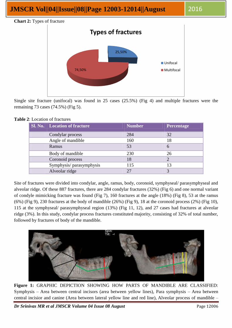
Chart 2: Types of fracture
Single site fracture (unifocal) was found in 25 cases (25.5%) (Fig 4) and multiple fractures were the
remaining 73 cases (74.5%) (Fig 5).
Table 2: Location of fractures
Sl. No. Location of fracture Number Percentage
Condylar process 284 32
Angle of mandible 160 18
Ramus 53 6
Body of mandible 230 26
Coronoid process 18 2
Symphysis/ parasymphysis 115 13
Alveolar ridge 27 3
Site of fractures were divided into condylar, angle, ramus, body, coronoid, symphyseal/ parasymphyseal and
alveolar ridge. Of these 887 fractures, there are 284 condylar fractures (32%) (Fig 6) and one normal variant
of condyle mimicking fracture was found (Fig 7), 160 fractures at the angle (18%) (Fig 8), 53 at the ramus
(6%) (Fig 9), 230 fractures at the body of mandible (26%) (Fig 9), 18 at the coronoid process (2%) (Fig 10),
115 at the symphyseal/ parasymphyseal region (13%) (Fig 11, 12), and 27 cases had fractures at alveolar
ridge (3%). In this study, condylar process fractures constituted majority, consisting of 32% of total number,
followed by fractures of body of the mandible.
Figure 1: GRAPHIC DEPICTION SHOWING HOW PARTS OF MANDIBLE ARE CLASSIFIED:
Symphysis – Area between central incisors (area between yellow lines), Para symphysis – Area between
central incisior and canine (Area between lateral yellow line and red line), Alveolar process of mandible –
25,50%
74,50%
Types of fractures
Unifocal
Multifocal

Dr Srinivas MR et al JMSCR Volume 04 Issue 08 August Page 12007
JMSCR Vol||04||Issue||08||Page 12003-12014||August 2016
Superior part of mandible consisting of tooth (Area above green line), Body of mandible – Area bounded
anteriorly by the parasymphysis (defined as a vertical line just distal to the canine tooth ) and posteriorly by
the anterior border of the masseter muscle (Area between red line and medial most black line), Angle of
mandible fractures are defined as those that involve a triangular region bounded by the anterior border
of masseter muscle and an oblique line extending from the lower third molar (wisdom tooth) region to the
posteroinferior attachment of the masseter muscle (Area bounded by medial most black line and black line
distal to it, Condylar process of mandible : Area of projected part of mandible involved in TMJ, Coronoid
process: Triangular area of projected bone of mandible anterior to condylar process, Ramus of mandible –
Remnant area between body, condylar and coronoid process of mandible.
Figure 2: GRAPHIC DEPICTION OF PARTS OF MANDIBLE – Symphysis of mandible – Area shaded in
yellow, Parasymphysis of mandible – Area shaded in red, Alveolar process of mandible – Area shaded with
oblique green lines, Body of mandible – Area shaded with transverse white lines, Angle of mandible – Area
Shaded with criss cross black lines, Ramus of mandible – Area shaded in pink, Coronoid process – Area
shaded in yellow, Condylar process – Area shaded in orange.
Figure 3: DENTITION OF RIGHT HALF OF MANDIBLE: Graphic depiction of mandible in A- lateral
view, B-Anterior view and C – Superior view - Showing Central incisior (Red arrow head), Lateral incisior
(Yellow arrow head), Canine (Blue arrow head), First Pre molar (Green arrow head), Second Pre molar
(White arrow head), First Molar (Black arrow head), Second Molar (Orange arrow head), Third molar (Dark
blue arrow head).

Dr Srinivas MR et al JMSCR Volume 04 Issue 08 August Page 12008
JMSCR Vol||04||Issue||08||Page 12003-12014||August 2016
Figure 4: Left hemi mandible alveolar process unifocal fracture at symphysis and para symphysis region,
left Le Fort I, II, III fracture, left tripod fracture, fracture of left frontal bone, bilateral maxillary sinuses wall
fracture: A – Axial section bone window – Showing unifocal fracture of alveolar process of mandible at left
symphysis and para symphysis region (outlined arrow), B,C,D – VRT AP, left lateral and right lateral view –
Showing associated fractures (Normal arrow), fronto – nasal junction fracture, left frontal bone fracture, left
tripod fracture, bilateral maxillary sinus wall fractures and unifocal left mandibular alveolar process fracture
(outlined arrow) and rest of mandible is normal.
Figure 5: Bilateral body and para symphysis fracture of mandible – A, Scannogram showing fracture of
body of mandible with significant anterior displacement (Long arrow), B, C- Axial section bone window
showing fracture lines lateral (outlined arrows) to canine (Arrow head) and fracture lines medial (Normal
arrows) to canine (Arrow head) - comminuted displaced fracture involving bilateral body and
parasymphysis of mandible.

Dr Srinivas MR et al JMSCR Volume 04 Issue 08 August Page 12009
JMSCR Vol||04||Issue||08||Page 12003-12014||August 2016
Figure 6: Axial section bone window - unifocal displaced fracture involving articular surface of right
condylar process (arrows).
Figure 7: Normal variant of right condylar head with bilateral le fort i, ii, iii fracture, left tripod fracture and
unifocal left sub condylar fracture : A, Axial section bone window showing suspicious fracture of articular
surface of right condylar head (Normal arrow), B, coronal section bone window – showing no fracture of
right condylar head but a normal variant of condylar head (Normal arrows), C,D and E – Axial section bone
window and VRT AP view – showing associated fractures (outlined arrow) fracture of walls of bilateral
maxillary sinuses, bilateral pterygo maxillary disjunction and fractured pterygoid plates, bilateral zygomatic
arch fracture, fronto – nasal disjunction, fracture of lateral wall of bilateral orbits and eft tripod fracture, F-G
– VRT left posterior view and oblique view of mandible - showing sub condylar fracture of left condyle (
Long arrows).

Dr Srinivas MR et al JMSCR Volume 04 Issue 08 August Page 12010
JMSCR Vol||04||Issue||08||Page 12003-12014||August 2016
Figure 8: Right longitudinal fracture of ramus and angle of mandible and left condylar fracture – A,B,C –
Coronal, right sagittal reconstructed image and VRT right lateral oblique view - showing longitudinal
fracture involving right ramus and angle of mandible (Normal arrows) – D,E,F – left sagittal, axial and VRT
left lateral view – showing left condylar fracture (Short arrows).
Figure 9: Displaced fracture involving bilateral body of mandible, mild left temporo mandibular joint
dislocation – A,B,C – Axial section bone window, fracture line (Normal arrow) is seen lateral to right canine
(Arrow head) and left canine (Arrow head) – suggestive of bilateral mandibular body fracture and Mild left
TMJ dislocation (C -outlined arrow), D,E,F – Oblique AP, left lateral and left lateral oblique view – showing
displaced bilateral mandibular body fracture (Normal arrow)

Dr Srinivas MR et al JMSCR Volume 04 Issue 08 August Page 12011
JMSCR Vol||04||Issue||08||Page 12003-12014||August 2016
Figure 10: Right coronoid process fracture, right tripod fracture, complex fracture of right temporal bone,
right hemi maxillary fracture, right Le Fort I fracture – A,B,C - Axial section bone window and VRT right
lateral view – showing fracture of right coronoid process of mandible (A&C- normal arrows), and
associated fractures like – complex fracture of right temporal bone, right tripod fracture, right hemi
maxillary fracture, right pterygo maxillary disjunction (A,B,C – outlined arrows).
Figure 11: Right para symphysis fracture with traumatic dislocation of right lateral incisior tooth, fracture of
right mandibular ramus, fracture of left condylar process – A, B – Axial and coronal section bone window –
showing right para symphysis oblique fracture (Short arrow) with traumatic dislocation of right lateral
incisior tooth (Arrow head), C – Axial section bone window - fracture of right mandibular ramus (normal
arrow) and fracture of left condylar process (Normal arrow), D,E,F – VRT right oblique AP, left lateral and
left posterior oblique view – Showing para symphysis fracture (Short arrow) with traumatic dislocation of
right lateral incisior tooth (Arrow head), fracture of right mandibular ramus (normal arrow) and displaced
fracture of left condylar process (Normal arrow)

Dr Srinivas MR et al JMSCR Volume 04 Issue 08 August Page 12012
JMSCR Vol||04||Issue||08||Page 12003-12014||August 2016
Figure 12: Symphysis and right para symphysis fracture of mandible with dislocation of bilateral central
incisiors, bilateral le fort I, II and III fractures, bilateral tripod fracture, depressed fracture of frontal bone,
bilateral tmj dislocation, bilateral nasal bone fracture – A – Scannogram showing depressed fracture of face
with short arrows depicting probable direction of impact, B-G – Axial and coronal section bone window –
showing Symphysis fracture and right para symphysis fracture (B,C,F and G – outlined arrow) with
traumatic dislocation of bilateral central incisiors (C,F,G - Arrow head), Bilateral TMJ dislocation (D&E –
Long arrow), H - VRT – AP view – showing symphysis and para symphysis fracture of mandible (Outlined
arrow) with traumatic dislocation of central incisiors (arrow head) and other associated fractures (normal
arrows) – bilateral tripod fracture, bilateral maxillary sinus wall fracture, bilateral nasal bone fracture with
fronto nasal disjunction, depressed frontal bone fracture and maxillary bone fracture.
Discussion
Facial trauma is the most common injury to the victims of motor vehicle accidents [3]
. Road traffic injuries
are a major public health problem worldwide. Every year, an estimated 1.2 million people die in road traffic
accidents and up to 50 million suffer non-fatal injuries [4].
A broken jaw is the second most common facial
injury- second only to a broken nose, Motor vehicle accidents (MVAs) cause 43% of mandibular fractures [5]
.
Male constituted 83% of total number of cases owing to the fact that there are more men driving the motor
vehicles. And the age group with maximum number of cases was between 21-30 years followed by 31-40
years as published in World Health Organization (2009) Global status report on road safety: time for action,
where road traffic morbidity and mortality rates were higher in men than women [4]
. Furthermore, a study
conducted by Elvik R et al., reviewed that young drivers, especially males, are involved in accidents more
than middle-aged drivers. The high accident rates of young drivers are attributable to deliberate risk taking
and overestimation of skills [6]
.
The gender difference in mortality and morbidity rates is likely related to both risk-taking behaviour and
increased exposure to risk [7]
.
A study conducted by Escott EJ et al., showed that multiple fractures in a single mandible are far more
common than unifocal fractures, where the incidence of multiple mandible fractures was 58% as in this
study where 74% of cases were multifocal [8]
. MVA-induced injuries are the result of the remarkable
amounts of kinetic energy released, when the steady state of a passenger is changed by sudden deceleration
or acceleration; both speed and stopping distance have a significant influence [9]
.
Condylar fractures are the most common site for injury in this study constituting 32%, followed immediately
by fracture of body of the mandible. Similar results were obtained in a study by Bag AK et al., in which the
prevalence of condylar process fractures ranges between 25% and 50% of all mandibular fractures [10]
.
Displacement of the condyle through the roof of glenoid fossa and into the middle cranial fossa is rare [11]
.
Malocclusion and restricted jaw movement are usually more severe in condylar fractures [1]
. Bilateral body
Dr Srinivas MR et al JMSCR Volume 04 Issue 08 August Page 12013
JMSCR Vol||04||Issue||08||Page 12003-12014||August 2016
or bilateral para-symphysis fractures are sometimes referred to as ‘flail mandible’, which can cause
involuntary movement of the tongue posteriorly with subsequent obstruction of the upper airway [12]
.
Multi-detector CT is an invaluable aid in assessing mandibular injuries [14]
. There is better clinician
agreement on the location and absence of fractures with CT [15]
. The capability of thin-section acquisition
improves detection of even minor pathologic details, and the high spatial resolution capability of the scanner
enables display in arbitrary planes, so that there is no need of manipulation of the head in prone or
hyperextended positions to acquire images [16]
. MDCT has revolutionized the imaging of suspected cases of
facial fracture and, furthermore has improved diagnosis [17]
.
Conclusion
Road traffic accidents are very common in metropolitan cities like Bangalore and facial injuries with
mandibular fractures constitute a significant number. Young and middle aged males are more prone for
injury before of increased habit of risk taking and more exposure to risk. MDCT plays a major role in
evaluation of patients with facial and mandibular trauma. It not only gives information about site and
displacement of the fracture; but also helps in detection of adjacent soft tissue injury and airway. In cases of
trauma, imaging is very essential in diagnosis, treatment planning, and in prognosticating.
References
1. Kamali U, Pohchi A. Mandibular fracture at HUSM: a 5-year retrospective study. Arch Orofac Sci.
2009;4(2):33-5.
2. Pricop M, Urechescu H, Sîrbu A. Fractura procesului coronoid al mandibulei—prezentare de caz și
recenzie a literaturii de specialitate. Revista de chirurgie oro-maxilo-facială și implantologie. 2012
Mar 1;3(1):1-4.
3. Scheyerer MJ, Döring R, Fuchs N, et al. Maxillofacial injuries in severely injured patients. Journal of
Trauma Management & Outcomes. 2015;9:4.
4. World Health Organization. Global status report on road safety: time for action. World Health
Organization; 2009.
5. MDGuidelines Directory for Medical Topics- Fracture Jaw Mandible And Maxilla, medical code:
ICD-9-CM
6. Elvik R. Why some road safety problems are more difficult to solve than others. Accid Anal Prev.
2010; 42(4)1089–1096.
7. Johnston BD. Road traffic injury prevention. Injury prevention. 2010 Feb 1;16(1):1.
8. Escott EJ, Branstetter BF. Incidence and characterization of unifocal mandible fractures on CT.
AJNR Am J Neuroradiol. 2008;29 (5): 890-4.
9. Sasser SM, Hunt RC, Sullivent EE, et at. Guidelines for field triage of injured patients.
Recommendations of the National Expert Panel on Field Triage. MMWR Recomm Rep. 2009; 58:1-
35.
10. Bag AK, Gaddikeri S, Singhal A, et al. Imaging of the temporomandibular joint: An update. World
Journal of Radiology. 2014;6(8):567-582. doi:10.4329/wjr.v6.i8.567.
11. Chao MT, Losee JE. Complications in Pediatric Facial Fractures.Craniomaxillofacial Trauma &
Reconstruction. 2009;2(2):103-112.
12. Mehta, Nisha et al. The Imaging of Maxillofacial Trauma and its Pertinence to Surgical Intervention,
Radiol Clin N Am 50 2012; 43-57
13. Shintaku WH, Venturin JS, Azevedo B et al, Applications of cone-beam computed tomography in
fractures of the maxillofacial complex. Dent Traumatol 2009; 25:358–366.

Dr Srinivas MR et al JMSCR Volume 04 Issue 08 August Page 12014
JMSCR Vol||04||Issue||08||Page 12003-12014||August 2016
14. Yildirim D, Tamam C, Gumus T. Three‐dimensional scanning with dual‐source computed
tomography in patients with acute skeletal trauma. Clinics. 2010; 65(10):991-1002.
15. Ersan N, Ilguy M. Diagnosis of unusual mandibular split fracture with cone-beam computed
tomography. J Oral Maxillofac Radiol 2015;3:67-9.
16. Lam S, Bux S, Kumar G, Ng K, Hussain A. A comparison between low-dose and standard-dose non-
contrasted multidetector CT scanning of the paranasal sinuses. Biomedical Imaging and Intervention
Journal. 2009;5(3):e13.
17. Ogura I, Kaneda T, Mori S, Sekiya K, Ogawa H, Tsukioka T. Characterization of mandibular
fractures using 64-slice multidetector CT. Dentomaxillofacial Radiology. 2012;41(5):392-395.