spectroscopic study of sulforhodamine 640-doped sol–gel silica
TRANSCRIPT

ARTICLE IN PRESS
0022-2313/$ - se
doi:10.1016/j.jlu
�Correspondifax: +9111 269
E-mail addre
Journal of Luminescence 114 (2005) 162–166
www.elsevier.com/locate/jlumin
Spectroscopic study of sulforhodamine 640-doped sol–gel silica
Haider Abbas�, K.P. Tiwary, L.S.S. Singh, Mohd. Zulfequar,Z.H. Zaidi, M. Husain
Department of Physics, Jamia Millia Islamia (Central University), New Delhi – 110025, India
Received 31 March 2004; received in revised form 3 September 2004; accepted 7 December 2004
Available online 8 February 2005
Abstract
The sol–gel process is a technique which provides a low-temperature route for the preparation of organic dye-doped
porous silica glass. Extended UV transmission was observed for HCl-catalysed sol–gel silica. Properties of a solute may
differ greatly between a free solution and that solution confined in pores of a sol–gel glass. Absorption and fluorescence
properties of sulforhodamine 640-doped silica samples prepared by sol–gel process were investigated. In the TEOS-
derived gel, the emission of the dye does not shift during aging but exhibits a large change during drying. The emission
maximum of sulforhodamine 640 blue shifts was discussed as a function of increasing rigidity of the surrounding
matrix.
r 2005 Elsevier B.V. All rights reserved.
PACS: 81.20.Fw; 42.55.Mv; 42.55.R
Keywords: Tunable solid state dye laser; Laser dye; Dye/matrix interaction
1. Introduction
During the past decades, the sol–gel process hasbeen extensively studied in many laboratories. In1984, Avnir et al. [1–3] have reported the prepara-tion and optical properties of alkoxide-derivedsilica gels doped with rhodamine 6G and pyrene[1–3]. They have also demonstrated that other
e front matter r 2005 Elsevier B.V. All rights reserve
min.2004.12.013
ng author. Tel.: +9111 26989965;
81753.
ss: [email protected] (H. Abbas).
organic molecules can be successfully incorporatedinto sol–gel matrices [1,4]. The advantages of thesol–gel process are lower temperature processing,excellent homogeneity at a molecular level, highcompositional purity and the possibility of incor-porating organic molecules at low temperaturethat will otherwise be destroyed in conventionalglass-forming process. The most useful feature ofdye lasers is their tunability, i.e., the lasingwavelength for a given dye may be varied over awide range. Taking advantage of the broadfluorescent line widths available in organic dyes,
d.

ARTICLE IN PRESS
H. Abbas et al. / Journal of Luminescence 114 (2005) 162–166 163
one can use a wavelength-selective resonator toperform selective tuning [5]. Organic dye moleculesas laser gain media enjoy the advantage of broadtunability ranging from 300–900 nm with highefficiency, high photostability and superior ther-mal–optical properties. Reports on the spectro-scopic behaviours [6,7], lasing properties [6,8–16]and optical non-linearities [17] of a large numberof dyes trapped in sol–gel-derived silica glasseshave already appeared in the literature. Theapplications of tunable solid-state laser in visibleare in remote sensing, isotope separation, highsensitivity analytical spectroscopy and underwatercommunications. Development of the above-men-tioned applications requires a good understandingof the structure of the doped sol–gel matrices, theproperties of the matrices on the molecular leveland the conditions that the oxide network imposeson the optical properties of the dopant.
Fig. 1. Molecular structure of sulforhodamine 640.
2. Experimental
Two types of sol–gel silica samples, one usingHCl and another using HNO3 as catalyst werefabricated following the sol–gel procedures out-lined in [12,14]. The initial solution is typicallycomposed of 15ml of TEOS, 12.5ml of ethylalcohol, 18ml of water, 12.5ml of formamide and2ml of acid serving as catalyst. Sulforhodamine640 was added to the initial solution undermagnetic stirring until the desired molar concen-tration was reached. The organic dye and chemicalused in these experiments were procured fromcommercial vendors (Exciton, Merk and SigmaAldrich, respectively). The concentration of sul-forhodamine 640 as suggested by the vendor(Exciton) is 1� 10�4molar in the initial solution.Kept at a temperature of 60 1C, gelling occurs inhours. After aging and drying for about 1 week,the containers were opened and the pore liquidexpelled during shrinkage was evacuated. Dryingand aging were continued for 3 weeks at 60 1C.The bulk density of the samples prepared usingHCl as a catalyst is 1.42 g/cm3 and that of usingHNO3 is 1.39 g/cm
3. For all data presented here.Cylindrical-shaped samples with a diameter of14mm and a thickness of 10mm were used for the
measurement. All samples were visually of goodsurface finish with end faces appearing to be planeparallel. Experiments were carried out at roomtemperature immediately after hydrolysis throughthe gelation stage until the sol–gel material dried.The absorption spectra were measured with aJasco V-570 spectrophotometer and the fluores-cence spectra were recorded on Hitachi fluores-cence spectrophotometer model F- 4500.
3. Results and discussion
When HCl was used as a catalyst during thesol–gel process, improved UV cutoffs can beachieved [12,13]. The improved UV transmissionis a result of smaller size concentrated poredistribution is effected by the HCl-catalysedhydrolysis, so we choose HCl-catalysed sol–gelsilica for our further experiments. To understandthe effects of dye/matrix interaction on thefluorescent properties, one needs to explore howa dye behaves in different molecular environment.A continuous red shift in absorption spectra wasobserved as the polarity of the solvent increasesbecause of the polar nature of dye (Fig. 1). Thelowest energy transition is the (p,p*) transition dueto dipole–dipole interaction with the solventmolecules. The (p,p*) excited state is more polarand more polarizable than the ground state,resulting in greater lowering of the excited stateand reducing the transition energy, and therefore itexperiences a red shift upon increase in solvent

ARTICLE IN PRESS
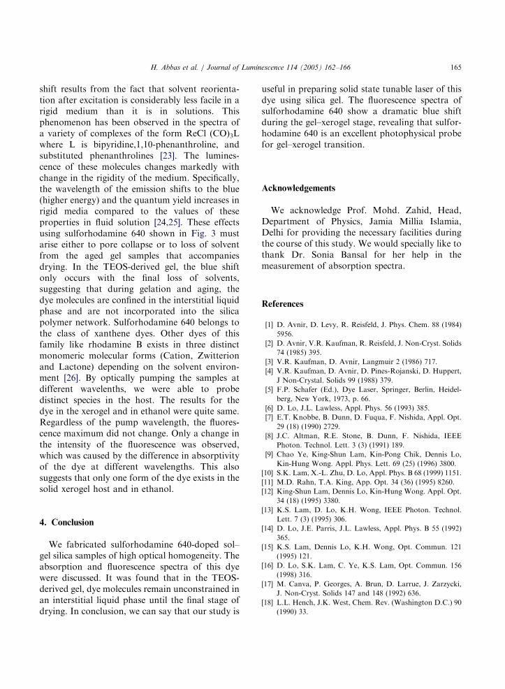
Fig. 3. Fluorescence spectra of the sulforhodamine 640 in
ethanol (curve a) , in HCl catalysed sol–gel glass (curve b), in
HCl-catalysed sol (curve c) and in xerogel (curve d).
H. Abbas et al. / Journal of Luminescence 114 (2005) 162–166164
polarity. From the inception of the work onencapsulated molecules in sol–gel matrices[18,19], it has been well accepted that themolecules are located in the pores of the networkrather being part of the network itself. The natureof the silica cage was examined in more detail bycarrying out spectroscopy during drying andgelling of one of the sol–gel solution. Absorptionspectra of the dye are shown in Fig. 2. Red shifts(�8 nm) is observed compared to ethanolic solu-tion. This may be attributed to the nature of thelocal environment of the gel. As water vaporizesfrom the sol solution, the colloidal sol particlescontact each other, and then they start to build upa three-dimensional network cage. The cage maybe composed of Si–OH and Si–O–Si groups [1,20].Therefore, the red shift described above mayindicate the slightly less polar nature of the silicacage. Hydrolysis leads to the formation of silanolgroups Si–OH. These species are only intermedi-ates as they react further to form siloxane Si–O–Sigroups. Emission spectroscopy offers sensitivemethods of reporting the local enviroment of theprobe molecules. Fig. 3. Shows the emission peakof the sulforhodamine 640 in HCl-catalysed sol at621 nm, representing red shift of 11 nm from thoseof ethanol. It is well known that the position of themaximum of the fluorescence spectrum is redshifted with increasing solvent polarity. Four verygeneral locations of the sol–gel matrix can bedifferentiated: the interiors of pores (often filledwith liquids), the interface between the liquids and
Fig. 2. Absorption spectra of (curve a) the sulforhodamine 640
in ethanol and (curve b) the doped-HCl catalysed sol–gel glass.
the solid pore wall, the pore wall itself, and theconstraining region where the distance betweenopposite sides of the pores is the same as the probemolecule itself [21]. The use of luminescentmolecules as optical probes of the sol–gel processhas been reported for over 17 years, in some cases,the desired dopant molecule like ReCl(CO)3–2,2
0-bipyridine acts as a probe [22]. If the solvent shellstrongly interacts with the dye molecules andsolvent–pore wall interactions are relatively weak,then the dye molecules will behave as it would insolution. It is known that the TEOS-gel glass is aporous solid, and the average radius of the pores inthis solid is about 17 A. The pore shape is similarto an ink bottle, which restricts the evaporation ofthe solvent (ethanol), owing to the residualsolvents and Si–OH groups in the pores. Thusthe environment inside a pore has some chemicalsimilarity to that offered by ethanol. That is whyin Fig. 3, the fluorescence of dye, peaks at roughlythe same wavelength for the sol–gel sample andethanol solution. We can conclude that theenvironment inside a pore has some chemicalsimilarity to the environment offered by ethanolfor sulforhodamine 640. Upon ambient drying ofthe samples, the blue shift continues during theaged gel to xerogel structural transformation. Atotal shift of 11 nm (610–599) can be attributed todrying effects. The phenomenon of blue shifting ofemission band with increasing rigidity of the localenvironment is known as rigidochromism. This
ARTICLE IN PRESS
H. Abbas et al. / Journal of Luminescence 114 (2005) 162–166 165
shift results from the fact that solvent reorienta-tion after excitation is considerably less facile in arigid medium than it is in solutions. Thisphenomenon has been observed in the spectra ofa variety of complexes of the form ReCl (CO)3Lwhere L is bipyridine,1,10-phenanthroline, andsubstituted phenanthrolines [23]. The lumines-cence of these molecules changes markedly withchange in the rigidity of the medium. Specifically,the wavelength of the emission shifts to the blue(higher energy) and the quantum yield increases inrigid media compared to the values of theseproperties in fluid solution [24,25]. These effectsusing sulforhodamine 640 shown in Fig. 3 mustarise either to pore collapse or to loss of solventfrom the aged gel samples that accompaniesdrying. In the TEOS-derived gel, the blue shiftonly occurs with the final loss of solvents,suggesting that during gelation and aging, thedye molecules are confined in the interstitial liquidphase and are not incorporated into the silicapolymer network. Sulforhodamine 640 belongs tothe class of xanthene dyes. Other dyes of thisfamily like rhodamine B exists in three distinctmonomeric molecular forms (Cation, Zwitterionand Lactone) depending on the solvent environ-ment [26]. By optically pumping the samples atdifferent wavelenths, we were able to probedistinct species in the host. The results for thedye in the xerogel and in ethanol were quite same.Regardless of the pump wavelength, the fluores-cence maximum did not change. Only a change inthe intensity of the fluorescence was observed,which was caused by the difference in absorptivityof the dye at different wavelengths. This alsosuggests that only one form of the dye exists in thesolid xerogel host and in ethanol.
4. Conclusion
We fabricated sulforhodamine 640-doped sol–gel silica samples of high optical homogeneity. Theabsorption and fluorescence spectra of this dyewere discussed. It was found that in the TEOS-derived gel, dye molecules remain unconstrained inan interstitial liquid phase until the final stage ofdrying. In conclusion, we can say that our study is
useful in preparing solid state tunable laser of thisdye using silica gel. The fluorescence spectra ofsulforhodamine 640 show a dramatic blue shiftduring the gel–xerogel stage, revealing that sulfor-hodamine 640 is an excellent photophysical probefor gel–xerogel transition.
Acknowledgements
We acknowledge Prof. Mohd. Zahid, Head,Department of Physics, Jamia Millia Islamia,Delhi for providing the necessary facilities duringthe course of this study. We would specially like tothank Dr. Sonia Bansal for her help in themeasurement of absorption spectra.
References
[1] D. Avnir, D. Levy, R. Reisfeld, J. Phys. Chem. 88 (1984)
5956.
[2] D. Avnir, V.R. Kaufman, R. Reisfeld, J. Non-Cryst. Solids
74 (1985) 395.
[3] V.R. Kaufman, D. Avnir, Langmuir 2 (1986) 717.
[4] V.R. Kaufman, D. Avnir, D. Pines-Rojanski, D. Huppert,
J Non-Crystal. Solids 99 (1988) 379.
[5] F.P. Schafer (Ed.), Dye Laser, Springer, Berlin, Heidel-
berg, New York, 1973, p. 66.
[6] D. Lo, J.L. Lawless, Appl. Phys. 56 (1993) 385.
[7] E.T. Knobbe, B. Dunn, D. Fuqua, F. Nishida, Appl. Opt.
29 (18) (1990) 2729.
[8] J.C. Altman, R.E. Stone, B. Dunn, F. Nishida, IEEE
Photon. Technol. Lett. 3 (3) (1991) 189.
[9] Chao Ye, King-Shun Lam, Kin-Pong Chik, Dennis Lo,
Kin-Hung Wong. Appl. Phys. Lett. 69 (25) (1996) 3800.
[10] S.K. Lam, X.-L. Zhu, D. Lo, Appl. Phys. B 68 (1999) 1151.
[11] M.D. Rahn, T.A. King, App. Opt. 34 (36) (1995) 8260.
[12] King-Shun Lam, Dennis Lo, Kin-Hung Wong. Appl. Opt.
34 (18) (1995) 3380.
[13] K.S. Lam, D. Lo, K.H. Wong, IEEE Photon. Technol.
Lett. 7 (3) (1995) 306.
[14] D. Lo, J.E. Parris, J.L. Lawless, Appl. Phys. B 55 (1992)
365.
[15] K.S. Lam, Dennis Lo, K.H. Wong, Opt. Commun. 121
(1995) 121.
[16] D. Lo, S.K. Lam, C. Ye, K.S. Lam, Opt. Commun. 156
(1998) 316.
[17] M. Canva, P. Georges, A. Brun, D. Larrue, J. Zarzycki,
J. Non-Cryst. Solids 147 and 148 (1992) 636.
[18] L.L. Hench, J.K. West, Chem. Rev. (Washington D.C.) 90
(1990) 33.

ARTICLE IN PRESS
H. Abbas et al. / Journal of Luminescence 114 (2005) 162–166166
[19] C.J. Brinker, G. Scherer, Sol–Gel Science. The Physics and
Chemistry of Sol–Gel Processing, Academic Press, San
Diego, 1990.
[20] Z. Grauer, D. Avnir, S. Yariv, Can. J. Chem. 62 (1984)
1889.
[21] B. Dunn, J.I. Zink, Chem. Mater 9 (11) (1997) 2280.
[22] S.D. Hanna, B. Dunn, J.I. Zink, J. Non-Cryst. Solids 167
(1994) 239.
[23] J. Mc Kiernan, J.-C. Pouxviel, B. Dunn, J.I. Zink, J. Phys.
Chem. 93 (1989) 2129.
[24] M.S. Wrighton, D.L. Morse, J. Am. Chem. Soc. 96 (1974)
998.
[25] P.J. Giordano, M.S. Wrighton, J. Am. Chem. Soc. 101
(1979) 2888.
[26] T.L. Chang, H.C. Cheung, J. Phys. Chem. 96 (1992)
4874.