specific protein markers - city assays portfolio seminars/2016... · non-specific, change in...
TRANSCRIPT

SPECIFIC PROTEIN MARKERS Jenna Waldron 7th June 2016

What is a protein? Proteins - large biological molecules,
or macromolecules, consisting of one or more long chains of amino acid residues.
Perform a vast array of functions within living organisms, including: Catalyzing metabolic reactions
(enzymes) Replicating DNA Responding to stimuli Transporting molecules from one
location to another
Proteins differ from one another primarily in their sequence of amino acids.
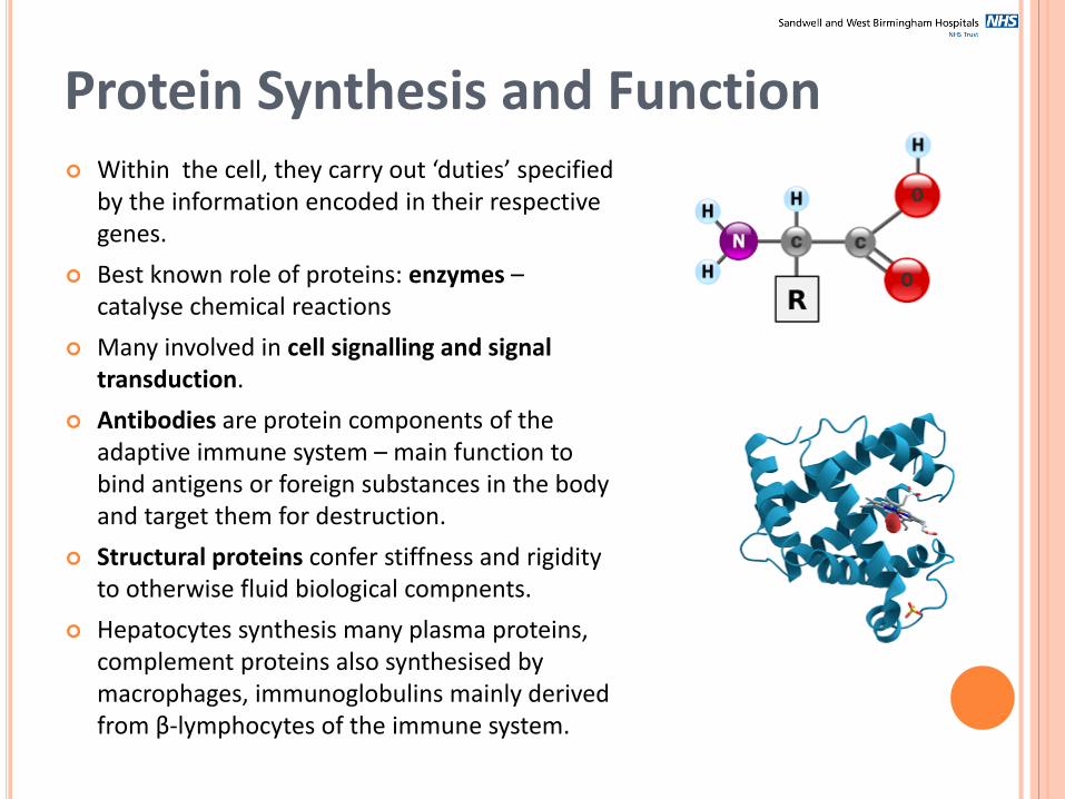
Protein Synthesis and Function Within the cell, they carry out ‘duties’ specified
by the information encoded in their respective genes.
Best known role of proteins: enzymes – catalyse chemical reactions
Many involved in cell signalling and signal transduction.
Antibodies are protein components of the adaptive immune system – main function to bind antigens or foreign substances in the body and target them for destruction.
Structural proteins confer stiffness and rigidity to otherwise fluid biological compnents.
Hepatocytes synthesis many plasma proteins, complement proteins also synthesised by macrophages, immunoglobulins mainly derived from β-lymphocytes of the immune system.

Proteins measured in the lab Enzymes - ALT, AST, CK, Lipase, Amylase etc… Acute phase proteins – C-reactive Protein (CRP) Transport proteins – Caeruloplasmin, transferrin, SHBG, Albumin Immune system components - Immunoglobulins (Ig A, G, M, D
and E), complement, β2 Microglobulin (B2M), cryoglobulin Protease Inhibitors – Alpha-1-Antitrypsin Hormones And many more…
Variety of different sample matrices:
Serum, plasma, urine, CSF, other fluids

Acute Phase Response (APR) The acute phase response is a complex
systemic early-defence system activated by trauma, infection, stress, neoplasia, and inflammation.
Series of cellular and humoral responses that act together to initiate and control the inflammatory reaction and remove damaged tissues/foreign substances
Defense mechanism divided into 2 main groups: The inflammatory response
Non-specific, change in permeability of cell membranes
Dependent on 1. humoral factors e.g. acute phase reactants, complement or cytokines released in response to inflammation and 2. phagocytic cellular response
The immune response…

The Immune response Immunoglobulins (Igs), or antibodies, are glycoprotein
molecules produced by plasma cells (white blood cells). Act as a critical part of the immune response (+
complement proteins and acute phase reactants e.g. CRP) by specifically recognizing and binding to particular antigens, such as bacteria or viruses and aiding in their destruction.
Structure: Y –shaped molecules composed of two different kinds of
polypeptide chain; heavy chain (x2) and light chain (x2) joined by disulfide bonds
Each heavy chain is linked to a light chain and the two heavy chains are linked together.
The 2 heavy chains and the 2 light chains are identical, giving an antibody molecule two identical antigen-binding sites and the ability to bind simultaneously to 2 identical structures.
Classified by isotype that differ in function and antigen responses primarily due to structure variability…

Immunoglobulins Five major isotypes :
IgA - Neutralizing antibody - preventing invading pathogens by attaching and penetrating epithelial surfaces (exists as monomer or dimer).
IgG - Key player in the humoral immune response. Can activate the complement system. Phagocytosis of microorganisms.
IgM - The first antibody built during an immune response and is responsible for agglutination and cytolytic reactions (exists as monomer or pentamer).
IgD – Functions to signal the B cells to be activated. By being activated, they are ready to take part in the defense of the body in the immune system.
IgE - Allergic reactions, parasitic infections, and hypersensitivity reactions.

Acute Phase Proteins Pronounced changes in concentrations of plasma proteins
in response to e.g. infection, tissue injury, inflammation. Due to increased protein synthesis in liver in response to
cytokines.
Increased concentration of positive acute phase proteins: C-Reactive protein (CRP) Fibrinogen Serum amyloid A. Also… Caeruloplasmin 1-Antitrypsin Haptoglobin Ferritin
Decreased concentration of negative acute phase proteins: Albumin Transferrin C3

C-Reactive Protein (CRP) So called because it reacts with C-polysaccharide of
pneumococci. Combines with bacterial polysaccharides or
phospholipids released from damaged tissue to become an activator of the complement pathway.
Plasma concentrations rise rapidly in response to acute inflammation (6h after insult) Conc. related to extent and severity of inflammation
Clinical uses of this test: To detect infection (useful in early detection of acute
infection, mostly bacterial) – more specific than ESR Guide to severity of connective tissue disease activity
e.g. rheumatoid arthritis Diagnosis/monitoring of IBD – Crohn’s/Ulcerative
Colitis Cardiovascular risk indicator – hsCRP (sub-clinical
inflammation).

Complement Macrophages and hepatocytes synthesis a group of proteins called the
complement system, as part of the innate immune system. Activation of complement system results in attraction of phagocytes to
area of inflammation (chemotaxis) Increases capillary wall permeability to cellular and chemical components,
allowing them to reach affected cells.
Results in lysis of foreign cell surfaces and, together with immunoglobulins, some complement proteins enhance phagocytosis.
Circulate in inactive form (due to inhibitors). Clinically most important complement protein
= C3. Two main pathways of activation:
Classsical pathway Alternative pathway Both result in low plasma C3 concentrations.

Other commonly measured specific proteins…

β2-Microglobulin Also known as B2M is a component of MHC class I
molecules, which are present on all nucleated cells (excludes red blood cells).
Cleared from plasma by glomerular filtration followed by tubular reabsorption and catabolism. Plasma concentrations reflect cell turnover and renal
function. Concentrations increase in conditions where there is
increased cell turnover e.g. malignancy (lymphoid), acquired immune deficiency syndromes, inflammation
Important prognostic indicator in myeloma (non-specific, therefore not useful for diagnosis).
May also help to determine disease severity, progression/tumour burden and to evaluate effectiveness of treatment.

α1-Antitrypsin A glycoprotein, part of a family of serine protease inhibitors (anti-
proteinease). Synthesised in the liver.
Constitutes 90% of serum α1 globulin seen in protein electrophoresis. Controls the inflammatory response to minimise damage to host tissue.
Protects tissues from effects of neutophils.
α1-antitrypsin deficiency associated with pulmonary and liver disease at any age. Accumulation of abnormal protein causing inflammation and damage, often
progressing to cirrhosis in liver (mechanism ill understood).
Genetic variants of α1-antitrypsin characterised by their different electrophoretic mobilities on Isoelectric Focusing: Normal phenotype = protease inhibitor MM (PiMM) Homozygote for Z variant = PiZZ (seen in most patients with clinical
disease) Heterozygote phenotype = PiMZ and PiSZ S phenotype mainly associated with lung disease Z phenotype strongly assocuated with liver and lung disease.
Initial screening test = plasma [α1-antitrypsin] – low or zero. NB: raised concentrations during acute phase

Caeruloplasmin Principle Cu-containing protein in plasma. Ferroxidase activity essential for oxidation of Fe(II) to Fe (III). Important role in Wilson’s Disease:
Rare inborn error of Cu metabolism, resulting in Cu deposition in organs – liver (acute hepatitis, cirrhosis), basal ganglia of brain, cornea of eye (colouration).
Autosomal recessive mutation of Cu-transporting ATPase gene ATP7B, >100 mutations worldwide
Result = failure to incorporate Cu into caeruloplasmin and biliary excretion of Cu, reduced circulating [caeruloplasmin]
Presentation in children is mainly hepatic problems, in young adults more neurological problems.
Investigation/diagnosis: Low serum Cu and caeruloplasmin (NB: Caer can be increased in acute phase), increased urine Cu excretion (24 hr urine Cu, penacillamine chelating test), abn LFTs, gold standard = liver biopsy.

Cryoglobulins Proteins that precipitate out of serum at temperatures below
body temperature (37°C) Most cryoglobulins are either:
Monoclonal immunoglobulins - typically IgM (but IgG, IgA or rheumatoid factors are also found), or
Polyclonal - usually immune complexes containing more than one class of immunoglobulins, e.g. IgM rheumatoid factor antibody bound to IgG.
Can occur in any conditions in which there are high concentrations of immunoglobulins e.g. Systemic Lupus Erythematosus (SLE).
The insoluble fibrillar protein complexes in Amyloid Disease may also cause Cryoglobulinaemia.
Cryoglobulinaemia can cause clinical symptoms (Raynaud’s Syndrome) when precipitation occurs above 21°C: Intolerance to cold, purpura, gangrene of the extremities and skin
sores.

Cryoglobulin Determination Blood collected, transported and separated at above 37°C
(flask filled with sand stored at 50°C). Serum divided into two aliquots; stored at 4°C to encourage
cryoglobulin precipitation, and at 37°C to prevent cryoglobulin precipitating from the serum.
Specimens visually inspected for up to 7 day for any signs of precipitation/gel formation.
If sample positive and forms adequate amount of cryo-precipitate than that sample will be further investigated and typed: Cryo-precipitate washed and reconstituted in saline. Protein electrophoresis (SPE) performed to identify any
monoclonal or polyclonal components. Immunotyping (IT) to identify the presence of IgM, IgG, IgA
or kappa/lambda light chains. SPE and IT by capillary electrophoresis Cryoglobulin classified according to the immunoglobulin
present in the precipitate (Type 1, 2, 3).

Paraproteinaemia Paraprotein – an immunoglobulin (protein) produced by a single clone of
myeloma cells (B cells), most frequently plasma cells, found in excess in the blood and/or urine. Since all molecules identical, seen as discrete band (usually in γ region) on serum electrophoresis.
Paraproteins can be found in both malignant and non-malignant conditions including: Multiple myeloma (associated with older adults). Waldenstrӧm’s Macroglobulinaemia (non-hodgkin lymphoma) -
macroglobulin = IgM, symptoms related to macroglobulin (increased plasma viscosity).
Monoclonal Gammopathy of Undetermined Significance (MGUS) - Pre-malignant (benign).
Hyperviscosity syndrome - a group of symptoms triggered by increase in the viscosity of the blood. Symptoms: spontaneous bleeding from mucous membranes, visual
disturbances due to retinopathy, neurologic symptoms (headache, vertigo, seizures, coma).
Occurs from pathologic changes of cellular/protein fractions of blood found in polycythaemias, multiple myeloma, leukaemia, monoclonal gammopathies, (e.g. Waldrenstrӧms, sickle cell anaemia, and sepsis.

Myeloma Malignancy of plasma cells, usually in marrow (of lumbar
spine, skull, long bones, pelvis). Variants:
IgG, IgA, IgM, IgD, IgE Light chain (kappa [κ] or Lambda [λ]) myeloma Heavy chain disease Biclonal gammopathy Non-secretory myeloma
Clinical Presentation: generally bone pain (back, ribs), pathological fracture/osteolytic lesions (“pepper pot skull” on x-ray, hypercalcaemia, recurrent infections, fatigue/anaemia
Diagnosis 2/3 of: Serum and/or urine paraprotein Osteolytic lesions on x-ray >25% plasma cells in marrow (or clonal population)
Treatment: (symptomatic, complications) Chemotherapy Consider BM transplant

Myeloma: Investigation Biochemistry:
Serum immunoglobulins (turbidimetry) IgG >20g/L, IgA/M >10 g/L, Ig D/E paraprotein @ any conc.
Serum protein electrophoresis (gel or capillary elect) if identify abnormally high/low immunoglobulins
Quantification of paraprotein by densitometry (relate density of band to measures total protein)
Urine electrophoresis for Bence Jones Protein (BJP) or light chains Confirmation/typing of paraprotein in serum or urine by immunofixation (if
present) ?serum Free Light Chains
Other Investigations: FBC/Hb (anaemia) ESR (inc. due to high [protein]) Calcium (hypercalcaemia) B2M (prognosis) BM biopsy/aspirate (to study plasma cells) Skeletal survey (bone/skull x-ray)

Myeloma: Complications & Prognosis Complications:
Increased risk of chronic renal failure - tubular damage due to accumulation of free light chains (BJP)
Increased risk of acute renal failure – hypercalcaemia due to localised action of cytokines stimulating osteoclastic bone resorption (therefore measure urea/cre to investigate poss. Renal impairment)
Fractures/lesions - Bone x-rays, skeletal survey, MRI or CT Immunoparesis/ increased risk of infection - due to suppressed Immunoglobulin levels Increased plasma viscosity
Poor prognosis indicated by: Presence of urine BJP Low serum albumin (<30 g/L – inverse correlation with tumour burden) Anaemia (normochromic, normocytic, BM failure) Increased B2M (increases as tumour mass increases) Also presence of hyperCa, renal dysfunction, progressive increase in [paraprotein]
Monitor by: Paraprotein quantification by densitometry (related to tumour burden)
Can’t use turbidimetry/nephlometry for Igs qualntification

MGUS “Monoclonal Gammopathy of Undetermined Significance”. Non-malignant, benign (pre-malignant). Presence of paraprotein in absence of features of myeloma:
Paraprotein <30 g/L, no associated FLCs. Plasma cells in BM <10%.
Repeat investigations in 6 months to see if paraprotein size has increased.

Urine Proteins Proteinuria – presence of excess serum proteins in the urine.
Often causes urine to become ‘foamy’.
Causes of proteinuria: Pre-renal – Heavy exercise, fever, hypertension, multiple
myeloma, pre-eclampsia Renal – Acute and chronic glomerulonephritis, renal tubular
dysfunction, polycystic kidney, nephrotic syndrome Post-renal – acut and chronic cystitis, tuberculosis
Key proteinuria mechanisms: Overflow – High conc. Of low MW protein filtered in quantities exceeding
tubular reabsorptive capacity (e.g. Urine BJP) Glomerular – Increased glomerular permeability (e.g. Albumin) Tubular – Impaired/saturated reabsorption of protein filtered by normal
glomeruli (e.g. B2M)
Measurement of protein in urine important to diagnose, stage and monitor chronic kidney disease (together with GFR). NICE: Albumin:Creatinine Ratio (ACR) should be used in preference to
Protein:Creatinine Ratio (PCR) or 24 hr urine TP and dipstick.

Urine Bence Jones Protein (BJP) Bence Jones protein is a monoclonal globulin or immunogobulin light
chain (kappa or lambda) found in the urine (MW 22-24 kDa). Free light chains (FLCs) have a low molecular weight and are freely filtered at
the glomeruli and largely reabsorbed by proximal tubular cells. However when there is overproduction the reabsorptive capacity is exceeded and they pass into the urine.
Detection of urine BJP may be suggestive of multiple myeloma or Waldernström’s macroglubulinaemia. Monoclonal free light chains may be found in myeloma in addition to a
serum paraprotein or may occur in isolation (i.e. in light chain only myeloma, seen in approximately 2% myeloma cases where undetectable in serum but detectable in urine).
These FLCs may eventually cause renal impairment due to deposition of the protein in renal tubular cells and can cause formation of large casts and the characteristic myeloma kidney.
Identified by urine electrophoresis and typed as either kappa (κ) or Lambda (λ) by urine immunofixation.

Protein Separation and Analysis
Variety of laboratory methods to separate/measure proteins…
Electophoresis (gel, capillary) Immunofixation
Chromatography Nephlometry Turbidimetry Immunoassay

Protein Separation Properties of proteins that aid separation:
Molecular weight (MW) Charge Solubility Affinity

Electrophoresis Definition: Migration of all charged particles in a liquid medium
under the influence of an electric field. Used to separate range of ionised analytes e.g. proteins, amino
acids, nucleic acids, organic acids. Rate of migration dependent on:
Net electrical charge of particle Particle size and shape Electric field strength Interaction with support medium (e.g. endo-osmosis, wick flow
effects) Temperature
Migration of proteins based on charge-to-mass ratio Proteins possess aa residues which can donate/accept protons
and alter their overall charge, depending on pH of surrounding environment (buffers).

Electrophoresis - Components
Support media (e.g. agarose, polyacrylamide gel) – in contact with buffer via wicks, where separation takes place
Buffers (pH/Ionic strength) Electrodes connected to power supply (provides electric field)
As current passes through support medium, proteins will migrate towards anode (+) or cathode (-), depending on charge
Protein visualisation method (stains, UV detection for CE) Quantification method (e.g. densitometry)
Capillary Electrophoresis
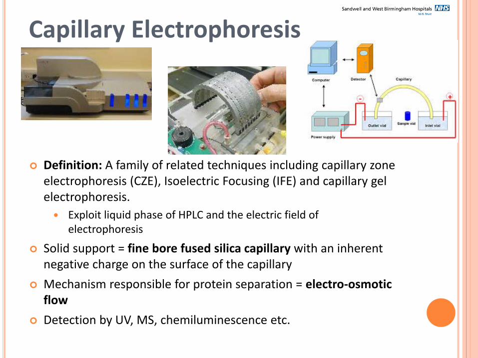
Definition: A family of related techniques including capillary zone electrophoresis (CZE), Isoelectric Focusing (IFE) and capillary gel electrophoresis. Exploit liquid phase of HPLC and the electric field of
electrophoresis
Solid support = fine bore fused silica capillary with an inherent negative charge on the surface of the capillary
Mechanism responsible for protein separation = electro-osmotic flow
Detection by UV, MS, chemiluminescence etc.

Electro-Osmotic Flow Negatively charged proteins attracted to the cathode (-) by a
strong endo-osmotic flow of positive buffer ions (put buffer through capillary with positive charge) and electrical field.
Separation of proteins results in differences in migration back to the anode (+)
Therefore gamma globulins elute first (good separation in gamma region) – ideal for Immunoglobulin separation I.e. cathodic movement of gamma globulins (despite neg.
charge) from point of origin

CE Electrophoresis: Results Interpretation Order of proteins detected is in the following manner, start with Gamma globulins and ending with albumin:
• Gamma(γ)-globulins: Immunoglobulins (some also found in the 2 and β regions)
• Beta(β)-globulins: β1 consists mainly of transferrin and some LDL, β2 consists of C3 component of complement.
• Alpha()2-globulins: mainly 2-macroglobulin and haptoglobin.
• Alpha()1-globulins: almost entirely 1-antitrypsin and 1-antichymotrypsin.
• Albumin: usually a single protein, makes up the obvious band.
Note: If plasma used rather than serum (i.e. not clotted): fibrinogen appears in β-γ region.

Serum Electrophoresis: Patterns in Disease
Acute Phase Reaction: Synthesis of acute phase proteins – increased 1 and 2 globulins Responsible for rise in ESR and increased
plasma viscosity.
Chronic Inflammation: Diffuse rise in γ-globulins.
Nephrotic syndrome: In established cases, reduced albumin, 1 and γ-globulins, increased 2 globulin.
1-antitrypsin deficiency: Absence or obvious reduction in density of 1 band.
Hypogammaglobulinaemia: Reduced γ-globulins.
Multiple Myeloma/Paraproteinaemia: Discrete band(s) in the in γ-globulin region.

CE Examples

CE Examples
Previously on Gel Electrophoresis it is difficult to determine when a band is over a beta 2 region.

Capillary vs Gel Electrophoresis Advantages of CE Disadvantages of CE
High efficiency of separation and high resolution of small and large molecules (in β and γ regions)
Different method of electrophoresis interpretation (“electrophoretograms”) – requires staff re-training
Multiple sample simultaneously/rapid analysis - multiple capillaries, fully automated
High capital cost of equipment
No gel staining procedures – measurement by direct UV at SWBH
No gel for record keeping – all computer records must be securely backed-up
Improved linearity and real-time quantification of paraproteins (during analysis)
Cannot identify IgD/E paraproteins by Immunosubtraction – requires Immunofixation
Small sample volumes (nL) Increased Immunofixation rate – Increased workload
Analyser interfacing with LIMS

Electrophoresis: Clinical Application E.g. Serum Immunotyping to identify the monoclonal
immunoglobulin composition of suspected paraproteins detected in serum by protein electrophoresis.
Essential for the complete investigation of Myeloma. Also used to monitor Monoclonal Gammopathy of Uncertain Significance (MGUS).
Immunotyping Capillary Electrophoresis currently used at SWBH: Diluted serum mixed with individual specific antisera Forms large insoluble complexes with heavy and light chains which
thus move more slowly then other components of the serum when electrophoresed.
The immune complex migrates anodically. After, treated and untreated curves are overlaid to see which peaks
have been removed under which antisera.

Serum Immunofixation Clinical Application: Used to identify the monoclonal immunoglobulin composition of
suspected paraproteins detected in serum by protein electrophoresis. Essential for the complete investigation of myeloma. Also used to monitor monoclonal gammopathy of uncertain significance
(MGUS).
Method: Following electrophoretic separation, individual heavy and light chain
immunoglobulins are identified by their reaction with specific antisera raised against gamma (IgG), alpha (IgA) and mu (IgM) heavy chains and kappa (free and bound) and lambda (free and bound) light chains, respectively.
The resulting insoluble complexes are trapped in the gel matrix (fixed). Non-reacted proteins are then removed by blotting and washing and the
insoluble complexes visualised with acid violet stain.

Serum Immunofixation: Results Interpretation

Urine Electrophoresis: BJP The demonstration of paraproteinaemia and immuneparesis by protein
capillary electrophoresis and urine BJP are diagnostic of multiple myeloma.
Urine protein gel electrophoresis (Sebia Hydrasys) currently used at SWBH for identification of urine BJP: Proteins separated and visualised by high resolution electrophoresis using
highly sensitive protein specific stain Acid Violet. Method provides a rapid initial screening method for detecting abnormal
bands in urine, in particular Bence Jones Protein.

Urine Electrophoresis: Results
• Lane 1/N – FLCs not detected (normal)
• Lane 2, 21 – Generalised proteinuria (FLCs not detected)
• Lane 11 - Abnormal band detected – hold results and investigate further by urine Immunofixation
• Lanes 14 15, 25, 27 – Abnormal bands detected, hold (as above)
• T – Would require further invx if patient
• Pos – Band in βγ region

Urine Immunofixation BJP immunofixation electrophoresis is used to identify the
monoclonal kappa (κ) or Lambda (λ) free light chains in urine following the detection of abnormal bands in serum by protein electrophoresis and urine by BJP electrophoresis.
Method: Electrophoretic separation of urine proteins. Individual heavy and light chain immunoglobulins identified by their
reaction with a gamma (IgG), alpha (IgA) and mu (IgM) combined trivalent antiserum, and antiserum raised against kappa and lambda light chains (both free and bound).
Resulting insoluble complexes trapped in the gel matrix (FIXED). Non-reacted proteins removed by blotting and washing. Insoluble complexes visualised with acid violet stain.

Urine Immunofixation: Results Interpretation
Free κ light chains detected Free light chains not detected Free λ light chains detected

Nephlometry & Turbidimetry Methods used to measure scattered light using ‘Light Scattering
Theory’: A physical phenomenon resulting from interaction of light with insoluble
particles in solution Transmitted light reduces as the reaction progresses Factors affecting: particle size, wavelength dependence, measuring
angle etc..
Incident light

Nephelometry vs Turbidimetry Nephelometry Turbidimetry
Definition Detection of scattered light that is not in the direct path of the transmitted light
Measures a decrease in intensity of incident light transmission caused by scattering
Type of light measured
Scattered light Transmitted light
Measuring angle/ arrangement of photometer
Measurement of light at right angle (90°) to direction of incident light. Could be movable detectors which allow operator to vary the angle of detection.
180° from the incident light (same direction as the propgation of the light from the source)
Instrument used Dedicated Nephelometry analyser (e.g. Siemens Dade Behring BN II Nephelometer)
Spectrophotometer
Advantages Better sensitivity than turbidimetry (particularly for small immune complexes) – superior at <20mg/L (no advantage over turbidimetry above this)
Performed on routine chem analysers, high throughput, accurate and precise
Disadvantages Requires dedicated analyser, cost and TAT implications, antigen (ag) excess/hook effect
Lower sensitivity compared with nephelometers (but improving with technological advances), ag excess
Clinical use/ examples
Ag-AB rxn, immunocomplex rxn, lipoprotein (e.g. serum FLCs, IgG subclasses)
Ag-Ab rxn, immunocomplex rxn (e.g. CRP, urine and CSF protein, IgA, IgG, IgM, C3, C4, A1AT, B2M)

CSF Proteins CSF is a clear body fluid produced by the choroid
plexuses that occupies the subarachnoid space and ventricular system around and inside the brain. Acts as a “cushion” for the cerebral cortex of the brain
CSF samples obtained by lumbar puncture. CSF composition:
Less protein/bilirubin and similar electrolyte composition when compared with plasma
CSF total protein (albumin, IgG, transthyretin) derived from plasma proteins (80%) and manufacture within the brain (20%). Increases as a result of a breakdown of the integrity of the blood-brain barrier
E.g. Froin’s syndrome (block to spinal circulation of CSF), Guillain Barre syndrome (deposition of immune complexes), multiple sclerosis.

Multiple Sclerosis (MS) Autoimmune condition in which immune system attacks
the central nervous system (CNS) leading to demyelination.
Neurological symptoms occur in discrete attacks or progressively.
Clinical diagnosis difficult, although MRI can form part of the diagnosis.
Biochemically, the hallmark of MS is the presence of oligoclonal IgG bands in CSF (absent from paired serum) Found in at least 98% of patients with MS BUT not specific for MS – can be found in other inflammatory and
autoimmune conditions affecting the brain
Presence of oligoclonal bands and a CSF protein >1g/L points to a diagnosis other than MS.

Oligoclonal Bands by IFE CSF oligoclonal band determination by
Isoelectric Focusing (IFE) coupled to nitrocellulose immunoblotting (Western blotting) or immunofixation.
IFE is a type of electrophoresis technique for separating different molecules by differences in their isoelectric point (dependent on pH of surroundings) Significantly more sensitive at detecting CSF IgG
than quantitative methods.
Results interpretation: No oligoclonal bands = normal Presence of oligoclonal bands in CSF and not
serum = MS (local synthesis) Presence of bands in both CSF and paired
serum = systemic response due to inflammation
Further Reading
Clinical Biochemistry: Metabolic and clinical aspects, William J Marshall, Stephen K. Bangert
Kit Inserts – CRP, A1AT, B2M, Urine/CSF protein, Caer, Ig A/G/M etc…
SOPs on iPassport: Immunotyping of Serum Proteins by Sebia Capillarys (Serum protein
electrophoresis) Serum/urine immunofixation electrophoresis Urinary Bence Jones Protein by Capillary Electrophoresis Bence Jones Protein - Sebia Hydrasys LC Cryoglobulin identification and typing