specific organ/system toxicity: the liver - carnegie · pdf filespecific organ/system...
TRANSCRIPT
Short-tenn Toxicity Tests for Non-genotoxic EffectsEdited by P. Bourdeau et al.@ 1990 SCOPE. Published by John Wiley & Sons Ltd
CHAPTER 9
SpecificOrgan/System Toxicity: the Liver
JAMES W. BRIDGES
9.1 INTRODUCTION
Mammalian liver consists of at least fourteen different types of cells. Hepatocytes,the most common site of toxic action, account for between 60 per cent (rat) and 85per cent (man) of the total number of cells in the liver, or 90-95 per cent of the totalweight of the liver. The reticuloendothelial cells (also called Kupffer, littoral orsinusoidal cells) only constitute 5-10 per cent of liver by weight but they cancomprise up to 35 per cent of the total cell population. Other cells include fat storingand pit cells.
Despite their morphological similarity, hepatocytes are not a homogeneous popu-lation. For example, those from the centrilobular region contain significantly higherlevels of cytochrome P-450 (and are much more responsive to induction by pheno-barbitone) than those derived from the mid-zonal or periportal regions. Cells of theperiportal area tend to be more metabolically active than those of mid-zonal andcentrilobular regions, probably because they are more richly endowed with bloodand consequently with nutrients and oxygen.
The liver has a wide range of important physiological functions and is the site of awide range of intermediary metabolite reactions. These functions include:
(1) synthesis of many serum proteins (such as albumin, fibronogen and clottingfactors);formation and secretion of bile;
storage of various substances (such as metals and vitamins);detoxification of endogenous and exogenous waste products (including drugs,environmental chemicals, active oxygen species and haem breakdownproducts ).
(2)(3)(4)
The liver, despite its remarkable regenerative ability, is vulnerable to injury frommany causes which may result in profound metabolic effects. These may be revers-ible or irreversible. Chemically-mediated liver toxicity in man may take various
forms including hepatocyte necrosis, hepatitis, jaundice, vascular injury, porphyria,
111
112 Short-term Toxicity Testsfor Non-genotoxic Effects
cirrhosis or neoplasia (see Table 9.1). Drug-induced liver damage is one of thecommonest fonns of iatrogenic disease. Jaundice, for example, occurs with manychemically-induced hepatic disease in man.
Pathological effects of disturbed hepatic function fall into three primary classes:
(1) Hepatocellular failure or impaired hepatocellular function. This results fromdirect injury to hepatocytes leading to selective or total loss of function. It mayalso arise through chronic impainnent of blood flow. Alterations in nitrogenmetabolism, failure to remove bilirubin from blood and/or conjugate it, buildup of porphyrins, reduced synthesis of plasma proteins, cyanosis and honnonaldisturbances are possible consequences of hepatocellular failure.Biliary obstruction. This may arise from blockage of the hepatic or commonbile ducts or impaired flow. Consequences may include leakage of conjugatedbilirubin and other bile constituents into the blood and reduced absorption offats and fat soluble vitamins from the intestine. Prolonged cholestasis is likelyto lead to hepatic necrosis.Portal hypertension. This may develop due to obstruction of liver blood flow.As a result, blood may be shunted from the portal blood supply directly into
(2)
(3)
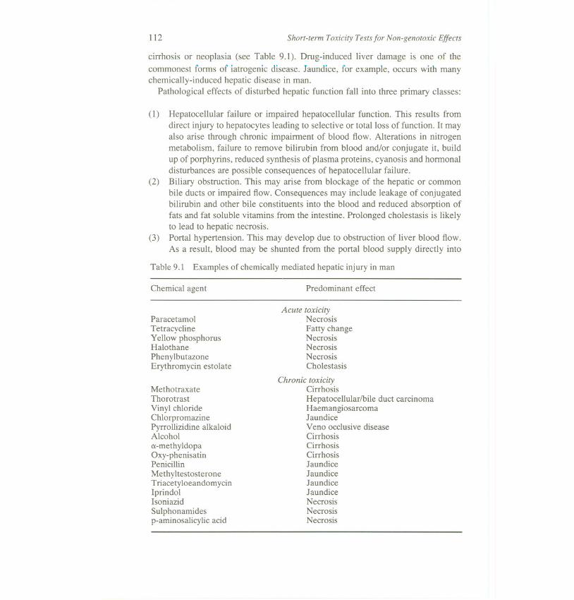
Table 9.1 Examples of chemically mediated hepatic injury in man
Chemical agent Predominant effect
MethotraxateThorotrastVinyl chlorideChlorpromazinePyrrollizidine alkaloidAlcohol(X-methyldopaOxy-phenisatinPenicillinMethyltestosteroneTriacetyloeandomycinIprindolIsoniazidSulphonamidesp-aminosalicylic acid
Acute toxicityNecrosisFatty changeNecrosisNecrosisNecrosisCholestasis
Chronic toxicityCirrhosisHepatocellular/bile duct carcinomaHaemangiosarcomaJaundiceVeno occlusive diseaseCirrhosisCirrhosisCirrhosisJaundiceJaundiceJaundiceJaundiceNecrosisNecrosisNecrosis
ParacetamolTetracyclineYellow phosphorusHalothanePhenylbutazoneErythromycin estolate
Specific Organ/System Toxicity: the Liver 113
the systemic circulation, thus by-passing the liver. These anastomoses mayrupture. Toxic materials absorbed from the gut may build up in the blood dueto failure of the liver to detoxify them.
9.1.1 Vulnerability of the liver
There are a number of reasons why the liver is a primary target for the toxic effectsof chemical agents:
(1) Ingestion is a common route of exposure to chemicals. Chemicals absorbed inthe stomach, small intestine or large intestine pass almost exclusively into thehepatic portal vein, and are transported to the liver in appreciable concentra-tions and amounts.The liver has a high capacity to non-specifically bind chemicals by virtue of itsrelatively large size and high cellular concentration of binding sites (includingproteins such as ligandin, and extensive amounts of lipid-rich endoplasmicreticulum).The liver contains higher concentrations of the drug-metabolizing enzymesthan any other tissue. Since these enzymes frequently cause the formation oftoxic metabolites as well as bringing about detoxification, the liver isespecially vulnerable to metabolite-mediated toxicity.The liver is the most biochemically diverse of all the organs, playing key rolesin the utilization of absorbed nutrients, detoxification and excretion on endo-genous and exogenous compounds and the synthesis of plasma proteins. Thisgreat range of activities renders the liver more vulnerable to toxins than manyother organs.
(2)
(3)
(4)
9.1.2 Routes of chemical exposure of the liver
Since most of the blood supply to the gastrointestinal tract feeds into the portalcirculation (it accounts for 80 per cent of the liver's blood supply), chemicals whichare absorbed from the gastrointestinal tract will, in theory, expose the liver to higherconcentrations of the agent than most other routes of administration.
Metabolism of some chemicals under the influence of gut microfloral metabolismwill result in exposure of the liver tissue to a range of metabolites and breakdownproducts. Interestingly, some substances (e.g. dimethline) exhibit greater hepatox-icity by gavage than when given in the diet while the converse is true in other cases(e.g. griseofulvin).
After intraperitoneal administration, a large part of the dosage of a chemical willdrain to the liver. The rate of this process will depend on the solubility of thechemical in the peritoneal fluid. The liver is also susceptible to the effects ofchemicals present in the systemic circulation since it receives approximately 30 per
cent of total cardiac output.
114 Short-term Toxicity Testsfor Non-genotoxic Effects
9.2 IN VIVO MODELS FOR LIVER TOXICITY
In vivo investigations, although usually physiologically more relevant, have a num-ber of practical as well as ethical drawbacks:
(1) Only gross control can be exerted over the concentration of xenobiotics reach-ing a particular tissue site, and the time of exposure of that site to thexenobiotics.
Monitoring of plasma or urine for toxic metabolites generally only picks upstable metabolites which tend to accumulate in these fluids. Metabolites which
are reactive and may contribute significantly to the toxicological properties ofthe xenobiotic will frequently not be detected.Obfuscating factors in vivo, such as stress and other environmental variables,may interfere with the correct interpretation of data.Information about toxicity in man is usually the goal of toxicity studies. Manyinvestigations cannot be made in vivo in man for ethical reasons.
(2)
(3)
(4)
9.2.1 Effectiveness of in vivo animal models for predicting hepatic toxicity inman
In some cases, in vivo tests using animals may fail to detect hepatic effects which dooccur in man (false negative response). Such is often the case with chemically-induced hepatic disease which has an immunological basis. Drugs which inducejaundice in man (e.g. chlorpromazine, halothane, erythromycin, estolate) are veryfrequently not identified as having potential to cause jaundice using current methodswith whole animals. Often, such effects are identified only in later clinical trials.Cirrhosis, caused by chronic alcohol poisoning, is another hepatic disease in man forwhich it is doubtful that a valid animal model yet exists.
There are also instances when animal tests produce hepatic disease which doesnot seem to occur in man (false positive responses). It would appear that a numberof chemicals (e.g. clofibrate, phenobarbitone, DDT) which produce hepatocar-cinoma in rodents (but are non-genotoxic in classical in vivo and in vitro tests)probably fall into this category. However, failure to identify a lesion in man mayalso be due to poor detection methods.
9.2.2 Methods to assess liver toxicity following in vivo tests
9.2.2.1 Clinical observations
In general, observations of animal behaviour are not helpful in distinguishing liverdamage from other forms of malaise. The development of whole body protonnuclear magnetic resonance scanners suitable for small conscious animals seemslikely to provide a powerful means for identifying changes in the liver and in other
Specific Organ/System Toxicity: the Liver 115
soft tissues. Already, such instruments are able to measure 4-mm 'slices' throughthe liver and measurement of l-mm 'slices' should be possible in the near future.
9.2.2.2 Morphology
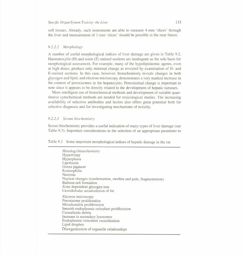
A number of useful morphological indices of liver damage are given in Table 9.2.Haematoxylin (H) and eosin (E) stained sections are inadequate as the sole basis formorphological assessment. For example, many of the hypolipidaemic agents, evenat high doses, produce only minimal change as revealed by examination of H- andE-stained sections. In this case, however, histochemistry reveals changes in bothglycogen and lipid, and electron microscopy demonstrates a very marked increase inthe content of peroxisomes in the hepatocytes. Peroxisomal change is important tonote since it appears to be directly related to the development of hepatic tumours.
More intelligent use of histochemical methods and development of suitable quan-titative cytochemical methods are needed for toxicological studies. The increasingavailability of selective antibodies and lectins also offers great potential both forselective diagnosis and for investigating mechanisms of toxicity.
9.2.2.3 Serum biochemistry
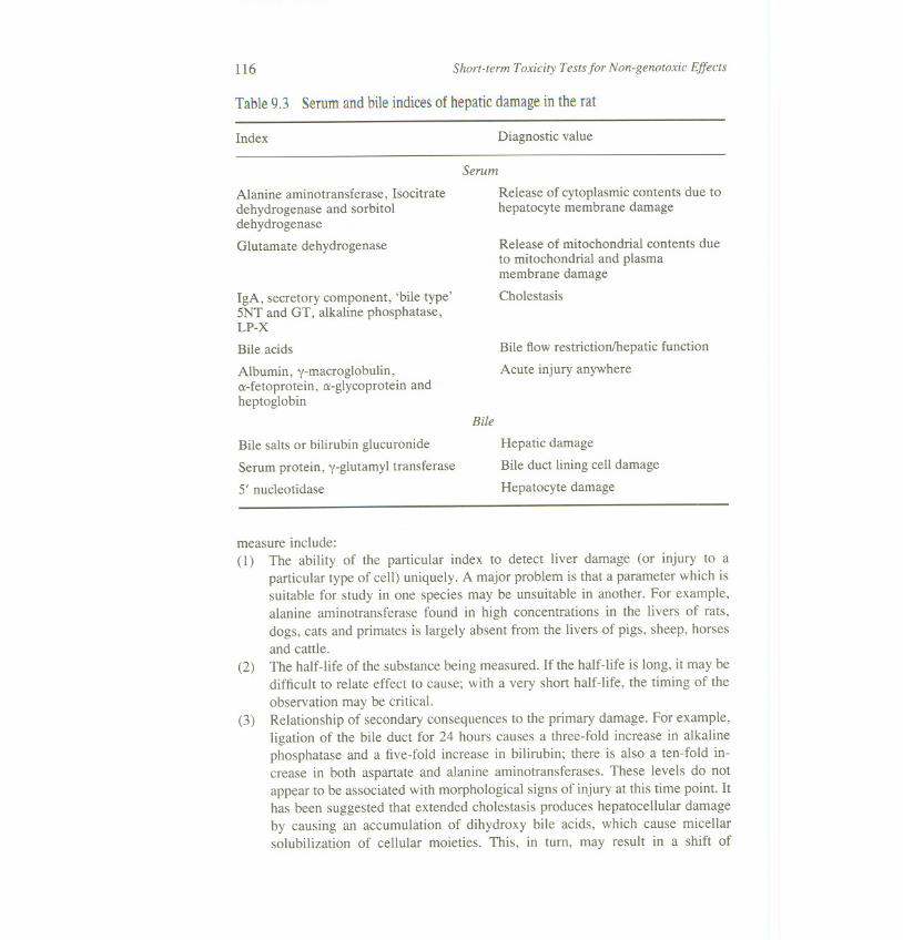
Serum biochemistry provides a useful indication of many types of liver damage (seeTable 9.3). Important considerations in the selection of an appropriate parameter to
Table 9.2 Some important morphological indices of hepatic damage in the rat
Histology /histochemistryHypertropyHyperplasiaLipofuscinGreen pigmentEosinophiliaNecrosis
Nuclear changes (condensation, swollen and pale, fragmentation)Balloon cell formationZone dependent glycogen lossCentrilobular accumulation of fat
Electron microscopyPeroxisome proliferationMitochondria proliferationSmooth endoplasmic reticulum proliferationCannulicula debrisIncrease in secondary lysosomesEndoplasmic reticulum vacuolizationLipid dropletsDisorganization of organelle relationships
116
Table 9.3 Serum and bile indices of hepatic damage in the rat
Short-term Toxicity Testsfor Non-genotoxic Effects
Index Diagnostic value
Alanine aminotransferase, Isocitratedehydrogenase and sorbitoldehydrogenase
Glutamate dehydrogenase
IgA, secretory component, 'bile type'5NT and GT, alkaline phosphatase,LP-X
Bile acids
Albumin, y-macroglobulin,ex-fetoprotein, ex-glycoprotein andheptoglobin
Bile salts or bilirubin glucuronide
Serum protein, y-glutamyl transferase
5' nucleotidase
Serum
Release of cytoplasmic contents due tohepatocyte membrane damage
Release of mitochondrial contents dueto mitochondrial and plasmamembrane damage
Cholestasis
Bile flow restriction/hepatic function
Acute injury anywhere
Bile
Hepatic damage
Bile duct lining cell damage
Hepatocyte damage
measure include:
(1) The ability of the particular index to detect liver damage (or injury to aparticular type of cell) uniquely. A major problem is that a parameter which issuitable for study in one species may be unsuitable in another. For example,alanine aminotransferase found in high concentrations in the livers of rats,
dogs, cats and primates is largely absent from the livers of pigs, sheep, horsesand cattle.
(2) The half-life of the substance being measured. If the half-life is long, it may bedifficult to relate effect to cause; with a very short half-life, the timing of the
observation may be critical.(3) Relationship of secondary consequences to the primary damage. For example,
ligation of the bile duct for 24 hours causes a three-fold increase in alkalinephosphatase and a five-fold increase in bilirubin; there is also a ten-fold in-crease in both aspartate and alanine aminotransferases. These levels do notappear to be associated with morphological signs of injury at this time point. Ithas been suggested that extended cholestasis produces hepatocellular damageby causing an accumulation of dihydroxy bile acids, which cause micellarsolubilization of cellular moieties. This, in turn, may result in a shift of
Specific Organ/System Toxicity: the Liver 117
(4)
enzymes from organelles to cytoplasm and, hence, increased leakage fromhepatocytes to the extracellular fluid.The relationship between the duration of damage and the release of a particularcomponent into the serum. In many instances, the severity of damage may notbe reflected in the levels of cellular components in the serum over prolongedperiods of time. Studies of enzyme release after acute toxic liver injury in therat have shown that there is frequently a distinct pattern of release of intra-cellular enzymes into the circulation. Cytoplasmic enzyme levels increase inserum within a few hours, followed by cytoplasmic and mitochondrialenzymes other than those of purely mitochondrial membrane origin. Subse-quently, the depletion of constituents in necrotic cells leads to a decrease in therelease of constituents into the serum. Hence, the direct relationship betweenmorphological damage and serum composition no longer holds.Its stability during sample storage and sample work-up.Its ease of assay, induding complexity of any sample work-up, possible pres-ence in serum of interfering agents, etc. The sensitivity, precision and specif-icity of the assay needs to be defined and checked regularly.
(5)(6)
Insufficient attention has been given to non-enzymatic proteins as an index ofhepatic damage. The promise of this approach is indicated by the fact that hypo-lipidaemic agents induce dose-related changes in six proteins in rats detected bycrossed immunoelectrophoresis. Two of these proteins appear to be specific forhypolipidaemic agents.
9.2.2.4 Tissue biochemistry
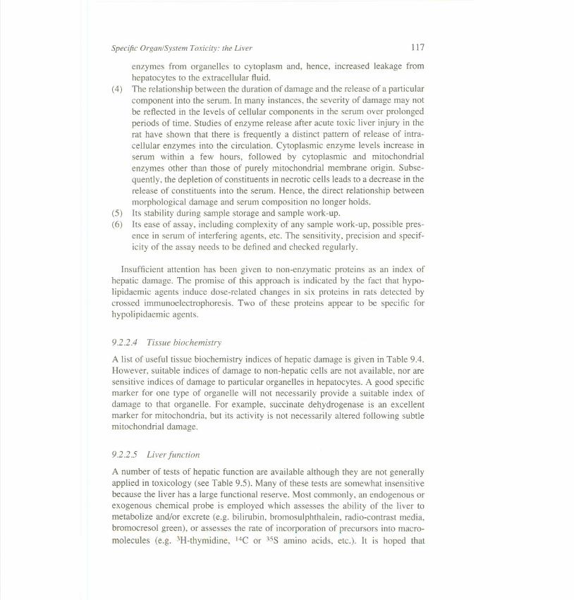
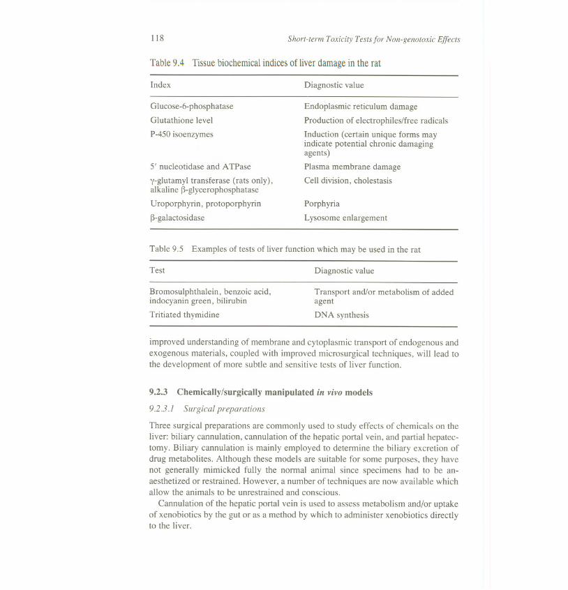
A list of useful tissue biochemistry indices of hepatic damage is given in Table 9.4.However, suitable indices of damage to non-hepatic cells are not available, nor aresensitive indices of damage to particular organelles in hepatocytes. A good specificmarker for one type of organelle will not necessarily provide a suitable index ofdamage to that organelle. For example, succinate dehydrogenase is an excellentmarker for mitochondria, but its activity is not necessarily altered following subtlemitochondrial damage.
9.2.2.5 Liver function
A number of tests of hepatic function are available although they are not generallyapplied in toxicology (see Table 9.5). Many of these tests are somewhat insensitivebecause the liver has a large functional reserve. Most commonly, an endogenous orexogenous chemical probe is employed which assesses the ability of the liver tometabolize and/or excrete (e.g. bilirubin, bromo suiphthalein, radio-contrast media,bromocresol green), or assesses the rate of incorporation of precursors into macro-
molecules (e.g. 3H-thymidine, 14C or 35S amino acids, etc.). It is hoped that
118 Short-term Toxicity Testsfor Non-genotoxic Effects
Table 9.4 Tissue biochemical indices of liver damage in the rat
Index Diagnostic value
Glucose-6-phosphatase
Glutathione level
P-450 isoenzymes
5' nucleotidase and ATPase
y-glutamyl transferase (rats only),alkaline ~-glycerophosphatase
Uroporphyrin, protoporphyrin
~-galactosidase
Endoplasmic reticulum damage
Production of electrophiles/free radicals
Induction (certain unique forms mayindicate potential chronic damagingagents)
Plasma membrane damage
Cell division, cholestasis
Porphyria
Lysosome enlargement
Test
Table 9.5 Examples of tests of liver function which may be used in the rat
Diagnostic value
Bromosulphthalein, benzoic acid,indocyanin green, bilirubin
Tritiated thymidine
Transport and/or metabolism of addedagent
DNA synthesis
improved understanding of membrane and cytoplasmic transport of endogenous andexogenous materials, coupled with improved microsurgical techniques, will lead tothe development of more subtle and sensitive tests of liver function.
9.2.3.1 Surgical preparations
Chemically/surgically manipulated in vivo models9.2.3
Three surgical preparations are commonly used to study effects of chemicals on theliver: biliary cannulation, cannulation of the hepatic portal vein, and partial hepatec-tomy. Biliary cannulation is mainly employed to determine the biliary excretion ofdrug metabolites. Although these models are suitable for some purposes, they havenot generally mimicked fully the normal animal since specimens had to be an-aesthetized or restrained. However, a number of techniques are now available whichallow the animals to be unrestrained and conscious.
Cannulation of the hepatic portal vein is used to assess metabolism and/or uptakeof xenobiotics by the gut or as a method by which to administer xenobiotics directlyto the liver.
Specific Organ/System Toxicity: the Liver 119
A favoured model for reducing the functional reserve of the liver is partialhepatectomy. This increases the liver's sensitivity to xenobiotics. Partial hepatec-tomy has also been used to study the effects of chemicals on cell division.
9.2.3.2 Chemically manipulated models
A wide range of chemicals have been employed to produce selective modificationsin hepatic function for investigating the effects of a toxic chemical on that function.For example, modification of drug metabolizing activity can be achieved usingselective enzyme inducers (such as phenobarbitone, 3-methylcholanthrene, iso-safrole, clofibrate and pregenolone carbonitrile) or inhibitors (such as cobalt chlor-ide, piperonyl butoxide and SKF525A). Diethylmaleate is only moderatelyhepatotoxic and may be used to deplete glutathione thereby enabling the toxiceffects of electrophiles to be more fully expressed. Chemicals such as carbontetrachloride and paracetamol have been employed to produce selective damage inorder to reduce the functional reserve of the liver. However, the effects of thesechemicals tend to be rather poorly reproducible.
9.3 IN VITRO MODELS
There is no single ideal in vitro hepatic preparation for toxicological investigations,nor is there likely to be. Since no in vitro system can mimic all of the intricate andcomplex interactions which occur within an organism, only a limited assessment canbe made of the adverse effects of xenobiotic in vitro. Therefore, the preparationmust be selected in the light of the particular purpose of the intended experiment.These data can then be compared with those obtained in vivo and a prediction madeof the compound's likely effect in a human being.
Some of the obvious limitations of the exclusively in vitro approach can beovercome by deriving the appropriate culture preparation from animals which have
been pretreated in various ways (see 9.2.3.2 above). This approach is particularlyuseful where pharmokinetic considerations are critical to the expression of toxicityor where the useful lifetime of the in vitro preparation is rather short.
9.3.1 Perfused liver
Isolated perfused organs are particularly useful for evaluating the complex interrela-tionships between heterogeneous cell types or between different metabolic path-ways. Variations in pharmacokinetics with time and dose are probably best studiedin the perfused organ as opposed to other in vitro preparations. As yet, other than
tissue slices, liver perfusion offers the only potential model for studying chemically-induced portal hypertension or biliary obstruction.
120 Short-term Toxicity Testsfor Non-genotoxic Effects
9.3.1.1 Perfusedorganviability
Bile flow is useful as a viability index. Bile flow tends to drop with the time of
perfusion since the endogenous bile acid pool is depleted during bile collection.Another easily recognizable viability criterion is perfusion flow rate. Once a stableflow is attained, the flow rate through the liver can be used as a readily availablecriterion for evaluating the organ's viability.
Another easily detectable criterion is visual appearance of the liver. Inadequatelyperfused liver gives a reddish appearance, (indicating anoxia) as well as a blotchyappearance on the surface of the liver. Oxygen consumption by the liver can also beused as a criterion for viability. Oxygen consumption can be measured by followingthe oxygen tension (P02) in the perfusate after it effuses from the liver.
A number of biochemical indices can also be used for ascertaining viability of the
perfused liver including concentrations of glycolytic intermediates, respiratory quo-tient, and adenine nucleotide levels. A variety of other biochemical parameters mayalso be used, such as surface luminescence changes (which indicate cofactor status
or lipid peroxidation), incorporation of 14C-Iysine into proteins, and activity ofmicrosomal mixed function oxidase (MFO) and cytochrome P-450 levels. Satisfac-
tory preservation of the hepatic MFO system is usually maintained after a 4-hourperfusion when a perfusate containing erythrocytes is used.
Biliary excretion of sulphobromophthalein (BSP) and indocyanine green havealso been useful as a measure of the functional status of the isolated perfused liver.
The disadvantage of using such markers for functional status is that, after establish-ing that they are indeed viable, the same perfused livers cannot be used for manytoxicological purposes.
Although routine histological examination of perfused livers is not conducted, thetechnique may be useful in setting up a perfused preparation. Several authors report-ing results of histological examinations of perfused livers have indicated the generalusefulness of morphological examination in ascertaining the viability of perfusedliver systems. However, one should be aware that morphological examinations maybe of limited usefulness, since cells that appear abnormal morphologically mayretain normal cellular function and, conversely, normal appearing cells may exhibitabnormal cellular functions.
9.3 .1.2 Considerations and limitations of perfused liver systems
Although perfusion is a very time-consuming and relatively expensive methodcompared with other procedures, it is clearly of great value for studying particularaspects of toxicity, such as biliary clearance and blood-flow effects. Unfortunately,problems of technique do exist which are serious drawbacks when considering themore general applicability of the perfused liver systems. These drawbacks include:the possible interference from hormones and vasoactive factors if blood is used asthe perfusate (particularly if the blood is heterologous); the problem of adequate gas
Specific Organ/System Toxicity: the Liver 121
exchange; the short time (less than 6 hours) during which each preparation isviable; the problem of choosing appropriate parameters of organ viability during per-fusion; the difficulty in setting up more than one perfusion at a time; poor repro-ducibility in the hands of many workers; and the problem of adequate controls.
The predominant use of liver perfusion in toxicology to date has been to studymetabolism of xenobiotics. Chemicals investigated include benzo(a)pyrene, chlor-promazine, imipramine, phenylbutazone, antipyrine, nortriptyline, p-nitrophenol,barbiturates and vinyl chloride.
Liver perfusion has also been used to examine the effects of: carbon tetrachloride
on hepatocellular architecture and vascular resistance; sporidesmin and icterogeninon the mechanisms of bile secretion; hypoxia on lysosomal enzymes; mirex on thebiliary excretion of metabolites of monochlorobiphenyl.
Particular problems exist with perfused liver systems which must be addressed.There is a need to prolong viability in order to study more than just immediate acutetoxicity. Improved reproducibility between preparations is required. Developmentof better synthetic perfusion media offer the best prospects of achieving this lattergoal. Also, better methods for handling single lobes and parts of lobes need to bedeveloped so that perfusion studies can be applied readily to larger species, includ-mg man.
9.3.2 Cell and tissue culture
Freshly isolated tissue slices and snips have been used for a number of toxicologicalstudies. Recently, these have been used to study the metabolism of xenobiotics and
their effects on cellular respiration. Such preparations are composed of a variety ofliver cell types and tend to retain the integrity of receptors. However, when cultured,
these preparations tend to become necrotic at their centre and their permeability toadded xenobiotics may be poor. Consequently, their useful lifetime is rather short.
Improvement of systems for extending culture viability is an essential prerequisitefor the more extensive adoption of tissue slices for toxicological purposes.
Isolated cells provide an excellent experimental model to study microsomalactivation/deactivation of chemicals and the subsequent reactivity of metaboliteswith cellular macromolecules such as DNA, RNA and protein. They are especiallyvaluable for studying mechanisms of action.
The advantages of isolated or cultured cells for investigating hepatotoxicityinclude:
(1) Freedom from major variables such as hormones or nutrients. This enablesproblems such as hormone effects on toxicity to be investigated.Prolonged viability of cell cultures, compared with organ perfusions.Ready and equal access to the chemical by each cell. There is much more rigidcontrol of dosing, concentration and time of exposure to the xenobiotic withthese systems.
(2)(3)
122 Short-term Toxicity Testsfor Non-genotoxic Effects
(4) The possibility of making direct studies of changes going on within the cells(e.g. binding of substrates to cytochrome P-4S0, cytochemical assessment ofenzyme activity in the cells). Accumulation of metabolites or other productsmay be measured in the cells as well as in the medium.Direct investigation of the relationship between kinetics, metabolism and tox-icity. For example, changes within the cells (such as DNA damage, DNA-repair activity, enzyme leakage, or cell-growth characteristics) can be corre-lated with metabolite production or metabolic inhibition. As an example of theuse of metabolic inhibition, incubating cells under an atmosphere of carbonmonoxide permits the identification of cell effects which are mediated by theP-4S0 oxidase system.Reduction of the time scale for the detection of a toxic effect.
(5)
(6)
For routine toxicological studies, it would obviously be most convenient to use apreparation of cells which readily reproduces itself in vitro. Dividing 'hepatocyte-like' populations may be derived from the livers of partially hepatectomized ani-mals, from very young animals or from liver tumours.
Although culturing allows the hepatocytes to recover from the trauma of theisolation procedure (which is manifested, for example, by the initial loss of someglutathione in freshly isolated cells), the more prolonged the culture period, thegreater the likelihood of some loss of normal in vivo characteristics. This dedifferen-tiation is particularly likely with dividing cell populations.
Other questions to be considered in the preparation of cell cultures include: Doesthe isolation procedure impair the functional capabilities of the cell? What is theviability of the cells (i.e. to what extent are the plasma membranes intact afterisolation)? Of what origin are the isolated cells (are they fibroblasts, parenchymalcells or cells of the reticuloendothelial system)?
9.3.2.1 Methods of preparation
Tissue dispersion involves both the dissolution of the extracellular matrix and thebreaking of cell-to-cell contacts without severe damage to the plasma membrane. Arange of chemical, mechanical and enzymic methods have been used to separatehepatocytes. The best method involves the enzyme collagenase and starved animals.The enzyme may either be perfused through the portal vein and vena cava or addedslices of liver. Perfusion produces better yields but treatment of slices is technicallyless demanding, cheaper and is especially suited for small biopsy samples.
Hepatocytes can be separated from other cells by differential centrifugation usinggradients such as metrizamide. To check the purity of separated cells, isoenzymes ofcytoplasmic pyruvate kinase have been recommended to identify hepatocytes fromnon-hepatocytes.
To prepare non-hepatocyte preparations, the favoured method is to incubate withpronase, which results in suspensions almost free of hepatocytes. Kupffer cells can
Specific Organ/System Toxicity: the Liver 123
be isolated from such preparations, since other non-hepatocytes tend not to attach tothe walls of plastic culture vessels. This property of Kupffer cells can also beadvantageous for removing them from cell preparations. Unfortunately, Kupffercells tend to phagocytose during the separation and purification process resulting inaccumulation of cell debris which may interfere with the assessment of the effects ofexogenous chemicals. Alternatively, iron-sorbitol or triton WR 1339can be used toload Kupffer cells to increase their density prior to centrifugation. Endothelial cellsof 90 per Centpurity can be produced by this approach. A centrifugal elutriator canalso be employed for cell separation. Effective methods have yet to be developed forisolating bile duct lining cells.
9.3.2.2 Culture viability
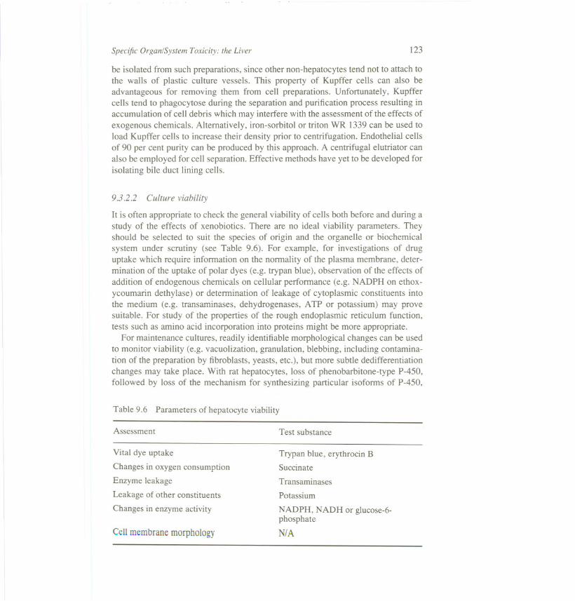
It is often appropriate to check the general viability of cells both before and during astudy of the effects of xenobiotics. There are no ideal viability parameters. Theyshould be selected to suit the species of origin and the organelle or biochemicalsystem under scrutiny (see Table 9.6). For example, for investigations of druguptake which require infonnation on the nonnality of the plasma membrane, deter-mination of the uptake of polar dyes (e.g. trypan blue), observation of the effects ofaddition of endogenous chemicals on cellular perfonnance (e.g. NADPH on ethox-ycoumarin dethylase) or detennination of leakage of cytoplasmic constituents intothe medium (e.g. transaminases, dehydrogenases, ATP or potassium) may provesuitable. For study of the properties of the rough endoplasmic reticulum function,tests such as amino acid incorporation into proteins might be more appropriate.
For maintenance cultures, readily identifiablemorphological changes can be usedto monitor viability (e.g. vacuolization, granulation, blebbing, including contamina-tion of the preparation by fibroblasts, yeasts, etc.), but more subtle dedifferentiationchanges may take place. With rat hepatocytes, loss of phenobarbitone-type P-4S0,followed by loss of the mechanism for synthesizing particular isofonns of P-4S0,
Table 9.6 Parameters of hepatocyte viability
Assessment Test substance
Vital dye uptake
Changes in oxygen consumption
Enzyme leakage
Leakage of other constituents
Changes in enzyme activity
Trypan blue, erythrocin B
Succinate
Transaminases
Potassium
Cell membrane morphology
NADPH, NADH or glucose-6-phosphate
N/A
124 Short-term Toxicity Testsfor Non-genotoxic Effects
appears to be one of the earliest changes to take place and might, therefore, beemployed as a viability parameter.The relative contributionof fetal type cells can bemonitored by biochemical indicatorssuch as oc-fetoprotein,alkaline phosphatase,andoc-glutamyltranspeptidase.No single viabilityparameter is likely to be adequate.
Antibiotics such as penicillin and streptomycin are usually added to cell culturesfor prolonged investigations. However, in particular studies where antibiotics mayinterfere (investigation of the transport of anti-cancer drugs or mixed-cell studieswith mutant bacteria, for example) it is possible to work without antibiotics, pro-vided that rigorous sterile techniques are employed.
Most maintenance-culture preparations use a solid surface onto which the cellsadhere and flatten. The material used may depend on the species from which thecells were removed. For rat hepatocytes, polystyrene or collagen-coated glass orfloats are generally used. Polycarbonate appears to be more effective for rabbithepatocytes.
The requirements for maintaining dividing hepatocyte-derived lines tend to berather similar to those for maintenance cultures, although it is important to remem-ber that dividing cells do utilize nutrients much more rapidly than non-dividingcells.
9.3.2.3 Current and potential in vitro techniques using liver cells
Hepatocyte cell preparations have several uses in toxicology. They can be used toexamine 'liver-specific' effects of chemicals. For this purpose, the hepatocytes mustrepresent as closely as possible their state in vivo. They can also serve as a generalmodel of the toxic effects in many other cell types.
Hepatocytes are a particular relevant model for chemicals which mediate theireffects wholly or partly through the formation of active metabolites. Hepatocytescan be used to provide a source of toxic metabolites, the biological effects of whichare monitored in a second cell type, or in the hepatocytes themselves.
Primary-maintenance cultures of adult rat hepatocytes frequently become pro-gressively overgrown with fibroblasts. A cytotoxicity test has been devised whichuses inhibition of fibroblast growth as an endpoint. Hepatocytes have also been usedsuccessfully as the metabolizing system in a number of mutagenicity tests.
A variety of methods may be used to characterize toxic effects. These methodsinclude:
(1) membrane change (permeability, active transport or surface-chemistrychanges);alterations in the synthesis, degradation, conformation or availability of macr-omolecules or growth-related factors;modifications in intermediary metabolism;changes in activity, growth, morphological (including organelle changes) orbehavioural characteristics.
(2)
(3)(4)
Specific Organ/System Toxicity: the Liver 125
Freshly isolated hepatocytes can often be used to identify and characterize rapidlydeveloping endpoints such as initial inhibition of protein and RNA synthesis orenzyme leakage. Primary-maintenance cultures, on the other hand, are employed tostudy more slowly developing lesions such as mitotic rate changes and identificationof sustained alterations in organelles.
It is oft~n possible to measure several endpoints produced by a chemical using asingle preparation of hepatocytes. Positive controls can be used to ascertain therelevance of any observed changes in a preparation.
The plasma membrane is the first part of the cell to be exposed to toxins and it is,therefore, not surprising that it is a common site of toxic damage. Determinants oftoxicity to the membrane include the uptake of polar dyes (such as trypan blue), co-factors (e.g. NADPH), or polar substrates (e.g. succinate). These are normally excludedfrom the cell by the plasma membrane. Leakage of cytoplasmic enzymes into thesurrounding media has also been used by many workers as an indicator of cytotoxicity.One great advantage of studying enzyme leakage in hepatocytes is that it is a parameterwhich is frequently studied in vivo. Morphological changes in the membrane (e.g.blebbing, or rounding up of cells) may also be used as cytotoxicity criteria.
Isolated hepatocytes provide a useful model for assessing the toxicological sig-nificance of lipid peroxidation reactions. Both the drug-metabolizing enzymes andthe various defense systems (e.g. catalase, superoxide dismutase, glutathione perox-idase and cellular antioxidants) bear the same quantitative relationship to one an-other as in vivo. However, few studies of lipid peroxidation have been made inisolated hepatocytes.
Many chemicals cause a significant inhibition of protein and RNA synthesisunder conditions in which little or no cytotoxicity is observed. Studies on proteinsynthesis inhibition in hepatocytes have so far been largely restricted to measuringreduced rates of 14C-Ieucine incorporation into acid-precipitable material.
Determination of the increased synthesis of macromolecules is liable to be a morespecific indicator of selective toxicity than measurement of synthesis inhibition. Manycarcinogens are known to damage DNA and therefore trigger increased unscheduledDNA repair. Hepatocytes provide a particularly relevant model in which to study sucheffects. DNA synthesis is minimal in hepatocytes in the absence of DNA-damagingagents (these cells have a very slow rate of cell division). This test is now widely used,with others, to identify genotoxic agents either in vitro or ex-in vivo.
Cells which have flattened out on a solid transparent surface such as collagen-coated glass or polystyrene are particularly amenable to the quantitative histochemicaldetermination of the activity of cellular enzymes. Application of quantitative histo-chemical techniques to the study of toxic effects of chemicals on enzyme activity incultured hepatocytes is still in its infancy, but early results are promising. For example,primary adult rat hepatocytes have been employed to investigate cytochrome P-450 in-duction by various barbiturates and benzanthracene. Primary maintenance cultures of rat
hepatocyteshavealso beenemployedto testfor peroxysomeproliferating agents.Sincethis class of chemical is known to cause hepatocarcinoma,but is not detected by
126 Short-term Toxicity Tests for Non-genotoxic Effects
presentgenotoxicitytests,the use of hepatocytesfor this purpose is important.
The identification of significant covalent binding often provides a valuablepointer to the role of active metabolites in toxicity. For example, the extent ofcovalent binding of 14C-labelled isosafrole metabolites to hepatocyte proteins iscomparable to that observed in vivo whereas covalent binding in liver microsomes ismuch higher. However, it is difficult to interpret the toxicological significance ofcovalent binding results unless indicators of cellular functional integrity are deter-mined in a parallel study.
Changes in cofactor levels often result from chemical insults. ATP may beespecially vulnerable. The formation of reactive metabolites may result in signifi-cant depletion of glutathione. For example, reactive metabolites of paracetamol andbromobenzene reduce glutathione levels in isolated adult rat hepatocytes, whichallows increased covalent binding to cellular proteins, thereby producing cytotox-icity. BCND also rapidly diminishes cellular glutathione. Isosafrole and safrole mayexert their toxic effects, at least in part, by a similar mechanism.
Rather little attention has been paid to the use of morphological end-points asindicators of toxicity in spite of the fact that histopathology is the predominanttechnique in conventional toxicology for identifying toxic changes. Hepatocytes inprimary culture display a very slow division rate. However, certain carcinogens canenhance the mitotic index.
A number of areas require further attention in order to improve in vitro techniquesfor toxicity studies. These areas include: the development of improved proceduresfor preparing and storing human hepatocytes; the extension of the useful lifetime ofprimary maintenance cultures in order to study the mechanisms of slow onsethepatotoxicity; the development of effective, synthetic culture media forhepatocytes; the identification of a means of preserving the constitutive P-450isoenzymes and other drug metabolizing enzymes at the levels and activities foundin vivo (this is particularly important for studies of metabolite mediated toxicity);development of suitable cell purification methods for the minor hepatic cell types, inparticular, bile duct lining cells (this may be especially relevant to the understandingof some forms of drug-induced jaundice); improved methods for following themovement of xenobiotics and xenobiotic-endogenous macromolecular complexes(e.g. drug-receptor complexes) within cells. Modem molecular biology techniquesalso allow the transference of hepatic drug metabolizing enzymes to other rapidlydividing cell types.
9.3.3 Cell homogenates and fractions
The primary hepatotoxic effects of a chemical are in many cases rather specific to aparticular macromolecule or organelle. Moreover, the ultimate toxin which manifeststhis primary effect is often created by particular isoenzymes of the drug metabolizingenzymes. Details of the processes involved are often best investigated using cellhomogenates or cell fractions. This approach can be applied to the investigation of
Specific Organ/System Toxicity: the Liver 127
toxicity in both in vivo and in vitro systems and has been used extensively to examinethe nature of the enzymes involved in the biotransfonnation of xenobiotics.
Methodological constraints involved in the use of cell homogenates and fractions are:(1) Homogenization of a tissue which contains several cell types may obfuscate
understanding of the contribution of minority cell types to the process underinvestigation. It may also lead to the unjustified assumption that all the cells ofthe majority cell type are homogeneous in function.
(2) Tissue homogenization can lead to artefactual changes in the homogenate due tohydrolytic destruction of membrane components and co-factors through theliberation of lysosomal enzymes. The homogenization procedure may also leadto activation or inhibition of various enzymes by other means. For example, thenature of the homogenizing medium used can greatly affect the functional stateof mitochondria and the mitochondrial inhibition of microsomal biphenyl 2- and4-hydroxylase activity when assayed in homogenates. Furthennore, compart-mentalized endogenous inhibitors may be liberated by homogenization.
(3) The levels of co-factors used in the incubations are generally optimal only forthe enzyme(s) under study and in all probability do not correspond with levelsexisting within the cell. Therefore, they may give an undue emphasis to thefonnation of certain metabolites which are of minor significance in vivo. Agood example here is the extremely high co-factor levels used in nitroreduc-tase assays. Also, fonnation of a toxic metabolite may be of little consequenceto the cell in which it is fonned if it is immediately bound to a non-essentialprotein or is rapidly conjugated and excreted.
(4) The use of isolated subcellular fractions will not necessarily reflect the totalcellular effects of a chemical if its effects involve interactions between a
number of different organelles.(5) Many toxic reactions depend on the entry of the xenobiotic into the cell and
cannot be assessed in isolated cell fractions. Hepatocytes are readily penneableto most lipophilic xenobiotics and, therefore, it is probably uncommon foruptake to be rate limiting for toxicity. However, for metals and certain drugs,this is an important consideration. For methotrexate, for example, active trans-port processes govern cellular uptake.
(6) Due consideration must be given to the importance, for the purpose of the exper-iment, of freedom from contamination and viability of the preparation being used.
(7) Where in vivo pretreatment with a chemical precedes the liver homogenizationand fractionation, attention must be given to the possibility that the treatmentmay modify the homogenization and separation conditions. For example, or-ganelle membranes may become more fragile and, therefore, destroyed by thehomogenization process.
Although techniques exist to separate and purify cellular components andenzymes, a number of difficulties must be overcome to improve their use in toxicol-ogy. These problems include:
128 Short-term Toxicity Tests for Non-genotoxic Effects
(I) Improved purity and storage of organelle preparations, particularly plasmamembranes, Golgi apparatus and CURL;Identification of sensitive indices of damage for each organelle;Development of techniques for transferring chemically pretreated organellesbetween cells in order to identify the relationship between primary and second-ary lesions;Improved techniques for investigating organelle interactions;Improved techniques for enzyme and receptor extraction and purification.
(2)(3)
(4)(5)
9.3.3.1 Mitochondria
Enzymes involved in energy production, carbohydrate metabolism, haem bio-synthesis, and the urea cycle are found in the mitochondria. There are a variety ofbiochemical parameters that can be used to assess mitochondrial function. In part,the effectiveness of these depends on the procedures used to isolate themitochondria. Assessment of ion transport or coupling of oxidative phosphorylationwith respiration are favoured methods of assessing viability.
Many of the enzymes involved in intermediary metabolism, including de-hydrogenases for pyruvate, malate and glutamate, are localized in the mitochondrialmatrix. Toxicant damage here has been demonstrated for agents such as arsenic,carbon tetrachloride, and methyl mercury. Trace metals such as arsenic, lead, mer-cury and cadmium inhibit mitochondrial respiration. For lead and arsenic, thisinhibition is relatively specific for NAD-linked substrates such as pyruvate/malate.
Three of the key enzymes in the haem biosynthetic pathway are associated withthe inner mitochondrial membrane. Ferrochelatase and o-aminolevulinic acid syn-thetase are highly sensitive to the action of toxic trace metals (resultant increases inthe urinary excretion of porphyrin precursors have proved to be useful in vivobiological indicators of this toxicity).
Mitochondrial swelling and contraction is caused by agents such as arsenic,carbon tetrachloride, and phosphate. These effects can be detected by measurementof light scattering in a spectrophotometer. This method is based on the increasedoptical density of mitochondria in a contracted state and decreased density in aswollen or orthodox configuration due to cation influx.
Movement of H+, Na+, K+ or Ca2+between isolated mitochondria and the sur-rounding medium may be monitored by specific ion electrodes. Changes in thetransport of these cations have resulted from exposure to mercurials and lead.Energy-dependent mitochondrial uptake of arsenic has also been demonstrated.
Biochemical changes resulting from in vivo exposure to arsenate, cortisone andmethyl mercury, as well as vitamin E deficiency, have been related to mitochondrialmorphometry.
Specific Organ/System Toxicity: the Liver 129
9.3.3.2 Lysosomes
Lysosomes are primarily involved in hydrolysis of endogenous materials. Activelysosomes (secondary lysosomes) may be cytochemically distinguished from inac-tive (teleolysosomes) or autophagic vacuoles by the presence of acid phosphataseactivity. Lability may be determined by assessing the leakage of hydro lases from thelysosomes. The various acid hydrolase activities in lysosomes are an importantmeans of assessing lysosome functionality. These assays are frequently performedon lysed lysosomes so that activities of the lysosomal enzymes may be more clearlyseparated from those present in the microsomal fraction. Marker enzymes frequentlymeasured are the cathepsins A, B, C and D, acid phosphatase, aryl sulphatase,glycosidases, and acid RNAase.
X-ray microanalysis has been used by several investigators to demonstrate thepresence of toxic trace metals within lysosomes following in vivo administration ofthe metal. Histochemical staining methods have also been used to deOmonstratelysosomal uptake of metals in cells of metal-exposed animals.
In vitro exposure of lysosomes to agents such as toxic metals or mycotoxins hasbeen found to alter the ability of lysosomes to perform the basic function of proteindegradation. Protein degradation by lysosomes has been followed by monitoring therelease of 1251from labelled proteins following either in vitro or in vivo incubation.Alterations of lysosomal membrane stability has been shown to occur on exposureof lysosomes to chemicals such as the retenoids.
9.3.3.3 Endoplasmic reticulum
The main function of the endoplasmic reticulum is protein synthesis. The endo-plasmic reticulum contains a series of ftavoproteins and cytochromes that functionin electron transport, ultimately resulting in the activation and reduction of molecu-lar oxygen. The endoplasmic reticulum incorporates a number of oxidative (e.g.P-450 isoenzymes) and conjugative (e.g. glucuronyl transferases) drug metabolizingenzymes. Oxidative, reductive and occasionally conjugation reactions may lead tothe formation of reactive electrophilic intermediates.
Viability of the endoplasmic reticulum can be assessed by measurement of mal-ondialdehyde levels and the activity of glucose-6-phosphatase, P-450 or cytochromeP-450 reductase.
Toxicological interest in the endoplasmic reticulum relates particularly to thepotential use of the proteins synthesized there as sensitive biochemical indicators of
toxicity and the insight it may give into the mechanism of toxic actions of particularchemicals. A common approach is to inject radiolabelled amino acids into intactanimals. The incorporation of the radiolabel into specific proteins is then deter-
mined. Subsequent protein purification is undertaken to assess synthesis/degradationof individual proteins.
Measurement of lipid peroxidation may be used as an index of oxygen or organic
130 Short-term Toxicity Testsfor Non-genotoxic Effects
free-radical production. Malondialdehyde is the most widely measured index oflipid peroxidation, although it is insufficient on its own as an index of the peroxida-tion process.
Covalent binding of radiolabelled chemical to the microsomes themselves or toadded macromolecules is an index of the reactivity of the chemical and/or itsmetabolites. In assessing covalent binding, it is most important to use exhaustivemethods (e.g. solvent extraction) to distinguish accurately between true covalentbinding and non-specific adsorption of the radiolabel.
9.3.3.4 Reconstituted and purified subcellular systems
The use of crude cell fractions allows only limited characterization of individualcomponents. Research in respect of xenobiotics has focused particularly on thepurification of the drug metabolizing enzymes. Studies have been or can be under-taken to assess substrate specificity of individual enzymes or identification of spe-cific sites of covalent binding, for example. A variety of techniques have been usedto purify individual components of the system and to reconstitute the electrontransport chain.
The main methods to purify cell fractions are detergent extraction, column chro-matography, and preparative electrophoresis. The success of the purification pro-cedure can be assessed in a number of ways but no single technique is sufficient.Sodium dodecyl sulphate (SDS)-polyacrylamide gel electrophoresis is a powerfultechnique -but has some limitations. Evidence has been presented that differentmicrosomal enzymes cannot always be distinguished by this technique. Further-more, the results obtained with this technique are rather dependent upon the exactprocedure used.
Isoelectric focusing offers a great potential for resolution of enzymes and hasbeen used in studying cytochrome P-450. However, a number of artefacts are relatedto the use of this methodology and, at the present time, one should view dataobtained using isoelectric focusing of membrane proteins with caution.
Antibodies have been used to examine the homogeneity of isolated enzymefractions, the multiplicity of enzymes in microsomes, and the amounts of individualenzyme forms in microsomal preparations. Other immunological techniques thatcan be used include double-diffusion analysis, radial diffusion quantitation, inhibi-tion of enzyme activity complement fixation, radioimmune assay, crossed gel elec-trophoresis, immunoprecipitation, immunoaffinity column chromatography andimmunohistochemical localization.
9.4 SUMMARY
No single test is now or is ever likely to be able to provide all the information that isrequired in order to assess the hepatotoxic properties of a chemical. Rather, a rangeof viable liver preparations is required from which can be selected the most appro-
Specific Organ/System Toxicity: the Liver 131
priate model(s) for the particular question being asked. As yet, in vitro methodswould not appear to be of much value in identifying delayed onset/idiosyncratichepatotoxicity. Liver fractions and hepatocytes are presently most usefully employ-ed once a hepatotoxic property of a chemical has been identified in vivo. These invitro preparations are used to determine the contribution of reactive metabolites tothe toxic process and ascertain the mechanism(s) of toxicity.
The rapid developments in methodology for handling isolated cells indicate that,in the near future, such in vitro preparations may also be suitable for the initial
screening of chemicals for acute hepatotoxic properties and for obtaining informa-tion on the relative response to a chemical of human liver compared with otherspecies.
9.5 RECOMMENDAnONS
In vitro methods to identify and characterize chemicals whose primary toxicity is tonon-hepatocyte liver cells should be approached by the development of cell isola-tion and separation techniques which will enable such cells to be cultured in a viablestate. This must be accompanied by improved liver perfusion methods in order toprolong the viability and reproducibility of perfusion systems. This work on tech-niques should be allied with detailed studies of the mechanisms of both acute anddelayed cholestasis and portal hypertension.
Improvements are needed in the methods for producing and storing viable pre-parations of human liver cells so that they reflect the properties of these cells in vivo.
Also required is standardization of criteria for reporting the purity, viability andreproducibility of in vitro preparations used in toxicology.
In view of the increasing evidence of large variations in human liver response dueto genetic (interindividual) differences, the development of human liver bank(s)should be encouraged in order to identify likely types of susceptible individuals.
Extending the lifetime of cultures and the development of totally synthetic culturemedia is needed. Unless this is achieved, delayed onset hepatotoxicity cannot beinvestigated with confidence in vitro. There is also a need for incorporation of thehepatic drug metabolizing system into a rapidly dividing cell type in order toexamine the effects of active metabolite generation on growth-related factors. Re-cent advances in genetic manipulation techniques should enable this to be achieved.
Improved monitoring techniques are required for both in vivo and in vitro sys-tems. For in vivo systems, the principal requirement is for identifying the mostappropriate indices of hepatic damage for each species which can be ascertainedfrom blood. Also, whole body NMR offers great potential to detect hepatic andother tissue changes at an early stage. For in vitro systems, the development ofquantitative cytochemical, immunochemical and lectin based identification methods
merit priority because of their specificity and their facility to provide information at
the individual cell level. Major progress here will depend on increasing understand-ing of mechanisms of hepatotoxicity.
132 Short-term Toxicity Testsfor Non-genotoxic Effects
Finally, effective training in in vitro methods is required for toxicologists whohave been exclusively concerned with in vivo tests.
BIBLIOGRAPHY
Bein, H.J. (1963). Rational and irrational numbers in toxicology. Proceedings of the Euro-pean Societyfor the Study of Drug Toxicity, 2, 15-26.
Bridges, J.W. (1980). Monooxygenase reactions glucuronic acid sulphate conjugation inisolated hepatocytes. Toxicology, 18, 195-204.
Bridges, J.W. (1981). The use ofhepatocytes in toxicological investigations. In: Gorrod, J.W.(Ed.), Testingfor Toxicity, Taylor and Francis, London, pp. 125-43.
Bridges, J.W., and Fry, J.R. (1978). Mammalian short-term tests for carcinogens. In: Dayan,A.D., and Brimblecombe, R.W. (Eds), Carcinogenicity Testing: Principles and Problems,Baltimore University Park Press, pp. 29-52.
Chayen, J., Bitensky, L., Johnstone, U., Gooding, P.E., and Slater, T.F. (1979). The applica-tion of microspectrophotometry to the measurement of cytochrome P-450. In: Pattison,J.R., Bitensky, L., and Chayen, J. (Eds) Quantitative Cytochemistry and Its Applications,Academic Press, London, pp. 129-37.
Elcombe, e. (1976). Ph.D. Thesis, University of Surrey.Enderlin, F.E., and Honohan, T. (1977). Long term bile collection in the rat. Lab. Anim. Sci.,
27,490-93.Fowler, B.A., Lucier, G.W., and Hayes, A.W. (1982). Organelles as tools in toxicology. In:
Hayes, A.W. (Ed.), Principles and Methods of Toxicology, Raven Press, New York,pp.509-60.
Home, D.W., Briggs, W.T., and Wagner, e. (1976). A functional, active transport system formethotrexate in freshly isolated hepatocytes. Biochem. Biophys. Res. Commun., 68, 70-76.
Johnson, P., and Rising, P.A. (1978). Techniques for assessment of biliary excretion andenterohepatic circulation in the rat. Xenobiotica, 8, 27-36.
Jones, D.P., Thor, H., Andersson, B., and Orrenius, S. (1978). Role of glutathione peroxidase,catalase and formaldehyde dehydrogenase in reactions relating to N-demethylation by thecytochrome P-450 system. J. Bioi. Chem., 253, 6031.
Knook, D.L., and Sleyser, E.C. (1979). Liver sinusoidal cells: Isolation and purification bycentrifugal elutriator. In: Reid, E. (Ed.), Cell Populations, Ellis Horwood, Chichester, pp.47-52.
Lentz, P.E., and Di Luzio, N.R. (1971). Biochemical characterization of Kupffer and par-enchymal cells isolated from rat liver. Exptl. Cell Res., 67, 17-26.
Lowing, R.K., Fry, J.R., Jones, e.A., Wiebkin, P., King, LJ., and Bridges, J.W. (1979). Theearly effects of chemical carcinogens on adult rat hepatocytes in primary culture: 1.Quan-titative changes in intracellular enzyme activities following a single dose of carcinogen.Chem.-Biol. Interactions, 24, 121-31.
Lowing, R.K., Fry, J.R., King, LJ., and Bridges, J.W. (1979). The early effects of chemicalcarcinogens on adult rat hepatocytes in primary culture: II. Effects on unscheduled DNAsynthesis, cell division and oc-fetoproteinproduction. Chem.-Biol. Interactions, 25, 303-19.
MacSween, R.N.M. (1980). Liver, biliary tract and exocrine pancreas. In: Anderson, J.R.(Ed.), Muir's Textbook of Pathology, Edward Arnold, London.
Mehendale, H.M. (1982). Application of isolated organ techniques in toxicology. In: Hayes,A.W. (Ed.), Principles and Methods of Toxicology, Raven Press, New York, pp. 509-59.
Miller, L.L., Bly, e.G., Watson, M.L., and Bale, W.F. (1951). The dominant role of the liverin lasma protein synthesis. A direct study of the isolated perfused rat liver with the aid oflysine-E-14-e. J. Exp. Med., 94, 431-53.
Specific Organ/System Toxicity: the Liver 133
Plaa, G.L., and Hewitt, W.R. (1982). Detection and evaluation of chemically induced liverinjury. In: Hayes, A.W. (Ed.), Principles and Methods of Toxicology, Raven Press, NewYork, pp. 407-45.
San, R.H.C., and Stich, H.F. (1975). DNA repair synthesis of cultured human cells as a rapidbioassay for chemical carcinogens. Int. 1. Cancer, 16,284-91.
Schmidt, F.W. (1977). Enzymes in cholestasis. In: Bianchi, L., Gerok, W., and Sickinger, K.(Eds), Liver and Bile, Falk Symposium, vol. 23, Baltimore University Park Press, pp. 203-14.
Seglen, P.O. (1979). Separation approaches for liver and other cell sources. In: Reid, E. (Ed.),Cell Populations, Ellis Horwood, Chichester, pp. 25-46.
Thor, H., Moldeus, P., and Orrenius, S. (1979). Metabolic activation and hepatotoxicity:effect of cysteine, n-acetylcystein and methionine on glutathione biosynthesis and brom-obenzene toxicity in isolated rat hepatocytes. Arch. Biochem. Biophys., 192, 405-13.
Tomlinson, P.W., Jeffery, D.J., and Filer, C.W. (1981). A novel technique for assessment ofbiliary secretion and enterohepatic circulation in the unrestrained conscious rat.Xenobiotica. 11, 863-70.
Wiebkin, P., Fry, J.R., and Bridges, J.W. (1978). Metabolism-mediated cytotoxicity of chemi-cal carcinogens and non-carcinogens. Biochem. Pharmacal., 27, 1849-51.
Woodman, D.D. (1981). Plasma enzymes in drug toxicity. In: Gorrod, J.W. (Ed.), Testing forToxicity, Taylor and Francis, London, pp. 145-56.