specialty training curriculum for clinical radiology: 2010 · radiology trainees will be expected...
TRANSCRIPT
1
SPECIALTY TRAINING CURRICULUM
FOR
CLINICAL RADIOLOGY
May 2010
The Faculty of Clinical Radiology The Royal College of Radiologists
38 Portland Place London W1B 1JQ
Telephone: 020 7636 4432
2
CONTENTS
1 INTRODUCTION 4
1.1 AIMS AND VALUES 6
1.2 CURRICULUM RATIONALE 8
1.3 HOW TO USE THE CURRICULUM 9
2 SYLLABUS AND COMPETENCES – CONTENT 13
2.1 THE SYLLABUS IN PRACTICE 15
3 SYLLABUS AND COMPETENCES 16
3.1 PHYSICS 16
3.2 ANATOMY 22
3.3 GENERIC CONTENT 27
A Behaviours in the Workplace 27 B Good clinical care 32 C Managing Long-term Conditions 42 D Infection control 44 E Patient safety within clinical governance 46 F Leadership/Management development 49 G Ethical and legal issues 53 H Maintaining good medical practice 59 I Teaching and training 65
3.4 RADIOLOGY SPECIFIC CONTENT 67
Breast Radiology 68 Cardiac Radiology 72 Emergency Radiology 78 Gastro-intestinal Radiology 83 Head and Neck Radiology 90 Musculoskeletal Radiology 95 Neuroradiology 100 Oncological Radiology 105 Paediatric Radiology 109 Radiology Procedural Skills 116 Radionuclide Radiology 121 Thoracic Radiology 132 Uro-gynaecological Radiology 138 Vascular Radiology 144 Academic Radiology 149
4 SUPPORT FOR LEARNING, SUPERVISION AND FEEDBACK 151
Work-based Experiential Learning 151
5 APPRAISAL 157
6 ASSESSMENT 160
3
7 ANNUAL REVIEW OF COMPETENCY PROGRESSION (ARCP) 164
8 APPENDICES 168
APPENDIX A: CURRICULUM IMPLEMENTATION AND MANAGEMENT 168
APPENDIX B: CURRICULUM REVIEW 170
APPENDIX C: STANDARDS FOR TRAINING IN CLINICAL RADIOLOGY 173
APPENDIX D: EXAMINATION POLICIES IN CLINICAL RADIOLOGY 175
APPENDIX E: ENSURING QUALITY IN CLINICAL RADIOLOGY 180
APPENDIX F: EQUALITY AND DIVERSITY 181
APPENDIX G: CHANGES SINCE 2007 183
APPENDIX H: CURRICULUM DEVELOPMENT 185
4
1 INTRODUCTION The Radiology Curriculum sets out the framework for educational progression that will support professional development throughout Specialty Training in Clinical Radiology. The curriculum defines the process of training and the competences needed for the successful completion of training in Clinical Radiology. The aim is to ensure that trainees are fully competent to provide a high quality service at consultant level in the NHS. The curriculum has been designed in line with the GMC Standards for Curricula and Assessment Systems (July 2008). There are sections detailing the planning, content, delivery, outcomes and review of the curriculum. With respect to the content, generic, professional and specialty specific areas are included.
Good Medical Practice The Generic Syllabus Content is based on Good Medical Practice (GMP) 2006, as outlined by the General Medical Council (GMC). Radiology trainees will have a chance to show both the confidence and competences necessary to develop increasing levels of expertise in their subsequent clinical and professional practice.
Outcomes of radiology training This programme will allow radiologists in training to apply their knowledge and skills in the workplace and demonstrate improving performance to the level that will satisfy the needs of the GMC for completion of training and fulfil the requirements for a Certificate of Completion of Training in Clinical Radiology, making them eligible to apply for entry to the GMC Specialist Register and then to take up consultant posts.
This curriculum is intended to be used by radiologists in training, those delivering their education and those responsible for quality assurance (national), quality management (deanery) and quality control (local education provider).
How to use this Curriculum
It is strongly recommended that the section How to use this Curriculum
is read thoroughly by all.
Key messages of the Curriculum
Patient Safety • Must be placed at the centre of healthcare
• High quality patient care depends, among other aspects of practice, on effective multidisciplinary team working
• Learning in, and from, clinical practice is the most effective way for professionals to develop much of their expertise.
Personal development • Radiologists are committed to lifelong learning in, and from, the practice of
radiology in the clinical environment and through repeated clinical experience.
5
Radiology trainees will be expected to develop critical thinking and professional judgement, especially where there is clinical uncertainty
• Every clinical experience is a learning opportunity and should be reflected upon from the perspective of developing skills, acquiring clinical/radiological acumen and improving performance. By doing this, an individual demonstrates their commitment to lifelong learning and continuing professional development.
• Doctors must continuously work to improve performance, ie improve what they actually do as distinct from what they are capable of doing.
Assessment The emphasis of Radiology training is on developing radiologists who are safe in their judgements, patient-focused and accountable to the public for delivering evidence based, effective medical care. The concept of "competent" requires the integration of different types of knowledge, skills and attitude in a pressurised, but supervised, clinical environment.
Objective assessments Workplace based assessments (WpBA) will take place at regular intervals throughout training. The assessment tools are designed to help doctors develop and improve their performance. Feedback is a key factor to enable this to happen.
Throughout their careers, doctors should strive to improve their performance to ensure their progression from competence, through proficiency, to expertise. The vast majority of radiology trainees will have no difficulty with their assessments. When problems are identified, the trainee will be encouraged to work to find solutions with the support of their clinical and educational supervisors.
ePortfolio The ePortfolio will be a record of a trainee’s progress and development through radiology training. It will provide a record of objective evidence of competence to work in a range of clinical settings and a record of satisfactory performance. This means that ePortfolio completion will contribute to the end of year report, annual review of competence progression (ARCP) and may also be used in interviews. Successful completion of the curriculum requires the achievement of competence in a variety of domains relating to generic medical practice, radiological and clinical practice. The assessments of these competences will be recorded in the ePortfolio. This revised curriculum updates the document revised in 2007. It emphasises the importance of supervised, practice-based learning. Dr David Lindsell Warden of the Faculty of Clinical Radiology The Royal College of Radiologists
6
1.1 AIMS AND VALUES
Aims The over-arching aims of the curriculum are to represent a distillation of the values and attributes attainable by radiologists passing through training programmes implementing and embracing the educational potential of clinical radiology. These can be summarised as follows:
• Sufficient knowledge and skills to undertake the practice of Clinical Radiology at Consultant level.
• A professional attitude to all aspects of clinical practice, which places good conduct at its centre.
• Sound judgement through intelligent application of knowledge.
• A sense of team-working within all spheres of practice.
• An insightful approach: knowing individual/collective strengths and limitations, when to be decisive and when to seek help.
• An enthusiasm for knowledge and understanding to support lifelong learning.
• A reflective attitude allowing accurate self-assessment and learning from practice.
• The abilities necessary to provide improved patient care.
Values Set out below are the values considered to be of importance in the teaching, learning and practice of Clinical Radiology. In clinical practice, there is little or no distinction between the sub-headings of practical, educational and professional values. The sub-division is simply for emphasis and clarity.
Practice Values for Clinical Radiology • A recognition that Clinical Radiology is not merely technical specialty but a
specialty of medical practice concerned with diagnosing and treating patients and, therefore, requires practitioners with all the attributes of a good doctor.
• Clinical Radiology has good conduct at the heart of its practice.
• Through sound judgement Clinical Radiological practice can improve patient management and outcome.
• Good communication is an essential component of sound practice.
• Clinical Radiology is a dynamic medical specialty that must work with other medical and surgical specialties to respond to the needs of patients.
7
• Clinical Radiology relies on a multi-professional team and so radiologists should work with other healthcare professionals to put the needs of the patient above their own.
• As a medical practitioner, clinical radiologists have a responsibility to question the decisions of others if they believe it undermines the best care of the patient.
Educational Values for Clinical Radiology • Recognition of the importance of nurturing a professional attitude (see below)
to complement the knowledge and skills required for good practice.
• Teaching that recognises the importance of understanding in the creation of knowledge.
• Knowledge should not be assumed.
• The need and desire to establish educational partnerships.
• Flexibility to tailor teaching to the needs of the learner as agreed between both teacher and learner.
• Recognition of the need for a variety of educational methods to suit the learner and the context of learning.
• Wherever practical, set teaching in the practice setting and teach theory within practice.
• Establish early learner motivation towards an attitude of self-sufficient life-long learning and development.
• Recognition of the educational potential of reflective practice with self, peers and teachers as a means to constructive self criticality.
• Recognition of the multi-faceted nature of Clinical Radiological practice so exposing learners to the many special interests within Clinical Radiology as well as those that support it, such as research, audit, management and teaching.
• Understanding and recognition that knowledge is not merely acquired for perpetuity but is a developmental process of increasing sophistication.
• A desire to commit to the dynamic nature of radiological practice and its teaching, so seeing the curriculum as an evolutionary document.
• Recognition that hierarchy can be detrimental to education.
Professional Values for Clinical Radiology: • To be accountable for individual/collective actions.
• Develop a clear understanding of individual abilities and limitations.
8
• Be honest in all aspects of Clinical Radiological practice even, and especially, in times of adversity.
• To strive to develop and practise sound judgment.
• Show respect towards patients and colleagues.
• Maintain individual skills, knowledge and values throughout one’s career
1.2 CURRICULUM RATIONALE Radiology trainees are developing professionals and need to deepen and broaden their understanding and expertise. This means
• revisiting clinical and professional practice, and studying at increasingly complex levels
• practising with decreasing supervision
• recognising that levels of expertise generally increase with practice and reflection.
In order to become effective, clinical radiologists must improve the diagnosis and management of their patients. To do this, radiology trainees need a broad knowledge base and extensive experience. The curriculum needs to take account of the fact that "common things commonly occur" and that these need to be within the repertoire of all radiologists. Uncommon diagnoses are possible and need to be suspected when there is something unusual in the patient’s presentation. These higher level competences are addressed within level 1/2 training. As with all doctors, radiology trainees should never stop learning and continuing their professional development. They should continue to refine their clinical skills and techniques and the quality of their interactions with others. This includes encouraging self care and shared decision making with patients, relatives and colleagues. It is probably as important for them to understand their personal style, assumptions and beliefs (and to change them when appropriate), as it is to develop their procedural and clinical/radiological skills. Radiology trainees are responsible for their own learning. At the same time, they must understand the needs of the patient and of the organisation in which they work. They should understand the complexities, constraints and opportunities they find in their practice, and be able to choose how to make best use of these. They also need to understand that, as well as engaging in more formal educational activities, they learn by working with other team members. They must learn how to contribute to the safe practice of radiology. Good educational practice acknowledges the private and public aspects of professional development, and gives due importance to the key relationships that inform professional development. Effective learners will achieve their aims, acknowledging that who they are and what they believe affects what they do. Effective educational practice will help radiology trainees to understand the relationship between theory and reality, which will enable them to exercise better judgement in complex situations. They will also be encouraged to understand other
9
roles within the team and show how they can adapt and collaborate in emergency situations. They will need to become aware of the different perspectives and expertise that can improve problem solving, clinical reasoning, patient management and decision-making. Acquiring expertise that can be adapted to new situations depends on the development of clinical /radiological and ethical reasoning and professional judgement. Much learning occurs in teams and much knowledge and expertise is found in groups rather than in individuals. This strengthens the principle that learning in Clinical Radiology should take place in team-based practice. Expertise is more than knowledge or a tool kit of skills. The radiology trainee will learn similar skills in different settings, facilitating the development of transferable skills.
1.3 HOW TO USE THE CURRICULUM
Trainee radiologist To make the most of the opportunities available in radiology training you need to have an appreciation of how the curriculum works. The curriculum assumes that all doctors will be proactive and organised in managing their continuing education. The first steps are to understand • The purpose of radiology training
Please read the Introduction and Aims and Values sections. • How you will be supported educationally
Read the sections on Support for Learning, Assessment and Feedback and Appraisal. Understand the system of workplace based learning and other educational opportunities that should be made available to you.
• Radiology training
Most training programmes offer a variety of training opportunities badged according to imaging modality and/or body systems. Not every trainee is expected to rotate through every attachment. Trainees, educational supervisors and training programme directors should compile rotations that cover the core and, wherever possible, reflect each trainee’s special interests. Trainees will have the opportunity to cover many aspects of the neurology and oncology curriculum during other attachments, such as CT, MR and ultrasound, or vice versa.
• Focussed individualised training (FIT)
Trainees with a particular area of special interest, on entering training in radiology, following discussion with their training programme director, can be offered focussed individualised training (FIT), if suitable and educationally deliverable. This will ensure that they can sample their preferred area early in training to confirm/affirm their interest and ability. In addition, they can, where possible, spend time in their special interest area during each rotation (eg one or two sessions a week).
• What you are expected to achieve
Review the Syllabus and Competences section, looking at the main domains/headings applied to groups of competences in relation to the relevant presentations and diagnoses. Get an idea of what you should be aiming to achieve over the programme. You should distinguish between core, level 1 and level 2 competence.
10
• How your competence will be assessed in the workplace
Competency assessment in radiology training is outlined in the Support for Learning, Supervision and Feedback and Assessment sections. You should familiarise yourself with this especially the ethos of reflective learning and feedback.
• Workplace based assessments (WpBA)
Participation in workplace based assessment (WpBA) is mandatory. A minimum number of WpBA is specified in order to progress. It is expected that most trainees will undergo many more assessments demonstrating their engagement with reflective learning in practice. Workplace based assessors will include all those individuals involved in the delivery of training. This includes consultants, senior trainees and advanced radiographic/sonographic practitioners. It is expected that at least 50% of WpBAs will be undertaken with consultants. Each WpBA should also be considered developmental and an opportunity for learning and feedback.
• How to record your progress in the ePortfolio
You should enrol with the Royal College of Radiologists prior to the commencement of your training. This will, amongst other things, allow you access to your ePortfolio. You need to become familiar with the ePortfolio as a record of learning.
• Reflective Practice
Radiologists should learn from both their positive and negative experiences, demonstrate consistent good performance and record their achievements and concerns in their ePortfolio. Reflective practice has the potential for demonstrating evidence of on-going self appraisal of aspects of clinical practice, not currently assessed in the syllabus.
Educational Supervision At the start of your specialty training, and of every rotation to a new education provider, there should be a local induction, which further introduces the programme and how it is delivered and assessed by the education provider. There should be further induction sessions at the start of each placement. At the first Educational Supervision session, you may wish to discuss aspects of curriculum delivery with your educational supervisor. These might include
• known strengths from undergraduate and early clinical training • particular areas of interest to you • any potential weaknesses that you feel may need addressing.
You should agree to follow the appraisal system and associated timelines for ongoing educational supervision, as well as undertaking the required assessments. This is signed off by both trainee and educational supervisor in the form of an educational agreement.
Core, Level 1 and Level 2 competences The curriculum recognises core, level 1 and level 2 competences. It is expected that you will acquire more competences as you progress through training. It is important to monitor the progression and the achievement of competences from the
11
outset of training. Trainees should familiarise themselves with the ARCP decision aid at the start of training so they are aware of what is required of them throughout each stage of their training. See the Assessment and ARCP sections below. Each trainee should strive to achieve as highly as possible but it is recognised that learning occurs at different rates in each individual. Many trainees are expected to achieve level 1 or 2 in some areas during core training. It is not expected that every trainee acquires every competence or covers every area. 1. Core training (indicative Years 1-3)
All trainees are expected to reach core competence, as this reflects what is likely to be required by any radiologist performing acute imaging.
2. Higher training (indicative Years 4-5)
Levels 1 and 2 competence indicate the greater degree of expertise to be achieved by those intending to practice with multiple or mono-special interest areas.
Level 1 All radiologists would probably hold level 1 in at least two areas. They would be able to practice as a consultant with a special interest in these areas. Radiologists with other specialist interests would be expected to consult them for advice within their disciplines. Level 2 A radiologist with level 2 competence would be likely to be a mono-specialist and an expert in their field. He/she is likely to be consulted by radiologists within the same discipline.
When engaged in reflection, formal assessment or self assessment, it is recommended that you again refer to the framework of competences to check your progress against the range of competences that you are expected to achieve. If you experience any difficulties with this, your educational and clinical supervisors are there to help you.
Trainer Please read the Introduction and How to use the curriculum: Trainee Radiologist sections. The definition of roles can be found at: http://www.rcr.ac.uk/docs/radiology/pdf/SAC%20Definition%20of%20Roles.pdf Your roles will vary and may involve teaching and making available other learning opportunities in the workplace, contributing to other forms of learning, providing workplace based assessments and clinical supervision, providing educational supervision and ensuring patient safety within the learning environment. You should be supported in your role by your Local Education Provider (LEP) and your Radiology School and should have received training for all the different roles that contribute to postgraduate education. There should be adequate time within your job plan to carry out your agreed postgraduate training roles to a high quality standard.
12
Learning in the radiology department
Overview The main themes of the curriculum are core competency (Years 1-3), and development of special interest (Years 4 and 5). Satisfactory performance in professional practice will be expected throughout. Formative workplace based assessments will enable overall competency and performance to be judged and will be the basis of much of the assessment of generic skills and competences such as good medical practice, clinical care, professionalism and leadership. Other learning environments, such as e-learning, textbooks, journals, short courses and simulation activities, should also be used. During a radiological attachment, the trainee radiologist should select topics on which to be assessed from the relevant list of presentations/diagnoses contained within the syllabus. A range of assessment tools will be used. The trainee radiologist and clinical or educational supervisor should ensure that a wide selection of core problems is formally assessed over the course of each attachment. More details about the assessment methodology appear in the Support for Learning, Supervision and Feedback and Assessment sections.
Practical procedures Radiologists perform many practical procedures during their day to day work. Some of these relate to imaging techniques such as ultrasound; others are peripheral to the technique, such as insertion of intravenous canulae, nasogastric tubes etc. Other procedures are interventions or therapies in their own right. Throughout the curriculum, interventions are included in the Procedural Skills section. Thus, trainees and trainers should refer to this section to find details of core and levels 1 and 2 procedural competences.
13
2 SYLLABUS AND COMPETENCES – CONTENT
PHYSICS
ANATOMY
GENERIC CONTENT
A Behaviours in the workplace
A.1 Professionalism A.2 Working with colleagues A.3 Relations with patients A.4 Personal qualities
B Good clinical care
B.1 History taking B.2 Written records B.3 Overall clinical judgement B.4 Time management and decision-making B.5 Therapeutics and safe prescribing B.6 The use of sedation and analgesia B.7 Breaking bad news
C Managing long-term conditions
D Infection control
E Patient safety within clinical governance
E.1 Risk management E.2 Quality improvement and patient safety
F Leadership/Management development F.1 Leadership F.2 NHS structure F.3 Media awareness
14
G Ethical and legal issues
G.1 Medical ethics and confidentiality G.2 Valid consent G.3 Legal framework of medical practice G.4 Equality and diversity
H Maintaining good medical practice
H.1 Insight H.2 Lifelong learning H.3 Ethical research H.4 Evidence based practice H.5 Clinical governance and audit H.6 Information technology
I Teaching and training
RADIOLOGY SPECIFIC CONTENT
Breast Radiology
Cardiac Radiology
Emergency Radiology
Gastro-intestinal Radiology
Head and Neck Radiology
Musculoskeletal Radiology
Neuroradiology
Oncological Radiology
Paediatric Radiology
Radiology Procedural Skills
Radionuclide Radiology
Thoracic Radiology
Uro-gynaecological Radiology
Vascular Radiology
Academic Radiology
15
2.1 THE SYLLABUS IN PRACTICE The syllabus sets out what radiologists need to learn in order to be able to manage a wide and varied caseload and to work adaptively in healthcare teams. These competences may be acquired in a variety of radiological settings. Radiology trainees should emerge with the professional qualities, understanding, critical perspective and ability to reflect on and in practice. Throughout their training, it is important that radiology trainees should be encouraged to reflect on decisions, management plans and actions taken. In discussion with their supervisors, they will be expected to discuss the thinking and reasoning behind them. At all times radiology trainees will: • practise within their competence level • practise in accordance with the standards expected of them in the unit in which
they are placed • always refer to more experienced radiology colleagues/teachers/mentors when
they are uncertain as to the best management of a particular patient • practise according to prevailing professional standards and requirements. Outcomes The outcomes and competences described for core training should be achieved by the end of the third year of training. The appropriate level 1 and 2 outcomes and competences should be achieved by the end of the fifth year of training. A key feature of the clinical radiology curriculum is that all radiology trainees must develop competences at an ever increasingly higher level during the course of their training. Radiology trainees will need to find out about the specific learning opportunities offered by the various specialty placements. Evidence of the radiology trainee’s learning, development and achievements will be recorded in the ePortfolio. Further information and declaration forms for probity, professional behaviour and personal health can be found in the ePortfolio. The following section outlines what needs to be learnt in the Clinical Radiology Training Programme. Throughout this section, the terms "patient" or "carer" should be understood to mean "patient", "patient and parent", "guardian", "carer", and/or "supporter" or "advocate" as appropriate in the context.
16
3 SYLLABUS AND COMPETENCES
3.1 PHYSICS 1 INTRODUCTION
1.1 The purpose of the curriculum is to provide those undertaking specialty
training in clinical radiology with appropriate knowledge of the physical principles that underpin diagnostic medical imaging. When linked with other training in clinical radiology, this should lead to the safe and effective application of diagnostic imaging for the benefit of patients.
1.2 It is intended that the curriculum should be delivered during the first year of
specialty training. This is expected to take about 40-45 hours of formal physics teaching, during the early months of specialty training, supplemented by practical training and private study of material recommended by trainers. Basic knowledge of physics and mathematics is assumed.
1.3 Assessment is in the form of a written multiple choice question (MCQ)
paper, which is a component of the First FRCR Examination in Clinical Radiology. Further detail is available on the College's website: www.rcr.ac.uk/content.aspx?PageID=175.
2 AIMS OF THE CURRICULUM
2.1 Provide appropriate knowledge of the physical principles that underpin the
following diagnostic medical imaging modalities: planar (projection) x-radiography, x-ray fluoroscopy, x-ray computed tomography (CT), ultrasound imaging, magnetic resonance imaging (MRI), planar (projection) radionuclide imaging, single photon emission computed tomography (SPECT) and positron emission tomography (PET).
2.2 Describe how the concepts of risk, safety and quality apply in these
imaging modalities including the responsibilities of individuals and organisations.
2.3 Provide sufficient understanding of the principles underlying each imaging
modality to enable selection of the most appropriate modality for a particular clinical situation, to select the optimal operating factors, to interpret the images produced, to communicate the results and to discuss the complete imaging process with professional colleagues.
2.4 Assist trainees to satisfy the requirements for adequate training in order to
carry out professional roles in medical diagnostic imaging as specified by UK legislation and guidance.
3 LEARNING OBJECTIVES Those who have followed the curriculum should be able to:
17
3.1 Describe the structure and properties of matter, the phenomena of radioactivity and magnetism, the nature of ionising radiation, radiofrequency radiation and ultrasound and how they interact with matter.
3.2 Distinguish between different types of diagnostic medical image and
understand how such images are created, reconstructed, processed, transmitted, stored and displayed.
3.3 Describe the construction and function of medical imaging equipment
including the radiation or ultrasound source, image-forming components and image or signal receptor.
3.4 Indicate how imaging equipment is operated and describe the imaging
techniques that are performed with such equipment. 3.5 Identify the type of information contained in images from different
modalities. 3.6 Distinguish between different indices of image quality, explain how they
are inter-related and indicate how they are affected by changing the operating factors of imaging equipment.
3.7 Identify agents that are used to enhance image contrast and explain their
action. 3.8 Explain how the performance of imaging equipment is measured and
expressed. 3.9 Describe the principles of quality assurance and outline how quality control
tests of imaging equipment are performed and interpreted. 3.10 Recognise artefacts in medical images and identify how they are removed
or their impact is reduced. 3.11 Recognise the hazards and risks to patients, members of staff and
members of the public associated with medical imaging and describe how their impact is reduced without compromising diagnostic image quality.
3.12 Identify the major pieces of UK legislation and guidance that affect the
practice of medical imaging and interpret their requirements. 4 SYLLABUS CONTENT
The syllabus is intended as a guide and general indication to the breadth of the topics that may appear in the examination questions. It is not a teaching plan and the bullet points do not relate to equal amounts of study time. The syllabus should be studied to a depth sufficient to allow the learning objectives in Section 3 above to be achieved.
4.1 Principles of medical diagnostic imaging
• Projection (planar) and tomographic images • Analogue and digital images • Structure of digital images • Digital image processing, fusion, transmission and storage
18
• Display and viewing of analogue and digital images • Picture Archiving and Communications Systems (PACS) • Quality assurance
4.2 Common themes for all imaging modalities
• Image formation • Image quality - contrast, noise, contrast resolution and spatial resolution • Contrast agents • Image processing and analysis • Equipment performance measurement, test objects and quality control • Image artefacts • Hazards, risks and safety
4.3 Matter and radiation
• Structure of matter, the atom and the nucleus • Nature and properties of charged particle and electromagnetic radiation • Interaction of electrons with matter • Production of x-rays • Interaction of high energy photons with matter • Filtration of x-ray beams • Electron energy in solids • Luminescence
4.4 Ionising radiation dose
• Absorbed dose and kinetic energy released to matter • Effects of ionising radiation on living tissue • Equivalent dose and effective dose • Radiation risk • Population dose from natural and artificial sources
4.5 Radiography with x-rays
• Construction, function and operation of computed and digital radiographic systems
• X-ray tube and x-ray beam • Image receptors for computed and digital radiography • Scatter rejection • Contrast media – iodine, barium and air • Dual energy radiography • Film-screen radiography • Mammography • Radiographic tomography and tomosynthesis
4.6 Fluoroscopy with x-rays
• Construction, function and operation of a fluoroscopy system • Image receptor – image intensifier and flat panel detector • Scatter rejection • Automatic brightness control • Image digitisation • Angiography with contrast media, including digital subtraction
techniques 4.7 Safety in radiography and fluoroscopy with x-rays
• Radiation detectors and dose meters
19
• Measurement of absorbed dose and dose rate in air • Estimation of patient absorbed dose • Typical dose-area products, entrance surface doses and effective doses
in radiography and fluoroscopy • Detector dose indicators • Factors affecting radiation dose • Time, distance and shielding for dose reduction • Children and pregnant patients • Estimation and control of radiation dose to staff and members of the
public • Operational dose quantities • Personal dosimetry • Pregnant staff
4.8 Radioactivity
• Nuclear stability • Mechanisms of radioactive transformation • Nuclear energy states and gamma emission • Activity and radioactive decay • Natural radioactivity • Artificial radionuclides and their production • Radiopharmaceuticals and their production
4.9 Planar radionuclide imaging
• Construction, function and operation of a digital gamma camera • Imaging collimators • Image receptor – scintillation detector • Scatter rejection • Mechanisms and quantification of radiopharmaceutical localisation • Static, whole-body, dynamic and gated imaging
4.10 Safety in planar radionuclide imaging
• Activity measurement with radionuclide calibrator • Estimation of patient absorbed dose • Typical activities and effective doses • Factors affecting radiation dose • Time, distance and shielding for dose reduction • Children and conception, pregnancy and breast-feeding in patients • Estimation and control of radiation dose to staff and members of the
public • Pregnant staff • Contamination and environmental dose rate monitoring • Storage, handling and transportation of radioactive substances • Storage and disposal of radioactive waste
4.11 UK framework for ionising radiation protection
• Hierarchy of recommendations, legislation and guidance • Justification, optimisation and dose limitation • Ionising Radiations Regulations 1999 and Approved Code of Practice • Risk assessment, restriction of exposure and dose monitoring • Radiation Protection Adviser and Radiation Protection Supervisor • Local Rules and work procedures • Designation of working areas and classification of workers
20
• Dose limits and dose constraints • Comforters and carers • Ionising Radiation (Medical Exposure) Regulations 2000, Notes on
Good Practice and 2006 amendment • Duty holders and their training and responsibilities • Employer’s procedures • Diagnostic reference levels • Exposures for research, health screening and medico-legal purposes • Medicines (Administration of Radioactive Substances) Regulations
1978 and 1995 and 2006 amendments • Administration of Radioactive Substances Advisory Committee and
Notes for Guidance • Radioactive Substances Act 1993 • Registration to hold radioactive substances • Authorisation to store and dispose of radioactive waste • Medical and Dental Guidance Notes • Notification and reporting of radiation incidents
4.12 Tomographic reconstruction
• Angular and linear sampling of projection data • Filtered back-projection and reconstruction filters • Iterative reconstruction
4.13 X-ray computed tomography
• Construction, function and operation of a CT scanner • Helical and multi-slice scanners • Image reconstruction • CT angiography, CT fluoroscopy and gated imaging • CT perfusion • Radiation dose to patients, staff and the public • Radiation safety and factors affecting radiation dose
4.14 Single photon emission computed tomography
• Construction, function and operation of a rotating multi-head gamma camera
• Image reconstruction • SPECT/CT • Radiation safety and factors affecting radiation dose • Typical activities and effective doses to patients, staff and the public
4.15 Positron emission tomography
• Construction, function and operation of a multi-detector ring system • 2D and 3D acquisition • Image reconstruction • Standardised uptake value (SUV) • PET/CT • Radiation safety and factors affecting radiation dose • Typical activities and effective doses to patients, staff and the public
4.16 Nuclear magnetic resonance
• Nuclear spin angular momentum and nuclear magnetic moment • Bulk magnetisation and the effect of magnetic field strength • Precession in a magnetic field and the Larmor equation
21
• Resonance with radiofrequency pulses • Relaxation mechanisms and relaxation times • Free induction decay signal
4.17 Magnetic resonance imaging
• Construction, function and operation of a superconducting MRI scanner • Permanent and resistive magnets • Radiofrequency receiver coils • Spin-echo pulse sequence • Spatial localisation of the signal • K-space, image acquisition and image reconstruction • Multi-echo, fast spin-echo and single shot techniques • Gradient echo imaging – basic spoiled and non-spoiled techniques • Tissue suppression methods – short TI inversion recovery (STIR), fluid
attenuated inversion recovery (FLAIR) and fat saturation • Standard gadolinium extracellular space contrast agents • Magnetic resonance angiography (MRA) • Basic principles of diffusion techniques and diffusion weighted imaging • Dynamic contrast enhancement and perfusion imaging • Principles of magnetic resonance spectroscopy (MRS) • Spatial misregistration, chemical shift, susceptibility, motion, flow and
other artefacts 4.18 Safety in magnetic resonance imaging
• Static magnetic field – projectiles, induced voltage, implants • Fringe field and controlled area • Time-varying gradient fields – eddy currents, stimulation, implanted
devices, acoustic noise • Radiofrequency fields – specific absorption rate, heating • Safety of patients, staff and members of the public • Pregnant patients • Shielding and imaging room design • Safety Guidelines for Magnetic Resonance Imaging Equipment in
Clinical Use
4.19 Physics of ultrasound • Nature and properties of ultrasound • Propagation and interaction of ultrasound in matter • Scattering of ultrasound waves • Piezoelectric effect • Design and construction of ultrasound transducers • Continuous and pulsed wave ultrasound • Beam shape from a single transducer and an annular array • The Doppler effect
4.20 Ultrasound imaging
• A-mode and B-mode imaging • Time-gain compensation • Construction, function and operation of a real-time B-mode scanner • Image acquisition and reconstruction • M-mode • Microbubble and particle suspension contrast agents • Harmonic imaging
22
• Measurement of flow with continuous and pulsed Doppler ultrasound • Duplex scanners • Colour-flow and power Doppler imaging
4.21 Safety in ultrasound imaging
• Physical effects - heating, streaming, cavitation and mechanical damage
• Intensity and energy limits • Thermal and mechanical indices • Measurement of power output • Safety of patients, staff and members of the public • Safety guidance
4.22 Functional and molecular imaging (FMI) • Meaning and principles of functional imaging and molecular imaging • Biological and physiological processes – flow, perfusion, diffusion,
uptake, excretion etc • Comparison of imaging modalities for FMI – sensitivity, spatial
resolution etc
3.2 ANATOMY 1 INTRODUCTION 1.1 The purpose of the curriculum is to provide those undertaking specialty
training in clinical radiology with appropriate knowledge of the anatomy needed to perform and interpret radiological studies. When linked to other training in clinical radiology, this will lead to the safe and effective application of diagnostic imaging for the benefit of patients.
1.2 It is intended that the curriculum should be delivered during the first year of
specialty training. This is expected to take about 30 hours of focused anatomy teaching, over a period of about six months, supplemented by practical training and private study of material recommended by teachers. Basic knowledge of anatomy is assumed.
1.3 Assessment is in the form of an electronic image viewing session, which is
a component of the First FRCR Examination in Clinical Radiology. Further detail is available on the College's website: www.rcr.ac.uk/content.aspx?PageID=175.
1.4 A knowledge of radiological anatomy is fundamental to the study of
radiology. The standard and level of anatomical knowledge tested and expected reflect the time available for training. The assessment is of knowledge of radiological anatomy – not surgical anatomy, surface anatomy or cadaveric anatomy – but applied anatomy that is relevant to clinical radiology.
2 AIMS OF THE CURRICULUM 2.1 Provide appropriate knowledge of the anatomy that underpins all
radiological imaging including radiography, fluoroscopy, computed
23
tomography (CT), ultrasound imaging and magnetic resonance imaging (MRI).
2.2 Provide sufficient understanding of the radiological anatomy that is visible
on each imaging modality to perform and interpret studies including communicating the results and discussion with clinical colleagues.
3 LEARNING OBJECTIVES Those who have followed the curriculum should be able to: 3.1 Describe and recognise the bony and soft tissue anatomy visible on
radiographs, including common normal variants. This will include children of all ages.
3.2 Describe and recognise the radiological anatomy visible on CT, including
multiplanar reformats. This will include solid organs such as the heart and lungs, bones, vessels and muscles.
3.3 Describe and recognise the radiological anatomy visible on ultrasound
imaging, including first trimester antenatal ultrasound. This will include solid viscera such as the liver and spleen, bones, vessels, major ligaments and tendons. Endocavity ultrasound, such as transvaginal, transrectal and endoscopic ultrasound, will be excluded.
3.4 Describe and recognise the radiological anatomy of MRI, including solid
viscera such as the brain and abdominal organs, bones, joints, muscles and vessels.
3.5 Describe and recognise the radiological anatomy of fluoroscopic studies of
the gastro-intestinal, biliary, genito-urinary and vascular systems. NB: Nuclear medicine, including positron emission tomography, is excluded
from the anatomy curriculum. 4 SYLLABUS CONTENT This syllabus is intended as a guide and general indication to the breadth of the
topics that may appear in the examination questions. It is not a teaching plan and the bullet points do not relate to equal amounts of study time. The syllabus should be read in conjunction with the learning objectives in Section 10 above.
1 Head & Neck 1.1 Brain • Ventricles and CSF spaces • Arteries and venous sinuses • Basal nuclei and major white matter tracts • Cerebrum and cerebellum • Cranial nerves • Pituitary and juxtasellar structures
24
1.2 Skull • Calvaria and base of skull 1.3 Face and neck • Arteries and veins • Sinuses • Orbit and contents • Facial skeleton • Tongue and oral cavity • Lymph node groups • Larynx and pharynx • Thyroid and parathyroid • Salivary glands 2 Thorax 2.1 Cardiac • Mediastinum, pericardium and lymph node groups • Cardiac chambers, valves, arteries and veins • Great vessels and azygos/hemi-azygos system 2.2 Bronchopulmonary • Trachea and major bronchi • Pulmonary vasculature • Pleura and fissures 2.3 Chest wall and diaphragm 2.4 Breast and axilla 3 Abdomen and Pelvis 3.1 Bowel • Oesophagus and stomach • Duodenum, small bowel and appendix • Colon, rectum and anus 3.2 Upper Abdominal Viscera • Liver segments and blood vessels • Biliary tree and gall bladder • Pancreas, adrenals and spleen 3.3 Abdominal wall 3.4 Spaces and planes • Perirenal and pararenal spaces and fasciae • Peritoneal reflections and spaces 3.5 Genitourinary tract • Kidneys and pelvicalyceal systems • Ureters and bladder • Prostate, seminal vesicles and urethra • Testes and epididymides
25
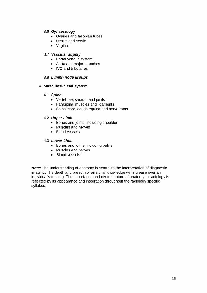
3.6 Gynaecology • Ovaries and fallopian tubes • Uterus and cervix • Vagina 3.7 Vascular supply • Portal venous system • Aorta and major branches • IVC and tributaries 3.8 Lymph node groups 4 Musculoskeletal system 4.1 Spine • Vertebrae, sacrum and joints • Paraspinal muscles and ligaments • Spinal cord, cauda equina and nerve roots 4.2 Upper Limb • Bones and joints, including shoulder • Muscles and nerves • Blood vessels 4.3 Lower Limb • Bones and joints, including pelvis • Muscles and nerves
• Blood vessels Note: The understanding of anatomy is central to the interpretation of diagnostic imaging. The depth and breadth of anatomy knowledge will increase over an individual’s training. The importance and central nature of anatomy to radiology is reflected by its appearance and integration throughout the radiology specific syllabus.
26
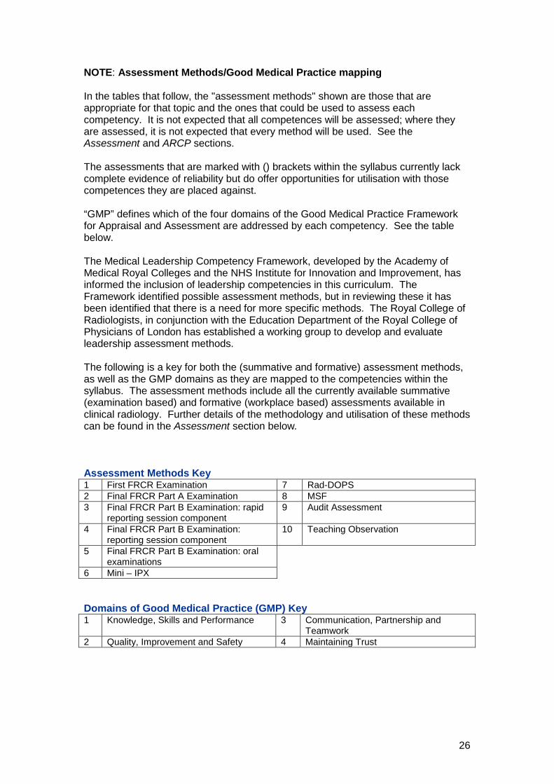
NOTE: Assessment Methods/Good Medical Practice mapping In the tables that follow, the "assessment methods" shown are those that are appropriate for that topic and the ones that could be used to assess each competency. It is not expected that all competences will be assessed; where they are assessed, it is not expected that every method will be used. See the Assessment and ARCP sections. The assessments that are marked with () brackets within the syllabus currently lack complete evidence of reliability but do offer opportunities for utilisation with those competences they are placed against. “GMP” defines which of the four domains of the Good Medical Practice Framework for Appraisal and Assessment are addressed by each competency. See the table below. The Medical Leadership Competency Framework, developed by the Academy of Medical Royal Colleges and the NHS Institute for Innovation and Improvement, has informed the inclusion of leadership competencies in this curriculum. The Framework identified possible assessment methods, but in reviewing these it has been identified that there is a need for more specific methods. The Royal College of Radiologists, in conjunction with the Education Department of the Royal College of Physicians of London has established a working group to develop and evaluate leadership assessment methods. The following is a key for both the (summative and formative) assessment methods, as well as the GMP domains as they are mapped to the competencies within the syllabus. The assessment methods include all the currently available summative (examination based) and formative (workplace based) assessments available in clinical radiology. Further details of the methodology and utilisation of these methods can be found in the Assessment section below. Assessment Methods Key 1 First FRCR Examination 7 Rad-DOPS 2 Final FRCR Part A Examination 8 MSF 3 Final FRCR Part B Examination: rapid
reporting session component 9 Audit Assessment
4 Final FRCR Part B Examination: reporting session component
10 Teaching Observation
5 Final FRCR Part B Examination: oral examinations
6 Mini – IPX Domains of Good Medical Practice (GMP) Key 1 Knowledge, Skills and Performance 3 Communication, Partnership and
Teamwork 2 Quality, Improvement and Safety 4 Maintaining Trust
27
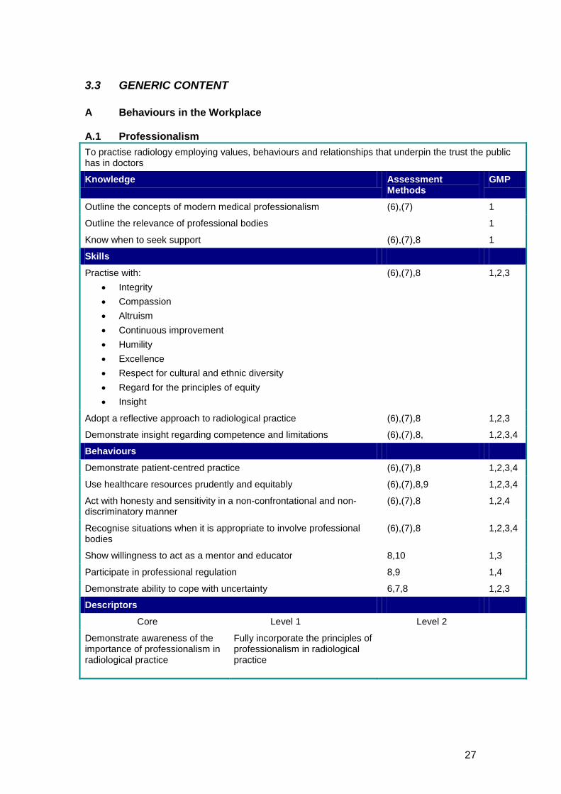
3.3 GENERIC CONTENT A Behaviours in the Workplace A.1 Professionalism To practise radiology employing values, behaviours and relationships that underpin the trust the public has in doctors
Knowledge Assessment Methods
GMP
Outline the concepts of modern medical professionalism (6),(7) 1
Outline the relevance of professional bodies 1
Know when to seek support (6),(7),8 1
Skills
Practise with: • Integrity • Compassion • Altruism • Continuous improvement • Humility • Excellence • Respect for cultural and ethnic diversity • Regard for the principles of equity • Insight
(6),(7),8 1,2,3
Adopt a reflective approach to radiological practice (6),(7),8 1,2,3
Demonstrate insight regarding competence and limitations (6),(7),8, 1,2,3,4
Behaviours
Demonstrate patient-centred practice (6),(7),8 1,2,3,4
Use healthcare resources prudently and equitably (6),(7),8,9 1,2,3,4
Act with honesty and sensitivity in a non-confrontational and non-discriminatory manner
(6),(7),8 1,2,4
Recognise situations when it is appropriate to involve professional bodies
(6),(7),8 1,2,3,4
Show willingness to act as a mentor and educator 8,10 1,3
Participate in professional regulation 8,9 1,4
Demonstrate ability to cope with uncertainty 6,7,8 1,2,3
Descriptors
Core Level 1 Level 2
Demonstrate awareness of the importance of professionalism in radiological practice
Fully incorporate the principles of professionalism in radiological practice
28
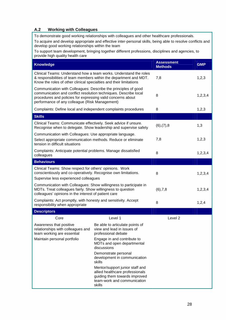
A.2 Working with Colleagues To demonstrate good working relationships with colleagues and other healthcare professionals. To acquire and develop appropriate and effective inter-personal skills, being able to resolve conflicts and develop good working relationships within the team To support team development, bringing together different professions, disciplines and agencies, to provide high quality health care
Knowledge Assessment Methods GMP
Clinical Teams: Understand how a team works. Understand the roles & responsibilities of team members within the department and MDT. Know the roles of other clinical specialties and their limitations
7,8 1,2,3
Communication with Colleagues: Describe the principles of good communication and conflict resolution techniques. Describe local procedures and policies for expressing valid concerns about performance of any colleague (Risk Management)
8 1,2,3,4
Complaints: Define local and independent complaints procedures 8 1,2,3
Skills
Clinical Teams: Communicate effectively. Seek advice if unsure. Recognise when to delegate. Show leadership and supervise safely (6),(7),8 1,3
Communication with Colleagues: Use appropriate language. Select appropriate communication methods. Reduce or eliminate tension in difficult situations
7,8 1,2,3
Complaints: Anticipate potential problems. Manage dissatisfied colleagues 8 1,2,3,4
Behaviours
Clinical Teams: Show respect for others’ opinions. Work conscientiously and co-operatively. Recognise own limitations. Supervise less experienced colleagues
8 1,2,3,4
Communication with Colleagues: Show willingness to participate in MDTs. Treat colleagues fairly. Show willingness to question colleagues’ opinions in the interest of patient care
(6),7,8 1,2,3,4
Complaints: Act promptly, with honesty and sensitivity. Accept responsibility when appropriate 8 1,2,4
Descriptors
Core Level 1 Level 2
Awareness that positive relationships with colleagues and team working are essential Maintain personal portfolio
Be able to articulate points of view and lead in issues of professional debate Engage in and contribute to MDTs and open departmental discussions Demonstrate personal development in communication skills Mentor/support junior staff and allied healthcare professionals guiding them towards improved team-work and communication skills
29
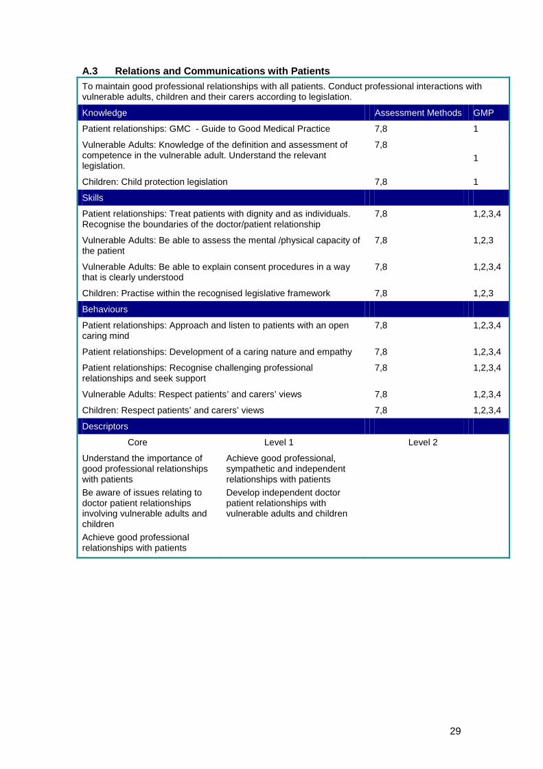
A.3 Relations and Communications with Patients To maintain good professional relationships with all patients. Conduct professional interactions with vulnerable adults, children and their carers according to legislation.
Knowledge Assessment Methods GMP
Patient relationships: GMC - Guide to Good Medical Practice 7,8 1
Vulnerable Adults: Knowledge of the definition and assessment of competence in the vulnerable adult. Understand the relevant legislation.
7,8 1
Children: Child protection legislation 7,8 1
Skills
Patient relationships: Treat patients with dignity and as individuals. Recognise the boundaries of the doctor/patient relationship
7,8 1,2,3,4
Vulnerable Adults: Be able to assess the mental /physical capacity of the patient
7,8 1,2,3
Vulnerable Adults: Be able to explain consent procedures in a way that is clearly understood
7,8 1,2,3,4
Children: Practise within the recognised legislative framework 7,8 1,2,3
Behaviours
Patient relationships: Approach and listen to patients with an open caring mind
7,8 1,2,3,4
Patient relationships: Development of a caring nature and empathy 7,8 1,2,3,4
Patient relationships: Recognise challenging professional relationships and seek support
7,8 1,2,3,4
Vulnerable Adults: Respect patients’ and carers’ views 7,8 1,2,3,4
Children: Respect patients’ and carers’ views 7,8 1,2,3,4
Descriptors
Core Level 1 Level 2
Understand the importance of good professional relationships with patients Be aware of issues relating to doctor patient relationships involving vulnerable adults and children Achieve good professional relationships with patients
Achieve good professional, sympathetic and independent relationships with patients Develop independent doctor patient relationships with vulnerable adults and children
30
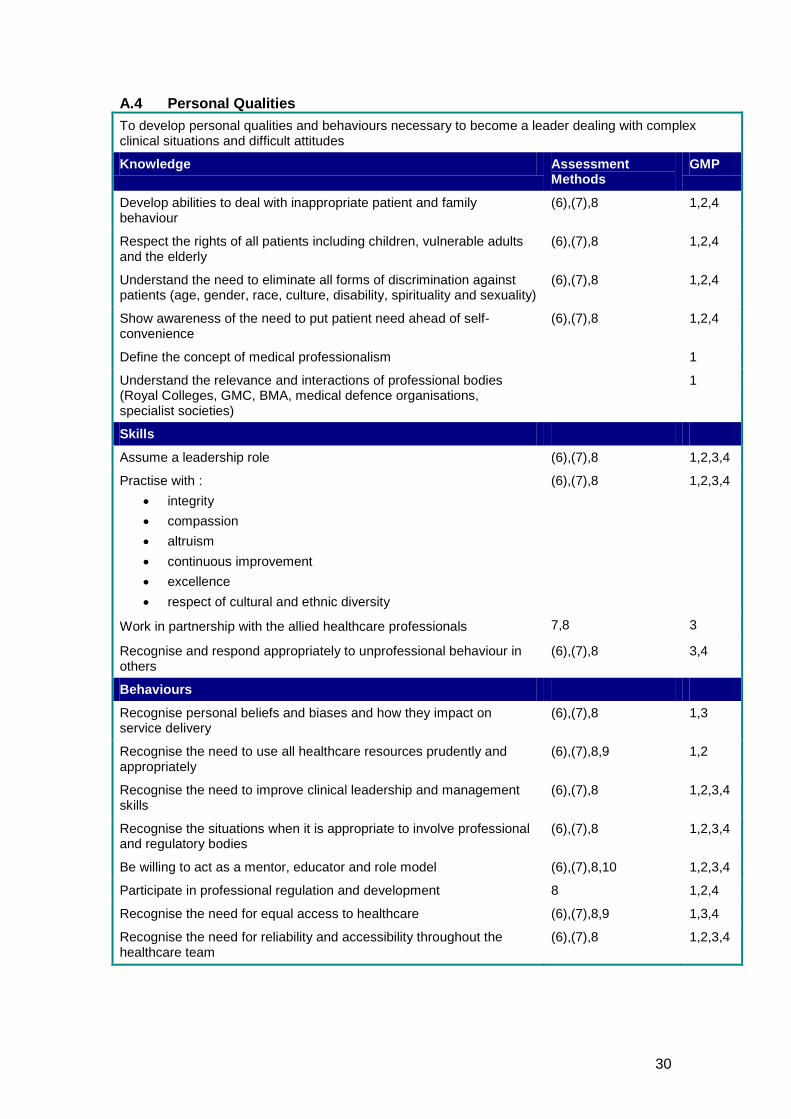
A.4 Personal Qualities To develop personal qualities and behaviours necessary to become a leader dealing with complex clinical situations and difficult attitudes
Knowledge Assessment Methods
GMP
Develop abilities to deal with inappropriate patient and family behaviour
(6),(7),8 1,2,4
Respect the rights of all patients including children, vulnerable adults and the elderly
(6),(7),8 1,2,4
Understand the need to eliminate all forms of discrimination against patients (age, gender, race, culture, disability, spirituality and sexuality)
(6),(7),8 1,2,4
Show awareness of the need to put patient need ahead of self- convenience
(6),(7),8 1,2,4
Define the concept of medical professionalism 1
Understand the relevance and interactions of professional bodies (Royal Colleges, GMC, BMA, medical defence organisations, specialist societies)
1
Skills
Assume a leadership role (6),(7),8 1,2,3,4
Practise with : • integrity • compassion • altruism • continuous improvement • excellence • respect of cultural and ethnic diversity
(6),(7),8 1,2,3,4
Work in partnership with the allied healthcare professionals 7,8 3
Recognise and respond appropriately to unprofessional behaviour in others
(6),(7),8 3,4
Behaviours
Recognise personal beliefs and biases and how they impact on service delivery
(6),(7),8 1,3
Recognise the need to use all healthcare resources prudently and appropriately
(6),(7),8,9 1,2
Recognise the need to improve clinical leadership and management skills
(6),(7),8 1,2,3,4
Recognise the situations when it is appropriate to involve professional and regulatory bodies
(6),(7),8 1,2,3,4
Be willing to act as a mentor, educator and role model (6),(7),8,10 1,2,3,4
Participate in professional regulation and development 8 1,2,4
Recognise the need for equal access to healthcare (6),(7),8,9 1,3,4
Recognise the need for reliability and accessibility throughout the healthcare team
(6),(7),8 1,2,3,4
31
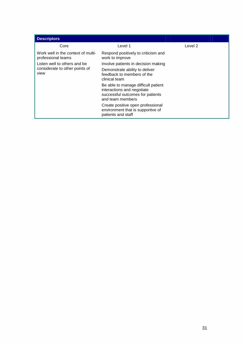
Descriptors
Core Level 1 Level 2
Work well in the context of multi-professional teams Listen well to others and be considerate to other points of view
Respond positively to criticism and work to improve Involve patients in decision making Demonstrate ability to deliver feedback to members of the clinical team Be able to manage difficult patient interactions and negotiate successful outcomes for patients and team members Create positive open professional environment that is supportive of patients and staff
32
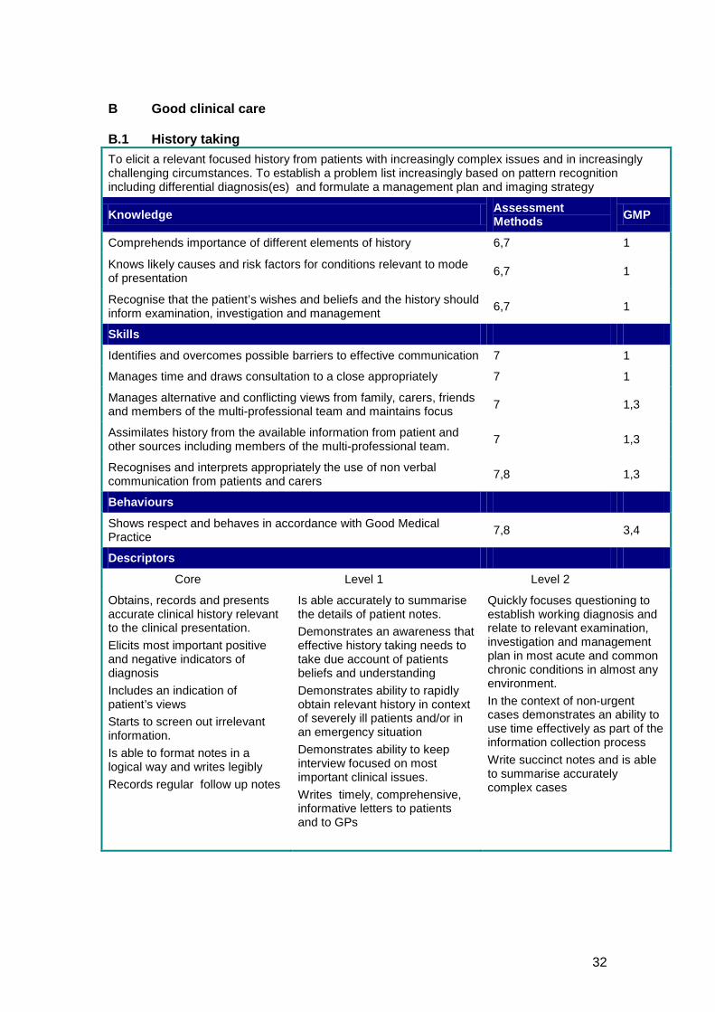
B Good clinical care B.1 History taking To elicit a relevant focused history from patients with increasingly complex issues and in increasingly challenging circumstances. To establish a problem list increasingly based on pattern recognition including differential diagnosis(es) and formulate a management plan and imaging strategy
Knowledge Assessment Methods GMP
Comprehends importance of different elements of history 6,7 1
Knows likely causes and risk factors for conditions relevant to mode of presentation 6,7 1
Recognise that the patient’s wishes and beliefs and the history should inform examination, investigation and management 6,7 1
Skills
Identifies and overcomes possible barriers to effective communication 7 1
Manages time and draws consultation to a close appropriately 7 1
Manages alternative and conflicting views from family, carers, friends and members of the multi-professional team and maintains focus 7 1,3
Assimilates history from the available information from patient and other sources including members of the multi-professional team. 7 1,3
Recognises and interprets appropriately the use of non verbal communication from patients and carers 7,8 1,3
Behaviours
Shows respect and behaves in accordance with Good Medical Practice 7,8 3,4
Descriptors
Core Level 1 Level 2
Obtains, records and presents accurate clinical history relevant to the clinical presentation. Elicits most important positive and negative indicators of diagnosis Includes an indication of patient’s views Starts to screen out irrelevant information. Is able to format notes in a logical way and writes legibly Records regular follow up notes
Is able accurately to summarise the details of patient notes. Demonstrates an awareness that effective history taking needs to take due account of patients beliefs and understanding Demonstrates ability to rapidly obtain relevant history in context of severely ill patients and/or in an emergency situation Demonstrates ability to keep interview focused on most important clinical issues. Writes timely, comprehensive, informative letters to patients and to GPs
Quickly focuses questioning to establish working diagnosis and relate to relevant examination, investigation and management plan in most acute and common chronic conditions in almost any environment. In the context of non-urgent cases demonstrates an ability to use time effectively as part of the information collection process Write succinct notes and is able to summarise accurately complex cases
33
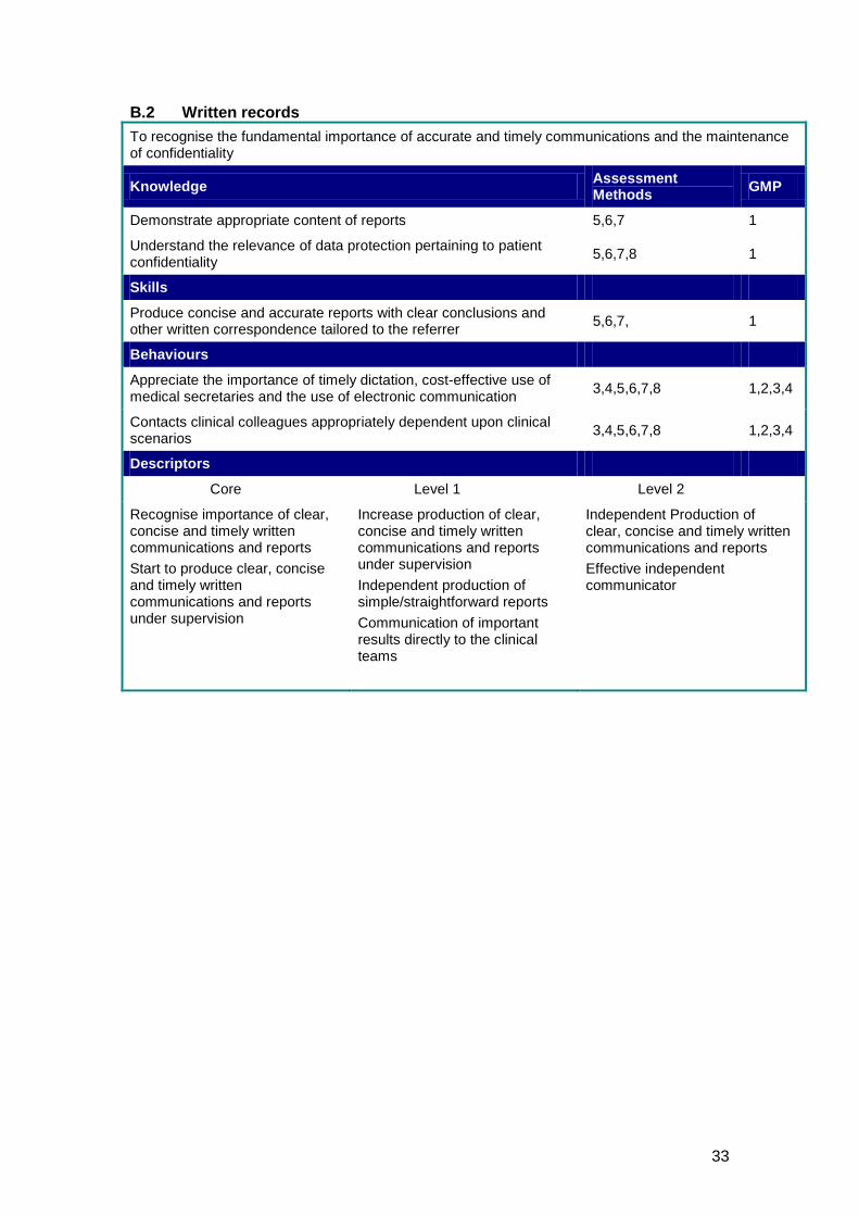
B.2 Written records To recognise the fundamental importance of accurate and timely communications and the maintenance of confidentiality
Knowledge Assessment Methods GMP
Demonstrate appropriate content of reports 5,6,7 1
Understand the relevance of data protection pertaining to patient confidentiality 5,6,7,8 1
Skills
Produce concise and accurate reports with clear conclusions and other written correspondence tailored to the referrer 5,6,7, 1
Behaviours
Appreciate the importance of timely dictation, cost-effective use of medical secretaries and the use of electronic communication 3,4,5,6,7,8 1,2,3,4
Contacts clinical colleagues appropriately dependent upon clinical scenarios 3,4,5,6,7,8 1,2,3,4
Descriptors
Core Level 1 Level 2
Recognise importance of clear, concise and timely written communications and reports Start to produce clear, concise and timely written communications and reports under supervision
Increase production of clear, concise and timely written communications and reports under supervision Independent production of simple/straightforward reports Communication of important results directly to the clinical teams
Independent Production of clear, concise and timely written communications and reports Effective independent communicator
34
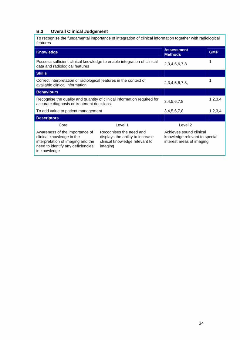
B.3 Overall Clinical Judgement To recognise the fundamental importance of integration of clinical information together with radiological features
Knowledge Assessment Methods GMP
Possess sufficient clinical knowledge to enable integration of clinical data and radiological features 2,3,4,5,6,7,8 1
Skills
Correct interpretation of radiological features in the context of available clinical information 2,3,4,5,6,7,8, 1
Behaviours
Recognise the quality and quantity of clinical information required for accurate diagnosis or treatment decisions. 3,4,5,6,7,8 1,2,3,4
To add value to patient management 3,4,5,6,7,8 1,2,3,4
Descriptors
Core Level 1 Level 2
Awareness of the importance of clinical knowledge in the interpretation of imaging and the need to identify any deficiencies in knowledge
Recognises the need and displays the ability to increase clinical knowledge relevant to imaging
Achieves sound clinical knowledge relevant to special interest areas of imaging
35
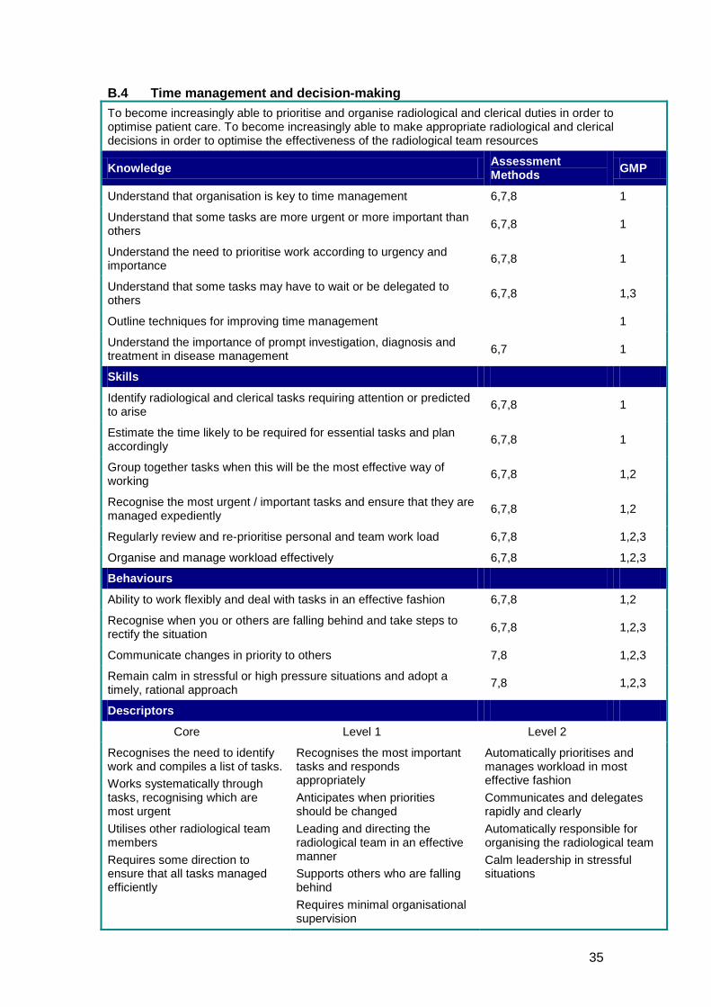
B.4 Time management and decision-making To become increasingly able to prioritise and organise radiological and clerical duties in order to optimise patient care. To become increasingly able to make appropriate radiological and clerical decisions in order to optimise the effectiveness of the radiological team resources
Knowledge Assessment Methods GMP
Understand that organisation is key to time management 6,7,8 1
Understand that some tasks are more urgent or more important than others 6,7,8 1
Understand the need to prioritise work according to urgency and importance 6,7,8 1
Understand that some tasks may have to wait or be delegated to others 6,7,8 1,3
Outline techniques for improving time management 1
Understand the importance of prompt investigation, diagnosis and treatment in disease management 6,7 1
Skills
Identify radiological and clerical tasks requiring attention or predicted to arise 6,7,8 1
Estimate the time likely to be required for essential tasks and plan accordingly 6,7,8 1
Group together tasks when this will be the most effective way of working 6,7,8 1,2
Recognise the most urgent / important tasks and ensure that they are managed expediently 6,7,8 1,2
Regularly review and re-prioritise personal and team work load 6,7,8 1,2,3
Organise and manage workload effectively 6,7,8 1,2,3
Behaviours
Ability to work flexibly and deal with tasks in an effective fashion 6,7,8 1,2
Recognise when you or others are falling behind and take steps to rectify the situation 6,7,8 1,2,3
Communicate changes in priority to others 7,8 1,2,3
Remain calm in stressful or high pressure situations and adopt a timely, rational approach 7,8 1,2,3
Descriptors
Core Level 1 Level 2
Recognises the need to identify work and compiles a list of tasks. Works systematically through tasks, recognising which are most urgent Utilises other radiological team members Requires some direction to ensure that all tasks managed efficiently
Recognises the most important tasks and responds appropriately Anticipates when priorities should be changed Leading and directing the radiological team in an effective manner Supports others who are falling behind Requires minimal organisational supervision
Automatically prioritises and manages workload in most effective fashion Communicates and delegates rapidly and clearly Automatically responsible for organising the radiological team Calm leadership in stressful situations
36
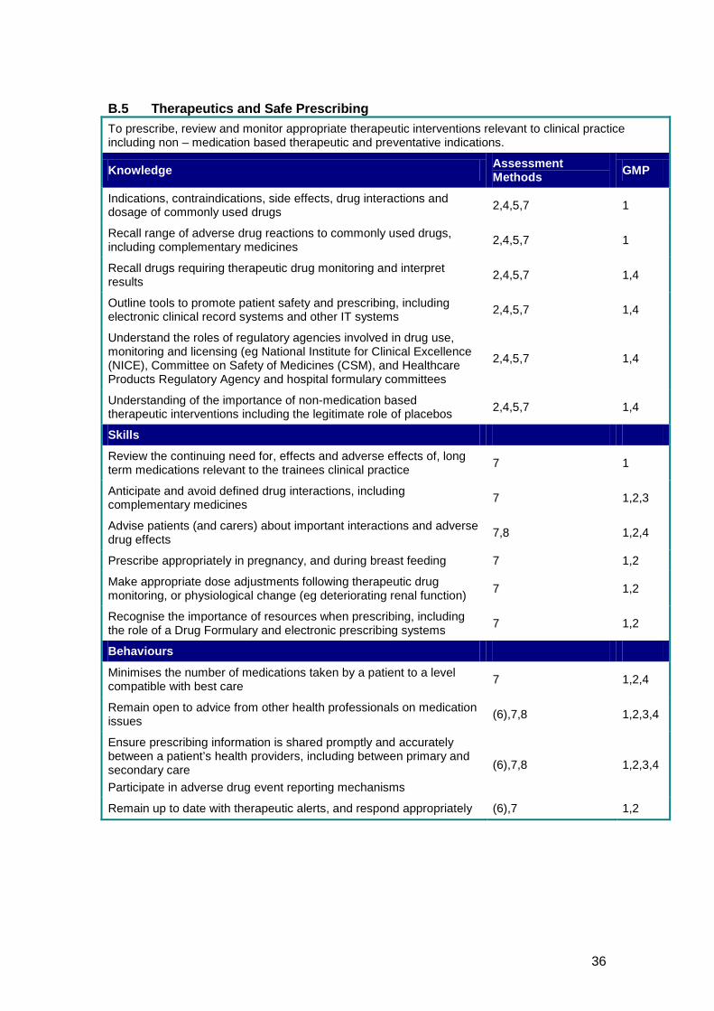
B.5 Therapeutics and Safe Prescribing To prescribe, review and monitor appropriate therapeutic interventions relevant to clinical practice including non – medication based therapeutic and preventative indications.
Knowledge Assessment Methods GMP
Indications, contraindications, side effects, drug interactions and dosage of commonly used drugs 2,4,5,7 1
Recall range of adverse drug reactions to commonly used drugs, including complementary medicines 2,4,5,7 1
Recall drugs requiring therapeutic drug monitoring and interpret results 2,4,5,7 1,4
Outline tools to promote patient safety and prescribing, including electronic clinical record systems and other IT systems 2,4,5,7 1,4
Understand the roles of regulatory agencies involved in drug use, monitoring and licensing (eg National Institute for Clinical Excellence (NICE), Committee on Safety of Medicines (CSM), and Healthcare Products Regulatory Agency and hospital formulary committees
2,4,5,7 1,4
Understanding of the importance of non-medication based therapeutic interventions including the legitimate role of placebos 2,4,5,7 1,4
Skills
Review the continuing need for, effects and adverse effects of, long term medications relevant to the trainees clinical practice 7 1
Anticipate and avoid defined drug interactions, including complementary medicines 7 1,2,3
Advise patients (and carers) about important interactions and adverse drug effects 7,8 1,2,4
Prescribe appropriately in pregnancy, and during breast feeding 7 1,2
Make appropriate dose adjustments following therapeutic drug monitoring, or physiological change (eg deteriorating renal function) 7 1,2
Recognise the importance of resources when prescribing, including the role of a Drug Formulary and electronic prescribing systems 7 1,2
Behaviours
Minimises the number of medications taken by a patient to a level compatible with best care 7 1,2,4
Remain open to advice from other health professionals on medication issues (6),7,8 1,2,3,4
Ensure prescribing information is shared promptly and accurately between a patient’s health providers, including between primary and secondary care Participate in adverse drug event reporting mechanisms
(6),7,8 1,2,3,4
Remain up to date with therapeutic alerts, and respond appropriately (6),7 1,2
37
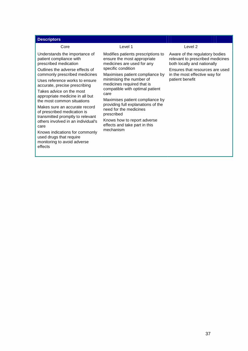
Descriptors
Core Level 1 Level 2
Understands the importance of patient compliance with prescribed medication Outlines the adverse effects of commonly prescribed medicines Uses reference works to ensure accurate, precise prescribing Takes advice on the most appropriate medicine in all but the most common situations Makes sure an accurate record of prescribed medication is transmitted promptly to relevant others involved in an individual's care Knows indications for commonly used drugs that require monitoring to avoid adverse effects
Modifies patients prescriptions to ensure the most appropriate medicines are used for any specific condition Maximises patient compliance by minimising the number of medicines required that is compatible with optimal patient care Maximises patient compliance by providing full explanations of the need for the medicines prescribed Knows how to report adverse effects and take part in this mechanism
Aware of the regulatory bodies relevant to prescribed medicines both locally and nationally Ensures that resources are used in the most effective way for patient benefit
38
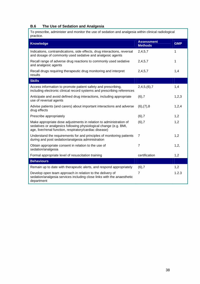
B.6 The Use of Sedation and Analgesia To prescribe, administer and monitor the use of sedation and analgesia within clinical radiological practice.
Knowledge Assessment Methods GMP
Indications, contraindications, side effects, drug interactions, reversal and dosage of commonly used sedative and analgesic agents
2,4,5,7 1
Recall range of adverse drug reactions to commonly used sedative and analgesic agents
2,4,5,7 1
Recall drugs requiring therapeutic drug monitoring and interpret results
2,4,5,7 1,4
Skills
Access information to promote patient safety and prescribing, including electronic clinical record systems and prescribing references
2,4,5,(6),7 1,4
Anticipate and avoid defined drug interactions, including appropriate use of reversal agents
(6),7 1,2,3
Advise patients (and carers) about important interactions and adverse drug effects
(6),(7),8 1,2,4
Prescribe appropriately (6),7 1,2
Make appropriate dose adjustments in relation to administration of sedatives or analgesics following physiological change (e.g. BMI, age, liver/renal function, respiratory/cardiac disease)
(6),7 1,2
Understand the requirements for and principles of monitoring patients during and post sedation/analgesia administration
7 1,2
Obtain appropriate consent in relation to the use of sedation/analgesia
7 1,2,
Formal appropriate level of resuscitation training certification 1,2
Behaviours
Remain up to date with therapeutic alerts, and respond appropriately (6),7 1,2
Develop open team approach in relation to the delivery of sedation/analgesia services including close links with the anaesthetic department
7 1.2.3
39
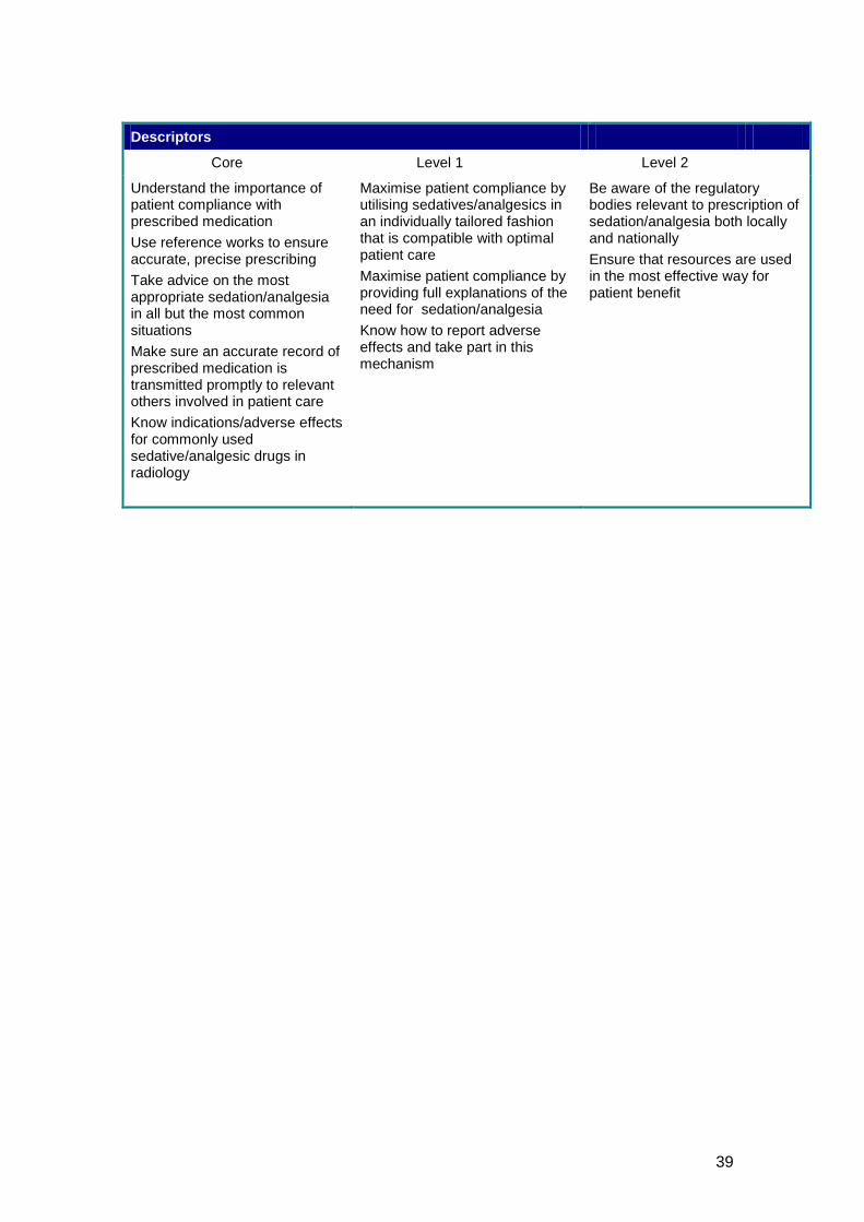
Descriptors
Core Level 1 Level 2
Understand the importance of patient compliance with prescribed medication Use reference works to ensure accurate, precise prescribing Take advice on the most appropriate sedation/analgesia in all but the most common situations Make sure an accurate record of prescribed medication is transmitted promptly to relevant others involved in patient care Know indications/adverse effects for commonly used sedative/analgesic drugs in radiology
Maximise patient compliance by utilising sedatives/analgesics in an individually tailored fashion that is compatible with optimal patient care Maximise patient compliance by providing full explanations of the need for sedation/analgesia Know how to report adverse effects and take part in this mechanism
Be aware of the regulatory bodies relevant to prescription of sedation/analgesia both locally and nationally Ensure that resources are used in the most effective way for patient benefit
40
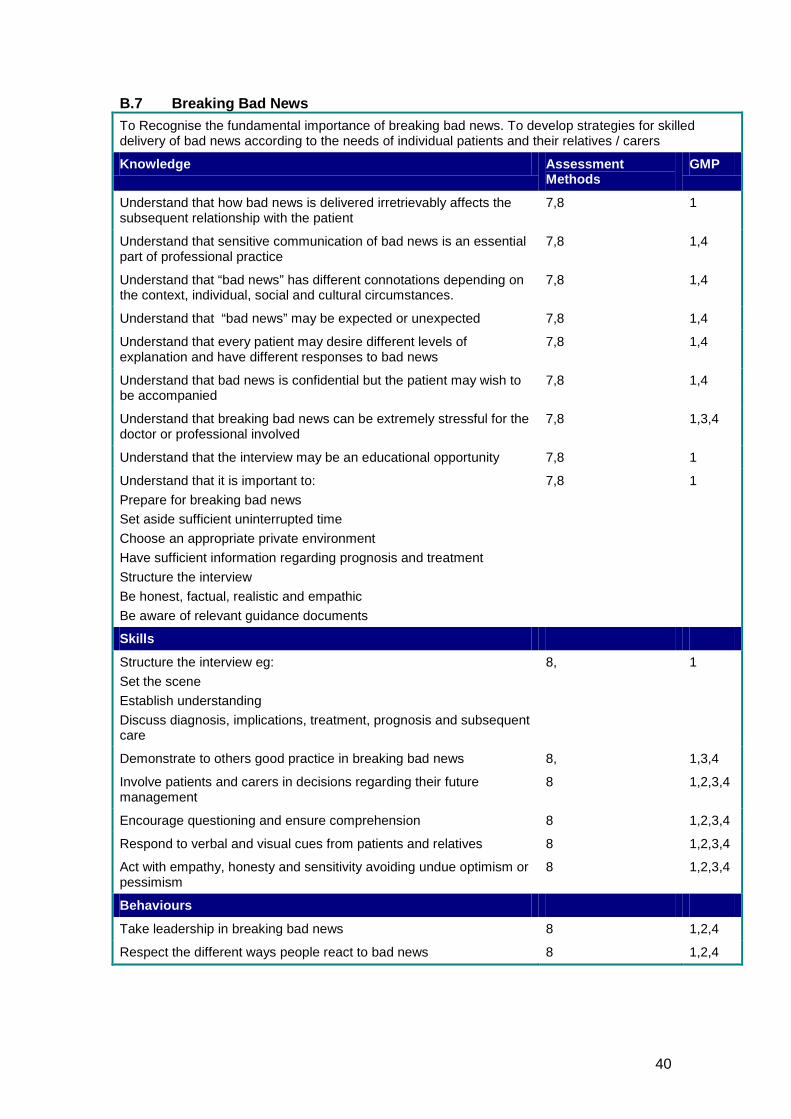
B.7 Breaking Bad News To Recognise the fundamental importance of breaking bad news. To develop strategies for skilled delivery of bad news according to the needs of individual patients and their relatives / carers
Knowledge Assessment Methods
GMP
Understand that how bad news is delivered irretrievably affects the subsequent relationship with the patient
7,8 1
Understand that sensitive communication of bad news is an essential part of professional practice
7,8 1,4
Understand that “bad news” has different connotations depending on the context, individual, social and cultural circumstances.
7,8 1,4
Understand that “bad news” may be expected or unexpected 7,8 1,4
Understand that every patient may desire different levels of explanation and have different responses to bad news
7,8 1,4
Understand that bad news is confidential but the patient may wish to be accompanied
7,8 1,4
Understand that breaking bad news can be extremely stressful for the doctor or professional involved
7,8 1,3,4
Understand that the interview may be an educational opportunity 7,8 1
Understand that it is important to: Prepare for breaking bad news Set aside sufficient uninterrupted time Choose an appropriate private environment Have sufficient information regarding prognosis and treatment Structure the interview Be honest, factual, realistic and empathic Be aware of relevant guidance documents
7,8 1
Skills
Structure the interview eg: Set the scene Establish understanding Discuss diagnosis, implications, treatment, prognosis and subsequent care
8, 1
Demonstrate to others good practice in breaking bad news 8, 1,3,4
Involve patients and carers in decisions regarding their future management
8 1,2,3,4
Encourage questioning and ensure comprehension 8 1,2,3,4
Respond to verbal and visual cues from patients and relatives 8 1,2,3,4
Act with empathy, honesty and sensitivity avoiding undue optimism or pessimism
8 1,2,3,4
Behaviours
Take leadership in breaking bad news 8 1,2,4
Respect the different ways people react to bad news 8 1,2,4
41
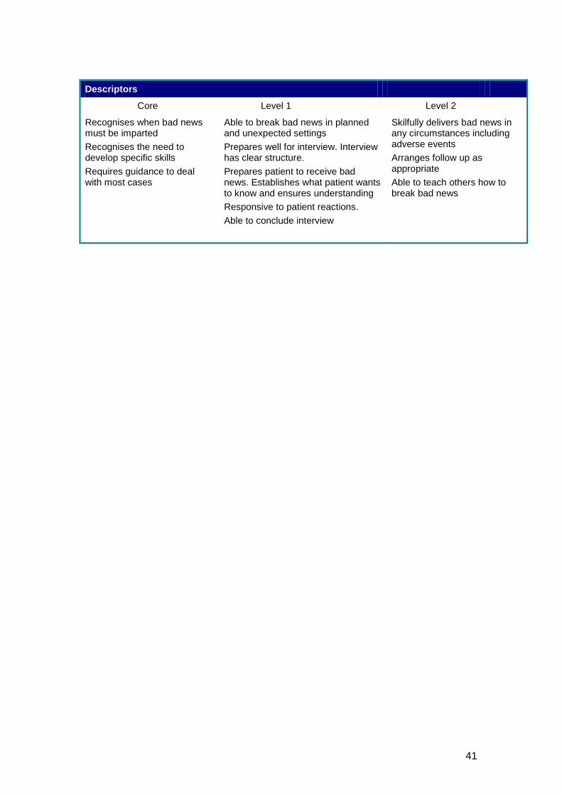
Descriptors
Core Level 1 Level 2
Recognises when bad news must be imparted Recognises the need to develop specific skills Requires guidance to deal with most cases
Able to break bad news in planned and unexpected settings Prepares well for interview. Interview has clear structure. Prepares patient to receive bad news. Establishes what patient wants to know and ensures understanding Responsive to patient reactions. Able to conclude interview
Skilfully delivers bad news in any circumstances including adverse events Arranges follow up as appropriate Able to teach others how to break bad news
42
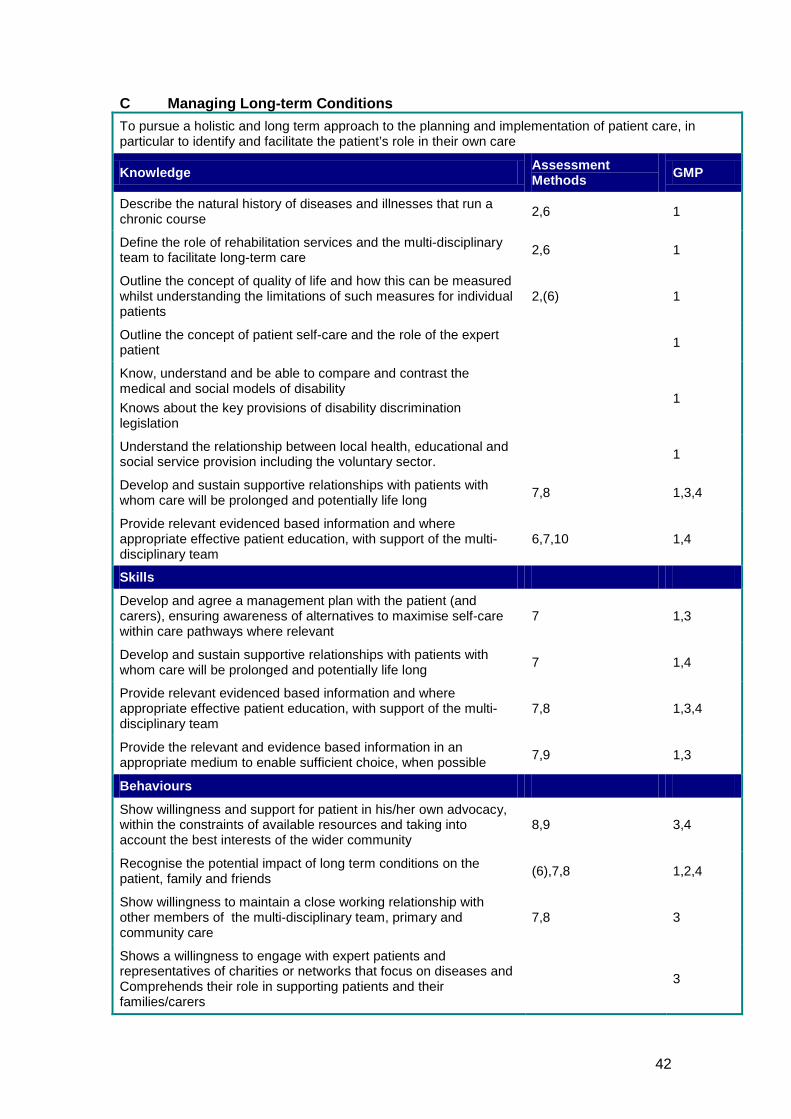
C Managing Long-term Conditions To pursue a holistic and long term approach to the planning and implementation of patient care, in particular to identify and facilitate the patient’s role in their own care
Knowledge Assessment Methods GMP
Describe the natural history of diseases and illnesses that run a chronic course 2,6 1
Define the role of rehabilitation services and the multi-disciplinary team to facilitate long-term care 2,6 1
Outline the concept of quality of life and how this can be measured whilst understanding the limitations of such measures for individual patients
2,(6) 1
Outline the concept of patient self-care and the role of the expert patient 1
Know, understand and be able to compare and contrast the medical and social models of disability Knows about the key provisions of disability discrimination legislation
1
Understand the relationship between local health, educational and social service provision including the voluntary sector. 1
Develop and sustain supportive relationships with patients with whom care will be prolonged and potentially life long 7,8 1,3,4
Provide relevant evidenced based information and where appropriate effective patient education, with support of the multi-disciplinary team
6,7,10 1,4
Skills
Develop and agree a management plan with the patient (and carers), ensuring awareness of alternatives to maximise self-care within care pathways where relevant
7 1,3
Develop and sustain supportive relationships with patients with whom care will be prolonged and potentially life long 7 1,4
Provide relevant evidenced based information and where appropriate effective patient education, with support of the multi-disciplinary team
7,8 1,3,4
Provide the relevant and evidence based information in an appropriate medium to enable sufficient choice, when possible 7,9 1,3
Behaviours
Show willingness and support for patient in his/her own advocacy, within the constraints of available resources and taking into account the best interests of the wider community
8,9 3,4
Recognise the potential impact of long term conditions on the patient, family and friends (6),7,8 1,2,4
Show willingness to maintain a close working relationship with other members of the multi-disciplinary team, primary and community care
7,8 3
Shows a willingness to engage with expert patients and representatives of charities or networks that focus on diseases and Comprehends their role in supporting patients and their families/carers
3
43
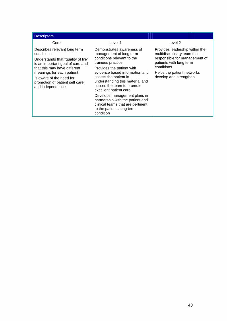
Descriptors
Core Level 1 Level 2
Describes relevant long term conditions Understands that “quality of life” is an important goal of care and that this may have different meanings for each patient Is aware of the need for promotion of patient self care and independence
Demonstrates awareness of management of long term conditions relevant to the trainees practice Provides the patient with evidence based information and assists the patient in understanding this material and utilises the team to promote excellent patient care Develops management plans in partnership with the patient and clinical teams that are pertinent to the patients long term condition
Provides leadership within the multidisciplinary team that is responsible for management of patients with long term conditions Helps the patient networks develop and strengthen
44
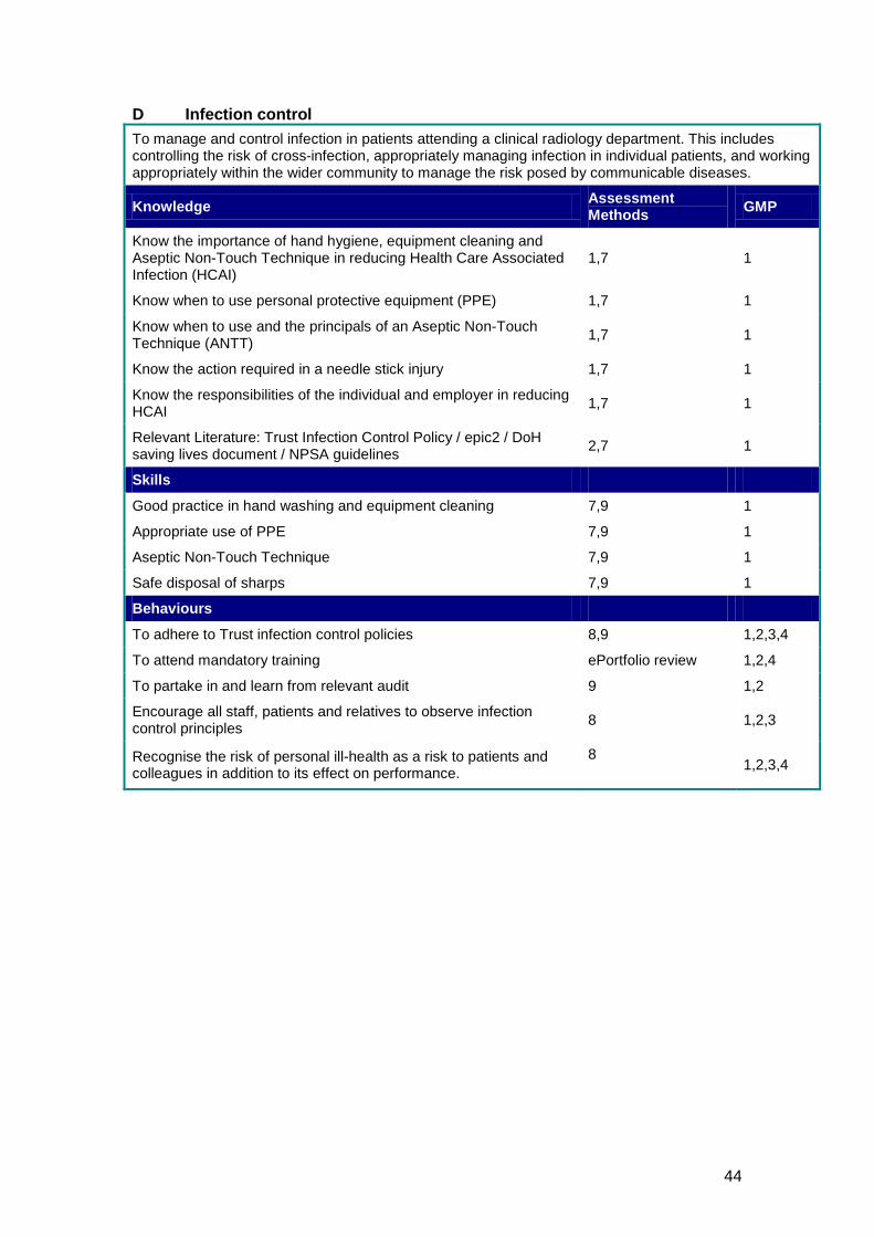
D Infection control To manage and control infection in patients attending a clinical radiology department. This includes controlling the risk of cross-infection, appropriately managing infection in individual patients, and working appropriately within the wider community to manage the risk posed by communicable diseases.
Knowledge Assessment Methods GMP
Know the importance of hand hygiene, equipment cleaning and Aseptic Non-Touch Technique in reducing Health Care Associated Infection (HCAI)
1,7 1
Know when to use personal protective equipment (PPE) 1,7 1
Know when to use and the principals of an Aseptic Non-Touch Technique (ANTT) 1,7 1
Know the action required in a needle stick injury 1,7 1
Know the responsibilities of the individual and employer in reducing HCAI 1,7 1
Relevant Literature: Trust Infection Control Policy / epic2 / DoH saving lives document / NPSA guidelines 2,7 1
Skills
Good practice in hand washing and equipment cleaning 7,9 1
Appropriate use of PPE 7,9 1
Aseptic Non-Touch Technique 7,9 1
Safe disposal of sharps 7,9 1
Behaviours
To adhere to Trust infection control policies 8,9 1,2,3,4
To attend mandatory training ePortfolio review 1,2,4
To partake in and learn from relevant audit 9 1,2
Encourage all staff, patients and relatives to observe infection control principles 8 1,2,3
Recognise the risk of personal ill-health as a risk to patients and colleagues in addition to its effect on performance.
8
1,2,3,4
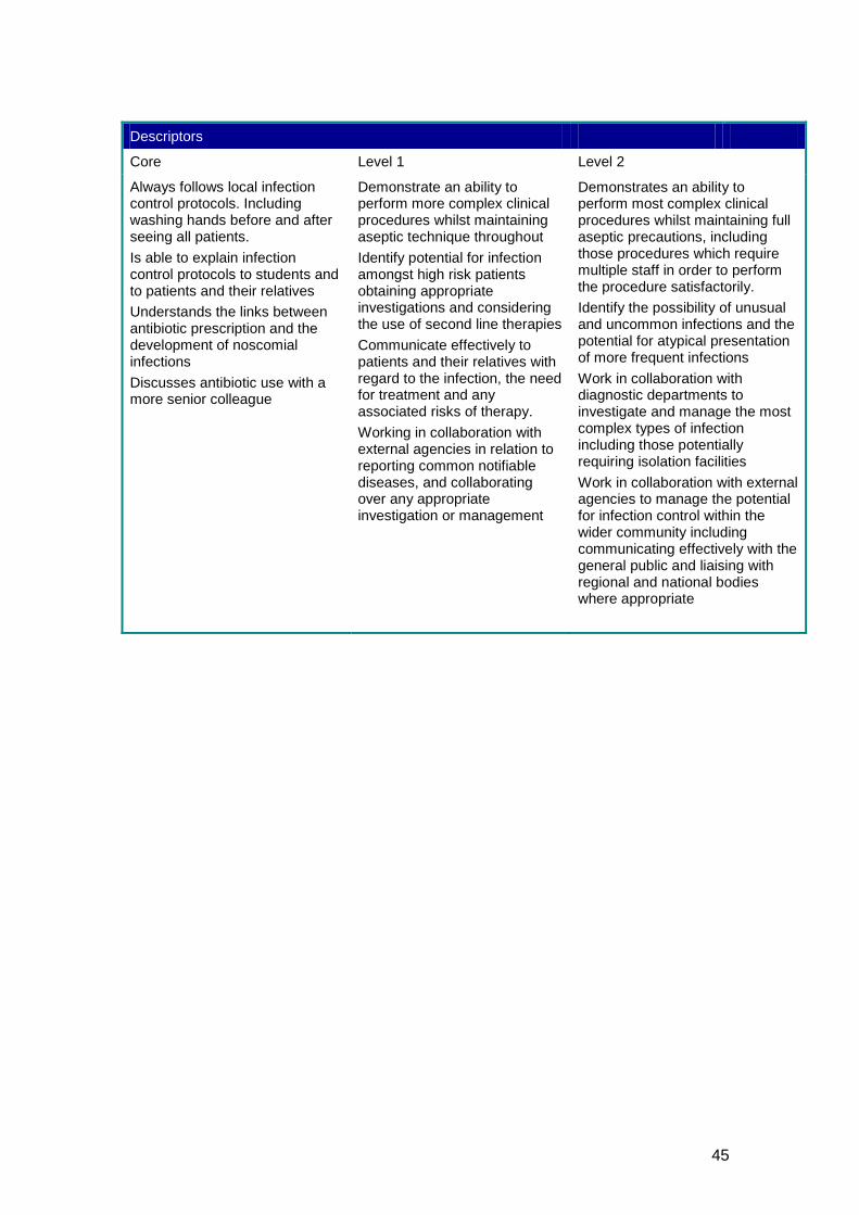
45
Descriptors
Core Level 1 Level 2
Always follows local infection control protocols. Including washing hands before and after seeing all patients. Is able to explain infection control protocols to students and to patients and their relatives Understands the links between antibiotic prescription and the development of noscomial infections Discusses antibiotic use with a more senior colleague
Demonstrate an ability to perform more complex clinical procedures whilst maintaining aseptic technique throughout Identify potential for infection amongst high risk patients obtaining appropriate investigations and considering the use of second line therapies Communicate effectively to patients and their relatives with regard to the infection, the need for treatment and any associated risks of therapy. Working in collaboration with external agencies in relation to reporting common notifiable diseases, and collaborating over any appropriate investigation or management
Demonstrates an ability to perform most complex clinical procedures whilst maintaining full aseptic precautions, including those procedures which require multiple staff in order to perform the procedure satisfactorily. Identify the possibility of unusual and uncommon infections and the potential for atypical presentation of more frequent infections Work in collaboration with diagnostic departments to investigate and manage the most complex types of infection including those potentially requiring isolation facilities Work in collaboration with external agencies to manage the potential for infection control within the wider community including communicating effectively with the general public and liaising with regional and national bodies where appropriate
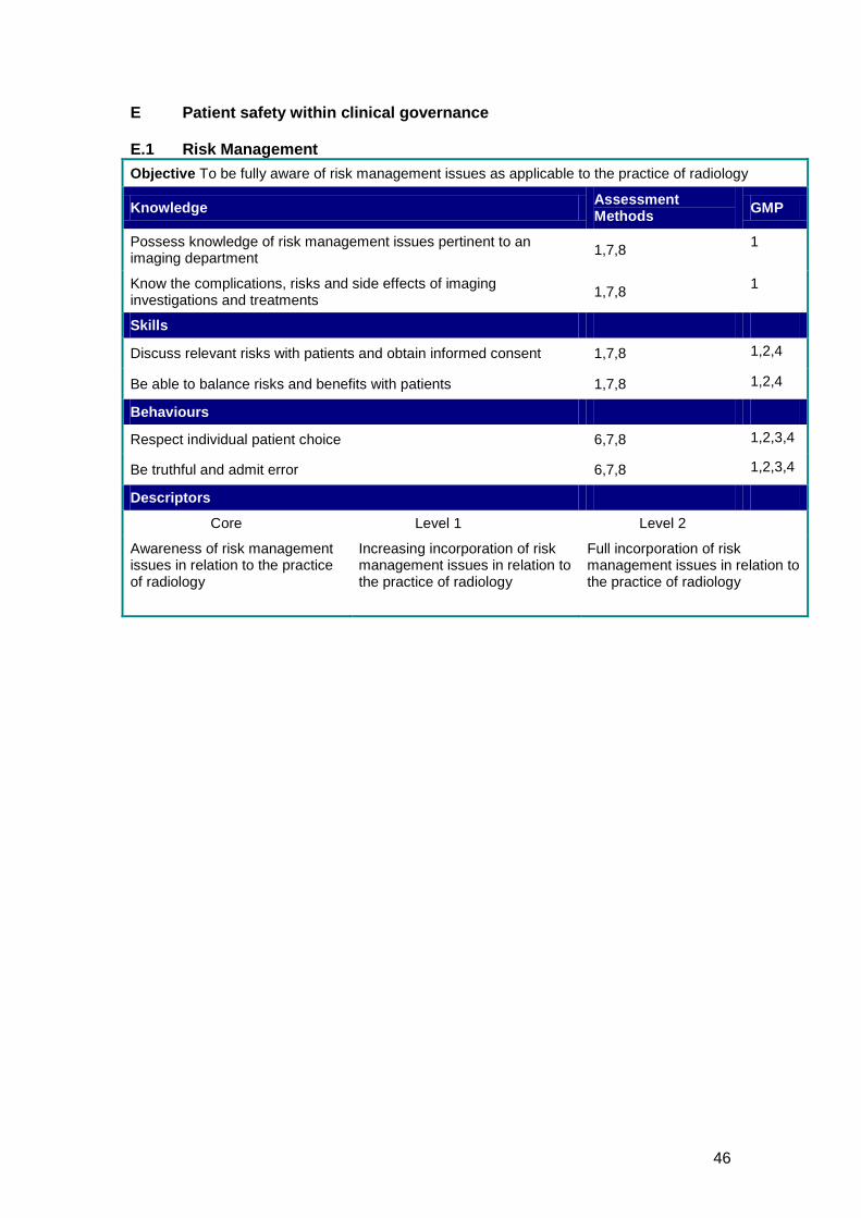
46
E Patient safety within clinical governance E.1 Risk Management Objective To be fully aware of risk management issues as applicable to the practice of radiology
Knowledge Assessment Methods GMP
Possess knowledge of risk management issues pertinent to an imaging department 1,7,8 1
Know the complications, risks and side effects of imaging investigations and treatments 1,7,8 1
Skills
Discuss relevant risks with patients and obtain informed consent 1,7,8 1,2,4
Be able to balance risks and benefits with patients 1,7,8 1,2,4
Behaviours
Respect individual patient choice 6,7,8 1,2,3,4
Be truthful and admit error 6,7,8 1,2,3,4
Descriptors
Core Level 1 Level 2
Awareness of risk management issues in relation to the practice of radiology
Increasing incorporation of risk management issues in relation to the practice of radiology
Full incorporation of risk management issues in relation to the practice of radiology
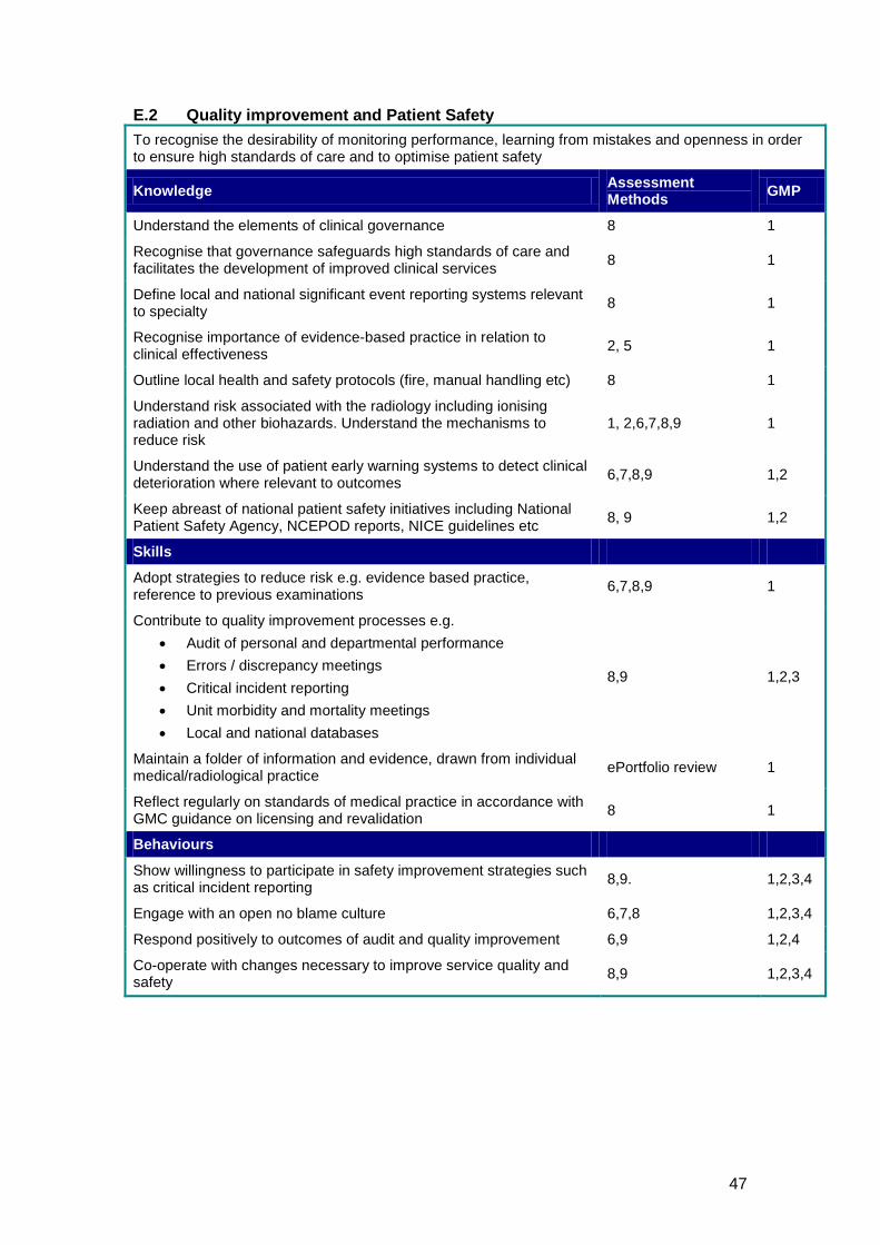
47
E.2 Quality improvement and Patient Safety To recognise the desirability of monitoring performance, learning from mistakes and openness in order to ensure high standards of care and to optimise patient safety
Knowledge Assessment Methods GMP
Understand the elements of clinical governance 8 1
Recognise that governance safeguards high standards of care and facilitates the development of improved clinical services 8 1
Define local and national significant event reporting systems relevant to specialty 8 1
Recognise importance of evidence-based practice in relation to clinical effectiveness 2, 5 1
Outline local health and safety protocols (fire, manual handling etc) 8 1
Understand risk associated with the radiology including ionising radiation and other biohazards. Understand the mechanisms to reduce risk
1, 2,6,7,8,9 1
Understand the use of patient early warning systems to detect clinical deterioration where relevant to outcomes 6,7,8,9 1,2
Keep abreast of national patient safety initiatives including National Patient Safety Agency, NCEPOD reports, NICE guidelines etc 8, 9 1,2
Skills
Adopt strategies to reduce risk e.g. evidence based practice, reference to previous examinations 6,7,8,9 1
Contribute to quality improvement processes e.g. • Audit of personal and departmental performance • Errors / discrepancy meetings • Critical incident reporting • Unit morbidity and mortality meetings • Local and national databases
8,9 1,2,3
Maintain a folder of information and evidence, drawn from individual medical/radiological practice ePortfolio review 1
Reflect regularly on standards of medical practice in accordance with GMC guidance on licensing and revalidation 8 1
Behaviours
Show willingness to participate in safety improvement strategies such as critical incident reporting 8,9. 1,2,3,4
Engage with an open no blame culture 6,7,8 1,2,3,4
Respond positively to outcomes of audit and quality improvement 6,9 1,2,4
Co-operate with changes necessary to improve service quality and safety 8,9 1,2,3,4
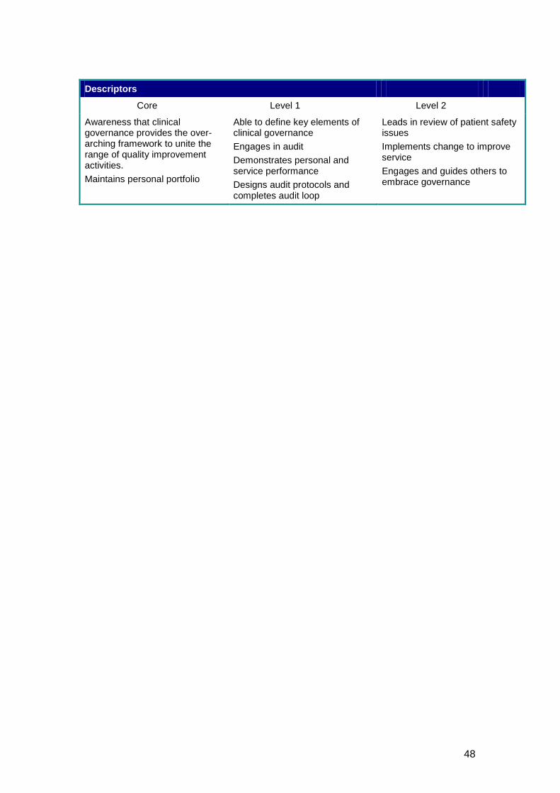
48
Descriptors
Core Level 1 Level 2
Awareness that clinical governance provides the over-arching framework to unite the range of quality improvement activities. Maintains personal portfolio
Able to define key elements of clinical governance Engages in audit Demonstrates personal and service performance Designs audit protocols and completes audit loop
Leads in review of patient safety issues Implements change to improve service Engages and guides others to embrace governance
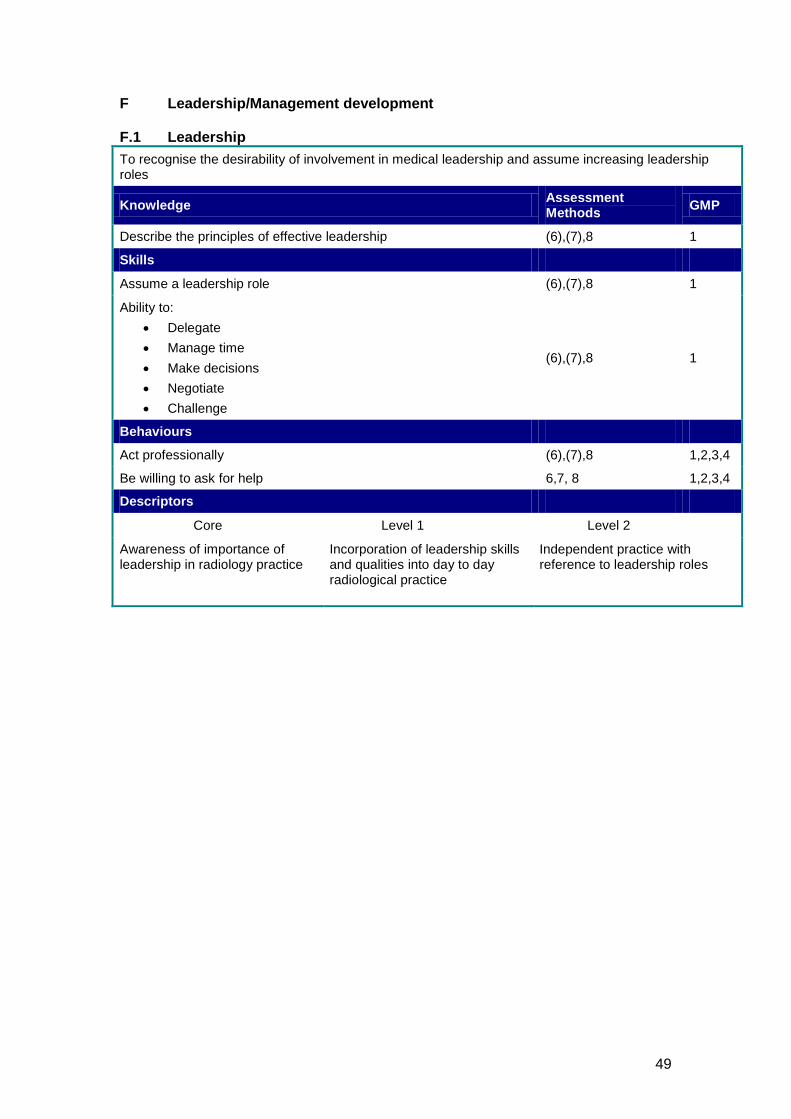
49
F Leadership/Management development F.1 Leadership To recognise the desirability of involvement in medical leadership and assume increasing leadership roles
Knowledge Assessment Methods GMP
Describe the principles of effective leadership (6),(7),8 1
Skills
Assume a leadership role (6),(7),8 1
Ability to: • Delegate • Manage time • Make decisions • Negotiate • Challenge
(6),(7),8 1
Behaviours
Act professionally (6),(7),8 1,2,3,4
Be willing to ask for help 6,7, 8 1,2,3,4
Descriptors
Core Level 1 Level 2
Awareness of importance of leadership in radiology practice
Incorporation of leadership skills and qualities into day to day radiological practice
Independent practice with reference to leadership roles
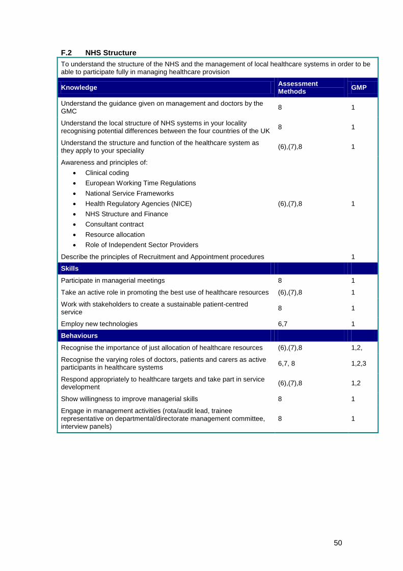
50
F.2 NHS Structure To understand the structure of the NHS and the management of local healthcare systems in order to be able to participate fully in managing healthcare provision
Knowledge Assessment Methods GMP
Understand the guidance given on management and doctors by the GMC 8 1
Understand the local structure of NHS systems in your locality recognising potential differences between the four countries of the UK 8 1
Understand the structure and function of the healthcare system as they apply to your speciality (6),(7),8 1
Awareness and principles of: • Clinical coding • European Working Time Regulations • National Service Frameworks • Health Regulatory Agencies (NICE) • NHS Structure and Finance • Consultant contract • Resource allocation • Role of Independent Sector Providers
(6),(7),8 1
Describe the principles of Recruitment and Appointment procedures 1
Skills
Participate in managerial meetings 8 1
Take an active role in promoting the best use of healthcare resources (6),(7),8 1
Work with stakeholders to create a sustainable patient-centred service 8 1
Employ new technologies 6,7 1
Behaviours
Recognise the importance of just allocation of healthcare resources (6),(7),8 1,2,
Recognise the varying roles of doctors, patients and carers as active participants in healthcare systems 6,7, 8 1,2,3
Respond appropriately to healthcare targets and take part in service development (6),(7),8 1,2
Show willingness to improve managerial skills 8 1
Engage in management activities (rota/audit lead, trainee representative on departmental/directorate management committee, interview panels)
8 1
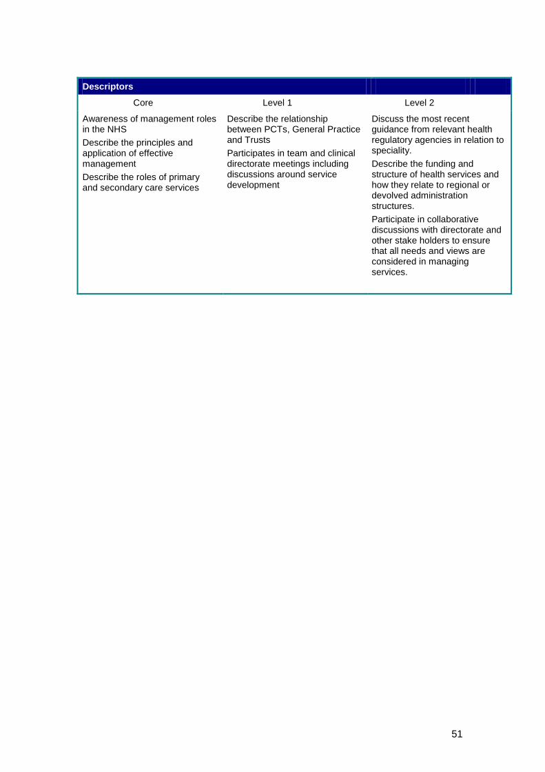
51
Descriptors
Core Level 1 Level 2
Awareness of management roles in the NHS Describe the principles and application of effective management Describe the roles of primary and secondary care services
Describe the relationship between PCTs, General Practice and Trusts Participates in team and clinical directorate meetings including discussions around service development
Discuss the most recent guidance from relevant health regulatory agencies in relation to speciality. Describe the funding and structure of health services and how they relate to regional or devolved administration structures. Participate in collaborative discussions with directorate and other stake holders to ensure that all needs and views are considered in managing services.
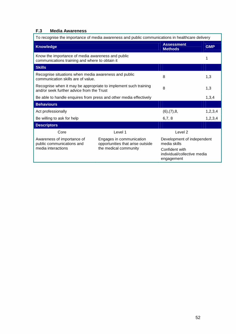
52
F.3 Media Awareness To recognise the importance of media awareness and public communications in healthcare delivery
Knowledge Assessment Methods GMP
Know the importance of media awareness and public communications training and where to obtain it 1
Skills
Recognise situations when media awareness and public communication skills are of value. 8 1,3
Recognise when it may be appropriate to implement such training and/or seek further advice from the Trust 8 1,3
Be able to handle enquires from press and other media effectively 1,3,4
Behaviours
Act professionally (6),(7),8, 1,2,3,4
Be willing to ask for help 6,7, 8 1,2,3,4
Descriptors
Core Level 1 Level 2
Awareness of importance of public communications and media interactions
Engages in communication opportunities that arise outside the medical community
Development of independent media skills Confident with individual/collective media engagement
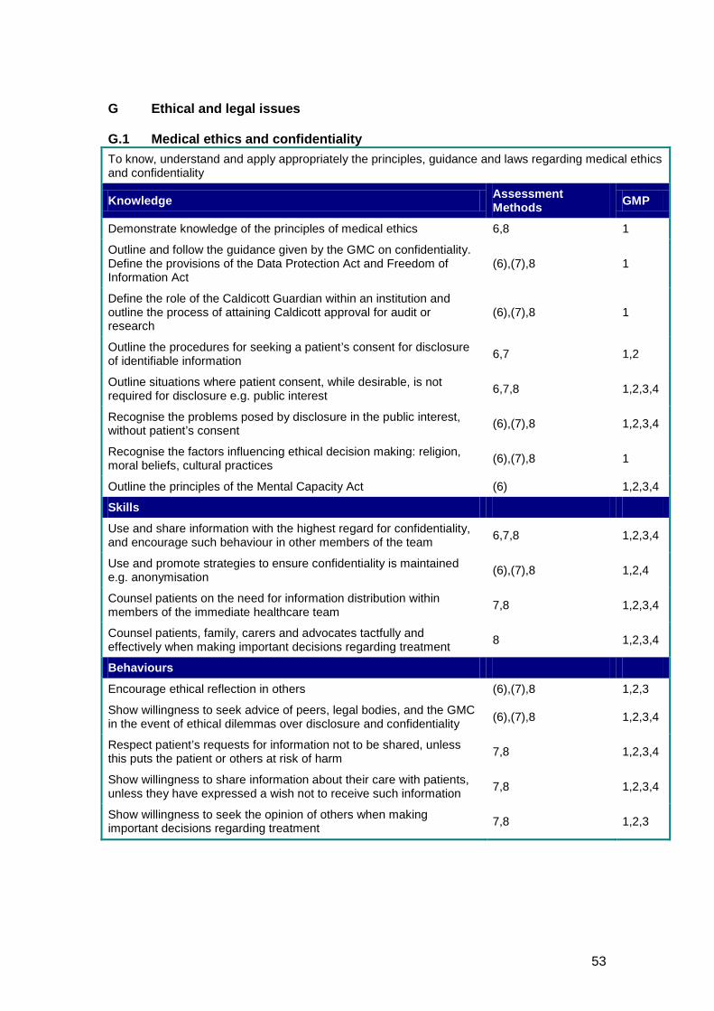
53
G Ethical and legal issues G.1 Medical ethics and confidentiality To know, understand and apply appropriately the principles, guidance and laws regarding medical ethics and confidentiality
Knowledge Assessment Methods GMP
Demonstrate knowledge of the principles of medical ethics 6,8 1
Outline and follow the guidance given by the GMC on confidentiality. Define the provisions of the Data Protection Act and Freedom of Information Act
(6),(7),8 1
Define the role of the Caldicott Guardian within an institution and outline the process of attaining Caldicott approval for audit or research
(6),(7),8 1
Outline the procedures for seeking a patient’s consent for disclosure of identifiable information 6,7 1,2
Outline situations where patient consent, while desirable, is not required for disclosure e.g. public interest 6,7,8 1,2,3,4
Recognise the problems posed by disclosure in the public interest, without patient’s consent (6),(7),8 1,2,3,4
Recognise the factors influencing ethical decision making: religion, moral beliefs, cultural practices (6),(7),8 1
Outline the principles of the Mental Capacity Act (6) 1,2,3,4
Skills
Use and share information with the highest regard for confidentiality, and encourage such behaviour in other members of the team 6,7,8 1,2,3,4
Use and promote strategies to ensure confidentiality is maintained e.g. anonymisation (6),(7),8 1,2,4
Counsel patients on the need for information distribution within members of the immediate healthcare team 7,8 1,2,3,4
Counsel patients, family, carers and advocates tactfully and effectively when making important decisions regarding treatment 8 1,2,3,4
Behaviours
Encourage ethical reflection in others (6),(7),8 1,2,3
Show willingness to seek advice of peers, legal bodies, and the GMC in the event of ethical dilemmas over disclosure and confidentiality (6),(7),8 1,2,3,4
Respect patient’s requests for information not to be shared, unless this puts the patient or others at risk of harm 7,8 1,2,3,4
Show willingness to share information about their care with patients, unless they have expressed a wish not to receive such information 7,8 1,2,3,4
Show willingness to seek the opinion of others when making important decisions regarding treatment 7,8 1,2,3
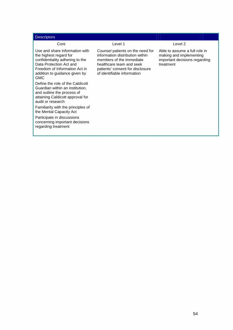
54
Descriptors
Core Level 1 Level 2
Use and share information with the highest regard for confidentiality adhering to the Data Protection Act and Freedom of Information Act in addition to guidance given by GMC Define the role of the Caldicott Guardian within an institution, and outline the process of attaining Caldicott approval for audit or research Familiarity with the principles of the Mental Capacity Act Participate in discussions concerning important decisions regarding treatment
Counsel patients on the need for information distribution within members of the immediate healthcare team and seek patients’ consent for disclosure of identifiable information
Able to assume a full role in making and implementing important decisions regarding treatment
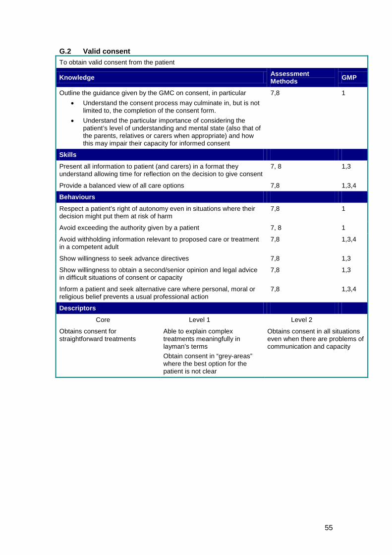
55
G.2 Valid consent To obtain valid consent from the patient
Knowledge Assessment Methods GMP
Outline the guidance given by the GMC on consent, in particular • Understand the consent process may culminate in, but is not
limited to, the completion of the consent form. • Understand the particular importance of considering the
patient’s level of understanding and mental state (also that of the parents, relatives or carers when appropriate) and how this may impair their capacity for informed consent
7,8 1
Skills
Present all information to patient (and carers) in a format they understand allowing time for reflection on the decision to give consent
7, 8 1,3
Provide a balanced view of all care options 7,8 1,3,4
Behaviours
Respect a patient’s right of autonomy even in situations where their decision might put them at risk of harm
7,8 1
Avoid exceeding the authority given by a patient 7, 8 1
Avoid withholding information relevant to proposed care or treatment in a competent adult
7,8 1,3,4
Show willingness to seek advance directives 7,8 1,3
Show willingness to obtain a second/senior opinion and legal advice in difficult situations of consent or capacity
7,8 1,3
Inform a patient and seek alternative care where personal, moral or religious belief prevents a usual professional action
7,8 1,3,4
Descriptors
Core Level 1 Level 2
Obtains consent for straightforward treatments
Able to explain complex treatments meaningfully in layman’s terms Obtain consent in “grey-areas” where the best option for the patient is not clear
Obtains consent in all situations even when there are problems of communication and capacity
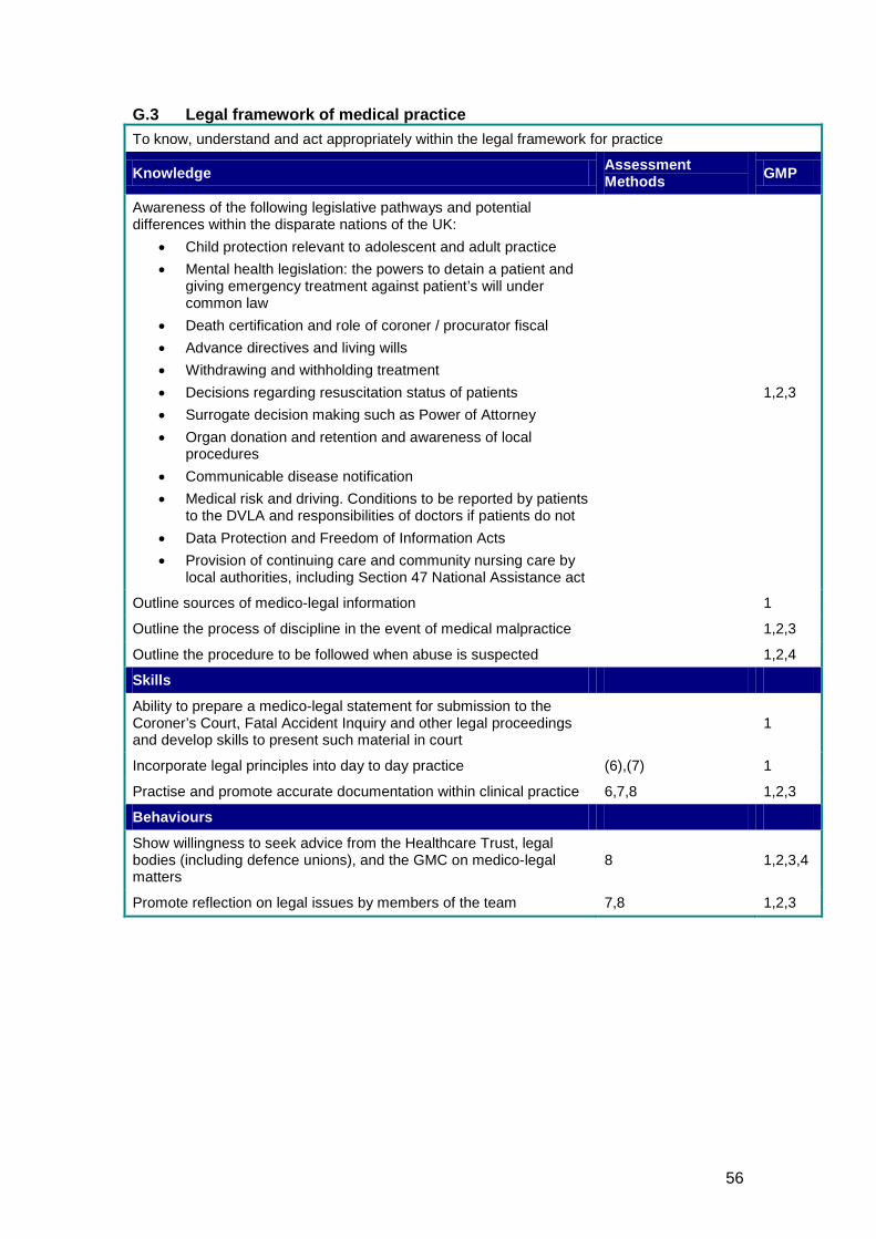
56
G.3 Legal framework of medical practice To know, understand and act appropriately within the legal framework for practice
Knowledge Assessment Methods GMP
Awareness of the following legislative pathways and potential differences within the disparate nations of the UK:
• Child protection relevant to adolescent and adult practice • Mental health legislation: the powers to detain a patient and
giving emergency treatment against patient’s will under common law
• Death certification and role of coroner / procurator fiscal • Advance directives and living wills • Withdrawing and withholding treatment • Decisions regarding resuscitation status of patients • Surrogate decision making such as Power of Attorney • Organ donation and retention and awareness of local
procedures • Communicable disease notification • Medical risk and driving. Conditions to be reported by patients
to the DVLA and responsibilities of doctors if patients do not • Data Protection and Freedom of Information Acts • Provision of continuing care and community nursing care by
local authorities, including Section 47 National Assistance act
1,2,3
Outline sources of medico-legal information 1
Outline the process of discipline in the event of medical malpractice 1,2,3
Outline the procedure to be followed when abuse is suspected 1,2,4
Skills
Ability to prepare a medico-legal statement for submission to the Coroner’s Court, Fatal Accident Inquiry and other legal proceedings and develop skills to present such material in court
1
Incorporate legal principles into day to day practice (6),(7) 1
Practise and promote accurate documentation within clinical practice 6,7,8 1,2,3
Behaviours
Show willingness to seek advice from the Healthcare Trust, legal bodies (including defence unions), and the GMC on medico-legal matters
8 1,2,3,4
Promote reflection on legal issues by members of the team 7,8 1,2,3
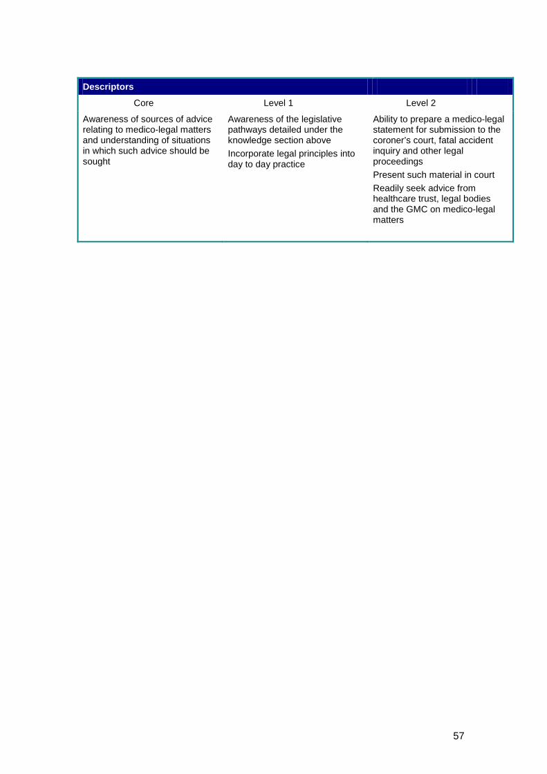
57
Descriptors
Core Level 1 Level 2
Awareness of sources of advice relating to medico-legal matters and understanding of situations in which such advice should be sought
Awareness of the legislative pathways detailed under the knowledge section above Incorporate legal principles into day to day practice
Ability to prepare a medico-legal statement for submission to the coroner’s court, fatal accident inquiry and other legal proceedings Present such material in court Readily seek advice from healthcare trust, legal bodies and the GMC on medico-legal matters
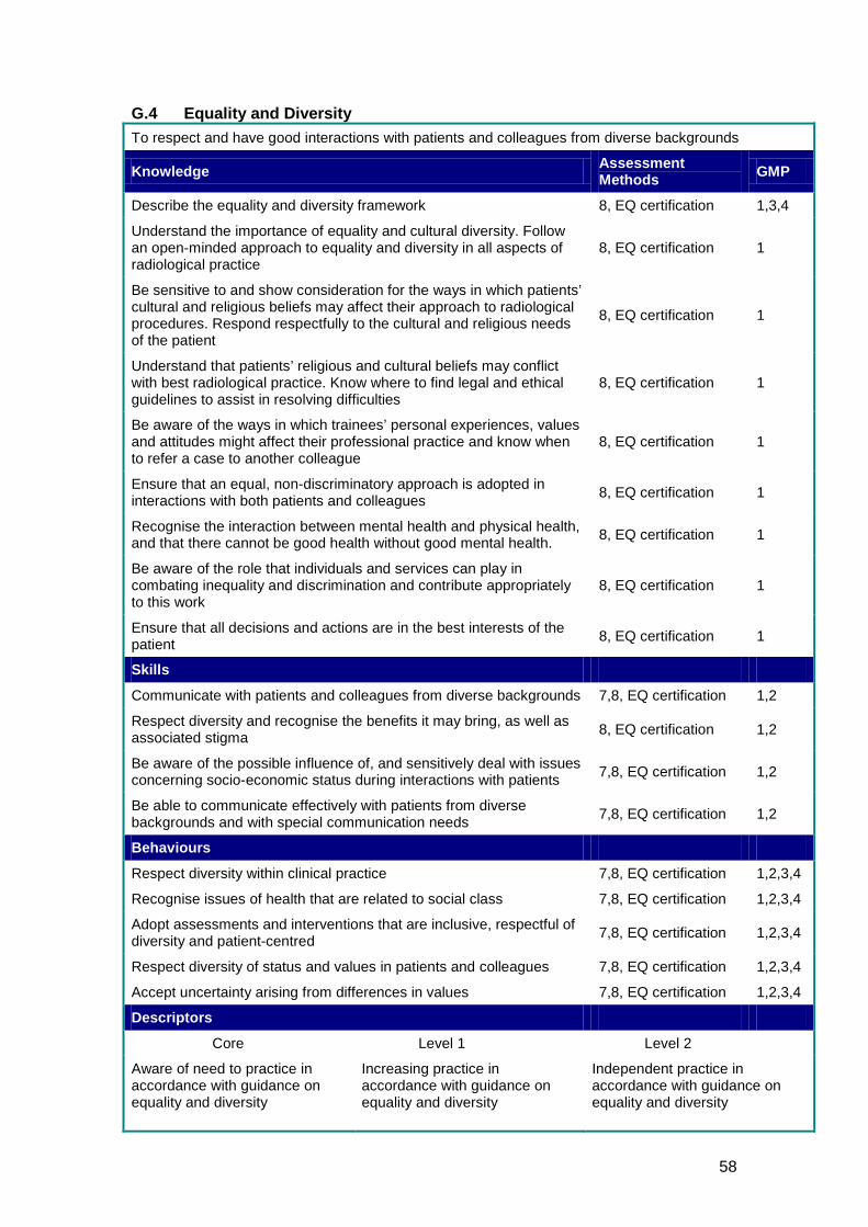
58
G.4 Equality and Diversity To respect and have good interactions with patients and colleagues from diverse backgrounds
Knowledge Assessment Methods GMP
Describe the equality and diversity framework 8, EQ certification 1,3,4
Understand the importance of equality and cultural diversity. Follow an open-minded approach to equality and diversity in all aspects of radiological practice
8, EQ certification 1
Be sensitive to and show consideration for the ways in which patients’ cultural and religious beliefs may affect their approach to radiological procedures. Respond respectfully to the cultural and religious needs of the patient
8, EQ certification 1
Understand that patients’ religious and cultural beliefs may conflict with best radiological practice. Know where to find legal and ethical guidelines to assist in resolving difficulties
8, EQ certification 1
Be aware of the ways in which trainees’ personal experiences, values and attitudes might affect their professional practice and know when to refer a case to another colleague
8, EQ certification 1
Ensure that an equal, non-discriminatory approach is adopted in interactions with both patients and colleagues 8, EQ certification 1
Recognise the interaction between mental health and physical health, and that there cannot be good health without good mental health. 8, EQ certification 1
Be aware of the role that individuals and services can play in combating inequality and discrimination and contribute appropriately to this work
8, EQ certification 1
Ensure that all decisions and actions are in the best interests of the patient 8, EQ certification 1
Skills
Communicate with patients and colleagues from diverse backgrounds 7,8, EQ certification 1,2
Respect diversity and recognise the benefits it may bring, as well as associated stigma 8, EQ certification 1,2
Be aware of the possible influence of, and sensitively deal with issues concerning socio-economic status during interactions with patients 7,8, EQ certification 1,2
Be able to communicate effectively with patients from diverse backgrounds and with special communication needs 7,8, EQ certification 1,2
Behaviours
Respect diversity within clinical practice 7,8, EQ certification 1,2,3,4
Recognise issues of health that are related to social class 7,8, EQ certification 1,2,3,4
Adopt assessments and interventions that are inclusive, respectful of diversity and patient-centred 7,8, EQ certification 1,2,3,4
Respect diversity of status and values in patients and colleagues 7,8, EQ certification 1,2,3,4
Accept uncertainty arising from differences in values 7,8, EQ certification 1,2,3,4
Descriptors
Core Level 1 Level 2
Aware of need to practice in accordance with guidance on equality and diversity
Increasing practice in accordance with guidance on equality and diversity
Independent practice in accordance with guidance on equality and diversity
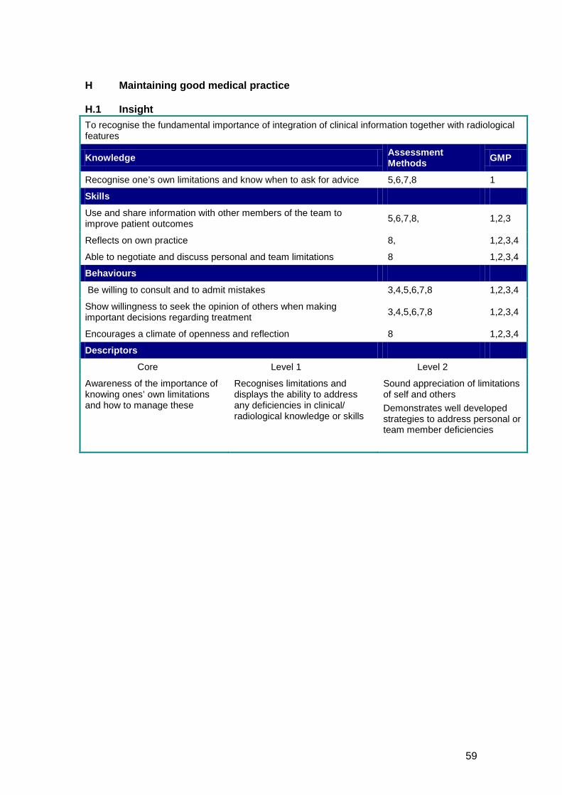
59
H Maintaining good medical practice H.1 Insight To recognise the fundamental importance of integration of clinical information together with radiological features
Knowledge Assessment Methods GMP
Recognise one’s own limitations and know when to ask for advice 5,6,7,8 1
Skills
Use and share information with other members of the team to improve patient outcomes 5,6,7,8, 1,2,3
Reflects on own practice 8, 1,2,3,4
Able to negotiate and discuss personal and team limitations 8 1,2,3,4
Behaviours
Be willing to consult and to admit mistakes 3,4,5,6,7,8 1,2,3,4
Show willingness to seek the opinion of others when making important decisions regarding treatment 3,4,5,6,7,8 1,2,3,4
Encourages a climate of openness and reflection 8 1,2,3,4
Descriptors
Core Level 1 Level 2
Awareness of the importance of knowing ones’ own limitations and how to manage these
Recognises limitations and displays the ability to address any deficiencies in clinical/ radiological knowledge or skills
Sound appreciation of limitations of self and others Demonstrates well developed strategies to address personal or team member deficiencies
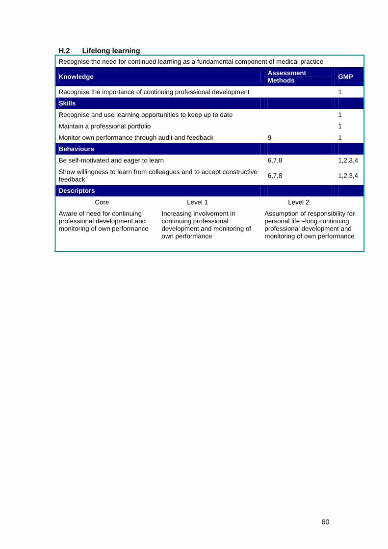
60
H.2 Lifelong learning Recognise the need for continued learning as a fundamental component of medical practice
Knowledge Assessment Methods GMP
Recognise the importance of continuing professional development 1
Skills
Recognise and use learning opportunities to keep up to date 1
Maintain a professional portfolio 1
Monitor own performance through audit and feedback 9 1
Behaviours
Be self-motivated and eager to learn 6,7,8 1,2,3,4
Show willingness to learn from colleagues and to accept constructive feedback 6,7,8 1,2,3,4
Descriptors
Core Level 1 Level 2
Aware of need for continuing professional development and monitoring of own performance
Increasing involvement in continuing professional development and monitoring of own performance
Assumption of responsibility for personal life –long continuing professional development and monitoring of own performance
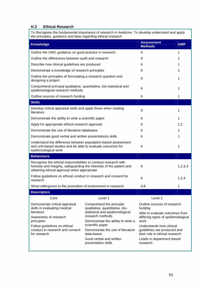
61
H.3 Ethical Research To Recognise the fundamental importance of research in medicine. To develop understand and apply the principles, guidance and laws regarding ethical research
Knowledge Assessment Methods GMP
Outline the GMC guidance on good practice in research 6 1
Outline the differences between audit and research 9 1
Describe how clinical guidelines are produced 6 1
Demonstrate a knowledge of research principles 6 1
Outline the principles of formulating a research question and designing a project 6 1
Comprehend principal qualitative, quantitative, bio-statistical and epidemiological research methods 6 1
Outline sources of research funding 6 1
Skills
Develop critical appraisal skills and apply these when reading literature 6 1
Demonstrate the ability to write a scientific paper 6 1
Apply for appropriate ethical research approval 6 1,2
Demonstrate the use of literature databases 6 1
Demonstrate good verbal and written presentations skills 6 1
Understand the difference between population-based assessment and unit-based studies and be able to evaluate outcomes for epidemiological work
6 1
Behaviours
Recognise the ethical responsibilities to conduct research with honesty and integrity, safeguarding the interests of the patient and obtaining ethical approval when appropriate
6 1,2,3,4
Follow guidelines on ethical conduct in research and consent for research 6 1,2,4
Show willingness to the promotion of involvement in research 6,8 1
Descriptors
Core Level 1 Level 2
Demonstrate critical appraisal skills in evaluating medical literature Awareness of research principles Follow guidelines on ethical conduct in research and consent for research
Comprehend the principle qualitative, quantitative, bio-statistical and epidemiological research methods Demonstrate the ability to write a scientific paper Demonstrate the use of literature data-bases Good verbal and written presentation skills
Outline sources of research funding Able to evaluate outcomes from differing types of epidemiological work Understands how clinical guidelines are produced and their role in ethical research Leads in department based research
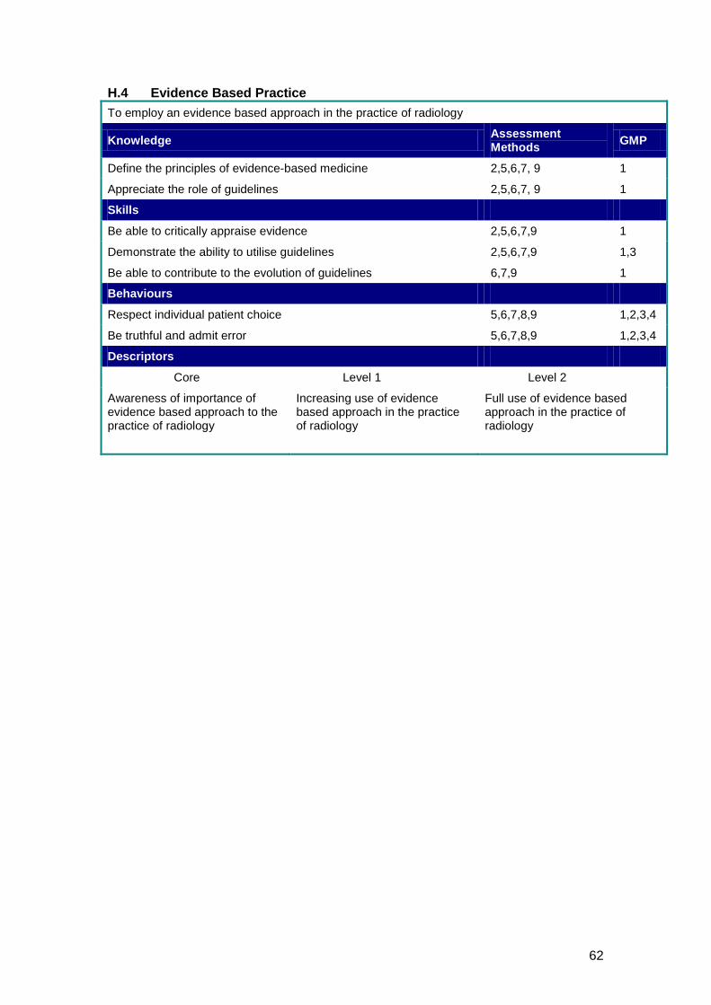
62
H.4 Evidence Based Practice To employ an evidence based approach in the practice of radiology
Knowledge Assessment Methods GMP
Define the principles of evidence-based medicine 2,5,6,7, 9 1
Appreciate the role of guidelines 2,5,6,7, 9 1
Skills
Be able to critically appraise evidence 2,5,6,7,9 1
Demonstrate the ability to utilise guidelines 2,5,6,7,9 1,3
Be able to contribute to the evolution of guidelines 6,7,9 1
Behaviours
Respect individual patient choice 5,6,7,8,9 1,2,3,4
Be truthful and admit error 5,6,7,8,9 1,2,3,4
Descriptors
Core Level 1 Level 2
Awareness of importance of evidence based approach to the practice of radiology
Increasing use of evidence based approach in the practice of radiology
Full use of evidence based approach in the practice of radiology
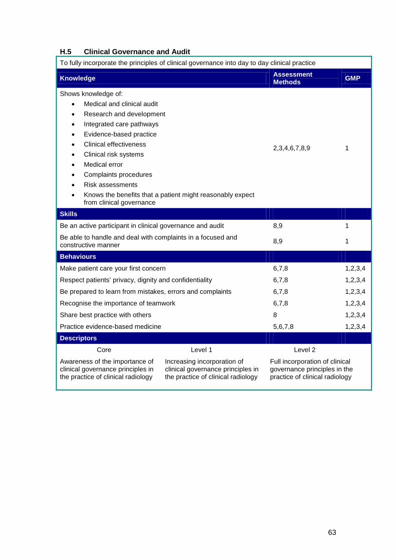
63
H.5 Clinical Governance and Audit To fully incorporate the principles of clinical governance into day to day clinical practice
Knowledge Assessment Methods GMP
Shows knowledge of: • Medical and clinical audit • Research and development • Integrated care pathways • Evidence-based practice • Clinical effectiveness • Clinical risk systems • Medical error • Complaints procedures • Risk assessments • Knows the benefits that a patient might reasonably expect
from clinical governance
2,3,4,6,7,8,9 1
Skills
Be an active participant in clinical governance and audit 8,9 1
Be able to handle and deal with complaints in a focused and constructive manner 8,9 1
Behaviours
Make patient care your first concern 6,7,8 1,2,3,4
Respect patients’ privacy, dignity and confidentiality 6,7,8 1,2,3,4
Be prepared to learn from mistakes, errors and complaints 6,7,8 1,2,3,4
Recognise the importance of teamwork 6,7,8 1,2,3,4
Share best practice with others 8 1,2,3,4
Practice evidence-based medicine 5,6,7,8 1,2,3,4
Descriptors
Core Level 1 Level 2
Awareness of the importance of clinical governance principles in the practice of clinical radiology
Increasing incorporation of clinical governance principles in the practice of clinical radiology
Full incorporation of clinical governance principles in the practice of clinical radiology
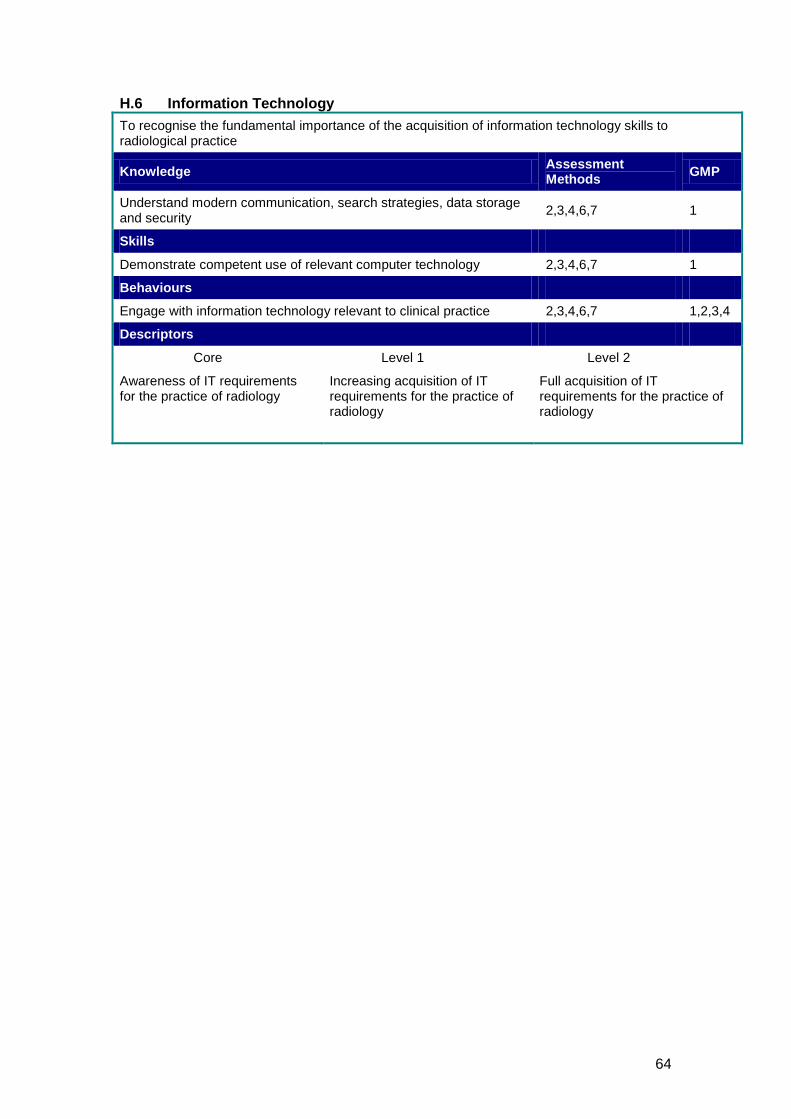
64
H.6 Information Technology To recognise the fundamental importance of the acquisition of information technology skills to radiological practice
Knowledge Assessment Methods GMP
Understand modern communication, search strategies, data storage and security 2,3,4,6,7 1
Skills
Demonstrate competent use of relevant computer technology 2,3,4,6,7 1
Behaviours
Engage with information technology relevant to clinical practice 2,3,4,6,7 1,2,3,4
Descriptors
Core Level 1 Level 2
Awareness of IT requirements for the practice of radiology
Increasing acquisition of IT requirements for the practice of radiology
Full acquisition of IT requirements for the practice of radiology
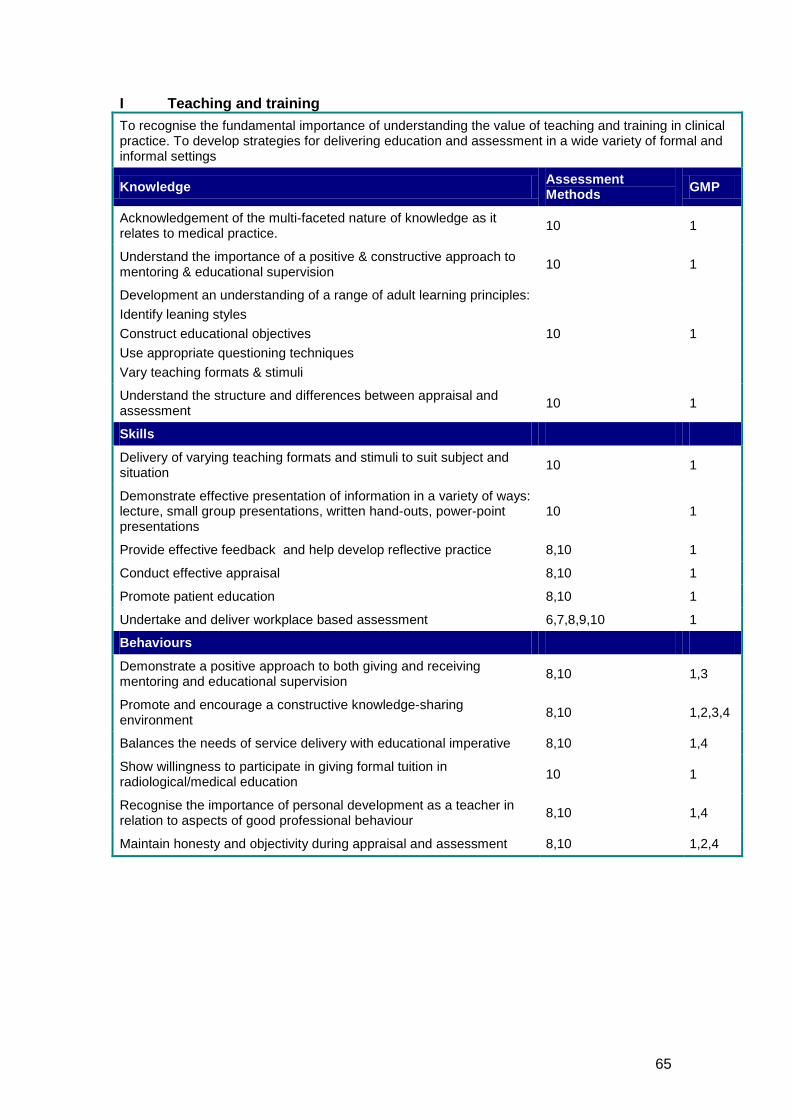
65
I Teaching and training To recognise the fundamental importance of understanding the value of teaching and training in clinical practice. To develop strategies for delivering education and assessment in a wide variety of formal and informal settings
Knowledge Assessment Methods GMP
Acknowledgement of the multi-faceted nature of knowledge as it relates to medical practice. 10 1
Understand the importance of a positive & constructive approach to mentoring & educational supervision 10 1
Development an understanding of a range of adult learning principles: Identify leaning styles Construct educational objectives Use appropriate questioning techniques Vary teaching formats & stimuli
10 1
Understand the structure and differences between appraisal and assessment 10 1
Skills
Delivery of varying teaching formats and stimuli to suit subject and situation 10 1
Demonstrate effective presentation of information in a variety of ways: lecture, small group presentations, written hand-outs, power-point presentations
10 1
Provide effective feedback and help develop reflective practice 8,10 1
Conduct effective appraisal 8,10 1
Promote patient education 8,10 1
Undertake and deliver workplace based assessment 6,7,8,9,10 1
Behaviours
Demonstrate a positive approach to both giving and receiving mentoring and educational supervision 8,10 1,3
Promote and encourage a constructive knowledge-sharing environment 8,10 1,2,3,4
Balances the needs of service delivery with educational imperative 8,10 1,4
Show willingness to participate in giving formal tuition in radiological/medical education 10 1
Recognise the importance of personal development as a teacher in relation to aspects of good professional behaviour 8,10 1,4
Maintain honesty and objectivity during appraisal and assessment 8,10 1,2,4
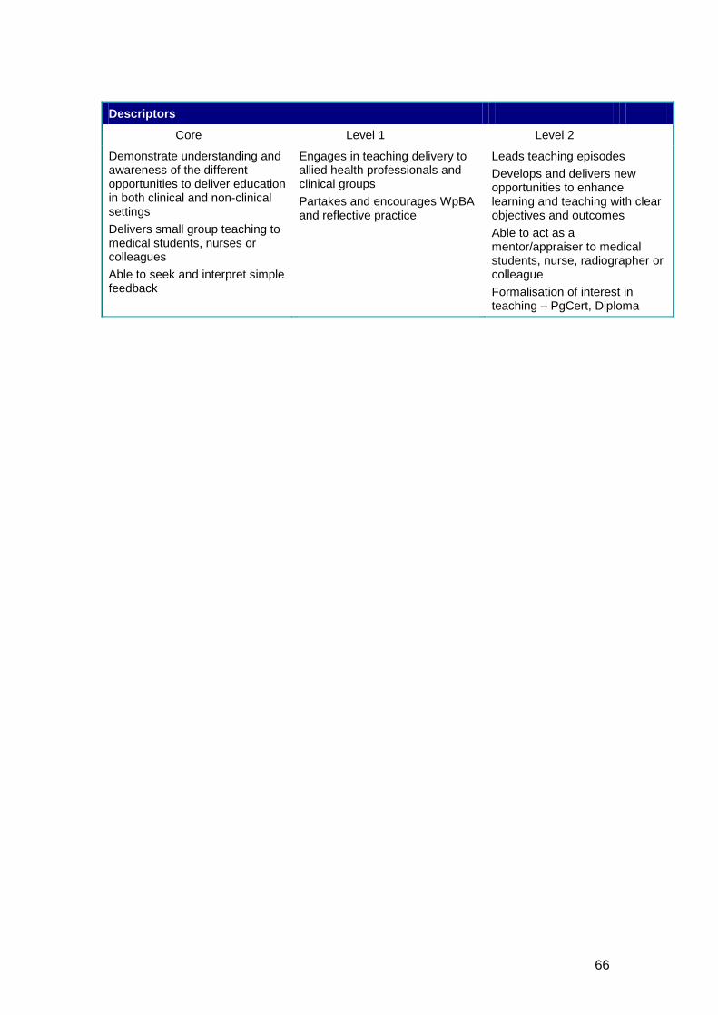
66
Descriptors
Core Level 1 Level 2
Demonstrate understanding and awareness of the different opportunities to deliver education in both clinical and non-clinical settings Delivers small group teaching to medical students, nurses or colleagues Able to seek and interpret simple feedback
Engages in teaching delivery to allied health professionals and clinical groups Partakes and encourages WpBA and reflective practice
Leads teaching episodes Develops and delivers new opportunities to enhance learning and teaching with clear objectives and outcomes Able to act as a mentor/appraiser to medical students, nurse, radiographer or colleague Formalisation of interest in teaching – PgCert, Diploma
67
3.4 RADIOLOGY SPECIFIC CONTENT All aspects of the radiology specific syllabus are referenced to illustrative examples of presentations and diagnoses. These examples are a guide to help both the trainer and trainee ensure sufficient curriculum coverage is obtained. In delivery of core training, the syllabus content is referenced to common presentations. These reflect the need for trainees to show competences across the breadth of the curriculum with particular emphasis on the most important/common topics within the curriculum. It is expected that trainees will produce evidence of at least one satisfactory assessment, either formative (WpBA) or summative (aspects of the FRCR), from all the common presentation topics by the completion of core training. During higher training, the syllabus is linked to a range of common and uncommon diagnoses. These lists are neither exhaustive nor complete as higher training is an integral part of lifelong learning with no upper limits. These lists are recommended topic areas from within which the trainee should produce evidence of assessments for the attainment of CCT in clinical radiology.
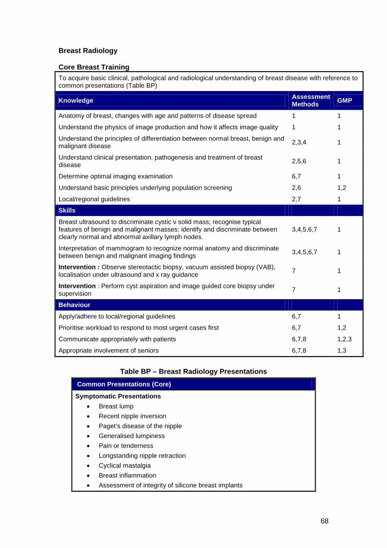
68
Breast Radiology Core Breast Training To acquire basic clinical, pathological and radiological understanding of breast disease with reference to common presentations (Table BP)
Knowledge Assessment Methods GMP
Anatomy of breast, changes with age and patterns of disease spread 1 1
Understand the physics of image production and how it affects image quality 1 1
Understand the principles of differentiation between normal breast, benign and malignant disease 2,3,4 1
Understand clinical presentation, pathogenesis and treatment of breast disease 2,5,6 1
Determine optimal imaging examination 6,7 1
Understand basic principles underlying population screening 2,6 1,2
Local/regional guidelines 2,7 1
Skills
Breast ultrasound to discriminate cystic v solid mass; recognise typical features of benign and malignant masses; identify and discriminate between clearly normal and abnormal axillary lymph nodes.
3,4,5,6,7 1
Interpretation of mammogram to recognize normal anatomy and discriminate between benign and malignant imaging findings 3,4,5,6,7 1
Intervention : Observe stereotactic biopsy, vacuum assisted biopsy (VAB), localisation under ultrasound and x ray guidance 7 1
Intervention : Perform cyst aspiration and image guided core biopsy under supervision 7 1
Behaviour
Apply/adhere to local/regional guidelines 6,7 1
Prioritise workload to respond to most urgent cases first 6,7 1,2
Communicate appropriately with patients 6,7,8 1,2,3
Appropriate involvement of seniors 6,7,8 1,3
Table BP – Breast Radiology Presentations
Common Presentations (Core)
Symptomatic Presentations • Breast lump • Recent nipple inversion • Paget’s disease of the nipple • Generalised lumpiness • Pain or tenderness • Longstanding nipple retraction • Cyclical mastalgia • Breast inflammation • Assessment of integrity of silicone breast implants
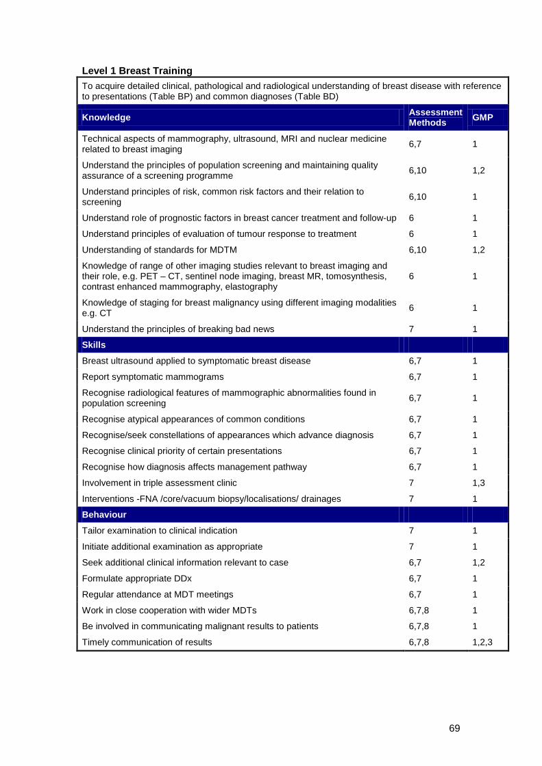
69
Level 1 Breast Training To acquire detailed clinical, pathological and radiological understanding of breast disease with reference to presentations (Table BP) and common diagnoses (Table BD)
Knowledge Assessment Methods GMP
Technical aspects of mammography, ultrasound, MRI and nuclear medicine related to breast imaging 6,7 1
Understand the principles of population screening and maintaining quality assurance of a screening programme 6,10 1,2
Understand principles of risk, common risk factors and their relation to screening 6,10 1
Understand role of prognostic factors in breast cancer treatment and follow-up 6 1
Understand principles of evaluation of tumour response to treatment 6 1
Understanding of standards for MDTM 6,10 1,2
Knowledge of range of other imaging studies relevant to breast imaging and their role, e.g. PET – CT, sentinel node imaging, breast MR, tomosynthesis, contrast enhanced mammography, elastography
6 1
Knowledge of staging for breast malignancy using different imaging modalities e.g. CT 6 1
Understand the principles of breaking bad news 7 1
Skills
Breast ultrasound applied to symptomatic breast disease 6,7 1
Report symptomatic mammograms 6,7 1
Recognise radiological features of mammographic abnormalities found in population screening 6,7 1
Recognise atypical appearances of common conditions 6,7 1
Recognise/seek constellations of appearances which advance diagnosis 6,7 1
Recognise clinical priority of certain presentations 6,7 1
Recognise how diagnosis affects management pathway 6,7 1
Involvement in triple assessment clinic 7 1,3
Interventions -FNA /core/vacuum biopsy/localisations/ drainages 7 1
Behaviour
Tailor examination to clinical indication 7 1
Initiate additional examination as appropriate 7 1
Seek additional clinical information relevant to case 6,7 1,2
Formulate appropriate DDx 6,7 1
Regular attendance at MDT meetings 6,7 1
Work in close cooperation with wider MDTs 6,7,8 1
Be involved in communicating malignant results to patients 6,7,8 1
Timely communication of results 6,7,8 1,2,3
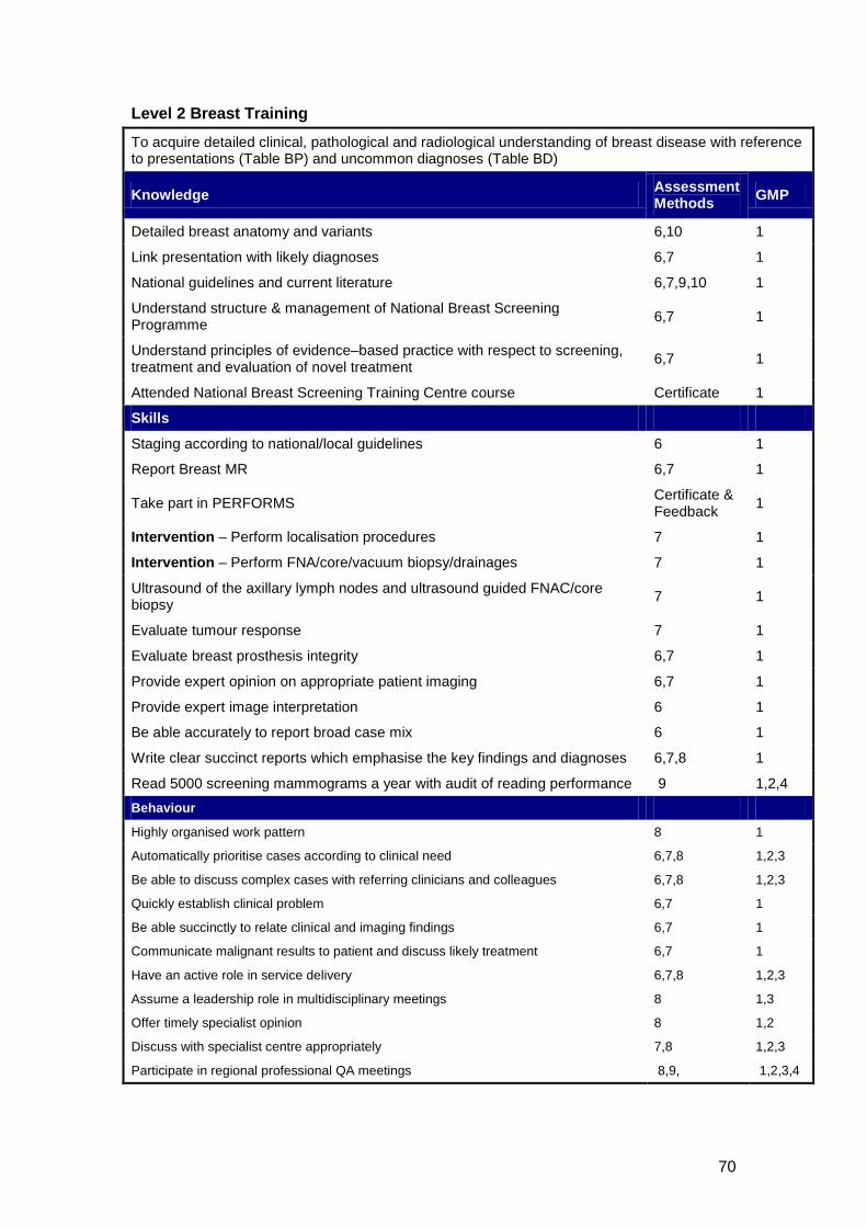
70
Level 2 Breast Training
To acquire detailed clinical, pathological and radiological understanding of breast disease with reference to presentations (Table BP) and uncommon diagnoses (Table BD)
Knowledge Assessment Methods GMP
Detailed breast anatomy and variants 6,10 1
Link presentation with likely diagnoses 6,7 1
National guidelines and current literature 6,7,9,10 1
Understand structure & management of National Breast Screening Programme 6,7 1
Understand principles of evidence–based practice with respect to screening, treatment and evaluation of novel treatment 6,7 1
Attended National Breast Screening Training Centre course Certificate 1
Skills
Staging according to national/local guidelines 6 1
Report Breast MR 6,7 1
Take part in PERFORMS Certificate & Feedback 1
Intervention – Perform localisation procedures 7 1
Intervention – Perform FNA/core/vacuum biopsy/drainages 7 1
Ultrasound of the axillary lymph nodes and ultrasound guided FNAC/core biopsy 7 1
Evaluate tumour response 7 1
Evaluate breast prosthesis integrity 6,7 1
Provide expert opinion on appropriate patient imaging 6,7 1
Provide expert image interpretation 6 1
Be able accurately to report broad case mix 6 1
Write clear succinct reports which emphasise the key findings and diagnoses 6,7,8 1
Read 5000 screening mammograms a year with audit of reading performance 9 1,2,4 Behaviour
Highly organised work pattern 8 1
Automatically prioritise cases according to clinical need 6,7,8 1,2,3
Be able to discuss complex cases with referring clinicians and colleagues 6,7,8 1,2,3
Quickly establish clinical problem 6,7 1
Be able succinctly to relate clinical and imaging findings 6,7 1
Communicate malignant results to patient and discuss likely treatment 6,7 1
Have an active role in service delivery 6,7,8 1,2,3
Assume a leadership role in multidisciplinary meetings 8 1,3
Offer timely specialist opinion 8 1,2
Discuss with specialist centre appropriately 7,8 1,2,3
Participate in regional professional QA meetings 8,9, 1,2,3,4
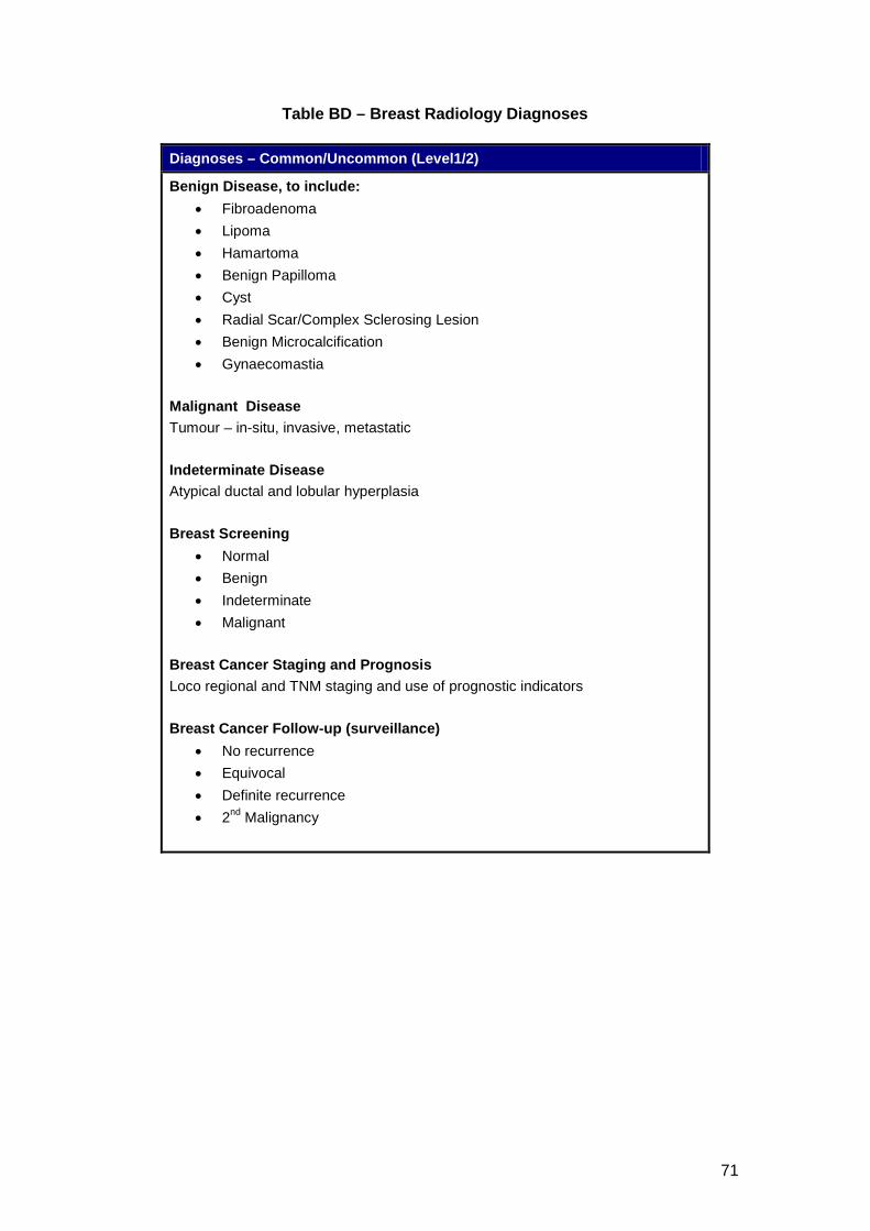
71
Table BD – Breast Radiology Diagnoses
Diagnoses – Common/Uncommon (Level1/2)
Benign Disease, to include: • Fibroadenoma • Lipoma • Hamartoma • Benign Papilloma • Cyst • Radial Scar/Complex Sclerosing Lesion • Benign Microcalcification • Gynaecomastia
Malignant Disease Tumour – in-situ, invasive, metastatic Indeterminate Disease Atypical ductal and lobular hyperplasia Breast Screening
• Normal • Benign • Indeterminate • Malignant
Breast Cancer Staging and Prognosis Loco regional and TNM staging and use of prognostic indicators Breast Cancer Follow-up (surveillance)
• No recurrence • Equivocal • Definite recurrence • 2nd
Malignancy
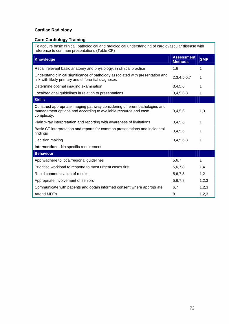
72
Cardiac Radiology Core Cardiology Training To acquire basic clinical, pathological and radiological understanding of cardiovascular disease with reference to common presentations (Table CP)
Knowledge Assessment Methods GMP
Recall relevant basic anatomy and physiology, in clinical practice 1,6 1
Understand clinical significance of pathology associated with presentation and link with likely primary and differential diagnoses 2,3,4,5,6,7 1
Determine optimal imaging examination 3,4,5,6 1
Local/regional guidelines in relation to presentations 3,4,5,6,8 1
Skills
Construct appropriate imaging pathway considering different pathologies and management options and according to available resource and case complexity.
3,4,5,6 1,3
Plain x-ray interpretation and reporting with awareness of limitations 3,4,5,6 1
Basic CT interpretation and reports for common presentations and incidental findings 3,4,5,6 1
Decision making 3,4,5,6,8 1
Intervention – No specific requirement
Behaviour
Apply/adhere to local/regional guidelines 5,6,7 1
Prioritise workload to respond to most urgent cases first 5,6,7,8 1,4
Rapid communication of results 5,6,7,8 1,2
Appropriate involvement of seniors 5,6,7,8 1,2,3
Communicate with patients and obtain informed consent where appropriate 6,7 1,2,3
Attend MDTs 8 1,2,3
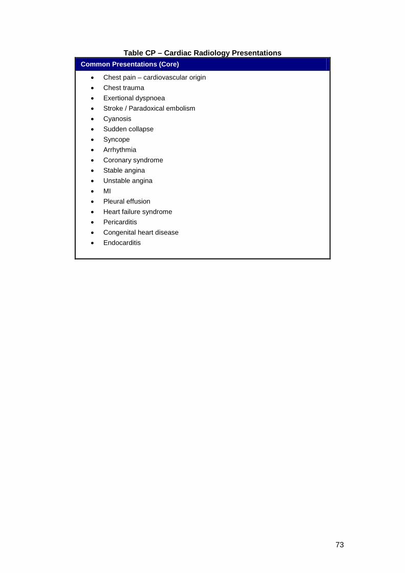
73
Table CP – Cardiac Radiology Presentations Common Presentations (Core)
• Chest pain – cardiovascular origin • Chest trauma • Exertional dyspnoea • Stroke / Paradoxical embolism • Cyanosis • Sudden collapse • Syncope • Arrhythmia • Coronary syndrome • Stable angina • Unstable angina • MI • Pleural effusion • Heart failure syndrome • Pericarditis • Congenital heart disease • Endocarditis
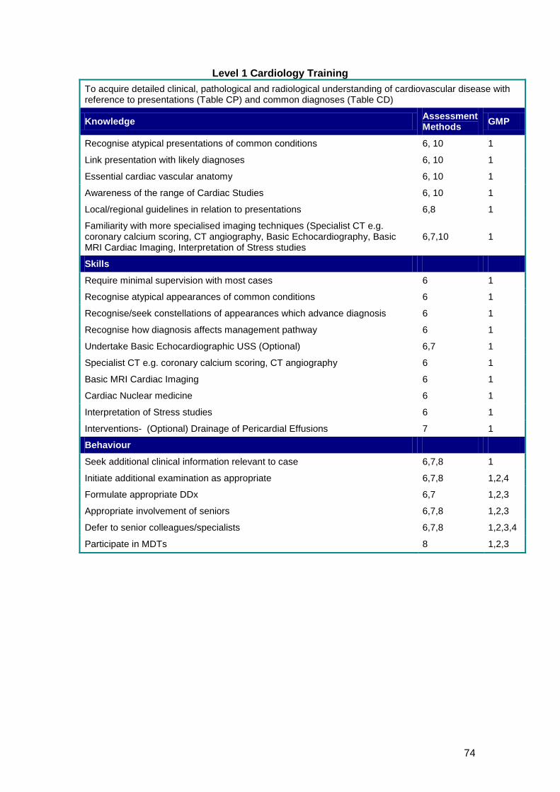
74
Level 1 Cardiology Training To acquire detailed clinical, pathological and radiological understanding of cardiovascular disease with reference to presentations (Table CP) and common diagnoses (Table CD)
Knowledge Assessment Methods GMP
Recognise atypical presentations of common conditions 6, 10 1
Link presentation with likely diagnoses 6, 10 1
Essential cardiac vascular anatomy 6, 10 1
Awareness of the range of Cardiac Studies 6, 10 1
Local/regional guidelines in relation to presentations 6,8 1
Familiarity with more specialised imaging techniques (Specialist CT e.g. coronary calcium scoring, CT angiography, Basic Echocardiography, Basic MRI Cardiac Imaging, Interpretation of Stress studies
6,7,10 1
Skills
Require minimal supervision with most cases 6 1
Recognise atypical appearances of common conditions 6 1
Recognise/seek constellations of appearances which advance diagnosis 6 1
Recognise how diagnosis affects management pathway 6 1
Undertake Basic Echocardiographic USS (Optional) 6,7 1
Specialist CT e.g. coronary calcium scoring, CT angiography 6 1
Basic MRI Cardiac Imaging 6 1
Cardiac Nuclear medicine 6 1
Interpretation of Stress studies 6 1
Interventions- (Optional) Drainage of Pericardial Effusions 7 1
Behaviour
Seek additional clinical information relevant to case 6,7,8 1
Initiate additional examination as appropriate 6,7,8 1,2,4
Formulate appropriate DDx 6,7 1,2,3
Appropriate involvement of seniors 6,7,8 1,2,3
Defer to senior colleagues/specialists 6,7,8 1,2,3,4
Participate in MDTs 8 1,2,3
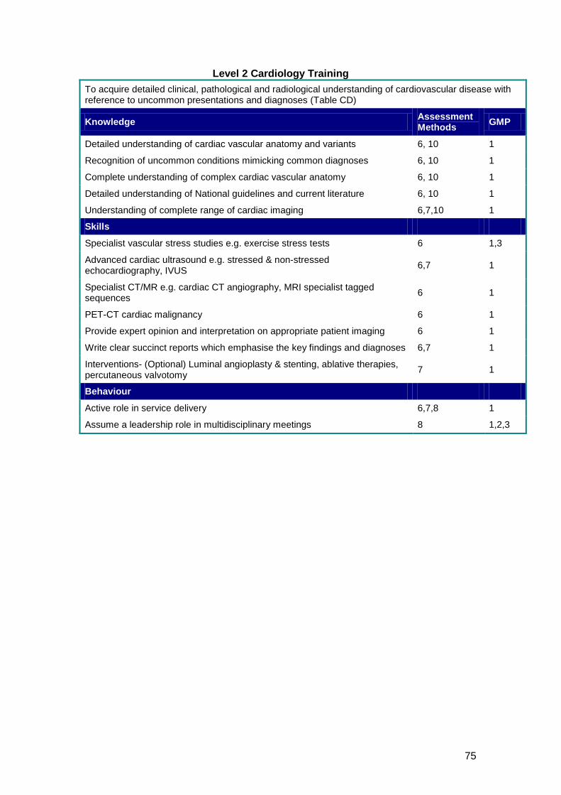
75
Level 2 Cardiology Training To acquire detailed clinical, pathological and radiological understanding of cardiovascular disease with reference to uncommon presentations and diagnoses (Table CD)
Knowledge Assessment Methods GMP
Detailed understanding of cardiac vascular anatomy and variants 6, 10 1
Recognition of uncommon conditions mimicking common diagnoses 6, 10 1
Complete understanding of complex cardiac vascular anatomy 6, 10 1
Detailed understanding of National guidelines and current literature 6, 10 1
Understanding of complete range of cardiac imaging 6,7,10 1
Skills
Specialist vascular stress studies e.g. exercise stress tests 6 1,3
Advanced cardiac ultrasound e.g. stressed & non-stressed echocardiography, IVUS 6,7 1
Specialist CT/MR e.g. cardiac CT angiography, MRI specialist tagged sequences 6 1
PET-CT cardiac malignancy 6 1
Provide expert opinion and interpretation on appropriate patient imaging 6 1
Write clear succinct reports which emphasise the key findings and diagnoses 6,7 1
Interventions- (Optional) Luminal angioplasty & stenting, ablative therapies, percutaneous valvotomy 7 1
Behaviour
Active role in service delivery 6,7,8 1
Assume a leadership role in multidisciplinary meetings 8 1,2,3
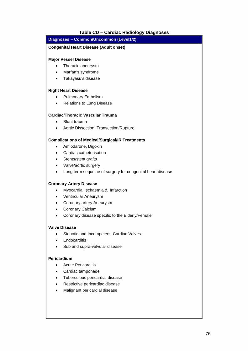
76
Table CD – Cardiac Radiology Diagnoses Diagnoses – Common/Uncommon (Level1/2)
Congenital Heart Disease (Adult onset) Major Vessel Disease
• Thoracic aneurysm • Marfan’s syndrome • Takayasu’s disease
Right Heart Disease
• Pulmonary Embolism • Relations to Lung Disease
Cardiac/Thoracic Vascular Trauma
• Blunt trauma • Aortic Dissection, Transection/Rupture
Complications of Medical/Surgical/IR Treatments
• Amiodarone, Digoxin • Cardiac catheterisation • Stents/stent grafts • Valve/aortic surgery • Long term sequelae of surgery for congenital heart disease
Coronary Artery Disease
• Myocardial Ischaemia & Infarction • Ventricular Aneurysm • Coronary artery Aneurysm • Coronary Calcium • Coronary disease specific to the Elderly/Female
Valve Disease
• Stenotic and Incompetent Cardiac Valves • Endocarditis • Sub and supra-valvular disease
Pericardium
• Acute Pericarditis • Cardiac tamponade • Tuberculous pericardial disease • Restrictive pericardiac disease • Malignant pericardial disease
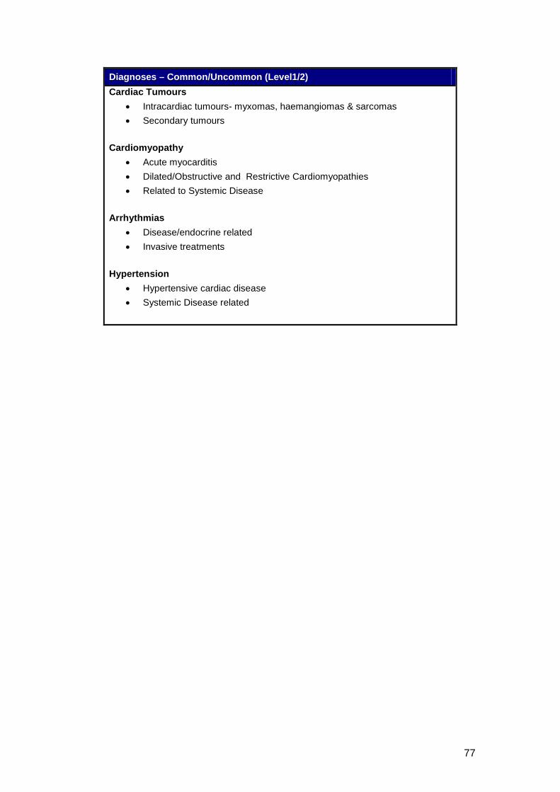
77
Diagnoses – Common/Uncommon (Level1/2) Cardiac Tumours
• Intracardiac tumours- myxomas, haemangiomas & sarcomas • Secondary tumours
Cardiomyopathy
• Acute myocarditis • Dilated/Obstructive and Restrictive Cardiomyopathies • Related to Systemic Disease
Arrhythmias
• Disease/endocrine related • Invasive treatments
Hypertension
• Hypertensive cardiac disease • Systemic Disease related
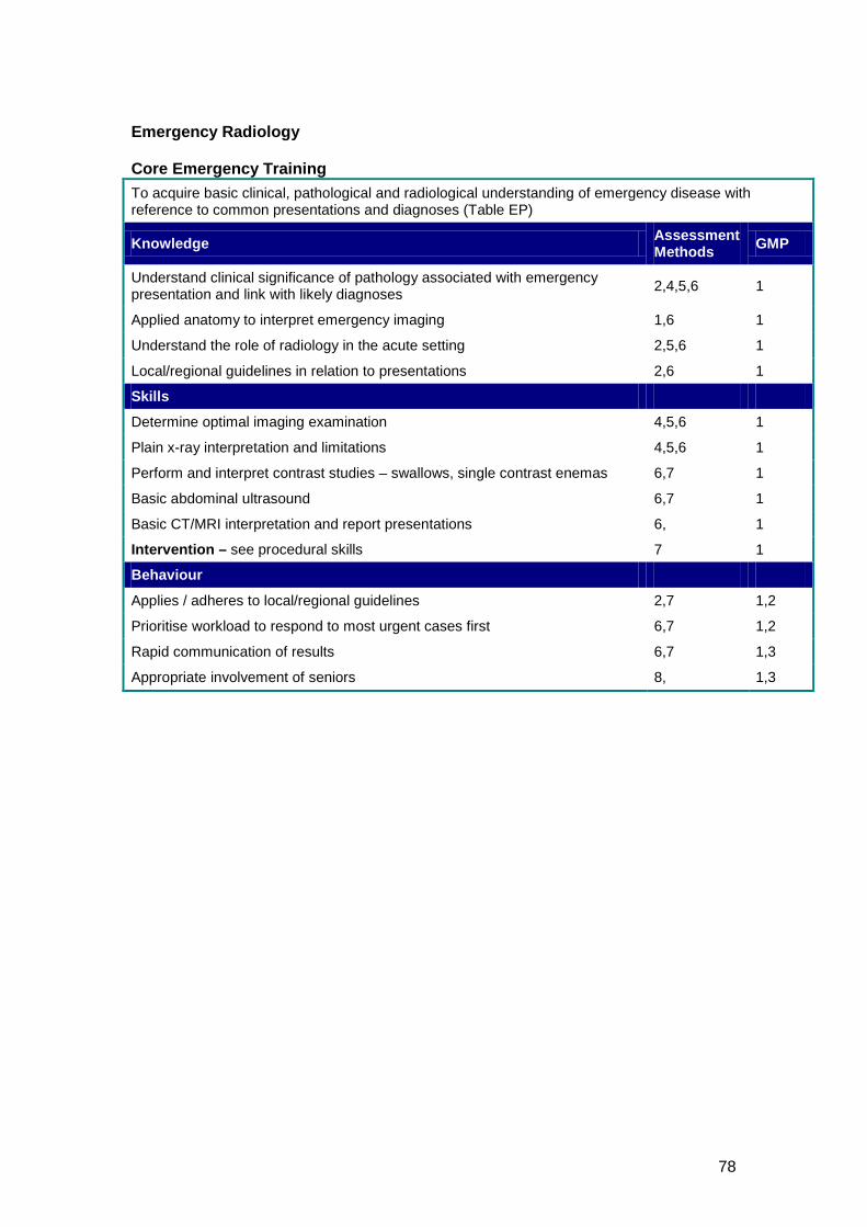
78
Emergency Radiology Core Emergency Training To acquire basic clinical, pathological and radiological understanding of emergency disease with reference to common presentations and diagnoses (Table EP)
Knowledge Assessment Methods GMP
Understand clinical significance of pathology associated with emergency presentation and link with likely diagnoses 2,4,5,6 1
Applied anatomy to interpret emergency imaging 1,6 1
Understand the role of radiology in the acute setting 2,5,6 1
Local/regional guidelines in relation to presentations 2,6 1
Skills
Determine optimal imaging examination 4,5,6 1
Plain x-ray interpretation and limitations 4,5,6 1
Perform and interpret contrast studies – swallows, single contrast enemas 6,7 1
Basic abdominal ultrasound 6,7 1
Basic CT/MRI interpretation and report presentations 6, 1
Intervention – see procedural skills 7 1
Behaviour
Applies / adheres to local/regional guidelines 2,7 1,2
Prioritise workload to respond to most urgent cases first 6,7 1,2
Rapid communication of results 6,7 1,3
Appropriate involvement of seniors 8, 1,3
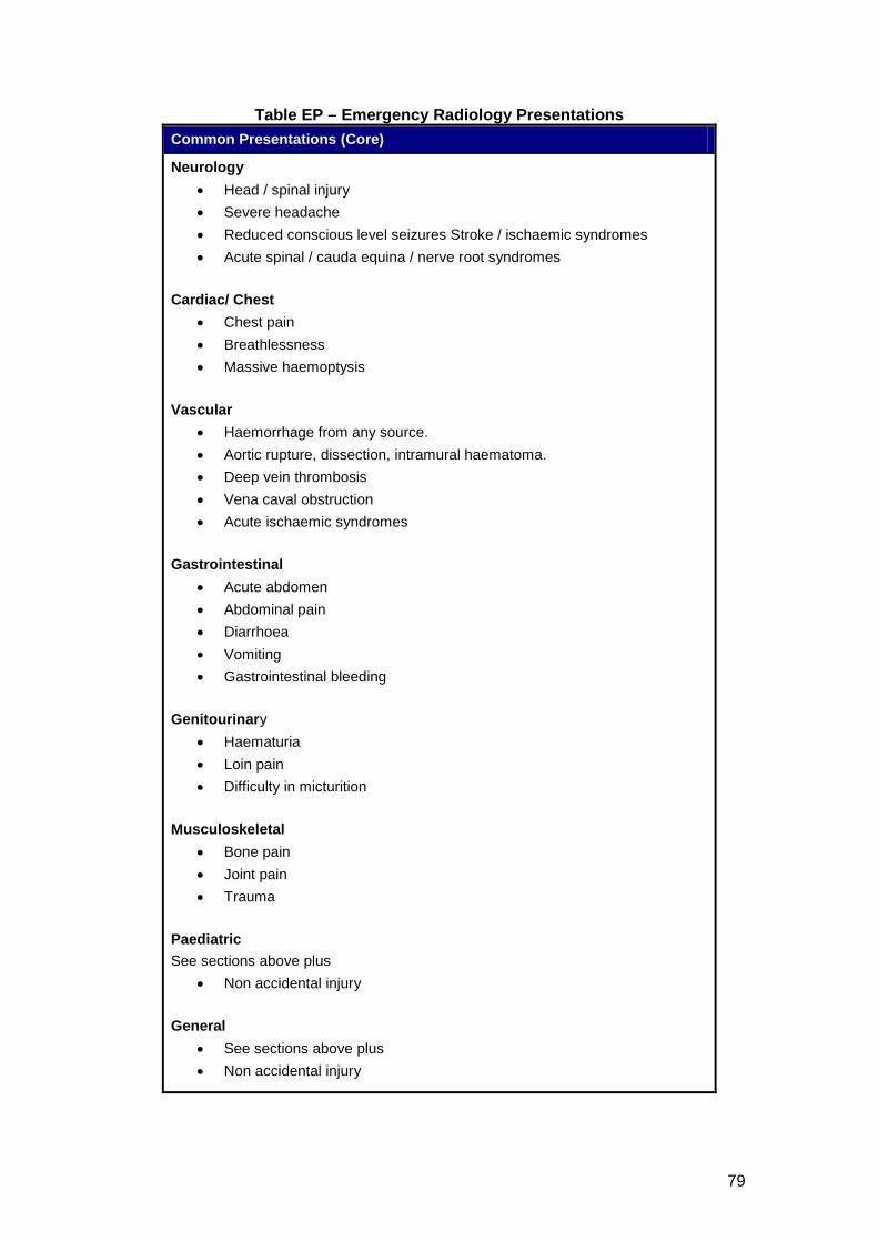
79
Table EP – Emergency Radiology Presentations Common Presentations (Core)
Neurology • Head / spinal injury • Severe headache • Reduced conscious level seizures Stroke / ischaemic syndromes • Acute spinal / cauda equina / nerve root syndromes
Cardiac/ Chest
• Chest pain • Breathlessness • Massive haemoptysis
Vascular
• Haemorrhage from any source. • Aortic rupture, dissection, intramural haematoma. • Deep vein thrombosis • Vena caval obstruction • Acute ischaemic syndromes
Gastrointestinal
• Acute abdomen • Abdominal pain • Diarrhoea • Vomiting • Gastrointestinal bleeding
Genitourinary
• Haematuria • Loin pain • Difficulty in micturition
Musculoskeletal
• Bone pain • Joint pain • Trauma
Paediatric See sections above plus
• Non accidental injury General
• See sections above plus • Non accidental injury
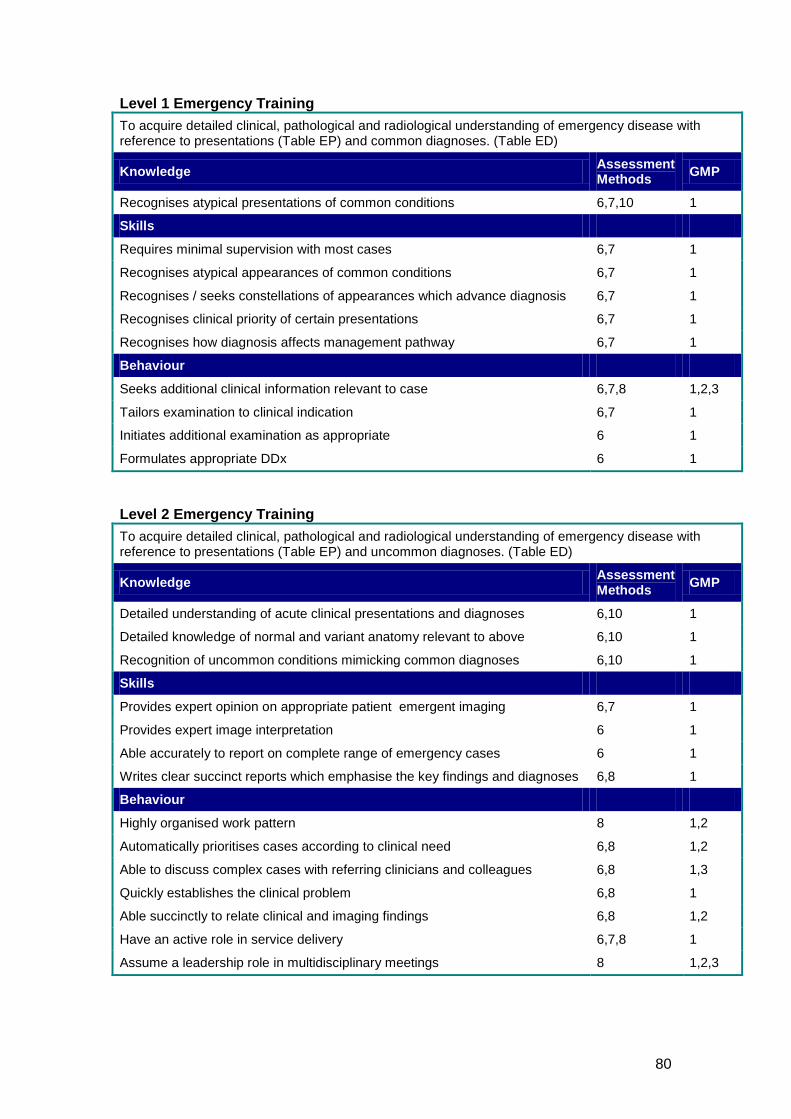
80
Level 1 Emergency Training To acquire detailed clinical, pathological and radiological understanding of emergency disease with reference to presentations (Table EP) and common diagnoses. (Table ED)
Knowledge Assessment Methods GMP
Recognises atypical presentations of common conditions 6,7,10 1
Skills
Requires minimal supervision with most cases 6,7 1
Recognises atypical appearances of common conditions 6,7 1
Recognises / seeks constellations of appearances which advance diagnosis 6,7 1
Recognises clinical priority of certain presentations 6,7 1
Recognises how diagnosis affects management pathway 6,7 1
Behaviour
Seeks additional clinical information relevant to case 6,7,8 1,2,3
Tailors examination to clinical indication 6,7 1
Initiates additional examination as appropriate 6 1
Formulates appropriate DDx 6 1
Level 2 Emergency Training To acquire detailed clinical, pathological and radiological understanding of emergency disease with reference to presentations (Table EP) and uncommon diagnoses. (Table ED)
Knowledge Assessment Methods GMP
Detailed understanding of acute clinical presentations and diagnoses 6,10 1
Detailed knowledge of normal and variant anatomy relevant to above 6,10 1
Recognition of uncommon conditions mimicking common diagnoses 6,10 1
Skills
Provides expert opinion on appropriate patient emergent imaging 6,7 1
Provides expert image interpretation 6 1
Able accurately to report on complete range of emergency cases 6 1
Writes clear succinct reports which emphasise the key findings and diagnoses 6,8 1
Behaviour
Highly organised work pattern 8 1,2
Automatically prioritises cases according to clinical need 6,8 1,2
Able to discuss complex cases with referring clinicians and colleagues 6,8 1,3
Quickly establishes the clinical problem 6,8 1
Able succinctly to relate clinical and imaging findings 6,8 1,2
Have an active role in service delivery 6,7,8 1
Assume a leadership role in multidisciplinary meetings 8 1,2,3
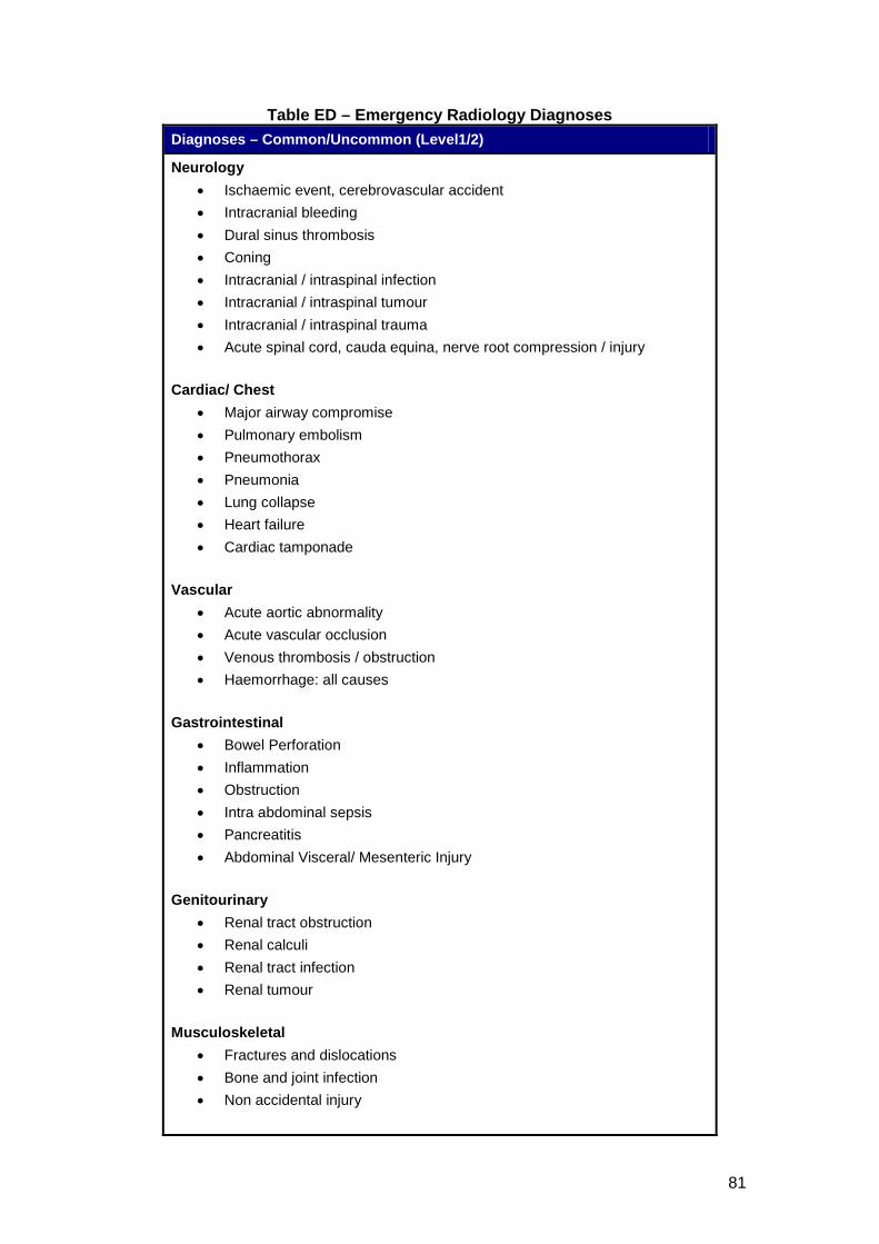
81
Table ED – Emergency Radiology Diagnoses Diagnoses – Common/Uncommon (Level1/2)
Neurology • Ischaemic event, cerebrovascular accident • Intracranial bleeding • Dural sinus thrombosis • Coning • Intracranial / intraspinal infection • Intracranial / intraspinal tumour • Intracranial / intraspinal trauma • Acute spinal cord, cauda equina, nerve root compression / injury
Cardiac/ Chest
• Major airway compromise • Pulmonary embolism • Pneumothorax • Pneumonia • Lung collapse • Heart failure • Cardiac tamponade
Vascular
• Acute aortic abnormality • Acute vascular occlusion • Venous thrombosis / obstruction • Haemorrhage: all causes
Gastrointestinal
• Bowel Perforation • Inflammation • Obstruction • Intra abdominal sepsis • Pancreatitis • Abdominal Visceral/ Mesenteric Injury
Genitourinary
• Renal tract obstruction • Renal calculi • Renal tract infection • Renal tumour
Musculoskeletal
• Fractures and dislocations • Bone and joint infection • Non accidental injury
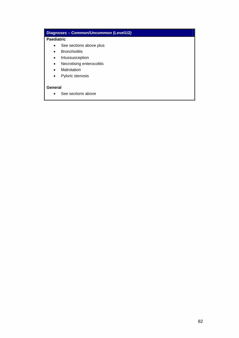
82
Diagnoses – Common/Uncommon (Level1/2) Paediatric
• See sections above plus • Bronchiolitis • Intussusception • Necrotising enterocolitis • Malrotation • Pyloric stenosis
General
• See sections above
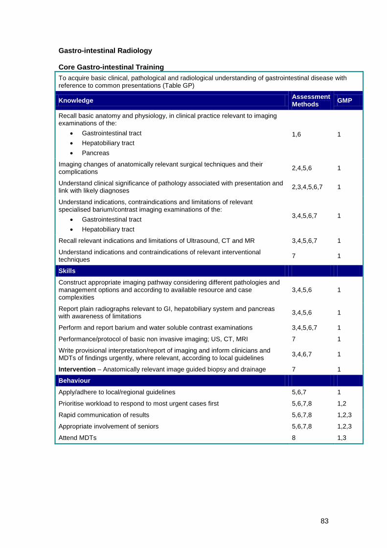
83
Gastro-intestinal Radiology Core Gastro-intestinal Training To acquire basic clinical, pathological and radiological understanding of gastrointestinal disease with reference to common presentations (Table GP)
Knowledge Assessment Methods GMP
Recall basic anatomy and physiology, in clinical practice relevant to imaging examinations of the:
• Gastrointestinal tract • Hepatobiliary tract • Pancreas
1,6 1
Imaging changes of anatomically relevant surgical techniques and their complications 2,4,5,6 1
Understand clinical significance of pathology associated with presentation and link with likely diagnoses 2,3,4,5,6,7 1
Understand indications, contraindications and limitations of relevant specialised barium/contrast imaging examinations of the:
• Gastrointestinal tract • Hepatobiliary tract
3,4,5,6,7 1
Recall relevant indications and limitations of Ultrasound, CT and MR 3,4,5,6,7 1
Understand indications and contraindications of relevant interventional techniques 7 1
Skills
Construct appropriate imaging pathway considering different pathologies and management options and according to available resource and case complexities
3,4,5,6 1
Report plain radiographs relevant to GI, hepatobiliary system and pancreas with awareness of limitations 3,4,5,6 1
Perform and report barium and water soluble contrast examinations 3,4,5,6,7 1
Performance/protocol of basic non invasive imaging; US, CT, MRI 7 1
Write provisional interpretation/report of imaging and inform clinicians and MDTs of findings urgently, where relevant, according to local guidelines 3,4,6,7 1
Intervention – Anatomically relevant image guided biopsy and drainage 7 1
Behaviour
Apply/adhere to local/regional guidelines 5,6,7 1
Prioritise workload to respond to most urgent cases first 5,6,7,8 1,2
Rapid communication of results 5,6,7,8 1,2,3
Appropriate involvement of seniors 5,6,7,8 1,2,3
Attend MDTs 8 1,3
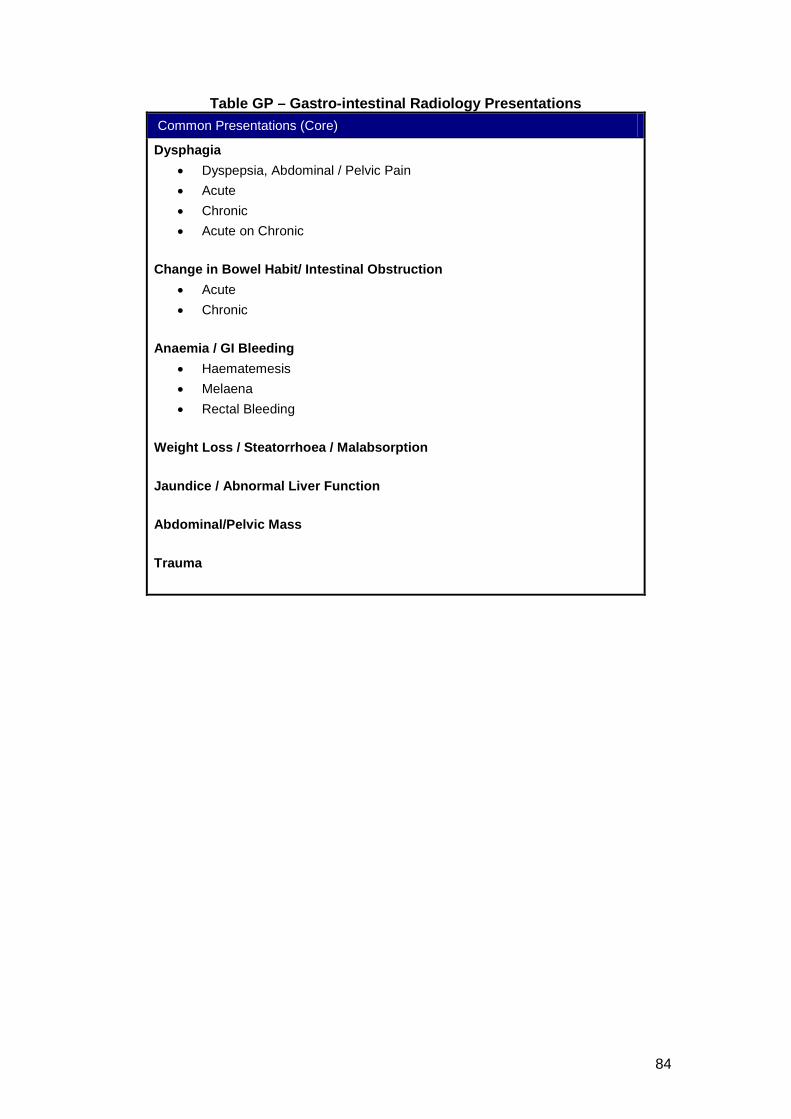
84
Table GP – Gastro-intestinal Radiology Presentations Common Presentations (Core)
Dysphagia • Dyspepsia, Abdominal / Pelvic Pain • Acute • Chronic • Acute on Chronic
Change in Bowel Habit/ Intestinal Obstruction
• Acute • Chronic
Anaemia / GI Bleeding
• Haematemesis • Melaena • Rectal Bleeding
Weight Loss / Steatorrhoea / Malabsorption Jaundice / Abnormal Liver Function Abdominal/Pelvic Mass Trauma
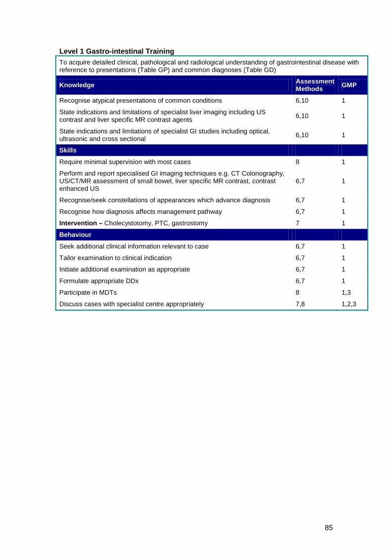
85
Level 1 Gastro-intestinal Training To acquire detailed clinical, pathological and radiological understanding of gastrointestinal disease with reference to presentations (Table GP) and common diagnoses (Table GD)
Knowledge Assessment Methods GMP
Recognise atypical presentations of common conditions 6,10 1
State indications and limitations of specialist liver imaging including US contrast and liver specific MR contrast agents 6,10 1
State indications and limitations of specialist GI studies including optical, ultrasonic and cross sectional 6,10 1
Skills
Require minimal supervision with most cases 8 1
Perform and report specialised GI imaging techniques e.g. CT Colonography, US/CT/MR assessment of small bowel, liver specific MR contrast, contrast enhanced US
6,7 1
Recognise/seek constellations of appearances which advance diagnosis 6,7 1
Recognise how diagnosis affects management pathway 6,7 1
Intervention – Cholecystotomy, PTC, gastrostomy 7 1
Behaviour
Seek additional clinical information relevant to case 6,7 1
Tailor examination to clinical indication 6,7 1
Initiate additional examination as appropriate 6,7 1
Formulate appropriate DDx 6,7 1
Participate in MDTs 8 1,3
Discuss cases with specialist centre appropriately 7,8 1,2,3
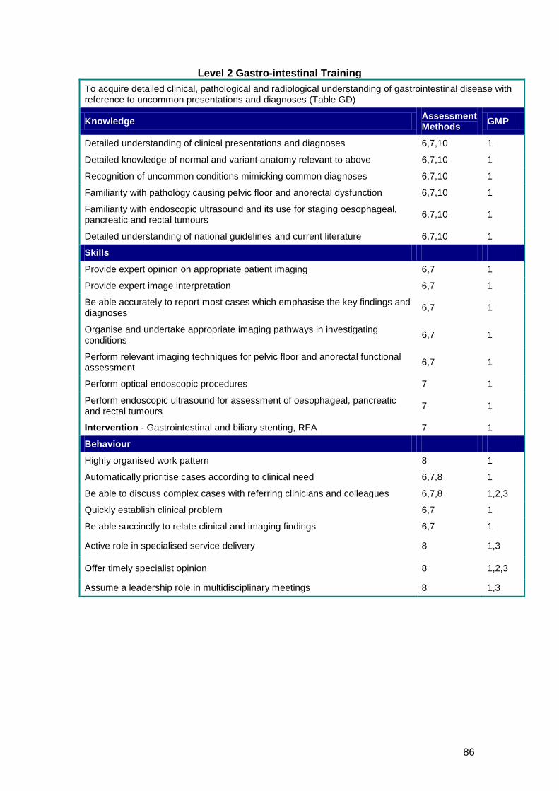
86
Level 2 Gastro-intestinal Training To acquire detailed clinical, pathological and radiological understanding of gastrointestinal disease with reference to uncommon presentations and diagnoses (Table GD)
Knowledge Assessment Methods GMP
Detailed understanding of clinical presentations and diagnoses 6,7,10 1
Detailed knowledge of normal and variant anatomy relevant to above 6,7,10 1
Recognition of uncommon conditions mimicking common diagnoses 6,7,10 1
Familiarity with pathology causing pelvic floor and anorectal dysfunction 6,7,10 1
Familiarity with endoscopic ultrasound and its use for staging oesophageal, pancreatic and rectal tumours 6,7,10 1
Detailed understanding of national guidelines and current literature 6,7,10 1
Skills
Provide expert opinion on appropriate patient imaging 6,7 1
Provide expert image interpretation 6,7 1
Be able accurately to report most cases which emphasise the key findings and diagnoses 6,7 1
Organise and undertake appropriate imaging pathways in investigating conditions 6,7 1
Perform relevant imaging techniques for pelvic floor and anorectal functional assessment 6,7 1
Perform optical endoscopic procedures 7 1
Perform endoscopic ultrasound for assessment of oesophageal, pancreatic and rectal tumours 7 1
Intervention - Gastrointestinal and biliary stenting, RFA 7 1
Behaviour
Highly organised work pattern 8 1
Automatically prioritise cases according to clinical need 6,7,8 1
Be able to discuss complex cases with referring clinicians and colleagues 6,7,8 1,2,3
Quickly establish clinical problem 6,7 1
Be able succinctly to relate clinical and imaging findings 6,7 1
Active role in specialised service delivery 8 1,3
Offer timely specialist opinion 8 1,2,3
Assume a leadership role in multidisciplinary meetings 8 1,3
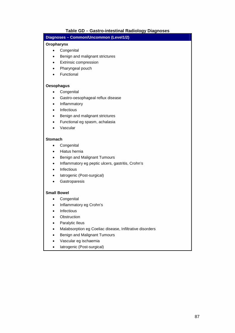
87
Table GD – Gastro-intestinal Radiology Diagnoses Diagnoses – Common/Uncommon (Level1/2)
Oropharynx • Congenital • Benign and malignant strictures • Extrinsic compression • Pharyngeal pouch • Functional
Oesophagus
• Congenital • Gastro-oesophageal reflux disease • Inflammatory • Infectious • Benign and malignant strictures • Functional eg spasm, achalasia • Vascular
Stomach
• Congenital • Hiatus hernia • Benign and Malignant Tumours • Inflammatory eg peptic ulcers, gastritis, Crohn’s • Infectious • Iatrogenic (Post-surgical) • Gastroparesis
Small Bowel
• Congenital • Inflammatory eg Crohn’s • Infectious • Obstruction • Paralytic Ileus • Malabsorption eg Coeliac disease, Infiltrative disorders • Benign and Malignant Tumours • Vascular eg ischaemia • Iatrogenic (Post-surgical)
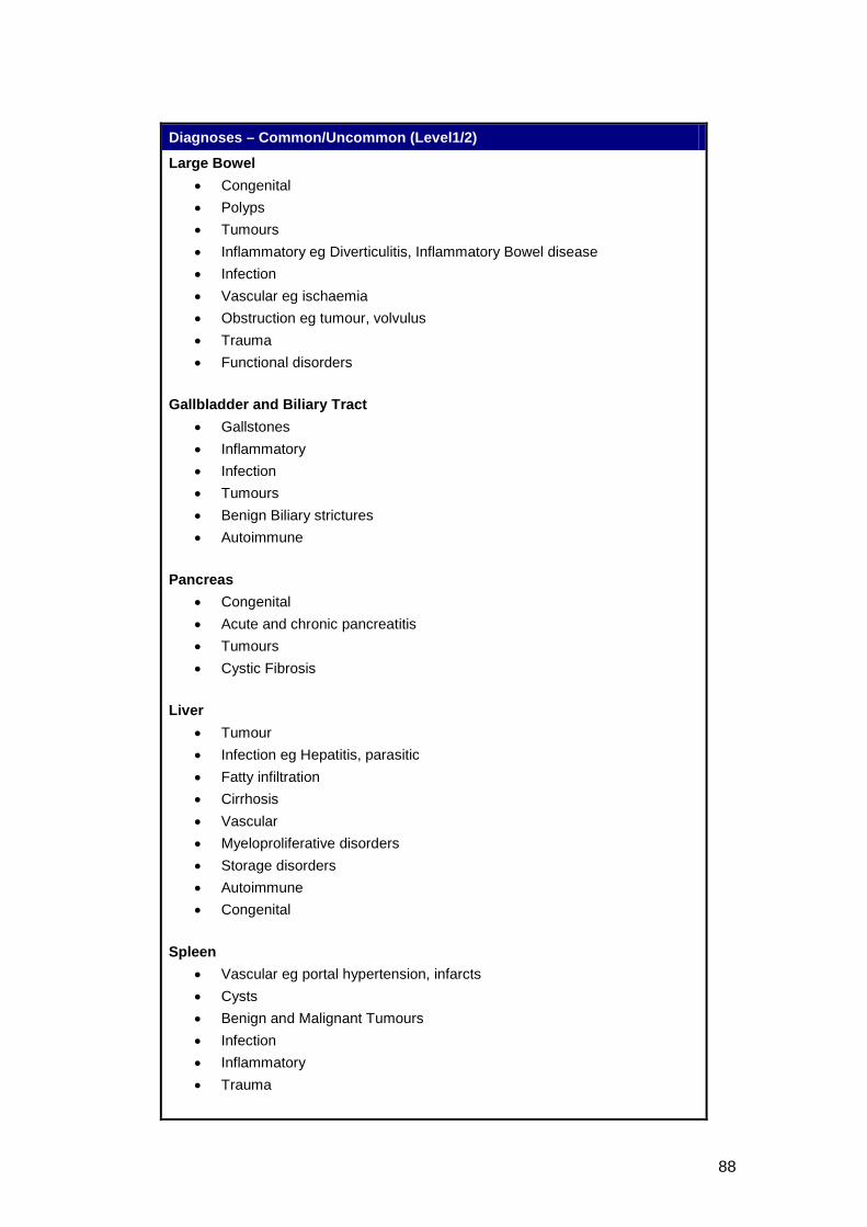
88
Diagnoses – Common/Uncommon (Level1/2)
Large Bowel • Congenital • Polyps • Tumours • Inflammatory eg Diverticulitis, Inflammatory Bowel disease • Infection • Vascular eg ischaemia • Obstruction eg tumour, volvulus • Trauma • Functional disorders
Gallbladder and Biliary Tract
• Gallstones • Inflammatory • Infection • Tumours • Benign Biliary strictures • Autoimmune
Pancreas
• Congenital • Acute and chronic pancreatitis • Tumours • Cystic Fibrosis
Liver
• Tumour • Infection eg Hepatitis, parasitic • Fatty infiltration • Cirrhosis • Vascular • Myeloproliferative disorders • Storage disorders • Autoimmune • Congenital
Spleen
• Vascular eg portal hypertension, infarcts • Cysts • Benign and Malignant Tumours • Infection • Inflammatory • Trauma
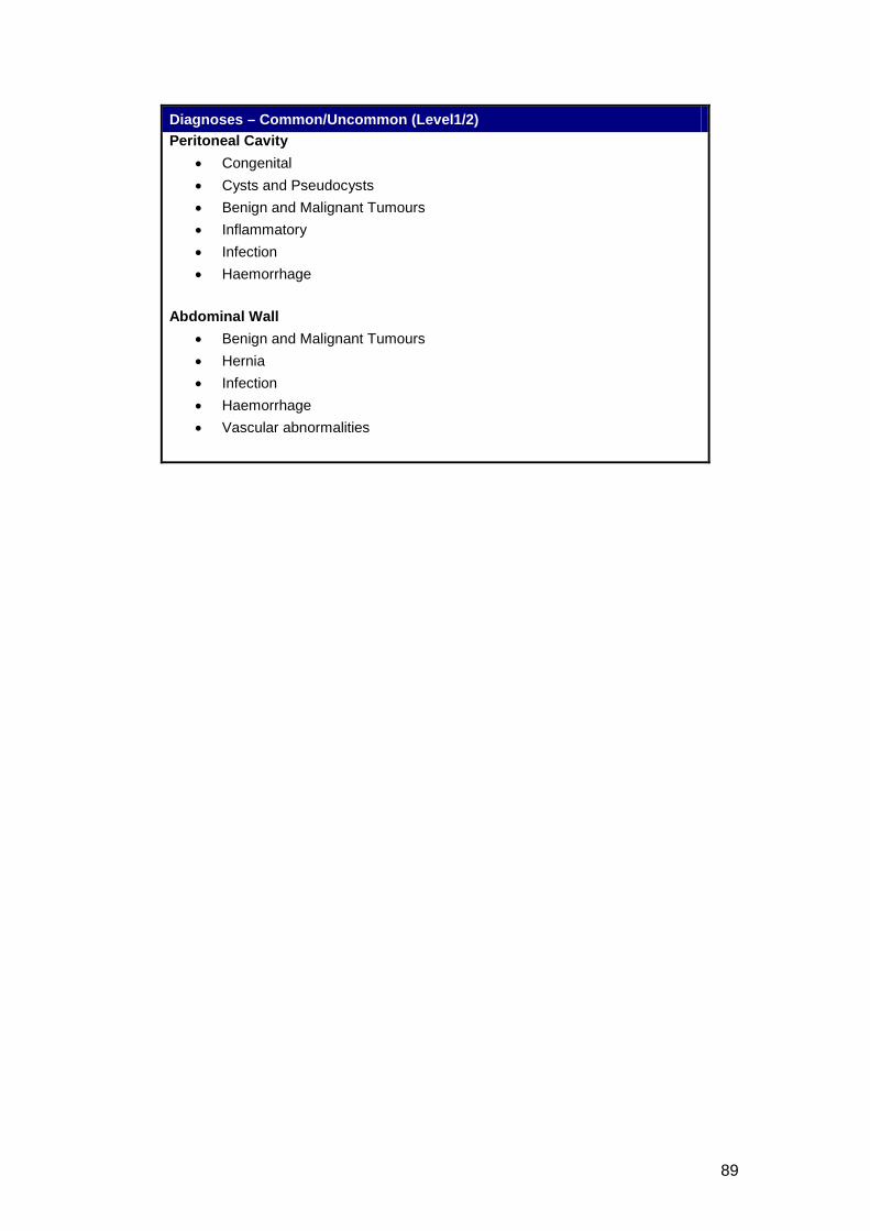
89
Diagnoses – Common/Uncommon (Level1/2) Peritoneal Cavity
• Congenital • Cysts and Pseudocysts • Benign and Malignant Tumours • Inflammatory • Infection • Haemorrhage
Abdominal Wall
• Benign and Malignant Tumours • Hernia • Infection • Haemorrhage • Vascular abnormalities
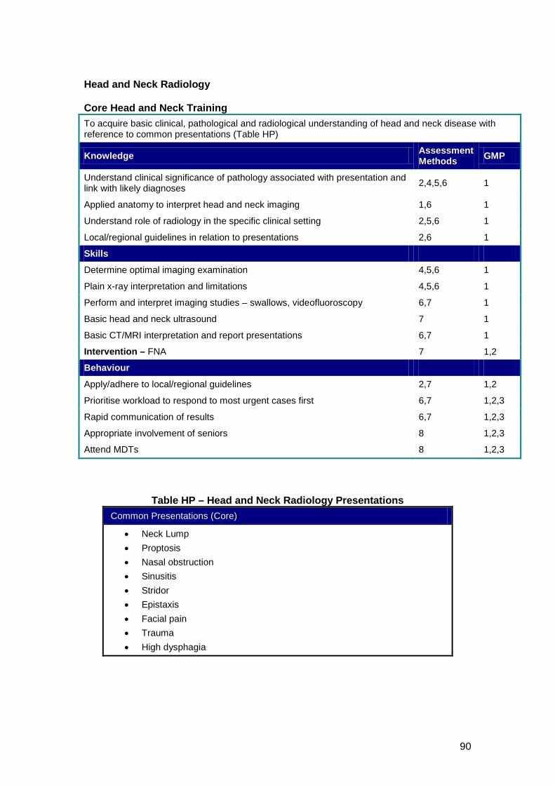
90
Head and Neck Radiology Core Head and Neck Training To acquire basic clinical, pathological and radiological understanding of head and neck disease with reference to common presentations (Table HP)
Knowledge Assessment Methods GMP
Understand clinical significance of pathology associated with presentation and link with likely diagnoses 2,4,5,6 1
Applied anatomy to interpret head and neck imaging 1,6 1
Understand role of radiology in the specific clinical setting 2,5,6 1
Local/regional guidelines in relation to presentations 2,6 1
Skills
Determine optimal imaging examination 4,5,6 1
Plain x-ray interpretation and limitations 4,5,6 1
Perform and interpret imaging studies – swallows, videofluoroscopy 6,7 1
Basic head and neck ultrasound 7 1
Basic CT/MRI interpretation and report presentations 6,7 1
Intervention – FNA 7 1,2
Behaviour
Apply/adhere to local/regional guidelines 2,7 1,2
Prioritise workload to respond to most urgent cases first 6,7 1,2,3
Rapid communication of results 6,7 1,2,3
Appropriate involvement of seniors 8 1,2,3
Attend MDTs 8 1,2,3
Table HP – Head and Neck Radiology Presentations Common Presentations (Core)
• Neck Lump • Proptosis • Nasal obstruction • Sinusitis • Stridor • Epistaxis • Facial pain • Trauma • High dysphagia
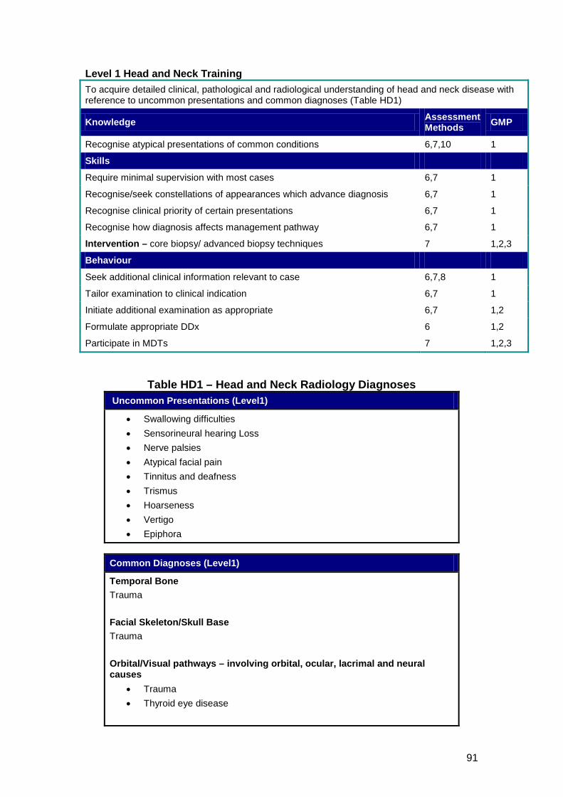
91
Level 1 Head and Neck Training To acquire detailed clinical, pathological and radiological understanding of head and neck disease with reference to uncommon presentations and common diagnoses (Table HD1)
Knowledge Assessment Methods GMP
Recognise atypical presentations of common conditions 6,7,10 1
Skills
Require minimal supervision with most cases 6,7 1
Recognise/seek constellations of appearances which advance diagnosis 6,7 1
Recognise clinical priority of certain presentations 6,7 1
Recognise how diagnosis affects management pathway 6,7 1
Intervention – core biopsy/ advanced biopsy techniques 7 1,2,3
Behaviour
Seek additional clinical information relevant to case 6,7,8 1
Tailor examination to clinical indication 6,7 1
Initiate additional examination as appropriate 6,7 1,2
Formulate appropriate DDx 6 1,2
Participate in MDTs 7 1,2,3
Table HD1 – Head and Neck Radiology Diagnoses Uncommon Presentations (Level1)
• Swallowing difficulties • Sensorineural hearing Loss • Nerve palsies • Atypical facial pain • Tinnitus and deafness • Trismus • Hoarseness • Vertigo • Epiphora
Common Diagnoses (Level1)
Temporal Bone Trauma Facial Skeleton/Skull Base Trauma Orbital/Visual pathways – involving orbital, ocular, lacrimal and neural causes
• Trauma • Thyroid eye disease
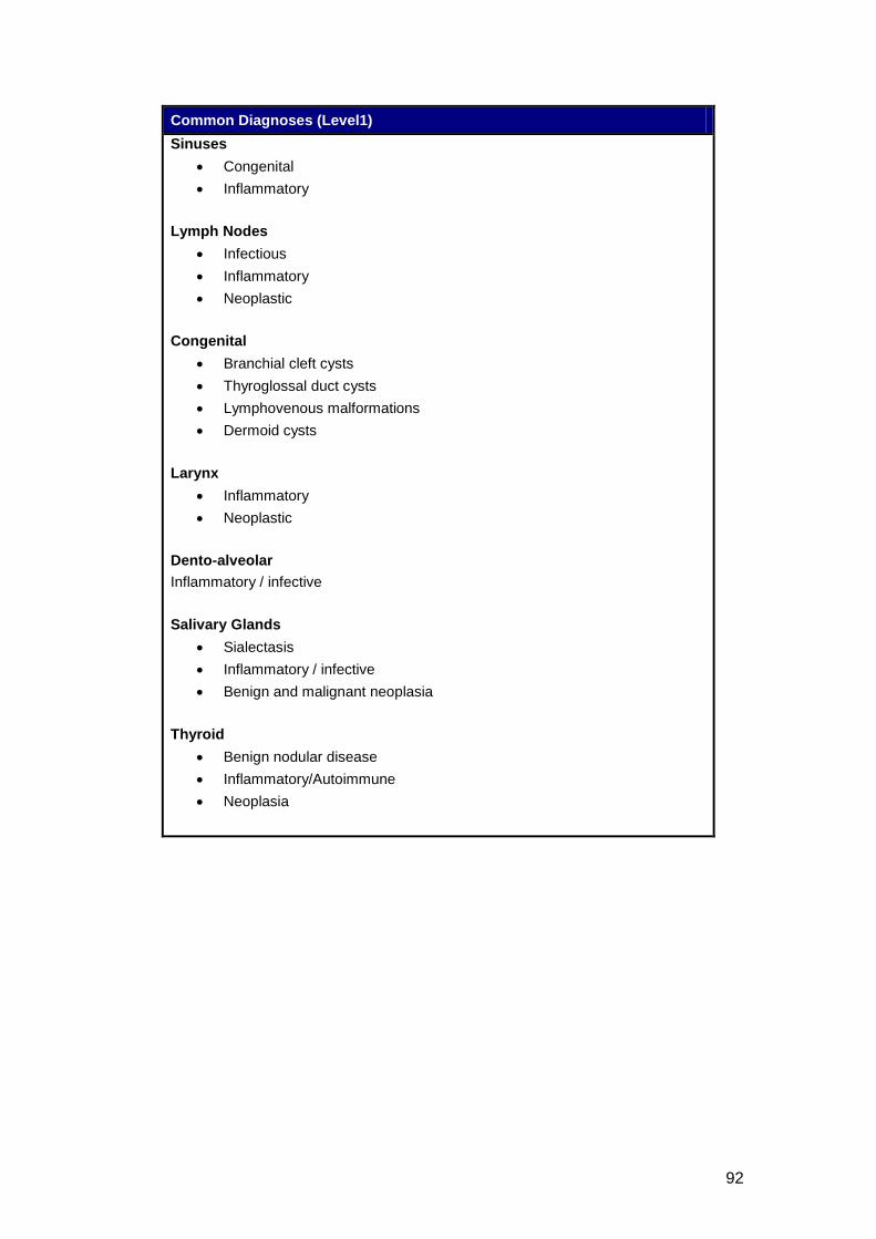
92
Common Diagnoses (Level1) Sinuses
• Congenital • Inflammatory
Lymph Nodes
• Infectious • Inflammatory • Neoplastic
Congenital
• Branchial cleft cysts • Thyroglossal duct cysts • Lymphovenous malformations • Dermoid cysts
Larynx
• Inflammatory • Neoplastic
Dento-alveolar Inflammatory / infective Salivary Glands
• Sialectasis • Inflammatory / infective • Benign and malignant neoplasia
Thyroid
• Benign nodular disease • Inflammatory/Autoimmune • Neoplasia
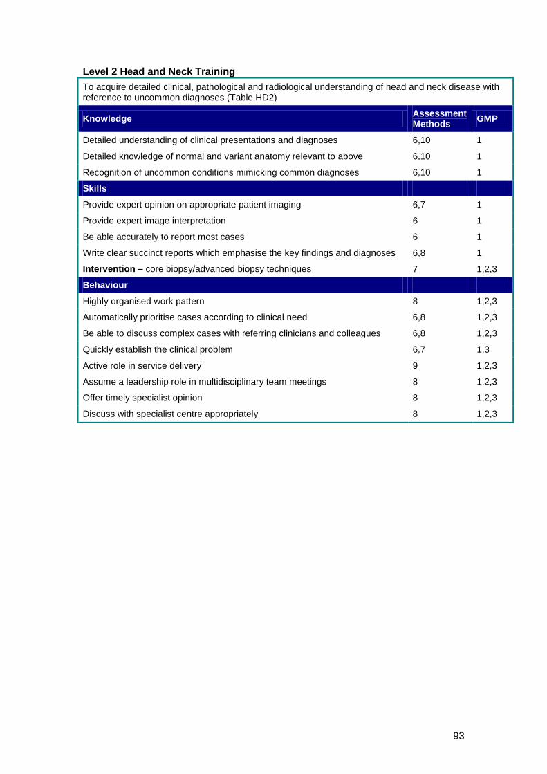
93
Level 2 Head and Neck Training To acquire detailed clinical, pathological and radiological understanding of head and neck disease with reference to uncommon diagnoses (Table HD2)
Knowledge Assessment Methods GMP
Detailed understanding of clinical presentations and diagnoses 6,10 1
Detailed knowledge of normal and variant anatomy relevant to above 6,10 1
Recognition of uncommon conditions mimicking common diagnoses 6,10 1
Skills
Provide expert opinion on appropriate patient imaging 6,7 1
Provide expert image interpretation 6 1
Be able accurately to report most cases 6 1
Write clear succinct reports which emphasise the key findings and diagnoses 6,8 1
Intervention – core biopsy/advanced biopsy techniques 7 1,2,3
Behaviour
Highly organised work pattern 8 1,2,3
Automatically prioritise cases according to clinical need 6,8 1,2,3
Be able to discuss complex cases with referring clinicians and colleagues 6,8 1,2,3
Quickly establish the clinical problem 6,7 1,3
Active role in service delivery 9 1,2,3
Assume a leadership role in multidisciplinary team meetings 8 1,2,3
Offer timely specialist opinion 8 1,2,3
Discuss with specialist centre appropriately 8 1,2,3
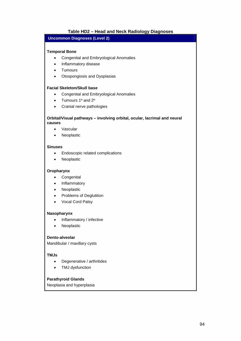
94
Table HD2 – Head and Neck Radiology Diagnoses Uncommon Diagnoses (Level 2)
Temporal Bone
• Congenital and Embryological Anomalies • Inflammatory disease • Tumours • Otospongiosis and Dysplasias
Facial Skeleton/Skull base
• Congenital and Embryological Anomalies • Tumours 1º and 2º • Cranial nerve pathologies
Orbital/Visual pathways – involving orbital, ocular, lacrimal and neural causes
• Vascular • Neoplastic
Sinuses
• Endoscopic related complications • Neoplastic
Oropharynx
• Congenital • Inflammatory • Neoplastic • Problems of Deglutition • Vocal Cord Palsy
Nasopharynx
• Inflammatory / infective • Neoplastic
Dento-alveolar Mandibular / maxillary cysts TMJs
• Degenerative / arthritides • TMJ dysfunction
Parathyroid Glands Neoplasia and hyperplasia
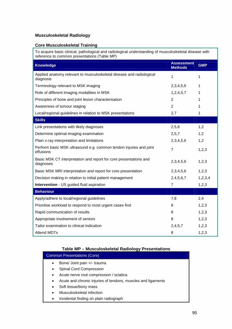
95
Musculoskeletal Radiology Core Musculoskeletal Training To acquire basic clinical, pathological and radiological understanding of musculoskeletal disease with reference to common presentations (Table MP)
Knowledge Assessment Methods GMP
Applied anatomy relevant to musculoskeletal disease and radiological diagnosis 1 1
Terminology relevant to MSK imaging 2,3,4,5,6 1
Role of different imaging modalities in MSK 1,2,4,5,7 1
Principles of bone and joint lesion characterisation 2 1
Awareness of tumour staging 2 1
Local/regional guidelines in relation to MSK presentations 2,7 1
Skills
Link presentations with likely diagnoses 2,5,6 1,2
Determine optimal imaging examination 2,5,7 1,2
Plain x-ray interpretation and limitations 2,3,4,5,6 1,2
Perform basic MSK ultrasound e.g. common tendon injuries and joint effusions 7 1,2,3
Basic MSK CT interpretation and report for core presentations and diagnoses 2,3,4,5,6 1,2,3
Basic MSK MRI interpretation and report for core presentation 2,3,4,5,6 1,2,3
Decision making in relation to initial patient management 2,4,5,6,7 1,2,3,4
Intervention - US guided fluid aspiration 7 1,2,3
Behaviour
Apply/adhere to local/regional guidelines 7,8 2,4
Prioritise workload to respond to most urgent cases first 8 1,2,3
Rapid communication of results 8 1,2,3
Appropriate involvement of seniors 8 1,2,3
Tailor examination to clinical indication 2,4,5,7 1,2,3
Attend MDTs 8 1,2,3
Table MP – Musculoskeletal Radiology Presentations Common Presentations (Core)
• Bone/ Joint pain +/- trauma • Spinal Cord Compression • Acute nerve root compression / sciatica • Acute and chronic injuries of tendons, muscles and ligaments • Soft tissue/bony mass • Musculoskeletal infection • Incidental finding on plain radiograph
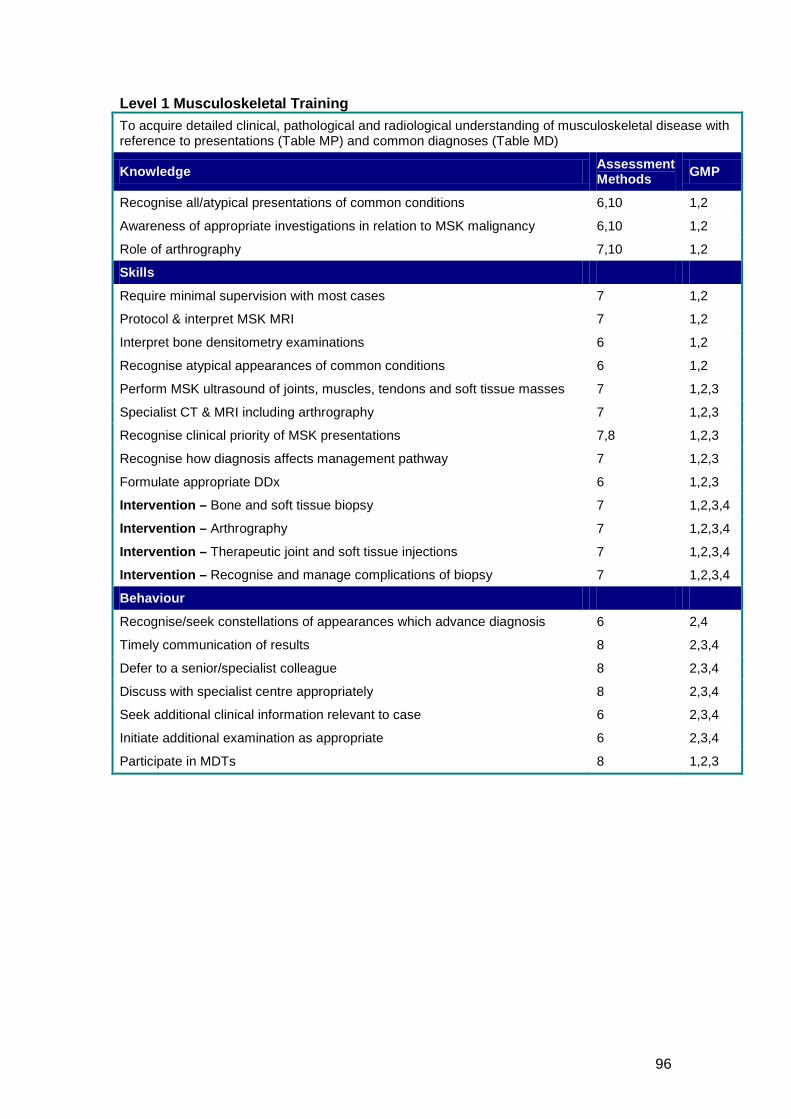
96
Level 1 Musculoskeletal Training To acquire detailed clinical, pathological and radiological understanding of musculoskeletal disease with reference to presentations (Table MP) and common diagnoses (Table MD)
Knowledge Assessment Methods GMP
Recognise all/atypical presentations of common conditions 6,10 1,2
Awareness of appropriate investigations in relation to MSK malignancy 6,10 1,2
Role of arthrography 7,10 1,2
Skills
Require minimal supervision with most cases 7 1,2
Protocol & interpret MSK MRI 7 1,2
Interpret bone densitometry examinations 6 1,2
Recognise atypical appearances of common conditions 6 1,2
Perform MSK ultrasound of joints, muscles, tendons and soft tissue masses 7 1,2,3
Specialist CT & MRI including arthrography 7 1,2,3
Recognise clinical priority of MSK presentations 7,8 1,2,3
Recognise how diagnosis affects management pathway 7 1,2,3
Formulate appropriate DDx 6 1,2,3
Intervention – Bone and soft tissue biopsy 7 1,2,3,4
Intervention – Arthrography 7 1,2,3,4
Intervention – Therapeutic joint and soft tissue injections 7 1,2,3,4
Intervention – Recognise and manage complications of biopsy 7 1,2,3,4
Behaviour
Recognise/seek constellations of appearances which advance diagnosis 6 2,4
Timely communication of results 8 2,3,4
Defer to a senior/specialist colleague 8 2,3,4
Discuss with specialist centre appropriately 8 2,3,4
Seek additional clinical information relevant to case 6 2,3,4
Initiate additional examination as appropriate 6 2,3,4
Participate in MDTs 8 1,2,3
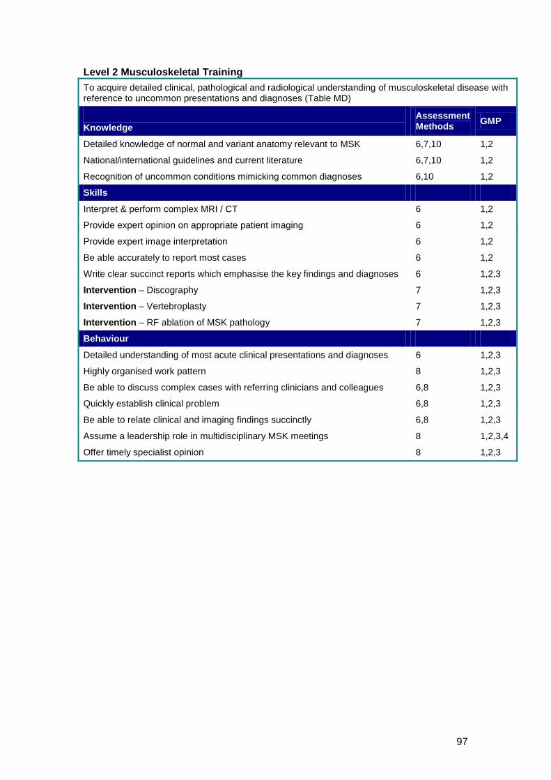
97
Level 2 Musculoskeletal Training To acquire detailed clinical, pathological and radiological understanding of musculoskeletal disease with reference to uncommon presentations and diagnoses (Table MD)
Knowledge
Assessment Methods GMP
Detailed knowledge of normal and variant anatomy relevant to MSK 6,7,10 1,2
National/international guidelines and current literature 6,7,10 1,2
Recognition of uncommon conditions mimicking common diagnoses 6,10 1,2
Skills
Interpret & perform complex MRI / CT 6 1,2
Provide expert opinion on appropriate patient imaging 6 1,2
Provide expert image interpretation 6 1,2
Be able accurately to report most cases 6 1,2
Write clear succinct reports which emphasise the key findings and diagnoses 6 1,2,3
Intervention – Discography 7 1,2,3
Intervention – Vertebroplasty 7 1,2,3
Intervention – RF ablation of MSK pathology 7 1,2,3
Behaviour
Detailed understanding of most acute clinical presentations and diagnoses 6 1,2,3
Highly organised work pattern 8 1,2,3
Be able to discuss complex cases with referring clinicians and colleagues 6,8 1,2,3
Quickly establish clinical problem 6,8 1,2,3
Be able to relate clinical and imaging findings succinctly 6,8 1,2,3
Assume a leadership role in multidisciplinary MSK meetings 8 1,2,3,4
Offer timely specialist opinion 8 1,2,3
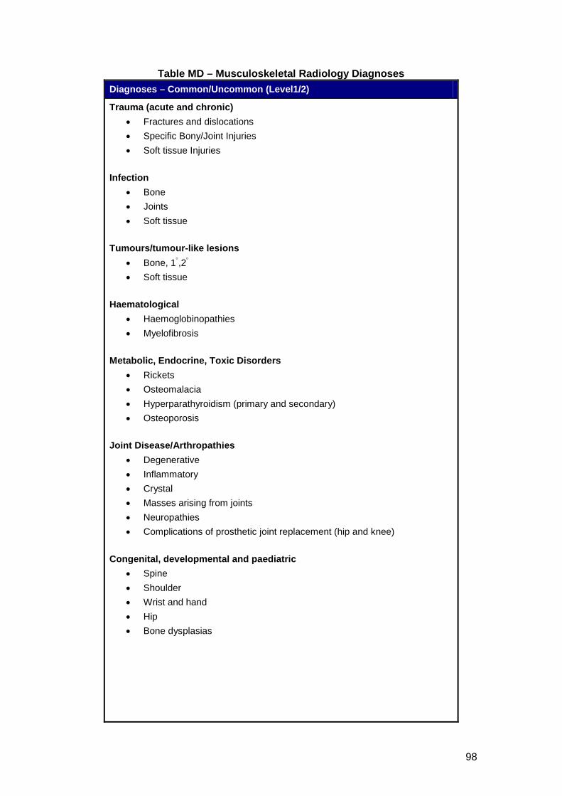
98
Table MD – Musculoskeletal Radiology Diagnoses Diagnoses – Common/Uncommon (Level1/2)
Trauma (acute and chronic) • Fractures and dislocations • Specific Bony/Joint Injuries • Soft tissue Injuries
Infection
• Bone • Joints • Soft tissue
Tumours/tumour-like lesions
• Bone, 1°,2• Soft tissue
°
Haematological
• Haemoglobinopathies • Myelofibrosis
Metabolic, Endocrine, Toxic Disorders
• Rickets • Osteomalacia • Hyperparathyroidism (primary and secondary) • Osteoporosis
Joint Disease/Arthropathies
• Degenerative • Inflammatory • Crystal • Masses arising from joints • Neuropathies • Complications of prosthetic joint replacement (hip and knee)
Congenital, developmental and paediatric
• Spine • Shoulder • Wrist and hand • Hip • Bone dysplasias

99
Diagnoses – Common/Uncommon (Level1/2) Miscellaneous
• Sarcoidosis • Paget’s disease • Hypertrophic osteoarthropathy • Transient/regional migratory osteoporosis • Osteonecrosis • Characterisation of soft tissue calcification/ossification
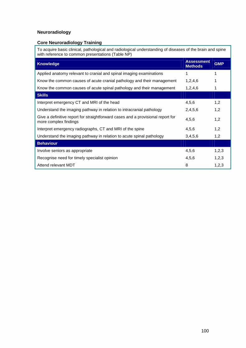
100
Neuroradiology Core Neuroradiology Training To acquire basic clinical, pathological and radiological understanding of diseases of the brain and spine with reference to common presentations (Table NP)
Knowledge Assessment Methods GMP
Applied anatomy relevant to cranial and spinal imaging examinations 1 1
Know the common causes of acute cranial pathology and their management 1,2,4,6 1
Know the common causes of acute spinal pathology and their management 1,2,4,6 1
Skills
Interpret emergency CT and MRI of the head 4,5,6 1,2
Understand the imaging pathway in relation to intracranial pathology 2,4,5,6 1,2
Give a definitive report for straightforward cases and a provisional report for more complex findings 4,5,6 1,2
Interpret emergency radiographs, CT and MRI of the spine 4,5,6 1,2
Understand the imaging pathway in relation to acute spinal pathology 3,4,5,6 1,2
Behaviour
Involve seniors as appropriate 4,5,6 1,2,3
Recognise need for timely specialist opinion 4,5,6 1,2,3
Attend relevant MDT 8 1,2,3
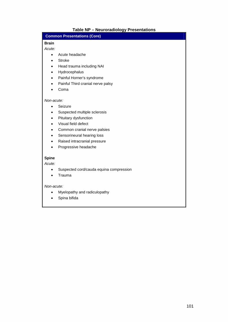
101
Table NP – Neuroradiology Presentations
Common Presentations (Core)
Brain Acute:
• Acute headache • Stroke • Head trauma including NAI • Hydrocephalus • Painful Horner’s syndrome • Painful Third cranial nerve palsy • Coma
Non-acute:
• Seizure • Suspected multiple sclerosis • Pituitary dysfunction • Visual field defect • Common cranial nerve palsies • Sensorineural hearing loss • Raised intracranial pressure • Progressive headache
Spine Acute:
• Suspected cord/cauda equina compression • Trauma
Non-acute:
• Myelopathy and radiculopathy • Spina bifida
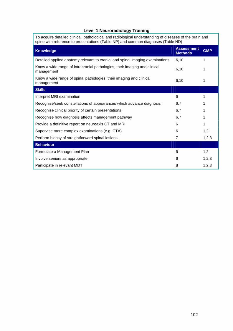
102
Level 1 Neuroradiology Training To acquire detailed clinical, pathological and radiological understanding of diseases of the brain and spine with reference to presentations (Table NP) and common diagnoses (Table ND)
Knowledge Assessment Methods GMP
Detailed applied anatomy relevant to cranial and spinal imaging examinations 6,10 1
Know a wide range of intracranial pathologies, their imaging and clinical management 6,10 1
Know a wide range of spinal pathologies, their imaging and clinical management 6,10 1
Skills
Interpret MRI examination 6 1
Recognise/seek constellations of appearances which advance diagnosis 6,7 1
Recognise clinical priority of certain presentations 6,7 1
Recognise how diagnosis affects management pathway 6,7 1
Provide a definitive report on neuroaxis CT and MRI 6 1
Supervise more complex examinations (e.g. CTA) 6 1,2
Perform biopsy of straightforward spinal lesions. 7 1,2,3
Behaviour
Formulate a Management Plan 6 1,2
Involve seniors as appropriate 6 1,2,3
Participate in relevant MDT 8 1,2,3
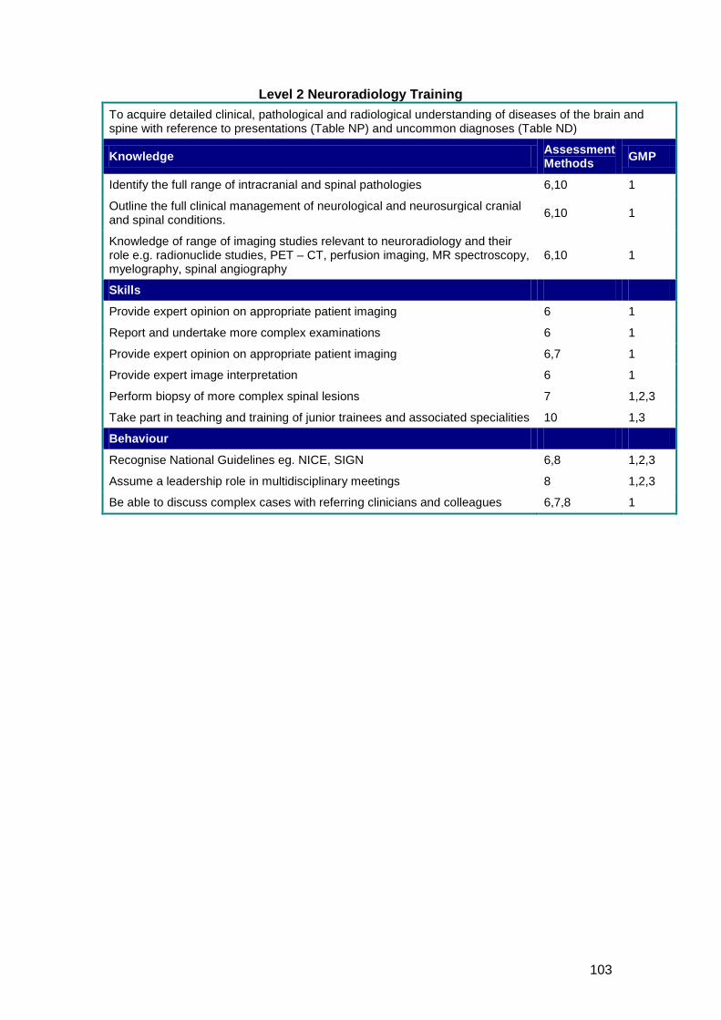
103
Level 2 Neuroradiology Training To acquire detailed clinical, pathological and radiological understanding of diseases of the brain and spine with reference to presentations (Table NP) and uncommon diagnoses (Table ND)
Knowledge Assessment Methods GMP
Identify the full range of intracranial and spinal pathologies 6,10 1
Outline the full clinical management of neurological and neurosurgical cranial and spinal conditions. 6,10 1
Knowledge of range of imaging studies relevant to neuroradiology and their role e.g. radionuclide studies, PET – CT, perfusion imaging, MR spectroscopy, myelography, spinal angiography
6,10 1
Skills
Provide expert opinion on appropriate patient imaging 6 1
Report and undertake more complex examinations 6 1
Provide expert opinion on appropriate patient imaging 6,7 1
Provide expert image interpretation 6 1
Perform biopsy of more complex spinal lesions 7 1,2,3
Take part in teaching and training of junior trainees and associated specialities 10 1,3
Behaviour
Recognise National Guidelines eg. NICE, SIGN 6,8 1,2,3
Assume a leadership role in multidisciplinary meetings 8 1,2,3
Be able to discuss complex cases with referring clinicians and colleagues 6,7,8 1
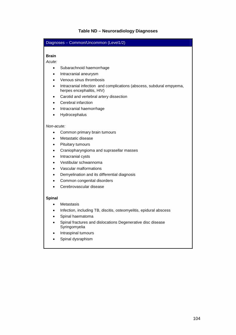
104
Table ND – Neuroradiology Diagnoses
Diagnoses – Common/Uncommon (Level1/2)
Brain Acute:
• Subarachnoid haemorrhage • Intracranial aneurysm • Venous sinus thrombosis • Intracranial infection and complications (abscess, subdural empyema,
herpes encephalitis, HIV) • Carotid and vertebral artery dissection • Cerebral infarction • Intracranial haemorrhage • Hydrocephalus
Non-acute:
• Common primary brain tumours • Metastatic disease • Pituitary tumours • Craniopharyngioma and suprasellar masses • Intracranial cysts • Vestibular schwannoma • Vascular malformations • Demyelination and its differential diagnosis • Common congenital disorders • Cerebrovascular disease
Spinal
• Metastasis • Infection, including TB, discitis, osteomyelitis, epidural abscess • Spinal haematoma • Spinal fractures and dislocations Degenerative disc disease
Syringomyelia • Intraspinal tumours • Spinal dysraphism
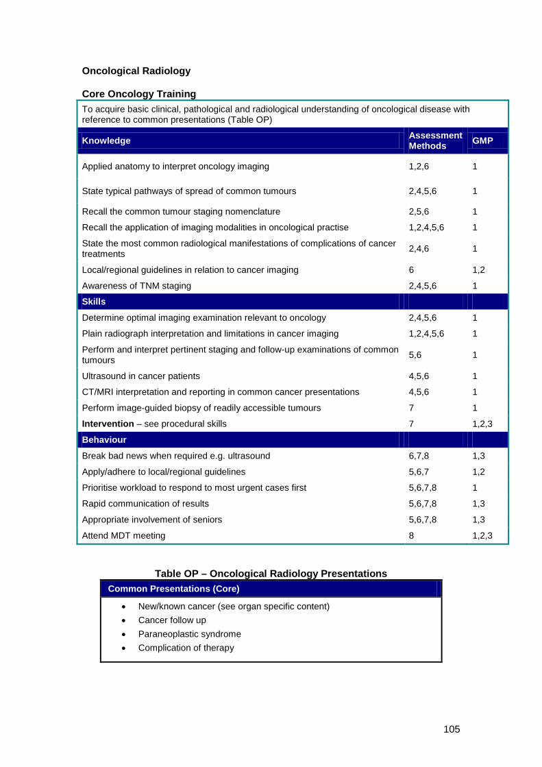
105
Oncological Radiology Core Oncology Training To acquire basic clinical, pathological and radiological understanding of oncological disease with reference to common presentations (Table OP)
Knowledge Assessment Methods GMP
Applied anatomy to interpret oncology imaging 1,2,6 1
State typical pathways of spread of common tumours 2,4,5,6 1
Recall the common tumour staging nomenclature 2,5,6 1
Recall the application of imaging modalities in oncological practise 1,2,4,5,6 1
State the most common radiological manifestations of complications of cancer treatments 2,4,6 1
Local/regional guidelines in relation to cancer imaging 6 1,2
Awareness of TNM staging 2,4,5,6 1
Skills
Determine optimal imaging examination relevant to oncology 2,4,5,6 1
Plain radiograph interpretation and limitations in cancer imaging 1,2,4,5,6 1
Perform and interpret pertinent staging and follow-up examinations of common tumours 5,6 1
Ultrasound in cancer patients 4,5,6 1
CT/MRI interpretation and reporting in common cancer presentations 4,5,6 1
Perform image-guided biopsy of readily accessible tumours 7 1
Intervention – see procedural skills 7 1,2,3
Behaviour
Break bad news when required e.g. ultrasound 6,7,8 1,3
Apply/adhere to local/regional guidelines 5,6,7 1,2
Prioritise workload to respond to most urgent cases first 5,6,7,8 1
Rapid communication of results 5,6,7,8 1,3
Appropriate involvement of seniors 5,6,7,8 1,3
Attend MDT meeting 8 1,2,3
Table OP – Oncological Radiology Presentations Common Presentations (Core)
• New/known cancer (see organ specific content) • Cancer follow up • Paraneoplastic syndrome • Complication of therapy
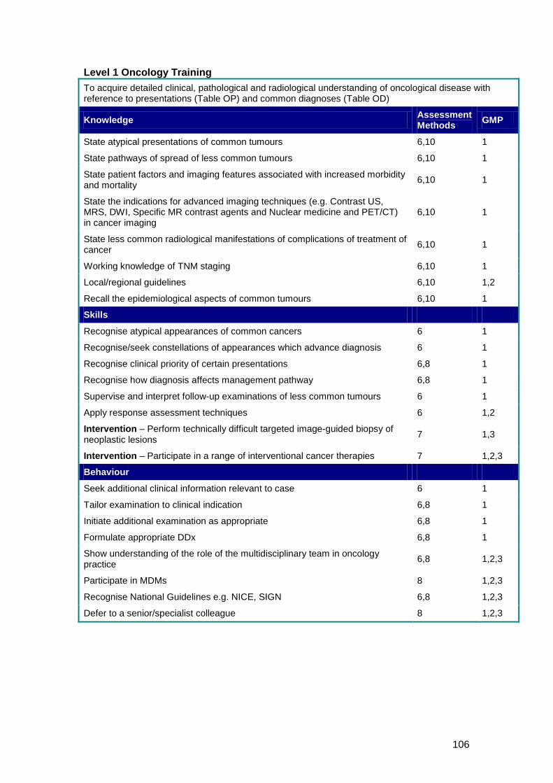
106
Level 1 Oncology Training To acquire detailed clinical, pathological and radiological understanding of oncological disease with reference to presentations (Table OP) and common diagnoses (Table OD)
Knowledge Assessment Methods GMP
State atypical presentations of common tumours 6,10 1
State pathways of spread of less common tumours 6,10 1
State patient factors and imaging features associated with increased morbidity and mortality 6,10 1
State the indications for advanced imaging techniques (e.g. Contrast US, MRS, DWI, Specific MR contrast agents and Nuclear medicine and PET/CT) in cancer imaging
6,10 1
State less common radiological manifestations of complications of treatment of cancer 6,10 1
Working knowledge of TNM staging 6,10 1
Local/regional guidelines 6,10 1,2
Recall the epidemiological aspects of common tumours 6,10 1
Skills
Recognise atypical appearances of common cancers 6 1
Recognise/seek constellations of appearances which advance diagnosis 6 1
Recognise clinical priority of certain presentations 6,8 1
Recognise how diagnosis affects management pathway 6,8 1
Supervise and interpret follow-up examinations of less common tumours 6 1
Apply response assessment techniques 6 1,2
Intervention – Perform technically difficult targeted image-guided biopsy of neoplastic lesions 7 1,3
Intervention – Participate in a range of interventional cancer therapies 7 1,2,3
Behaviour
Seek additional clinical information relevant to case 6 1
Tailor examination to clinical indication 6,8 1
Initiate additional examination as appropriate 6,8 1
Formulate appropriate DDx 6,8 1
Show understanding of the role of the multidisciplinary team in oncology practice 6,8 1,2,3
Participate in MDMs 8 1,2,3
Recognise National Guidelines e.g. NICE, SIGN 6,8 1,2,3
Defer to a senior/specialist colleague 8 1,2,3
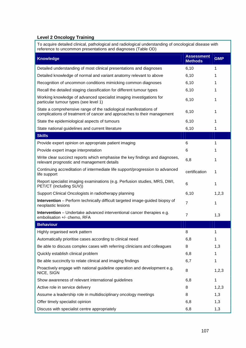
107
Level 2 Oncology Training To acquire detailed clinical, pathological and radiological understanding of oncological disease with reference to uncommon presentations and diagnoses (Table OD)
Knowledge Assessment Methods GMP
Detailed understanding of most clinical presentations and diagnoses 6,10 1
Detailed knowledge of normal and variant anatomy relevant to above 6,10 1
Recognition of uncommon conditions mimicking common diagnoses 6,10 1
Recall the detailed staging classification for different tumour types 6,10 1
Working knowledge of advanced specialist imaging investigations for particular tumour types (see level 1) 6,10 1
State a comprehensive range of the radiological manifestations of complications of treatment of cancer and approaches to their management 6,10 1
State the epidemiological aspects of tumours 6,10 1
State national guidelines and current literature 6,10 1
Skills
Provide expert opinion on appropriate patient imaging 6 1
Provide expert image interpretation 6 1
Write clear succinct reports which emphasise the key findings and diagnoses, relevant prognostic and management details 6,8 1
Continuing accreditation of intermediate life support/progression to advanced life support certification 1
Report specialist imaging examinations (e.g. Perfusion studies, MRS, DWI, PET/CT (including SUV)) 6 1
Support Clinical Oncologists in radiotherapy planning 6,10 1,2,3
Intervention – Perform technically difficult targeted image-guided biopsy of neoplastic lesions 7 1
Intervention – Undertake advanced interventional cancer therapies e.g. embolisation +/- chemo, RFA 7 1,3
Behaviour
Highly organised work pattern 8 1
Automatically prioritise cases according to clinical need 6,8 1
Be able to discuss complex cases with referring clinicians and colleagues 8 1,3
Quickly establish clinical problem 6,8 1
Be able succinctly to relate clinical and imaging findings 6,7 1
Proactively engage with national guideline operation and development e.g. NICE, SIGN 8 1,2,3
Show awareness of relevant international guidelines 6,8 1
Active role in service delivery 8 1,2,3
Assume a leadership role in multidisciplinary oncology meetings 8 1,3
Offer timely specialist opinion 6,8 1,3
Discuss with specialist centre appropriately 6,8 1,3
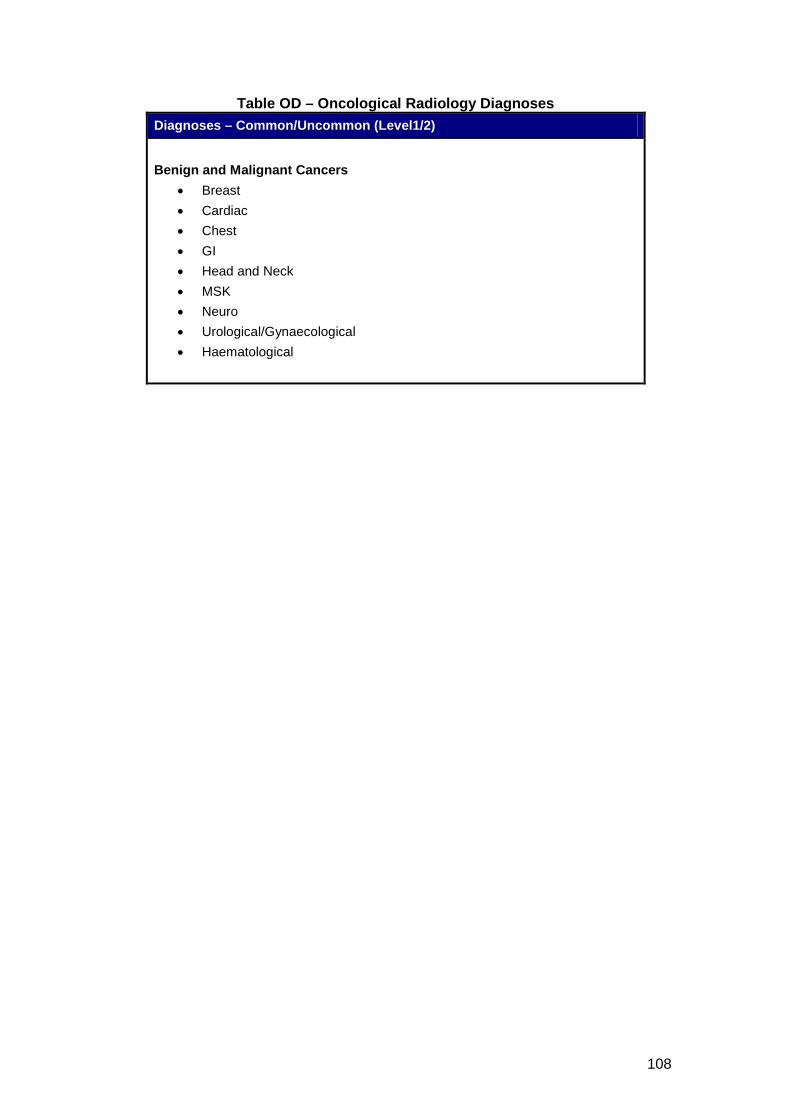
108
Table OD – Oncological Radiology Diagnoses Diagnoses – Common/Uncommon (Level1/2)
Benign and Malignant Cancers
• Breast • Cardiac • Chest • GI • Head and Neck • MSK • Neuro • Urological/Gynaecological • Haematological
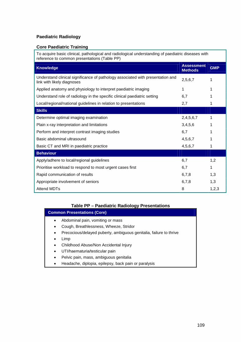
109
Paediatric Radiology Core Paediatric Training To acquire basic clinical, pathological and radiological understanding of paediatric diseases with reference to common presentations (Table PP)
Knowledge Assessment Methods GMP
Understand clinical significance of pathology associated with presentation and link with likely diagnoses 2,5,6,7 1
Applied anatomy and physiology to interpret paediatric imaging 1 1
Understand role of radiology in the specific clinical paediatric setting 6,7 1
Local/regional/national guidelines in relation to presentations 2,7 1
Skills
Determine optimal imaging examination 2,4,5,6,7 1
Plain x-ray interpretation and limitations 3,4,5,6 1
Perform and interpret contrast imaging studies 6,7 1
Basic abdominal ultrasound 4,5,6,7 1
Basic CT and MRI in paediatric practice 4,5,6,7 1
Behaviour
Apply/adhere to local/regional guidelines 6,7 1,2
Prioritise workload to respond to most urgent cases first 6,7 1
Rapid communication of results 6,7,8 1,3
Appropriate involvement of seniors 6,7,8 1,3
Attend MDTs 8 1,2,3
Table PP – Paediatric Radiology Presentations Common Presentations (Core)
• Abdominal pain, vomiting or mass • Cough, Breathlessness, Wheeze, Stridor • Precocious/delayed puberty, ambiguous genitalia, failure to thrive • Limp • Childhood Abuse/Non Accidental Injury • UTI/haematuria/testicular pain • Pelvic pain, mass, ambiguous genitalia • Headache, diplopia, epilepsy, back pain or paralysis
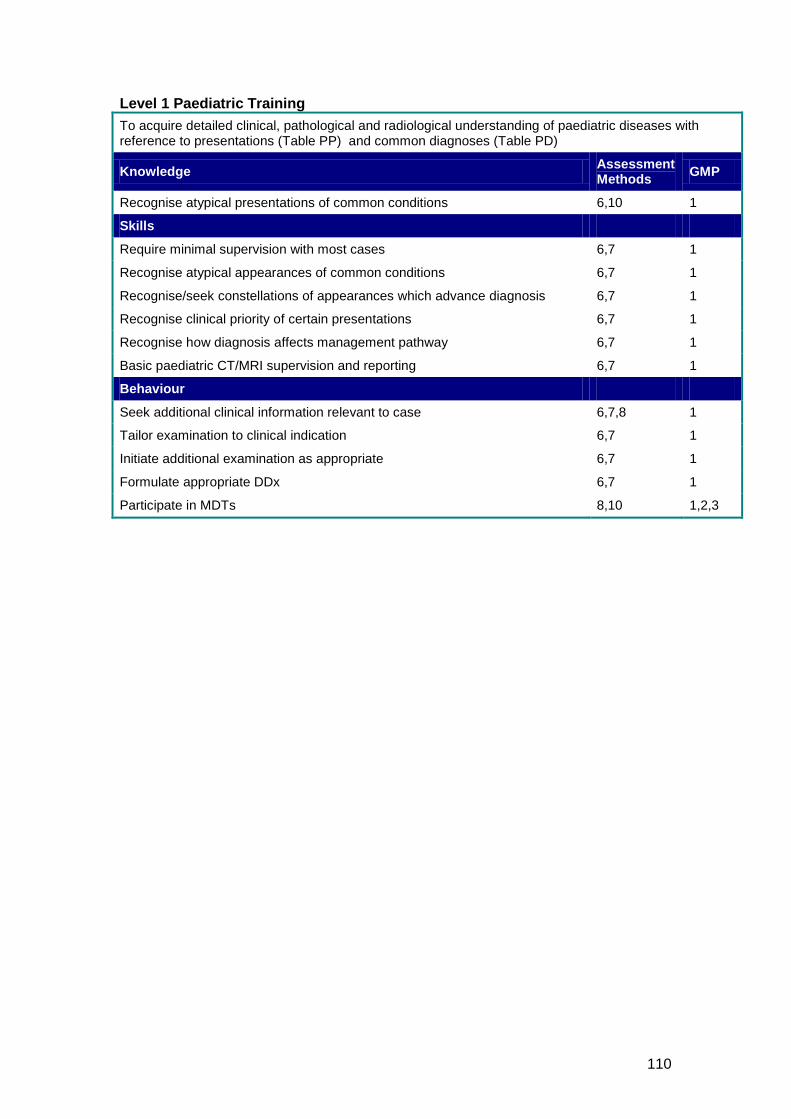
110
Level 1 Paediatric Training To acquire detailed clinical, pathological and radiological understanding of paediatric diseases with reference to presentations (Table PP) and common diagnoses (Table PD)
Knowledge Assessment Methods GMP
Recognise atypical presentations of common conditions 6,10 1
Skills
Require minimal supervision with most cases 6,7 1
Recognise atypical appearances of common conditions 6,7 1
Recognise/seek constellations of appearances which advance diagnosis 6,7 1
Recognise clinical priority of certain presentations 6,7 1
Recognise how diagnosis affects management pathway 6,7 1
Basic paediatric CT/MRI supervision and reporting 6,7 1
Behaviour
Seek additional clinical information relevant to case 6,7,8 1
Tailor examination to clinical indication 6,7 1
Initiate additional examination as appropriate 6,7 1
Formulate appropriate DDx 6,7 1
Participate in MDTs 8,10 1,2,3
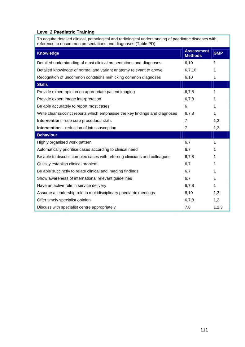
111
Level 2 Paediatric Training To acquire detailed clinical, pathological and radiological understanding of paediatric diseases with reference to uncommon presentations and diagnoses (Table PD)
Knowledge Assessment Methods GMP
Detailed understanding of most clinical presentations and diagnoses 6,10 1
Detailed knowledge of normal and variant anatomy relevant to above 6,7,10 1
Recognition of uncommon conditions mimicking common diagnoses 6,10 1
Skills
Provide expert opinion on appropriate patient imaging 6,7,8 1
Provide expert image interpretation 6,7,8 1
Be able accurately to report most cases 6 1
Write clear succinct reports which emphasise the key findings and diagnoses 6,7,8 1
Intervention – see core procedural skills 7 1,3
Intervention – reduction of intussusception 7 1,3
Behaviour
Highly organised work pattern 6,7 1
Automatically prioritise cases according to clinical need 6,7 1
Be able to discuss complex cases with referring clinicians and colleagues 6,7,8 1
Quickly establish clinical problem 6,7 1
Be able succinctly to relate clinical and imaging findings 6,7 1
Show awareness of international relevant guidelines 6,7 1
Have an active role in service delivery 6,7,8 1
Assume a leadership role in multidisciplinary paediatric meetings 8,10 1,3
Offer timely specialist opinion 6,7,8 1,2
Discuss with specialist centre appropriately 7,8 1,2,3
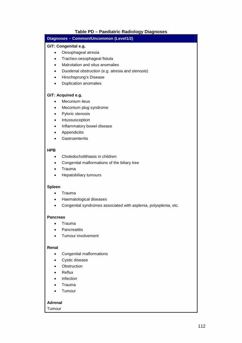
112
Table PD – Paediatric Radiology Diagnoses Diagnoses – Common/Uncommon (Level1/2)
GIT: Congenital e.g. • Oesophageal atresia • Tracheo-oesophageal fistula • Malrotation and situs anomalies • Duodenal obstruction (e.g. atresia and stenosis) • Hirschsprung’s Disease • Duplication anomalies
GIT: Acquired e.g.
• Meconium ileus • Meconium plug syndrome • Pyloric stenosis • Intussusception • Inflammatory bowel disease • Appendicitis • Gastroenteritis
HPB
• Choledocholithiasis in children • Congenital malformations of the biliary tree • Trauma • Hepatobiliary tumours
Spleen
• Trauma • Haematological diseases • Congenital syndromes associated with asplenia, polysplenia, etc.
Pancreas
• Trauma • Pancreatitis • Tumour involvement
Renal
• Congenital malformations • Cystic disease • Obstruction • Reflux • Infection • Trauma • Tumour
Adrenal Tumour
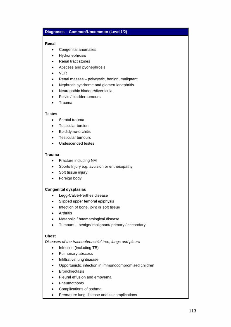
113
Diagnoses – Common/Uncommon (Level1/2) Renal
• Congenital anomalies • Hydronephrosis • Renal tract stones • Abscess and pyonephrosis • VUR • Renal masses – polycystic, benign, malignant • Nephrotic syndrome and glomerulonephritis • Neuropathic bladder/diverticula • Pelvic / bladder tumours • Trauma
Testes
• Scrotal trauma • Testicular torsion • Epididymo-orchitis • Testicular tumours • Undescended testes
Trauma
• Fracture including NAI • Sports Injury e.g. avulsion or enthesopathy • Soft tissue injury • Foreign body
Congenital dysplasias
• Legg-Calvé-Perthes disease • Slipped upper femoral epiphysis • Infection of bone, joint or soft tissue • Arthritis • Metabolic / haematological disease • Tumours – benign/ malignant/ primary / secondary
Chest Diseases of the tracheobronchial tree, lungs and pleura
• Infection (including TB) • Pulmonary abscess • Infiltrative lung disease • Opportunistic infection in immunocompromised children • Bronchiectasis • Pleural effusion and empyema • Pneumothorax • Complications of asthma • Premature lung disease and its complications
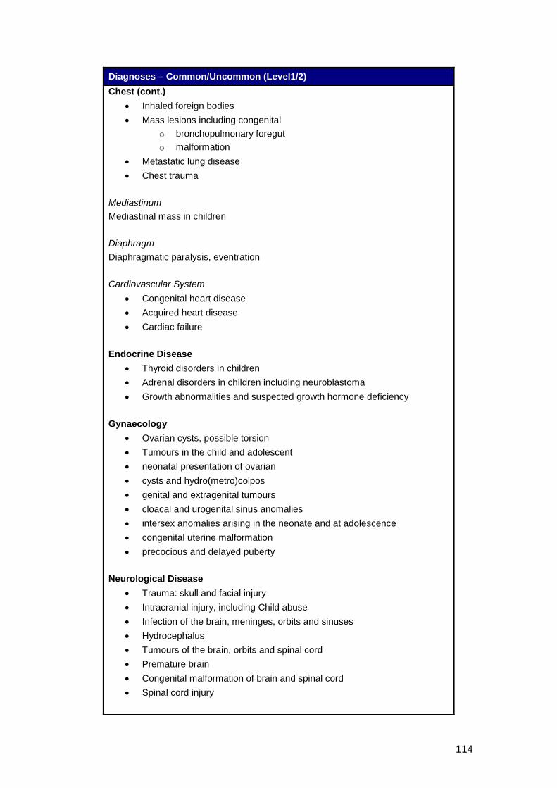
114
Diagnoses – Common/Uncommon (Level1/2) Chest (cont.)
• Inhaled foreign bodies • Mass lesions including congenital
o bronchopulmonary foregut o malformation
• Metastatic lung disease • Chest trauma
Mediastinum Mediastinal mass in children Diaphragm Diaphragmatic paralysis, eventration Cardiovascular System
• Congenital heart disease • Acquired heart disease • Cardiac failure
Endocrine Disease
• Thyroid disorders in children • Adrenal disorders in children including neuroblastoma • Growth abnormalities and suspected growth hormone deficiency
Gynaecology
• Ovarian cysts, possible torsion • Tumours in the child and adolescent • neonatal presentation of ovarian • cysts and hydro(metro)colpos • genital and extragenital tumours • cloacal and urogenital sinus anomalies • intersex anomalies arising in the neonate and at adolescence • congenital uterine malformation • precocious and delayed puberty
Neurological Disease
• Trauma: skull and facial injury • Intracranial injury, including Child abuse • Infection of the brain, meninges, orbits and sinuses • Hydrocephalus • Tumours of the brain, orbits and spinal cord • Premature brain • Congenital malformation of brain and spinal cord • Spinal cord injury
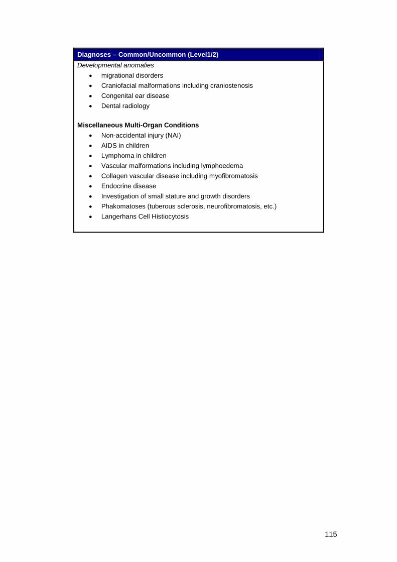
115
Diagnoses – Common/Uncommon (Level1/2) Developmental anomalies
• migrational disorders • Craniofacial malformations including craniostenosis • Congenital ear disease • Dental radiology
Miscellaneous Multi-Organ Conditions
• Non-accidental injury (NAI) • AIDS in children • Lymphoma in children • Vascular malformations including lymphoedema • Collagen vascular disease including myofibromatosis • Endocrine disease • Investigation of small stature and growth disorders • Phakomatoses (tuberous sclerosis, neurofibromatosis, etc.) • Langerhans Cell Histiocytosis
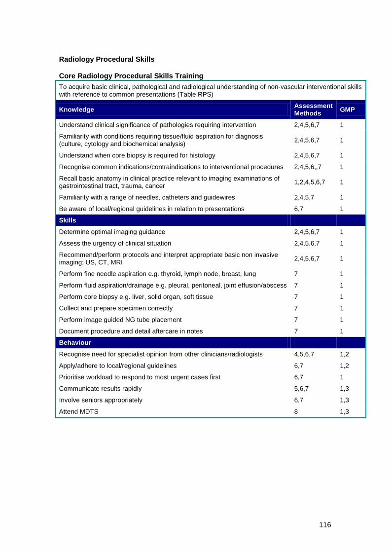
116
Radiology Procedural Skills Core Radiology Procedural Skills Training To acquire basic clinical, pathological and radiological understanding of non-vascular interventional skills with reference to common presentations (Table RPS)
Knowledge Assessment Methods GMP
Understand clinical significance of pathologies requiring intervention 2,4,5,6,7 1
Familiarity with conditions requiring tissue/fluid aspiration for diagnosis (culture, cytology and biochemical analysis) 2,4,5,6,7 1
Understand when core biopsy is required for histology 2,4,5,6,7 1
Recognise common indications/contraindications to interventional procedures 2,4,5,6,,7 1
Recall basic anatomy in clinical practice relevant to imaging examinations of gastrointestinal tract, trauma, cancer 1,2,4,5,6,7 1
Familiarity with a range of needles, catheters and guidewires 2,4,5,7 1
Be aware of local/regional guidelines in relation to presentations 6,7 1
Skills
Determine optimal imaging guidance 2,4,5,6,7 1
Assess the urgency of clinical situation 2,4,5,6,7 1
Recommend/perform protocols and interpret appropriate basic non invasive imaging; US, CT, MRI 2,4,5,6,7 1
Perform fine needle aspiration e.g. thyroid, lymph node, breast, lung 7 1
Perform fluid aspiration/drainage e.g. pleural, peritoneal, joint effusion/abscess 7 1
Perform core biopsy e.g. liver, solid organ, soft tissue 7 1
Collect and prepare specimen correctly 7 1
Perform image guided NG tube placement 7 1
Document procedure and detail aftercare in notes 7 1
Behaviour
Recognise need for specialist opinion from other clinicians/radiologists 4,5,6,7 1,2
Apply/adhere to local/regional guidelines 6,7 1,2
Prioritise workload to respond to most urgent cases first 6,7 1
Communicate results rapidly 5,6,7 1,3
Involve seniors appropriately 6,7 1,3
Attend MDTS 8 1,3

117
Table RPS – Radiology Procedural Skills Common Presentations (Core)
• Fluid collections requiring aspiration or drainage • Situations requiring tissue diagnosis • Acute and chronic renal tract obstruction including renal colic and renal
dysfunction • Ureteric leak • Bile tract Obstruction • Nutritional Disorders • Gastrointestinal tract obstruction
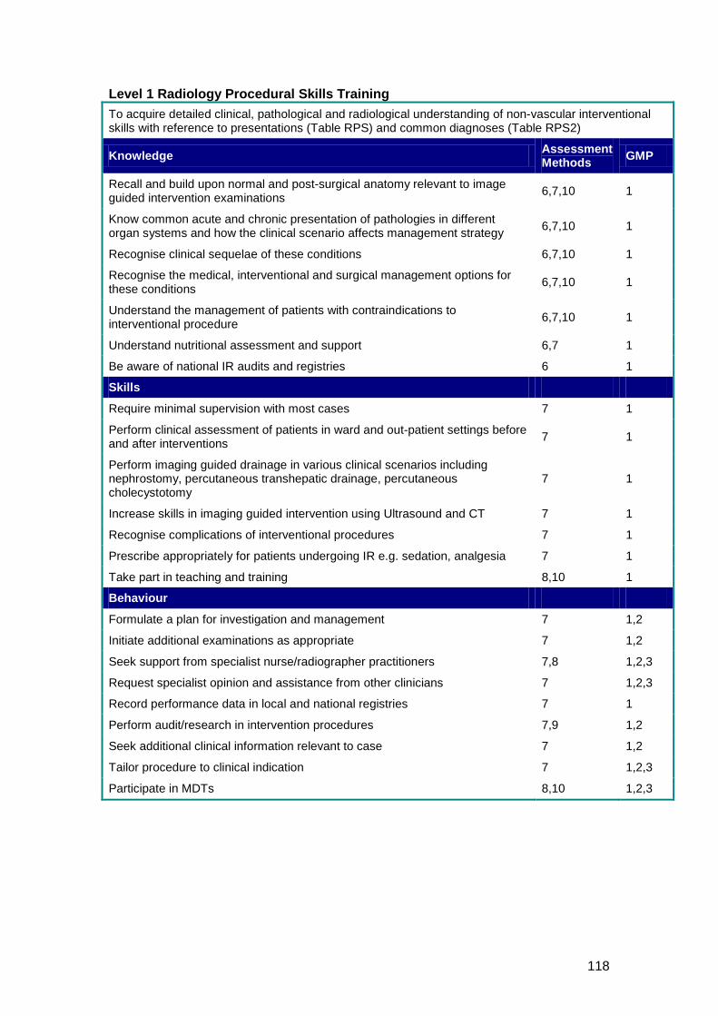
118
Level 1 Radiology Procedural Skills Training To acquire detailed clinical, pathological and radiological understanding of non-vascular interventional skills with reference to presentations (Table RPS) and common diagnoses (Table RPS2)
Knowledge Assessment Methods GMP
Recall and build upon normal and post-surgical anatomy relevant to image guided intervention examinations 6,7,10 1
Know common acute and chronic presentation of pathologies in different organ systems and how the clinical scenario affects management strategy 6,7,10 1
Recognise clinical sequelae of these conditions 6,7,10 1
Recognise the medical, interventional and surgical management options for these conditions 6,7,10 1
Understand the management of patients with contraindications to interventional procedure 6,7,10 1
Understand nutritional assessment and support 6,7 1
Be aware of national IR audits and registries 6 1
Skills
Require minimal supervision with most cases 7 1
Perform clinical assessment of patients in ward and out-patient settings before and after interventions 7 1
Perform imaging guided drainage in various clinical scenarios including nephrostomy, percutaneous transhepatic drainage, percutaneous cholecystotomy
7 1
Increase skills in imaging guided intervention using Ultrasound and CT 7 1
Recognise complications of interventional procedures 7 1
Prescribe appropriately for patients undergoing IR e.g. sedation, analgesia 7 1
Take part in teaching and training 8,10 1
Behaviour
Formulate a plan for investigation and management 7 1,2
Initiate additional examinations as appropriate 7 1,2
Seek support from specialist nurse/radiographer practitioners 7,8 1,2,3
Request specialist opinion and assistance from other clinicians 7 1,2,3
Record performance data in local and national registries 7 1
Perform audit/research in intervention procedures 7,9 1,2
Seek additional clinical information relevant to case 7 1,2
Tailor procedure to clinical indication 7 1,2,3
Participate in MDTs 8,10 1,2,3
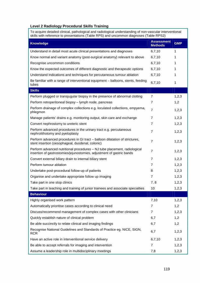
119
Level 2 Radiology Procedural Skills Training To acquire detailed clinical, pathological and radiological understanding of non-vascular interventional skills with reference to presentations (Table RPS) and uncommon diagnoses (Table RPS2)
Knowledge Assessment Methods GMP
Understand in detail most acute clinical presentations and diagnoses 6,7,10 1
Know normal and variant anatomy (post-surgical anatomy) relevant to above 6,7,10 1
Recognise uncommon conditions 6,7,10 1
Know the expected outcomes of different diagnostic and therapeutic options 6,7,10 1
Understand indications and techniques for percutaneous tumour ablation 6,7,10 1
Be familiar with a range of interventional equipment – balloons, stents, feeding tubes 6,7,10 1
Skills
Perform plugged or transjugular biopsy in the presence of abnormal clotting 7 1,2,3
Perform retroperitoneal biopsy – lymph node, pancreas 7 1,2
Perform drainage of complex collections e.g. loculated collections, empyema, phlegmon 7 1,2,3
Manage patients’ drains e.g. monitoring output, skin care and exchange 7 1,2,3
Convert nephrostomy to ureteric stent 7 1,2,3
Perform advanced procedures in the urinary tract e.g. percutaneous nephrolithotomy and pyeloplasty 7 1,2,3
Perform advanced procedures in GI tract – balloon dilatation of strictures, stent insertion (oesophageal, duodenal, colonic) 7 1,2,3
Perform advanced nutritional procedures – NJ tube placement, radiological insertion of gastrostomies/jejunostomies, adjustment of gastric bands 7 1,2,3
Convert external biliary drain to internal biliary stent 7 1,2,3
Perform tumour ablation 7 1,2,3
Undertake post-procedural follow-up of patients 8 1,2,3
Organise and undertake appropriate follow up imaging 7 1,2,3
Take part in one stop clinics 7, 8 1,2,3
Take part in teaching and training of junior trainees and associate specialties 10 1,2,3
Behaviour
Highly organised work pattern 7,10 1,2,3
Automatically prioritise cases according to clinical need 7 1,2
Discuss/recommend management of complex cases with other clinicians 7 1,2,3
Quickly establish nature of clinical problem 6,7 1,2
Be able succinctly to relate clinical and imaging findings 6,7 1,2
Recognise National Guidelines and Standards of Practice eg. NICE, SIGN, RCR 6,7 1,2,3
Have an active role in interventional service delivery 6,7,10 1,2,3
Be able to accept referrals for imaging and intervention 7 1,2,3
Assume a leadership role in multidisciplinary meetings 7,8 1,2,3
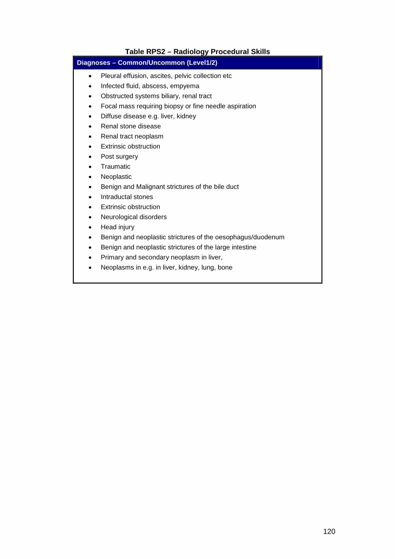
120
Table RPS2 – Radiology Procedural Skills Diagnoses – Common/Uncommon (Level1/2)
• Pleural effusion, ascites, pelvic collection etc • Infected fluid, abscess, empyema • Obstructed systems biliary, renal tract • Focal mass requiring biopsy or fine needle aspiration • Diffuse disease e.g. liver, kidney • Renal stone disease • Renal tract neoplasm • Extrinsic obstruction • Post surgery • Traumatic • Neoplastic • Benign and Malignant strictures of the bile duct • Intraductal stones • Extrinsic obstruction • Neurological disorders • Head injury • Benign and neoplastic strictures of the oesophagus/duodenum • Benign and neoplastic strictures of the large intestine • Primary and secondary neoplasm in liver, • Neoplasms in e.g. in liver, kidney, lung, bone
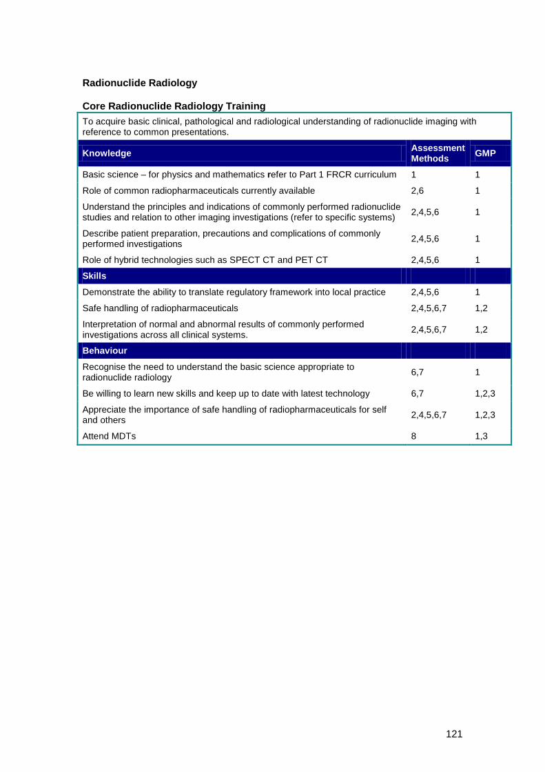
121
Radionuclide Radiology Core Radionuclide Radiology Training To acquire basic clinical, pathological and radiological understanding of radionuclide imaging with reference to common presentations.
Knowledge Assessment Methods GMP
Basic science – for physics and mathematics refer to Part 1 FRCR curriculum 1 1
Role of common radiopharmaceuticals currently available 2,6 1
Understand the principles and indications of commonly performed radionuclide studies and relation to other imaging investigations (refer to specific systems) 2,4,5,6 1
Describe patient preparation, precautions and complications of commonly performed investigations 2,4,5,6 1
Role of hybrid technologies such as SPECT CT and PET CT 2,4,5,6 1
Skills
Demonstrate the ability to translate regulatory framework into local practice 2,4,5,6 1
Safe handling of radiopharmaceuticals 2,4,5,6,7 1,2
Interpretation of normal and abnormal results of commonly performed investigations across all clinical systems. 2,4,5,6,7 1,2
Behaviour
Recognise the need to understand the basic science appropriate to radionuclide radiology 6,7 1
Be willing to learn new skills and keep up to date with latest technology 6,7 1,2,3
Appreciate the importance of safe handling of radiopharmaceuticals for self and others 2,4,5,6,7 1,2,3
Attend MDTs 8 1,3
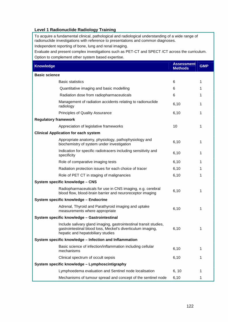
122
Level 1 Radionuclide Radiology Training To acquire a fundamental clinical, pathological and radiological understanding of a wide range of radionuclide investigations with reference to presentations and common diagnoses. Independent reporting of bone, lung and renal imaging. Evaluate and present complex investigations such as PET-CT and SPECT /CT across the curriculum. Option to complement other system based expertise.
Knowledge Assessment Methods GMP
Basic science
Basic statistics 6 1
Quantitative imaging and basic modelling 6 1
Radiation dose from radiopharmaceuticals 6 1
Management of radiation accidents relating to radionuclide radiology 6,10 1
Principles of Quality Assurance 6,10 1
Regulatory framework
Appreciation of legislative frameworks 10 1
Clinical Application for each system
Appropriate anatomy, physiology, pathophysiology and biochemistry of system under investigation 6,10 1
Indication for specific radiotracers including sensitivity and specificity 6,10 1
Role of comparative imaging tests 6,10 1
Radiation protection issues for each choice of tracer 6,10 1
Role of PET CT in staging of malignancies 6,10 1
System specific knowledge – CNS
Radiopharmaceuticals for use in CNS imaging, e.g. cerebral blood flow, blood-brain barrier and neuroreceptor imaging 6,10 1
System specific knowledge – Endocrine
Adrenal, Thyroid and Parathyroid imaging and uptake measurements where appropriate 6,10 1
System specific knowledge – Gastrointestinal
Include salivary gland imaging, gastrointestinal transit studies, gastrointestinal blood loss, Meckel’s diverticulum imaging, hepatic and hepatobiliary studies
6,10 1
System specific knowledge – Infection and Inflammation
Basic science of infection/inflammation including cellular mechanisms 6,10 1
Clinical spectrum of occult sepsis 6,10 1
System specific knowledge – Lymphoscintigraphy
Lymphoedema evaluation and Sentinel node localisation 6, 10 1
Mechanisms of tumour spread and concept of the sentinel node 6,10 1
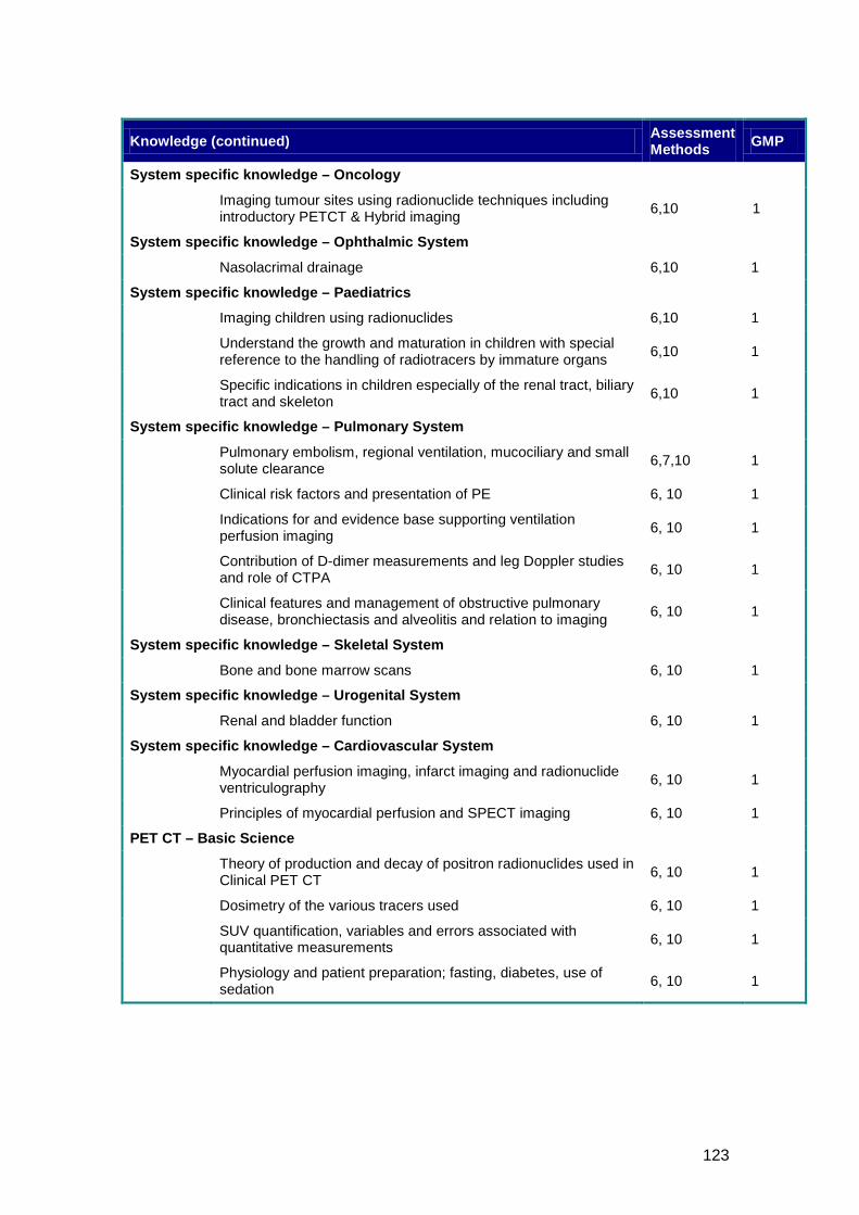
123
Knowledge (continued) Assessment Methods GMP
System specific knowledge – Oncology
Imaging tumour sites using radionuclide techniques including introductory PETCT & Hybrid imaging 6,10 1
System specific knowledge – Ophthalmic System
Nasolacrimal drainage 6,10 1
System specific knowledge – Paediatrics
Imaging children using radionuclides 6,10 1
Understand the growth and maturation in children with special reference to the handling of radiotracers by immature organs 6,10 1
Specific indications in children especially of the renal tract, biliary tract and skeleton 6,10 1
System specific knowledge – Pulmonary System
Pulmonary embolism, regional ventilation, mucociliary and small solute clearance 6,7,10 1
Clinical risk factors and presentation of PE 6, 10 1
Indications for and evidence base supporting ventilation perfusion imaging 6, 10 1
Contribution of D-dimer measurements and leg Doppler studies and role of CTPA 6, 10 1
Clinical features and management of obstructive pulmonary disease, bronchiectasis and alveolitis and relation to imaging 6, 10 1
System specific knowledge – Skeletal System
Bone and bone marrow scans 6, 10 1
System specific knowledge – Urogenital System
Renal and bladder function 6, 10 1
System specific knowledge – Cardiovascular System
Myocardial perfusion imaging, infarct imaging and radionuclide ventriculography 6, 10 1
Principles of myocardial perfusion and SPECT imaging 6, 10 1
PET CT – Basic Science
Theory of production and decay of positron radionuclides used in Clinical PET CT 6, 10 1
Dosimetry of the various tracers used 6, 10 1
SUV quantification, variables and errors associated with quantitative measurements 6, 10 1
Physiology and patient preparation; fasting, diabetes, use of sedation 6, 10 1
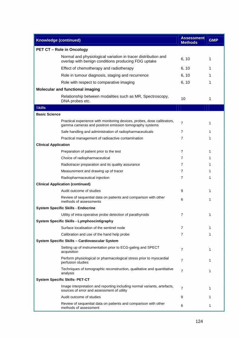
124
Knowledge (continued) Assessment Methods GMP
PET CT – Role in Oncology
Normal and physiological variation in tracer distribution and overlap with benign conditions producing FDG uptake 6, 10 1
Effect of chemotherapy and radiotherapy 6, 10 1
Role in tumour diagnosis, staging and recurrence 6, 10 1
Role with respect to comparative imaging 6, 10 1
Molecular and functional imaging
Relationship between modalities such as MR, Spectroscopy, DNA probes etc. 10 1
Skills
Basic Science
Practical experience with monitoring devices, probes, dose calibrators, gamma cameras and positron emission tomography systems 7 1
Safe handling and administration of radiopharmaceuticals 7 1
Practical management of radioactive contamination 7 1
Clinical Application
Preparation of patient prior to the test 7 1
Choice of radiopharmaceutical 7 1
Radiotracer preparation and its quality assurance 7 1
Measurement and drawing up of tracer 7 1
Radiopharmaceutical injection 7 1
Clinical Application (continued)
Audit outcome of studies 9 1
Review of sequential data on patients and comparison with other methods of assessments 6 1
System Specific Skills - Endocrine
Utility of intra-operative probe detection of parathyroids 7 1
System Specific Skills - Lymphoscintigraphy
Surface localisation of the sentinel node 7 1
Calibration and use of the hand help probe 7 1
System Specific Skills – Cardiovascular System
Setting up of instrumentation prior to ECG-gating and SPECT acquisition 7 1
Perform physiological or pharmacological stress prior to myocardial perfusion studies 7 1
Techniques of tomographic reconstruction, qualitative and quantitative analysis 7 1
System Specific Skills- PET-CT
Image interpretation and reporting including normal variants, artefacts, sources of error and assessment of utility 7 1
Audit outcome of studies 9 1
Review of sequential data on patients and comparison with other methods of assessment 6 1
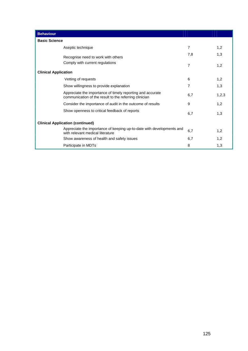
125
Behaviour
Basic Science
Aseptic technique 7 1,2
Recognise need to work with others Comply with current regulations
7,8 7
1,3 1,2
Clinical Application
Vetting of requests 6 1,2
Show willingness to provide explanation 7 1,3
Appreciate the importance of timely reporting and accurate communication of the result to the referring clinician 6,7 1,2,3
Consider the importance of audit in the outcome of results 9 1,2
Show openness to critical feedback of reports
6,7 1,3
Clinical Application (continued)
Appreciate the importance of keeping up-to-date with developments and with relevant medical literature 6,7 1,2
Show awareness of health and safety issues 6,7 1,2
Participate in MDTs 8 1,3
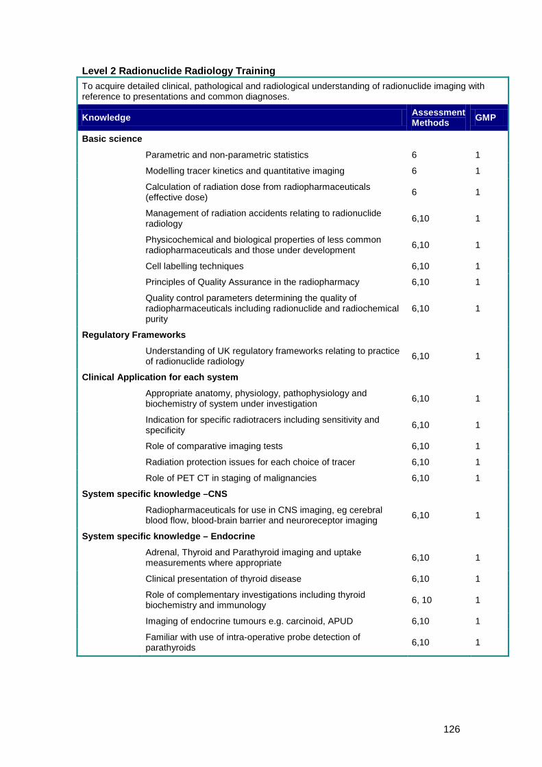
126
Level 2 Radionuclide Radiology Training To acquire detailed clinical, pathological and radiological understanding of radionuclide imaging with reference to presentations and common diagnoses.
Knowledge Assessment Methods GMP
Basic science
Parametric and non-parametric statistics 6 1
Modelling tracer kinetics and quantitative imaging 6 1
Calculation of radiation dose from radiopharmaceuticals (effective dose) 6 1
Management of radiation accidents relating to radionuclide radiology 6,10 1
Physicochemical and biological properties of less common radiopharmaceuticals and those under development 6,10 1
Cell labelling techniques 6,10 1
Principles of Quality Assurance in the radiopharmacy 6,10 1
Quality control parameters determining the quality of radiopharmaceuticals including radionuclide and radiochemical purity
6,10 1
Regulatory Frameworks
Understanding of UK regulatory frameworks relating to practice of radionuclide radiology 6,10 1
Clinical Application for each system
Appropriate anatomy, physiology, pathophysiology and biochemistry of system under investigation 6,10 1
Indication for specific radiotracers including sensitivity and specificity 6,10 1
Role of comparative imaging tests 6,10 1
Radiation protection issues for each choice of tracer 6,10 1
Role of PET CT in staging of malignancies 6,10 1
System specific knowledge –CNS
Radiopharmaceuticals for use in CNS imaging, eg cerebral blood flow, blood-brain barrier and neuroreceptor imaging 6,10 1
System specific knowledge – Endocrine
Adrenal, Thyroid and Parathyroid imaging and uptake measurements where appropriate 6,10 1
Clinical presentation of thyroid disease 6,10 1
Role of complementary investigations including thyroid biochemistry and immunology 6, 10 1
Imaging of endocrine tumours e.g. carcinoid, APUD 6,10 1
Familiar with use of intra-operative probe detection of parathyroids 6,10 1
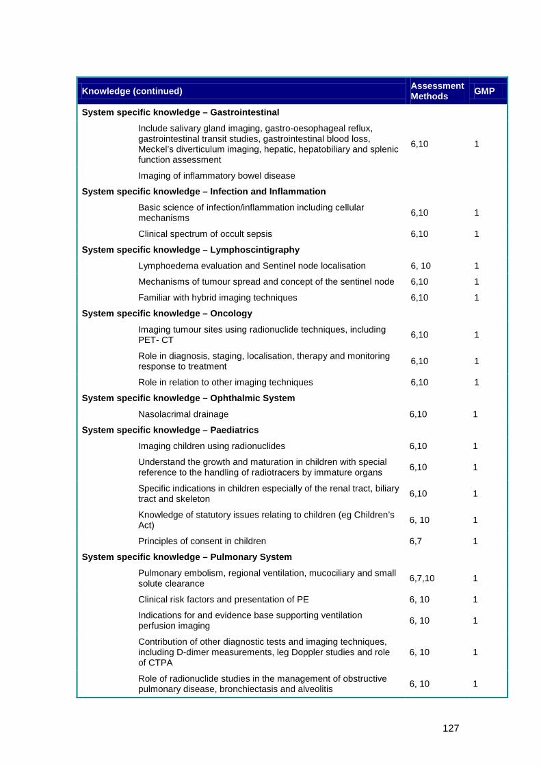
127
Knowledge (continued) Assessment Methods GMP
System specific knowledge – Gastrointestinal
Include salivary gland imaging, gastro-oesophageal reflux, gastrointestinal transit studies, gastrointestinal blood loss, Meckel’s diverticulum imaging, hepatic, hepatobiliary and splenic function assessment
6,10 1
Imaging of inflammatory bowel disease
System specific knowledge – Infection and Inflammation
Basic science of infection/inflammation including cellular mechanisms 6,10 1
Clinical spectrum of occult sepsis 6,10 1
System specific knowledge – Lymphoscintigraphy
Lymphoedema evaluation and Sentinel node localisation 6, 10 1
Mechanisms of tumour spread and concept of the sentinel node 6,10 1
Familiar with hybrid imaging techniques 6,10 1
System specific knowledge – Oncology
Imaging tumour sites using radionuclide techniques, including PET- CT 6,10 1
Role in diagnosis, staging, localisation, therapy and monitoring response to treatment 6,10 1
Role in relation to other imaging techniques 6,10 1
System specific knowledge – Ophthalmic System
Nasolacrimal drainage 6,10 1
System specific knowledge – Paediatrics
Imaging children using radionuclides 6,10 1
Understand the growth and maturation in children with special reference to the handling of radiotracers by immature organs 6,10 1
Specific indications in children especially of the renal tract, biliary tract and skeleton 6,10 1
Knowledge of statutory issues relating to children (eg Children’s Act) 6, 10 1
Principles of consent in children 6,7 1
System specific knowledge – Pulmonary System
Pulmonary embolism, regional ventilation, mucociliary and small solute clearance 6,7,10 1
Clinical risk factors and presentation of PE 6, 10 1
Indications for and evidence base supporting ventilation perfusion imaging 6, 10 1
Contribution of other diagnostic tests and imaging techniques, including D-dimer measurements, leg Doppler studies and role of CTPA
6, 10 1
Role of radionuclide studies in the management of obstructive pulmonary disease, bronchiectasis and alveolitis 6, 10 1
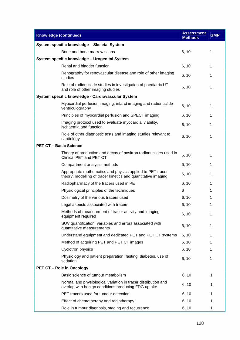
128
Knowledge (continued) Assessment Methods GMP
System specific knowledge – Skeletal System
Bone and bone marrow scans 6, 10 1
System specific knowledge – Urogenital System
Renal and bladder function 6, 10 1
Renography for renovascular disease and role of other imaging studies 6, 10 1
Role of radionuclide studies in investigation of paediatric UTI and role of other imaging studies 6, 10 1
System specific knowledge - Cardiovascular System
Myocardial perfusion imaging, infarct imaging and radionuclide ventriculography 6, 10 1
Principles of myocardial perfusion and SPECT imaging 6, 10 1
Imaging protocol used to evaluate myocardial viability, ischaemia and function 6, 10 1
Role of other diagnostic tests and imaging studies relevant to cardiology 6, 10 1
PET CT – Basic Science
Theory of production and decay of positron radionuclides used in Clinical PET and PET CT 6, 10 1
Compartment analysis methods 6, 10 1
Appropriate mathematics and physics applied to PET tracer theory, modelling of tracer kinetics and quantitative imaging 6, 10 1
Radiopharmacy of the tracers used in PET 6, 10 1
Physiological principles of the techniques 6 1
Dosimetry of the various tracers used 6, 10 1
Legal aspects associated with tracers 6, 10 1
Methods of measurement of tracer activity and imaging equipment required 6, 10 1
SUV quantification, variables and errors associated with quantitative measurements 6, 10 1
Understand equipment and dedicated PET and PET CT systems 6, 10 1
Method of acquiring PET and PET CT images 6, 10 1
Cyclotron physics 6, 10 1
Physiology and patient preparation; fasting, diabetes, use of sedation 6, 10 1
PET CT – Role in Oncology
Basic science of tumour metabolism 6, 10 1
Normal and physiological variation in tracer distribution and overlap with benign conditions producing FDG uptake 6, 10 1
PET tracers used for tumour detection 6, 10 1
Effect of chemotherapy and radiotherapy 6, 10 1
Role in tumour diagnosis, staging and recurrence 6, 10 1
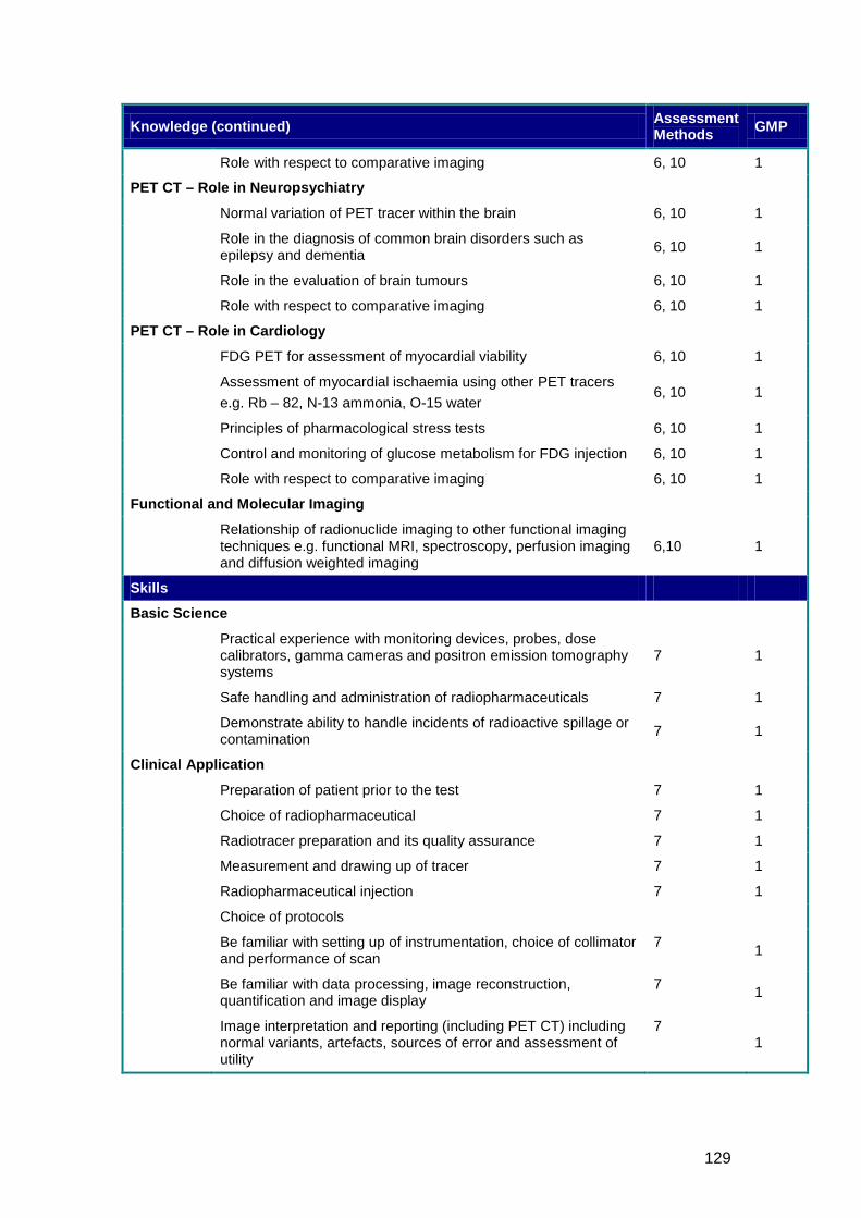
129
Knowledge (continued) Assessment Methods GMP
Role with respect to comparative imaging 6, 10 1
PET CT – Role in Neuropsychiatry
Normal variation of PET tracer within the brain 6, 10 1
Role in the diagnosis of common brain disorders such as epilepsy and dementia 6, 10 1
Role in the evaluation of brain tumours 6, 10 1
Role with respect to comparative imaging 6, 10 1
PET CT – Role in Cardiology
FDG PET for assessment of myocardial viability 6, 10 1
Assessment of myocardial ischaemia using other PET tracers e.g. Rb – 82, N-13 ammonia, O-15 water
6, 10 1
Principles of pharmacological stress tests 6, 10 1
Control and monitoring of glucose metabolism for FDG injection 6, 10 1
Role with respect to comparative imaging 6, 10 1
Functional and Molecular Imaging
Relationship of radionuclide imaging to other functional imaging techniques e.g. functional MRI, spectroscopy, perfusion imaging and diffusion weighted imaging
6,10 1
Skills
Basic Science
Practical experience with monitoring devices, probes, dose calibrators, gamma cameras and positron emission tomography systems
7 1
Safe handling and administration of radiopharmaceuticals 7 1
Demonstrate ability to handle incidents of radioactive spillage or contamination 7 1
Clinical Application
Preparation of patient prior to the test 7 1
Choice of radiopharmaceutical 7 1
Radiotracer preparation and its quality assurance 7 1
Measurement and drawing up of tracer 7 1
Radiopharmaceutical injection 7 1
Choice of protocols
Be familiar with setting up of instrumentation, choice of collimator and performance of scan
7 1
Be familiar with data processing, image reconstruction, quantification and image display
7 1
Image interpretation and reporting (including PET CT) including normal variants, artefacts, sources of error and assessment of utility
7 1
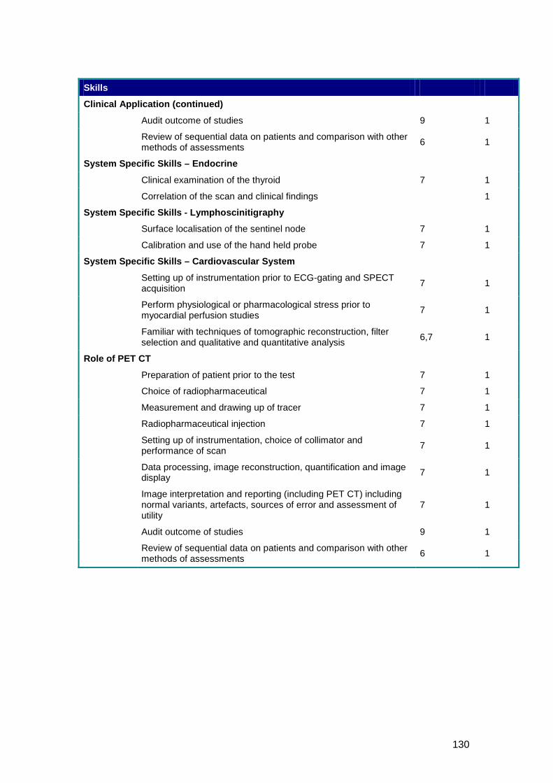
130
Skills
Clinical Application (continued)
Audit outcome of studies 9 1
Review of sequential data on patients and comparison with other methods of assessments 6 1
System Specific Skills – Endocrine
Clinical examination of the thyroid 7 1
Correlation of the scan and clinical findings 1
System Specific Skills - Lymphoscinitigraphy
Surface localisation of the sentinel node 7 1
Calibration and use of the hand held probe 7 1
System Specific Skills – Cardiovascular System
Setting up of instrumentation prior to ECG-gating and SPECT acquisition 7 1
Perform physiological or pharmacological stress prior to myocardial perfusion studies 7 1
Familiar with techniques of tomographic reconstruction, filter selection and qualitative and quantitative analysis 6,7 1
Role of PET CT
Preparation of patient prior to the test 7 1
Choice of radiopharmaceutical 7 1
Measurement and drawing up of tracer 7 1
Radiopharmaceutical injection 7 1
Setting up of instrumentation, choice of collimator and performance of scan 7 1
Data processing, image reconstruction, quantification and image display 7 1
Image interpretation and reporting (including PET CT) including normal variants, artefacts, sources of error and assessment of utility
7 1
Audit outcome of studies 9 1
Review of sequential data on patients and comparison with other methods of assessments 6 1
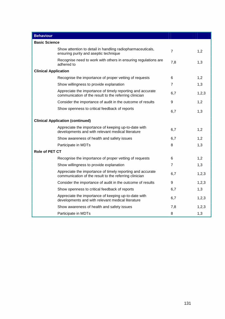
131
Behaviour
Basic Science
Show attention to detail in handling radiopharmaceuticals, ensuring purity and aseptic technique 7 1,2
Recognise need to work with others in ensuring regulations are adhered to 7,8 1,3
Clinical Application
Recognise the importance of proper vetting of requests 6 1,2
Show willingness to provide explanation 7 1,3
Appreciate the importance of timely reporting and accurate communication of the result to the referring clinician 6,7 1,2,3
Consider the importance of audit in the outcome of results 9 1,2
Show openness to critical feedback of reports
6,7 1,3
Clinical Application (continued)
Appreciate the importance of keeping up-to-date with developments and with relevant medical literature 6,7 1,2
Show awareness of health and safety issues 6,7 1,2
Participate in MDTs 8 1,3
Role of PET CT
Recognise the importance of proper vetting of requests 6 1,2
Show willingness to provide explanation 7 1,3
Appreciate the importance of timely reporting and accurate communication of the result to the referring clinician 6,7 1,2,3
Consider the importance of audit in the outcome of results 9 1,2,3
Show openness to critical feedback of reports 6,7 1,3
Appreciate the importance of keeping up-to-date with developments and with relevant medical literature 6,7 1,2,3
Show awareness of health and safety issues 7,8 1,2,3
Participate in MDTs 8 1,3
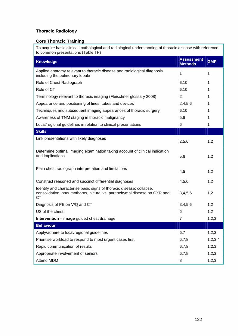
132
Thoracic Radiology Core Thoracic Training To acquire basic clinical, pathological and radiological understanding of thoracic disease with reference to common presentations (Table TP)
Knowledge Assessment Methods GMP
Applied anatomy relevant to thoracic disease and radiological diagnosis including the pulmonary lobule 1 1
Role of Chest Radiograph 6,10 1
Role of CT 6,10 1
Terminology relevant to thoracic imaging (Fleischner glossary 2008) 2 1
Appearance and positioning of lines, tubes and devices 2,4,5,6 1
Techniques and subsequent imaging appearances of thoracic surgery 6,10 1
Awareness of TNM staging in thoracic malignancy 5,6 1
Local/regional guidelines in relation to clinical presentations 6 1
Skills
Link presentations with likely diagnoses
2,5,6 1,2
Determine optimal imaging examination taking account of clinical indication and implications
5,6 1,2
Plain chest radiograph interpretation and limitations
4,5 1,2
Construct reasoned and succinct differential diagnoses 4,5,6 1,2
Identify and characterise basic signs of thoracic disease: collapse, consolidation, pneumothorax, pleural vs. parenchymal disease on CXR and CT
3,4,5,6 1,2
Diagnosis of PE on V/Q and CT 3,4,5,6 1,2
US of the chest 6 1,2
Intervention – image guided chest drainage 7 1,2,3
Behaviour
Apply/adhere to local/regional guidelines 6,7 1,2,3
Prioritise workload to respond to most urgent cases first 6,7,8 1,2,3,4
Rapid communication of results 6,7,8 1,2,3
Appropriate involvement of seniors 6,7,8 1,2,3
Attend MDM 8 1,2,3
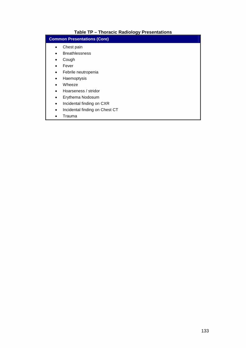
133
Table TP – Thoracic Radiology Presentations Common Presentations (Core)
• Chest pain • Breathlessness • Cough • Fever • Febrile neutropenia • Haemoptysis • Wheeze • Hoarseness / stridor • Erythema Nodosum • Incidental finding on CXR • Incidental finding on Chest CT • Trauma
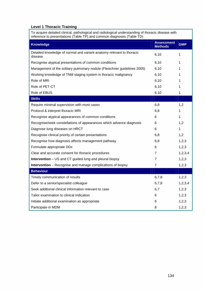
134
Level 1 Thoracic Training To acquire detailed clinical, pathological and radiological understanding of thoracic disease with reference to presentations (Table TP) and common diagnoses (Table TD)
Knowledge Assessment Methods GMP
Detailed knowledge of normal and variant anatomy relevant to thoracic disease 6,10 1
Recognise atypical presentations of common conditions 6,10 1
Management of the solitary pulmonary nodule (Fleischner guidelines 2005) 6,10 1
Working knowledge of TNM staging system in thoracic malignancy 6,10 1
Role of MRI 6,10 1
Role of PET-CT 6,10 1
Role of EBUS 6,10 1
Skills
Require minimal supervision with most cases 6,8 1,2
Protocol & interpret thoracic MRI 6,8 1
Recognise atypical appearances of common conditions 6 1
Recognise/seek constellations of appearances which advance diagnosis 6 1,2
Diagnose lung diseases on HRCT 6 1
Recognise clinical priority of certain presentations 6,8 1,2
Recognise how diagnosis affects management pathway 6,8 1,2,3
Formulate appropriate DDx 6 1,2,3
Clear and accurate consent for thoracic procedures 7 1,2,3,4
Intervention – US and CT guided lung and pleural biopsy 7 1,2,3
Intervention – Recognise and manage complications of biopsy 7 1,2,3
Behaviour
Timely communication of results 6,7,8 1,2,3
Defer to a senior/specialist colleague 6,7,8 1,2,3,4
Seek additional clinical information relevant to case 6,7 1,2,3
Tailor examination to clinical indication 6 1,2,3
Initiate additional examination as appropriate 6 1,2,3
Participate in MDM 8 1,2,3
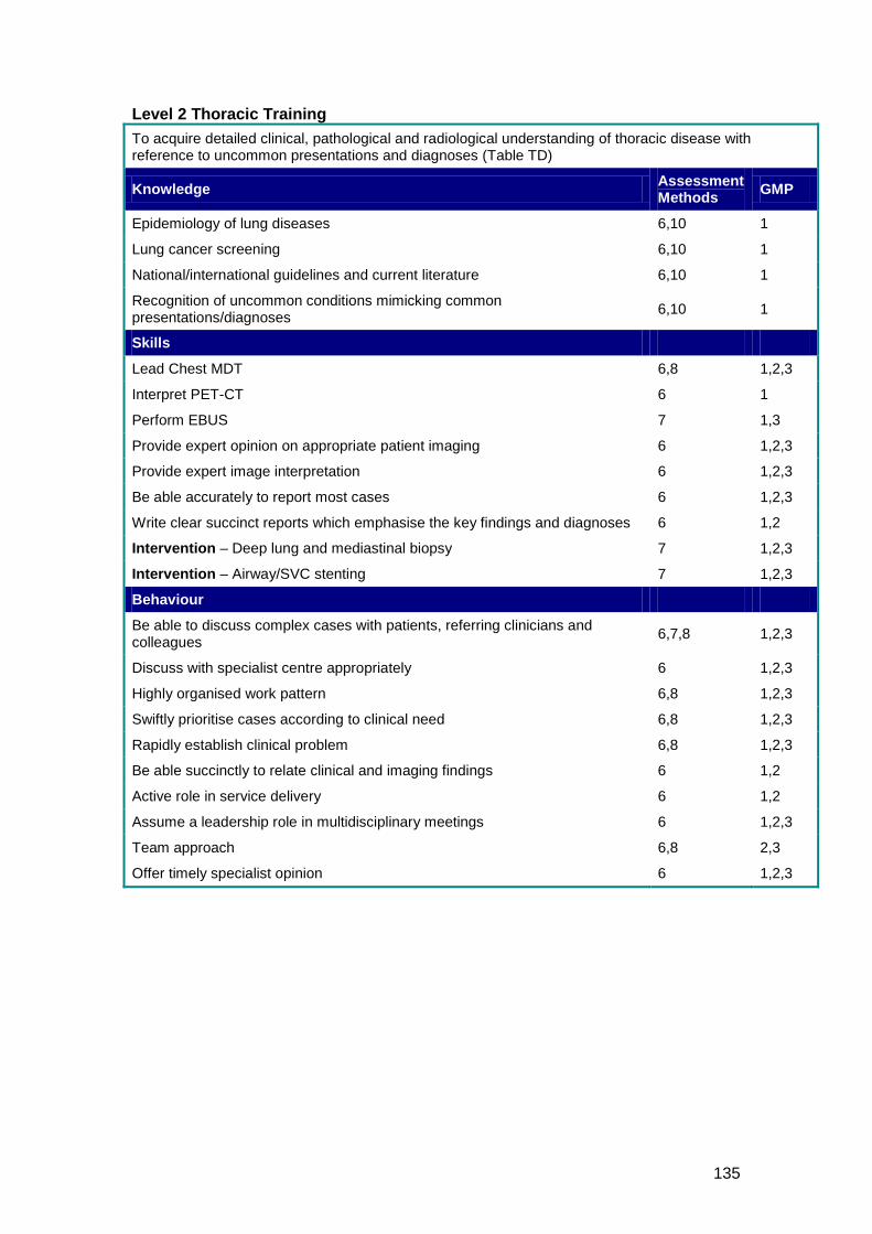
135
Level 2 Thoracic Training To acquire detailed clinical, pathological and radiological understanding of thoracic disease with reference to uncommon presentations and diagnoses (Table TD)
Knowledge Assessment Methods GMP
Epidemiology of lung diseases 6,10 1
Lung cancer screening 6,10 1
National/international guidelines and current literature 6,10 1
Recognition of uncommon conditions mimicking common presentations/diagnoses 6,10 1
Skills
Lead Chest MDT 6,8 1,2,3
Interpret PET-CT 6 1
Perform EBUS 7 1,3
Provide expert opinion on appropriate patient imaging 6 1,2,3
Provide expert image interpretation 6 1,2,3
Be able accurately to report most cases 6 1,2,3
Write clear succinct reports which emphasise the key findings and diagnoses 6 1,2
Intervention – Deep lung and mediastinal biopsy 7 1,2,3
Intervention – Airway/SVC stenting 7 1,2,3
Behaviour
Be able to discuss complex cases with patients, referring clinicians and colleagues 6,7,8 1,2,3
Discuss with specialist centre appropriately 6 1,2,3
Highly organised work pattern 6,8 1,2,3
Swiftly prioritise cases according to clinical need 6,8 1,2,3
Rapidly establish clinical problem 6,8 1,2,3
Be able succinctly to relate clinical and imaging findings 6 1,2
Active role in service delivery 6 1,2
Assume a leadership role in multidisciplinary meetings 6 1,2,3
Team approach 6,8 2,3
Offer timely specialist opinion 6 1,2,3
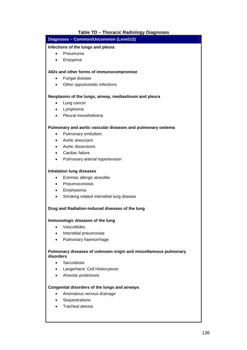
136
Table TD – Thoracic Radiology Diagnoses Diagnoses – Common/Uncommon (Level1/2)
Infections of the lungs and pleura • Pneumonia • Empyema
AIDs and other forms of immunocompromise
• Fungal disease • Other opportunistic infections
Neoplasms of the lungs, airway, mediastinum and pleura
• Lung cancer • Lymphoma • Pleural mesothelioma
Pulmonary and aortic vascular diseases and pulmonary oedema
• Pulmonary embolism • Aortic aneurysm • Aortic dissections • Cardiac failure • Pulmonary arterial hypertension
Inhalation lung diseases
• Extrinsic allergic alveolitis • Pneumoconiosis • Emphysema • Smoking related interstitial lung disease
Drug and Radiation-induced diseases of the lung Immunologic diseases of the lung
• Vasculitides • Interstitial pneumonias • Pulmonary haemorrhage
Pulmonary diseases of unknown origin and miscellaneous pulmonary disorders
• Sarcoidosis • Langerhans’ Cell Histiocytosis • Alveolar proteinosis
Congenital disorders of the lungs and airways
• Anomalous venous drainage • Sequestrations • Tracheal atresia
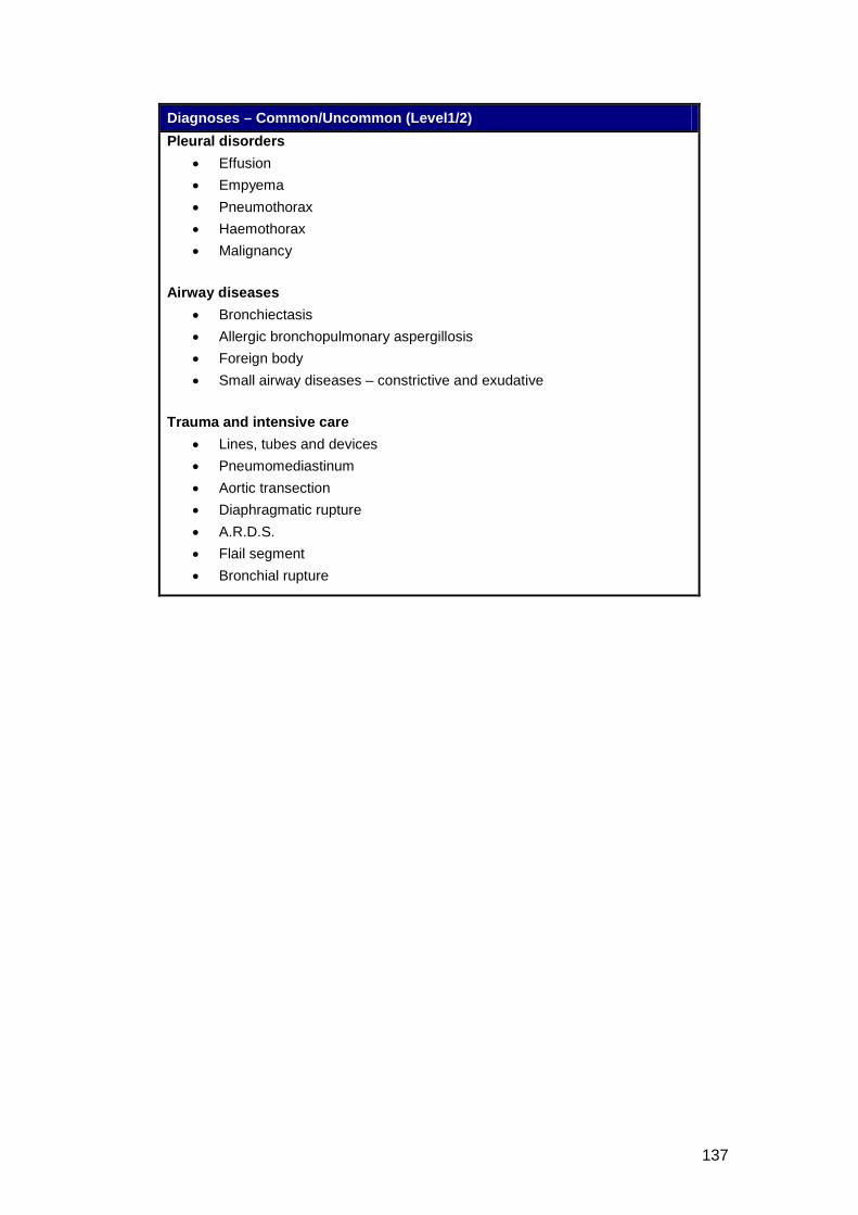
137
Diagnoses – Common/Uncommon (Level1/2) Pleural disorders
• Effusion • Empyema • Pneumothorax • Haemothorax • Malignancy
Airway diseases
• Bronchiectasis • Allergic bronchopulmonary aspergillosis • Foreign body • Small airway diseases – constrictive and exudative
Trauma and intensive care
• Lines, tubes and devices • Pneumomediastinum • Aortic transection • Diaphragmatic rupture • A.R.D.S. • Flail segment • Bronchial rupture
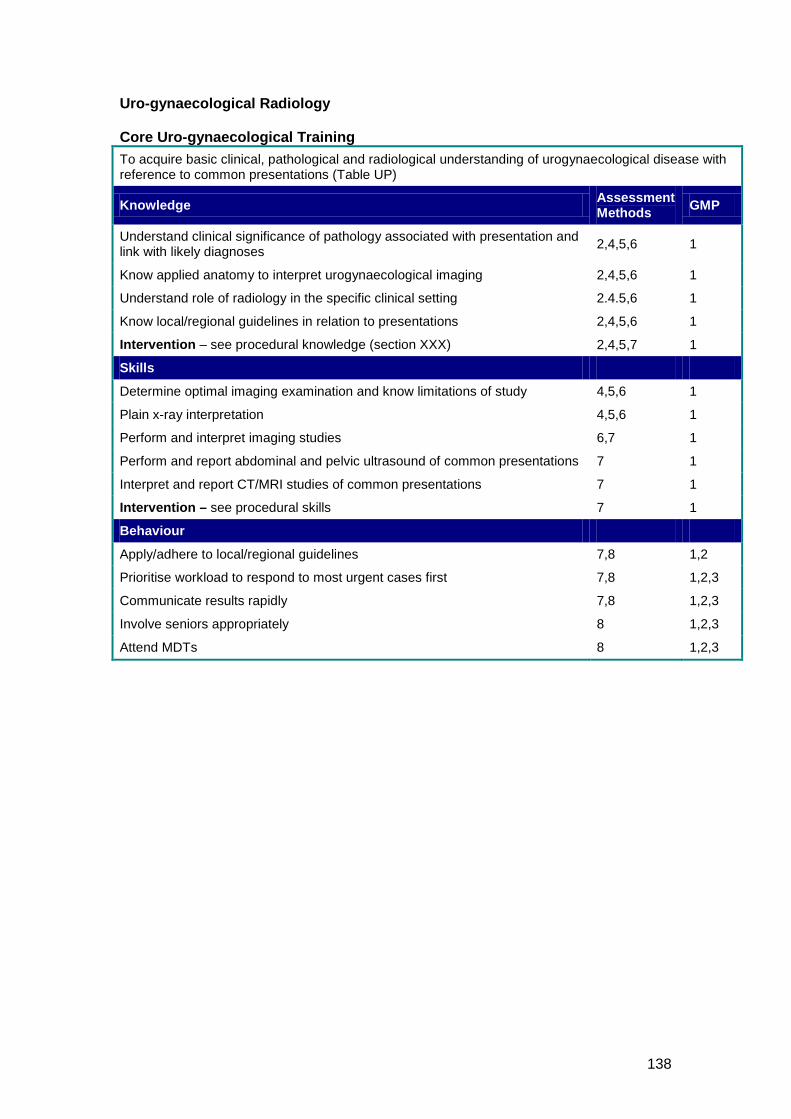
138
Uro-gynaecological Radiology Core Uro-gynaecological Training To acquire basic clinical, pathological and radiological understanding of urogynaecological disease with reference to common presentations (Table UP)
Knowledge Assessment Methods GMP
Understand clinical significance of pathology associated with presentation and link with likely diagnoses 2,4,5,6 1
Know applied anatomy to interpret urogynaecological imaging 2,4,5,6 1
Understand role of radiology in the specific clinical setting 2.4.5,6 1
Know local/regional guidelines in relation to presentations 2,4,5,6 1
Intervention – see procedural knowledge (section XXX) 2,4,5,7 1
Skills
Determine optimal imaging examination and know limitations of study 4,5,6 1
Plain x-ray interpretation 4,5,6 1
Perform and interpret imaging studies 6,7 1
Perform and report abdominal and pelvic ultrasound of common presentations 7 1
Interpret and report CT/MRI studies of common presentations 7 1
Intervention – see procedural skills 7 1
Behaviour
Apply/adhere to local/regional guidelines 7,8 1,2
Prioritise workload to respond to most urgent cases first 7,8 1,2,3
Communicate results rapidly 7,8 1,2,3
Involve seniors appropriately 8 1,2,3
Attend MDTs 8 1,2,3
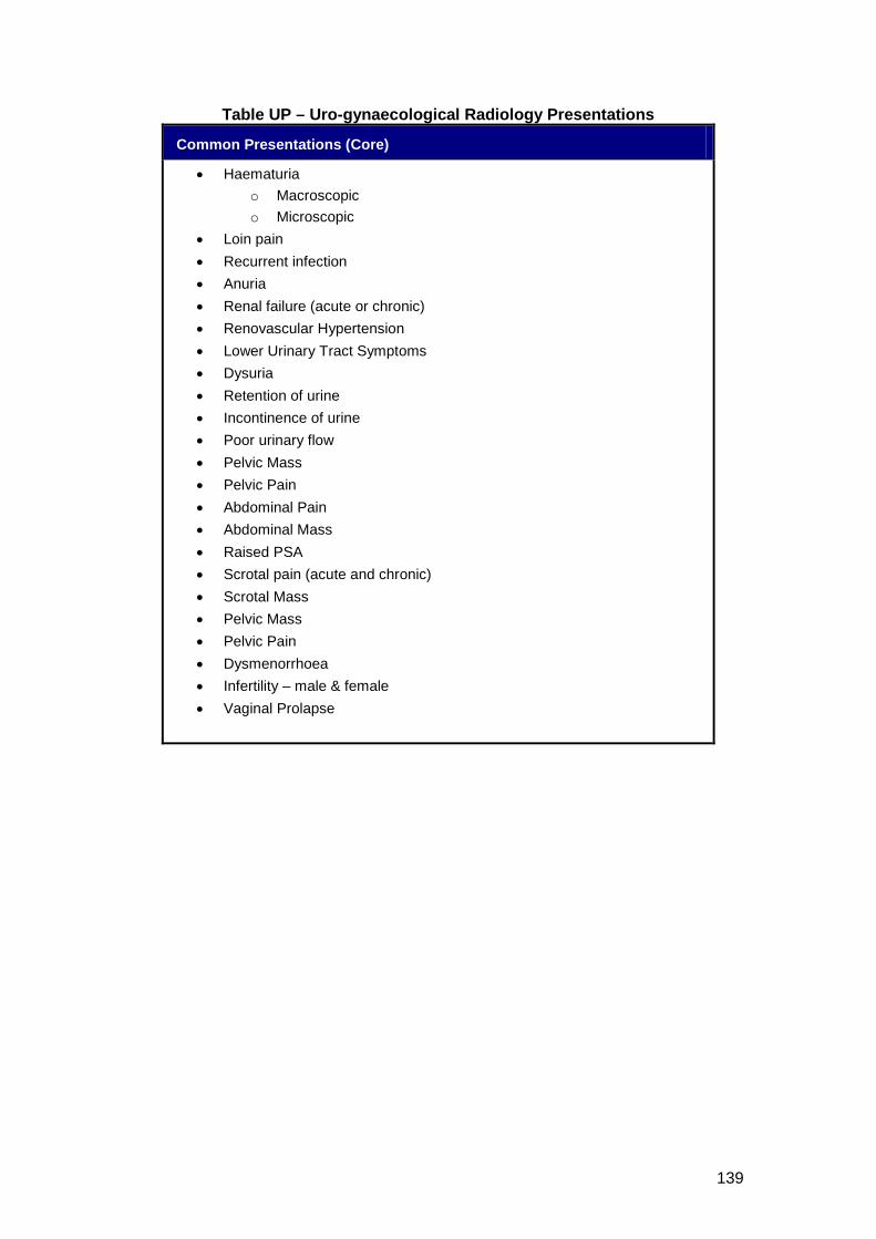
139
Table UP – Uro-gynaecological Radiology Presentations
Common Presentations (Core)
• Haematuria o Macroscopic o Microscopic
• Loin pain • Recurrent infection • Anuria • Renal failure (acute or chronic) • Renovascular Hypertension • Lower Urinary Tract Symptoms • Dysuria • Retention of urine • Incontinence of urine • Poor urinary flow • Pelvic Mass • Pelvic Pain • Abdominal Pain • Abdominal Mass • Raised PSA • Scrotal pain (acute and chronic) • Scrotal Mass • Pelvic Mass • Pelvic Pain • Dysmenorrhoea • Infertility – male & female • Vaginal Prolapse
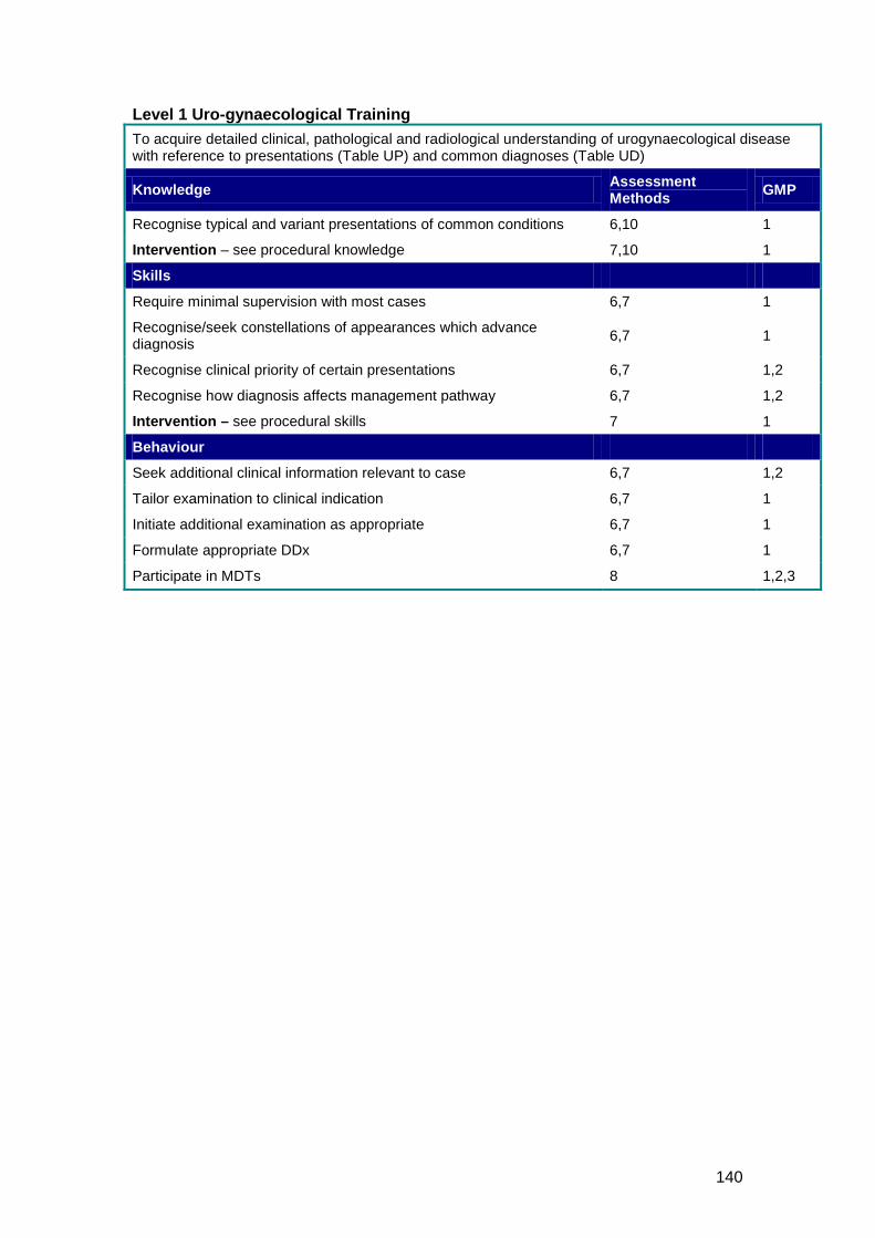
140
Level 1 Uro-gynaecological Training To acquire detailed clinical, pathological and radiological understanding of urogynaecological disease with reference to presentations (Table UP) and common diagnoses (Table UD)
Knowledge Assessment Methods GMP
Recognise typical and variant presentations of common conditions 6,10 1
Intervention – see procedural knowledge 7,10 1
Skills
Require minimal supervision with most cases 6,7 1
Recognise/seek constellations of appearances which advance diagnosis 6,7 1
Recognise clinical priority of certain presentations 6,7 1,2
Recognise how diagnosis affects management pathway 6,7 1,2
Intervention – see procedural skills 7 1
Behaviour
Seek additional clinical information relevant to case 6,7 1,2
Tailor examination to clinical indication 6,7 1
Initiate additional examination as appropriate 6,7 1
Formulate appropriate DDx 6,7 1
Participate in MDTs 8 1,2,3
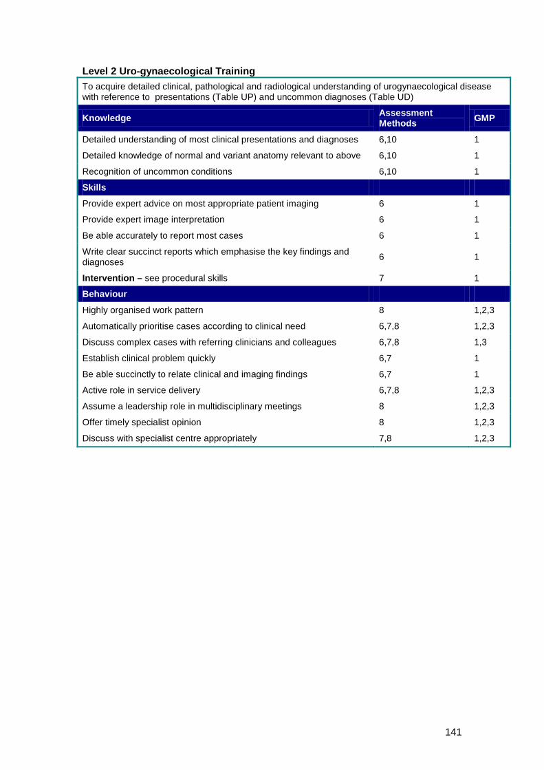
141
Level 2 Uro-gynaecological Training To acquire detailed clinical, pathological and radiological understanding of urogynaecological disease with reference to presentations (Table UP) and uncommon diagnoses (Table UD)
Knowledge Assessment Methods GMP
Detailed understanding of most clinical presentations and diagnoses 6,10 1
Detailed knowledge of normal and variant anatomy relevant to above 6,10 1
Recognition of uncommon conditions 6,10 1
Skills
Provide expert advice on most appropriate patient imaging 6 1
Provide expert image interpretation 6 1
Be able accurately to report most cases 6 1
Write clear succinct reports which emphasise the key findings and diagnoses 6 1
Intervention – see procedural skills 7 1
Behaviour
Highly organised work pattern 8 1,2,3
Automatically prioritise cases according to clinical need 6,7,8 1,2,3
Discuss complex cases with referring clinicians and colleagues 6,7,8 1,3
Establish clinical problem quickly 6,7 1
Be able succinctly to relate clinical and imaging findings 6,7 1
Active role in service delivery 6,7,8 1,2,3
Assume a leadership role in multidisciplinary meetings 8 1,2,3
Offer timely specialist opinion 8 1,2,3
Discuss with specialist centre appropriately 7,8 1,2,3
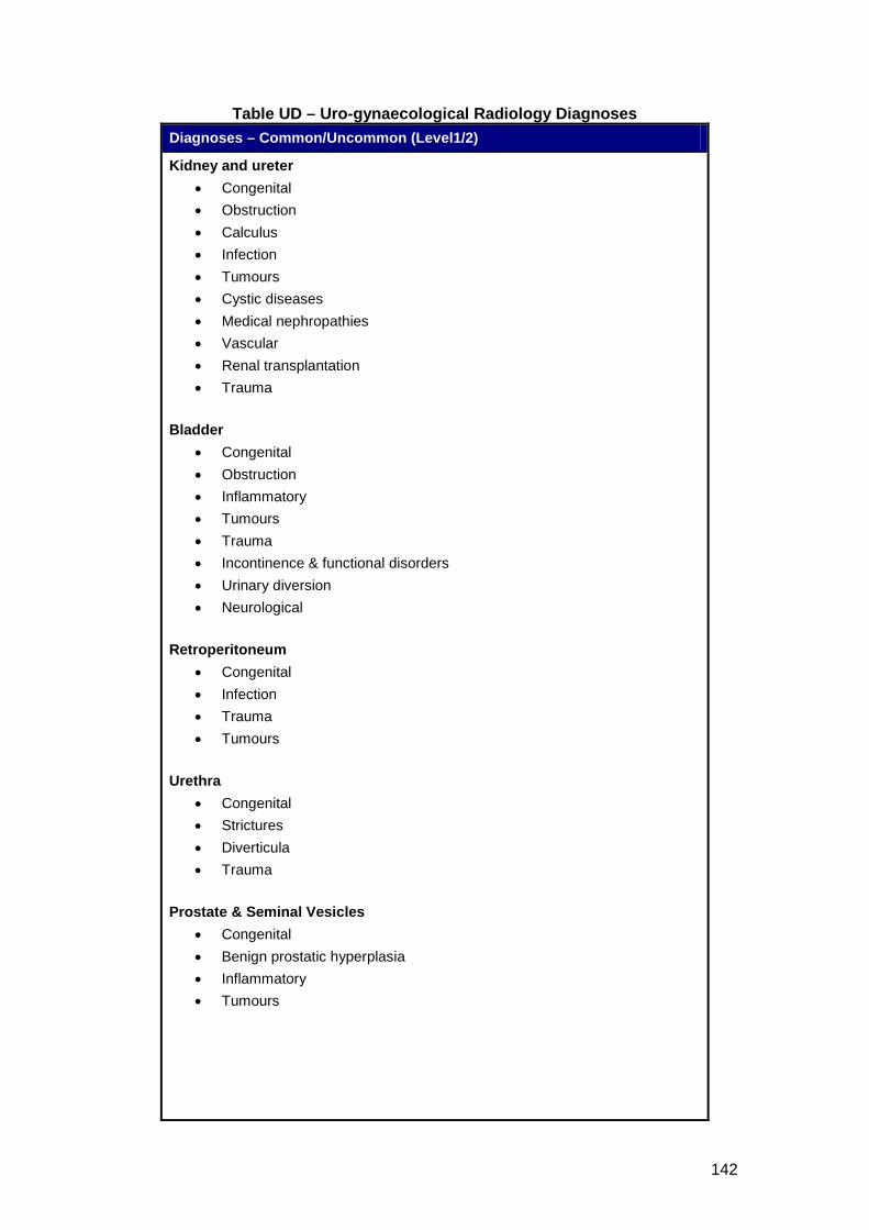
142
Table UD – Uro-gynaecological Radiology Diagnoses Diagnoses – Common/Uncommon (Level1/2)
Kidney and ureter • Congenital • Obstruction • Calculus • Infection • Tumours • Cystic diseases • Medical nephropathies • Vascular • Renal transplantation • Trauma
Bladder
• Congenital • Obstruction • Inflammatory • Tumours • Trauma • Incontinence & functional disorders • Urinary diversion • Neurological
Retroperitoneum
• Congenital • Infection • Trauma • Tumours
Urethra
• Congenital • Strictures • Diverticula • Trauma
Prostate & Seminal Vesicles
• Congenital • Benign prostatic hyperplasia • Inflammatory • Tumours
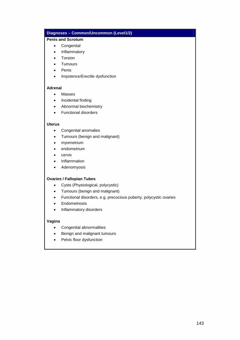
143
Diagnoses – Common/Uncommon (Level1/2) Penis and Scrotum
• Congenital • Inflammatory • Torsion • Tumours • Penis • Impotence/Erectile dysfunction
Adrenal
• Masses • Incidental finding • Abnormal biochemistry • Functional disorders
Uterus
• Congenital anomalies • Tumours (benign and malignant) • myometrium • endometrium • cervix • Inflammation • Adenomyosis
Ovaries / Fallopian Tubes
• Cysts (Physiological, polycystic) • Tumours (benign and malignant) • Functional disorders, e.g. precocious puberty, polycystic ovaries • Endometriosis • Inflammatory disorders
Vagina
• Congenital abnormalities • Benign and malignant tumours • Pelvic floor dysfunction
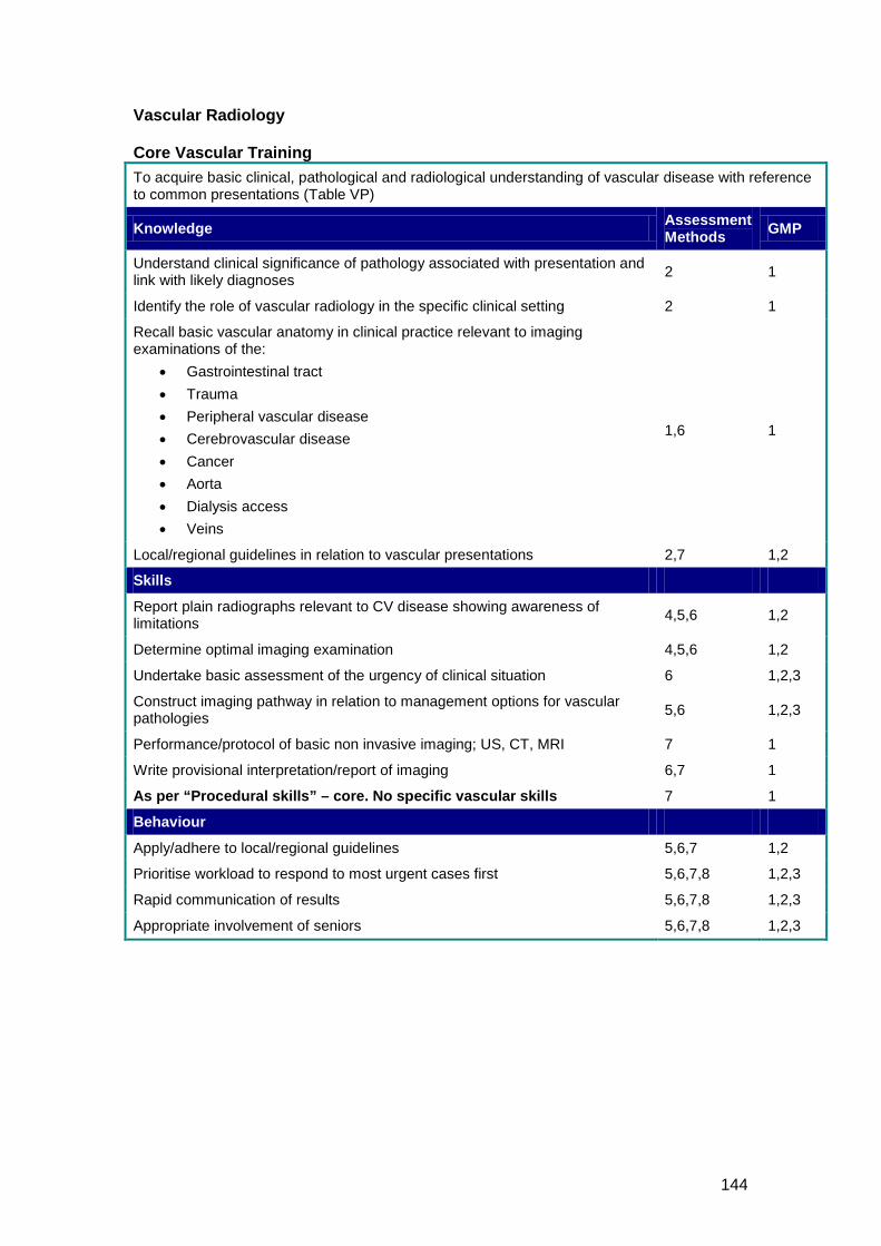
144
Vascular Radiology Core Vascular Training To acquire basic clinical, pathological and radiological understanding of vascular disease with reference to common presentations (Table VP)
Knowledge Assessment Methods GMP
Understand clinical significance of pathology associated with presentation and link with likely diagnoses 2 1
Identify the role of vascular radiology in the specific clinical setting 2 1
Recall basic vascular anatomy in clinical practice relevant to imaging examinations of the:
• Gastrointestinal tract • Trauma • Peripheral vascular disease • Cerebrovascular disease • Cancer • Aorta • Dialysis access • Veins
1,6 1
Local/regional guidelines in relation to vascular presentations 2,7 1,2
Skills
Report plain radiographs relevant to CV disease showing awareness of limitations 4,5,6 1,2
Determine optimal imaging examination 4,5,6 1,2
Undertake basic assessment of the urgency of clinical situation 6 1,2,3
Construct imaging pathway in relation to management options for vascular pathologies 5,6 1,2,3
Performance/protocol of basic non invasive imaging; US, CT, MRI 7 1
Write provisional interpretation/report of imaging 6,7 1
As per “Procedural skills” – core. No specific vascular skills 7 1
Behaviour
Apply/adhere to local/regional guidelines 5,6,7 1,2
Prioritise workload to respond to most urgent cases first 5,6,7,8 1,2,3
Rapid communication of results 5,6,7,8 1,2,3
Appropriate involvement of seniors 5,6,7,8 1,2,3
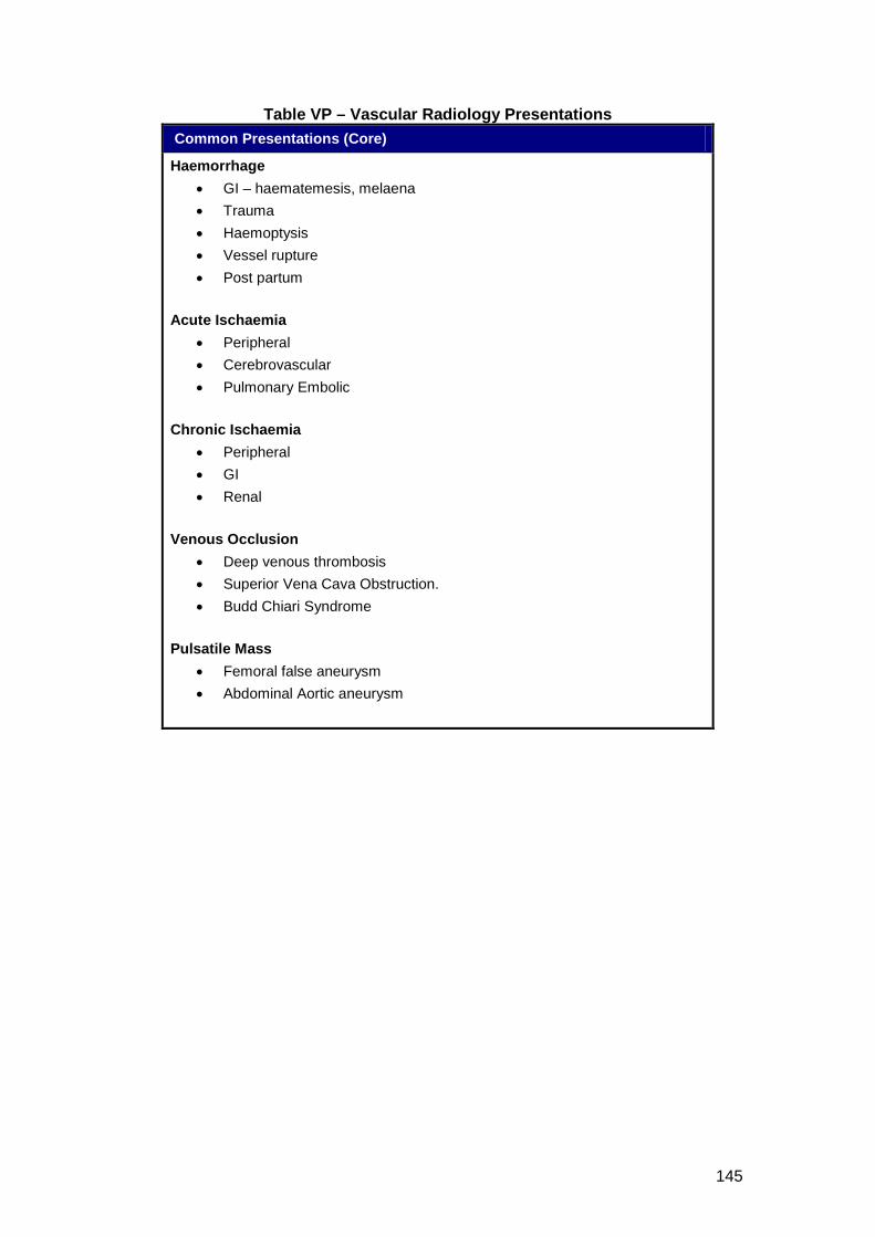
145
Table VP – Vascular Radiology Presentations Common Presentations (Core)
Haemorrhage • GI – haematemesis, melaena • Trauma • Haemoptysis • Vessel rupture • Post partum
Acute Ischaemia
• Peripheral • Cerebrovascular • Pulmonary Embolic
Chronic Ischaemia
• Peripheral • GI • Renal
Venous Occlusion
• Deep venous thrombosis • Superior Vena Cava Obstruction. • Budd Chiari Syndrome
Pulsatile Mass
• Femoral false aneurysm • Abdominal Aortic aneurysm
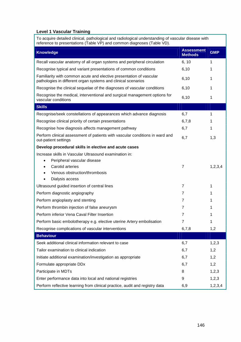
146
Level 1 Vascular Training To acquire detailed clinical, pathological and radiological understanding of vascular disease with reference to presentations (Table VP) and common diagnoses (Table VD).
Knowledge Assessment Methods GMP
Recall vascular anatomy of all organ systems and peripheral circulation 6, 10 1
Recognise typical and variant presentations of common conditions 6,10 1
Familiarity with common acute and elective presentation of vascular pathologies in different organ systems and clinical scenarios 6,10 1
Recognise the clinical sequelae of the diagnoses of vascular conditions 6,10 1
Recognise the medical, interventional and surgical management options for vascular conditions 6,10 1
Skills
Recognise/seek constellations of appearances which advance diagnosis 6,7 1
Recognise clinical priority of certain presentations 6,7,8 1
Recognise how diagnosis affects management pathway 6,7 1
Perform clinical assessment of patients with vascular conditions in ward and out-patient settings 6,7 1,3
Develop procedural skills in elective and acute cases
Increase skills in Vascular Ultrasound examination in: • Peripheral vascular disease • Carotid arteries • Venous obstruction/thrombosis • Dialysis access
7 1,2,3,4
Ultrasound guided insertion of central lines 7 1
Perform diagnostic angiography 7 1
Perform angioplasty and stenting 7 1
Perform thrombin injection of false aneurysm 7 1
Perform inferior Vena Caval Filter Insertion 7 1
Perform basic embolotherapy e.g. elective uterine Artery embolisation 7 1
Recognise complications of vascular interventions 6,7,8 1,2
Behaviour
Seek additional clinical information relevant to case 6,7 1,2,3
Tailor examination to clinical indication 6,7 1,2
Initiate additional examination/investigation as appropriate 6,7 1,2
Formulate appropriate DDx 6,7 1,2
Participate in MDTs 8 1,2,3
Enter performance data into local and national registries 9 1,2,3
Perform reflective learning from clinical practice, audit and registry data 6,9 1,2,3,4

147
Level 2 Vascular Training To acquire detailed clinical, pathological and radiological understanding of vascular disease with reference to uncommon presentations and diagnoses (Table VD)
Knowledge Assessment Methods GMP
Detailed understanding of clinical presentations and diagnoses 6,10 1
Detailed knowledge of normal and variant vascular anatomy relevant to above 6,10 1
Recognition of uncommon conditions 6,10 1
Skills
Provide expert opinion on appropriate patient imaging 6,7 1
Provide expert image interpretation 6,7 1
Be able accurately to report most cases and emphasise the key findings and diagnoses 6,7 1
Participate in acute interventional rota 8 1,2,3
Organise and undertake appropriate imaging pathways in investigating vascular conditions 6,7 1,3
Take part in one stop clinics 7,8 1,2,3
Increase procedural skills in elective and acute cases
Complex central line insertion 7 1
Mesenteric angiography, embolisation/chemo-embolisation techniques 7 1
Perform high stakes embolotherapy e.g. emergency for haemorrhage 7
Perform high stakes angioplasty and stenting 7 1
Perform thrombolysis and thrombectomy 7 1
Perform endovascular stent grafting e.g. EVAR, tEVAR 7 1
Recognise and manage complications of vascular interventions 6,7,8 1
Behaviour
Highly organised work pattern 8 1,2,3
Automatically prioritise cases according to clinical need 6,7,8 1,2,3
Discuss complex cases with referring clinicians and colleagues 6,7,8 1,2,3
Establishes clinical problem quickly 6,7 1
Be able succinctly to relate clinical and imaging findings 6,7 1
Show awareness of international relevant guidelines 6,7 1
Active role in service delivery 8 1,2,3
Assume a leadership role in multidisciplinary meetings 8 1,2,3
Offer timely specialist opinion 8 1,2,3
Discuss with specialist centre appropriately 7,8 1,2,3
Enter performance data into local and national registries 9 1,2,3
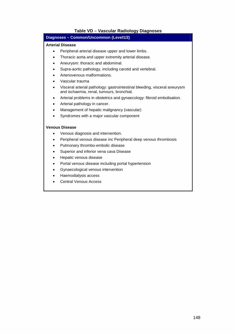
148
Table VD – Vascular Radiology Diagnoses Diagnoses – Common/Uncommon (Level1/2)
Arterial Disease • Peripheral arterial disease upper and lower limbs. • Thoracic aorta and upper extremity arterial disease. • Aneurysm: thoracic and abdominal. • Supra-aortic pathology, including carotid and vertebral. • Arteriovenous malformations. • Vascular trauma • Visceral arterial pathology: gastrointestinal bleeding, visceral aneurysm
and ischaemia, renal, tumours, bronchial. • Arterial problems in obstetrics and gynaecology: fibroid embolisation. • Arterial pathology in cancer. • Management of hepatic malignancy (vascular) • Syndromes with a major vascular component
Venous Disease
• Venous diagnosis and intervention. • Peripheral venous disease inc Peripheral deep venous thrombosis • Pulmonary thrombo-embolic disease • Superior and inferior vena cava Disease • Hepatic venous disease • Portal venous disease including portal hypertension • Gynaecological venous intervention • Haemodialysis access • Central Venous Access
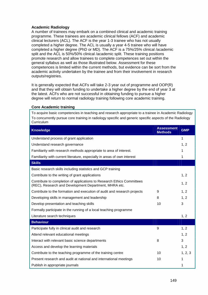
149
Academic Radiology A number of trainees may embark on a combined clinical and academic training programme. These trainees are academic clinical fellows (ACF) and academic clinical lecturers (ACL). The ACF is the year 1-3 trainee who has not usually completed a higher degree. The ACL is usually a year 4-5 trainee who will have completed a higher degree (PhD or MD). The ACF is a 75%/25% clinical /academic split and the ACL is 50%/50% clinical /academic split. These training positions promote research and allow trainees to complete competences set out within the general syllabus as well as those illustrated below. Assessment for these competences is limited within the current methods, but evidence can be sort from the academic activity undertaken by the trainee and from their involvement in research outputs/registries. It is generally expected that ACFs will take 2-3 year out of programme and OOP(R) and that they will obtain funding to undertake a higher degree by the end of year 3 at the latest. ACFs who are not successful in obtaining funding to pursue a higher degree will return to normal radiology training following core academic training. Core Academic training To acquire basic competencies in teaching and research appropriate to a trainee in Academic Radiology To concurrently pursue core training in radiology specific and generic specific aspects of the Radiology Curriculum
Knowledge Assessment Methods GMP
Understand process of grant application 1
Understand research governance 1, 2
Familiarity with research methods appropriate to area of interest. 1
Familiarity with current literature, especially in areas of own interest 1
Skills
Basic research skills including statistics and GCP training
Contribute to the writing of grant applications 1, 2
Contribute to completion of applications to Research Ethics Committees (REC), Research and Development Department, MHRA etc. 1, 2
Contribute to the formation and execution of audit and research projects 9 1, 2
Developing skills in management and leadership 8 1, 2
Develop presentation and teaching skills 10 3
Formally participate in the running of a local teaching programme
Literature search techniques 1, 2
Behaviour
Participate fully in clinical audit and research 9 1, 2
Attend relevant educational meetings 1, 2
Interact with relevant basic science departments 8 3
Access and develop the learning materials 1, 2
Contribute to the teaching programme of the training centre 10 1, 2, 3
Present research and audit at national and international meetings 10 1
Publish in appropriate journals 1

150
Level 1 Academic Training Objective Acquire increasing competencies in teaching and research appropriate to a trainee in Academic Radiology
Knowledge Assessment Methods GMP
Detailed knowledge of undertaking a major research project 1, 2, 3
Maintain familiarity with the literature and searching techniques 1
Maintain and develop knowledge of relevant methodology including epidemiology and statistics 1
Skills
Write own grant application 1, 2, 3
Prepare and present application to REC etc 1, 2
Participate in management and leadership of research team 1, 2, 3
Help to run local teaching programme 10 1, 2, 3
Present regularly at national and international meetings 10 1, 2, 3
Active role in audit 9 1, 2, 3
Be able to perform a detailed literature search 1
Maintain and develop relevant radiology expertise 1
Behaviour
Develop confidence as a clinical supervisor 1, 2, 3
Level 2 Academic Training Objective Acquire full competencies in teaching and research
Knowledge Assessment Methods GMP
Know how to plan and execute research projects independently 1, 2, 3
Know how to organise and manage a teaching programme 10 1, 2, 3
Skills
Be able to perform complex literature searches 1
Maintain and develop relevant radiology expertise 1
Help to organise and manage a teaching programme 10, 1, 2, 3
Plan and execute research projects independently 1, 2, 3
Assume a lead role in audit 9 1, 2, 3
Behaviour
Provide appropriate clinical supervision of other healthcare professionals 1, 2, 3
Full competence as a clinical supervisor 1, 2, 3
151
4 SUPPORT FOR LEARNING, SUPERVISION AND FEEDBACK
The Model of Learning Trainees will achieve the competences described in the curriculum through a variety of learning methods. There will be a balance of different modes of learning, from formal teaching programmes to experiential learning ‘on the job’. The proportion of time allocated to different learning methods may vary depending on the nature of the attachment within a rotation. There must be robust arrangements for quality assurance in place to ensure consistent implementation of the curriculum. Work-based Experiential Learning The content of work-based experiential learning is decided by the local faculty but includes active participation in
• Radiological attachments with gradual reduction in supervision according to increasing competence as judged by trainers (apprenticeship model). A major component of training in clinical radiology is achieved by the apprenticeship system with the trainee undertaking an increasing number of radiological tasks.
• Multidisciplinary team meetings: These inter-disciplinary meetings provide excellent learning opportunities.
• On-call and emergency provision.
• The degree of responsibility taken by the trainee will increase as competency increases. There should be appropriate levels of supervision throughout training with increasing independence and responsibility as learning outcomes are achieved.
Formal Postgraduate Teaching • A programme of formal, regular teaching sessions to cohorts of trainees • Case presentations • Journal clubs • Research and audit projects • Lectures and small group teaching • Grand Rounds • Radiological skills demonstrations and teaching • Joint meetings with clinical specialties • Attendance at training programmes organised on a deanery or regional basis,
which are designed to cover aspects of the training programme outlined in this curriculum
Independent Self-Directed Learning Trainees will use this time in a variety of ways depending upon their stage of learning. Suggested activities include
• Preparation for assessment and examinations • Reading, including web-based material • Maintenance of personal portfolio (self-assessment, reflective learning,
personal development plan)
152
• Audit and research projects • Reading journals • Achieving personal learning goals beyond normal expectation
Formal Study Courses Time to be made available for formal courses is encouraged, subject to local conditions of service. Examples include management courses and communication courses.
Learning Experiences Clinical and educational supervisors will be encouraged to identify learner centred educational opportunities in the course of clinical work. Radiology trainees and their teachers will recognise the importance of maximising the wide variety of learning opportunities in the clinical radiological workplace.
• Learning from Practice: Trainees will spend a large proportion of work-based experiential learning involved in supervised radiological practice in a hospital setting. Learning will involve closely supervised practice until competencies are achieved. The learning environment will be in all areas of the imaging department and in other areas where imaging services are provided (eg bedside ultrasound)
• Learning with Peers: There are many opportunities for trainees to learn with their peers. Local postgraduate teaching opportunities allow trainees of varied levels of experience to come together for small group sessions. Examination preparation encourages the formation of self-help groups and learning sets.
• Learning in Formal Situations: There are many opportunities for formal teaching in the local postgraduate teaching sessions and at regional, national and international meetings.
• Personal Study: Time will be provided during training for personal study. It may be possible for longer periods of private study to be offered as part of study leave.
• Specific Teacher Inputs: Individual units within a teaching programme will identify where specific teacher inputs will be provided. These will vary from programme to programme. Recommendations for good practice are identified in the ePortfolio. Examples include
o Each trainee having a radiological supervisor for each attachment for work-based experiential teaching
o Special interest teaching in a radiological environment from a recognised specialist
o Structured teaching sessions
Supervising and supporting workplace-based learning Educators need to identify their own professional development needs in order to carry out their role effectively, and develop the confidence and expertise to support workplace learning. The roles of the educators needed to support learning activities include adviser, appraiser, assessor, clinical supervisor, coach, co-learner, critical friend, educational supervisor, expert, facilitator, mentor, teacher, trainer and tutor.
153
Requirement for Trainers
All trainers are expected to:
• have demonstrated an interest in and developed a knowledge of training
• have appropriate equipment available
• have a sufficiently large spectrum of cases to ensure curriculum coverage
• have appropriate teaching resources
• be up-to-date with the requirements of the RCR continuing professional development scheme and be in possession of appropriate supporting documents
• have substantial expertise and knowledge in their area(s) of clinical practice
When learning in and from practice, it is important to understand that the roles of trainers may overlap and differ in subtle ways. In these cases supervision provides essential support. However
• the needs of the learner should determine which role is adopted, and these change over time and in different situations
• skilled educators move in and among these roles according to identified need
• enough time should be allocated to develop these roles and relationships
• those involved should aspire to mutually negotiated and fair outcomes, but they should also recognise that supervision involves a power relationship
• good educational practice requires a balance of the following aspects:
o support o challenge o clarification of the standards to be achieved o clarification of the consequences of non-achievement
Educational supervisor All radiology trainees will have an educational supervisor. An educational supervisor is appropriately trained to be responsible for the overall supervision and management of a specified trainee’s educational progress during a training placement or series of placements. The educational supervisor is integral to the appraisal process. A trainee appraisal with the educational supervisor will include feedback on performance, review of outcomes of assessments, induction to posts and career advice. The postgraduate deaneries should recognise the active role of educational supervisor in training and offer appropriate support. Local education providers must ensure that educational supervisors have adequate support and resources to undertake their training role. This will include training in equality and diversity.
154
The educational supervisor (ES) will
• ensure that the programme is appropriate for the doctor’s needs
• be responsible for the radiology trainee’s educational agreement
• meet with the radiology trainee at the beginning of each placement to agree how the learning objectives for this period of training will be met and confirm how formative feedback and summative judgements will be made.
• help radiology trainees by reviewing their learning needs in the light of achieved goals
• collate and/or carry out assessments from clinical supervisors, trainers and other assessors
• review the radiology trainee’s learning ePortfolio
• conduct appraisals and give supportive feedback on the results of MSF
• complete the structured supervisor’s report at the end of each year of training prior to the ARCP.
• support the trainee through any difficulty
• tell the clinical director, head of service or medical director and those responsible for training, of serious weaknesses in their trainee’s performance that have not been dealt with.
• tell the radiology trainee the content of any information about them that is given to someone else
• ensure that all training opportunities meet the requirements of equality and diversity legislation
• give appropriate handover to the next educational supervisor, with the radiology trainee’s knowledge.
The educational supervisor, when meeting with the trainee, should discuss issues of clinical governance, risk management and the report of any untoward clinical incidents involving the trainee. The educational supervisor is part of the clinical speciality team. Thus, if the clinical directorate (clinical director) should have any concerns about the performance of the trainee, or there were issues of doctor or patient safety, these would be discussed with the educational supervisor. These processes, which are integral to trainee development, must not detract from the statutory duty of the Employer to deliver effective clinical governance through its management systems.
155
Clinical supervisor A trainer is selected and appropriately trained to be responsible for overseeing a specified radiology trainee’s clinical work and providing constructive feedback during a training placement. Clinical supervisors may/will change on a day-to-day basis depending on the rota for each radiology trainee. Some training programmes appoint an educational supervisor for each placement. The roles of clinical and educational supervisor may then be merged. A clinical supervisor will usually be the consultant to whom a radiology trainee is directly responsible for their clinical work. There will be frequent contact between them. The educational supervisor may see the radiology trainee much less often. Local education providers must ensure that clinical supervisors have adequate support and resources to undertake their training role. This will include training in equality and diversity. The clinical supervisor is responsible for
• ensuring that their radiology trainees are never put in a situation where they are asked to work beyond their competence without appropriate support and supervision. Patient safety must be paramount at all times.
• guaranteeing suitable induction to the radiology department
• meeting with the radiology trainee at the beginning of each placement to discuss what is expected in the placement, learning opportunities available and the trainee’s learning needs
• ensuring that the clinical experience available to the trainee is appropriate and properly supervised
• ensuring that all training opportunities meet the requirements of equality and diversity legislation
• monitoring, supporting and assessing the radiology trainee's day-to-day clinical and professional work
• providing regular feedback on the trainee’s performance
• undertaking and facilitating WpBA
• allowing the trainee to give feedback on the experience, quality of training and supervision provided
• discussing serious concerns with the educational supervisor about a trainee’s performance, health or conduct
• meet with the radiology trainee to assess whether they have met the necessary outcomes and complete an end of placement review form for each placement
156
The first year in clinical radiology can be a difficult year of transition for trainees. Training programme directors (TPDs) and College tutors are encouraged to offer advice, a mentor system and a counselling service during the year. The following milestones should be acknowledged: The trainee should meet with their Educational Supervisor (ES), the College tutor (CT) in the hospital where they are working and their TPD at the start of their appointment, and again after three months in it. Some individuals may undertake more than one of these roles simultaneously. The trainee’s practice must be closely supervised and patient safety is of paramount importance. Such aspects are monitored by the clinical supervisor for each individual rotation and documented in the formal ePortfolio. Formal mechanisms for feeding back any concerns raised by the clinical supervisor, to the trainee, and the ES, CT and TPD, should be in place. There should be a formal mechanism for counselling trainees who are unsuccessful in the First FRCR Examination. All training in postgraduate radiology should be conducted in institutions with appropriate standards of clinical governance and that meet relevant Health and Safety standards for clinical areas. Training placements must also comply with the European Working Time Directive for trainee doctors. Trainees must work with a level of clinical supervision commensurate with their clinical experience and level of competence. This is the responsibility of the relevant clinical supervisor after discussion with the trainee’s educational supervisor and the designated clinical governance lead. In keeping with the principles of Good Medical Practice, trainees should know that they must limit their clinical practice to within their level of clinical competence and seek help and support without hesitation.
Feedback Frequent and timely feedback on performance is essential for successful work-based experiential learning. To train as a radiologist, a doctor must develop the ability to seek and respond to feedback on clinical practice from a range of individuals to meet the requirements of Good Medical Practice and revalidation. Constructive feedback should be provided throughout training in both formal and informal settings. Opportunities for feedback will arise during appraisal meetings, when trainees are undergoing workplace-based assessments, in the workplace setting, and through discussions with supervisors, trainers, assessors and those within the team.
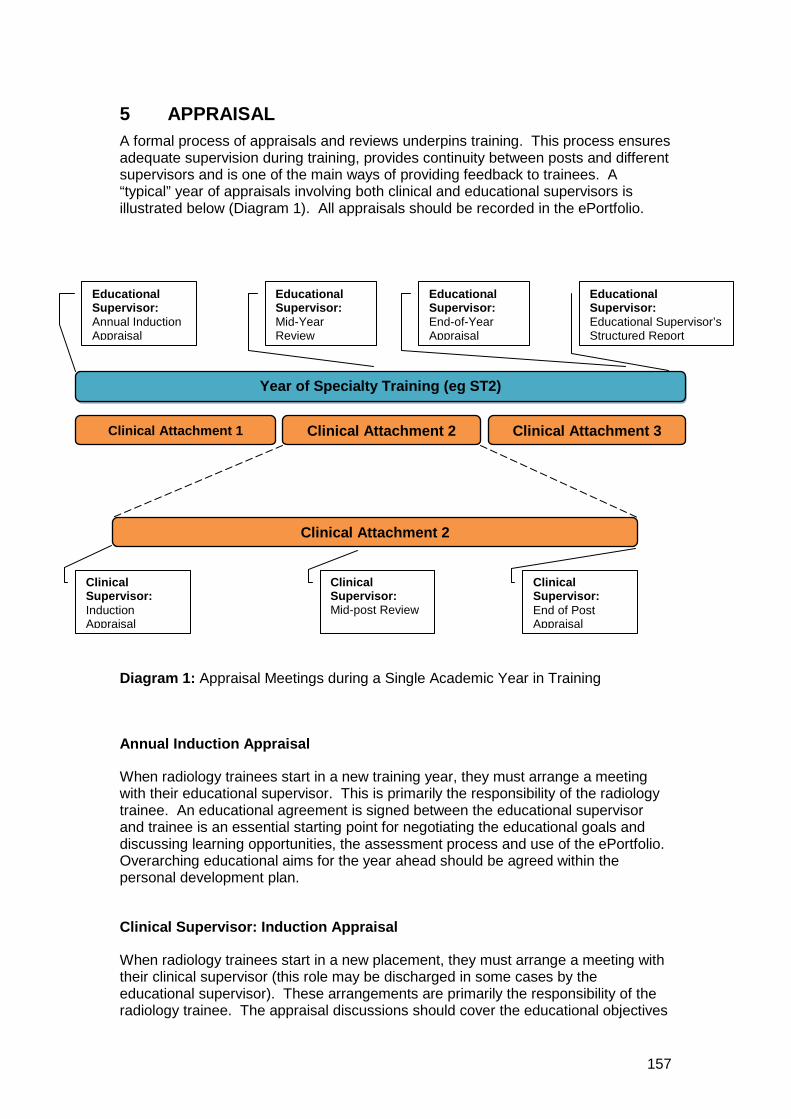
157
5 APPRAISAL A formal process of appraisals and reviews underpins training. This process ensures adequate supervision during training, provides continuity between posts and different supervisors and is one of the main ways of providing feedback to trainees. A “typical” year of appraisals involving both clinical and educational supervisors is illustrated below (Diagram 1). All appraisals should be recorded in the ePortfolio.
Diagram 1: Appraisal Meetings during a Single Academic Year in Training
Annual Induction Appraisal
When radiology trainees start in a new training year, they must arrange a meeting with their educational supervisor. This is primarily the responsibility of the radiology trainee. An educational agreement is signed between the educational supervisor and trainee is an essential starting point for negotiating the educational goals and discussing learning opportunities, the assessment process and use of the ePortfolio. Overarching educational aims for the year ahead should be agreed within the personal development plan.
Clinical Supervisor: Induction Appraisal
When radiology trainees start in a new placement, they must arrange a meeting with their clinical supervisor (this role may be discharged in some cases by the educational supervisor). These arrangements are primarily the responsibility of the radiology trainee. The appraisal discussions should cover the educational objectives
Year of Specialty Training (eg ST2)
Clinical Attachment 1 Clinical Attachment 3 Clinical Attachment 2
Clinical Attachment 2
Educational Supervisor: Annual Induction Appraisal
Educational Supervisor: Mid-Year Review
Educational Supervisor: End-of-Year Appraisal
Educational Supervisor: Educational Supervisor’s Structured Report
Clinical Supervisor: Induction Appraisal
Clinical Supervisor: Mid-post Review
Clinical Supervisor: End of Post Appraisal
158
for the clinical attachment and be used to inform the personal development plan (PDP).
Clinical Supervisor: Mid-Post Appraisal
A mid-point meeting during a clinical attachment, although not mandatory is highly recommended. It gives the trainee and clinical supervisor the opportunity to look at the achievements of the trainee and highlights areas for future development, in terms of the PDP and curriculum competences.
Clinical Supervisor: End of Post Appraisal
Towards the end of a placement, the radiology trainee and clinical supervisor will meet again for an appraisal. They will need to review the ePortfolio, the PDP and the results of assessments made during the placement. This process will involve review of colleagues’ comments, who have observed the doctor’s performance in practice and/or in individual assessments. If the educational supervisor is different from the clinical supervisor, there should be a robust communication system to ensure a continuous, appropriate, and timely flow of evidence. This should include a ‘supervisor’s report sign off’ document confirming satisfactory performance and progress. It should detail any outstanding issues that still need to be addressed.
Educational Supervisor’s Mid-Year Appraisal A mid-year appraisal with the educational supervisor is an opportunity to look at the trainee’s progress against the agreed educational objectives within the ePortfolio. It is at/around this meeting that the MSF is undertaken.
Feedback and debriefing
Feedback is a key component of the interactions between supervisors and radiology trainees. Giving and receiving feedback and engaging in constructive conversations about learning, successes, difficulties and progress are all part of an effective professional learning environment. Improvement in clinical radiological practice will only happen if regular review leads to constructive feedback. Unscheduled assessments are a good opportunity for immediate feedback. This is particularly true of Rad-DOPS and Mini-IPX, which may be opportunistic. It is essential that trainers provide, and radiology trainees receive, structured feedback.
Log Book Logbooks should be used for documenting the skills and experience attained and to facilitate reflective learning. Logbooks are mandatory for all interventional procedures irrespective of special interest.
The training objectives identified in this curriculum document (encompassing knowledge, skills and behaviours) are mapped with the appropriate assessment tools, which can be used to illustrate proof of learning across the curriculum. All these methods and tools are included in the ePortfolio.

159
These training objectives are used to assist trainee appraisal and assessment during specialty training and when achieved can verify that training has taken place to the required standard for a Certificate of Completion of Training (CCT) to be awarded.
ePortfolio The ePortfolio is the record for documenting assessments and other achievements. It is essential that radiology trainees populate the ePortfolio as it will be used to inform the end of year report by the education supervisor. End of Training Year Appraisal The results of educational activities for an academic year will be drawn together and included in a formal structured educational supervisor’s report. This will cover the overall performance of the trainee in each placement. The overall judgment of a trainee will include a triangulated view of the doctor’s performance, which will include their participation in educational activities, appraisals, the assessment process and recording of this in the ePortfolio. The outcome of the final appraisal discussion should be agreed by both the radiology trainee and the educational supervisor and recorded in the trainee’s ePortfolio in the structured supervisor’s report.
Placement reports put together in an annual structured educational supervisor’s report will form the basis of the education supervisor’s recommendations of satisfactory completion of the year of training.
160
6 ASSESSMENT
Purpose of assessment
The assessment system included in this curriculum is intended to
• enhance learning by providing formative assessment, enabling trainees to receive immediate feedback, measure their own performance and identify areas for development;
• drive learning and enhance the training process by making it clear what is required of trainees and motivating them to ensure they receive suitable training and experience;
• provide robust, summative evidence that trainees are meeting the curriculum standards during the training programme;
• ensure trainees are acquiring competencies within the domains of Good Medical Practice;
• assess trainees’ actual performance in the workplace;
• ensure that trainees possess the essential underlying knowledge required for Clinical Radiology;
• inform the Annual Review of Competence Progression (ARCP) is the culminating part of the assessment system;
• identify trainees who should be advised to consider changes of career direction.
Assessment methodology
Continuous assessment Improvement in clinical practice will only happen if regular review leads to constructive feedback. Thus, continuous review and assessment is a fundamental part of clinical radiology training. Radiology trainees are expected to demonstrate improvement and progression during each attachment. It is anticipated that radiology trainees will increasingly reach higher levels of attainments as they progress through their training. It is important that they arrange and undertake assessments in a timely and educationally appropriate manner spread throughout the year. Arriving at the overall assessment and judgement of the radiology trainee must be based on multiple assessments by many assessors, on multiple occasions. During core training, within a typical three/four month placement, an individual consultant/assessor is unlikely to build up a coherent picture of competences, let alone performance, of an individual trainee. Therefore, the training programme director (TPD) will ensure that there is a local faculty of trainers capable of building a balanced judgement of a trainee’s performance supported by the workplace based assessment results. Such an approach will prevent any individual having undue influence regarding a trainee’s progression.
161
Self Assessment Radiology trainees have a personal responsibility to undertake self assessment an integral part of their professional life. It is good educational practice for this to be stated clearly and discussed fully during induction.
As s es s ment Sys tem and Tools Radiological practice will be assessed using an integrated package of workplace based assessments and summative examination of knowledge and radiological skills, which will sample across the domains of the curriculum. The assessment methods are fit for purpose and mapped onto the curriculum in an integrated way. The assessments will generate structured feedback for trainees within core radiological training and level1/2 training. The assessment tools have been selected on the basis of their fitness for purpose.
Summative Assessment The First FRCR Examination (Physics module) and Final FRCR Part A Examination test knowledge through single best answer (SBA) questions. The First FRCR Examination (Anatomy module) tests knowledge by requiring the identification of normal anatomical structures on images. The Final FRCR Part B Examination assesses clinical competence (interpretative, analytical and communication skills)
Formative Assessment Workplace based assessment will be the cornerstone of assessment for day-to-day practice. There is a range of tools available for this use. These have undergone or are undergoing evaluation in terms of their feasibility, reliability, validity and reproducibility. The generic and radiologically specific workplace based assessment tools are
A. Multisource Feedback
• The multisource feedback (MSF) tool assesses generic skills across the domains of Good Medical Practice. It consists of the collated views from a range of co-workers (previously described as 360° assessment). It will be mapped to a self assessment tool with identical domains
• MSF should usually take place once a year, although the educational supervisor may choose to recommend and additional MSF to investigate a relevant behavioural issue or check progress after an adverse MSF.
• For each assessment, the radiology trainee should nominate 15 raters. A minimum of 12 returns are required.
• Most raters/assessors should be supervising consultants, doctors in training more senior than the trainee under assessment and experienced radiographic, nursing or allied health professional colleagues.
The recommended mix of raters/assessors is
• 2–4 senior doctors • 2–4 doctors in training
162
• 2–4 radiographers • 2–4 nurses/allied health professionals • 2–4 other team members including clerks, secretaries and auxiliary staff
B. Direct observation of doctor/patient encounter Two tools can be used to assess radiologist/patient encounters:
• Mini-imaging interpretation exercise (Mini-IPX)
• Radiology- Direct observation of procedural skills (Rad-DOPS) Radiology trainees are required to undertake a minimum of twelve observed encounters in each year of training although it is anticipated that they may/will undertake many more, as the WpBA are the vehicles by which the trainee will guarantee one-to-one teaching and ensure appropriate curriculum coverage during their clinical attachments.
Mini-imaging interpretation exercise (Mini-IPX) This is a structured assessment of an observed radiology interpretation/reporting episode:
• trainees should complete a minimum of six mini-IPX in each year of training. These should be spaced out during the year with at least two mini-IPX completed in each four month period.
• a different assessor should be used for each mini-IPX wherever possible, including at least one of consultant level, per four month placement
• assessors must
• mini-IPXs should sample across different clinical radiological problems from the radiology specific content (categories listed in the Syllabus and Competences section
be trained in giving feedback and understand the role of assessment.
• trainees should agree the timing, problem and assessor.
)
• assessors may also carry out unscheduled assessments. Radiology-Direct observation of procedural skills (Rad-DOPS) This is a structured checklist for assessing the radiology trainee’s interaction with the patient when performing a practical procedure:
• trainees must submit a minimum of six Rad-DOPS per annum
• different assessors should be used for each encounter wherever possible
• assessors must be trained both in the procedure and feedback methodology. They could include consultants, more senior doctors in training, advanced practitioner radiographers, qualified nurses or allied health professionals
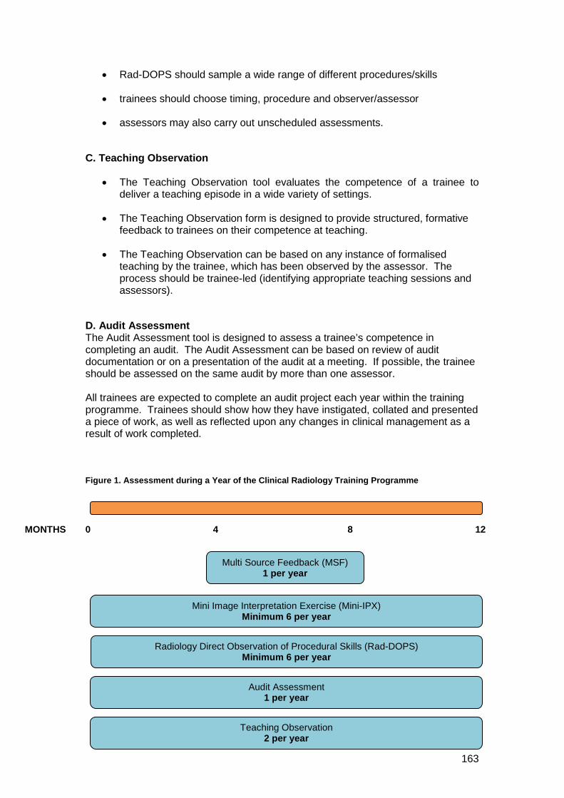
163
• Rad-DOPS should sample a wide range of different procedures/skills
• trainees should choose timing, procedure and observer/assessor
• assessors may also carry out unscheduled assessments.
C. Teaching Observation
• The Teaching Observation tool evaluates the competence of a trainee to deliver a teaching episode in a wide variety of settings.
• The Teaching Observation form is designed to provide structured, formative feedback to trainees on their competence at teaching.
• The Teaching Observation can be based on any instance of formalised teaching by the trainee, which has been observed by the assessor. The process should be trainee-led (identifying appropriate teaching sessions and assessors).
D. Audit Assessment The Audit Assessment tool is designed to assess a trainee’s competence in completing an audit. The Audit Assessment can be based on review of audit documentation or on a presentation of the audit at a meeting. If possible, the trainee should be assessed on the same audit by more than one assessor. All trainees are expected to complete an audit project each year within the training programme. Trainees should show how they have instigated, collated and presented a piece of work, as well as reflected upon any changes in clinical management as a result of work completed. Figure 1. Assessment during a Year of the Clinical Radiology Training Programme
MONTHS 0 4 8 12
Multi Source Feedback (MSF) 1 per year
Mini Image Interpretation Exercise (Mini-IPX) Minimum 6 per year
Radiology Direct Observation of Procedural Skills (Rad-DOPS) Minimum 6 per year
Audit Assessment 1 per year
Teaching Observation 2 per year
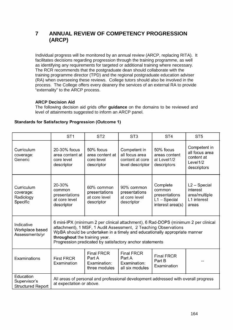
164
7 ANNUAL REVIEW OF COMPETENCY PROGRESSION (ARCP)
Individual progress will be monitored by an annual review (ARCP, replacing RITA). It facilitates decisions regarding progression through the training programme, as well as identifying any requirements for targeted or additional training where necessary. The RCR recommends that the postgraduate dean should collaborate with the training programme director (TPD) and the regional postgraduate education adviser (RA) when overseeing these reviews. College tutors should also be involved in the process. The College offers every deanery the services of an external RA to provide “externality” to the ARCP process.
ARCP Decision Aid The following decision aid grids offer guidance on the domains to be reviewed and level of attainments suggested to inform an ARCP panel.
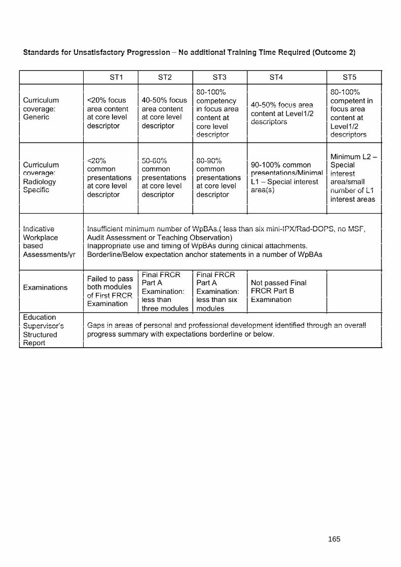
165

166

167
The main possible outcomes of this assessment and the ARCP process are listed below:
• Progress into the next year of training. Indicative of satisfactory progression across all domains within the decision aid grid.
• Unsatisfactory progression will be informed by some or all of the following (the decision being undertaken by the ARCP panel): lack of curriculum coverage, inadequate or poor outcomes in workplace based assessments and/or examinations and areas of concern within the structures supervisor's report. This will result in one of two outcomes.
Conditional progress into the next year of training. A specific action plan will be formulated with the trainee to redress deficiencies in performance. Progress will be re-assessed as appropriate within the next year of training.
Directed training without progression. If the trainee is so far short of the objectives for their year of training such as to prevent them continuing into the next year of training, directed training is recommended to achieve those objectives. The RCR recommends that repetition of the entire year should only be recommended for exceptional reasons.
168
8 APPENDICES
APPENDIX A: CURRICULUM IMPLEMENTATION AND MANAGEMENT Summary of the management of curriculum implementation:
• The new curriculum has already been discussed with all Regional Postgraduate Education Advisers and Training Programme Directors, who meet twice yearly at the Royal College of Radiologists.
• Local training programmes will develop rotations that deliver the curriculum, which is checked by:
Regional Postgraduate Education Advisers at the workplace and through the Deanery led RITA/ARCP process
The training accreditation process supervised by local Deaneries and the GMC with input from the RCR
Local Curriculum Management The organisation of training programmes for core radiological training and special interest training is the responsibility of Postgraduate Deaneries and their Schools of Radiology or equivalents. The Deaneries have established appropriate programmes for postgraduate radiological training in their regions. In England, Wales and Northern Ireland, Deanery Schools of Radiology have been set up to achieve this. As the term "School of Radiology" is not applicable to Scotland, in this curriculum, the general term "local faculties for radiological training" will be used. There may be more than one training programme within a local faculty for radiological training. Each of these is managed by a Training Programme Director (TPD) who is accountable to the Deanery Head of School (or their equivalent in Scotland). Within each participating local education provider (hospital) within a training scheme there is a College tutor who is responsible for the delivery of training and the curriculum within that hospital and is accountable for this to the TPD. The deaneries, through their local faculties and training programmes, will together with the TPDs and college tutors co-ordinate local postgraduate radiological training, with terms of reference as follows:
• Oversee recruitment and induction of trainees from Foundation and core training in other specialties into core radiological training.
• Allocate trainees into particular rotations for core radiological training, and then at ST4 and ST5 levels into rotations appropriate to their special interest training needs where possible.
• Oversee the quality of training posts provided locally.
• Interface with other Deanery Specialty Training faculties (General Practice, Anaesthesia etc).
• Ensure adequate provision of appropriate educational events.
• Ensure curricula implementation across training programmes.
169
• Oversee the workplace-based assessment process and programmes.
• Co-ordinate the ARCP process for trainees.
• Provide adequate and appropriate career advice.
• Provide systems to identify and assist trainee radiologists with training difficulties.
• Provide flexible training.
• Ensure the appropriate provision of potential to progress into an academic career
Intended Use of Curriculum by Trainers and Trainees Each trainee will be given access to download the curriculum and portfolio upon enrolling as a radiological trainee with the Royal College of Radiologists. Each trainee will engage with the curriculum by maintaining a portfolio. The trainee will use the curriculum to develop learning objectives, self-assess accomplishments in disparate areas of the curriculum, and reflect on learning experiences.
ePortfolio (https://www.nhseportfolios,org) The ePortfolio will be a record of a trainee’s progress and development through radiology training. It will provide a record of objective evidence of competence to work in a range of clinical settings and a record of satisfactory performance. This means that ePortfolio completion will contribute to the end of year report, annual review of competence progression (ARCP) and may also be used in interviews. Successful completion of the curriculum requires the achievement of competence in a variety of domains relating to generic medical practice, radiological and clinical practice. The assessments of these competences will be recorded in the ePortfolio.
Ensuring Curriculum Coverage The details of how the curriculum is covered in any individual training programme and training unit is the responsibility of the local faculty in consultation with the Royal College of Radiologists. The need to show how trainees are progressing in their attainment of competencies will be a strong driver in ensuring that all the curriculum objectives are met.
Curriculum Management Local management of the curriculum is the responsibility of the local faculty of education. Coordination of the curriculum at a national and regional level is the joint responsibility of the Deaneries and the Royal College of Radiologists, with robust arrangements for quality assurance of training.
170
APPENDIX B: CURRICULUM REVIEW The way in which this curriculum has evolved is set out in the Introduction and in the How to Use the Curriculum section. The Specialty Training Board of the Faculty of Clinical Radiology of the Royal College of Radiologists is responsible for review of the curriculum. Formal review will take place every two years. Clinical radiology, as a technology supported specialty, is a rapidly changing and evolving specialty. The curriculum needs to be able to respond appropriately to these changes to ensure that radiology training and education reflect modern practice. The regular meetings of Special Interest Groups, the Specialty Training Advisory Committee, the Professional Support and Standards Board and the Specialty Training Board allow opportunities for the curriculum to be discussed and amendments to be proposed and considered in advance of formal review. Curriculum evaluation should establish how trainees have responded to the curriculum and that the curriculum facilitates practical delivery of the required training. The curriculum will be evaluated by means of a range of qualitative and quantitative data. Trainees and lay representatives have been involved in the preparation of this curriculum and will continue to be involved in reviews, through representation from the Faculty’s Junior Radiologists’ Forum and the Patient Liaison Group. Trainers, tutors, Regional Advisers and Programme Directors will also continue to be involved in reviews through their membership of relevant working parties and committees.
Curriculum evaluation and monitoring Evaluation of Training The curriculum is an educational guide, which will be interpreted and shaped locally. Evaluation is an essential element of the curriculum for monitoring and developing local implementation. Training programmes will vary in the extent to which they currently evaluate teaching, learning and supervision. Changes in the specialty training curriculum offer new opportunities for evaluation. Whilst there are GMC surveys of trainees and trainers that provide insights into the performance of training programmes, an evaluation process is a more forensic and locally relevant enquiry than can be achieved through national questionnaire. Local evaluation will provide the evidence for training programme development, as well as material with which to respond to the findings of the annual GMC survey. The Faculty of Clinical Radiology is not intending to conduct local evaluation at the present time. It is, therefore, the responsibilities of individual training programmes to carry out this necessary work. The sections below are intended as a guide. Who is involved in evaluation? Anyone involved in the programme will have a contribution to make to its evaluation. Out of these experiences, new ideas and ways of delivery could be sought. The Training Programme Director would typically co-ordinate the process with administrative support. Local training programme committees, radiology schools and local faculties are best placed to analyse the results of evaluation.
171
Trainee involvement in curriculum review will be facilitated through:
• Involvement of trainees in local faculties of education
• Trainee involvement in the Specialty Training Board and Specialty Training Advisory Committee (STAC)
• Informal feedback during appraisal, ARCP and College meetings
When should evaluation occur? Evaluation should be embedded in the training programme. It is an annual process. Some ‘data’ will be collected throughout the year, eg evaluation of teaching sessions, whilst other ‘data’ can be collected at a single time point, eg review of e-portfolios. The various strands of evidence will need to be summarised, analysed and formulated in time for response to the GMC survey, deanery report and recommendations for programme development.
What to look for? Evaluation should be broad ranging in terms of individuals and sources without obstructing education or compromising confidentiality. There should be ample opportunities for qualitative and quantitative data analysis. The following are suggestions for sources of information to inform the evaluation process: Category Sub-category Sources/methods of data collection Stakeholder Views
Trainees GMC Survey/Local Trainee Questionnaire/Structured Interview/Trainee Representation at LEB
Trainers GMC Survey/Local Trainer Questionnaire/Structured Interview/LEB minutes/review of MSF
Support Staff
Structured Interview/Local Support Staff Questionnaire
Clinicians Audit of Opinions
Patients Departmental Patient Satisfaction Survey
Teaching Formal Trainee Attendance Record/Teacher Attendance
Record/Evaluation Forms/GMC Survey
Opportunistic GMC Survey/Quality and ease of completion of WpBA
Self Directed GMC Survey/Local Trainee Questionnaire / Study Leave Records / Access to e-learning / Structured Interview /
Teaching Experience
Review of Outputs of Teaching Observation Tool / Structured Interview / Local Trainee Questionnaire
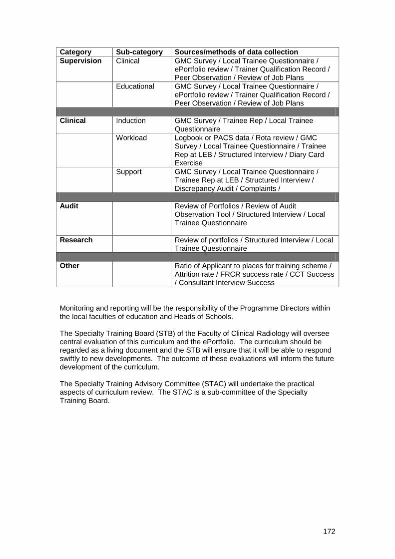
172
Category Sub-category Sources/methods of data collection Supervision Clinical GMC Survey / Local Trainee Questionnaire /
ePortfolio review / Trainer Qualification Record / Peer Observation / Review of Job Plans
Educational GMC Survey / Local Trainee Questionnaire / ePortfolio review / Trainer Qualification Record / Peer Observation / Review of Job Plans
Clinical Induction GMC Survey / Trainee Rep / Local Trainee
Questionnaire Workload Logbook or PACS data / Rota review / GMC
Survey / Local Trainee Questionnaire / Trainee Rep at LEB / Structured Interview / Diary Card Exercise
Support GMC Survey / Local Trainee Questionnaire / Trainee Rep at LEB / Structured Interview / Discrepancy Audit / Complaints /
Audit
Review of Portfolios / Review of Audit Observation Tool / Structured Interview / Local Trainee Questionnaire
Research
Review of portfolios / Structured Interview / Local Trainee Questionnaire
Other Ratio of Applicant to places for training scheme /
Attrition rate / FRCR success rate / CCT Success / Consultant Interview Success
Monitoring and reporting will be the responsibility of the Programme Directors within the local faculties of education and Heads of Schools. The Specialty Training Board (STB) of the Faculty of Clinical Radiology will oversee central evaluation of this curriculum and the ePortfolio. The curriculum should be regarded as a living document and the STB will ensure that it will be able to respond swiftly to new developments. The outcome of these evaluations will inform the future development of the curriculum. The Specialty Training Advisory Committee (STAC) will undertake the practical aspects of curriculum review. The STAC is a sub-committee of the Specialty Training Board.
173
APPENDIX C: STANDARDS FOR TRAINING IN CLINICAL RADIOLOGY Entry and Indicative training Summary of Standard Training for a CCT in Clinical Radiology:
• Primary Medical Qualification
• 2 Years of Foundation Years Training (FY 1&2) or equivalent and, for some, additional experience in other programmes (medicine, surgery, etc)
• Core Radiology Training as a specialty registrar (ST1-3) over an indicative period of three years followed by:
• Advanced (special interest) Radiology Training (ST 4-5) over an indicative period of two years
These indicative training periods represent whole-time equivalent. There is no requirement for less than full-time trainees to work for a specified percentage of the FT programme although 50% or more would be desirable. Less than full-time trainees should be involved in an on-call rota on a pro-rata basis.
Specia l c ircums tances
Absences from training Absence on sick leave or maternity leave reduces the time spent in training. In appropriate circumstances, an absence for sick or maternity leave of up to three months may occur without necessarily affecting the expected date for completion of specialty training. Such absences must be notified to the Training Office of the Royal College of Radiologists as soon as is feasible, preferably in advance. The Training Office will provide guidance to the trainee and his/her training programme director about the effect of the absence upon the trainee's expected CCT date and how use of the absence allow might be considered and recommended.
Acting-up as a consultant A trainee who has passed the Final FRCR Part B Examination may spend up to three months, during the final year of specialist training, "acting-up" as a consultant without affecting his/her expected CCT date, provided that a consultant supervisor is identified for the post, prospective approval has been obtained from the College's Training Office, and satisfactory progress is made.
Alternative Entry Points Those trainees who move into a specialty training programme from a Locum Appointment – Training (LAT) post or a Fixed Term Training Post will be eligible to have the earlier post and trainee accepted towards their CCT, provided that the original post has been approved by GMC, that their training is relevant to the CCT programme in clinical radiology and that progress and performance have been satisfactory. It is a legal requirement that a CCT can be awarded only to a person who has completed an entire course of training approved by GMC. Those with training and/or qualifications from outside the UK will be required to complete the full
174
duration of the CCT training programme, including success in all parts of the FRCR Examination if they wish to acquire a CCT in clinical radiology, or to apply under Article 14 of current GMC legislation for assessment of equivalence to the CCT. Out of Programme Activities Permission to spend time out of programme, such as for a Fellowship post or research, should be obtained in advance from the trainee's deanery and the College's Training Office. It should be noted that any time spent outside the trainee’s own training programme that is to be counted towards his/her CCT, either as Out of Programme Research (OOPR) or Out of Programme Training (OOPT), will require prospective approval to be obtained from the GMC by the trainee's deanery. Further guidance can be found on the College's website: http://www.rcr.ac.uk/content.aspx?PageID=955
Appeals There are formal mechanisms for appealing against decisions taken at all stages of training. Appeals against decisions of the Deanery Specialty Training Committee are conducted locally under the supervision of the Postgraduate Dean. Appeals related to examination results are conducted by the RCR; information can be obtained from the Examinations section of the College's website. Appeals against a failure to award a CCT may be made to GMC. It is important to be aware that the relevant regulations specify strict time limits within which appeals must be lodged.
175
APPENDIX D: EXAMINATION POLICIES IN CLINICAL RADIOLOGY The Fellowship Examination in Clinical Radiology (FRCR)
Overview The FRCR Examination in Clinical Radiology has three parts: First FRCR Examination, Final FRCR Part A Examination and Final FRCR Part B Examination. The First FRCR Examination is usually taken towards the end of the first year of specialty training, after formal training. The examination comprises two modules: Physics and Anatomy. The Final FRCR Part A Examination has six radiological modules. The Final FRCR Part B examination may be taken after three years of specialty training has also been completed. A trainee has completed core training once they have passed all three parts of the FRCR Examination and satisfactorily completed their end of third year ARCP. There is no limit on the number of times that any part of the FRCR examination can be taken nor are there any time constraints on how long a pass is valid for. Satisfactory progression through the different parts of the FRCR examination is monitored through the ARCP process and is guided by the ARCP decision aid (Section 7; Page 164). The FRCR Examination has undergone extensive review and redesign in the last year. Radiology trainees generally commence specialty training during the period August to October each year. The First FRCR Examination tests knowledge using MCQs for Physics and radiological images for Radiological Anatomy. These can be taken separately. The pass rate for Physics is over 90% for UK trainees. The success rate for Anatomy is unknown as the first sitting will be Spring 2010. Standard setting exercises, using the modified Angoff method, are used for both subjects. There are three sittings each year. No limit is set for the number of attempts but a trainee has to pass the First FRCR Examination before entering the Final FRCR Part A Examination. Unsuccessful candidates are informed of the mark they achieved in each subject, as well as the mark required for success. The Final FRCR Part A Examination uses single best answer (SBA) format questions. There are six separate modules sampling the whole of the radiology core syllabus with test specification sample grids. The six modules are Cardiothoracic and Vascular, Musculoskeletal, Gastro-intestinal, Genito-urinary, Adrenal, Obstetrics & Gynaecology and Breast, Paediatric and Central Nervous and Head & Neck. The Final FRCR Part A Examination is a knowledge test but also assesses the application of knowledge using clinical scenarios. No images are included in this part of the examination. The modified Angoff method is used for standard setting. Unsuccessful candidates are informed of the mark they achieved in each module, as well as the mark required for success. The six modules are marked separately and have to be passed independently. Once a trainee has completed three years of clinical radiological training and passed all six modules of the Final FRCR Part A Examination they are eligible to enter the Final FRCR Part B Examination.
176
The Final FRCR Part B Examination has four separate components: two written assessments – the rapid reporting session and the reporting session – and two structured oral examinations. The rapid reporting comprises 30 cases and mirrors the cases seen in an Accident and Emergency Department. The candidate has to correctly identify the normal cases and correctly describe the abnormality in the abnormal cases. A 10% error rate is acceptable, so up to three errors are allowed. The performance of each set of cases is reviewed by the Examining Board. The reporting section uses six clinical cases with a brief clinical history and the candidates have to write a structured answer describing the abnormalities seen in each case, together with a principal diagnosis and any relevant differential diagnoses. The cases chosen are predominately cross-sectional imaging, such as CT and MR, with a comprehensive selection of cases to cover many aspects of the curriculum. The candidates’ answers are marked according to a rigid marking scheme created in advance for each case. There are two separate structured oral examinations. Each examiner has to prepare material in advance. This material has to follow a specification set down by the Examining Board and includes the history, modality, body system and the radiological findings. This is reviewed by the Examining Board Chairman before it can be used. The cases used have to be suitable for inclusion and to cover the whole of the curriculum. All modalities have to be included and an examiner has to create a number of sets to ensure candidates see a variety of material to assess their clinical radiological skills. A brief clinical history is given and the candidate has to evaluate the case and offer a correct interpretation with any suitable management suggestions. Each examiner works as a pair with the trainee being assessed by both examiners and a consensus score is achieved using a close marking scheme and predetermined anchor statements. The two structured oral examinations are assessed separately but significant differences in scores are reviewed at the end of each day. A new examiner is paired with an experienced examiner and has to observe for a year before starting the structured oral examination. Each pair of examiners is independently assessed by the Examining Board Chairman to ensure consistency in the process and for standard setting. Examiner performance is reviewed by the Examining Board. All four components of the examination are marked separately and the examiners’ marking is blinded to the other components. A close marking scheme and a fixed pass mark are used. Borderline candidates are reviewed with all results in the examination disclosed. Good performance in one component of the examination may offset underperformance in another component. Examination pass rates are analysed in a variety of ways. The two written components have a recycle rate that allows reuse of the material. The performance data is recorded and reviewed when cases are being considered for re-use. Poorly performing cases are removed and replaced. Standard setting is more consistent by reusing cases after an appropriate interval. Pass rates overall are assessed using raw data and separate analyses are made of UK and non-UK trained candidates. The cohort of first attempt UK trainees is fairly consistent and this subset is usually the most reliable in terms of performance. The pass rate of this group is usually around 70%. Non-UK trained candidates have a variable success rate.
177
All examiners have to prove satisfactory completion of equality and diversity training before they can examine.
A policy for exceptional circumstances in candidates undertaking the FRCR Examination The RCR has established methods for consistent standard setting in the examination and has a threshold for acceptable performance. The RCR recognises that some candidates may experience very unsettling events that could undermine performance in an examination. However, adjustment to a candidate’s performance in such circumstances cannot be justified. The RCR, therefore, accepts that a candidate may withdraw from the examination at short notice if they suffer exceptional circumstances that could adversely affect their performance. The candidate should write to the Examinations Manager explaining the situation, and enclosing a supporting letter from their training programme director and, where appropriate, other supporting documentation such as a letter from their General Practitioner. If the circumstances are clearly exceptional and unexpected and such that the candidate’s performance would be likely to suffer, then the RCR may refund the examination fee.
An Appeals policy for the FRCR Examination Any appeal about the conduct of the examination must be made in writing within 20 days of the publication of the result of the examination. Letters should be addressed to the Examinations Manager. Appeals based on, or arising from, the following will be deemed invalid and not upheld:
• Academic judgement of College Examiners • Extenuating circumstances (such as ill health, personal issues) affecting
performance (see policy on exceptional circumstances above) • Lack of candidate awareness of examination regulations and procedures
Appeals will only be deemed valid for consideration when based on procedural irregularity in the conduct, or determination of the result, of the examination.
Summary of examiner application, training and appointment processes Nomination and application – process
• Applications invited via the College’s website, mailshot and ebulletin • Standard application form • Self nomination, including a statement of suitability • Application form must be supported by a signed statement from the
applicant’s training programme director or regional adviser • Applicants considered by the Examining Board and ranked • Applicants appointed on the basis of the quality of their application and their
areas of special interest
178
Training – process • New examiners are sent written notes covering all aspects of the examination
plus training. • Required to attend two compulsory days of training separated by six months • Participate in shadow marking of written and oral examinations and scores
assessed by experienced examiners highlighting any significant differences. • Submit radiological material in advance of examination to ensure coverage of
all modalities and sample widely across the curriculum. Approval following training: process
• Sign off by Examining Board Chairman • Each new examiner is paired with a more experienced examiner and the new
examiner is assessed twice in the first full year of examining by the Examining Board Chairman
• All examiners scores are disclosed and assessed after each sitting of the examination to display the hawk and dove effect and allow self-audit
• All examiners are assessed to ensure they understand the anchor statements and marking schemes
• All marks are initially blinded and then assessed by the examiner pairings. • At each sitting, a senior examiner reviews all the written answers and marks
for consistency pointing out any errors and deficiencies. • The Examining Board Chairman assesses each examiner pair to ensure that
the material used in the structured oral examination follows the standards agreed
Standards for appointment, training and performance monitoring of examiners for the Final FRCR Examination Mandatory Standards on appointment • On GMC Medical Register and have a Licence to Practice • Have undergone Equality and Diversity Training within the last year, including
an assessment of performance • Commit to fulfilling the RCR’s CPD requirements every five years • Be practicing in an area of clinical radiology that provides experience of a
broad range of radiological problems. • Have involvement in the supervision or training of UK trainees on a regular
basis. • Be a Fellow by examination • Prepared to train in an induction year • Have the support of their training programme director • Hold a substantive consultant post in the UK for at least ten years Initial Training Before attending training New Examiners must
• Read the instructions to new examiners • Undertake Equality and Diversity training (if update required)
On the training day New examiners undergoing training must
• Meet either the Senior Examiner or the Examining Board Chairman
179
• Participate in written assessments • Personally shadow assess at least five candidates in the oral examination • Shadow mark a minimum of five candidates during the rapid reporting and at
least five candidates in the reporting sessions, including receiving feedback from the senior examiner.
• Attain a satisfactory level of concordance (+ or – 1 mark) with paired co-examiners for each candidate assessed.
• Discuss clinical material with senior examiners • Attend at least one end of day review meeting, including the assessment of
borderline candidates and discrepant marks. • Commit to attending for five years with at least 20 days of professional leave
per year. • New examiners must attend two training days
Further performance monitoring • Examine with more senior examiner during first two years • Regular audit of material shown in the structured oral examination • Audit of scores, including reasons • Audit of modalities and body systems covered • Audit of completed mark sheets • Audit of marks awarded by examiner pairs • Concordance with co-examiner, initially blinded and then with consensus • Assessment by co-examiner • Concordance with co-examiner • Regular assessment by Examining Board Chairman during first year • Review of candidate appeals involving individual examiners Training and Selection of Examiners for the First FRCR Examination
The First FRCR Examination is set by two separate groups of examiners: the Anatomy Working Group and the Physics Working Group. The former group is comprised entirely of clinical radiologists; the latter is half clinical radiologists and half medical physicists. Nomination and application – process
• Applications invited via the College’s website, mailshot and ebulletin • Standard application form • Self nomination, including a statement of suitability • Application form must be supported by a signed statement from the
applicant’s training programme director or regional adviser • Applicants considered by the relevant working group and ranked • Applicants appointed on the basis of the quality of their application and their
areas of special interest Each of the working groups is co-ordinated by the most senior serving clinical radiologist, who is responsible for ensuring that examiners are appropriately trained and discharge their duties appropriately.
180
APPENDIX E: ENSURING QUALITY IN CLINICAL RADIOLOGY Responsibility for the approval of the training provided in the Clinical Radiology rests with the GMC as regulator (and UK competent authorities with regard to EU legislation).
Quality assurance - carried out by the regulatory authorities. Quality assurance encompasses all the policies, standards, systems and processes directed to ensuring maintenance and enhancement of the quality of postgraduate medical education in the UK. The regulators undertake planned and systematic activities to provide public and patient confidence that postgraduate medical education satisfies given requirements for quality within the principles of good regulation.
Quality management - carried out by the postgraduate deanery Quality management refers to the arrangements by which the postgraduate deanery discharges its responsibility for the standards and quality of postgraduate medical education. It satisfies itself that local education and training providers are meeting the regulator’s standards through robust reporting and monitoring mechanisms.
Quality control - carried out at local education provider (LEP) level Quality control relates to the arrangements (procedures, organisation) within local education providers (Health Boards, NHS Trusts, independent sectors) that ensure postgraduate medical trainees receive education and training that meet local, national and professional standards. A guide of evidence for local quality control is outlined in Curriculum Evaluation and Monitoring. These processes are interdependent. Regulators’ QA is a systematic educational audit of the deanery quality management systems; the latter must include review of LEP quality control measures. The regulators have set national standards for the delivery and outcomes of Clinical Radiology and deaneries are required to demonstrate through reports and visits that the standards have been met. There are nine domains of activity described:
• patient safety • quality assurance, review and evaluation • equality, diversity and opportunity • recruitment, selection and appointment • delivery of curriculum including assessment • support and development of trainees, trainers and local faculty • management of education and training • educational resources and capacity • outcomes
In each domain, the regulators have described who is responsible for its achievement, the standard(s) to be reached, and the criteria by which its achievement is judged. The standards set by the regulators are mandatory, but the processes by which deaneries quality manage, and LEP quality control, the programme provision are not specified.
181
APPENDIX F: EQUALITY AND DIVERSITY The Royal College of Radiologists will comply, and ensure compliance, with the requirements of equality and diversity legislation, such as the Sex Discrimination Act 1975 Race Relations Act 1976 Data Protection Acts 1984 and 1998 Disability Discrimination Act 1995 Race Relations (Amendment) Act 2000 Special Educational needs and Disabilities Act 2001 Employment Equality (Sexual Orientation) and (Religious Belief) Regulations 2003 Employment Equality (Age) Regulations 2006 Equality Act 2006 (covers service delivery in relation to sexual orientation and religious belief) Equality Act 2010 The Royal College of Radiologists believes that equality of opportunity is fundamental to all radiological practice and to the many and varied ways in which individuals become involved with the College, either as members of staff and Officers; as advisers from the medical profession or in a lay capacity; as members of the College’s professional bodies or as radiologists in training and examination candidates. Accordingly, it warmly welcomes contributions and applications from as diverse a population as possible, and actively seeks to recruit people to all its activities regardless of race, religion, ethnic origin, disability, age, gender or sexual orientation. Deanery quality assurance will ensure that each training programme complies with the equality and diversity standards in postgraduate medical training as set by GMC. Compliance with anti-discriminatory practice will be assured through
• monitoring of recruitment processes;
• ensuring all College representatives and TPDs have attended appropriate training sessions prior to appointment or within 12 months of taking up post;
• ensuring trainees have an appropriate, confidential and supportive route to report examples of inappropriate behaviour of a discriminatory nature;
• monitoring of College Examinations;
• ensuring all assessments discriminate on objective and appropriate criteria and do not unfairly disadvantage trainees because of gender, ethnicity, sexual orientation or disability (other than that which would make it impossible to practise safely as a radiologist). All efforts shall be made to ensure the participation of people with a disability in training.
The Royal College of Radiologists takes its obligations under the relevant equal opportunities legislation, such as the Race Relations (Amendment) Act 2000, seriously. This includes ensuring that members of staff involved in the delivery of
182
examinations receive appropriate briefing on the implications of race equality in the treatment of candidates. All those appointed as examiners are required to sign up to the following statement in the Examiner application form “I have read and accept the conditions with regard to the UK Race Relations Act 1976, as amended by the Race Relations (Amendment) Act 2000, and the Disabilities Discrimination Acts of 1995 and 2005 as documented above.” In order to meet its obligations under the relevant equal opportunities legislation, such as the Disability Discrimination Acts 1995 and 2005, the RCR considers and adopts appropriate standard operating procedures to deal with the common problems as they arise. These may include dyslexia/learning disability, mobility difficulties, chronic progressive condition, upper limb or back problem, repetitive strain injury (RSI), chronic recurrent condition (eg asthma, epilepsy), deafness/hearing loss, mental health difficulty, autism spectrum disorder (including Asperger’s syndrome) and others as appropriate. The Specialty Training Board is responsible for policy and regulations in respect of decisions about accommodations to be offered to candidates with disabilities. The Regulations introduced to update the Disability Discrimination Acts and to ensure that they are in line with EU Directives have been considered by the RCR. For implementation see Generic curriculum – Maintaining GMP: Equality & Diversity
183
APPENDIX G: CHANGES SINCE 2007 The curriculum has undergone wholesale re-design since 2007. There are fundamental changes in terms of the underpinning educational ethos, the development of mapped assessments and incorporation of the domains of Good Medical Practice. These significant overarching changes have come after feedback from GMC surveys, the Tooke Report and as a result of wide consultation. Structural changes
• The whole curriculum has been developed in educational terms in a spiral fashion (core, level1 & 2) with full integration of both generic and radiology specific content.
• There is incorporation and integration of the medical leadership and health inequalities competences.
• The updated layout brings the syllabus, competences and accompanying assessment to the forefront. This will facilitate easier navigation for the principal users, ie trainees and trainers.
Assessment
• Workplace based assessment methodology has been developed and radiology specific tools introduced and piloted.
• Specified assessments have been directly linked to each competence, where possible with existing tools.
• In the assessment tools, separate descriptors have been written for all grades core, level 1 and level 2.
• Generic assessment tools for teaching skills and audit assessment have been included.
• Generic skills across the domains of Good Medical Practice are to be assessed by the inclusion of a Multi-Source Feedback tool (MSF)
• Clarification of educational and clinical supervisor roles and responsibilities.
• A logbook has been incorporated into the ePortfolio to record competence in procedural skills.
Syllabus and competences
• Inclusion of a new generic competencies section, which underpins all medical practice and brings together attitudes and behaviours desirable in all radiologists.
• Development of the rationale of common presentations/diagnoses as providing the way of bringing the curriculum to life.

184
• Comprehensive mapping of all assessments (summative and formative) onto the syllabus contents.
185
APPENDIX H: CURRICULUM DEVELOPMENT The physics syllabus has been developed by the Physics Working Group and agreed by the Fellowship Examining Board and the Specialty Training Board of the Faculty of Clinical Radiology. The anatomy syllabus has been developed by the Anatomy Working Group and agreed by the Fellowship Examination board and the Specialty Training Board of the Faculty of Clinical Radiology. This curriculum was produced by members of the Specialty Training Advisory Committee of the Faculty of Clinical Radiology. The group has a broad UK representation and includes trainees and laypersons, as well as consultants who are actively involved in teaching and training. This curriculum applies to those who commence specialty training in clinical radiology from August 2010. Compared to the most recent curriculum (January 2007), it includes changes to ensure that this curriculum meets the seventeen GMC Standards for Curricula and Assessment. It incorporates significant revisions to the content and delivery of the training programme. The major changes from the previous curriculum include the incorporation of generic competencies together with new descriptor levels. This curriculum is trainee-centred and outcome-based. I t is designed to allow Core and level1/2 radiology training to be delivered in continuum and in a spiral fashion. A spiral curriculum describes a learning experience that revisits topics and themes, each time expanding the sophistication of the knowledge, skills and behaviours regarding that topic. This approach aids reinforcement of principles, the integration of topics and the achievement of higher levels of competency. The revisiting of topics is key to ensuring deep learning. This principle underpins the ethos of a spiral curriculum and effective life-long learning beyond specialty training. In this way, an individual progresses from being “competent” to becoming an “expert”.