special senses - sites.jackson.k12.ga.us · special senses people are responsive creatures. hold...
TRANSCRIPT

Special Senses
People are responsive creatures. Hold freshly baked bread before us, and our mouths water. A sudden clap of thunder makes us jump. These “ir-ritants” (the bread and the thunderclap) and many others are the stimuli that continually greet us and are interpreted by our nervous system.
We are usually told that we have five senses that keep us in touch with what is going on in the external world: touch, taste, smell, sight, and hearing. Actually, touch is a mixture of the general senses that we consider in the nervous system chapter (Chapter 7)—the temperature, pressure, and pain receptors of the skin and the propriocep-tors of muscles and joints. The other four “tradi-
tional” senses—smell, taste, sight, and hearing—are called special senses. Receptors for a fifth special sense, equilibrium, are housed in the ear, along with the organ of hearing. In contrast to the small and widely distributed general recep-tors, the special sense receptors are either large, complex sensory organs (eyes and ears) or local-ized clusters of receptors (taste buds and olfactory epithelium).
In this chapter we focus on the functional anatomy of each of the special sense organs indi-vidually, but keep in mind that sensory inputs are overlapping. What we finally experience—our “feel” of the world—is a blending of stimulus effects.
FUNCTION PREVIEW
◗ The special senses respond to different types of energetic stimuli involved in vision, hearing, balance, smell, and taste.
8

Chapter 8: Special Senses 279
8The adult eye is a sphere that measures about 1 inch (2.5 cm) in diameter. Only the anterior one-sixth of the eye’s surface can normally be seen. The rest of it is enclosed and protected by a cushion of fat and the walls of the bony orbit. The accessory struc-tures of the eye include the extrinsic eye muscles, eyelids, conjunctiva, and lacrimal apparatus.
Anteriorly the eyes are protected by the eyelids, which meet at the medial and lateral corners of the eye, the medial and lateral com-missure (canthus), respectively (Figure 8.1). The space between the eyelids in an open eye is called the palpebral fissure. Projecting from the bor-der of each eyelid are the eyelashes. Modified sebaceous glands associated with the eyelid edges are the tarsal glands. These glands produce an oily secretion that lubricates the eye (Figure 8.2a, p. 280). Ciliary glands, modified sweat glands, lie between the eyelashes (cilium = eyelash).
A delicate membrane, the conjunctiva (kon-junk″ti′vah), lines the eyelids and covers part of the outer surface of the eyeball (Figures 8.1 and 8.2). It ends at the edge of the cornea by fusing with the corneal epithelium. The conjunctiva se-cretes mucus, which helps to lubricate the eyeball and keep it moist.
Homeostatic Imbalance 8.1 Inflammation of the conjunctiva, called conjuncti-vitis, results in reddened, irritated eyes. Pinkeye, its infectious form caused by bacteria or viruses, is highly contagious. .................................................✚
Recall the three basic functions of the nervous system (Figure 7.1, p. 226). Each of the special senses gathers unique sensory information that, once integrated, will influence motor output. For example, if you saw a ball moving toward your head, this sensory input might result in a motor output that would move your body completely out of the path of the ball. Additionally, recall that each type of sensory information is processed in a specialized area of the cerebrum (Figure 7.13c, p. 242).
PART I: THE EYE AND VISIONHow we see has captured the curiosity of many researchers. Vision is the sense that has been stud-ied most. Of all the sensory receptors in the body, 70 percent are in the eyes. The optic tracts that carry information from the eyes to the brain are massive bundles, containing over a million nerve fibers. Vision is the sense that requires the most “learning,” and the eye appears to delight in be-ing fooled. The old expression “You see what you expect to see” is often very true.
Anatomy of the EyeExternal and Accessory Structures 8-1 When provided with a model or diagram, identify
the accessory eye structures, and list the functions of each.
Eyelashes
Sclera(covered byconjunctiva)
Site whereconjunctivamerges withcornea
Lateralcommissure(canthus)
Iris
Medialcommissure(canthus)
Lacrimalcaruncle
Eyelid
Eyelid
Eyebrow
PupilPalpebralfissure
Figure 8.1 Surface anatomy of the eye and accessory structures.

280 Essentials of Human Anatomy and Physiology
of the eyeball through several small ducts. The tears flush across the eyeball into the lacrimal canaliculi medially, then into the lacrimal sac, and finally into the nasolacrimal duct, which empties into the nasal cavity (see Figure 8.2b). Lacrimal secretion also contains mucus, antibod-ies, and lysozyme (li′so-zım), an enzyme that de-stroys bacteria. Thus, it cleanses and protects the eye surface as it moistens and lubricates it. When lacrimal secretion increases substantially, tears spill over the eyelids and fill the nasal cavities, caus-ing congestion and the “sniffles.” This happens when the eyes are irritated by foreign objects or chemicals and when we are emotionally upset. In the case of irritation, the enhanced tearing acts to wash away or dilute the irritating substance. The importance of “emotional tears” is poorly under-stood, but some suspect that crying is important in reducing stress. Anyone who has had a good cry would probably agree, but this has been difficult to prove scientifically.
Homeostatic Imbalance 8.2 Because the nasal cavity mucosa is continuous with that of the lacrimal duct system, a cold or nasal inflammation often causes the lacrimal mu-cosa to become inflamed and swell. This impairs the drainage of tears from the eye surface, causing “watery” eyes. ........................................................✚
Six extrinsic, or external, eye muscles are attached to the outer surface of each eye. These muscles produce gross eye movements and make it possible for the eyes to follow a moving object. (The names, locations, actions, and cranial nerve serving each of the extrinsic muscles are given in Figure 8.3).
Did You Get It? 1. What is the role of the eyelids?
2. Which structure of the eye forms tears?
3. What are tears?
4. What is the visual role of the external eye muscles?
(For answers, see Appendix D.)
Internal Structures: The Eyeball 8-2 Name the layers of the wall of the eye, and
indicate the major function of each.
8-3 Explain how the functions of rods and cones differ.
8-4 Define: blind spot, cataract, and glaucoma.
The lacrimal apparatus (Figure 8.2b) con-sists of the lacrimal gland and a number of ducts that drain the lacrimal secretions into the nasal cavity. The lacrimal glands are located above the lateral end of each eye. They continually release a dilute salt solution (tears) onto the anterior surface
(b)
Excretory ductof lacrimal gland
Eyelid
Eyelid
Eyelashes
Lacrimal gland
(a)
Anterioraspect
Tarsal glands
Conjunctiva
Lacrimal sacLacrimalgland
Excretory ductsof lacrimal gland
Lacrimal canaliculus
Nasolacrimal duct
Inferior meatusof nasal cavity
Nostril
Figure 8.2 Accessory structures of the eye. (a) Sagittal section of the accessory structures associated with the anterior part of the eye. (b) Anterior view of the lacrimal apparatus.

Chapter 8: Special Senses 281
8
the eye, is supported upright within the eye cavity, dividing it into two chambers.
Layers Forming the Wall of the EyeballNow that we have covered the general anatomy of the eyeball, we are ready to get specific.
Fibrous Layer The outermost layer, called the fibrous layer, consists of the protective sclera (skle′rah) and the transparent cornea (kor′ne-ah).
8-5 Trace the pathway of light through the eye to the retina.
8-6 Discuss the importance of an ophthalmoscopic examination.
The eye itself, commonly called the eyeball, is a hollow sphere (Figure 8.4, p. 282). Its wall is composed of three layers, and its interior is filled with fluids called humors that help to maintain its shape. The lens, the main focusing apparatus of
Superior oblique muscle
Trochlea
Superior oblique tendon
Superior rectus muscle
Lateral rectus muscle
Conjunctiva
Opticnerve
Inferiorrectusmuscle
Inferiorobliquemuscle
Medialrectus muscle
Inferiorrectus muscle
Axis atcenter ofeye
Lateralrectus muscle
(a) (b)
(c)
Lateral rectus
Medial rectus
Superior rectus
Inferior rectus
Inferior oblique
Superior oblique
Name Action
Moves eye laterally
Moves eye medially
Elevates eye and turns it medially
Depresses eye and turns it medially
Elevates eye and turns it laterally
Depresses eye and turns it laterally
VI (abducens)
III (oculomotor)
III (oculomotor)
III (oculomotor)
III (oculomotor)
IV (trochlear)
Controllingcranial nerve
Figure 8.3 Extrinsic muscles of the eye. (a) Lateral view of the right eye. (b) Superior view of the right eye. The four rectus muscles originate from the annular ring, a ringlike tendon at the back of the eye socket. (c) Summary of cranial nerve supply and actions of the extrinsic eye muscles.
Practice art labeling >Study Area>Chapter 8

282 Essentials of Human Anatomy and Physiology
(b)
Ciliary zonule CorneaLens
Aqueous humor(in anteriorsegment)
Marginof pupil
Iris
Ciliary body Vitreous humorin posteriorsegment
Choroid
Fovea centralisOptic discOptic nerve
Retina
Sclera
(a)
Ciliary body
Ciliary zonule
Cornea
Iris
Pupil
Aqueous humor(in anteriorsegment)
Lens
Scleral venous sinus(canal of Schlemm)
Vitreous humor(in posterior segment)
Optic nerve
Fovea centralis
Retina
Choroid
Sclera
Optic disc(blind spot)
Central artery and vein of the retina
Figure 8.4 Internal anatomy of the eye (sagittal section). (a) Diagrammatic view. (b) Photograph.
Q: Which layer of the eye would be the first to be affected by deficient tear production?
The outermost fibrous layer (the sclera and especially its cornea), which normally is continuously washed by tears.A:
The sclera, thick, glistening white connective tissue, is seen anteriorly as the “white of the eye.” The central anterior portion of the fibrous layer is crystal clear. This “window” is the cornea through which light enters the eye. The cornea is well supplied
with nerve endings. Most are pain fibers, and when the cornea is touched, blinking and increased tear-ing occur. Even so, the cornea is the most exposed part of the eye, and it is very vulnerable to damage. Luckily, its ability to repair itself is extraordinary.
Practice art labeling >Study Area>Chapter 8

Chapter 8: Special Senses 283
8
signals pass from the photoreceptors via a two-neuron chain—bipolar cells and then ganglion cells—before leaving the retina via the optic nerve as nerve impulses that are transmitted to the optic cortex. The result is vision.
Neural layerof retina
Pigmentedlayer ofretina
Centralarteryand veinof retina
Opticnerve
Sclera
Choroid
Optic disc
(b)
Rod
Cone
Pigmentedlayer of retina
Bipolarcells
Ganglioncells
Pathwayof light
(a)
Figure 8.5 The three major types of neurons composing the retina. (a) Notice that light must pass through the thickness of the retina to excite the rods and cones. Electrical signals flow in the opposite direction: from the rods and cones to the bipolar cells and finally to the ganglion cells. The ganglion cells generate the nerve impulses that leave the eye via the optic nerve. (b) Schematic view of the posterior part of the eyeball illustrating how the axons of the ganglion cells form the optic nerve.
Furthermore, the cornea is the only tissue in the body that is transplanted from one person to an-other without the worry of rejection. Because the cornea has no blood vessels, it is beyond the reach of the immune system.
Vascular Layer The middle layer of the eye-ball, the vascular layer, has three distinguishable regions. Most posterior is the choroid (ko′roid), a blood-rich nutritive tunic that contains a dark pig-ment. The pigment prevents light from scattering inside the eye. Moving anteriorly, the choroid is modified to form two smooth muscle structures, the ciliary (sil′e-er-e) body, to which the lens is attached by a suspensory ligament called the ciliary zonule, and then the iris. The pigmented iris has a rounded opening, the pupil, through which light passes. Circularly and radially arranged smooth muscle fibers form the iris, which acts like the diaphragm of a camera. That is, it regulates the amount of light entering the eye so that we can see as clearly as possible in the available light. In close vision and bright light, the circular muscles contract, and the pupil constricts. In distant vision and dim light, the radial fibers contract to enlarge (dilate) the pupil, which allows more light to enter the eye.
Sensory Layer The innermost sensory layer of the eye is the delicate two-layered retina (ret′ı-nah), which extends anteriorly only to the ciliary body. The outer pigmented layer of the retina is com-posed of pigmented cells that, like those of the choroid, absorb light and prevent light from scat-tering inside the eye. They also act as phagocytes to remove dead or damaged receptor cells and store vitamin A needed for vision.
The transparent inner neural layer of the ret-ina contains millions of receptor cells, the rods and cones, which are called photoreceptors be-cause they respond to light (Figure 8.5). Electrical

284 Essentials of Human Anatomy and Physiology
Cones are discriminatory receptors that allow us to see the details of our world in color under bright light conditions. They are densest in the center of the retina and decrease in number toward the retinal edge. Lateral to each blind spot is the fovea centralis (fo′ve-ah sen-tra′lis), a tiny pit that contains only cones (see Figure 8.4). Consequently, this is the area of greatest visual acuity, or point of sharpest vision, and anything we wish to view criti-cally is focused on the fovea centralis.
There are three varieties of cones. Each type is most sensitive to particular wavelengths of visible light (Figure 8.6). One type responds most vigor-ously to blue light, another to green light. The third cone variety responds to a range including both green and red wavelengths of light. However, this is the only cone population to respond to red light at all, so these are called the “red cones.” Impulses received at the same time from more than one type of cone by the visual cortex are interpreted as intermediate colors. For example, simultaneous impulses from blue and red color receptors are seen as purple or violet tones. When all three cone types are being stimulated, we see white. If someone shines red light into one of your eyes and green into the other, you will see yellow, indicating that the “mixing” and interpretation of colors occurs in the brain, not in the retina.
Homeostatic Imbalance 8.4Lack of all three cone types results in total color blindness, whereas lack of one cone type leads to partial color blindness. Most common is the lack of red or green receptors, which leads to two varieties of red-green color blindness. Red and green are seen as the same color—either red or green, depending on the cone type present. Many color-blind people are unaware of their condition because they have learned to rely on other cues—such as differences in intensities of the same color—to distinguish some-thing green from something red, for example on traffic signals. Because the genes regulating color vision are on the X (female) sex chromosome, color blindness is a sex-linked condition. It occurs almost exclusively in males. .....................................................✚
LensLight entering the eye is focused on the retina by the lens, a flexible biconvex crystal-like structure. The lens is held upright in the eye by a suspen-sory ligament, the ciliary zonule, attached to the ciliary body (see Figure 8.4).
The photoreceptor cells are distributed over the entire retina, except where the optic nerve (composed of ganglion cell axons) leaves the eyeball; this site is called the optic disc, or blind spot. When light from an object is focused on the optic disc, the object dis-appears from our view and we cannot see it.
The rods and cones are not evenly distrib-uted in the retina. The rods are most dense at the periphery, or edge, of the retina and decrease in number as the center of the retina is approached. The rods allow us to see in gray tones in dim light, and they provide our peripheral vision.
Homeostatic Imbalance 8.3Anything that interferes with rod function hin-ders our ability to see at night, a condition called night blindness. Night blindness dangerously im-pairs the ability to drive safely at night. Its most common cause is prolonged vitamin A deficiency, which eventually results in deterioration of much of the neural retina. Vitamin A is one of the building blocks of the pigments the photoreceptor cells need to respond to light (see “A Closer Look”). Vitamin A supplements will restore function if taken before degenerative changes occur. ......................................✚
Ligh
t abs
orpt
ion
by c
one
popu
latio
ns
380 450 500 550 600 650 700 750
Wavelength (nanometers)
Visible light
420 nm(blue cones)
530 nm(green cones)
560 nm(red cones)
Figure 8.6 Sensitivities of the three cone types to the different wavelengths of visible light.

The tiny photoreceptor cells of the retina have names that reflect their general shapes. As shown to the left, rods are slender, elongated neurons, whereas the fatter cones taper to pointed tips. In each type of photoreceptor, there is a region called an outer segment, attached to the cell body. The outer segment corresponds to a light-trapping dendrite, in which the discs containing the visual pigments are stacked like a row of pennies.
The behavior of the visual pigments is dramatic. When light strikes them, they lose their color, or are “bleached”; shortly afterward, they regenerate their pigment. Absorption of light and pigment bleaching cause electrical changes in the photoreceptor cells that ultimately cause nerve impulses to be transmitted to the brain for visual interpretation. Pigment regeneration ensures that you are not blinded and unable to see in bright sunlight.
A good deal is known about the structure and function of rhodopsin, the purple pigment found in rods (see figure below). It is formed from the union of a protein (opsin) and a modified vitamin A product (retinal). When combined in rhodopsin, retinal has a kinked shape that allows it to bind to opsin. But when light strikes rhodopsin, retinal straightens out and releases the protein. Once straightened out, the retinal continues its conversion until it is once again vitamin A. As these changes occur, the purple color of rhodopsin changes to the yellow of retinal and finally becomes colorless as the change to vitamin A occurs. Thus the term “bleaching of the pigment” accurately describes the color changes that occur when light hits the pigment. Rhodopsin is regenerated as vitamin A is again converted to the kinked form of retinal and recombined with opsin in an ATP-requiring process. The cone pigments, although similar to rhodopsin, differ in the specific kinds of proteins they contain.
A CLOSER LOOK Visual Pigments— The Actual Photoreceptors
285
Process ofbipolar cell
Ligh
t
Ligh
t
Ligh
t
Discscontainingvisual pigments
Melaningranules
Pigment cellnucleus
Innerfibers
Rodcellbody
Conecellbody
Synapticendings
Rod cellbody
Nuclei
Mitochondria
Outer segment
Pig
men
ted
laye
r
Outer fiber
Inne
r se
gmen
t
Retinal(visual yellow)
Releases
Opsin
Light absorptioncauses
Rhodopsin(visual purple)
Bleaching of the pigment

286 Essentials of Human Anatomy and Physiology
Homeostatic Imbalance 8.5In youth, the lens is perfectly transparent and has the consistency of hardened jelly, but as we age it becomes increasingly hard and opaque. Cataracts, which result from this process, cause vision to become hazy and distorted, and they eventually cause blindness in the affected eye (Figure 8.7). Other risk factors for forming cata-racts include diabetes mellitus, frequent exposure to intense sunlight, and heavy smoking. Current treatment of cataracts is either surgical removal of the lens and replacement with a lens implant or special cataract glasses. .............................................✚
The lens divides the eye into two segments, or chambers. The anterior (aqueous) segment, ante-rior to the lens, contains a clear watery fluid called aqueous humor. The posterior (vitreous) segment, posterior to the lens, is filled with a gel-like sub-stance called either vitreous (vit′re-us) humor or the vitreous body (see Figure 8.4). Vitreous humor helps prevent the eyeball from collaps-ing inward by reinforcing it internally. Aqueous humor is similar to blood plasma and is continu-ally secreted by a special area of the choroid. Like the vitreous humor, it helps maintain intraocular (in″trah-ok′u-lar) pressure, or the pressure inside the eye. It also provides nutrients for the avascular lens and cornea. Aqueous humor is reabsorbed into the venous blood through the scleral venous sinus, or canal of Schlemm (shlem), which is located at the junction of the sclera and cornea.
Homeostatic Imbalance 8.6If drainage of aqueous humor is blocked, fluid backs up like a clogged sink. Pressure within the eye may increase to dangerous levels and compress the delicate retina and optic nerve. The resulting condition, glaucoma (glaw-ko′mah; “vision going gray”), eventually causes pain and possibly blind-ness unless detected early. Glaucoma is a common cause of blindness in the elderly. Unfortunately, many forms of glaucoma progress slowly and have almost no symptoms at first. Thus, it steals sight slowly and painlessly until the damage is done. Later signs include seeing halos around lights, headaches, and blurred vision. A simple instru-ment called a tonometer (to-nom′e-ter) is used to measure the intraocular pressure. This examina-tion should be performed yearly in people over 40. Glaucoma is commonly treated with eyedrops that increase the rate of aqueous humor drain-age. Laser or surgical enlargement of the drainage channel can also be used. ........................................✚
The ophthalmoscope (of-thal′mo-skop) is an instrument that illuminates the interior of the eye-ball, allowing the retina, optic disc, and internal blood vessels at the fundus, or posterior wall of the eye, to be viewed and examined (Figure 8.8).
Figure 8.7 Photograph of a cataract. The cataract appears as a milky structure that seems to fill the pupil.
Figure 8.8 The posterior wall (fundus) of the retina as seen with an ophthalmoscope. Notice the optic disc, from which the blood vessels radiate.
Lateral
Blood vessels
Macula Optic disc Retina
Medial
Foveacentralis

Chapter 8: Special Senses 287
8
is able to accommodate properly. However, vision problems occur when a lens is too strong or too weak (overconverging and underconverging, re-spectively) or from structural problems of the eye-ball (as described in “A Closer Look” on near- and farsightedness on p. 289–290).
Visual Fields and Visual Pathways to the Brain8-9 Trace the visual pathway to the visual cortex.
Axons carrying impulses from the retina are bun-dled together at the posterior aspect of the eyeball
Such an examination can detect certain pathologi-cal conditions, such as diabetes, arteriosclerosis, and degeneration of the optic nerve and retina.
Did You Get It? 5. What is the meaning of the term blind spot in
relation to the eye?
6. What function does the choroid of the vascular layer have in common with the pigmented layer of the retina?
7. How do the rods and cones differ from each other?
(For answers, see Appendix D.)
Physiology of VisionPathway of Light through the Eye and Light Refraction8-7 Describe image formation on the retina.
8-8 Define the following terms: accommodation, astigmatism, emmetropia, hyperopia, myopia, and refraction.
When light passes from one substance to another substance that has a different density, its speed changes and its rays are bent, or refracted. Light rays are bent in the eye as they encounter the cor-nea, aqueous humor, lens, and vitreous humor.
The refractive, or bending, power of the cor-nea and humors is constant. However, that of the lens can be changed by changing its shape—that is, by making it more or less convex, so that light can be properly focused on the retina. The greater the lens convexity, or bulge, the more it bends the light. The flatter the lens, the less it bends the light.
The resting eye is “set” for distant vision. In gen-eral, light from a distant source (over 20 feet away) approaches the eye as parallel rays (Figure 8.9a), and the lens does not need to change shape to focus properly on the retina. However, light from a close object tends to scatter and to diverge, or spread out, and the lens must bulge more to make close vision possible (Figure 8.9b). To achieve this, the ciliary body contracts, allowing the lens to become more convex. This ability of the eye to focus specifically for close objects (those less than 20 feet away) is called accommodation. The image formed on the retina as a result of the light-bending activity of the lens is a real image—that is, it is reversed from left to right, upside down (inverted), and smaller than the object (Figure 8.10, p. 288). The normal eye
Q: As you look at this figure, are your lenses relatively thick or relatively thin?
You would be using your close vision, so your lenses would be bulged and thus relatively thick.A:
Retina
Retina
(a)
(b)
Light from distant source
Light from near source
Focal point
Focal point
Figure 8.9 Relative convexity of the lens during focusing for distant and close vision. (a) Light rays from a distant object are nearly parallel as they reach the eye and can be focused without requiring changes in lens convexity. (b) Diverging light rays from close objects require that the lens bulge more to focus the image sharply on the retina.

288 Essentials of Human Anatomy and Physiology
Eye Reflexes8-10 Discuss the importance of the convergence and
pupillary reflexes.
Both the internal and the external (extrinsic) eye muscles are necessary for proper eye function. The internal muscles are controlled by the autonomic nervous system. As mentioned earlier, these mus-cles include those of the ciliary body, which alters lens curvature, and the radial and circular muscles of the iris, which control pupil size. The external muscles are the rectus and oblique muscles at-tached to the eyeball exterior (see Figure 8.3). The external muscles control eye movements and make it possible to follow moving objects. They are also responsible for convergence, which is
and issue from the back of the eye as the optic nerve. At the optic chiasma (ki-as′mah; chiasm = cross) the fibers from the medial side of each eye cross over to the opposite side of the brain. The fiber tracts that result are the optic tracts. Each optic tract contains fibers from the lateral side of the eye on the same side and the medial side of the opposite eye. The optic tract fibers synapse with neurons in the thalamus, whose axons form the optic radiation, which runs to the occipital lobe of the brain. There they synapse with the cortical cells, and visual interpretation, or seeing, occurs. (The visual pathway from the eye to the brain is shown in Figure 8.11). Each side of the brain receives visual input from both eyes—from the lateral field of vision of the eye on its own side and from the medial field of the other eye. Also notice that each eye “sees” a slightly different view, but their visual fields overlap quite a bit. As a result of these two facts, humans have binocular vision. Binocular vision, literally “two-eyed vision,” provides for depth perception, also called “three-dimensional” vision, as our visual cortex fuses the two slightly different images delivered by the two eyes.
Homeostatic Imbalance 8.7Hemianopia (hem″e-ah-no′pe-ah) is the loss of the same side of the visual field of both eyes, which results from damage to the visual cortex on one side only (as occurs in some CVAs). Thus, the person would not be able to see things past the middle of his or her visual field on either the right or left side, depending on the site of the CVA. Such individuals should be carefully attended and warned of objects in the nonfunctional (nonseeing) side of the visual field. Their food and personal objects should always be placed on their functional side, or they might miss them. .................................✚
Figure 8.10 Real image (reversed left to right, and upside down) formed on the retina. Notice that the farther away the object, the smaller its image on the retina.
Fixation point
Right eye Left eye
Optic nerve
Optic tract
Optic chiasma
Occipital lobe(visual cortex)
Opticradiation
Thalamus
Figure 8.11 Visual fields of the eyes and visual pathway to the brain. Notice that the visual fields overlap considerably (area of binocular vision). Notice also the retinal sites at which a real image would be focused when both eyes are fixed on a close, pointlike object.

It seems that whenever people who wear glasses or contact lenses discuss their vision, one of them says something like, “Nearby objects appear blurry to me, but I can’t remember if that means I’m nearsighted or farsighted.” Or, someone else may say, “With my glasses I see faraway objects more clearly, so does that mean I am farsighted?”
The eye that focuses images correctly on the retina is said to have emmetropia (em″e -tro′pe-ah), literally, “harmonious vision.” Such an eye is shown in part (a) of the figure.
Nearsightedness is formally called myopia (mi″o′pe-ah; “short vision”). It occurs when the parallel light rays from distant objects fail to reach the retina and instead are focused in front of it; see part (b) in the figure. Therefore, distant objects appear blurry to myopic people. Nearby objects are in focus, however, because the lens “accommodates” (bulges) to focus the image properly on the retina. Myopia results from an eyeball that is too long, a lens that is too strong, or a cornea that is too curved. Correction requires concave corrective lenses that diverge the light rays before they enter the eye, so that
they converge farther back. To answer the first question posed previously, near sighted people see near objects clearly and need corrective lenses to focus distant objects.
Farsightedness is formally called hyperopia (hi″per-o′pe-ah; “far vision”). It occurs when the parallel light rays from distant objects are focused behind the retina—at least in the resting eye, in which the lens is flat and the ciliary muscle is relaxed; see part (c) in the figure. Hyperopia usually results from an eyeball that is too short or from a “lazy” lens. People with hyperopia see distant objects clearly because
A CLOSER LOOK Bringing Things into Focus
Correction
None required
Concave lens
Focalplane
(a) Emmetropic eye
(b) Myopic eye(nearsighted)
(c) Hyperopic eye(farsighted)
Convex lens
!
289

the reflexive movement of the eyes medially when we view close objects. When convergence occurs, both eyes are aimed toward the near object being viewed. The extrinsic muscles are controlled by somatic fibers of cranial nerves III, IV, and VI (see Figure 8.3).
When the eyes are suddenly exposed to bright light, the pupils immediately constrict; this is the photopupillary reflex. This protective reflex prevents excessively bright light from damaging the delicate photoreceptors. The pupils also con-strict reflexively when we view close objects; this accommodation pupillary reflex provides for more acute vision.
Reading requires almost continuous work by both sets of muscles. The muscles of the ciliary body bring about the lens bulge, and the circular (or constrictor) muscles of the iris produce the ac-commodation pupillary reflex. In addition, the ex-trinsic muscles must converge the eyes as well as move them to follow the printed lines. This is why long periods of reading tire the eyes and often re-sult in what is commonly called eyestrain. When you read for an extended time, it is helpful to look up from time to time and stare into the distance. This temporarily relaxes all the eye muscles.
Did You Get It? 8. What are the refractory media of the eye?
9. What name is given to the ability of the eye to focus on close objects?
10. What is the difference between the optic tract and the optic nerve?
11. In what way does the photopupillary reflex protect the eyes?
(For answers, see Appendix D.)
PART II: THE EAR: HEARING AND BALANCEAt first glance, the machinery for hearing and balance appears very crude. Fluids must be stirred to stimulate the receptors of the ear: sound vi-brations move fluid to stimulate hearing receptors, whereas gross movements of the head disturb f luids surrounding the balance organs. Receptors that re-spond to such physical forces are called mechano-receptors (mek″ah-no-re-sep′terz).
Our hearing apparatus allows us to hear an extraordinary range of sound, and our highly sensi-tive equilibrium receptors keep our nervous sys-tem continually up to date on the position and movements of the head. Without this information, it would be difficult if not impossible to maintain our balance. Although these two sense organs are housed together in the ear, their receptors respond to different stimuli and are activated independently of one another.
Anatomy of the Ear8-11 Identify the structures of the external, middle,
and internal ear, and list the functions of each.
Anatomically, the ear is divided into three major areas: the external, or outer, ear; the middle ear;
A CLOSER LOOK Bringing Things into Focus (continued)
their ciliary muscles contract continuously to increase the light-bending power of the lens, which moves the focal point forward onto the retina. However, the diverging rays from nearby objects are focused so far behind the retina that even at full “bulge,” the lens cannot focus the image on the retina. Therefore, nearby objects appear blurry, and hyperopic individuals are subject
to eyestrain as their endlessly contracting ciliary muscles tire from overwork. Correction of hyperopia requires convex corrective lenses that converge the light rays before they enter the eye. To answer the second question posed at the beginning of this essay, far sighted people can see far away objects clearly and require corrective lenses to focus on nearby objects.
Unequal curvatures in different parts of the cornea or lens cause astigmatism (ah-stig′mah-tizm). In this condition, blurry images occur because points of light are focused not as points on the retina but as lines (astigma = not a point). Special cylindrically ground lenses or contacts are used to correct this problem.
290

Chapter 8: Special Senses 291
8
and the internal, or inner, ear (Figure 8.12). The external and middle ear structures are involved with hearing only. The internal ear functions in both equilibrium and hearing.
External (Outer) EarThe external, or outer, ear is composed of the auricle and the external acoustic meatus. The au-ricle (aw′ri-kul), or pinna (pin′nah), is what most people call the “ear”—the shell-shaped structure surrounding the auditory canal opening. In many animals, the auricle collects and directs sound waves into the auditory canal, but in humans this function is largely lost.
The external acoustic meatus (or auditory canal) is a short, narrow chamber (about 1 inch long by ¼ inch wide) carved into the tempo-ral bone of the skull. In its skin-lined walls are
the ceruminous (se-roo′mı-nus) glands, which secrete waxy yellow cerumen or earwax, which provides a sticky trap for foreign bodies and repels insects.
Sound waves entering the auditory canal even-tually hit the tympanic (tim-pan′ik; tympanum = drum) membrane, or eardrum, and cause it to vibrate. The canal ends at the eardrum, which separates the external from the middle ear.
Middle EarThe middle ear, or tympanic cavity, is a small, air-filled, mucosa-lined cavity within the tempo-ral bone. It is flanked laterally by the eardrum and medially by a bony wall with two openings, the oval window and the inferior, membrane- covered round window. The pharyngotympanic (think throat-eardrum: pharynx-tympanic) tube, or
Figure 8.12 Anatomy of the ear.
Auricle(pinna)
External (outer) ear Middle ear
Internal (inner) ear
Semicircularcanals
Vestibulocochlearnerve
External acousticmeatus(auditory canal)
Pharyngotympanic(auditory) tube
Tympanicmembrane(eardrum) Stirrup
(stapes)
Oval window
Round window
Cochlea
Vestibule
Anvil(incus)
Auditory ossicles
Hammer(malleus)
Practice art labeling >Study Area>Chapter 8

292 Essentials of Human Anatomy and Physiology
Internal (Inner) EarThe internal ear is a maze of bony chambers called the bony, or osseous, labyrinth (lab′ ı-rinth; “maze”), located deep within the temporal bone behind the eye socket. The three subdivi-sions of the bony labyrinth are the spiraling, pea-sized cochlea (kok′le-ah, “snail”), the vestibule (ves′ti-bul), and the semicircular canals. The vestibule is situated between the semicircular canals and the cochlea. The views of the bony labyrinth typically seen in textbooks, including this one, are somewhat misleading because we are really talking about a cavity. The image (see Figure 8.12) can be compared to a cast of the bony labyrinth; that is, a labyrinth that was filled with plaster of paris and then had the bony walls removed after the plaster hardened. The shape of the plaster then reveals the shape of the cavity that worms through the temporal bone.
The bony labyrinth is filled with a plasmalike fluid called perilymph (per′ ı-limf). Suspended in the perilymph is a membranous labyrinth, a system of membrane sacs that more or less follows the shape of the bony labyrinth. The membra-nous labyrinth itself contains a thicker fluid called endolymph (en′do-limf).
Did You Get It? 12. Which region(s) of the ear (external, middle, or
internal) serve hearing only?
13. Which structures of the ear transmit sound vibrations from the eardrum to the oval window?
(For answers, see Appendix D.)
Equilibrium 8-12 Distinguish between static and dynamic
equilibrium.
8-13 Describe how the equilibrium organs help maintain balance.
The equilibrium sense is not easy to describe be-cause it does not “see,” “hear,” or “feel.” What it does is respond (frequently without our aware-ness) to various head movements. The equilibrium receptors of the inner ear, collectively called the vestibular apparatus, can be divided into two functional arms—one arm responsible for moni-toring static equilibrium, and the other involved with dynamic equilibrium.
auditory tube, runs obliquely downward to link the middle ear cavity with the throat, and the mucosae lining the two regions are continuous. Normally, the pharyngotympanic tube is flattened and closed, but swallowing or yawning can open it briefly to equalize the pressure in the mid-dle ear cavity with the external, or atmospheric, pressure. This is an important function because the eardrum does not vibrate freely unless the pressure on both of its surfaces is the same. When the pressures are unequal, the eardrum bulges inward or outward, causing hearing diffi-culty (voices may sound far away) and sometimes earaches. The ear-popping sensation of the pres-sures equalizing is familiar to anyone who has flown in an airplane.
Homeostatic Imbalance 8.8Inflammation of the middle ear, otitis media (o-ti′tis me′de-ah), is a fairly common result of a sore throat, especially in children, whose pharyn-gotympanic tubes run more horizontally. In otitis media, the eardrum bulges and often becomes inflamed. When large amounts of fluid or pus accumulate in the cavity, an emergency myrin-gotomy (lancing of the eardrum) may be required to relieve the pressure. A tiny tube is implanted in the eardrum that allows pus to drain into the external ear canal. The tube usually falls out by itself within the year. ................................................✚
The more horizontal course of the pharyngo-tympanic tube in infants also explains why it is never a good idea to “prop” a bottle or feed them when they are lying flat (a condition that favors the entry of the food into that tube).
The tympanic cavity is spanned by the three smallest bones in the body, the ossicles (os′s ı-kulz), which transmit the vibratory motion of the eardrum to the fluids of the inner ear (see Figure 8.12). These bones, named for their shape, are the hammer, or malleus (ma′le-us); the anvil, or incus (in′kus); and the stirrup, or stapes (sta′pez). When the eardrum moves, the hammer moves with it and transfers the vibra-tion to the anvil. The anvil, in turn, passes the vibration on to the stirrup, which presses on the oval window of the inner ear. The movement at the oval window sets the fluids of the inner ear into motion, eventually exciting the hearing receptors.

Chapter 8: Special Senses 293
8
Static EquilibriumWithin the membrane sacs of the vestibule are receptors called maculae (mak′u-le; “spots”) that are essential to our sense of static equilibrium (Figure 8.13). The maculae report on changes in the position of the head in space with respect to the pull of gravity when the body is not moving (static = at rest). Because they provide informa-tion on which way is up or down, they help us keep our head erect. The maculae are extremely important to divers swimming in the dark depths (where most other orienting cues are absent), enabling them to tell which way is up (to the sur-face). Each macula is a patch of receptor (hair) cells with their “hairs” embedded in the otolithic membrane, a jellylike mass studded with otoliths (o′to-lithz), tiny stones made of calcium salts. As the head moves, the otoliths roll in response to changes in the pull of gravity. This movement creates a pull on the gel, which in turn slides like a greased plate over the hair cells, bending their hairs. This event activates the hair cells, which send impulses along the vestibular nerve (a division of cranial nerve VIII) to the cerebellum of the brain, informing it of the position of the head in space.
Dynamic EquilibriumThe dynamic equilibrium receptors, found in the semicircular canals, respond to angular or rotatory movements of the head rather than to straight-line movements. When you twirl on the dance floor or suffer through a rough boat ride, these receptors are working overtime. The semicircular canals (each about ½ inch, or 1.3 cm, around) are oriented in the three planes of space. Thus, re-gardless of which plane you move in, there will be receptors to detect the movement.
Within the ampulla, a swollen region at the base of each membranous semicircular canal (Figure 8.14a, p. 294), is a receptor region called a crista ampullaris (kris′tah am″pu-lar′is), or sim-ply crista, which consists of a tuft of hair cells covered with a gelatinous cap called the cupula (ku′pu-lah) (Figure 8.14b). When your head moves in an arclike or angular direction, the endolymph in the canal lags behind. Then, as the cupula drags against the stationary endolymph, the cupula bends—like a swinging door—with the body’s
Figure 8.13 Structure and function of maculae (static equilibrium receptors). (a) Diagrammatic view of part of a macula. (b) When the head is tipped, the otoliths in the gelatinous otolithic membrane move in the direction of gravitational pull, stimulating the maculae. This creates a pull on the hair cells.
Hair cell
Head upright Head tilted
OtolithsOtolithicmembrane
Force ofgravity
Membranes in vestibule
Otoliths
Otolithic membrane
Hair tuft
Hair cellSupporting cell
Nerve fibers of vestibular division of cranial nerve VIII
(a)
(b)

294 Essentials of Human Anatomy and Physiology
tendons are also important in providing the cer-ebellum with information used to control balance.
Did You Get It? 14. What sense do the vestibule and semicircular canals
serve?
15. Benji is enjoying a boat ride until a storm suddenly descends on the bay. Soon he is nauseated and can barely stand up. Which equilibrium receptors—static or dynamic—are operating furiously during such a rough voyage?
16. What are otoliths, and what is their role in equilibrium?
(For answers, see Appendix D.)
motion. This stimulates the hair cells, and im-pulses are transmitted up the vestibular nerve to the cerebellum. Bending the cupula in the oppo-site direction reduces impulse generation. When you are moving at a constant rate, the receptors gradually stop sending impulses, and you no lon-ger have the sensation of motion until your speed or direction of movement changes.
Although the receptors of the semicircular canals and vestibule are responsible for dynamic and static equilibrium, respectively, they usually act together. Besides these equilibrium senses, sight and the proprioceptors of the muscles and
Semicircularcanals
Vestibule
Vestibularnerve
Ampulla
AmpullaEndolymph
Cupula of cristaampullaris
(a) (b)
(c)
Flow ofendolymph
Cupula
Nervefibers
Direction of bodymovement
Figure 8.14 Structure and function of the crista ampullaris (dynamic equilibrium receptor region).(a) Arranged in the three spatial planes, the semicircular ducts in the semicircular canals each have a swelling called an ampulla at their base. (b) Each ampulla contains a crista ampullaris, a receptor that is a cluster of hair cells that project into a gelatinous cap called the cupula. (c) When head position changes during rotation or in an angular direction, inertia causes the endolymph in the semicircular ducts to lag behind, and as the cupula moves it drags across the endolymph, which bends the hair cells in the opposite direction. The bending results in increased impulse transmission in the sensory neurons. This mechanism adjusts quickly if the angular motion (or rotation) continues at a constant speed.
295
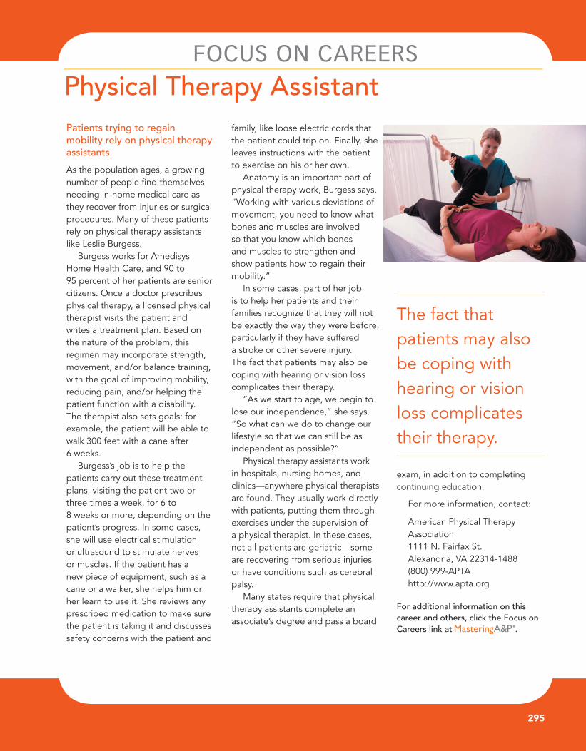
family, like loose electric cords that the patient could trip on. Finally, she leaves instructions with the patient to exercise on his or her own.
Anatomy is an important part of physical therapy work, Burgess says. “Working with various deviations of movement, you need to know what bones and muscles are involved so that you know which bones and muscles to strengthen and show patients how to regain their mobility.”
In some cases, part of her job is to help her patients and their families recognize that they will not be exactly the way they were before, particularly if they have suffered a stroke or other severe injury. The fact that patients may also be coping with hearing or vision loss complicates their therapy.
“As we start to age, we begin to lose our independence,” she says. “So what can we do to change our lifestyle so that we can still be as independent as possible?”
Physical Therapy Assistant
exam, in addition to completing continuing education.
For more information, contact:
American Physical Therapy Association1111 N. Fairfax St.Alexandria, VA 22314-1488(800) 999-APTA http://www.apta.org
For additional information on this career and others, click the Focus on Careers link at .
The fact that patients may also be coping with hearing or vision loss complicates their therapy.
Patients trying to regain mobility rely on physical therapy assistants.
As the population ages, a growing number of people find themselves needing in-home medical care as they recover from injuries or surgical procedures. Many of these patients rely on physical therapy assistants like Leslie Burgess.
Burgess works for Amedisys Home Health Care, and 90 to 95 percent of her patients are senior citizens. Once a doctor prescribes physical therapy, a licensed physical therapist visits the patient and writes a treatment plan. Based on the nature of the problem, this regimen may incorporate strength, movement, and/or balance training, with the goal of improving mobility, reducing pain, and/or helping the patient function with a disability. The therapist also sets goals: for example, the patient will be able to walk 300 feet with a cane after 6 weeks.
Burgess’s job is to help the patients carry out these treatment plans, visiting the patient two or three times a week, for 6 to 8 weeks or more, depending on the patient’s progress. In some cases, she will use electrical stimulation or ultrasound to stimulate nerves or muscles. If the patient has a new piece of equipment, such as a cane or a walker, she helps him or her learn to use it. She reviews any prescribed medication to make sure the patient is taking it and discusses safety concerns with the patient and
FOCUS ON CAREERS
Physical therapy assistants work in hospitals, nursing homes, and clinics—anywhere physical therapists are found. They usually work directly with patients, putting them through exercises under the supervision of a physical therapist. In these cases, not all patients are geriatric—some are recovering from serious injuries or have conditions such as cerebral palsy.
Many states require that physical therapy assistants complete an associate’s degree and pass a board
296 Essentials of Human Anatomy and Physiology
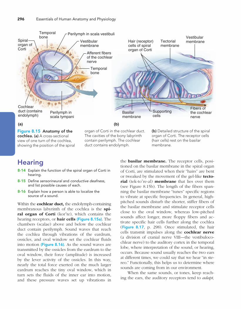
the basilar membrane. The receptor cells, posi-tioned on the basilar membrane in the spiral organ of Corti, are stimulated when their “hairs” are bent or tweaked by the movement of the gel-like tecto-rial (tek-to′re-al) membrane that lies over them (see Figure 8.15b). The length of the fibers span-ning the basilar membrane “tunes” specific regions to vibrate at specific frequencies. In general, high-pitched sounds disturb the shorter, stiffer fibers of the basilar membrane and stimulate receptor cells close to the oval window, whereas low-pitched sounds affect longer, more floppy fibers and ac-tivate specific hair cells further along the cochlea (Figure 8.17, p. 298). Once stimulated, the hair cells transmit impulses along the cochlear nerve (a division of cranial nerve VIII—the vestibuloco-chlear nerve) to the auditory cortex in the temporal lobe, where interpretation of the sound, or hearing, occurs. Because sound usually reaches the two ears at different times, we could say that we hear “in ste-reo.” Functionally, this helps us to determine where sounds are coming from in our environment.
When the same sounds, or tones, keep reach-ing the ears, the auditory receptors tend to adapt,
Hearing 8-14 Explain the function of the spiral organ of Corti in
hearing.
8-15 Define sensorineural and conductive deafness, and list possible causes of each.
8-16 Explain how a person is able to localize the source of a sound.
Within the cochlear duct, the endolymph-containing membranous labyrinth of the cochlea is the spi-ral organ of Corti (kor′te), which contains the hearing receptors, or hair cells (Figure 8.15a). The chambers (scalae) above and below the cochlear duct contain perilymph. Sound waves that reach the cochlea through vibrations of the eardrum, ossicles, and oval window set the cochlear fluids into motion (Figure 8.16). As the sound waves are transmitted by the ossicles from the eardrum to the oval window, their force (amplitude) is increased by the lever activity of the ossicles. In this way, nearly the total force exerted on the much larger eardrum reaches the tiny oval window, which in turn sets the fluids of the inner ear into motion, and these pressure waves set up vibrations in
Figure 8.15 Anatomy of the cochlea. (a) A cross-sectional view of one turn of the cochlea, showing the position of the spiral
organ of Corti in the cochlear duct. The cavities of the bony labyrinth contain perilymph. The cochlear duct contains endolymph.
(b) Detailed structure of the spiral organ of Corti. The receptor cells (hair cells) rest on the basilar membrane.
Basilarmembrane
Supportingcells
Hair (receptor)cells of spiralorgan of Corti
Tectorialmembrane
Fibers ofthe cochlearnerve
Vestibularmembrane
Temporalbone
Perilymph in scala vestibuli
Vestibularmembrane
Afferent fibersof the cochlearnerve
Spiral organ ofCorti
Cochlearduct (containsendolymph)
Perilymph inscala tympani
Temporal bone
(b)(a)

Chapter 8: Special Senses 297
8
Figure 8.16 Route of sound waves through the ear. To excite the hair cells in the spiral organ of Corti in the inner ear, sound wave vibrations must pass through air, membranes, bone, and fluid.
EXTERNAL EAR MIDDLE EAR INTERNAL EAR
Pinna Auditorycanal
Ear-drum
Hammer,anvil, stirrup
Fluids in cochlear canalsUpper and middle lower
Pre
ssur
e
Amplificationin middle ear
AmplitudeOnevibration
Spiral organof Corti
stimulated
Time
Ovalwindow
or stop responding, to those sounds, and we are no longer aware of them. This is why the drone of a continuously running motor does not demand our attention after the first few seconds. However, hearing is the last sense to leave our awareness when we fall asleep or receive anesthesia (or die) and is the first to return as we awaken.
Hearing and Equilibrium Deficits
Homeostatic Imbalance 8.9Children with ear problems or hearing deficits often pull on their ears or fail to respond when spoken to. Under such conditions, tuning fork or audiometry testing is done to try to diagnose the problem. Deafness is defined as hearing loss of any degree—from a slight loss to a total inability to hear sound. Generally speaking, there are two kinds of deafness, conduction and sensorineural. Temporary or permanent conduction deafness results when something interferes with the con-duction of sound vibrations to the fluids of the inner ear. Something as simple as a buildup of earwax may be the cause. Other causes of con-duction deafness include fusion of the ossicles (a problem called otosclerosis [o″to-skle-ro′sis]), a ruptured eardrum, and otitis media.
Sensorineural deafness occurs when there is degeneration or damage to the receptor cells in the spiral organ of Corti, to the cochlear nerve, or to neurons of the auditory cortex. This often results from extended listening to excessively loud sounds. Thus, whereas conduction deafness re-sults from mechanical factors, sensorineural deaf-ness is a problem of nervous system structures.
A person who has a hearing loss due to con-duction deafness will still be able to hear by bone conduction, even though his or her ability to hear air-conducted sounds (the normal conduction route) is decreased or lost. In contrast, individuals with sensorineural deafness cannot hear better by either conduction route. Hearing aids, which use skull bones to conduct sound vibrations to the in-ner ear, are generally very successful in helping people with conduction deafness to hear. They are less helpful for sensorineural deafness.
Equilibrium problems are usually obvious. Nausea, dizziness, and problems in maintaining balance are common symptoms, particularly when impulses from the vestibular apparatus “disagree” with what we see (visual input). There also may be strange (jerky or rolling) eye movements.
A serious pathology of the inner ear is Ménière’s (man″e-airz′) syndrome. The exact cause of this condition is not fully known, but suspected causes are arteriosclerosis, degeneration of cranial nerve VIII, and increased pressure of the

298 Essentials of Human Anatomy and Physiology
PART III: CHEMICAL SENSES: SMELL AND TASTE 8-17 Describe the location, structure, and function of
the olfactory and taste receptors.
8-18 Name the five basic taste sensations, and list factors that modify the sense of taste.
The receptors for taste and olfaction are classified as chemoreceptors (ke″mo-re-sep′terz) because they respond to chemicals in solution. Five types of taste receptors have been identified, but the olfactory receptors (for smell) are believed to be sensitive to a much wider range of chemicals. The receptors for smell and taste complement each other and respond to many of the same stimuli.
Olfactory Receptors and the Sense of SmellEven though our sense of smell is far less acute than that of many other animals, the human nose
inner ear fluids. In Ménière’s syndrome, progres-sive deafness occurs. Affected individuals become nauseated and often have howling or ringing sounds in their ears and vertigo (a sensation of spinning) that is so severe that they cannot stand up without extreme discomfort. Anti–motion sick-ness drugs are often prescribed to decrease the discomfort. ............................................................. ✚
Did You Get It? 17. From the air outside the body, through what
substances do sound waves travel to excite the receptor cells of the cochlea?
18. Which nerve transmits impulses from the spiral organ of Corti to the brain?
19. Do high-pitched sounds peak close to or far from the oval window?
20. How do sensorineural and conductive deafness differ from each other?
(For answers, see Appendix D.)
Figure 8.17 Activation of the cochlear hair cells. (a) The cochlea is drawn as though it were uncoiled to make the events of sound transmission occurring there easier to follow. Sound waves of low frequency below the level of hearing travel entirely around the cochlear duct without exciting hair cells. But sounds of higher frequency penetrate through the cochlear duct and basilar membrane to reach the scala tympani. This causes the basilar membrane to vibrate maximally in certain areas in response to certain frequencies of sound, stimulating particular hair cells and sensory neurons. The differential stimulation of hair cells is perceived in the brain as sound of a certain pitch. (b) The length and stiffness of the fibers spanning the basilar membrane tune specific regions to vibrate at specific frequencies. The higher notes—20,000 Hertz (Hz)—are detected by shorter, stiffer hair cells along the base of the basilar membrane.
Stapes
Ovalwindow
Scalavestibuli
Cochlearduct
Scalatympani
Basilarmembrane
Roundwindow
(a)
(b)
Perilymph
Fibers ofsensoryneurons
Fibers of basilar membrane
Base (short,stiff fibers)
20,000(High notes)
20(Low notes)
2,000 200
Frequency (Hz)
Apex(long,floppyfibers)
Practice art labeling >Study Area>Chapter 8

Chapter 8: Special Senses 299
8
olfactory cortex of the brain. There the odor is interpreted, and an “odor snapshot” is made. The olfactory pathways are closely tied into the lim-bic system (emotional-visceral part of the brain). Thus, olfactory impressions are long-lasting and very much a part of our memories and emotions. For example, the smell of chocolate chip cookies may remind you of your grandmother, and the smell of a special pipe tobacco may make you think of your father. There are hospital smells, school smells, baby smells, travel smells. The list can be continued almost without end. Our reactions to odors are rarely neutral. We tend to either like or dislike certain odors, and we change, avoid, or add odors according to our preferences.
The olfactory receptors are exquisitely sensitive—just a few molecules can activate them. Like the auditory receptors, the olfactory neurons tend to adapt rather quickly when they are exposed to an unchanging stimulus, in this case, an odor. This is why a woman stops smelling her own perfume after a while but will quickly pick up the scent of another perfume on someone else.
is still no slouch in picking up small differences in odors. Some people capitalize on this ability by becoming tea and coffee blenders, perfumers, or wine tasters.
The thousands of olfactory receptors, re-ceptors for the sense of smell, occupy a postage stamp–sized area in the roof of each nasal cavity (Figure 8.18). Air entering the nasal cavities must make a hairpin turn to enter the respiratory pas-sageway below, so sniffing, which causes more air to flow superiorly across the olfactory receptors, intensifies the sense of smell.
The olfactory receptor cells are neurons equipped with olfactory hairs, long cilia that protrude from the nasal epithelium and are con-tinuously bathed by a layer of mucus secreted by underlying glands. When the olfactory receptors located on the cilia are stimulated by chemicals dissolved in the mucus, they transmit impulses along the olfactory filaments, which are bun-dled axons of olfactory neurons that collectively make up the olfactory nerve (cranial nerve I). The olfactory nerve conducts the impulses to the
Q: How does sniffing help to identify scents?
It brings more odor-containing air into contact with the olfactory receptors in the superior part
of the nasal cavity.
A:
Figure 8.18 Location and cellular makeup of the olfactory epithelium.
Olfactory bulb
Cribriform plateof ethmoid bone
Olfactory tract
Supporting cell
Olfactory filaments ofthe olfactory nerve
Olfactory receptorcell
Olfactory hairs(cilia)
Olfactorymucosa
Mucus layer
Route of inhaled aircontaining odor molecules
(a)
(b)
Practice art labeling
>Study Area>Chapter 8
300 Essentials of Human Anatomy and Physiology
our environment in an intimate way, and many of us consider the sense of taste to be the most plea-surable of our special senses.
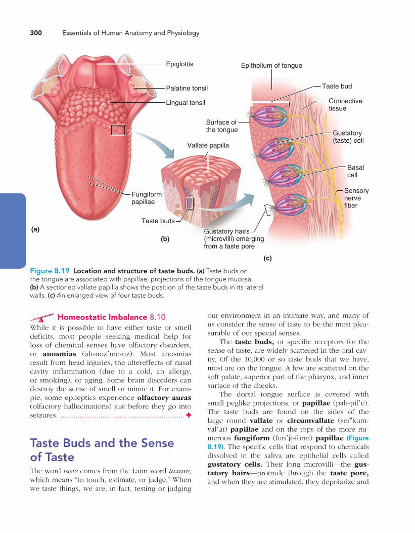
The taste buds, or specific receptors for the sense of taste, are widely scattered in the oral cav-ity. Of the 10,000 or so taste buds that we have, most are on the tongue. A few are scattered on the soft palate, superior part of the pharynx, and inner surface of the cheeks.
The dorsal tongue surface is covered with small peglike projections, or papillae (pah-pil′e). The taste buds are found on the sides of the large round vallate or circumvallate (ser″kum-val′at) papillae and on the tops of the more nu-merous fungiform (fun′jı -form) papillae (Figure 8.19). The specific cells that respond to chemicals dissolved in the saliva are epithelial cells called gustatory cells. Their long microvilli—the gus-tatory hairs—protrude through the taste pore, and when they are stimulated, they depolarize and
Homeostatic Imbalance 8.10While it is possible to have either taste or smell deficits, most people seeking medical help for loss of chemical senses have olfactory disorders, or anosmias (ah-noz′me-uz). Most anosmias result from head injuries, the aftereffects of nasal cavity inflammation (due to a cold, an allergy, or smoking), or aging. Some brain disorders can destroy the sense of smell or mimic it. For exam-ple, some epileptics experience olfactory auras (olfactory hallucinations) just before they go into seizures. ................................................................. ✚
Taste Buds and the Sense of TasteThe word taste comes from the Latin word taxare, which means “to touch, estimate, or judge.” When we taste things, we are, in fact, testing or judging
(a)(b)
Fungiformpapillae
Taste buds
Surface of the tongue
Vallate papilla
Gustatory hairs(microvilli) emergingfrom a taste pore
(c)
Epithelium of tongue
Taste bud
Connective tissue
Gustatory(taste) cell
Basalcell
Sensorynervefiber
Epiglottis
Palatine tonsil
Lingual tonsil
Figure 8.19 Location and structure of taste buds. (a) Taste buds on the tongue are associated with papillae, projections of the tongue mucosa. (b) A sectioned vallate papilla shows the position of the taste buds in its lateral walls. (c) An enlarged view of four taste buds.
Chapter 8: Special Senses 301
8
when your nasal passages are congested by a cold. Without the sense of smell, our morning cof-fee would simply taste bitter. In addition, the tem-perature and texture of food can enhance or spoil its taste for us. For example, some people will not eat foods that have a pasty texture (avocados) or that are gritty (pears), and almost everyone con-siders a cold greasy hamburger unfit to eat. “Hot” foods such as chili peppers actually excite pain receptors in the mouth.
Did You Get It? 21. What name is used to describe both taste and smell
receptors? Why?
22. Where, relative to specific structures, are most taste buds located?
23. Why does it help to sniff substances you are smelling?
(For answers, see Appendix D.)
PART IV: DEVELOPMENTAL ASPECTS OF THE SPECIAL SENSES 8-19 Describe changes that occur with age in the
special sense organs.
The special sense organs, essentially part of the nervous system, are formed very early in embryonic development. For example, the eyes, which are literally outgrowths of the brain, are developing by the fourth week. All of the special senses are func-tional, to a greater or lesser degree, at birth.
Homeostatic Imbalance 8.11Congenital eye problems are relatively uncom-mon, but we can give some examples. Strabismus (strah-biz′mus), which is commonly called “crossed eyes,” results from unequal pulls by the external eye muscles that prevent the baby from coordinat-ing movement of the two eyes. First, exercises are used to strengthen the weaker eye muscles, and/or the stronger eye may be covered with an eye patch to force the weaker muscles to become stronger. If these measures are not successful, surgery is al-ways used to correct the condition because if it is allowed to persist, the brain may stop recognizing signals from the deviating eye, causing that eye to become functionally blind.
impulses are transmitted to the brain. Three cra-nial nerves—VII, IX, and X—carry taste impulses from the various taste buds to the gustatory cortex. The facial nerve (VII) serves the anterior part of the tongue. The other two cranial nerves—the glossopharyngeal and vagus—serve the other taste bud–containing areas. Because of their loca-tion, taste bud cells are subjected to huge amounts of friction and are routinely burned by hot foods. Luckily, they are among the most dynamic cells in the body, and they are replaced every 7 to 10 days by basal cells (stem cells) found in the deeper regions of the taste buds.
There are five basic taste sensations, each corresponding to stimulation of one of the five major types of taste buds. The sweet receptors respond to substances such as sugars, saccharine, some amino acids, and some lead salts (such as those found in lead paint). Sour receptors respond to hydrogen ions (H+), or the acidity of the solu-tion; bitter receptors to alkaloids; and salty recep-tors to metal ions in solution. Umami (u-mah′me; “delicious”), a taste discovered by the Japanese, is elicited by the amino acid glutamate, which appears to be responsible for the “beef taste” of steak and the flavor of monosodium glutamate, a food additive.
Historically, the tip of the tongue was be-lieved to be most sensitive to sweet and salty sub-stances, its sides to sour, the back of the tongue to bitter, and the pharynx to umami. Actually there are only slight differences in the locations of the taste receptors in different regions of the tongue, but the bitter receptors do seem to be clustered more at the rear of the tongue. Most taste buds respond to two, three, four, or even all five taste modalities.
Taste likes and dislikes have homeostatic value. A liking for sugar and salt will satisfy the body’s need for carbohydrates and minerals (as well as some amino acids). Many sour, naturally acidic foods (such as oranges, lemons, and toma-toes) are rich sources of vitamin C, an essential vitamin. Umami guides the intake of proteins, and because many natural poisons and spoiled foods are bitter, our dislike for bitterness is protective.
Many factors affect taste, and what is com-monly referred to as our sense of taste depends heavily on stimulation of our olfactory receptors by aromas. Think of how bland food tastes is

302 Essentials of Human Anatomy and Physiology
depth perception is present, providing a readi-ness to begin reading. By school age, the earlier hyperopia has usually been replaced by emmetro-pia. This condition continues until about age 40, when presbyopia (pres″be-o′pe-ah) begins to set in. Presbyopia (literally, “old vision”) results from decreasing lens elasticity that accompanies aging. This condition makes it difficult to focus for close vision; it is basically farsightedness. The person who holds the newspaper at arm’s length to read it provides the most familiar example of this devel-opmental change in vision.
As aging occurs, the lacrimal glands become less active, and the eyes tend to become dry and more vulnerable to bacterial infection and irrita-tion. The lens loses its crystal clarity and becomes discolored. As a result, it begins to scatter light, causing a glare that is distressing when the per-son drives at night. The dilator muscles of the iris become less efficient; thus, the pupils are always somewhat constricted. These last two conditions work together to decrease the amount of light reaching the retina, and visual acuity is dramati-cally lower by one’s seventies. In addition to these changes, older people are susceptible to certain conditions that may result in blindness, such as glaucoma, cataracts, arteriosclerosis, and diabetes.
Homeostatic Imbalance 8.12Congenital abnormalities of the ears are fairly common. Examples include partly or completely missing pinnas and closed or absent external acoustic meatuses. Maternal infections can have a devastating effect on ear development, and maternal rubella during the early weeks of preg-nancy results in sensorineural deafness. ........... ✚
A newborn infant can hear after his or her first cry, but early responses to sound are mostly reflexive—for example, crying and clenching the eyelids in response to a loud noise. By the age of 3 or 4 months, the infant is able to localize sounds and will turn to the voices of family members. The toddler listens critically as he or she begins to imitate sounds, and good language skills are very closely tied to an ability to hear well.
Except for ear inflammations (otitis) resulting from bacterial infections or allergies, few prob-lems affect the ears during childhood and adult life. By the sixties, however, a gradual deterioration
Maternal infections, particularly rubella (German measles), that occur during early pregnancy may lead to congenital blindness or cataracts. If the mother has a type of sexually transmitted infection called gonorrhea (gon″o-re′ah), the baby’s eyes will be infected by the bacteria during delivery. In the resulting conjunctivitis, specifically called ophthal-mia neonatorum (of-thal′me-ah ne″o-na-to′rum), the baby’s eyelids become red and swollen, and pus is produced. All states legally require that all newborn babies’ eyes be routinely treated with silver nitrate or antibiotics shortly after birth. .........✚
Generally speaking, vision is the only special sense that is not fully functional when the baby is born, and many years of “learning” are needed before the eyes are fully mature. The eyeballs continue to enlarge until the age of 8 or 9, but the lens grows throughout life. At birth, the eyeballs are foreshortened, and all babies are hyperopic (farsighted). As the eyes grow, this condition usu-ally corrects itself. The newborn infant sees only in gray tones, makes uncoordinated eye movements, and often uses only one eye at a time. Because the lacrimal glands are not fully developed until about 2 weeks after birth, the baby is tearless for this period, even though he or she may cry lustily.
By 5 months, the infant is able to focus on articles within easy reach and to follow moving objects, but visual acuity is still poor. For example, an object that someone with mature vision can see clearly 200 feet away has to be a mere 20 feet away before an infant can see it clearly. (Such vision is said to be 20/200.) By the time the child is 5 years old, color vision is well developed, visual acuity has improved to about 20/30, and
This infant with strabismus is unable to focus both eyes simultaneously on the same object.
Chapter 8: Special Senses 303
8
breast. However, very young children seem indif-ferent to odors and can play happily with their own feces. As they get older, their emotional re-sponses to specific odors increase.
There appear to be few problems with the chemical senses throughout childhood and young adulthood. Beginning in the midforties, our ability to taste and smell diminishes, which reflects the gradual decrease in the number of these recep-tor cells. Almost half of people over the age of 80 cannot smell at all, and their sense of taste is poor. This may explain their inattention to formerly dis-agreeable odors, and why older adults often prefer highly seasoned (although not necessarily spicy) foods or lose their appetite entirely.
Did You Get It? 24. Fifty-year-old Mrs. Bates is complaining that she
can’t read without holding the newspaper out at arm’s length. What name is given to her problem?
25. Which of the special senses is least mature at birth?
26. What is presbycusis?
(For answers, see Appendix D.)
and atrophy of the spiral organ of Corti begins and leads to a loss in the ability to hear high tones and speech sounds. This condition, pres-bycusis (pres″ bı-ku′sis), is a type of sensorineu-ral deafness. In some cases, the ear ossicles fuse (otosclerosis), which compounds the hearing problem by interfering with sound conduction to the inner ear. Because many elderly people re-fuse to accept their hearing loss and resist using hearing aids, they begin to rely more and more on their vision for clues as to what is going on around them and may be accused of ignoring people. Although presbycusis was once consid-ered a disability of old age, it is becoming much more common in younger people as our world grows noisier day by day. The damage caused by excessively loud sounds is progressive and cumu-lative. Music played and heard at deafening levels definitely contributes to the deterioration of the hearing receptors.
The chemical senses, taste and smell, are sharp at birth, and infants relish some food that adults consider bland or tasteless. Some researchers claim the sense of smell is just as important as the sense of touch in guiding a newborn baby to its mother’s
SUMMARY
For more chapter study tools, go to the Study Area of MasteringA&P. There you will find:
Essentials of Interactive Physiology
A&PFlix Available at www.masteringaandp.com
Practice Anatomy Lab
Get Ready for A&P
Flashcards, Quizzes, Crossword Puzzles, Art-labeling Activities, Practice Tests, and more!
PART I: THE EYE AND VISION (pp. 279–290)
1. External/accessory structures of the eye:
a. Extrinsic eye muscles aim the eyes for following moving objects and for convergence.
b. The lacrimal apparatus includes a series of ducts and the lacrimal glands that produce a saline so-lution, which washes and lubricates the eyeball.
c. Eyelids protect the eyes. Associated with the eyelashes are the ciliary glands (modified sweat glands) and the tarsal glands (which produce an oily secretion that helps keep the eye lubricated).
d. The conjunctiva is a mucous membrane that covers the anterior eyeball and lines the eyelids. It produces a lubricating mucus.
2. Three layers form the eyeball.
a. The sclera forms most of the outer, tough, pro-tective fibrous layer. The anterior portion is the cornea, which is transparent to allow light to enter the eye.
b. The vascular layer, or middle coat, provides nutri-tion to the internal eye structures. Its posterior por-tion, the choroid, is pigmented and prevents light’s scattering in the eye. Anterior modifications include two smooth muscle structures: the ciliary body and the iris (which controls the size of the pupil).

304 Essentials of Human Anatomy and Physiology
c. The internal ear, or bony labyrinth, consists of bony chambers (cochlea, vestibule, and semi-circular canals) in the temporal bone. The bony labyrinth contains perilymph and membranous sacs filled with endolymph. Within the mem-branous sacs of the vestibule and semicircu-lar canals are equilibrium receptors. Hearing receptors are found within the membranes of the cochlea.
2. Receptors of the semicircular canals (cristae ampul-lares) are dynamic equilibrium receptors, which respond to angular or rotational body movements. Receptors of the vestibule (maculae) are static equi-librium receptors, which respond to the pull of gravity and report on head position. Visual and proprioceptor input to the brain are also necessary for normal balance.
3. Symptoms of equilibrium apparatus problems in-clude involuntary rolling of the eyes, nausea, vertigo, and an inability to stand erect.
4. Hair cells of the spiral organ of Corti (the recep-tor for hearing within the cochlea) are stimulated by sound vibrations transmitted through air, mem-branes, bone, and fluids.
5. Deafness is any degree of hearing loss. Conduction deafness results when the transmission of sound vibrations through the external and middle ears is hindered. Sensorineural deafness occurs when there is damage to the nervous system structures involved in hearing.
PART III: CHEMICAL SENSES: SMELL AND TASTE (pp. 298–301)
1. Chemical substances must be dissolved in aqueous solution to excite the receptors for smell and taste.
2. The olfactory (smell) receptors are located in the superior aspect of each nasal cavity. Sniffing helps to bring more air (containing odors) over the olfactory mucosa.
3. Olfactory pathways are closely linked to the limbic system; odors stimulate the recall of memories and arouse emotional responses.
4. Gustatory (taste) cells are located in the taste buds, primarily on the tongue. The five major taste sensa-tions are sweet, salt, sour, bitter, and umami.
5. Taste and appreciation of foods is influenced by the sense of smell and the temperature and texture of foods.
c. The sensory layer consists of the two-layered retina—a pigmented epithelium and the inner-most (neural) coat, which contains the photore-ceptors. Rods are dim light receptors. Cones are receptors that provide for color vision and high visual acuity. The fovea centralis, on which acute focusing occurs, contains only cones.
3. The blind spot (optic disc) is the point where the optic nerve leaves the back of the eyeball.
4. The lens is the major light-bending (refractory) structure of the eye. Its convexity is increased by the ciliary body for close focus. Anterior to the lens is the aqueous humor; posterior to the lens is the vitreous humor. Both humors reinforce the eye internally. The aqueous humor also provides nutri-ents to the avascular lens and cornea.
5. Errors of refraction include myopia, hyperopia, and astigmatism. All are correctable with specially ground lenses.
6. The pathway of light through the eye is cornea → aqueous humor → (through pupil) → aqueous humor → lens → vitreous humor → retina.
7. Overlap of the visual fields and inputs from both eyes to each optic cortex provide for depth perception.
8. The pathway of nerve impulses from the retina of the eye is optic nerve → optic chiasma → optic tract → thalamus → optic radiation → visual cortex in occipital lobe of brain.
9. Eye reflexes include the photopupillary, accommo-dation pupillary, and convergence.
PART II: THE EAR: HEARING AND BALANCE (pp. 290–298)
1. The ear is divided into three major areas.
a. External ear structures are the pinna, external acoustic meatus, and tympanic membrane, or eardrum. Sound entering the external acoustic meatus sets the eardrum into vibration. These structures are involved with sound transmission only.
b. Middle ear structures are the ossicles and pharyngotympanic tube within the tympanic cavity. Auditory ossicles transmit the vibratory motion from the eardrum to the oval window. The pharyngotympanic tube allows pressure to be equalized on both sides of the eardrum. These structures are involved with sound trans-mission only.

Chapter 8: Special Senses 305
8
4. Which cranial nerve controls contraction of the circular smooth muscle of the iris?
a. Trigeminal
b. Facial
c. Oculomotor
d. Abducens
5. The cornea is nourished by
a. corneal blood vessels.
b. aqueous humor.
c. vitreous humor.
d. scleral blood vessels.
6. When the eye focuses for far vision,
a. the lens is at its thinnest.
b. the ciliary muscles contract.
c. the light rays are nearly parallel.
d. suspensory fibers of the ciliary zonule are slack.
7. Convergence
a. requires contraction of the medial rectus muscles of both eyes.
b. is needed for near vision.
c. involves transmission of impulses along the abducens nerves.
d. can promote eyestrain.
8. Which of the following are paired incorrectly ?
a. Cochlear duct—cupula
b. Vestibule—macula
c. Ampulla—otoliths
d. Semicircular duct—ampulla
9. Movement of the _______ membrane triggers bend-ing of hairs of the hair cells in the spiral organ of Corti.
a. tympanic
b. tectorial
PART IV: DEVELOPMENTAL ASPECTS OF THE SPECIAL SENSES (pp. 301–303)
1. Special sense organs are formed early in embryonic development. Maternal infections during the first five or six weeks of pregnancy may cause visual abnormalities as well as sensorineural deafness in the developing child. An important congenital eye problem is strabismus. The most important con-genital ear problem is lack of the external acoustic meatus.
2. Vision requires the most learning. The infant has poor visual acuity (is farsighted) and lacks color vision and depth perception at birth. The eye con-tinues to grow and mature until age 8 or 9.
3. Problems of aging associated with vision include presbyopia, glaucoma, cataracts, and arteriosclero-sis of the eye’s blood vessels.
4. The newborn infant can hear sounds, but initial responses are reflexive. By the toddler stage, the child is listening critically and beginning to imitate sounds as language development begins.
5. Sensorineural deafness (presbycusis) is a normal consequence of aging.
6. Taste and smell are most acute at birth and de-crease in sensitivity after age 40 as the number of olfactory and gustatory receptors decreases.
REVIEW QUESTIONSMultiple Choice
More than one choice may apply.
1. Gustatory cells are
a. bipolar neurons.
b. multipolar neurons.
c. unipolar neurons.
d. epithelial cells.
2. Alkaloids excite gustatory hairs mostly at the
a. tip of the tongue.
b. back of the tongue.
c. circumvallate papillae.
d. fungiform papillae.
3. Cranial nerves that are part of the gustatory path-way include
a. trigeminal.
b. facial.
c. hypoglossal.
d. glossopharyngeal.
c. basilar
d. vestibular
10. Sounds entering the external acoustic meatus are eventually converted to nerve impulses via a chain of events including
a. vibration of the eardrum.
b. vibratory motion of the ossicles against the oval window.
c. stimulation of hair cells in the spiral organ of Corti.
d. resonance of the cupula.

306 Essentials of Human Anatomy and Physiology
30. What name is given to the taste receptors?
31. Name the five primary taste sensations.
32. Where are the olfactory receptors located, and why is that site poorly suited for their job?
33. Describe the effects or results of aging on the spe-cial sense organs.
34. Which special sense requires the most learning?
35. For each of the following statements, indicate whether it applies to a macula or a crista ampul-laris: inside a semicircular canal; contains otoliths; responds to linear acceleration and deceleration; has a cupula; responds to rotational acceleration and deceleration; inside the vestibule.
CRITICAL THINKING AND CLINICAL APPLICATION QUESTIONS
36. An engineering student has been working in con-struction to earn money to pay for his education. After about eight months, he notices that he is hav-ing problems hearing high-pitched tones. What is the cause-and-effect relationship here?
37. Nine children attending the same day-care center developed red, inflamed eyes and eyelids. What is the most likely cause and name of this condition?
38. Dr. Nguyen used an instrument to press on Mr. Cruz’s eye during his annual physical examination on his sixtieth birthday. The eye deformed very little, indicating the intraocular pressure was too high. What was Mr. Cruz’s probable condition?
39. Brandon suffered a ruptured artery in his middle cranial fossa, and a pool of blood compressed his left optic tract, destroying its axons. What part of the visual field was blinded?
40. Sylvia Marcus, age 70, recently had surgery for oto-sclerosis. The operation was a failure and did not improve her condition. What was the purpose of the surgery, and exactly what was it trying to ac-complish?
41. Janie is referred to the eye clinic by her teacher, who suspects a need for glasses. Examination dem-onstrates that Janie is myopic. Will she need con-cave or convex lenses? Explain.
Short Answer Essay
11. Name three accessory eye structures that help to lu-bricate the eyeball, and name the secretion of each.
12. Why do you often have to blow your nose after crying?
13. Diagram and label the internal structures of the eye, and give the major function of each structure.
14. Name the extrinsic eye muscles that allow you to direct your eyes.
15. Locate and describe the functions of the two hu-mors of the eye.
16. What is the blind spot, and why is it called this?
17. What name is given to the structure that controls light entry into the eye?
18. What is the fovea centralis, and why is it important?
19. Trace the pathway of light from the time it hits the cornea until it excites the rods and cones.
20. Trace the pathway of nerve impulses from the pho-toreceptors in the retina to the visual cortex of the brain.
21. Define hyperopia, myopia, and emmetropia.
22. Why do most people develop presbyopia as they age? Which of the conditions in question 21 does it most resemble?
23. There are only three types of cones. How can you explain the fact that we see many more colors?
24. Why are ophthalmoscopic examinations important?
25. Many students struggling through mountains of reading assignments are told that they need glasses for eyestrain. Why is it more of a strain on the ex-trinsic and intrinsic muscles to look at close objects than at far objects?
26. Name the structures of the outer, middle, and inner ears and give the general function of each structure and each group of structures.
27. Sound waves hitting the eardrum set it into motion. Trace the pathway of vibrations from the eardrum to the spiral organ of Corti, where the hair cells are stimulated.
28. Give two causes of conduction deafness.
29. Normal balance depends on information transmit-ted from a number of sensory receptor types. Name at least three of these receptors.

Chapter 8: Special Senses 307
8
Later she told her father that she felt like she “kept on going” when the elevator stopped. Explain her sensation.
44. Mrs. Garson has an immune disorder that causes dry mouth, and she complains to her doctor that she’s lost her sense of taste. How might her symp-toms be explained?
45. Four-year-old Aiden is brought to the ophthalmol-ogist for a routine vision checkup. Aiden is an albino. How do you think albinism affects vision?
42. Julie and her father loved to find the constella-tions in the sky on starry nights. One evening, Julie came running into the house and whispered excitedly to her mother. “Mom, I’ve got power! When I look hard at a star it disappears!” What was happening?
43. While visiting at her father’s office on the 25th floor of the Harris Building, five-year-old Emma wandered away into the hall. Fascinated by the but-tons in the elevator, she entered, pressed 1, and the high-speed elevator plummeted to the first floor.