special senses (2)
TRANSCRIPT

SPECIAL SENSES Cornea
Retina
Optic nerve
Prof.J.Anbalagan

Special sensory organs
Olfactory mucosa
Gustatory cells
Retina
Internal ear
– cochlea
– Semicircular canal


Eyeball

Eye lid Skin – Stratified squamous
keratinized
– dermis
Connective tissue layer
Skeletal muscle (orbicularis oculi)
Tarsal plate with glands
Conjunctiva – Stratified squamous non
keratinized

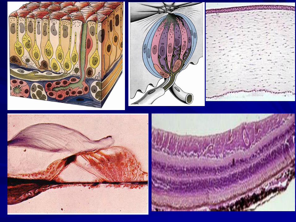
Cornea
Five layered structure
– Outer corneal epithelium
– Bowman’s membrane
– Substantia propria
– Descemet’s membrane
– Inner endothelium

Corneal epithelium – Thin Stratified squamous non
keratinized-5 LAYER
– Papilla absent
– Straight plane avoids refraction
Bowman’s membrane – Anterior limiting membrane
– Homogenous amorphous with fine collagen
– Forms basement membrane for corneal epithelium
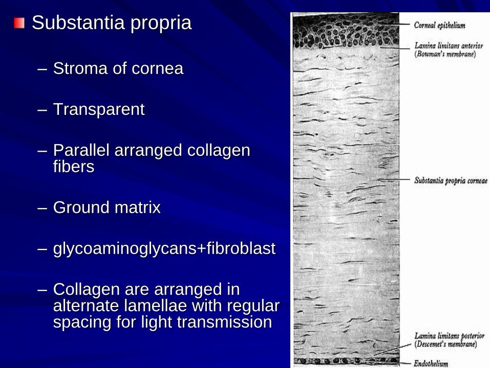
Substantia propria
– Stroma of cornea
– Transparent
– Parallel arranged collagen fibers
– Ground matrix
– glycoaminoglycans+fibroblast
– Collagen are arranged in alternate lamellae with regular spacing for light transmission

Descemet’s membrane
– Posterior limiting
membrane
– Thin and homogenous
– Layers continues into irido
corneal angle
Endothelium
– Single polygonal cells on
a basement membrane

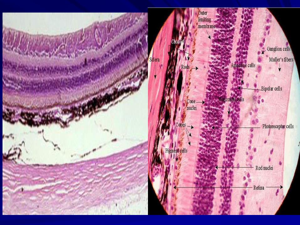
RETINA Nervous coat
Extends from optic disc
to ora serrata
Posterior part of retina
shows yellow spot
macula lutea
Optic disc – area where
optic nerve emerges

Layers of retina 1. PIGMENT LAYER
2. LAMINA OF RODS AND CONES
3. EXTERNAL LIMITING MEMBRANE
4. OUTER NUCLEAR LAYER
5. OUTER PLEXIFORM LAYER
6. INNER NUCLEAR LAYER
7. INNER PLEXIFORM LAYER
8. GANGLION CELL LAYER
9. LAMINA OF NERVE FIBERS
10. INTERNAL LIMITING MEMBRANE

PIGMENT EPITHELIUM
Low cubical cells Basement membrane known as Bruch’s membrane Cells faces towards rods and cones Appear black due to melanin pigments One cell contacts > 12 rods and cones functions – Phagocyte, – antireflection of light, – blood retinal barrier
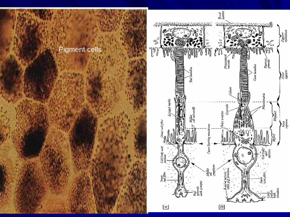
Pigment cells

Rods and Cones
Photoreceptors
Cones are wider and tapers at the end
Rods are narrow and cylindrical
Under EM they exhibit outer and inner segments
Inner segments contains mitochondria for energy
Outer segment shows discoidal membrane

External limiting membrane
– Sieve like membrane
– Supports rods and cones
– Appears pink linear marking
– Represents zona adherens
of Muller cells
Outer nuclear layer
– Nucleus of rods and cones
– Several layers
ONL
OLM

Outer plexiform layer
– Synaptic area between rods and cones with
– Bipolar neurons
– Horizontal cells
– Amacrine cells
Inner nuclear layer
– Shows nucleus of bipolar, horizontal cells, amacrine cells, Muller cells

Inner plexiform layer
– Synaptic process of
bipolar, amacrine, Muller
with ganglion cells
– Plexiform appearance
Ganglion layer
– Cell body and nucleus of
large ganglion cells and
amacrine cells

Nerve fibers
– axons of ganglion
cells
Internal limiting
membrane
– Homogenous layer
– Formed by end feet of
Muller cell and
astrocytes
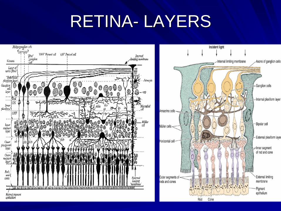
RETINA- LAYERS


Microstructure of Retina

OPTIC NERVE
MENINGES
– DURA
– ARACHNOID
– PIA
NERVE FIBERS
– MYELINATED
– ARRANGED IN FASCICULI
– CENTRAL ARTERY OF RETINA

OPTIC NERVE
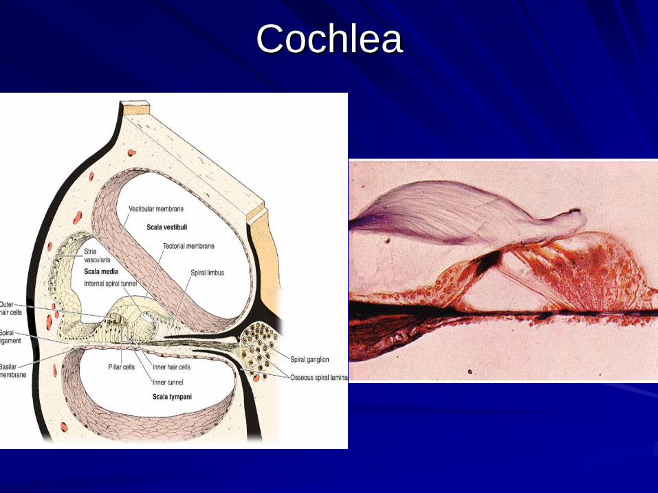
Cochlea