special section - rhode island medical society · and her children, diana oehrli, guillaume de...
TRANSCRIPT

M E D I C A L J O U R N A LR H O D E I S LA N D
V O L U M E 97 • N U M B E R 5 I S S N 2 3 2 7 - 2 2 2 8M A Y 2 0 1 4
NORMAN PRINCE NEUROSCIENCES INSTITUTE JOHN A. ROBSON, PhD; KAREN FURIE, MD; STEVEN RASMUSSEN, MD
GUEST EDITORS
SPECIAL SECTION

M E D I C A L J O U R N A LR H O D E I S LA N D
18 Neurotechnology: A New Approach for Treating Brain DisordersJOHN A. ROBSON, PhD; R. JOHN DAVENPORT, PhD
22 The Brown University Traumatic Brain Injury Research Consortium and the Norman Prince Neurosciences InstituteJEFFREY ROGG, MD; HEATHER SPADER, MD; BETHANY J. WILCOX, BS;
ANNA ELLERMEIER, MD; STEVE CORREIA, PhD; ADAM CHODOBSKI, PhD;
JOANNA SZMYDYNGER-CHODOBSKA, PhD; NEHA RAUKAR, MD;
JASON T. MACHAN, PhD; W. CURT LAFRANCE, JR, MD, MPH;
JOSEPH J. CRISCO, PhD
27 Advances in Stroke Over the Past DecadeBRIAN SILVER, MD
31 The Rhode Island Consortium for Autism Research and Treatment (RI-CART): A New Statewide Autism CollaborativeALAN GERBER, MA; ERIC MORROW, MD, PhD; STEPHEN J. SHEINKOPF, PhD;
THOMAS ANDERS, MD
15 The Norman Prince Neurosciences Institute: Linking Research to Clinical CareJOHN A. ROBSON, PhD; KAREN FURIE, MD; STEVEN RASMUSSEN, MD
GUEST EDITORS

The Norman Prince Neurosciences Institute: Linking Research to Clinical CareJOHN A. ROBSON, PhD; KAREN FURIE, MD; STEVEN RASMUSSEN, MD
Over the past 50 years the expansion and advances in neuro-science have been astonishing. The Society for Neuroscience illustrates this growth. It is the world’s largest organization of scientists and physicians devoted to understanding the brain and nervous system. In 1969 it had 500 members. To-day it has almost 42,000 and its annual meeting attracts over 30,000 researchers. This tremendous growth in research has led to discoveries that have fundamentally changed our understanding of the nervous system.
We are gaining an ever-more sophisticated understanding of the functioning of neurons through the development of advanced imaging technologies in combination with genetic manipulations, and the human genome project is providing new insights about diseases and disorders of the nervous system. Advances in brain imaging technologies, such as Positron Emission Tomography (PET) and Magnetic Reso-nance Imaging (MRI), have revolutionized the ways that we diagnose disorders of the nervous system and they are show-ing, on a global scale, how information is processed in the brain. At the same time researchers are developing devices, like electrodes for deep brain stimulation and implantable
multi-electrode arrays that take advantage of progress in computational neuroscience and computer science to better diagnose and treat a variety of neurological disorders, rang-ing from depression to epilepsy to Parkinson’s disease and paralysis.
In 2013 the White House announced a new national initia-tive in neuroscience – BRAIN (Brain Research through Ad-vancing Innovative Neurotechnologies). It is targeted at one of the great mysteries of the brain – how networks of neu-rons interact to create sensations, movements and thoughts. It will lead to even more breakthroughs in our understanding of how populations of neurons work together normally and how changes in network interplay lead to symptoms associ-ated with neurological and psychiatric disease. These recent and anticipated future advances are creating opportunities for devising new treatments for diseases of the brain and nervous system that are greater than at any time in history.
In Rhode Island This excitement about brain science is having a profound impact in Rhode Island. In the past few years the Alpert
CO
UR
TE
SY
OF
NO
RM
AN
PR
INC
E N
EU
RO
SC
IEN
CE
S I
NS
TIT
UT
E
The cover image is an artist’s semitransparent rendering of the brain looking from the back. It shows features of the vascular system on the surface,
the cerebral cortex on the left, deep structures of the cerebral hemisphere on the right, and brain stem in the lower center.
R H O D E I S L A N D M E D I C A L J O U R N A L 15W W W. R I M E D . O R G | R I M J A R C H I V E S | M A Y W E B P A G E M A Y 2 0 1 4
15
17
EN

NEUROSCIENCES
Medical School at Brown University, in partnership with its affiliated hospitals, has been moving toward the creation of a coordinated academic medical center in Providence. One goal of such a center is to create links between research sci-entists and clinicians to encourage disease-targeted research and to create a pipeline that will facilitate the conversion of research findings into benefits for patients. In this area neuroscience is leading the way.
In December 2009 descendants of Frederick Henry Prince, a New England entrepreneur who made his fortune during the Gilded Age, around the turn of the 20th century, ap-proached Dr. Timothy Babineau, President of Rhode Island Hospital, about making a gift from the Frederick Henry Prince 1932 Trust. Over the next several months that idea crystallized into a $15-million donation, the largest in the history of Rhode Island Hospital, from Elizabeth J.M. Prince and her children, Diana Oehrli, Guillaume de Ramel and Re-gis de Ramel, to endow the Norman Prince Neurosciences Institute (NPNI), named after the son of Henry Frederick Prince, who died from a head injury suffered in a plane crash during World War 1.
Although the gift was made to Rhode Island Hospital, the Institute was expected to develop as a collaborative venture with Brown University and other hospitals affiliated with the Alpert Medical School, including Bradley Hospital, Butler Hospital, Women and Infants Hospital, The Miriam Hospital and the Providence VA Medical Center. In that re-spect, the timing could not have been better. The chairs of the clinical neuroscience departments of neurology, neuro-surgery and psychiatry were vacant, creating an opportunity to recruit new leadership with a shared vision for building interdisciplinary programs. In addition, Brown University was planning a major expansion of brain science on its campus through its own neuroscience institute, the Brown
Institute for Brain Science (BIBS). The fit was obvious and these initiatives have coalesced into a broad-based, inter- disciplinary and inter-institutional effort.
Launching the Norman Prince Neurosciences InstituteIn 2011 NPNI began to develop a vision for growth. A steering committee was created that included the leaders in neu-rology, neurosurgery, psychiatry and basic neuroscience at Rhode Island Hospital, Brown and its other affiliated hos-pitals. Dr. G. Rees Cosgrove was recruited from Boston in 2010 to serve as chair of neurosurgery and take a major leadership role in the NPNI, which he did until resigning in 2014. Dr. Cosgrove did his neurosurgical training at the Montreal Neurological Institute, a self-contained, highly in-tegrated clinical and research institute at McGill University that, in several respects, serves as a model for NPNI. In 2011 he was joined by Dr. John Robson as executive director. He is a neuroscientist who also worked at the Montreal Neu-rological Institute as associate director from 1997–2007. In 2012 Dr. Karen Furie, a Brown graduate and stroke specialist at Harvard University and the Massachusetts General Hos-pital, was recruited to be the chair of neurology and Dr. Steven Rasmussen accepted the chair of psychiatry and human behavior. Dr. Rasmussen has been on the Brown fac-ulty for almost 30 years and is a leading expert on the treat-ment of obsessive-compulsive disorder. Both Drs. Furie and Rasmussen also serve as co-clinical directors of NPNI.
Building on Excellence: Brown Institute for Brain Science Brown University is well known for its strengths in neu-roscience research. It was one of the first universities in the United States to establish a Department of Neurosci-ence and, as a group, its neuroscientists have been very suc-cessful building excellence and attracting research funds.
PH
OT
OS
: B
RO
WN
UN
IVE
RS
ITY
R H O D E I S L A N D M E D I C A L J O U R N A L 16W W W. R I M E D . O R G | R I M J A R C H I V E S | M A Y W E B P A G E M A Y 2 0 1 4
John A. Robson, PhD Karen Furie, MD Steven Rasmussen, MD

NEUROSCIENCES
However, Brown’s neuroscientists are not confined to one department; they are found across the campus. Consequent-ly the Brown Institute for Brain Science (BIBS) was estab-lished as an umbrella organization to advocate for all “brain scientists” and to facilitate interdisciplinary research. This institute now lists more than 100 faculty members from 15 different departments. Represented disciplines range from applied math, engineering and computer science, to cell and molecular biology and physiology, to cognitive neuro-science and brain imaging. Similarly, the clinical neurosci-ences are distributed across several departments, including neurology, neurosurgery, pathology, psychiatry and radiolo-gy, and they are found in many different hospitals affiliated with the Alpert Medical School. Like BIBS, NPNI strives to unite their efforts and create new opportunities for growth and collaboration.
From the onset there has been a major emphasis on col-laboration between NPNI and BIBS. The two organizations have worked closely together to create joint programs in-tended to benefit all of brain science across the campuses of Brown and its affiliated hospitals. These efforts have includ-ed funding for collaborative research projects, a symposium, workshops and seminar speakers. BIBS and NPNI also part-nered with the Rhode Island Medical Society for its highly successful “200th Anniversary Lecture Series.”
Efforts are also underway to find ways to collaborate with scientists at the University of Rhode Island, which started an interdisciplinary neuroscience graduate program in 2012. That program and others related to neuroscience at URI are sure to grow in size and prominence in the coming years due to the recent creation of the Ryan Institute for Neuroscience.
These are exciting times for neuroscience in Rhode Island.
There is a real opportunity to make Providence a nationally recognized center of excellence in this area. In some areas we are already there but further investment will be needed. The clinicians and scientists are eager and many of the parts are in place.
This issue of the Rhode Island Medical Journal contains articles by members of NPNI. They provide examples of programs being developed that focus on important clini-cal issues. It is not a comprehensive review of all programs that fall under the NPNI umbrella. However, these articles describe programs that illustrate the approach that we are developing. They focus on autism, stroke, traumatic brain injury and emerging uses of technology to treat a variety of neurological and psychiatric disorders. Each of these efforts involves teams that are collaborative, interdisciplinary and inter-institutional.
AuthorsJohn A. Robson, PhD, is Administrative Director of the Norman
Prince Neurosciences Institute, Rhode Island Hospital and Associate Director, Brown Institute for Brain Science, Brown University.
Karen Furie, MD, is Co-Clinical Director of the Norman Prince Neurosciences Institute, Rhode Island Hospital, and Professor and Chair, Department of Neurology, The Alpert Medical School of Brown University.
Steven Rasmussen, MD, is Co-Clinical Director of the Norman Prince Neurosciences Institute, Rhode Island Hospital and the Mary E. Zucker Professor and Chair of the Department of Psychiatry and Human Behavior, The Alpert Medical School of Brown University.
R H O D E I S L A N D M E D I C A L J O U R N A L 17W W W. R I M E D . O R G | R I M J A R C H I V E S | M A Y W E B P A G E M A Y 2 0 1 4
NEUROSCIENCES
Neurotechnology: A New Approach for Treating Brain DisordersJOHN A. ROBSON, PhD; R. JOHN DAVENPORT, PhD
ABSTRACT Advances in neuroscience, engineering and computer technologies are creating opportunities to connect the brain directly to devices to treat a variety of disorders, both neurological and psychiatric. They are opening a new field of neuroscience called “neurotechnology.” This article reviews efforts in this area that are ongoing at Brown University and the hospitals affiliated with Brown’s Alpert Medical School. Two general approaches are being used. One uses advanced electrodes to “sense” the activity of many individual neurons in the cerebral cortex and then use that activity for therapeutic purposes. The other uses various types of devices to stimulate spe-cific networks in the brain in order to restore normal function and alleviate symptoms.
KEYWORDS: Neurotechnology, neuroscience advances, BrainGate
INTRODUCTION
Diseases and disorders of the nervous system have proven to be especially difficult to treat in part because of the complex-ity of the brain. Most efforts have focused on the develop-ment of behavioral or pharmacological therapies. However, with advances in engineering and computer technologies, alternative “device-based” approaches are being explored as potential tools for treating a number of conditions ranging from paralysis to movement disorders to mental illness.
Researchers at Brown University and its affiliated hos-pitals are international leaders in this approach, known as “neurotechnology.” This collaborative, interdisciplinary effort has followed two approaches. One takes advantage of devices to detect brain activity and then uses that informa-tion for therapeutic purposes. The second uses devices to change brain activity in ways that can restore normal func-tion. The work has benefited from the collaboration between the Norman Prince Neurosciences Institute (NPNI) and the Brown Institute for Brain Science (BIBS).
SENSING THE BRAIN
BrainGate research The BrainGate research project is a multi-institutional effort based at Brown that is focused on improving the ability of
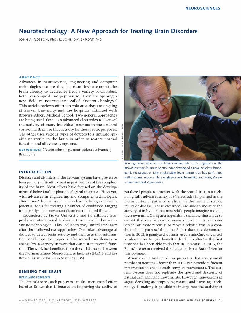
paralyzed people to interact with the world. It uses a tech-nologically advanced array of 96 electrodes implanted in the motor cortex of patients paralyzed as the result of stroke, injury or disease. These electrodes are able to measure the activity of individual neurons while people imagine moving their own arm. Computer algorithms translate that input to output that can be used to move a cursor on a computer screen1 or, more recently, to move a robotic arm in a coor-dinated and purposeful manner.2 In a dramatic demonstra-tion in 2012, a paralyzed woman used BrainGate to control a robotic arm to give herself a drink of coffee2 – the first time she has been able to do that in 15 years! In 2013, the BrainGate team received the inaugural Israel Brain Prize for this advance.
A remarkable finding of this project is that a very small number of neurons – fewer than 100 – can provide sufficient information to encode such complex movements. The cur-rent system does not replicate the speed and dexterity of natural arm and hand movements. However, innovations in signal decoding are improving control and “sensing” tech-nology is making it possible to incorporate the activity of
FR
ED
FIE
LD
FO
R B
RO
WN
UN
IVE
RS
ITY
In a significant advance for brain-machine interfaces, engineers in the
Brown Institute for Brain Science have developed a novel wireless, broad-
band, rechargeable, fully implantable brain sensor that has performed
well in animal models. Here engineers Arto Nurmikko and Ming Yin ex-
amine their prototype device.
R H O D E I S L A N D M E D I C A L J O U R N A L 18W W W. R I M E D . O R G | R I M J A R C H I V E S | M A Y W E B P A G E M A Y 2 0 1 4
18
21
EN

NEUROSCIENCES
FDA recently approved such a device based on ECoG recording technology and implanted stimulating electrodes (NeuroPace RNS System®).
ALTERING ACTIVITY
Deep brain stimulation (DBS) use in various disordersPhysicians have been altering brain activity to treat psychiatric disorders since the 1930s in those patients with
the most severe and intractable symptoms.6 However, in recent years, these techniques have become progressively more refined.
In the 1980s a new form of stimulation was developed primarily to treat tremors and other abnormal movements in patients with Parkinson’s disease7,8 who had severe medi-cation-related problems. Neurosurgeons had discovered that small lesions deep in the brain, in the sub-thalamus and the globus pallidus, could greatly reduce certain symptoms in patients whose responses to drug therapy were problematic. Subsequently, they discovered that high-frequency stimula-tion in these same areas had similar effects. Since lesions are not reversible and can cause complications, if not properly placed, deep brain stimulation (DBS) has become increasing-ly common as it causes minimal brain damage, can be ad-justed with changes in stimulation and the electrodes can be removed. Today there are close to 100,000 people worldwide with DBS electrodes. The vast majority has been implant-ed for the treatment of Parkinson’s disease and other move-ment disorders, including essential tremor and dystonia.
DBS is also being tested as a potential treatment for a number of other conditions. These include epilepsy,9 Tourette’s syndrome,10 motor problems of multiple scle-rosis11 and several others. Within NPNI and BIBS, most research using DBS has focused on mood disorders – depres-sion12 and obsessive-compulsive disorder (OCD).13 The ratio-nale behind this is a growing body of evidence that these disorders are related to dysfunctions of networks of neurons involving the prefrontal cortex,14-16 much like the symptoms of Parkinson’s disease are related to networks involving the motor cortex.
Researchers do not yet agree on the best target for treating depression with DBS. A group from Butler Hospital and the Providence Veterans Affairs Medical Center has focused on an area deep in the forebrain that includes the ventral por-tion of the anterior limb of the internal capsule and the adja-cent striatum (VC/VS). In 2009 they reported that about half of a group of 15 patients with refractive major depression benefitted from DBS in this region with no adverse effects.12
Of those who responded, all showed significant improve-ment in standard mood-rating scales and about 40% were in remission when last examined (up to 4 years postopera-tively). In comparison, others have stimulated the medial
R H O D E I S L A N D M E D I C A L J O U R N A L 19W W W. R I M E D . O R G | R I M J A R C H I V E S | M A Y W E B P A G E M A Y 2 0 1 4
more neurons into the system. As this trend progresses, the ability of paralyzed patients to control robotic devices should improve dramatically.
Engineers at Brown are also mak-ing rapid progress in developing a next-generation BrainGate device that transmits neural signals wirelessly.3 That technology will enable patients to use the BrainGate approach in more ambulatory, real-life situations, untethered from a computer. It will also advance the possibility of amputees using this approach to better control prosthetic limbs. Other studies, using non- human primates, suggest that it will eventually be possible to use this approach to control movements of a patient’s paralyzed limb by stimulating muscles directly.4
Multi-electrode arraysMulti-electrode arrays are also providing new insights into brain activity during epileptic seizures. To plan for epilepsy surgery, doctors often record activity from the brains of patients using electrocorticography (ECoG). In this procedure a large array of 50 or more electrodes is placed directly on the surface of the cortex in the region suspected to be the source of the seizures. Recordings can be made over a week or more while the patients are alert and off anti-seizure medications in an effort to record seizure activity and localize its source. These standard ECoG arrays do not, however, reveal the activity of single neurons.
As part of this procedure it is now possible to insert the same 96-electrode array used in the BrainGate system into the region suspected to be the source of seizure activity. This is being done by a team of clinicians, scientists and engineers at Brown and Rhode Island Hospital, in collabo-ration with colleagues at Massachusetts General Hospital. Thus, for the first time, the activity of individual neurons is being recorded and analyzed before, during, and after sei-zures. An initial report of the results from four research par-ticipants illustrates the potential power of this approach.5 This study showed that seizures are not comprised of hy-per-synchronized neuronal firing as previously suspected. Instead the patterns of activity are quite heterogeneous during the seizure. In comparison, at the end of the seizure almost all neural activity is suppressed for several seconds.
Perhaps the most surprising and significant finding in this study was the discovery that many neurons, even ones well outside the area of seizure origin, showed significant changes in activity minutes prior to the onset of the seizure. Thus, chronically implanted electrodes that record individ-ual neurons could become reliable tools for identifying sei-zures prior to their onset. If this proves to be the case, it could lead to closed-loop devices able to treat epilepsy by stopping seizures before they start, by injecting a drug or an electrical current into the region of the seizure’s onset. The
VIDEO: Paralyzed woman uses thoughts to
sip coffee

NEUROSCIENCES
surface of the cortex in an area known as the sub-callosal cingulate gyrus (area 25) – also part of the mood disorder circuitry – and they report similar benefits.17
Efforts are also underway to evaluate the effectiveness of DBS in the VC/VS for treating severe, unresponsive OCD. In a recent multi-center study, about two-thirds of patients responded positively to treatment for 12 months.13 When the stimulation was interrupted, the responders quickly fell into a severely depressive state, which was reversed when the stimulation resumed.
The reason that some patients do not respond to stimula-tion is not understood, although it is presumed to be related to the placement of the stimulating electrode. This is cur-rently being addressed by a large multi-institutional team, including researchers from Brown, Harvard, the University of Rochester, the University of Pittsburgh and the Univer-sity of Puerto Rico, that is supported by a grant from the National Institute of Mental Health. They are studying the neural mechanisms that underlie DBS stimulation and the cortical networks that are associated with OCD with the ex-pectation that the results will reveal more effective targets and stimulus parameters.
The use of DBS to treat Alzheimer’s disease has also re-ceived attention recently. Lozano and colleagues18 stimulated the fornix and hypothalamus in a patient who was part of a study using DBS to treat obesity and observed that stimula-tion invoked memories. This led to a preliminary study of six patients with early-stage symptoms of Alzheimer’s dis-ease. Of the six, two showed improved function on standard memory tests for a year. The performance of a third patient was unchanged although it would normally be expected to get worse during this period. The other three patients contin-ued to worsen as typical Alzheimer’s patients do. This study lacked controls but was suggestive of positive cognitive benefits from DBS.
Based on these preliminary results, a phase 1–2 clinical trial is now underway to test safety and efficacy in 20–30 pa-tients. Rhode Island and Butler Hospitals are collaborating as one of the sites. Surgery is being done at Rhode Island Hospi-tal and testing is being conducted at Butler. In this one-year trial, only half of the patients will be stimulated and neither the patients nor the testers will know who was stimulated until the end of the trial. After the results are known, all patients will have the option of turning on their stimula-tors if they want. This study design will greatly mitigate placebo effects and investigator bias.
DBS shows great promise for a number of conditions. How-ever, DBS is an invasive surgical technique that comes with small risks for bleeding and infection. It is also expensive. NPNI and BIBS researchers are exploring other techniques to stimulate the brain non-invasively and inexpensively. These techniques include transcranial magnetic stimula-tion (TMS) and transcranial direct or alternating current stimulation (tDCS or tACS). All involve the excitation or inhibition of brain activity by passing a current outside the
head. TMS uses a strong magnetic pulse, placed next to the skull, to induce an electric current in the adjacent cortical surface. TDCS and tACS apply a direct (tDCS) or alternating (tACS) current to the scalp, which causes subtle changes in the activity of the underlying region of the cerebral cortex. The equipment required for all three is relatively inexpen-sive and can be used on patients by trained technicians. All of these techniques have been shown capable of affecting mood19 and compulsions.20
In 2008 the FDA approved TMS as a treatment for severe, intractable depression and all three stimulation techniques are being actively explored for a variety of other applications by research teams at the Center for Neurorestoration and Neurotechnology at the Providence VA Medical Center. Dis-orders being studied include OCD, Post Traumatic Stress Disorder and chronic pain. In addition, evidence suggests that TMS and tDCS may enhance plasticity in the cortex. Thus, this research team is also investigating the possibil-ity that stimulation could be used to enhance the benefits of rehabilitation therapy following stroke or other forms of brain injury.
SUMMARY
Researchers in NPNI and BIBS are collaborating on all of the efforts described above. They are located at different insti-tutions in Providence, including Brown University and hos-pitals affiliated with Brown’s Alpert Medical School. They are members of teams that are using neurotechnology to develop novel treatments for patients suffering with a wide variety of neurological and psychiatric disorders. This area of research demands coordination and collaboration between clinicians, neuroscientists, engineers, mathematicians and computer scientists. It is also an area where Providence already stands out on the world stage and is poised to expand its prominence.
References1. Hochberg LR, Serruya MD, Friehs GM, Mukand JA, Saleh M,
Caplan AH, Branner A, Chen D, Penn RD, Donoghue JP. Neu-ronal ensemble control of prosthetic devices by a human with tetraplegia. Nature. 2006;442:164-71.
2. Hochberg LR, Bacher D, Jarosiewicz B, Masse NY, Simeral JD, Vogel J, Haddadin S Liu J, Cash SS, van der Smagt P, Donoghue JP. Reach and grasp by people with tetraplegia using a neurally controlled robotic arm. Nature. 2012;485:372–375.
3. Song Y-K, Borton DA, Park S, Patterson WR, Bull CW, Laiwalla F, Mislow J, Simeral JD, Donoghue JP, Nurmikko AV. Active Mi-croelectronic Neurosensor Arrays for Implantable Brain Com-munication Interfaces. IEEE Trans. Neural Syst. Rehabil. Eng. 2009;17:339-345.
4. Ethier C, Oby ER, Bauman MJ, Miller LE. Restoration of grasp following paralysis through brain-controlled stimulation of muscles. Nature. 2012;485:368-371.
5. Trucculo W, Donoghue JA, Hochberg LR, Eskandar EN, Madsen JR, Anderson WS, Brown EN, Halgren E, Cash SS. Single-neu-ron dynamics in human focal epilepsy. Nature Neuroscience. 2011;14:35-641.
R H O D E I S L A N D M E D I C A L J O U R N A L 20W W W. R I M E D . O R G | R I M J A R C H I V E S | M A Y W E B P A G E M A Y 2 0 1 4

NEUROSCIENCES
6. Shorter E. A History of Electroconvulsive Treatment in Mental Illness. New Brunswick, NJ: Rutgers University Press. 2007.
7. Limousin P, Krack P, Pollak P, Benazzouz A, Ardouin C, Hoff-man D, Benabid A-L. Electrical Stimulation of the Subthalam-ic Nucleus in Advanced Parkinson’s Disease. N Engl J Med. 1998;339:1105-1111.
8. Lang AE, Lozano AM. Parkinson’s disease. Part II: medical prog-ress. N Engl J Med. 1998; 339:1130-1143.
9. Andrade DM, Zumsteg D, Hamani C, Hodaie C, Sarkassian S, Lozano AM, Wennberg RA. Long-term follow-up of patients with thalamic deep brain stimulation for epilepsy. Neurology. 2006; 66:1571-1573.
10. Flaherty AW, Williams ZM, Amirnovin R, Kasper E, Rauch SL, Cosgrove GR, Eskandar EN. Deep Brain Stimulation of the An-terior Internal Capsule for the Treatment of Tourette Syndrome: Technical Case Report. Neurosurgery. 2005;57:E403.
11. Wishart HA, Roberts DW, Roth RM, McDonald BC, Coffey DJ, Mamourian AC, Hartley C, Flashman LA, Fadul CE. Chronic deep brain stimulation for the treatment of tremor in multiple sclerosis: review and case reports. J Neurol Neurosurg Psychia-try. 2003;74:1392-1397.
12. Malone DA, Dougherty DD, Rezai AR, Carpenter LL, Friehs GM, Eskandar EN, Rauch SL, Ramussen SA, Machado AG, Kubu CA, Tyrka AR, Price LH, Stypulkowski PH, Giftakis JE, Rise MT, Malloy PF, Salloway SP, Greenberg BD. Deep Brain Stimulation of the Ventral Capsule/Ventral Striatum for Treat-ment-Resistant Depression. Biol Psychiatry. 2009;4:267-275.
13. Greenberg BD, Malone DA, Friehs GM, Rezai AR, Kubu CS, Malloy PF, Salloway SP, Okun MS, Goodman WK, Rasmussen SA. Three-Year Outcomes in Deep Brain Stimulation for Highly Resistant Obsessive–Compulsive Disorder. Neuropsychophar-macology. 2006;31:2384-2393.
14. Kringelbach ML. The human orbitofrontal cortex: linking re-ward to hedonic experience. Nat Rev Neurosci. 2005;6:691-702.
15. Quirk GJ, Garcia R, Gonzales-Lima F. Prefrontal mechanisms in extinction of conditioned fear. Biol Psychiatry. 2006;60:337-343.
16. Haber SN, Knutson B. The reward circuit: linking primate anatomy and human imaging. Neuropsychopharmacology. 2009;35:4-26.
17. Mayberg HS, Lozano AM, Voon V, McNeely HE, Seminowicz D, Hamani C, Schwalb JM, Kennedy SH. Deep Brain Stimulation for Treatment-Resistant Depression. Neuron. 2005;45:651-660.
18. Laxton AW, Tang-Wai DF, McAndrews MP, Zumsteg D, Wen-nberg R, Keren R, Wherrett J, Naglie G, Hamani C, Smith GS, Lozano AM. A phase I trial of deep brain stimulation of memory circuits in Alzheimer’s disease. Ann Neurol. 2010;68:521-534. Deep Brain Stimulation of the Ventral Capsule/Ventral Striatum for Treatment-Resistant Depression.
19. Barrett J, Della-Maggiore V, Chouinard PA, Paus T. Mechanisms of action underlying the effect of repetitive transcranial magnet-ic stimulation on mood: behavioral and brain imaging studies. Neuropsychopharmacology. 2004;29:1172-89.
20. Greenberg BD, George MS, Martin JD, Benjamin J, Schlaepfer TE, Altemus M, Wassermann EM, Post RM, Murphy DL. Effect of prefrontal repetitive transcranial magnetic stimulation in ob-sessive-compulsive disorder: a preliminary study. Am J Psychi-atry. 1997;154:867-869.
AuthorsJohn A. Robson, PhD, is Administrative Director, Norman Prince
Neurosciences Institute, Rhode Island Hospital, and Associate Director, Brown Institute for Brain Science, Brown University.
R. John Davenport, PhD, is Associate Director, Brown Institute for Brain Science, and Adjunct Assistant Professor of Neuroscience, Brown University.
DisclosuresThe authors have no financial disclosures to report.
CorrespondenceJohn A. Robson, PhDAdministrative DirectorNorman Prince Neurosciences InstituteRhode Island Hospital593 Eddy StreetProvidence, RI [email protected]
R H O D E I S L A N D M E D I C A L J O U R N A L 21W W W. R I M E D . O R G | R I M J A R C H I V E S | M A Y W E B P A G E M A Y 2 0 1 4

NEUROSCIENCES
The Brown University Traumatic Brain Injury Research Consortium and the Norman Prince Neurosciences InstituteJEFFREY ROGG, MD; HEATHER SPADER, MD; BETHANY J. WILCOX, BS; ANNA ELLERMEIER, MD; STEVE CORREIA, PhD;
ADAM CHODOBSKI, PhD; JOANNA SZMYDYNGER-CHODOBSKA, PhD; NEHA RAUKAR, MD; JASON T. MACHAN, PhD;
JOSEPH J. CRISCO, PhD; W. CURT LAFRANCE, JR, MD, MPH, FOR THE BROWN UNIVERSITY TRAUMATIC BRAIN INJURY
RESEARCH CONSORTIUM*
ABSTRACT This article provides an overview of the Brown University Traumatic Brain Injury Research Consortium (TBIRC) and summarizes the multidisciplinary basic and clinical neuroscience work being conducted by investigators at Brown University and the affiliate hospitals in associa-tion with the Norman Prince Neurosciences Institute (NPNI).
KEYWORDS: Traumatic brain injury (TBI), concussion, biomechanics of head impact
INTRODUCTION
Traumatic brain injury (TBI) has become a health issue of major concern in recent years due to increasing numbers and evidence of long-lasting effects. Between 2000 and 2012 the US military reported more than 250,000 cases of TBI1 whereas, in the civilian population, there are an estimated
1.7 million cases per year, most of which are classified as mild TBI (mTBI) or concussion.2
The Brown TBIRC was established in 2012 at Rhode Island Hospital (RIH) under the NPNI umbrella, uniting basic and clinical neuroscience researchers at RIH, Providence Veter-ans Affairs Medical Center, Butler Hospital, the Brown Insti-tute for Brain Sciences and Alpert Medical School of Brown University. Members of the consortium consist of a diverse group of clinician scientists (including the Departments of Neurosurgery, Neurology, Psychiatry, Neuroradiology, Emergency Medicine, Orthopedics and Neuropsychology), biomechanical engineers, biostatisticians and basic neu-roscientists. TBIRC members are conducting studies to better understand the mechanisms of brain injury, to find methods to identify features that affect prognosis, and to develop treatments for patients with TBI. Linking clinical neuroscience and public health, members of the TBIRC also serve on the State of Rhode Island Governor’s Permanent Advisory Commission on TBI, the Sports Medicine Advi-sory Committee of the Rhode Island Interscholastic League
and have served on the Institute of Medicine’s Committee on Sports Related Concussion in Youth. The following describes several of the research projects currently underway.
MECHANISM OF INJURY
Biomechanics of Head Impact While concussions are a growing health care concern, the mechanism and the basis for pre-vention and treatment remain poorly under-stood. Head acceleration after impact is the primary mechanical factor in concussion inju-ry. However, the relationship between the bio-mechanics of impact and its clinical effect is unknown. The Head Impact Telemetry (HIT) System is an accelerometer-based head impact monitoring device (Simbex, Lebanon, NH) that
BR
OW
N U
NIV
ER
SIT
Y
R H O D E I S L A N D M E D I C A L J O U R N A L 22W W W. R I M E D . O R G | R I M J A R C H I V E S | M A Y W E B P A G E M A Y 2 0 1 4
22
26
EN
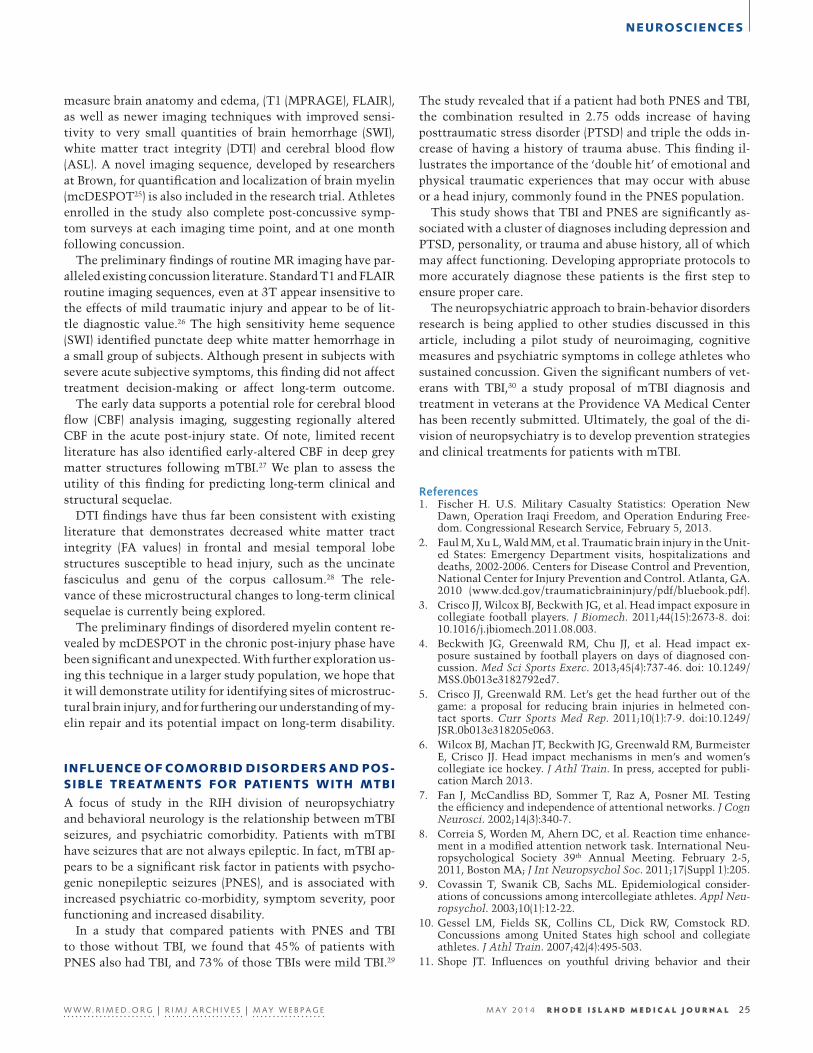
Contact sports: Helmets with special sensors allowed
researchers to gather data on the strength, number, and
direction of blows to the head in contact sports like foot-
ball. Data from the sensors shows that running backs and
quarterbacks suffer the hardest hits to the head, while
linemen and linebackers are hit on the head most often.

NEUROSCIENCES
allows researchers to record the frequency and severity of head impact sustained by helmeted athletes during play. This provides information toward understanding the biomechani-cal basis of concussion and repeated subconcussive impacts.
In our approach to understanding the biomechanics of concussions, we have used data collected by the HIT System to quantify head impact exposure, a multifactorial term that includes the frequency, magnitude, and location of head im-pacts. Our objective is to quantitatively measure head im-pact exposure in contact-sport athletes, in relation to their head impact mechanism. These data are then correlated with clinical outcome.
Previously, we have quantified and reported head impact exposure based on player position in collegiate football play-ers.3 In a subsequent study that evaluated impact associated with clinical concussion, we identified a relationship be-tween type of head impact exposure and incidence of con-cussion.4 We are now applying this analysis to men’s and women’s collegiate ice hockey to determine the addition-al role that gender may play in the athlete’s biomechanical tolerance to concussion.
We propose that reducing an athlete’s head impact expo-sure is a practical approach for reducing their risk for brain injury.5 In order to investigate strategies for reducing head impact exposure, we developed a tool that synchronized HIT data with game video footage to associate the biomechanics of head impact with specific impact mechanisms (e.g., head contact with the ice in hockey). Using this technique, we have identified the circumstances of play that result in the most frequent or high-magnitude head impact.6 By quantify-ing the biomechanics of concussion we have accomplished the first step in understanding concussion injury with practical application to furthering our exploration of early detection and prevention.
CLINICAL SEVERITY ASSESSMENT
Attention Network Task for Acute Concussion Modified The development of an easily administered, reliable, and valid measure of mTBI-related attention dysfunction was motivated by a need to understand when U.S. military vet-erans of Iraq and Afghanistan could be safely redeployed to combat after having sustained mild traumatic brain injury (mTBI). One such test would also have clear utility and ap-plication to the sports arena where augmentation of current return-to-play guidelines would have expected benefit for preventing severe or chronic brain injury.
In order to achieve this goal, investigators at Brown and the Providence VA Medical Center have modified the computer-administered Attention Network Task (ANT),7 a well-established visual flanker task, to serve as a screen-ing measure for changes in attention during the acute and near-term post-acute period following mTBI. The modified ANT (mANT) includes distracting sounds (e.g., beeps, buzz-es) paired with visual stimuli. The sounds are intended to
magnify mTBI-mediated attention dysfunction in military, sports, or other highly stimulating situations.
The ANT is a computer task designed to evaluate alert-ing, orienting, and executive aspects of attention. It requires the participant to rapidly determine the left-right direction of a central arrow surrounded by congruent or incongruent flankers (e.g., ß ß ß ß ß or ß ß à ß ß).The arrows are preceded either by no cue, an alerting cue or an orienting cue (indicating where the arrows will appear). Prior studies show reduced reaction times (RT) for congruent compared to incongruent arrows, alerting cue compared to no cue, and orienting cue compared to alerting cue.5
Using mANT, an initial validation study in 20 healthy young adults tested the hypothesis that RT would be lon-ger and accuracy poorer for sound vs. no sound conditions.8 However, results showed faster RT for sound compared to no-sound conditions with no differences in accuracy. This unexpected result could be due to additional alerting from the second sensory channel (auditory) since the sounds were designed to occur slightly (400 ms) before the visual presentation of the arrows.
This result raised the question of whether the RT enhancement effect of positive sound conditions would be attenuated following mTBI, and thereby serve as a poten-tially rapid, easily applied measure of post-concussion dis-ability. Early application of this technique to Brown football players diagnosed with sports-related mTBI where mANT was performed within 72 hours of injury showed that the RT advantage for sound compared to no-sound conditions was significantly smaller for the mTBI group compared to the control group. There were no significant group differences in accuracy. These results provide limited initial support for the mANT as a sensitive measure of acute mTBI.
EFFECT OF CONCUSSION ON THE YOUNG DRIVER
Participation in high school and collegiate sports is on the rise, with more than 7 million high school students partici-pating in 2005–2006 and almost 385,000 collegiate students participating in 2004–2005.9 Concussions represented 11.6% of all high school athletic injuries and 5.8% of all collegiate athletic injuries.10 Concurrently, novice drivers have the highest crash rate per miles driven of any age group, and it is not until age 25 that the rate starts to approach the rate seen throughout most of adulthood.11
The combination of inexperience and developmental and structural risk factors contribute to the statistic that motor vehicle collisions are a leading cause of death in this age group in both boys and girls.11 Young drivers exhibit dimin-ished ability to judge risk and inhibit impulses, have in-creased distractibility, and an increased propensity towards risky behavior.11 During the post-concussive phase, there is evidence of a reduction in visual memory, reaction time, impulse control composite score and processing speed.12 All of these brain functions are used during the act of driving.13
R H O D E I S L A N D M E D I C A L J O U R N A L 23W W W. R I M E D . O R G | R I M J A R C H I V E S | M A Y W E B P A G E M A Y 2 0 1 4

NEUROSCIENCES
It was purported that during the acute post-concussive period, these alterations will translate to deficits in driving ability. In an Australian study, concussed adult drivers demon-strated impaired hazard perception when compared to non- concussed aged matched controls.14,15
We conducted a pilot research study that enrolled male and female collegiate hockey players from Brown Univer-sity. The athletes underwent pre-season ImPACT testing, Trail-Maker B (TM-B) and Driving Simulator testing. TM-B is an assessment tool that provides information about visual search speed, scanning, speed of processing, mental flexibil-ity, as well as executive functioning. It is also sensitive to detecting several cognitive impairments. Following concus-sion, the athletes repeated both the Driving Simulator and TM-B within 48 hours and then serially until their symp-tom score and ImPACT tests normalized. Early results of comparison of preseason testing with concussed testing identified deficits in both the Trail-Maker B and the Driving Simulator sections.
Our intention is to expand our study population to include both high school and additional college athletes in order to better inform driving recommendations for our young drivers who have sustained mild traumatic brain injury.
ASSESSMENT OF MORBIDITY AND PROGNOSIS
Inflammatory Biomarkers for Mild Traumatic Brain InjuryMild TBI or concussion, which represents the majority of TBI cases, is increasingly being recognized in adolescents,16
and the associated morbidity can be significant in this age group. A substantial subset of these children has delayed recovery, resulting in the loss of productivity and psycho-social distress.17 Compared to adults, adolescents are more susceptible to repetitive injuries and post-concussion syn-drome (PCS). Currently, there is no established method for predicting the recovery period and determining the optimal treatment for individual mTBI patients. Certain patient characteristics or symptoms observed at admission appear to be predictive of PCS.18 However, more objective measures would improve clinicians’ ability to provide prognostic information and potentially guide therapy.
There has been a considerable interest in identifying serum biomarkers that would allow for diagnosis and prognosis in neurotrauma. However, defining such biomarkers for mTBI has been particularly challenging.19 Among the potential biomarkers S100B, a predominantly astrocyte-derived pro-tein, has been extensively studied but demonstrates low sensitivity and specificity as a biomarker for mTBI. Neu-ronal proteins, such as neuron-specific enolase, ubiquitin C-terminal hydrolase-L1, cleaved tau protein, and αII-spec-trin breakdown product of 145 kDa, were also found to have significant limitations as serum biomarkers in mTBI. It has been generally assumed that the levels of serum biomark-ers should reflect the magnitude of damage of neural tissue caused by injury. However, the extent of damage of neural
tissue in mTBI is likely to be quite limited, which may ex-plain the low sensitivity of serum biomarkers that have been studied. We are pursuing an alternative approach to identify proteins that are produced in the brain but whose serum lev-els would reflect functional changes in brain parenchymal cells rather than the cellular damage resulting from injury.
Our studies involve both pediatric (adolescent) and adult populations of mTBI patients, and include control groups of patients with long-bone fractures and healthy volunteers. These investigations focus on inflammatory biomarkers. Although the pathophysiological processes accompanying mTBI/concussion are not fully understood, studies in ani-mal models of mTBI suggest that neuroinflammation plays a significant pathophysiological role in mTBI.20 In this con-text, it is also important to note that the immature brain likely exhibits a much stronger inflammatory response to injury than the adult central nervous system.21 There is ample evidence of adverse effects of neuroinflammation on various aspects of brain function, which are highly relevant to mTBI and PCS. Neuroinflammation has a detrimental ef-fect on neurogenesis, learning and memory, and appears to play a part in the pathophysiology of depressive disorders. Our preliminary observations stress the importance of how the collected blood samples are processed. While the serum levels of circulating proteins are commonly assessed, the co-agulation process involved in harvesting serum may liberate some proinflammatory mediators that are carried by circu-lating leukocytes or bound to Duffy antigen receptors ex-pressed on erythrocytes.22,23 For example, we have found that the serum levels of CXCL1, a neutrophil chemoattractant, are considerably higher than those measured in plasma. This indicates that some published data should be evaluated with caution. Our studies have nearly completed enrollment. If our hypothesis is correct, the results may have prognostic potential for mTBI, may assist clinicians in tailoring recom-mendations to patients, and may provide the mechanistic basis for new therapeutic approaches in mTBI patients.
NOVEL MRI FINDINGS IN COLLEGIATE ATHLETES WITH MILD TRAUMATIC BRAIN INJURY
A team of clinical neuroscientists, radiologists, statisticians and brain imagers at RIH and Brown University is investi-gating the utility of routine, advanced and novel MR im-aging sequences in collegiate athletes for identifying early features that define subjects at risk for long-term sequelae of mTBI. In addition, we are studying subacute functional and microstructural brain MR changes that may correlate with the quality and severity of long-term post-mTBI disability.
To investigate this question we have designed a research trial that enrolls scholastic athletes with sports-related con-cussion (Glasgow Coma Scale24 of 13-15). 3T-MR imaging is performed at Brown University or RIH within 72 hours of concussion and then repeated three months later. The MR imaging protocol includes standard clinical sequences that
R H O D E I S L A N D M E D I C A L J O U R N A L 24W W W. R I M E D . O R G | R I M J A R C H I V E S | M A Y W E B P A G E M A Y 2 0 1 4
NEUROSCIENCES
measure brain anatomy and edema, (T1 (MPRAGE), FLAIR), as well as newer imaging techniques with improved sensi-tivity to very small quantities of brain hemorrhage (SWI), white matter tract integrity (DTI) and cerebral blood flow (ASL). A novel imaging sequence, developed by researchers at Brown, for quantification and localization of brain myelin (mcDESPOT25) is also included in the research trial. Athletes enrolled in the study also complete post-concussive symp-tom surveys at each imaging time point, and at one month following concussion.
The preliminary findings of routine MR imaging have par-alleled existing concussion literature. Standard T1 and FLAIR routine imaging sequences, even at 3T appear insensitive to the effects of mild traumatic injury and appear to be of lit-tle diagnostic value.26 The high sensitivity heme sequence (SWI) identified punctate deep white matter hemorrhage in a small group of subjects. Although present in subjects with severe acute subjective symptoms, this finding did not affect treatment decision-making or affect long-term outcome.
The early data supports a potential role for cerebral blood flow (CBF) analysis imaging, suggesting regionally altered CBF in the acute post-injury state. Of note, limited recent literature has also identified early-altered CBF in deep grey matter structures following mTBI.27 We plan to assess the utility of this finding for predicting long-term clinical and structural sequelae.
DTI findings have thus far been consistent with existing literature that demonstrates decreased white matter tract integrity (FA values) in frontal and mesial temporal lobe structures susceptible to head injury, such as the uncinate fasciculus and genu of the corpus callosum.28 The rele-vance of these microstructural changes to long-term clinical sequelae is currently being explored.
The preliminary findings of disordered myelin content re-vealed by mcDESPOT in the chronic post-injury phase have been significant and unexpected. With further exploration us-ing this technique in a larger study population, we hope that it will demonstrate utility for identifying sites of microstruc-tural brain injury, and for furthering our understanding of my-elin repair and its potential impact on long-term disability.
INFLUENCE OF COMORBID DISORDERS AND POS-SIBLE TREATMENTS FOR PATIENTS WITH MTBI
A focus of study in the RIH division of neuropsychiatry and behavioral neurology is the relationship between mTBI seizures, and psychiatric comorbidity. Patients with mTBI have seizures that are not always epileptic. In fact, mTBI ap-pears to be a significant risk factor in patients with psycho-genic nonepileptic seizures (PNES), and is associated with increased psychiatric co-morbidity, symptom severity, poor functioning and increased disability.
In a study that compared patients with PNES and TBI to those without TBI, we found that 45% of patients with PNES also had TBI, and 73% of those TBIs were mild TBI.29
The study revealed that if a patient had both PNES and TBI, the combination resulted in 2.75 odds increase of having posttraumatic stress disorder (PTSD) and triple the odds in-crease of having a history of trauma abuse. This finding il-lustrates the importance of the ‘double hit’ of emotional and physical traumatic experiences that may occur with abuse or a head injury, commonly found in the PNES population.
This study shows that TBI and PNES are significantly as-sociated with a cluster of diagnoses including depression and PTSD, personality, or trauma and abuse history, all of which may affect functioning. Developing appropriate protocols to more accurately diagnose these patients is the first step to ensure proper care.
The neuropsychiatric approach to brain-behavior disorders research is being applied to other studies discussed in this article, including a pilot study of neuroimaging, cognitive measures and psychiatric symptoms in college athletes who sustained concussion. Given the significant numbers of vet-erans with TBI,30 a study proposal of mTBI diagnosis and treatment in veterans at the Providence VA Medical Center has been recently submitted. Ultimately, the goal of the di-vision of neuropsychiatry is to develop prevention strategies and clinical treatments for patients with mTBI.
References 1. Fischer H. U.S. Military Casualty Statistics: Operation New
Dawn, Operation Iraqi Freedom, and Operation Enduring Free-dom. Congressional Research Service, February 5, 2013.
2. Faul M, Xu L, Wald MM, et al. Traumatic brain injury in the Unit-ed States: Emergency Department visits, hospitalizations and deaths, 2002-2006. Centers for Disease Control and Prevention, National Center for Injury Prevention and Control. Atlanta, GA. 2010 (www.dcd.gov/traumaticbraininjury/pdf/bluebook.pdf).
3. Crisco JJ, Wilcox BJ, Beckwith JG, et al. Head impact exposure in collegiate football players. J Biomech. 2011;44(15):2673-8. doi: 10.1016/j.jbiomech.2011.08.003.
4. Beckwith JG, Greenwald RM, Chu JJ, et al. Head impact ex-posure sustained by football players on days of diagnosed con-cussion. Med Sci Sports Exerc. 2013;45(4):737-46. doi: 10.1249/MSS.0b013e3182792ed7.
5. Crisco JJ, Greenwald RM. Let’s get the head further out of the game: a proposal for reducing brain injuries in helmeted con-tact sports. Curr Sports Med Rep. 2011;10(1):7-9. doi:10.1249/JSR.0b013e318205e063.
6. Wilcox BJ, Machan JT, Beckwith JG, Greenwald RM, Burmeister E, Crisco JJ. Head impact mechanisms in men’s and women’s collegiate ice hockey. J Athl Train. In press, accepted for publi-cation March 2013.
7. Fan J, McCandliss BD, Sommer T, Raz A, Posner MI. Testing the efficiency and independence of attentional networks. J Cogn Neurosci. 2002;14(3):340-7.
8. Correia S, Worden M, Ahern DC, et al. Reaction time enhance-ment in a modified attention network task. International Neu-ropsychological Society 39th Annual Meeting. February 2-5, 2011, Boston MA; J Int Neuropsychol Soc. 2011;17(Suppl 1):205.
9. Covassin T, Swanik CB, Sachs ML. Epidemiological consider-ations of concussions among intercollegiate athletes. Appl Neu-ropsychol. 2003;10(1):12-22.
10. Gessel LM, Fields SK, Collins CL, Dick RW, Comstock RD. Concussions among United States high school and collegiate athletes. J Athl Train. 2007;42(4):495-503.
11. Shope JT. Influences on youthful driving behavior and their
R H O D E I S L A N D M E D I C A L J O U R N A L 25W W W. R I M E D . O R G | R I M J A R C H I V E S | M A Y W E B P A G E M A Y 2 0 1 4

NEUROSCIENCES
potential for guiding interventions to reduce crashes. Inj Prev. 2006;12(Suppl 1):i9-14.
12. Schatz P, Pardini JE, Lovell MR, Collins MW, Podell K. Sensitiv-ity and specificity of the ImPACT Test Battery for concussion in athletes. Arch Clin Neuropsychol. 2006;21(1):91-9.
13. Hildreth EC, Beusmans JM, Boer ER, Royden CS. From vision to action: experiments and models of steering control during driv-ing. J Exp Psychol Hum Percept Perform. 2000;26(3):1106-32.
14. Preece MH, Horswill MS, Geffen GM. Driving after concussion: the acute effect of mild traumatic brain injury on drivers’ hazard perception. Neuropsychology. 2010;24(4):493-503. doi: 10.1037/a0018903.
15. Preece MH, Horswill MS, Geffen GM. Assessment of drivers’ ability to anticipate traffic hazard after traumatic brain injury. J Neurol Neurosurg Psychiatry. 2011;82(4):447-51. doi: 10.1136/jnnp.2010.215228.
16. Bazarian JJ, McClung J, Shah MN, Cheng YT, Flesher W, Kraus J. Mild traumatic brain injury in the United States, 1998-2000. Brain Inj. 2005;19(2):85-91.
17. Barlow KM, Crawford S, Stevenson A, Sandhu SS, Belanger F, Dewey D. Epidemiology of postconcussion syndrome in pediat-ric mild traumatic brain injury. Pediatrics. 2010;126(2):e374-81.
18. de Kruijk JR, Leffers P, Menheere PP, Meerhoff S, Rutten J, Twi-jnstra A. Prediction of post-traumatic complaints after mild traumatic brain injury: early symptoms and biochemical mark-ers. J Neurol Neurosurg Psychiatry. 2002;73(6):727-32.
19. Begaz T, Kyriacou DN, Segal J, Bazarian JJ. Serum biochemical markers for post-concussion syndrome in patients with mild traumatic brain injury. J Neurotrauma. 2006;23(8):1201-10.
20. Perez-Polo JR, Rea HC, Johnson KM, et al. Inflammatory con-sequences in a rodent model of mild traumatic brain injury. J Neurotrauma. 2013;30(9):727-40. doi: 10.1089/neu.2012.2650.
21. Bolton SJ, Perry VH. Differential blood-brain barrier breakdown and leucocyte recruitment following excitotoxic lesions in juve-nile and adult rats. Exp Neurol. 1998;154(1):231-40.
22. Scapini P, Lapinet-Vera JA, Gasperini S, Calzetti F, Bazzoni F, Cassatella MA. The neutrophil as a cellular source of chemok-ines. Immunol Rev. 2000;177:195-203.
23. Rot A, Horuk R. The Duffy antigen receptor for chemokines. Methods Enzymol. 2009;461:191-206.
24. Teasdale G, Jennett B. Assessment of coma and impaired con-sciousness. A practical scale. Lancet. 1974;2(7872):81-4.
25. Deoni SC, Mercure E, Blasi A, et al. Mapping infant brain myelination with magnetic resonance imaging. J Neurosci. 2011;31(2):784-91. doi: 10.1523/J Neuro Sci. 2106-10.2011.
26. Belanger HG, Vanderploeg R, Curtiss G, Warden DL. Recent neuroimaging techniques in mild traumatic brain injury. J Neu-ropsychiatry Clin Neurosci. 2007;19(1):5-20.
27. Grossman EJ, Jensen JH, Babb JS, et al. Cognitive impairment in mild traumatic brain injury: A longitudinal diffusional kurtosis and perfusion imaging study. Am J Neuroradiol. 2013;34(5):951-7,S1-3. doi: 10.3174/ajnr.A3358.
28. Shenton ME, Hamoda HM, Schneiderman JS, et al. A review of magnetic resonance imaging and diffusion tensor imaging findings in mild traumatic brain injury. Brain Imaging Behav. 2012;6(2):137-92. doi: 10.1007/s11682-012-9156-5.
29. LaFrance WC Jr., Deluca M, Machan JT, Fava JL. Traumatic brain injury and psychogenic nonepileptic seizures yield worse outcomes. Epilepsia.2013;54(4):718-25. doi: 10.1111/epi.12053.
30. Mernoff ST, Correia S. Military blast injury in Iraq and Afghan-istan: the Veterans Health Administration’s polytrauma system of care. Med Health RI. 2010;93(1):16-18,21.
Acknowledgements * Participants in the Brown TBIRC include: Jeffrey Rogg (Co-Chair), W. Curt LaFrance, Jr., (Co-Chair), John Robson, J. J. “Trey” Crisco, Bethany Wilcox, Steve Correia, Michael Worden, Neha Raukar, Adam Chodobski, Joanna Szmydynger-Chodobska, Anna
Ellermeier, Heather Spader, Elizabeth Morrell, Wendy Smith, Jason T. Machan, Grayson L. Baird, Glenn Tung, Stephen Mernoff, Peter Quesenberry, Michael Hulstyn, Paul Fadale, Russell Fiore, Sean Deoni, William Heindel, Sandra Mather, Philip Lieberman.
Novel MRI Findings study has been supported by grants from Brown Department of Diagnostic Imaging and the Brown Institute for Brain Science.
Inflammatory Biomarker Research efforts are supported by funds from the Department of Emergency Medicine at the Alpert Medi-cal School of Brown University and from Lifespan.
Driving while Concussed is supported by funds from the Depart-ment of Emergency Medicine at the Alpert Medical School of Brown University.
AuthorsJeffrey Rogg, MD, is Associate Professor, Department of Diagnostic
Imaging at the Warrren Alpert Medical School of Brown University where he is the Director of Neuroradiology.
Heather Spader, MD, is Chief Resident in the Department of Neurosurgery at the Warren Alpert Medical School of Brown University.
Bethany J. Wilcox, BS, is a PhD candidate working in the Bioengineering Lab directed by Joseph J. Crisco, PhD, Professor, Department of Orthopaedics at The Warren Alpert Medical of Brown University.
Anna Ellermeier, MD, is a Third-year Resident in the Department of Diagnostic Imaging at the Warren Alpert Medical School of Brown University.
Steven Correira, PhD, is Assistant Professor, Department of Psychiatry and Human Behavior, based at the Providence VA Medical Center.
Adam Chodobski, PhD, is Associate Professor, Department of Emergency Medicine, Warren Alpert Medical School of Brown University, where he is Director of Neurotrauma and Brain Barriers Research.
Joanna Szmydynger-Chodobska, PhD, is Assistant Professor of Emergency Medicine (Research), Emergency Medicine.
Neha Raukar, MD, is Assistant Professor, Department of Emergency Medicine, Warren Alpert Medical School of Brown University, where she is Director of the Division of Sports Medicine.
Jason T. Machan, PhD, is Assistant Professor in the Department of Orthopaedics (Research) at the Warren Alpert Medical School of Brown University, where he is in charge of the Rhode Island Hospital Biostatistics Core.
Joseph J. Crisco, PhD, is the Henry F. Lippitt Professor of Orthopedics in the Department of Orthopaedics at The Alpert Medical School of Brown University.
W. Curt LaFrance, Jr., MD, MPH, is Assistant Professor, Departments of Psychiatry and Neurology (Research) at the Warren Alpert Medical School of Brown University, Director of the Rhode Island Hospital Division of Neuropsychiatry and Behavioral Neurology and Staff Physician at the Providence VA Medical Center
DisclosuresJoseph J. Crisco and Simbex have a financial interest in the instruments (HIT System, Sideline Response System (Riddell, Inc)) that were used to collect the biomechanical data.The other authors have no financial interests to disclose.
CorrespondenceJeffrey Rogg, MDDepartment of Diagnostic Imaging, Rhode Island Hospital593 Eddy Street, Providence, RI 02903401-444-5184 [email protected]
R H O D E I S L A N D M E D I C A L J O U R N A L 26W W W. R I M E D . O R G | R I M J A R C H I V E S | M A Y W E B P A G E M A Y 2 0 1 4
NEUROSCIENCES
27
30
EN
Advances in Stroke Over the Past DecadeBRIAN SILVER, MD
ABSTRACT Over the last decade, a number of advances in the care of stroke and TIA patients have been made. These advances include prevention, acute management, and recovery. Some of this work has occurred in Rhode Island. This review will focus on the revised definition of stroke and TIA; short-term risk of TIA; rapid management of TIA; targeted use of medication and lifestyle changes; moni-toring for atrial fibrillation; novel anticoagulants for atri-al fibrillation; a better understanding of the limitations of intra-arterial therapy for acute ischemic stroke; clinical treatment trials for intracerebral hemorrhage; and the use of robotic, magnetic, and chemical interventions to improve function after stroke.
KEYWORDS: Stroke, TIA, risk factors, acute intervention, recovery
INTRODUCTION
In this brief review, some of the advances in stroke over the past decade will be reviewed. Of note, many of these advances occurred as a result of work done here in Rhode Island.
DEFINITION OF STROKE AND TIAThe definitions of stroke and transient ischemic attack (TIA) have evolved over the last decade. The term TIA was first used in the 1960s to designate a presumed ischemic neu-rologic event from which a complete recovery occurred in under 24 hours. In 2002, a panel of experts proposed a new, tissue-based, definition of TIA: “a brief episode of neurolog-ical dysfunction caused by focal brain or retinal ischemia, with clinical symptoms typically lasting less than one hour, and without evidence of acute infarction.” In 2009, the American Stroke Association proposed a modification of that definition which eliminated time altogether and also included spinal cord ischemia as follows: “a transient episode of neurological dysfunction caused by focal brain, spinal cord, or retinal ischemia, without acute infarction (on neuroimaging).”1
The basis for the change in these definitions comes from research over the last decade. Among 19 studies of 1,117 patients with the time-based definition of TIA, the rate
of positive findings on diffusion- weighted imaging (DWI) was 39%.1 DWI is an MRI sequence sensitive to the diffusion of water molecules. During acute ischemic stroke, there is a restriction of the nor-mal Brownian movement of water which manifests as brightness on DWI. The longer the event, the more likely DWI will be positive. Nevertheless, short-lasting events
can also result in a positive DWI. As imaging technology evolves, smaller areas of suspected tissue damage will also become apparent, further increasing the percentage of pa-tients who are reclassified as having had a stroke. Indeed, it is possible that all such events cause some tissue damage and would be obvious if we had the capability of performing non-invasive microscopic imaging.
SHORT-TERM RISK AFTER TIA AND ITS MODIFICATION
Though it was well known that stroke carried a substantial risk of recurrence, it was not until a study in 2000 that the high short-term risk of TIA became apparent.2 In that study, which used the 24-hour definition of TIA, approximately 10% of patients returned with stroke within 90 days, half within the first 48 hours. Patients in that study did not have
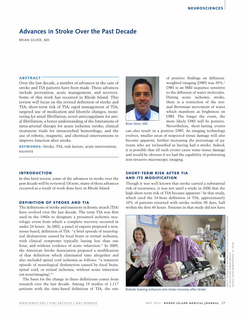
Robotic training enhances arm motor recovery after stroke.
PH
OT
O C
OU
RT
ES
Y O
F N
AT
ION
AL
IN
ST
ITU
TE
S O
F H
EA
LT
H/
DE
PA
RT
ME
NT
OF
HE
AL
TH
AN
D H
UM
AN
SE
RV
ICE
S
SO
UR
CE
: D
AV
ID R
EIN
KE
NS
ME
YE
R,
UC
IR
VIN
E
R H O D E I S L A N D M E D I C A L J O U R N A L 27W W W. R I M E D . O R G | R I M J A R C H I V E S | M A Y W E B P A G E M A Y 2 0 1 4
Brian Silver, MD

NEUROSCIENCES
urgent evaluation or treatment. A risk-stratification model called ABCD2 was then developed which allocated points for presenting variables (age, blood pressure at presentation, clinical symptoms, duration, diabetes).3 The range of scores is 0-7 with higher scores being associated with greater risk.
The finding of high risk of stroke following TIA led to clinical studies evaluating urgent intervention. The first study, called SOS-TIA, and conducted in France, evaluated an urgent management program including rapid carotid im-aging, rhythm monitoring, carotid revascularization when appropriate, anticoagulation when appropriate, and lipid management.4 Compared with the expected rate of stroke (based on ABCD2 risk stratification), the authors found an approximate 80% reduction in risk. Simultaneously, the EX-PRESS study, conducted in England, using a before-and-after design, found a similar 80% reduction in stroke recurrence using a rapid evaluation and treatment program.5 A 2012 study from Australia demonstrated a 1.5% risk of stroke at 90 days in patients with TIA who had all investigations and management conducted in the emergency department.6 The expected rate, based on the ABCD2 scheme, was 10%.
On the basis of these findings, Rhode Island Hospital de-veloped a TIA unit in the emergency department in March 2013. The rate of stroke at 7 days, based on telephone contact, has been less than 1% to date.
LONG-TERM RISK MODIFICATION
INTERSTROKE was a landmark case-control study which matched 3,000 stroke patients with 3,000 controls in 22 countries.7 The authors found that 10 risk factors were associated with 90% of all stroke [hypertension, current smoking, increased waist-to-hip ratio, poor diet, physical in-activity, diabetes mellitus, excessive alcohol intake, psycho-social stress and depression, cardiac causes and abnormal ratio of apolipoproteins B to A1. The authors concluded that interventions that targeted these factors could substantially reduce the burden of stroke. The newly formed School of Public Health at Brown University will focus on initiatives at improving risk factors that lead to cardiovascular disease. A recent study by Wing and colleagues at Brown University found that the addition of intensive lifestyle changes (diet and exercise) did not reduce the rates of death, stroke, or myocardial infarction compared with medication use alone.8 However, those assigned to intensive lifestyle changes used less medication.
Additional advances in the last decade include the ob-servation that longer heart rhythm monitoring leads to an increased detection of atrial fibrillation. In a Canadian study which randomly assigned patients to Holter monitor-ing versus 30 day monitoring in patients with cryptogenic stroke, detection rates of atrial fibrillation were 3% and 16%, respectively.9 What is unclear is what duration of atrial fibrillation on these monitors confers increased risk. For ex-ample, does a 20-second episode of atrial fibrillation during
30 days of monitoring suggest increased risk requiring anticoagulation? The standard definition of paroxysmal atrial fibrillation is at least 30 continuous seconds of the abnormal rhythm. Further study will be required to deter-mine prognosis and optimal medical treatment. There are now many options for anticoagulation for patients with atri-al fibrillation including vitamin K antagonists (warfarin), direct thrombin inhibitors (dabigatran), and factor Xa inhib-itors (apixaban, rivaroxaban).10 These agents can be expected to reduce the risk of embolism by approximately 60%-70% relative to no treatment and approximately 40%-50% rela-tive to aspirin. Individualized determination of risk can be accomplished with the CHADS2 and CHA2DS2Vasc scoring systems.11 Specific risk assessment for neurovascular pro-cesses may be helpful in shared decision-making processes, with careful attention to presentation format.
At this time, there does not appear to be a role for anti-coagulation in intracranial atherosclerosis, cervical arterial dissection, or patent foramen ovale (PFO)-related stroke. Recurrence risk of stroke is highest with intracranial ath-erosclerosis (approximately 12% per year) and much lower with PFO-related stroke (approximately 1%–2% per year), and cervical arterial dissection (3% in the 12 months after ictus). In addition, interventional approaches do not appear to mitigate risk in these conditions, and even if subgroup analyses suggest benefit, the absolute reduction is very small (less than 1%) with an increased risk of procedure- related complications.
ACUTE INTERVENTIONS
Despite the wide use of intra-arterial procedures for the treat-ment of acute stroke, definitive evidence of overall benefit is lacking at this time. Three trials failed to show net benefit for intra-arterial therapy added to intravenous thrombolytic therapy within 3 hours (IMS III), intra-arterial compared to intravenous therapy within 4.5 hours (SYNTHESIS), and imaging-guided intra-arterial therapy compared to placebo within 8 hours (MR RESCUE).12 Post-hoc analyses suggest that there are subgroups which may benefit. The most im-portant variable is time to treatment. There is a strong cor-relation between time to intra-arterial recanalization and outcome.13 Further, the completeness of recanalization at earlier time points is also important.14 Because approximate-ly 2 million neurons, 14 billion synapses, and 7.5 miles of myelinated fibers are lost every second during a large ves-sel ischemic stroke,15 recanalization at late time points may only serve to perfuse already destroyed tissue, analogous to putting out a fire after a house has already burned down.
Intracerebral hemorrhage carries a worse prognosis than ischemic stroke yet an acute treatment which improves out-come remains elusive. Potential promising interventions in-clude rapid control of blood pressure and targeted removal of clot. The INTERACT2 study failed to show a statistically significant benefit in favor of rapid blood pressure reduction
R H O D E I S L A N D M E D I C A L J O U R N A L 28W W W. R I M E D . O R G | R I M J A R C H I V E S | M A Y W E B P A G E M A Y 2 0 1 4

NEUROSCIENCES
below a target of 140 mmHg systolic within the first 6 hours of bleeding but sample size may have precluded detection of benefit.16 The ongoing ATACH-II study,17 which is also evaluating rapid reduction in blood pressure to less than 140 mmHg systolic should yield a definitive answer on the ques-tion of blood-pressure reduction, particularly when data are pooled with INTERACT2.
Another intriguing option for the treatment of intracere-bral hemorrhage is thrombolysis-assisted clot evacuation. The procedure consists of creation of a burr hole ipsilateral to the bleeding, insertion of a catheter into the center of the clot, injection of tPA into the center of the clot, and then evacuation of the dissolved material. Moreover, the proce-dure can be performed as late as 24 hours. MISTIE II was a small study which suggested benefit of this procedure with good recovery at one year, reduced length-of-stay in the hos-pital, and total cost of care.18 Mortality, however, was not decreased. MISTIE III,19 the phase III version of the study, will provide a definitive answer on whether this procedure is truly of value in the case of patients with intracerebral hemorrhage.
RECOVERY OPTIONS
Recovery after stroke is an exciting area of research oppor-tunity. The notion that the nervous system was incapable of regeneration was dispelled in the 1990s. Since that time, a number of potential interventions to augment recovery after stroke have been posited. These include robotic, electromag-netic, and pharmacological therapies. Cellular therapy re-mains an active area of interest but logistical and regulatory issues in the United States have not led to a trial in stroke at this time. In addition, the concept that electrical energy from the brain can be converted to kinetic action through an external device has now become a reality.
Lo and colleagues, from the Providence VA, published a randomized trial of robot-assisted therapy for upper-limb im-pairment in stroke in 2010.20 It was the first such study ever published in the New England Journal of Medicine. Though the study did not find that robot-assisted therapy was superi-or to intensive or usual care at 12 weeks, there was a sugges-tion of benefit over usual care at 36 weeks. Since then, the research group at the VA has continued to explore different robot options for the purpose of augmenting limb recovery.
Hochberg, Donoghue and colleagues from the Brown Institute of Brain Sciences made headlines worldwide with the publication of an article in Nature regarding the implan-tation of a 96-channel microelectrode array that allowed two patients with long-standing tetraplegia to control an exter-nal robot arm.21 In one patient, the arm was used to lift a bot-tle of coffee to her mouth. Remarkably, the complex robotic arm movements could be controlled by a very small pool of neurons. This groundbreaking research paves the way for next-generation devices that can be controlled through implanted chips.
Multiple studies now suggest that transcranial magnetic stimulation (TMS) may be used to augment both motor-ic and linguistic recovery after stroke. Of note, excitatory stimulation of the affected hemisphere appears to produce benefit while inhibitory stimulation of the unaffected hemi-sphere may produce benefit.22 A transcranial magnetic stim-ulation device now exists at Butler Hospital and will allow additional study in this area.
Pharmacological therapy, such as fluoxetine and PDE5 inhibitors, are also of potential benefit. The FLAME study suggested that fluoxetine not only improved depression but also improved motor function after stroke.23 Sildenafil has been shown in young and aged animals to improve neurolog-ical outcome though neurogenesis, angiogenesis, and synap-togenesis.24,25 Preliminary human studies26 have served as the basis for larger pilot randomized trials.
SUMMARY
Evolving understanding of the concept of cerebral ischemia and recurrent risk has led to improved treatments and short-term outcomes for patients. Large international studies of recurrent stroke have now helped focus the agenda for what needs to be done to lower long-term risk. Beneficial acute treatments of both ischemic and hemorrhagic stroke continue to be defined and many exciting options are now available. Strategies for improving recovery after disabling stroke are now entering an active phase of development. Rhode Island, the Brown Institute for Brain Sciences, and the Norman Prince Neurosciences Institute have the tools and expertise to be leaders in these areas.
References1. Easton JD, Saver JL, Albers GW, et al. Definition and evalua-
tion of transient ischemic attack: a scientific statement for healthcare professionals from the American Heart Association/American Stroke Association Stroke Council; Council on Car-diovascular Surgery and Anesthesia; Council on Cardiovascular Radiology and Intervention; Council on Cardiovascular Nurs-ing; and the Interdisciplinary Council on Peripheral Vascular Disease. The American Academy of Neurology affirms the value of this statement as an educational tool for neurologists. Stroke. 2009;40:2276-2293.
2. Johnston SC, Gress DR, Browner WS, Sidney S. Short-term prognosis after emergency department diagnosis of TIA. JAMA. 2000;284:2901-2906.
3. Johnston SC, Rothwell PM, Nguyen-Huynh MN, et al. Valida-tion and refinement of scores to predict very early stroke risk after transient ischaemic attack. Lancet. 2007;369:283-292.
4. Lavallee PC, Meseguer E, Abboud H, et al. A transient ischaemic attack clinic with round-the-clock access (SOS-TIA): feasibility and effects. Lancet Neurol. 2007;6:953-960.
5. Rothwell PM, Giles MF, Chandratheva A, et al. Effect of urgent treatment of transient ischaemic attack and minor stroke on early recurrent stroke (EXPRESS study): a prospective popula-tion-based sequential comparison. Lancet. 2007;370:1432-1442.
6. Sanders LM, Srikanth VK, Jolley DJ, et al. Monash transient ischemic attack triaging treatment: safety of a transient isch-emic attack mechanism-based outpatient model of care. Stroke. 2012;43:2936-2941.
R H O D E I S L A N D M E D I C A L J O U R N A L 29W W W. R I M E D . O R G | R I M J A R C H I V E S | M A Y W E B P A G E M A Y 2 0 1 4

NEUROSCIENCES
7. O’Donnell MJ, Xavier D, Liu L, et al. Risk factors for ischaemic and intracerebral haemorrhagic stroke in 22 countries (the INTER-STROKE study): a case-control study. Lancet. 2010;376:112-123.
8. Wing RR, et al. Cardiovascular Effects of Intensive Life-style Intervention in Type 2 Diabetes. N Engl J Med. 2013 Jul 11;369(2):145-54.
9. Gladstone DJ, Spring M, Dorian P, et al. Prolonged ambulato-ry cardiac monitoring improves the detection and treatment of atrial fibrillation in patients with cryptogenic stroke: Primary results from the EMBRACE multicenter randomized trial. Inter-national Stroke Conference, 2/7/2013.
10. Weitz JI, Eikelboom JW, Samama MM. New antithrombotic drugs: Antithrombotic Therapy and Prevention of Thrombosis, 9th ed: American College of Chest Physicians Evidence-Based Clinical Practice Guidelines. Chest. 2012;141:e120S-151S.
11. Lip GY, Nieuwlaat R, Pisters R, Lane DA, Crijns HJ. Refining clinical risk stratification for predicting stroke and thrombo-embolism in atrial fibrillation using a novel risk factor-based approach: the euro heart survey on atrial fibrillation. Chest. 2010;137:263-272.
12. Chimowitz MI. Endovascular treatment for acute ischemic stroke--still unproven. N Engl J Med 2013;368:952-955.
13. Khatri P, Abruzzo T, Yeatts SD, Nichols C, Broderick JP, Tom-sick TA. Good clinical outcome after ischemic stroke with successful revascularization is time-dependent. Neurology. 2009;73:1066-1072.
14. Jayaraman MV, Grossberg JA, Meisel KM, Shaikhouni A, Silver B. The clinical and radiographic importance of distinguishing partial from near-complete reperfusion following intra-arterial stroke therapy. Am J Neuroradiol. 2013;34:135-139.
15. Saver JL. Time is brain--quantified. Stroke. 2006;37:263-266.16. Anderson CS, Heeley E, Huang Y, et al. Rapid blood-pressure
lowering in patients with acute intracerebral hemorrhage. N Engl J Med. 2013;368:2355-2365.
17. ATACH-II [online]. Available at: http://clinicaltrials.gov/ct2/show/NCT01176565. Accessed June 30, 2013.
18. MISTIE II results [online]. Available at: http://my.american-heart.org/idc/groups/ahamah-public/@wcm/@sop/@scon/docu-ments/downloadable/ucm_449055.pdf. Accessed June 30, 2013.
19. MISTIE III [online]. Available at: http://clinicaltrials.gov/ct2/show/NCT01827046. Accessed June 30, 2013.
20. 20. Lo AC, Guarino PD, Richards LG, et al. Robot-assisted therapy for long-term upper-limb impairment after stroke. N Engl J Med. 2010;362:1772-1783.
21. Hochberg LR, Bacher D, Jarosiewicz B, et al. Reach and grasp by people with tetraplegia using a neurally controlled robotic arm. Nature. 2012;485:372-375.
22. Adeyemo BO, Simis M, Macea DD, Fregni F. Systematic review of parameters of stimulation, clinical trial design characteris-tics, and motor outcomes in non-invasive brain stimulation in stroke. Front Psychiatry. 2012;3:88.
23. Chollet F, Tardy J, Albucher JF, et al. Fluoxetine for motor recov-ery after acute ischaemic stroke (FLAME): a randomised place-bo-controlled trial. Lancet Neurol. 2011;10:123-130.
24. Zhang R, Wang Y, Zhang L, et al. Sildenafil (Viagra) induces neu-rogenesis and promotes functional recovery after stroke in rats. Stroke. 2002;33:2675-2680.
25. Zhang RL, Zhang Z, Zhang L, Wang Y, Zhang C, Chopp M. De-layed treatment with sildenafil enhances neurogenesis and im-proves functional recovery in aged rats after focal cerebral isch-emia. J Neurosci Res. 2006;83:1213-1219.
26. Silver B, McCarthy S, Lu M, et al. Sildenafil treatment of sub-acute ischemic stroke: a safety study at 25-mg daily for 2 weeks. J Stroke Cerebrovasc Dis. 2009;18:381-383.
AuthorBrian Silver, MD, is Director, Stroke Center, Rhode Island
Hospital; Associate Professor of Neurology, The Warren Alpert Medical School of Brown University.
DisclosuresThe author reports no conflicts of interest.
CorrespondenceBrian Silver, MDDepartment of Neurology110 Lockwood Street, Suite #324 Providence, RI 02903 401-444-8806 Fax [email protected]
R H O D E I S L A N D M E D I C A L J O U R N A L 30W W W. R I M E D . O R G | R I M J A R C H I V E S | M A Y W E B P A G E M A Y 2 0 1 4

NEUROSCIENCES
31
34
EN
The Rhode Island Consortium for Autism Research and Treatment (RI-CART): A New Statewide Autism CollaborativeALAN GERBER, MA; ERIC M. MORROW, MD, PhD; STEPHEN J. SHEINKOPF, PhD; THOMAS ANDERS, MD
ABSTRACT Autism is a neurodevelopmental disorder characterized by core deficits in social interaction, language and repet-itive behaviors. The need for services is rising sharply as the number of children identified with autism increases. The Rhode Island Consortium for Autism Research and Treatment (RI-CART) was founded in 2009 with the goal of increasing communication among autism researchers throughout the state and improving treatment for chil-dren with autism. RI-CART members have several ex-citing projects in progress, with its larger aim being the creation of a statewide research registry. A statewide reg-istry would benefit research in Rhode Island and allow for larger collaborations nationally.
KEYWORDS: RI-CART, autism, autism spectrum disorder, registry, Rhode Island
WHAT IS AUTISM?
Autism, usually referred to as an autism spectrum disorder (ASD), is a highly heterogeneous, neurodevelopmental dis-order with symptoms that typically appear by the second year of life.1 The main symptoms include core deficits in social interaction, communication and repetitive and inflex-ible behaviors.2 In order to receive a diagnosis of autism, the symptoms must be present by age three, and not be better explained through another developmental disorder. While the genetic underpinnings of autism are not completely un-derstood, it is clear from twin studies that autism is a highly heritable disorder.3 Recent advances in genetic research have been able to account for between 10%-20% of autism spectrum diagnoses.4
Current estimates indicate that 1 in 88 children have been diagnosed with an autism spectrum disorder, a sharp rise from the prior estimate of 1 in 110.5,6 Boys receive a diagno-sis of autism spectrum disorder at almost five times the rate of girls.6 Unfortunately, epidemiological work beyond the school years is almost nonexistent. In addition to the difficulties of raising a child with autism, the lifetime costs can be substantial for both families and communities as a whole.7,8 In order to meet these rising needs, the National Institutes of Health (NIH) has increased
federal funding for autism research from $132 million in 2009 to $192 million in 2012 and funding is projected to stay at that level for the next two years.9
AUTISM IN RHODE ISLAND
Similar to the national numbers, according to the Rhode Island Department of Elementary and Secondary Education there has been a constant rise in the number of school-age children receiving services for an autism spectrum disorder. (See Figure 1.) In the 2011–2012 school year, there were ap-proximately 140,143 children in all Rhode Island schools between preschool and 12th grade. Of those children, ap-proximately 2,099 were receiving support for an autism spectrum disorder, which totaled about 8% of the 24,836 children receiving special education services.10 If national prevalence estimates can be applied throughout the lifespan, we estimate that there are about 10,000 individuals living with an autism spectrum disorder across the state.6
Figure 1. Children with Autism in Rhode Island 1994-2012
0
500
1000
1500
2000
2500
1994 1996 1998 2000 2002 2004 2006 2008 2010 2012 Year
Children with Autism in Rhode Island (ages 3-‐21)
WHY RI-CART?Rhode Island has a proud and impressive history of support-ing children and families with special needs. It was the first
state to offer a public special education class and among the first to adopt regula-tions requiring special education in public schools. Additionally, Rhode Island is the nation’s leader in identifying special learn-ing needs among its students.11 Rhode Island is also among the nation’s leaders in caring for its children. According to the
R H O D E I S L A N D M E D I C A L J O U R N A L 31W W W. R I M E D . O R G | R I M J A R C H I V E S | M A Y W E B P A G E M A Y 2 0 1 4

NEUROSCIENCES
U.S. Census, 94% of its children are covered by insurance, which is the 7th best in the nation.12 Despite in-creasing uncertainty over health care reform, in 2011 Rhode Island became one of 31 states to mandate that in-surance covers autism. Benefits to families include coverage for identi-fication, assessment and treatment of autism spectrum disorders.
Rhode Island also has an excellent mixture of demographic factors and a unique population density that make it appealing to re-searchers. In addition to having a small population of a little over 1 million people, the state is the smallest in the United States by a significant margin, making it conceivable to reach a large percentage of its residents. These qualities reduce the burden for participants, as well as researchers, and reduce a bias to collect data from families who are capable of trav-eling the farthest for a study. Additionally, Rhode Island’s population is remarkably stable and diverse. Its population reflects the racial and ethnic diversity found in the Unit-ed States. Moreover, according to the U.S. Census, about 56% of its residents were born in Rhode Island, with 97% remaining in the state from year to year.13
This combination of demographic factors and progressive thinking has led to a rich history of successful longitu-dinal studies in Rhode Island such as the New England Family Study,14 the Neonatal Follow-Up Program,15 and several long-standing, longitudinal studies in psychiatry, including anxiety, depression, OCD and children born af-ter specific neonatal exposures such as amphetamine. What makes Rhode Island unique is that its numerous hospitals are united by one medical school. Despite its size, Rhode Island contains several well-known research institutions and is a national leader in medical research. Furthermore, its academic, research, and clinical programs have strong relationships with governmental representatives and state agencies such as the Department of Health and the Depart-ment of Education. This makes Rhode Island strongly po-sitioned to lead innovative research in autism assessment and treatment.
WHAT IS RI-CART?
In 2009, a small group of interested parties began to meet at Bradley Hospital to discuss autism research and treatment in Rhode Island. As this group of collaborators grew, it be-came known as the Rhode Island Consortium for Autism Research and Treatment (RI-CART). From the beginning, RI-CART researchers broke barriers by including treat-ment providers, community agencies, and parent advocacy groups in the discussion. RI-CART has since expanded to include a diverse array of disciplines such as psycholo-gists, psychiatrists, pediatricians, neurologists, geneticists,
neuroscientists, epidemiologists, and educators. Members represent many of the institutions throughout the state, including Brown University and its affiliated hospitals, Rhode Island College, the University of Rhode Is-land, parent advocacy groups (The Autism Project), community programs (The Groden Center, The Neurodevel-opment Center, Gateway Healthcare), and the RI Departments of Education and Health. A list of our members can
be found on our website.16 The group continues to search for further collaborators throughout the state.
This issue of the Rhode Island Medical Journal discusses the exciting contributions of the Norman Prince Neuro-sciences Institute to neuroscience and brain research. This model of cooperation across disciplines for a common goal directly mirrors the aims of RI-CART members. The Nor-man Prince Neurosciences Institute (NPNI) at Rhode Island Hospital and the Brown University Institute for Brain Sci-ence (BIBS) are spearheading an effort to bridge basic and clinical neuroscience research and bring laboratory discov-eries to the bedside. RI-CART is thrilled to be a partner in their focus on neurodevelopmental disorders and brain development.
What are RI-CART’s Goals?RI-CART members believe that the best way to improve the lives of children with autism spectrum disorder and their families is to improve and expand knowledge of these disor-ders. We believe that this needs to be accomplished through collaboration between all invested parties. Thus, RI-CART members are united by three overarching goals:
1. To foster research directed at improving treatments and a better understanding of the basic mechanisms of autism spectrum disorders.
2. To enhance communication among parents, educators, and researchers throughout Rhode Island.
3. To use data-driven advocacy and education to inform state health and education programs about the special needs of this population.
RI-CART Research ProjectsRI-CART members have several noteworthy projects under- way that will help to shed light on the complexities of autism. One of the first funded RI-CART research projects is a study of barriers to health care and potential improvements to care in an adult population with autism. This project is directed by Dr. Henry Sachs of Bradley Hospital and cur-rently funded by the Rhode Island Foundation. Typically, when a person with an intellectual or developmental dis-ability turns 21, their access to state and federal benefits becomes highly restricted, making this a transition marked
VIDEO: Eric M. Morrow, MD, PhD, discusses
his genetics and autism research
R H O D E I S L A N D M E D I C A L J O U R N A L 32W W W. R I M E D . O R G | R I M J A R C H I V E S | M A Y W E B P A G E M A Y 2 0 1 4

NEUROSCIENCES
by difficulty. Research has demonstrated that adults with intellectual and developmental disabilities have greater medical needs and decreased access to health care than the general population.17,18 The autism literature has generally focused on school-age children, ignoring the large number of adults, making this a vital project. One previous study that focused on the experiences of adults with autism in access-ing health care reported that adults with autism experienced significantly less satisfaction with patient-provider commu-nication, as well as significantly higher odds of unmet phys-ical and mental health care needs. This study utilized online survey methods adapted to facilitate the inclusion of adults with autism; however, researchers were unable to reach populations that were not able to use a computer.19 In the current study, researchers are working together with com-munity institutions and families to conduct interviews and examine medical records of adults with autism to further investigate barriers to health care and possible solutions for this population.
Another currently funded project is the Rhode Island Multi-site Genetic Study of Autism and Intellectual Disabil-ities. This project is directed by Dr. Eric Morrow and current-ly funded by the Simons Foundation for Autism Research Initiative (SFARI), representing a collaboration with the third largest national funding agency for autism research.20 While research has clearly demonstrated that autism spec-trum disorders have a strong genetic component,4 the utility of clinical testing requires a great deal more research.21 Dr. Morrow’s laboratory uses genome-wide strategies to identify genes involved in autism spectrum disorders. This research will contribute to the advancing understanding of the complex genetic underpinnings of autism spectrum dis-orders with the long term goal of improving diagnosis and treatments. Other complementary projects to examine the role of gene-environment interactions are also planned and follow-up projects that build on this initial SFARI-funded project are likely.
Future projects will be enabled through the creation of a statewide research registry. This model is drawn from oth-er pediatric illness, such as pediatric cancers, where all pa-tients in major centers were enrolled in clinical treatment trials. This approach has subsequently led to a number of curative treatments for cancers that previously carried a very high mortality rate. RI-CART’s aim is to enroll all individuals in Rhode Island with an autism spectrum dis-order diagnosis into this registry. Participants will be eval-uated using the Autism Diagnostic Observation Schedule (ADOS), a clinical interview considered the gold standard of autism assessment. Registrants and their immediate family members will also be invited to provide biological samples for genetic, stem cell, and environmental exposure stud-ies. The registry data will be stored on a secure web-based platform where de-identified data may be shared with qualified investigators in the future. The implementation of further research will be augmented by the existing ADOS
assessments and genetic data already collected. In the near future, RI-CART plans to welcome an educa-
tion specialist and a parent liaison to its staff and increase the number of clinicians conducting the ADOS assessment. Families will benefit from participation in RI-CART by vir-tue of receiving state-of-the-art autism assessments as well as long-term access to RI-CART staff and expertise. These efforts will allow us to provide an infrastructure of support for families in a range of domains, working collaboratively with community service providers and educators. Ultimately, RI-CART members have several larger goals as part of our long-term aims. These include the first complete statewide study of autism, large scale genetic and epidemiological stud-ies, the ability to provide high-quality targeted treatments to people with autism spectrum disorders and their fami-lies as well as potential collaborations with the National Database for Autism Research (NDAR) and Interactive Autism Network (IAN).
SUMMARY
Despite the rising rates of autism in Rhode Island and across the United States, there is reason for optimism. Through its unique multi-disciplinary approach that allows for wide collaborations among all interested parties in the state, RI-CART will serve as a national model for biomedical and treatment research. RI-CART members are already making strong progress towards our goal of improved communi-cation throughout the state and are working hard towards improving the lives of children with autism as well as their families.
References1. Zwaigenbaum L, Bryson S, Lord C, et al. Clinical Assessment
and Management of Toddlers With Suspected Autism Spectrum Disorder: Insights From Studies of High-Risk Infants. Pediat-rics. 2009;123(5):1383-1391.
2. Diagnostic and Statistical Manual of Mental Disorders, 4th edn., text rev. Washington, DC: American Psychiatric Associ-ation; 2000.
3. Rosenberg RE, Law J, Yenokyan G, McGready J, Kaufmann WE, Law PA. Characteristics and concordance of autism spectrum disorders among 277 twin pairs. Archives of Pediatrics & Ado-lescent Medicine. 2009;163(10):907-914.
4. Geschwind DH. Genetics of autism spectrum disorders. Trends in Cognitive Sciences. 2011;15(9):409-416.
5. CDC. Prevalence of autism spectrum disorders—Autism and Developmental Disabilities Monitoring Network, 11 sites, United States, 2006. MMWR 58(No. SS-10). 2009.
6. CDC. Prevalence of autism spectrum disorders—Autism and Developmental Disabilities Monitoring Network, 14 sites, United States, 2008. MMWR 61(No. 3). 2012.
7. Ganz ML. The lifetime distribution of the incremental societal costs of autism. Archives of Pediatrics & Adolescent Medicine. 2007;161(4):343-349.
8. Cidav Z, Marcus SC, Mandell DS. Implications of Childhood Autism for Parental Employment and Earnings. Pediatrics. 2012;129;617.
9. NIH Categorical Spending. http://report.nih.gov/categorical_spending.aspx#top. Accessed April 19th, 2013.
R H O D E I S L A N D M E D I C A L J O U R N A L 33W W W. R I M E D . O R G | R I M J A R C H I V E S | M A Y W E B P A G E M A Y 2 0 1 4

NEUROSCIENCES
10. Rhode Island Department of Elementary and Secondary Educa-tion Office of Student, Community and Academic Supports. An-nual Special Education Census Report, 1994-2012. Providence, RI: RI Department of Education; 2012.
11. Scull J, Winkler AM. Shifting Trends in Special Education. Washington, DC: Thomas E. Fordham Institute; 2011.
12. United States Census Bureau. The Annual Social and Econom-ic Supplement to the Current Population Survey. Washington, D.C.: U.S. Census Bureau; 2012.
13. United States Census Bureau. The American Community Sur-vey. Washington, D.C.: U.S. Census Bureau; 2012.
14. The New England Family Study. Retrieved from http://www.bidmc.org/Research/Departments/Psychiatry/New-En-gland-Family-Study.aspx. Accessed May 7th, 2013.
15. The Neonatal Follow-up Program. Retrieved from www.wom-enandinfants.org/Services/Neonatal-Follow-Up-Program.cfm. Accessed May 7th, 2013.
16. Rhode Island Consortium for Autism Research and Treatment website. Retrieved from http://www.bradleyhasbroresearch.org/oth/Page.asp?PageID=OTH132616. Accessed May 7th, 2013.
17. Krahn GL, Hammond L, Turner A. A cascade of disparities: Health and health care access for people with intellectual dis-abilities. Mental Retardation and Developmental Disabilities Research Reviews.2006;12(1):70-82.
18. Havercamp SM, Scandlin D, Roth M. Health disparities among adults with developmental disabilities, adults with other dis-abilities, and adults not reporting disability in North Carolina. Public Health Rep. 2004;119(4):418–426.
19. Nicolaidis C, Raymaker D, McDonald K, et al. Comparison of Healthcare Experiences in Autistic and Non-Autistic Adults: A Cross-Sectional Online Survey Facilitated by an Academ-ic-Community Partnership. J Gen Intern Med. 2012:1-9.
20. Office of Autism Research Coordination (OARC), National In-stitute of Mental Health and Thomson Reuters, Inc. on behalf of the Interagency Autism Coordinating Committee (IACC). IACC/OARC Autism Spectrum Disorder Research Publications Analysis Report: The Global Landscape of Autism Research. July 2012. Retrieved from the Department of Health and Human Services Interagency Autism Coordinating Committee website: http://iacc.hhs.gov/publications-analysis/july2012/index.shtml
21. Morrow EM. Genomic Copy Number Variation in Disorders of Cognitive Development. Journal of the American Academy of Child and Adolescent Psychiatry. 2010;49(11):1091-1104.
AuthorsAlan Gerber, MA, is a Clinical Research Assistant at Bradley
Hospital.
Eric M. Morrow, MD, PhD, is an Assistant Professor in Brown University’s Departments of Biology, and Psychiatry & Human Behavior. He is also the director of the Developmental Disorders Genetics Research Program at Bradley Hospital.
Stephen Sheinkopf, PhD, is an Assistant Professor in Brown University’s Department of Psychiatry & Human Behavior.
Thomas Anders, MD, is an Adjunct Professor in Brown University’s Department of Psychiatry & Human Behavior. He is also a Senior Research Scientist at Bradley Hospital.
DisclosuresThe authors report no conflict of interests.
CorrespondenceAlan Gerber, MABradley Hospital1011 Veterans Memorial ParkwayEast Providence, RI 02915401-432-1624Fax [email protected]
R H O D E I S L A N D M E D I C A L J O U R N A L 34W W W. R I M E D . O R G | R I M J A R C H I V E S | M A Y W E B P A G E M A Y 2 0 1 4